The Enterococcus faecalis fsr Two-Component System Controls Biofilm Development through Production...
11
JOURNAL OF BACTERIOLOGY, Sept. 2004, p. 5629–5639 Vol. 186, No. 17 0021-9193/04/$08.000 DOI: 10.1128/JB.186.17.5629–5639.2004 Copyright © 2004, American Society for Microbiology. All Rights Reserved. The Enterococcus faecalis fsr Two-Component System Controls Biofilm Development through Production of Gelatinase Lynn E. Hancock and Marta Perego* Division of Cellular Biology, Department of Molecular and Experimental Medicine, The Scripps Research Institute, La Jolla, California Received 2 April 2004/Accepted 3 June 2004 Bacterial growth as a biofilm on solid surfaces is strongly associated with the development of human infections. Biofilms on native heart valves (infective endocarditis) is a life-threatening disease as a consequence of bacterial resistance to antimicrobials in such a state. Enterococci have emerged as a cause of endocarditis and nosocomial infections despite being normal commensals of the gastrointestinal and female genital tracts. We examined the role of two-component signal transduction systems in biofilm formation by the Enterococcus faecalis V583 clinical isolate and identified the fsr regulatory locus as the sole two-component system affecting this unique mode of bacterial growth. Insertion mutations in the fsr operon affected biofilm formation on two distinct abiotic surfaces. Inactivation of the fsr-controlled gene gelE encoding the zinc-metalloprotease gela- tinase was found to prevent biofilm formation, suggesting that this enzyme may present a unique target for therapeutic intervention in enterococcal endocarditis. Bacterial virulence is one of many adaptive responses gen- erally believed to be controlled through signal transduction mechanisms (39). Signal transduction in bacteria is mainly the prerogative of the so-called two-component systems consisting of a sensory histidine kinase that senses the signal and relays the adaptive response through the transfer of a phosphoryl group to a response regulator, generally a transcriptional reg- ulator, that modulates gene expression in response to the sig- nal received. Two-component signal transduction pathways are responsible for controlling gene expression in a wide variety of cellular processes including sporulation, virulence, biofilm for- mation, and antibiotic production and resistance (for reviews, see references 15 and 28). A total of 17 two-component sys- tems and one orphan response regulator have been identified on the genome of the Enterococcus faecalis strain V583 (14). In a study aimed at the systematic inactivation of all two-compo- nent systems present in E. faecalis and the analysis of their role in virulence, we identified the fsr system as the only one af- fecting biofilm formation when inactivated (our unpublished data). The fsr regulatory locus is comprised of three genes, desig- nated fsrA, fsrB, and fsrC (Fig. 1) (33). Recently, this system has been identified as a quorum-sensing locus which responds to the extracellular accumulation of a peptide lactone encoded at the C terminus of the FsrB protein (27). Accumulation of the peptide in the extracellular space is likely sensed by the FsrC histidine kinase, leading to the activation of the response regulator and transcription factor FsrA. The fsr system and the products of the genes it regulates have been shown to be important for virulence in several infection models, including mouse peritonitis, Caenorhabditis elegans infection, and rabbit endophthalmitis (26, 32, 37). The FsrABC proteins are neces- sary for the production of two secreted proteases, gelatinase (GelE) and serine protease (SprE) (33). Here, we show that the E. faecalis fsr quorum-sensing system controls biofilm de- velopment through the production of gelatinase. (The data reported here were partially presented [L. E. Hancock and M. Perego, Abstr. 103rd Gen. Meet. Am. Soc. Microbiol., abstr. B078, 2003] and fully presented at the Func- tional Genomics of Gram-Positive Microorganisms, 12th In- ternational Conference on Bacilli, Baveno, Italy, 22 to 27 June 2003 [L. E. Hancock and M. Perego, abstr. T55]). MATERIALS AND METHODS Bacterial strains, plasmids, and growth conditions. E. faecalis strains and plasmids used in this study are listed in Tables 1 and 2. Strains were cultured in Todd-Hewitt broth (THB), brain heart infusion broth, or M17 medium (Difco Laboratories). Escherichia coli DH5 was used for plasmid constructions and propagation. Strains were cultivated in Luria-Bertani broth. Antibiotics used for selection in E. coli and E. faecalis were spectinomycin (150 and 1,000 g/ml, respectively) and tetracycline (15 g/ml). Screening for gelatinase production was carried out on THB agar plates containing 3% gelatin or 1.5% skim milk. Zymography to detect serine protease activity was carried out as described pre- viously by Qin et al. (32). Electroporation was carried out as described previously (8). Construction of insertion mutations. The insertional inactivation vector p3TET was constructed from p3ERM (4) by replacing an MfeI/NaeI fragment carrying the erythromycin resistance determinant with the tetM gene from plas- mid pFW16 (30). PCR products were obtained from the amplification of V583 genomic DNA with the primers indicated in Fig. 1 and Table 3. To obtain an internal fragment of fsrA, primers RR15P1 and RR15P4 were used, and a 622-bp internal HincII/RsaI fragment was ligated to p3TET digested with SmaI, result- ing in plasmid pML15. For fsrB, primers RR15P3 and RR15P4 were used for amplification. The product was digested with BsmAI followed by Klenow treat- ment and then restricted with Sau3AI to liberate a 460-bp internal fsrB fragment which was ligated to p3TET cut with BamHI and SmaI, resulting in pML19. For fsrC, the PCR product obtained with primers RR15P3 and GELE3 was digested with NheI and Sau3AI, and a 528-bp internal fragment was ligated to p3TET cut with BamHI and XbaI, resulting in pML20. For gelE, primers GELE5 and GELE3 were used for amplification. The product, cut with BglII and RsaI to generate a 522-bp internal gelE fragment, was ligated to p3TET cut with BamHI and SmaI, resulting in pML21. For sprE, primers GELE5 and SPRE3 were used for amplification. A 444-bp XmnI/EcoRV internal sprE fragment was purified and ligated to p3TET cut with SmaI, resulting in pML22. All constructs were verified by sequence analysis. The resulting constructs were electroporated in E. faecalis V583 followed by selection for tetracycline on THB plates. The insertions * Corresponding author. Mailing address: Department of Molecular and Experimental Medicine, MEM-116, The Scripps Research Insti- tute, 10550 North Torrey Pines Rd., La Jolla, CA 92037. Phone: (858) 784-7912. Fax: (858) 784-7966. E-mail: [email protected]. 5629 on September 19, 2015 by guest http://jb.asm.org/ Downloaded from
Transcript of The Enterococcus faecalis fsr Two-Component System Controls Biofilm Development through Production...

JOURNAL OF BACTERIOLOGY, Sept. 2004, p. 5629–5639 Vol. 186, No. 170021-9193/04/$08.00�0 DOI: 10.1128/JB.186.17.5629–5639.2004Copyright © 2004, American Society for Microbiology. All Rights Reserved.
The Enterococcus faecalis fsr Two-Component System ControlsBiofilm Development through Production of Gelatinase
Lynn E. Hancock and Marta Perego*Division of Cellular Biology, Department of Molecular and Experimental Medicine,
The Scripps Research Institute, La Jolla, California
Received 2 April 2004/Accepted 3 June 2004
Bacterial growth as a biofilm on solid surfaces is strongly associated with the development of humaninfections. Biofilms on native heart valves (infective endocarditis) is a life-threatening disease as a consequenceof bacterial resistance to antimicrobials in such a state. Enterococci have emerged as a cause of endocarditisand nosocomial infections despite being normal commensals of the gastrointestinal and female genital tracts.We examined the role of two-component signal transduction systems in biofilm formation by the Enterococcusfaecalis V583 clinical isolate and identified the fsr regulatory locus as the sole two-component system affectingthis unique mode of bacterial growth. Insertion mutations in the fsr operon affected biofilm formation on twodistinct abiotic surfaces. Inactivation of the fsr-controlled gene gelE encoding the zinc-metalloprotease gela-tinase was found to prevent biofilm formation, suggesting that this enzyme may present a unique target fortherapeutic intervention in enterococcal endocarditis.
Bacterial virulence is one of many adaptive responses gen-erally believed to be controlled through signal transductionmechanisms (39). Signal transduction in bacteria is mainly theprerogative of the so-called two-component systems consistingof a sensory histidine kinase that senses the signal and relaysthe adaptive response through the transfer of a phosphorylgroup to a response regulator, generally a transcriptional reg-ulator, that modulates gene expression in response to the sig-nal received. Two-component signal transduction pathways areresponsible for controlling gene expression in a wide variety ofcellular processes including sporulation, virulence, biofilm for-mation, and antibiotic production and resistance (for reviews,see references 15 and 28). A total of 17 two-component sys-tems and one orphan response regulator have been identifiedon the genome of the Enterococcus faecalis strain V583 (14). Ina study aimed at the systematic inactivation of all two-compo-nent systems present in E. faecalis and the analysis of their rolein virulence, we identified the fsr system as the only one af-fecting biofilm formation when inactivated (our unpublisheddata).
The fsr regulatory locus is comprised of three genes, desig-nated fsrA, fsrB, and fsrC (Fig. 1) (33). Recently, this systemhas been identified as a quorum-sensing locus which respondsto the extracellular accumulation of a peptide lactone encodedat the C terminus of the FsrB protein (27). Accumulation ofthe peptide in the extracellular space is likely sensed by theFsrC histidine kinase, leading to the activation of the responseregulator and transcription factor FsrA. The fsr system and theproducts of the genes it regulates have been shown to beimportant for virulence in several infection models, includingmouse peritonitis, Caenorhabditis elegans infection, and rabbitendophthalmitis (26, 32, 37). The FsrABC proteins are neces-sary for the production of two secreted proteases, gelatinase
(GelE) and serine protease (SprE) (33). Here, we show thatthe E. faecalis fsr quorum-sensing system controls biofilm de-velopment through the production of gelatinase.
(The data reported here were partially presented [L. E.Hancock and M. Perego, Abstr. 103rd Gen. Meet. Am. Soc.Microbiol., abstr. B078, 2003] and fully presented at the Func-tional Genomics of Gram-Positive Microorganisms, 12th In-ternational Conference on Bacilli, Baveno, Italy, 22 to 27 June2003 [L. E. Hancock and M. Perego, abstr. T55]).
MATERIALS AND METHODS
Bacterial strains, plasmids, and growth conditions. E. faecalis strains andplasmids used in this study are listed in Tables 1 and 2. Strains were cultured inTodd-Hewitt broth (THB), brain heart infusion broth, or M17 medium (DifcoLaboratories). Escherichia coli DH5� was used for plasmid constructions andpropagation. Strains were cultivated in Luria-Bertani broth. Antibiotics used forselection in E. coli and E. faecalis were spectinomycin (150 and 1,000 �g/ml,respectively) and tetracycline (15 �g/ml). Screening for gelatinase productionwas carried out on THB agar plates containing 3% gelatin or 1.5% skim milk.Zymography to detect serine protease activity was carried out as described pre-viously by Qin et al. (32). Electroporation was carried out as described previously(8).
Construction of insertion mutations. The insertional inactivation vectorp3TET was constructed from p3ERM (4) by replacing an MfeI/NaeI fragmentcarrying the erythromycin resistance determinant with the tetM gene from plas-mid pFW16 (30). PCR products were obtained from the amplification of V583genomic DNA with the primers indicated in Fig. 1 and Table 3. To obtain aninternal fragment of fsrA, primers RR15P1 and RR15P4 were used, and a 622-bpinternal HincII/RsaI fragment was ligated to p3TET digested with SmaI, result-ing in plasmid pML15. For fsrB, primers RR15P3 and RR15P4 were used foramplification. The product was digested with BsmAI followed by Klenow treat-ment and then restricted with Sau3AI to liberate a 460-bp internal fsrB fragmentwhich was ligated to p3TET cut with BamHI and SmaI, resulting in pML19. ForfsrC, the PCR product obtained with primers RR15P3 and GELE3 was digestedwith NheI and Sau3AI, and a 528-bp internal fragment was ligated to p3TET cutwith BamHI and XbaI, resulting in pML20. For gelE, primers GELE5 andGELE3 were used for amplification. The product, cut with BglII and RsaI togenerate a 522-bp internal gelE fragment, was ligated to p3TET cut with BamHIand SmaI, resulting in pML21. For sprE, primers GELE5 and SPRE3 were usedfor amplification. A 444-bp XmnI/EcoRV internal sprE fragment was purifiedand ligated to p3TET cut with SmaI, resulting in pML22. All constructs wereverified by sequence analysis. The resulting constructs were electroporated in E.faecalis V583 followed by selection for tetracycline on THB plates. The insertions
* Corresponding author. Mailing address: Department of Molecularand Experimental Medicine, MEM-116, The Scripps Research Insti-tute, 10550 North Torrey Pines Rd., La Jolla, CA 92037. Phone: (858)784-7912. Fax: (858) 784-7966. E-mail: [email protected].
5629
on Septem
ber 19, 2015 by guesthttp://jb.asm
.org/D
ownloaded from

were confirmed by diagnostic PCR. The growth curves and stability of single-crossover insertions were determined for cultures grown in liquid medium aspreviously described (42).
Complementation studies. To complement the fsrA mutation of strainJML101, a 2.0-kb EcoRI/SphI fragment was recovered from the PCR ampliconobtained by using primers RR15P1 and RR15P4. The fragment was cloned intothe shuttle vector pAT28 (44) cut with EcoRI and SphI, resulting in plasmidpML23. Transformation of E. faecalis JML101 with pML23 resulted in strainJML107. For complementing the gelE mutation of strain JML104, two separateplasmids were constructed by ligating the 2.4-kb NheI/BamHI restriction frag-ment, recovered from the PCR product generated by using primers RR15P3 andGELE3, and the 3.5-kb NheI/BamHI fragment, recovered from the PCR productgenerated by using primers RR15P3 and SPRE3, to pAT28 cut with XbaI andBamHI, obtaining plasmids pML26 and pML27, respectively. Transformation ofJML104 with pML26 and pML27 resulted in strains JML109 and JML110,respectively.
To complement E. faecalis strain FA2-2 for biofilm production, plasmidpML24 was constructed by ligating a 4.5-kb EcoRI/BglII fragment, obtainedfrom the amplicon generated by using primers RR15P1 and SPRE3, to pAT28digested with EcoRI and BamHI. Plasmid pML25 was obtained by ligating a6.8-kb EcoRI/BamHI fragment, generated by using primers RR15P1 andSPRE3, to pAT28. Plasmid pML29 was constructed by ligating the 1.6-kb BamHIfragment from the PCR product, obtained by using primers GELE5 and GELE3,to pML28 cut with BamHI. Plasmid pML28 is a derivative of pAT28 carrying a369-bp EcoRI-BamHI fragment containing the aphA-3 promoter from pTCV-lac(31). Plasmid pML30 was constructed by ligating the 2.8-kb BamHI fragment
from the PCR product obtained by using primers GELE5 and SPRE3 to pML28.FA2-2 was transformed with pAT28, pML23, pML24, pML25, pML28, pML29,and pML30, obtaining strains JML111, JML112, JML113, JML114, JML115,JML116, and JML117, respectively. These strains were screened for proteasecomplementation by streaking colonies onto THB skim milk agar plates.
Sequence analysis of PCR products. The fsr region was amplified from chro-mosomal DNA of E. faecalis strains FA2-2 and V583 by using PCR SupermixHigh Fidelity enzyme (Invitrogen, Carlsbad, Calif.) with primers RR15P3 andGELE3 (Table 3). Sequencing reactions were carried out directly on the PCRproducts, and the results were compared to the V583 genomic DNA sequenceavailable on the Institute for Genomic Research website (http://www.tigr.org).
Biofilm assay on polystyrene microtiter plates. Biofilm formation on polysty-rene was quantified with the crystal violet staining method essentially as previ-ously described (43). Each assay was performed in triplicate and repeated twice.For 48-h biofilms, cultures were grown in M17 medium for 24 h in the microtiterplate, at which time medium was removed and the adherent film was rinsed threetimes with phosphate-buffered saline (PBS) before the addition of fresh M17medium. After an additional 24 h of growth, the biofilms were processed asdescribed above.
Confocal laser scanning microscopy. Confocal microscopy was performed onE. faecalis biofilms grown on glass coverslips. Sterile glass coverslips were placedon the bottom of 6-well tissue culture plates and submerged with 5 ml of M17broth, seeded with a 1:100 dilution from an overnight culture (approximately 5 �106 to 10 � 106 CFU), and grown for 24 h at 37°C. For 48-h biofilms, cultureswere grown for 24 h, at which time medium was removed and the adherent filmwas rinsed three times with PBS before the addition of fresh M17 medium. The
FIG. 1. Genetic organization of the E. faecalis V583 fsr locus which includes fsrA (response regulator), fsrB (signaling peptide), fsrC (histidinekinase), gelE (gelatinase), and sprE (serine protease) (32). The open arrows specify the coding region of each gene. The gene designated orf1encodes a putative N-acetylmuramidase; only the 3� end of the gene is shown. The extents of the fragments cloned in the plasmids are indicatedby the solid lines. For plasmids pML29 and pML30, the aphA-3 promoter was cloned upstream of the fragment shown, and it is depicted as anelevated arrow. The small solid arrows represent the position of the oligonucleotide primers used in the study. The positions of the promotersidentified in this region are indicated by arrows and the symbols Pa, Pb, and Pe. Only restriction sites relevant to this study are shown. Restrictionsites in parenthesis were introduced by the oligonucleotides used in PCR amplification reactions. Restriction enzyme symbols: EI, EcoRI; H,HincII; R, RsaI; Sa, Sau3A; Sp, SphI; Bs, BsmAI; N, NdeI; Ba, BamHI; Bg, BglII; X, XmnI; EV, EcoRV.
5630 HANCOCK AND PEREGO J. BACTERIOL.
on Septem
ber 19, 2015 by guesthttp://jb.asm
.org/D
ownloaded from

optical density (OD) of the cultures was measured at the 24-h time point, and nodifferences were observed between the two strains. Both the 24- and 48-h biofilmswere washed three times with PBS and then stained with 5 ml of acridine orange(100 �g/ml) for 15 min at room temperature. Unbound stain was removed bygentle washing three times with PBS. The coverslips were mounted onto amicroscope slide by using a Prolong Antifade kit (Molecular Probes) and sealedwith clear nail polish. Slides were visualized by using an inverted confocal mi-croscope (Zeiss Instruments).
Gelatinase purification. Two liters of THB was inoculated with 20 ml of anovernight culture of E. faecalis strain JML116. Bacteria were grown as a standingculture at 37°C for 20 h. Cells were removed by centrifugation for 30 min at15,000 � g at 4°C. Nucleic acid present in the supernatant was removed byprecipitation with 0.9% protamine sulfate by the dropwise addition of 200 ml of10% protamine sulfate in 50 mM Tris-HCl and 1 mM CaCl2, pH 7.8. After 2 hof standing at 4°C, precipitants were removed by centrifugation (30 min at 27,500� g at 4°C). Gelatinase was precipitated from the remaining supernatant withammonium sulfate at 60% saturation by slowly adding 858 g of ammoniumsulfate powder to the 2.2 liters of supernatant. After overnight precipitation at4°C, the mixture was centrifuged for 30 min at 27,500 � g at 4°C. Pellets were
resuspended in 200 ml of 50 mM Tris-HCl and 1 mM CaCl2, pH 7.8. The 200-mlsample was applied to a phenyl-Sepharose CL-4B column (2.5 by 17 cm) andwashed with 6 column volumes of 50 mM Tris-HCl and 1 mM CaCl2, pH 7.8. Theenzyme was eluted from the column by washing with 3 column volumes of 50%(vol/vol) ethylene glycol in 50 mM Tris-HCl and 1 mM CaCl2, pH 7.8. Five-milliliter fractions were collected, and those fractions displaying protease activityon a 1.5% skim milk agar plate were pooled and dialyzed extensively against 50mM Tris-HCl and 1 mM CaCl2, pH 7.8, with an Mr cutoff of 12,000 to 14,000.The protein was concentrated to 4 ml with Centricon-10 membrane filters, andits purity was analyzed by sodium dodecyl sulfate (SDS)-acrylamide gel electro-phoresis (�90% pure) and mass spectrometry (only the peak for gelatinase wasdetected). Amino acid sequence analysis of the first six amino-terminal residuesrevealed the correct sequence, VGSEVT, of the mature protein.
Site-directed mutagenesis. Plasmid pML29 was subjected to site-directed mu-tagenesis by using a QuickChange II site-directed mutagenesis kit (Stratagene,La Jolla, Calif.) and mutagenic primers E137A (5� TAG ATG TAG TTG GTCATG CAA TGA CAC ATG GTG TGA C 3�) and E137A-antisense (5� GTCACA CCA TGT GTC ATT GCA TGA CCA ACT ACA TCT A 3�) accordingto the manufacturer’s recommendations. The resulting plasmid, pML31, whichcontains the nucleotide substitution GAA to GCA which results in a glutamicacid-to-alanine substitution at amino acid residue 137 in the mature gelatinasesequence, was confirmed by DNA sequence analysis.
Construction of expression plasmids. Plasmid vector pET21 (Novagen) wasused for constructing plasmids pET-MATH6 and pET-E137AH6. Plasmid pET-MATH6 was constructed by cloning the PCR-amplified mature gelE codingsequence obtained by using primers MGELNT-NdeI (5� ATT TAA CGC ATATGG TCG GTA GTG AAG TAA CG 3�) and MGELCT-XhoI (5� GAG ACTCGA GTT CAT TGA CCA GAA CAG ATT C 3�), modified to carry an NdeIsite at the 5� end and an XhoI site at the 3� end (underlined), into the NdeI-XhoIsites of pET21. The amplicon contains an artificially introduced Met codonfollowed by the codon for valine, as the mature protease sequence is known tobegin at Val-192 (40). The XhoI site replaced the stop codon of gelE generating
TABLE 1. E. faecalis strains used in this study
Strain Relevant genotype Complementation in pAT28 Relevant phenotype Origin
V583 Parental GelE� SprE� Clinical isolateJML101 fsrA GelE� SprE� Tetr pML153V583JML102 fsrB GelE� SprE� Tetr pML193V583JML103 fsrC GelE� SprE� Tetr pML203V583JML104 gelE GelE� SprE� Tetr pML213V583JML105 sprE SprE� Tetr pML223V583JML106 fsrA GelE� SprE� Tetr Spcr pAT283JML101JML107 fsrA fsrA GelE� SprE� Tetr Spcr pML233JML101JML108 gelE GelE� SprE� Tetr Spcr pAT283JML104JML109 gelE gelE GelE� SprE� Tetr Spcr pML263JML104JML110 gelE gelE sprE GelE� SprE� Tetr Spcr pML273JML104FA2-2 fsrC GelE� SprE� Lab strainJML111 fsrC GelE� SprE� Spcr pAT283FA2-2JML112 fsrC fsrA GelE� SprE� Spcr pML233FA2-2JML113 fsrC fsrABC GelE� SprE� Spcr pML243FA2-2JML114 fsrC fsrABC gelE sprE GelE�� SprE� Spcr pML253FA2-2JML115 fsrC aphA-3 promoter GelE� SprE� Spcr pML283FA2-2JML116 fsrC aphA-3 promoter-gelE GelE�� SprE� Spcr pML293FA2-2JML117 fsrC aphA-3 promoter-gelE sprE GelE�� SprE� Spcr pML303FA2-2JML120 fsrA aphA-3 promoter GelE� SprE� Tetr Spcr pML283JML101JML121 fsrA aphA-3 promoter-gelE GelE� SprE� Tetr Spcr pML293JML101JML122 fsrA aphA-3 promoter-gelE sprE GelE� SprE� Tetr Spcr pML303JML101
TABLE 2. Plasmids used in this study
Plasmid Description
p3TET ..........Integrational vector, Tetr derivative of p3ERMpML15..........p3TET containing a 622-bp internal fragment of fsrApML19..........p3TET containing a 460-bp internal fragment of fsrBpML20..........p3TET containing a 528-bp internal fragment of fsrCpML21..........p3TET containing a 522-bp internal fragment of gelEpML22..........p3TET containing a 444-bp internal fragment of sprEpAT28...........Shuttle vector for E. coli and E. faecalis, specr
pML23..........pAT28 containing fsrA on a 2.0-kb EcoRI/SphI fragmentpML24..........pAT28 containing fsrABC on a 4.5-kb EcoRI/BglII fragmentpML25..........pAT28 containing fsrABC, gelE, and sprE on a 6.8-kb
EcoRI/BamHI fragmentpML26..........pAT28 containing gelE on a 2.4-kb NheI/BamHI fragmentpML27..........pAT28 containing gelE and sprE on a 3.5-kb NheI/BamHI
fragmentpML28..........pAT28 plus aphA-3 promoter on a 369-kb EcoRI/BamHI
fragmentpML29..........pML28 containing a 1.6-kb BamHI fragment with gelE
fused to the aphA-3 promoterpML30..........pML28 containing a 2.8-kb BamHI fragment with gelE and
sprE fused to the aphA-3 promoter
TABLE 3. Oligonucleotides used in this study
Primer Sequence
RR15P1..........5�-GAGAGAATTCCGTTCGGAAGCCAARR15P4..........5�-GAGAAAGCTTCCTGTAAAAATAACGACTGRR15P3..........5�-GAGAATGCATTAGGGAGGGATAATGACTGELE3...........5�-TTTACGGATCCAACCATTTTTCTCACAATGCCGELE5...........5�-GGGAGGATCCAGCAATACTTTTGTTGGSPRE3............5�-TTGGGGATCCTTTTTCATTCATTGACC
VOL. 186, 2004 GELATINASE CONTROL OF BIOFILM DEVELOPMENT 5631
on Septem
ber 19, 2015 by guesthttp://jb.asm
.org/D
ownloaded from

a fusion to leucine and glutamic acid codons followed by six histidine codons inthe vector. Plasmid pET-E137AH6 was constructed by replacing the wild-typegelE sequence by digestion with NdeI-XhoI and replacement with the gelE geneamplified from plasmid pML31. Both constructs were verified by DNA sequenceanalysis.
Protein expression and purification. Plasmids pET-MATH6 and pET-E137AH6 were transformed into the E. coli expression strain BL21(DE3) (No-vagen). One-liter cultures were grown at 37°C with shaking at 220 rpm in LBmedium supplemented with 100 �g of ampicillin per ml to an OD at 600 nm(OD600) of 0.6 to 0.8. The cultures were induced with isopropyl-�-D-thiogalac-topyranoside (IPTG; 1 mM final concentration) for 3 h and then harvested bycentrifugation at 6,000 � g. Cell pellets were resuspended in 35 ml of cold lysisbuffer (5 mM imidazole, 150 mM NaCl, 1 mM phenylmethylsulfonyl fluoride, 50mM Tris-HCl, pH 8.0). The suspension was French pressed, and cellular debriswas removed by centrifugation at 35,000 � g for 30 min. The soluble lysate wasmixed with nickel-nitrilotriacetic acid agarose resin (QIAGEN) and incubatedovernight on a rocker before column packing. The column was washed with 10volumes of binding buffer (5 mM imidazole, 150 mM NaCl, 1 mM phenylmeth-ylsulfonyl fluoride, 50 mM Tris-HCl, pH 8.0). His-tagged proteins were eluted byusing a stepwise imidazole elution gradient in binding buffer (50 mM Tris-HCl,100 mM NaCl, 300 mM imidazole). Fractions were analyzed by 10% Tris-glycineSDS-polyacrylamide gel electrophoresis, and those fractions containing matureGelE were pooled and dialyzed against 50 mM Tris-Cl, pH 8.0. Proteins wereconcentrated by using Amicon Centriprep 10 centrifugal concentrators. Proteinswere 90% pure as assessed by SDS-polyacrylamide gel electrophoresis andCoomassie staining. Proteins were stored at 4°C.
Complementation of FA2-2 with purified proteins. E. faecalis strain FA2-2(biofilm negative) was grown overnight in M17 broth at 37°C and diluted 1:100into fresh M17 broth. Two hundred microliters of the diluted culture was used toinoculate a U-bottom polystyrene microtiter plate. Ten microliters of purifiedgelatinase or His-tagged mature wild-type or E137A mutant protein (range,0.001 to 1.0 �g) was added to triplicate wells, and the plates were incubated for24 h at 37°C prior to processing as described above. Based on the work ofMakinen et al. (24), the concentration of gelatinase in culture supernatants isapproximately 0.5 �g/ml.
RESULTS
Analysis of biofilm formation in fsr, gelE, and sprE mutants.In a systematic gene inactivation study of the 18 responseregulators identified in the E. faecalis strain V583 (reference 14and our unpublished data), we found that an fsrA insertionmutant was significantly impaired in its ability to form a biofilmon polystyrene microtiter plates. Since fsrA is part of a regu-latory operon (Fig. 1) known to control the production of twosecreted proteases, gelatinase and serine protease, we investi-gated whether the gene products of the fsrB, fsrC, gelE, andsprE genes also contributed to biofilm formation. Insertionmutants of each gene were obtained by single-crossover inte-gration of an internal fragment cloned into the insertion vectorp3TET (see Materials and Methods), and they were comparedto the parental strain V583. All strains had similar doublingtimes, reached similar cell densities at stationary phase, andstably maintained the tetracycline resistance associated withthe gene inactivation event when tested in THB medium at37°C (data not shown) (see Materials and Methods). An ex-amination of the biofilm-forming capacity of these mutants onpolystyrene revealed that all were attenuated in their ability toform a biofilm, except for the strain defective in serine pro-tease JML105 (sprE) (Fig. 2A). Detectable gelatinase andserine protease activity was absent from mutants in fsrB, fsrC,and gelE genes (data not shown). The fact that JML104 (gelE)lacked both gelatinase and serine protease activity confirmedthat the insertion in gelE exerted a polar effect on sprE expres-sion (33). Furthermore, strain JML105 (sprE) still exhibitedproteolytic activity on 1.5% skim milk agar plates, suggesting
that gelatinase accounts for the predominant proteolytic activ-ity on this substrate (data not shown).
To demonstrate that the phenotypes associated with theinsertion mutations were due to insertion in the intended tar-get sequence and not to mutations elsewhere on the chromo-some, complementation studies were performed for strainsJML101 (fsrA) and JML104 (gelE). JML101 was comple-mented with the replicative vector pAT28 (44) containing thefsrA gene (pML23) (Fig. 1) giving rise to strain JML107. Asstrain JML104 possessed an insertion in gelE which also pre-vented sprE expression, we constructed two separate deriva-tives of pAT28, plasmid pML26 containing gelE and plasmidpML27 containing both gelE and sprE (Fig. 1), to determinethe role of gelatinase with and without serine protease produc-tion in the complementation of strain JML104. Strain JML104complemented with pML26 and pML27 was designatedJML109 and JML110, respectively. An examination of theproteolytic activity of these complemented strains on gelatin orskim milk agar plates indicated that the proteolytic activitycould be complemented in trans (data not shown). The com-plemented strains also reacquired the ability to form biofilmson a polystyrene surface (Fig. 2A). The presence of the sprEgene in addition to gelE did not quantitatively affect the for-mation of biofilms. These results confirmed that the pheno-types observed were due to a mutation of the intended targetand that complementation by the gelE gene restores biofilmformation even in the double mutant gelE sprE.
Localization of strain FA2-2 biofilm defect to the fsrC gene.In the course of examining our E. faecalis strain collection forbiofilm formation on polystyrene, we found that strain FA2-2was unable to produce a biofilm. FA2-2 is also nonproteolyticon gelatin and casein substrates (40), suggesting a defect in thefsr or gelE gene or in their expression. To determine whetherfsr and gelE were present in strain FA2-2, we performed PCRwith DNA templates from strains FA2-2 and V583 with prim-ers RR15P1 and RR15P4 as well as primers RR15P3 andGELE3 (Fig. 1). Amplified fragments were of the predictedsize from both strains, suggesting that the fsr and gelE genesare present in FA2-2 (data not shown). To determine thelocation of the defect in these genetic loci in FA2-2, we per-formed complementation studies with a series of pAT28 de-rivatives containing fsrA, fsrABC, or fsrABC plus gelE and sprE(pML23, pML24, and pML25, respectively) (Fig. 1). Theseconstructs were chosen to include the three different RNAtranscripts found in this region (33). An analysis of comple-mentation of proteolytic activity demonstrated that fsrA alonewas insufficient to restore protease activity (strain JML112),whereas FA2-2 derivatives transformed with fsrABC or fsrABCgelE sprE carrying plasmids (strains JML113 and JML114, re-spectively) were restored for protease activity on gelatin andskim milk substrates (data not shown). These strains and thecontrol strain JML111 (FA2-2 complemented with vector only)were examined for their ability to form a biofilm on polysty-rene. The results depicted in Fig. 2B revealed a significantdifference (P 0.002) in adherence to polystyrene betweenstrains JML111 and JML112 compared to JML113 and JML114,with the latter strains exhibiting four- to eightfold better ad-herence to polystyrene as assessed by crystal violet staining.
The fact that fsrA alone did not complement the defect inFA2-2 but the presence of fsrABC restored both proteolytic
5632 HANCOCK AND PEREGO J. BACTERIOL.
on Septem
ber 19, 2015 by guesthttp://jb.asm
.org/D
ownloaded from

FIG. 2. Biofilm formation by parental and mutant E. faecalis strains. The OD550s of solubilized crystal violet from microtiter plate assays at 24 h(stippled) and 48 h (solid) are shown for the strains listed. (A) All strains are derivatives of the E. faecalis V583 clinical isolate. Indicated is thename of the strain, its relevant genotype, the name of the plasmid it carries, and the complementing gene present on the plasmid (in parentheses). StrainsJML102 and JML103 were omitted, as the level of biofilm formation was comparable to the level observed with strain JML101. (B) All strains arederivatives of the E. faecalis FA2-2 laboratory strain and thus carry the fsrC mutation. Complementing plasmids are indicated as described above.
5633
on Septem
ber 19, 2015 by guesthttp://jb.asm
.org/D
ownloaded from

and biofilm-positive phenotypes to FA2-2 indicated that the fsrdefect in FA2-2 localized to the fsrBC genes. We amplified andsequenced this region from strain FA2-2 with primers RR15P3and GELE3 (Fig. 1). Sequence analysis and comparison tothe V583 sequence (http://www.tigr.org) revealed four basechanges in the FA2-2 fsrB sequence, two of which resulted insilent mutations and the other two of which resulted in aleucine-to-phenylalanine substitution at amino acid position 39and a methionine-to-isoleucine substitution at position 111.However, the primary sequence of the peptide lactone (QNSPNIFGQWMG) (27) remained unchanged. In the fsrC gene, wefound three nucleotide changes that resulted in missense mu-tations. Since complementation studies with a plasmid express-
ing the fsrB gene alone from V583 did not rescue the gelatinaseproduction in FA2-2, it is likely that the mutations in the fsrCgene inactivate the FsrC protein in this strain (data not shown).Characterization of these mutations is ongoing.
Biofilm analysis by confocal laser scanning microscopy. Ex-periments with glass coverslips as a substrate on which to formbiofilms confirmed the defect of strain JML101 (fsrA) in bio-film formation compared to the parental strain V583. Usingconfocal laser scanning microscopy, we observed first a differ-ence in the formation of initial foci of attachment between theparental strain V583 and the fsrA mutant JML101, with theformer showing bigger and more numerous surface-attachedmicrocolonies than the latter (Fig. 3A and B). At 48 h we then
FIG. 3. Confocal laser scanning microscopy of E. faecalis biofilms. The 24-h (A and B) and 48-h (C and D) biofilms were stained with acridineorange and visualized by confocal laser scanning microscopy as described in Materials and Methods. Standard projections of the biofilm throughthe x-y plane for strain V583 (A and C) and strain JML101 (fsrA) (B and D) are shown. Bars (A to D) indicate size in microns. Projections of thebiofilms through the x-z plane at 48 h for strains V583 (E) and JML101 (fsrA) (F) are also shown. Bars (E and F), 10 �m.
5634 HANCOCK AND PEREGO J. BACTERIOL.
on Septem
ber 19, 2015 by guesthttp://jb.asm
.org/D
ownloaded from

observed a well-developed biofilm for V583 with a heteroge-neous distribution, consisting of microcolonies of bacterialcells encased in an extracellular matrix separated by interstitialvoids (water channels) (Fig. 3C). This was in contrast to theJML101 (fsrA) strain, which appeared as clusters of small num-bers of cells and lacked the three-dimensional architecturepossessed by V583 (Fig. 3D, E, and F). Thus, the fsrA mutationseems to affect both the primary attachment to a solid surfaceand the growth of the biofilm.
Expression of gelatinase is sufficient to restore biofilm for-mation. As gelatinase expression is dependent on an intact fsrregulatory system (33) and because FA2-2 possessed a defec-tive fsr system, we addressed the question of whether gelati-nase expression alone or in combination with serine proteasecould influence biofilm formation independent of fsr. To thisaim, we placed the gelE or gelE together with the sprE codingsequences under the control of the constitutive aphA-3 pro-moter in the replicative vector pAT28 (44). The aphA-3 geneencodes a 3�,5�-aminoglycoside phosphotransferase responsi-ble for kanamycin resistance, which is functional in both gram-positive and -negative bacteria (44). The plasmids obtained,pML29 and pML30, restored protease activity to strain FA2-2as well as to strain JML101 (fsrA) compared to the nonpro-teolytic strains JML120 and JML115 carrying the pAT28 plas-mid with the aphA-3 promoter alone (pML28) (data not shown).Thus, gelatinase and serine protease could be expressed inde-pendently of a functional fsr locus. Examination of the biofilm-forming capacity of these strains demonstrated that gelatinasealone, or in combination with serine protease, could also re-store a biofilm-positive phenotype to FA2-2 (strains JML116and JML117) (Fig. 2B) and JML101 (strains JML121 andJML122) (Fig. 2A), even in the absence of a functional fsrsystem.
Gelatinase enzymatic activity is required for biofilm forma-tion. As expression of the gelE gene was required for biofilmformation, we wondered whether the gelatinase was requiredfor its enzymatic activity or as a protein per se. If the formerwere the case, then one can envision the existence of a sub-strate(s) whose processing is necessary for the formation of abiofilm. If the latter possibility were the case, then either thefull-length gelatinase somehow contributes to the process ofbiofilms formation or it is itself a substrate for processing by anunknown enzyme with the resulting peptides having a role inattachment and/or development of the biofilm.
To address this question, we attempted to purify a maturewild-type form of the gelatinase from an overexpressing E. colistrain and a control gelatinase protein presumably inactive dueto a glutamate-to-alanine substitution at residue 137 in theactive site of the enzyme. Although both proteins were solubleand thus purified as described in Materials and Methods, thewild-type gelatinase did not show any enzymatic activity in acasein plate assay. The mutated form was not expected to haveany activity. The reason for this unexpected result is not knownat this time. In order to test an active gelatinase enzyme, wethen purified the protein from the supernatant of the gelati-nase producer strain JML116 (see Materials and Methods).The protein thus purified was active as determined by thecasein plate assay, and its N-terminal end sequence was asexpected for the mature form of the enzyme (i.e., VGSEVT) asdetermined by amino acid sequence analysis.
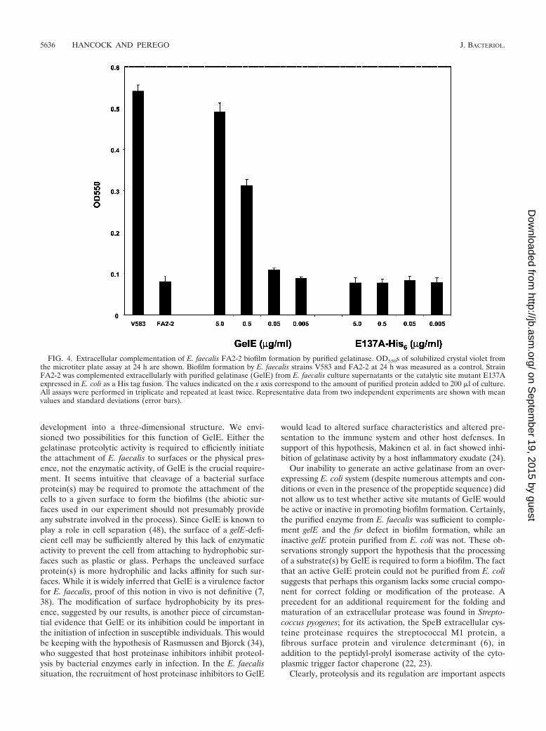
The active gelatinase enzyme purified from the E. faecalisculture supernatant and the two inactive forms isolated fromE. coli were then tested for their ability to promote biofilmformation when added to the culture medium of a strain un-able to make biofilm (FA2-2). As shown in Fig. 4, 5 �g of pu-rified active gelatinase/ml (final concentration) could inducebiofilm formation by strain FA2-2 to a level comparable to theone observed for strain V583. The wild-type inactive (data notshown) or the mutated gelatinase proteins from E. coli did notpromote biofilm formation when used at the same concentra-tions.
These results strongly suggest that the enzymatic activity ofgelatinase is required for its role in biofilm development.
DISCUSSION
Gelatinase is a member of the thermolysin-like M4 family ofproteases (TLPs) that includes enzymes from pathogens suchas Legionella, Listeria, Clostridium, Staphylococcus, Pseudomo-nas, and Vibrio (2). Many of these bacterial metalloproteaseshave been found to be associated with virulence, while eukary-otic members of the family (so-called matrix metalloproteases)(16) were shown to be involved in a number of processes inhumans, including the processing of precursors that play modu-lation roles in the formation of tumors (20, 25). Thus, metal-loproteases of the M4 family have attracted increasing at-tention as model proteins for the development of specificinhibitors that can be applied to disease treatment (41). Ourfinding of the role of gelatinase in biofilm development inE. faecalis raises the possibility that protease inhibitors mayalso be relevant, synergistically with antibiotics, in the treatmentof biofilm-based pathogeneses such as infective endocarditis.
Gelatinase has been implicated in E. faecalis virulence byboth epidemiological data and animal model studies (7, 11, 26,29, 37, 38). Biochemical analysis of purified GelE showed thatit cleaved a number of substrates in vitro, including insulin-�chain, Azocoll, insoluble collagen fragments, and endothelin-1,at primarily hydrophobic amino acids (24, 25, 48). It was alsofound to degrade the pheromone and inhibitor peptides in-volved in conjugative plasmid transfer in E. faecalis (24). Re-cently, it was shown that GelE functions to clear the bacterialcell surface of misfolded proteins (48). A role for GelE in theactivation of an autolysin and in the degradation of polymer-ized fibrin was also suggested by the same study (48). Thoseauthors proposed that GelE acts to increase the disseminationof E. faecalis in high-density environments by fitting the two-step model of Streptococcus pyogenes infection described byRasmussen and Bjorck (34). In the first phase, at low bacterialcell density, a low level of bacterial proteolytic activity leads toattachment mediated by surface proteins. In the second phase,at high bacterial cell density, high levels of proteolytic activitypromote bacterial dissemination by cleaving bacterial attach-ment proteins and host tissue proteins. Our finding that GelEis required for bacterial attachment to a surface and the growthin biofilms argues against that proposal. Certainly, the absenceof GelE in E. faecalis is highly pleiotropic and affects manyphenotypic aspects of the pathogenicity of this organism. Thepresent results show that GelE is required for the formation ofbiofilm, and it promotes the aggregation of the cells in micro-colonies to form the primary attachment site and subsequent
VOL. 186, 2004 GELATINASE CONTROL OF BIOFILM DEVELOPMENT 5635
on Septem
ber 19, 2015 by guesthttp://jb.asm
.org/D
ownloaded from
development into a three-dimensional structure. We envi-sioned two possibilities for this function of GelE. Either thegelatinase proteolytic activity is required to efficiently initiatethe attachment of E. faecalis to surfaces or the physical pres-ence, not the enzymatic activity, of GelE is the crucial require-ment. It seems intuitive that cleavage of a bacterial surfaceprotein(s) may be required to promote the attachment of thecells to a given surface to form the biofilms (the abiotic sur-faces used in our experiment should not presumably provideany substrate involved in the process). Since GelE is known toplay a role in cell separation (48), the surface of a gelE-defi-cient cell may be sufficiently altered by this lack of enzymaticactivity to prevent the cell from attaching to hydrophobic sur-faces such as plastic or glass. Perhaps the uncleaved surfaceprotein(s) is more hydrophilic and lacks affinity for such sur-faces. While it is widely inferred that GelE is a virulence factorfor E. faecalis, proof of this notion in vivo is not definitive (7,38). The modification of surface hydrophobicity by its pres-ence, suggested by our results, is another piece of circumstan-tial evidence that GelE or its inhibition could be important inthe initiation of infection in susceptible individuals. This wouldbe keeping with the hypothesis of Rasmussen and Bjorck (34),who suggested that host proteinase inhibitors inhibit proteol-ysis by bacterial enzymes early in infection. In the E. faecalissituation, the recruitment of host proteinase inhibitors to GelE
would lead to altered surface characteristics and altered pre-sentation to the immune system and other host defenses. Insupport of this hypothesis, Makinen et al. in fact showed inhi-bition of gelatinase activity by a host inflammatory exudate (24).
Our inability to generate an active gelatinase from an over-expressing E. coli system (despite numerous attempts and con-ditions or even in the presence of the propeptide sequence) didnot allow us to test whether active site mutants of GelE wouldbe active or inactive in promoting biofilm formation. Certainly,the purified enzyme from E. faecalis was sufficient to comple-ment gelE and the fsr defect in biofilm formation, while aninactive gelE protein purified from E. coli was not. These ob-servations strongly support the hypothesis that the processingof a substrate(s) by GelE is required to form a biofilm. The factthat an active GelE protein could not be purified from E. colisuggests that perhaps this organism lacks some crucial compo-nent for correct folding or modification of the protease. Aprecedent for an additional requirement for the folding andmaturation of an extracellular protease was found in Strepto-coccus pyogenes; for its activation, the SpeB extracellular cys-teine proteinase requires the streptococcal M1 protein, afibrous surface protein and virulence determinant (6), inaddition to the peptidyl-prolyl isomerase activity of the cyto-plasmic trigger factor chaperone (22, 23).
Clearly, proteolysis and its regulation are important aspects
FIG. 4. Extracellular complementation of E. faecalis FA2-2 biofilm formation by purified gelatinase. OD550s of solubilized crystal violet fromthe microtiter plate assay at 24 h are shown. Biofilm formation by E. faecalis strains V583 and FA2-2 at 24 h was measured as a control. StrainFA2-2 was complemented extracellularly with purified gelatinase (GelE) from E. faecalis culture supernatants or the catalytic site mutant E137Aexpressed in E. coli as a His tag fusion. The values indicated on the x axis correspond to the amount of purified protein added to 200 �l of culture.All assays were performed in triplicate and repeated at least twice. Representative data from two independent experiments are shown with meanvalues and standard deviations (error bars).
5636 HANCOCK AND PEREGO J. BACTERIOL.
on Septem
ber 19, 2015 by guesthttp://jb.asm
.org/D
ownloaded from

of bacterial pathogenesis, and the role of GelE in enterococcalbiofilm formation confirms this notion. Unlike GelE, the serineprotease encoded by the E. faecalis sprE gene is not involved inbiofilm formation and does not have an additive effect withGelE, at least in our assay conditions. However, SprE wasreported to have a role in enterococcal infection when a mouseperitonitis model (32) or the nematode C. elegans killing model(37) was used. In the latter model, an additive effect of GelEand SprE was also noted.
Our results represent the first documented link between atwo-component system (Fsr), a molecular entity (GelE), andbiofilm formation in E. faecalis. A previous report suggestedthe product of the esp gene, a cell surface protein, was criticalfor biofilm formation by E. faecalis based on epidemiologicalstudies (43). The same study, however, also showed that esp-deficient mutants of E. faecalis were fully capable of formingbiofilms, thus leaving open the question of whether Esp has arole in this process or not (19). The E. faecalis V583 strain usedin our study was shown to be esp deficient, as it lacks the regionwithin the pathogenicity island that carries this gene in otherstrains (36). Yet strain V583 is fully capable of forming bio-films on abiotic surfaces, thus confirming that Esp is not arequirement for this type of bacterial growth, at least in theconditions used here, although we cannot rule out the possi-bility of a synergistic effect with GelE. Esp-independent biofilmformation was also reported by Kristich et al. with strainOG1RF (19). The same study incidentally reported the en-hancement of biofilm formation by GelE.
Our study also indicates that through the control of gelEexpression, the fsr two-component system is the only oneamong the 17 systems identified from E. faecalis V583 as beinginvolved in biofilm formation (reference 14 and our unpub-lished data; Hancock and Perego, Abstr. 103rd Gen. Meet.Am. Soc. Microbiol. 2003). Deletion of the other nonessentialsystems indeed did not affect biofilm growth, at least not at adetectable level in our assay. Although synergistic effects oftwo or more systems cannot be ruled out, it appears that GelEis a major requirement for biofilm development, as its soleexpression can complement the lack of the FsrA responseregulator or the lack of the FsrC histidine kinase (Fig. 2).Additionally, if one assumes that the basal OD of the fsrAmutant or of the fsrC mutant strain FA2-2 in the microtiterplate assay is equal to an inability to form a biofilm, as sup-ported by the confocal microscopy images of Fig. 3B and D,then it is possible to conclude that the gelatinase enzymaticactivity is necessary and sufficient (in the presence of a hypo-thetical substrate[s] and in the absence of the fsr system) forbiofilm formation in E. faecalis V583. Furthermore, overex-pression of gelE from the kanamycin promoter in a multicopyplasmid resulted in higher biofilm accumulation than in thecontrol strain carrying a single copy of the gelE gene. (Fig. 2B).Accordingly, biofilm density was directly proportional to theconcentration of GelE in the medium, as shown in Fig. 4.These observations, together with the knowledge that gelEexpression is dependent upon an active FsrABC system (32),indicate that any physiological or environmental signal affect-ing the activation of the Fsr two-component system or theactivity of its quorum-sensing, regulating peptide FsrB, if any,will have an impact on the efficiency of biofilm formation.
To this regard, it should also be pointed out that the role of
the Fsr system in enterococcal biofilm formation does not seemto have any resemblance to the role of its counterpart systemsAgrABCD of Staphylococcus aureus and ComCDE of strepto-cocci in the respective ability of these organisms to developbiofilms. These three systems belong to the so-called agr-likesubfamily of two-component signal transduction systems iden-tified in gram-positive bacteria and are known as quorum-sensing systems for the presence of a third component, a se-creted signaling peptide, in addition to a membrane-boundhistidine kinase and an intracellular response regulator (18).This type of quorum-sensing system has been found to regulatea variety of physiological activities in gram-positive organisms,including competence development in streptococci and Bacil-lus subtilis, antibiotic biosynthesis in Lactococcus lactis, andinduction of virulence factors in S. aureus (1, 5, 9, 10, 17).Recently, it was reported that the inactivation of the genesencoding the ComD histidine kinase or the ComE responseregulator of Streptococcus mutans resulted in the formation ofa biofilm with a reduced mass, while the inactivation of thecomC gene encoding the signaling peptide gave rise to a bio-film with a defective architecture probably due to a defect incell separation. An additive effect of the comDE and comCmutations was also reported previously (21). These observa-tions suggested the existence of a second receptor for theComC extracellular peptide in addition to the ComD compe-tence histidine kinase. In contrast, the FsrB signaling peptideof E. faecalis does not seem to have additional targets otherthan the FsrC histidine kinase, at least for the biofilm pheno-type, since an fsrBC mutant is quantitatively similar to fsrC orfsrA single mutants in biofilm formation. Additionally, theFsrB-containing supernatant of strain JML104 (gelE) did notimprove the biofilm defect of the FA2-2 strain, thus ruling outthe existence of a secondary pathway, at least in these strains(our unpublished observations).
For the process of biofilm formation in staphylococci, thereis mounting evidence that the lack of a functional agr systemincreases the formation of biofilm despite the observation thatproteins required for biofilm formation, such as alpha-toxin,are positively regulated by the Agr system (3, 47). Additionally,there is increasing evidence that whereas secreted virulencefactors may be important for establishing infections, the loss ofagr function may enhance the long-term survival of staphylo-cocci in the host, thus contributing to persistent infectionsoften associated with biofilm formation (35). The observationsby Vuong et al. (46) that the disruption of the agr locus inStaphylococcus epidermidis resulted in increased biofilm forma-tion and that the S. epidermidis O-47 clinical isolate is an agrmutant further support the notion that the role of the staph-ylococcal quorum-sensing system in biofilm formation may notbe as obvious as generally anticipated. Furthermore, the ob-servation that the expression of agr seen in certain animalmodels of infections does not significantly affect the expressionof virulence factors, as would be expected from in vitro data,suggests that the role of quorum sensing in staphylococcalinfections is not as clear as originally presented (12, 13, 49).
Clearly, the involvement, if any, of peptide-controlled sys-tems in biofilm formation in gram-positive microorganisms isspecies specific and apparently requires the activation or inac-tivation of a myriad of known and unknown genes, making thegeneral understanding of the pathway a very complex task.
VOL. 186, 2004 GELATINASE CONTROL OF BIOFILM DEVELOPMENT 5637
on Septem
ber 19, 2015 by guesthttp://jb.asm
.org/D
ownloaded from

This also means that there will probably be the need for se-lective and targeted pharmaceutical approaches for the controlof bacterial infections in cases of biofilm development.
ACKNOWLEDGMENTS
This research was supported in part by Public Health Service grantGM55594 and GM19416 from the National Institute of General Med-ical Sciences, National Institutes of Health. The Stein Beneficial Trustsupported in part oligonucleotide synthesis and DNA sequencing.
This is publication 15858-MEM from The Scripps Research Insti-tute.
REFERENCES
1. Abdelnour, A., S. Arvidson, T. Bremell, C. Ryden, and A. Tarkowski. 1993.The accessory gene regulator (agr) controls Staphyloccoccus aureus virulencein a murine arthritis model. Infect. Immun. 61:3879–3885.
2. Barrett, A. J., N. D. Rawlings, and J. F. Woessner. 1998. Handbook ofproteolytic enzymes, p. 350–369. Academic Press, Inc., New York, N.Y.
3. Caiazza, N. C., and G. A. O’Toole. 2003. Alpha-toxin is required for biofilmformation by Staphylococcus aureus. J. Bacteriol. 185:3214–3217.
4. Callegan, M. C., B. D. Jett, L. E. Hancock, and M. S. Gilmore. 1999. Role ofhemolysin BL in the pathogenesis of extraintestinal Bacillus cereus infectionassessed in an endophthalmitis model. Infect. Immun. 67:3357–3366.
5. Cheung, A. L., K. J. Eberhardt, E. Chung, M. R. Yeaman, P. M. Sullam, M.Ramos, and A. S. Bayer. 1994. Diminished virulence of a sar�/agr� mutantof Staphylococcus aureus in the rabbit model of endocarditis. J. Clin. Investig.94:1815–1822.
6. Collin, M., and A. Olsen. 2000. Generation of a mature streptococcal cys-teine proteinase is dependent on cell wall-anchored M1 protein. Mol. Mi-crobiol. 36:1306–1318.
7. Coque, T. M., J. E. Patterson, J. M. Steckelberg, and B. E. Murray. 1995.Incidence of hemolysin, gelatinase, and aggregation substance among en-terococci isolated from patients with endocarditis and other infections andfrom feces of hospitalized and community-based persons. J. Infect. Dis. 171:1223–1229.
8. Cruz-Rodz, A. L., and M. S. Gilmore. 1990. High efficiency introduction ofplasmid DNA into glycine treated Enterococcus faecalis by electroporation.Mol. Gen. Genet. 224:152–154.
9. Cvitkovitch, D. G. 2001. Genetic competence and transformation in oralstreptococci. Crit. Rev. Oral Biol. Med. 12:217–243.
10. Dubnau, D., J. Hahn, M. Roggiani, F. Piazza, and Y. Weinrauch. 1994.Two-component regulators and genetic competence in Bacillus subtilis. Res.Microbiol. 145:403–411.
11. Garsin, D. A., C. A. Sifri, E. Mylonakis, X. Qin, K. V. Singh, B. E. Murray,S. B. Calderwood, and F. M. Ausubel. 2001. A simple model host for iden-tifying Gram-positive virulence factors. Proc. Natl. Acad. Sci. USA 98:10892–10897.
12. Goerke, C., S. Campana, M. G. Bayer, G. Doring, K. Botzenhart, and C.Wolz. 2000. Direct quantitative transcript analysis of the agr regulon ofStaphylococcus aureus during human infection in comparison to the expres-sion profile in vitro. Infect. Immun. 68:1304–1311.
13. Goerke, C., U. Fluckiger, A. Steinhuber, W. Zimmerli, and C. Wolz. 2001.Impact of the regulatory loci agr, sarA and sae of Staphylococcus aureus onthe induction of �-toxin during device-related infection resolved by directquantitative transcript analysis. Mol. Microbiol. 40:1439–1447.
14. Hancock, L., and M. Perego. 2002. Two-component signal transduction inEnterococcus faecalis. J. Bacteriol. 184:5819–5825.
15. Hoch, J. A., and T. J. Silhavy. 1995. Two-component signal transduction.ASM Press, Washington, D.C.
16. Johnson, L. L., Q. Z. Ye, L. L. Johnson, D. J. Hupe, D. F. Ortwine, J. B.Dunbar, Jr., J. B. Rubin, A. Pavlovsky, C. Humblet, and T. L. Blundell. 1996.Designing inhibitors of the metalloproteinase superfamily: comparative anal-ysis of representative structures. Drugs Des. Discov. 13:3–14.
17. Kleerebezem, M., and L. E. Quadri. 2001. Peptide pheromone-dependentregulation of antimicrobial peptide production in Gram-positive bacteria: acase of multicellular behavior. Peptides 22:1579–1596.
18. Kleerebezem, M., L. E. N. Quadri, O. P. Kuipers, and W. de Vos. 1997.Quorum sensing by peptide pheromones and two-component signal-trans-duction systems in Gram-positive bacteria. Mol. Microbiol. 24:895–904.
19. Kristich, C. J., Y. H. Li, D. G. Cvitkovitch, and G. M. Dunny. 2004. Esp-in-dependent biofilm formation by Enterococcus faecalis. J. Bacteriol. 186:154–163.
20. Lennarz, W. J., and W. J. Strittmatter. 1991. Cellular functions of metallo-endoproteinases. Biochim. Biophys. Acta 1071:149–158.
21. Li, Y.-H., N. Tang, M. B. Aspiras, P. C. Y. Lau, J. H. Lee, R. P. Ellen, andD. G. Cvitkovitch. 2002. A quorum-sensing signaling system essential forgenetic competence in Streptococcus mutans is involved in biofilm formation.J. Bacteriol. 184:2699–2708.
22. Lyon, W. R., and M. G. Caparon. 2003. Trigger factor-mediated prolyl
isomerization influences maturation of the Streptococcus pyogenes cysteineprotease. J. Bacteriol. 185:3661–3667.
23. Lyon, W. R., C. M. Gibson, and M. G. Caparon. 1998. A role for triggerfactor and an rgg-like regulator in the transcription, secretion and processingof the cysteine proteinase of Streptococcus pyogenes. EMBO J. 17:6263–6275.
24. Makinen, P.-L., D. B. Clewell, F. An, and K. K. Makinen. 1989. Purificationand substrate specificity of a strongly hydrophobic extracellular metalloen-dopeptidase (“gelatinase”) from Streptococcus faecalis (strain 0G1–10).J. Biol. Chem. 264:3325–3334.
25. Makinen, P., and K. K. Makinen. 1994. The Enterococcus faecalis extracel-lular metalloendopeptidase (EC 3.4.24.30; coccolysin) inactivates humanendothelin at bonds involving hydrophobic amino acid residues. Biochem.Biophys. Res. Commun. 200:981–985.
26. Mylonakis, E., M. Engelbert, X. Qin, C. D. Sifri, B. E. Murray, F. M.Ausubel, M. S. Gilmore, and S. B. Calderwood. 2002. The Enterococcusfaecalis fsrB gene, a key component of the fsr quorum-sensing system, isassociated with virulence in the rabbit endophthalmitis model. Infect. Im-mun. 70:4678–4681.
27. Nakayama, J., Y. Cao, T. Horii, S. Sakuda, A. D. L. Akkermans, W. de Vos,and H. Nagasawa. 2001. Gelatinase biosynthesis-activating pheromone: apeptide lactone that mediates a quorum sensing in Enterococcus faecalis.Mol. Microbiol. 41:145–154.
28. Olson, M. E., H. Ceri, D. W. Morck, A. G. Buret, and R. R. Read. 2002.Biofilm bacteria: formation and comparative susceptibility to antibiotics.Can. J. Vet. Res. 66:86–92.
29. Pillai, S. K., G. Sakoulas, H. S. Gold, C. Wennersten, G. M. Eliopoulos, R. C.Moellering, Jr., and R. T. Inouye. 2002. Prevalence of the fsr locus inEnterococcus faecalis infections. J. Clin. Microbiol. 40:2651–2652.
30. Podbielski, A., B. Spellerberg, M. Woischnik, B. Pohl, and R. Lutticken.1996. Novel series of plasmid vectors for gene inactivation and expressionanalysis in group A streptococci (GAS). Gene 177:137–147.
31. Poyart, C., and P. Trieu-Cuot. 1997. A broad-host-range mobilizable shuttlevector for the construction of transcriptional fusions to �-galactosidase inGram-positive bacteria. FEMS Microbiol. Lett. 156:193–198.
32. Qin, X., K. V. Singh, G. M. Weinstock, and B. E. Murray. 2000. Effects ofEnterococcus faecalis fsr genes on production of gelatinase and a serineprotease and virulence. Infect. Immun. 68:2579–2586.
33. Qin, X., K. V. Singh, G. M. Weinstock, and B. E. Murray. 2001. Character-ization of fsr, a regulator controlling expression of gelatinase and serineprotease in Enterococcus faecalis OG1RF. J. Bacteriol. 183:3372–3382.
34. Rasmussen, M., and L. Bjorck. 2002. Proteolysis and its regulation at thesurface of Streptococcus pyogenes. Mol. Microbiol. 43:537–544.
35. Schwan, W. R., M. H. Langhorne, H. D. Ritchie, and C. K. Stover. 2003. Lossof hemolysin expression in Staphylococcus aureus agr mutants correlates withselective survival during mixed inflections in murine abscesses and wounds.FEMS Immunol. Med. Microbiol. 38:23–28.
36. Shankar, N., A. S. Baghdayan, and M. S. Gilmore. 2002. Modulation ofvirulence within a pathogenicity island in vancomycin-resistant Enterococcusfaecalis. Nature 417:746–750.
37. Sifri, C. D., E. Mylonakis, K. V. Singh, X. Qin, D. A. Garsin, B. E. Murray,F. M. Ausubel, and S. B. Calderwood. 2002. Virulence effect of Enterococcusfaecalis protease genes and the quorum-sensing locus fsr in Caenorhabditiselegans and mice. Infect. Immun. 70:5647–5650.
38. Singh, K. V., X. Qin, G. M. Weinstock, and B. E. Murray. 1998. Generationand testing of mutants of Enterococcus faecalis in a mouse peritonitis model.J. Infect. Dis. 178:1416–1420.
39. Stephenson, K., and J. A. Hoch. 2002. Virulence- and antibiotic resistance-associated two-component signal transduction systems of Gram-positivepathogenic bacteria as targets for antimicrobial therapy. Pharmacol. Ther.93:293–305.
40. Su, Y. A., M. C. Sulavik, P. He, K. K. Makinen, P.-L. Makinen, S. Fiedler, R.Wirth, and D. B. Clewell. 1991. Nucleotide sequence of the gelatinase (gelE)from Enterococcus faecalis subsp. liquefaciens. Infect. Immun. 59:415–420.
41. Tamaki, M., K. Tanzawa, S. Kurihara, T. Oikawa, S. Monma, K. Shimada, andY. Sugimura. 1995. Synthesis and structure-activity relationships of gelatinaseinhibitors derived from matlystatins. Chem. Pharm. Bull. (Tokyo) 43:1883–1893.
42. Teng, F., L. Wang, K. V. Singh, B. E. Murray, and G. M. Weinstock. 2002.Involvement of PhoP-PhoS homologs in Enterococcus faecalis virulence.Infect. Immun. 70:1991–1996.
43. Toledo-Arana, A., J. Valle, C. Solano, M. J. Arrizubieta, C. Cucarella, M.Lamata, B. Amorena, J. Leiva, J. R. Penades, and I. Lasa. 2001. The en-terococcal surface protein, Esp, is involved in Enterococcus faecalis biofilmformation. Appl. Environ. Microbiol. 67:4538–4545.
44. Trieu-Cuot, P., C. Carlier, C. Poyart-Salmeron, and P. Courvalin. 1990. Apair of mobilizable shuttle vectors conferring resistance to spectinomycin formolecular cloning in Escherichia coli and in Gram-positive bacteria. NucleicAcids Res. 18:4296.
45. Trieu-Cuot, P., and P. Courvalin. 1983. Nucleotide sequence of the Strepto-coccus faecalis plasmid gene encoding the 3�5�-aminoglycoside phospho-transferase type III. Gene 23:331–341.
46. Vuong, C., C. Gerke, G. A. Somerville, E. R. Fischer, and M. Otto. 2003.
5638 HANCOCK AND PEREGO J. BACTERIOL.
on Septem
ber 19, 2015 by guesthttp://jb.asm
.org/D
ownloaded from

Quorum-sensing control of biofilm factors in Staphylococcus epidermidis.J. Infect. Dis. 188:706–718.
47. Vuong, C., H. L. Saenz, F. Gotz, and M. Otto. 2000. Impact of the agrquorum-sensing system on adherence to polystyrene in Staphylococcus au-reus. J. Infect. Dis. 182:1688–1693.
48. Waters, C. M., M. H. Antiporta, B. E. Murray, and G. M. Dunny. 2003. Role
of the Enterococcus faecalis GelE protease in determination of cellular chainlength, supernatant pheromone levels, and degradation of fibrin and mis-folded surface proteins. J. Bacteriol. 185:3613–3623.
49. Yarwood, J. M., J. K. McCormick, M. L. Paustian, V. Kapur, and P. M.Schlievert. 2002. Repression of the Staphylococcus aureus accessory generegulator in serum and in vivo. J. Bacteriol. 184:1095–1101.
VOL. 186, 2004 GELATINASE CONTROL OF BIOFILM DEVELOPMENT 5639
on Septem
ber 19, 2015 by guesthttp://jb.asm
.org/D
ownloaded from