The Effects of Copper (II) Ions on Enterococcus hirae Cell Growth and the Proton-Translocating FoF1...
8
ORIGINAL PAPER The Effects of Copper (II) Ions on Enterococcus hirae Cell Growth and the Proton-Translocating F o F 1 ATPase Activity Zaruhi Vardanyan • Armen Trchounian Published online: 30 March 2010 Ó Springer Science+Business Media, LLC 2010 Abstract Enterococcus hirae grow well under anaerobic conditions at alkaline pH (pH 8.0) producing acids by glucose fermentation. Bacterial growth was shown to be accompanied by decrease of redox potential from positive values (*?35 mV) to negative ones (*-220 mV). An oxidizer copper (II) ions (Cu 2? ) affected bacterial growth in a concentration-dependent manner (within the range of 0.05 mM to 1 mM) increasing lag phase duration and decreasing specific growth rate. These effects were observed with the wild-type strain ATCC9790 and the atpD mutant strain MS116 (with absent b subunit of F 1 of the F o F 1 ATPase) both. Also ATPase activity and proton– potassium ions exchange were assessed with and without N,N 0 -dicyclohexylcarbodiimide (DCCD), inhibitor of the F o F 1 ATPase. In both cases (DCCD ±), even low Cu 2? concentrations had noticeable effect on ATPase activity, but with less visible concentration-dependent manner. Changes in the number of accessible SH-groups were observed with E. hirae ATCC9790 and MS116 membrane vesicles. In both strains Cu 2? markedly decreased the number of SH-groups in the presence of K ? ions. The addition of ATP increased the amount of accessible SH- groups in ATCC9790 and decreased this number in MS116; Cu 2? blocked ATP-installed increase in SH- groups number in ATCC9790. H ? –K ? -exchange of bac- teria was markedly inhibited by Cu 2? , but stronger effects were detected together with DCCD. Moreover, discrimi- nation between Cu 2? and other bivalent cation—Ni 2? was shown. It is suggested that Cu 2? ions inhibit E. hirae cell growth by direct affect on the F o F 1 ATPase leading to conformational changes in this protein complex and decrease in its activity. Keywords Cu 2? Bacterial growth Proton transport F o F 1 ATPase SH-groups Enterococcus hirae Introduction Enterococcus hirae grow well under anaerobic conditions which is accompanied by acidification of the medium and changes in environment redox potential (E h ). It is known that positive values of E h inhibit the anaerobic bacteria growth (for reviews, see [10, 38]) while E h negative values are required for bacterial growth [4, 7, 22]. The latter can be inhibited by oxidizers, which maintain E h on positive level [4, 15], and stimulated by reducers, which decrease E h to negative values [14]. Moreover, E h affects proton-motive force by changing pH gradient across the membrane [31]. Low concentrations of oxidizing Cu 2? ions are required for Escherichia coli and the other bacteria (for reviews, see [11, 29, 32]) whereas in considerably higher concentrations they can cause a number of toxic cellular effects inhibiting E. coli cell growth [15, 39], which can be explained by alteration of H ? flux through the F o F 1 ATPase as installed by Kirakosyan and Trchounian [15] and inhibition of hydrogenase activity associated with the F o F 1 ATPase as determined by Kirakosyan et al. [16], by increase in surface charge density as shown by Volodina et al. [39] as well as by changes in membrane permeability as suggested for this bacterium by Lebedev et al. [18]. The change in H ? -per- meability and the other properties of bacterial membrane may be related to membrane proteins re-organization or changed functional activity, and that might depend on proteins thiol groups’ state and distribution. In accordance Z. Vardanyan A. Trchounian (&) Department of Biophysics of the Biological Faculty, Yerevan State University, 1 A. Manoukian Str, 0025 Yerevan, Armenia e-mail: [email protected] Cell Biochem Biophys (2010) 57:19–26 DOI 10.1007/s12013-010-9078-z
Transcript of The Effects of Copper (II) Ions on Enterococcus hirae Cell Growth and the Proton-Translocating FoF1...

ORIGINAL PAPER
The Effects of Copper (II) Ions on Enterococcus hirae Cell Growthand the Proton-Translocating FoF1 ATPase Activity
Zaruhi Vardanyan • Armen Trchounian
Published online: 30 March 2010
� Springer Science+Business Media, LLC 2010
Abstract Enterococcus hirae grow well under anaerobic
conditions at alkaline pH (pH 8.0) producing acids by
glucose fermentation. Bacterial growth was shown to be
accompanied by decrease of redox potential from positive
values (*?35 mV) to negative ones (*-220 mV). An
oxidizer copper (II) ions (Cu2?) affected bacterial growth
in a concentration-dependent manner (within the range of
0.05 mM to 1 mM) increasing lag phase duration and
decreasing specific growth rate. These effects were
observed with the wild-type strain ATCC9790 and the
atpD mutant strain MS116 (with absent b subunit of F1 of
the FoF1 ATPase) both. Also ATPase activity and proton–
potassium ions exchange were assessed with and without
N,N0-dicyclohexylcarbodiimide (DCCD), inhibitor of the
FoF1 ATPase. In both cases (DCCD ±), even low Cu2?
concentrations had noticeable effect on ATPase activity,
but with less visible concentration-dependent manner.
Changes in the number of accessible SH-groups were
observed with E. hirae ATCC9790 and MS116 membrane
vesicles. In both strains Cu2? markedly decreased the
number of SH-groups in the presence of K? ions. The
addition of ATP increased the amount of accessible SH-
groups in ATCC9790 and decreased this number in
MS116; Cu2? blocked ATP-installed increase in SH-
groups number in ATCC9790. H?–K?-exchange of bac-
teria was markedly inhibited by Cu2?, but stronger effects
were detected together with DCCD. Moreover, discrimi-
nation between Cu2? and other bivalent cation—Ni2? was
shown. It is suggested that Cu2? ions inhibit E. hirae cell
growth by direct affect on the FoF1 ATPase leading to
conformational changes in this protein complex and
decrease in its activity.
Keywords Cu2? � Bacterial growth � Proton transport �FoF1 ATPase � SH-groups � Enterococcus hirae
Introduction
Enterococcus hirae grow well under anaerobic conditions
which is accompanied by acidification of the medium and
changes in environment redox potential (Eh). It is known that
positive values of Eh inhibit the anaerobic bacteria growth
(for reviews, see [10, 38]) while Eh negative values are
required for bacterial growth [4, 7, 22]. The latter can be
inhibited by oxidizers, which maintain Eh on positive level
[4, 15], and stimulated by reducers, which decrease Eh to
negative values [14]. Moreover, Eh affects proton-motive
force by changing pH gradient across the membrane [31].
Low concentrations of oxidizing Cu2? ions are required
for Escherichia coli and the other bacteria (for reviews, see
[11, 29, 32]) whereas in considerably higher concentrations
they can cause a number of toxic cellular effects inhibiting
E. coli cell growth [15, 39], which can be explained by
alteration of H? flux through the FoF1 ATPase as installed
by Kirakosyan and Trchounian [15] and inhibition of
hydrogenase activity associated with the FoF1 ATPase as
determined by Kirakosyan et al. [16], by increase in surface
charge density as shown by Volodina et al. [39] as well as
by changes in membrane permeability as suggested for this
bacterium by Lebedev et al. [18]. The change in H?-per-
meability and the other properties of bacterial membrane
may be related to membrane proteins re-organization or
changed functional activity, and that might depend on
proteins thiol groups’ state and distribution. In accordance
Z. Vardanyan � A. Trchounian (&)
Department of Biophysics of the Biological Faculty, Yerevan
State University, 1 A. Manoukian Str, 0025 Yerevan, Armenia
e-mail: [email protected]
Cell Biochem Biophys (2010) 57:19–26
DOI 10.1007/s12013-010-9078-z

with these ideas, it is suggested that Cu2? can break
disulfides in membrane proteins [18], increasing the num-
ber of accessible SH-groups [16]. However, a role of Eh in
bacterial growth, effects of oxidizers and reducers on
bacterial growth would be clarified; the appropriate
mechanisms for Cu2? uptake and intracellular handling are
still not clear.
Kirakosyan et al. [16] have shown that in E. coli mem-
brane vesicles, prepared from anaerobically grown cells at
slightly alkaline pH, Cu2? ions alone increase the level of
SH-groups but block ATP-stimulated increase of these
groups when added together with ATP. The obtained effects
might be the result of Cu2? action on Eh or a direct effect of
these ions on proteins in bacterial membrane, probably on the
FoF1 ATPase. It is suggested that the energy of ATP can be
transferred through a dithiol–disulfide interchange between
the FoF1 ATPase and the other membrane proteins [5, 16],
which are forming the protein–protein associations within
the membrane [6, 22, 36]. And the marked increase in the
number of FoF1 ATPase SH-groups by ATP but not by ADP
has been shown with membrane vesicles [23]. Furthermore,
Cu2? can break these interactions thus increasing the level of
accessible SH-groups. The latter is proposed to be another
way which also depends on Eh. In our laboratory it has been
established [15] that the addition of 0.1 and 2 mM Cu2? into
the E. coli growth medium results in a delayed decrease of Eh
although a drop in Eh is less for 2 mM than for 0.1 mM of
these ions. All these findings can be taken into consideration
to explain the action mechanisms of oxidizers effects on
E. coli growth.
A closed relationship of the FoF1 ATPase with other
membrane proteins is also assumed in case of E. hirae [37].
This idea results from two findings at least: H?–K?-
exchange with the fixed stoichiometry through the FoF1
ATPase and via the K? uptake system [24, 37] and N,N0-dicyclohexylcarbodiimide (DCCD)-inhibited ATPase
activity depended on K? ions [37]. Moreover, the marked
increase in the number of FoF1 ATPase SH-groups by ATP
and nicotinamide adenine dinucleotides has been deter-
mined with E. hirae membrane vesicles [27]. However,
mechanisms of such relationship and thiol groups’ role in
the E. hirae FoF1 ATPase activity remain unclear. At
present there are no data on Cu2? effects with E. hirae.
Our present data show that Cu2? affect E. hirae cell
growth by increasing lag phase duration and decreasing
specific growth rate. These ions have noticeable effect on
H?–K?-exchange and ATPase activity and lower the
number of accessible SH-groups in a concentration-
dependent manner; discrimination between Cu2? and Ni2?
is shown. The results with data obtained by using the
E. hirae atpD mutant strain MS116 with defective FoF1
ATPase might indicate direct effects of Cu2? on this
ATPase.
Materials and Methods
Bacterial Strains and Growth, Whole Cells, Membrane
Vesicles, Redox Potential
The wild-type strain E. hirae ATCC9790 [17] and the atpD
mutant strain MS116 (having absent b subunit of F1 of the
FoF1 ATPase) [3, 37] were used in this study. MS116 strain
expresses the FoF1 ATPase to the level as wild-type one
[3], but it has significantly lowered H? efflux [2] and
ATPase activity [3, 37]. The strains were kindly supplied
by Prof. H. Kobayashi (Department of Biochemistry, Chiba
University, Chiba 263, Japan).
Bacteria were grown under anaerobic conditions at 37�C
in a 0.2% glucose containing growth medium (1% tryptone,
0.5% yeast extract, 1% K2HPO4; pH 8.0) as described earlier
[2, 24, 26, 27, 37]. The medium pH was measured by a pH-
meter with selective pH-electrode (HJ1131B, Hanna
Instruments, Portugal) and adjusted by means of 0.1 M
NaOH or HCl. Bacterial growth was assessed by measuring
optical density (OD) changes in bacterial suspension using a
Spectro UV–vis Auto spectrophotometer (Labomed, USA)
at wavelength of 600 nm. To study the effects of Cu2?,
0.05 mM, 0.1 mM, or 1 mM CuCl2 were added in bacterial
suspension, when mentioned. The latent (lag) growth phase
duration was determined as described before [14]. The spe-
cific growth rate was calculated over the interval, where the
logarithm of OD for the culture increased linearly with time,
and it was expressed as lg2 (lg2 = 0.693)/doubling time.
Whole cells [2, 15, 25] were prepared and right-side-out
membrane vesicles [16, 27, 37] were isolated as described
except that the buffers lacked K?.
Eh was measured by a platinum (Pt) (EPB-1, GSEEE, or
PT42BNC, Hanna Instruments, Portugal) or titanium-
silicate (Ti–Si) (EO-02, Electrometer Equipment State
Enterprise, Gomel, Belarus) electrode as described else-
where [4–7, 14, 16, 22]. When the Pt-electrode was used
together with a Ti–Si-electrode, which, unlike the former, is
insensitive to molecular oxygen and molecular hydrogen, no
significant differences in electrode readings were detected.
It was detected that Eh value in the conditions used was
changed on 35–40 mV only by *8–10-fold alteration of
bacterial count. It was also determined that Eh value was
not changed more than on 25–30 mV by *sixfold change
of Cu2? concentration within the concentration range used.
So the significant decrease of Eh during bacterial growth
(see ‘‘Results and Discussion’’ section) does not depend on
either bacterial count or Cu2? ions count change.
Accessible SH-Groups and ATPase Assays
Accessible SH-groups of membrane vesicles were deter-
mined by Ellmann’s reagent (5,50-dithiobis-2-nitrobenzoic
20 Cell Biochem Biophys (2010) 57:19–26

acid) [30] using spectroscopic method as described [16, 23,
27] and glutathione as a standard. Membrane vesicles were
treated with the reagent and OD was measured after *1.5–
2 h (OD became constant), corrections were made for
blanks without membrane vesicles and with different
reagents used. The level of SH-groups was expressed in
nmol per mg protein. Using this reagent gave the same data
for the number of SH-groups as the other reagents as shown
[16, 23, 28].
ATPase activity was measured by amount of inorganic
phosphate (Pi) liberated after adding 5 mM ATP (Tris salt)
[22, 28]. Pi was determined by the spectrophotometric
method of Taussky and Shorr [35], corrections were made
for blanks without ATP or membrane vesicles. ATPase
activity was expressed in nmol Pi per lg protein in 1 min.
The assay mixture was of 200 mM Tris-phosphate (pH
8.0) containing 0.4 mM MgSO4, 1 mM NaCl and
1 mM KCl; for ATPase activity determination 50 mM
Tris–HCl (pH 8.0), containing 0.4 mM MgSO4 with or
without 100 mM KCl was used. When used, membrane
vesicles were pre-incubated with Cu2? or DCCD for
10 min.
Proton and Potassium Ions Transport Study
H? and K? fluxes through the bacterial membrane in whole
cells were measured using appropriate selective electrodes
(HJ1131B, Hanna Instruments, Portugal; and PVC mem-
brane type, Cole Parmer Instruments Co., USA, or E-031,
‘‘Niko Analit’’ Sci.-Prod. Co., Russia) as described else-
where [7, 15, 22, 26]. Electrode readings were calibrated
by titration of the assay medium (200 mM Tris-phosphate
buffer (pH 8.0) containing 0.4 mM MgSO4, 1 mM NaCl
and 1 mM KCl) with 0.01 N HCl and 0.02 mM KCl. Ions
transport was determined after addition of glucose into the
assay medium. Ion fluxes are expressed as the change in
external activity of the ion in mM/min per number of cells
in a unit of medium volume (ml). For DCCD inhibition
studies, cells were treated with this reagent at 0.1 mM for
10 min prior assays; during treatment with DCCD bacterial
count was not changed. Note, Cu2? ions had no effects on
proton and potassium electrode readings.
Other and Chemicals
Bacterial count was determined by counting colony-form-
ing units grown on solid media with glucose (after plating
of diluted bacterial suspension). Protein was measured by
the method of Lowry et al. [20] using bovine serum albu-
min as a standard. All assays were routinely carried out
under anaerobic conditions and all measurements were
done at 37�C. At least three independent measurements
were made; standard errors were not more than 3% if not
represented. The Student’s validity criteria (p) was calcu-
lated to show statistically reliable difference between
changed values and control [16].
Trypton, yeast extract were from Roth (Germany), ATP,
DCCD, Ellmann’s reagent, glucose and glutathione were
from Sigma (USA) and other reagents of analytical grade
were used in experiments. For metal ions appropriate
chloride salts were used.
Results and Discussion
Bacterial Growth and Redox Potential in the Presence
of Copper (II) Ions
The wild-type strain E. hirae ATCC9790 and the atpD
mutant strain MS116 with defective FoF1 ATPase are
known to grow well under anaerobic conditions at pH 8.0
[2, 3, 26, 37].
The addition of Cu2? into the bacterial growth medium
resulted in an increased lag phase duration and decreased
A
0
0.5
1
1.5
2
2.5
3
3.5
4
Lag
ph
ase
du
rati
on
, h
ATCC9790 MS116
B
0
0.1
0.2
0.3
0.4
0.5
0.6
0.7
Control 0.05 mM 0.1 mM 1 mM
Control 0.05 mM 0.1 mM 1 mM
Sp
ecifi
c g
row
th r
ate,
h-1 ATCC9790 MS116
Fig. 1 Effects of Cu2? ions in different concentrations on the E.hirae ATCC9790 and MS116 cell growth. a Lag phase duration, bspecific growth rate. CuCl2 of 0.05 mM to 1 mM was added (if
specified, see x-axis) to the growth medium before inoculation of
bacteria; control was bacterial growth in the growth medium without
CuCl2 added. For the others, see ‘‘Materials and Methods’’ section
Cell Biochem Biophys (2010) 57:19–26 21

specific growth rate (Fig. 1). With low concentrations of
Cu2? (0.05 mM) no statistically reliable bacterial growth
differences were observed (in comparison with control
samples, P [ 0.05). In contrast, a higher concentration of
Cu2? (0.1 mM and 1 mM) notably prolonged lag phase
duration and significantly decreased the specific growth
rate (P \ 0.05).
The influence of Cu2? on MS116 cell growth was less
noticeable than that on ATCC9790 (see Fig. 1). The lag
phase duration with this atp mutant strain is *4.5-fold
higher than that with wild-type strain but specific growth
rate are almost the same (see Fig. 1). These findings point
out that the FoF1 ATPase is not essential for E. hirae
growth at alkaline pH. This contradicts with a common
idea that the FoF1 ATPase of bacteria is a main membrane
enzyme of bioenergetic meaning that is responsible for
generation of H?-motive force under anaerobic conditions
(for review, see [36]). However, this seems to be in favor
with data of Kobayashi with co-workers [17, 24] that E.
hirae can grow at alkaline pH in the presence of a pro-
tonophore which almost completely dissipates the H?-
motive force. However, the requirement of H?-motive
force for E. hirae growth and mechanisms of its generation
at alkaline pH if any are not clear yet. In addition, the
obtained results with Cu2? ions are in accordance with
those of Kirakosyan et al. [16] reported for E. coli.
Furthermore, the suppression of E. hirae growth at
alkaline pH can be resulted by Cu2? direct effect on bac-
terial membrane or on Eh.
During E. hirae ATCC9790 growth Eh measured with
Pt-electrode drops from positive values (35 ± 10 mV) to
negative ones (-220 ± 10 mV) (Fig. 2a). In the case of
MS116 the initial Eh value is 5 ± 10 mV which drops to
negative values (-170 ± 10 mV) as the culture passes to
the stationary growth phase (Fig. 2b). This drop of Eh
indicates that there are many reduction processes taking
place at anaerobic growth [14, 38]. At the stationary phase,
after 24 h of growth, Eh increases, but does not reach the
initial values (not shown).
Changes in Eh during E. hirae growth were also
observed in the presence of Cu2?. These ions were shown
to affect Eh values changes in a concentration-dependent
manner (Fig. 2). The effects of Cu2? were more expressed
in case of wild-type strain E. hirae ATCC9790 if com-
paring with MS116 (Fig. 2a, b). To have clear data with
Cu2?, we have detected that copper (I) ions had a little bit
less noticeable effect on Eh change (see Fig. 2c). This
proves that Cu2? more than Cu? ions have significant
action on Eh changes during E. hirae growth, although Cu?
rather than Cu2? could be accumulated in spite of some
balance between influx and efflux systems for these ions
[32]. These results may be related to changes in the number
of SH-groups which depends on Eh and redox-regulated
H?-translocating ATPase activity [8, 38]. Besides, changes
in Eh decrease upon bacterial growth are suggested could
be related with changes in bacterial growth [14, 31].
Effects of Cu2? on Accessible SH-Groups Number, Its
Changes, and ATPase Activity
Accessible SH-groups were determined in membrane ves-
icles from E. hirae ATCC9790 and MS116 both. As it is
A
-250
-200
-150
-100
-50
0
50
Time, h
Eh
, mV
Eh
, mV
Eh
, mV
Control0.05mM0.1 mM1 mM
B
-250
-200
-150
-100
-50
0
50
Time, h
Control0.05 mM
0.1 mM1 mM
C
-250
-200
-150
-100
-50
0
50
0 1 2 3 4 5 6
0 1 2 3 4 5 6
0 1 2 3 4 5 6
Time, h
Control0.05 mM0.01 mM1 mM
Fig. 2 Changes in redox potential during E. hirae ATCC9790 growth
in the presence of Cu2? (a) and Cu? ions (c), and MS116 growth in
the presence of Cu2? ions (b). Redox potential was measured with a
Pt-electrode. For details, see the legends in Fig. 1 and ‘‘Materials and
Methods’’ section
22 Cell Biochem Biophys (2010) 57:19–26
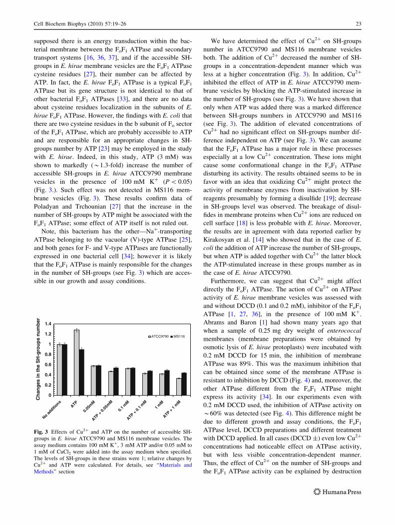
supposed there is an energy transduction within the bac-
terial membrane between the FoF1 ATPase and secondary
transport systems [16, 36, 37], and if the accessible SH-
groups in E. hirae membrane vesicles are the FoF1 ATPase
cysteine residues [27], their number can be affected by
ATP. In fact, the E. hirae FoF1 ATPase is a typical FoF1
ATPase but its gene structure is not identical to that of
other bacterial FoF1 ATPases [33], and there are no data
about cysteine residues localization in the subunits of E.
hirae FoF1 ATPase. However, the findings with E. coli that
there are two cysteine residues in the b subunit of Fo sector
of the FoF1 ATPase, which are probably accessible to ATP
and are responsible for an appropriate changes in SH-
groups number by ATP [23] may be employed in the study
with E. hirae. Indeed, in this study, ATP (3 mM) was
shown to markedly (*1.3-fold) increase the number of
accessible SH-groups in E. hirae ATCC9790 membrane
vesicles in the presence of 100 mM K? (P \ 0.05)
(Fig. 3.). Such effect was not detected in MS116 mem-
brane vesicles (Fig. 3). These results confirm data of
Poladyan and Trchounian [27] that the increase in the
number of SH-groups by ATP might be associated with the
FoF1 ATPase; some effect of ATP itself is not ruled out.
Note, this bacterium has the other—Na?-transporting
ATPase belonging to the vacuolar (V)-type ATPase [25],
and both genes for F- and V-type ATPases are functionally
expressed in one bacterial cell [34]; however it is likely
that the FoF1 ATPase is mainly responsible for the changes
in the number of SH-groups (see Fig. 3) which are acces-
sible in our growth and assay conditions.
We have determined the effect of Cu2? on SH-groups
number in ATCC9790 and MS116 membrane vesicles
both. The addition of Cu2? decreased the number of SH-
groups in a concentration-dependent manner which was
less at a higher concentration (Fig. 3). In addition, Cu2?
inhibited the effect of ATP in E. hirae ATCC9790 mem-
brane vesicles by blocking the ATP-stimulated increase in
the number of SH-groups (see Fig. 3). We have shown that
only when ATP was added there was a marked difference
between SH-groups numbers in ATCC9790 and MS116
(see Fig. 3). The addition of elevated concentrations of
Cu2? had no significant effect on SH-groups number dif-
ference independent on ATP (see Fig. 3). We can assume
that the FoF1 ATPase has a major role in these processes
especially at a low Cu2? concentration. These ions might
cause some conformational change in the FoF1 ATPase
disturbing its activity. The results obtained seems to be in
favor with an idea that oxidizing Cu2? might protect the
activity of membrane enzymes from inactivation by SH-
reagents presumably by forming a disulfide [19]; decrease
in SH-groups level was observed. The breakage of disul-
fides in membrane proteins when Cu2? ions are reduced on
cell surface [18] is less probable with E. hirae. Moreover,
the results are in agreement with data reported earlier by
Kirakosyan et al. [14] who showed that in the case of E.
coli the addition of ATP increase the number of SH-groups,
but when ATP is added together with Cu2? the latter block
the ATP-stimulated increase in these groups number as in
the case of E. hirae ATCC9790.
Furthermore, we can suggest that Cu2? might affect
directly the FoF1 ATPase. The action of Cu2? on ATPase
activity of E. hirae membrane vesicles was assessed with
and without DCCD (0.1 and 0.2 mM), inhibitor of the FoF1
ATPase [1, 27, 36], in the presence of 100 mM K?.
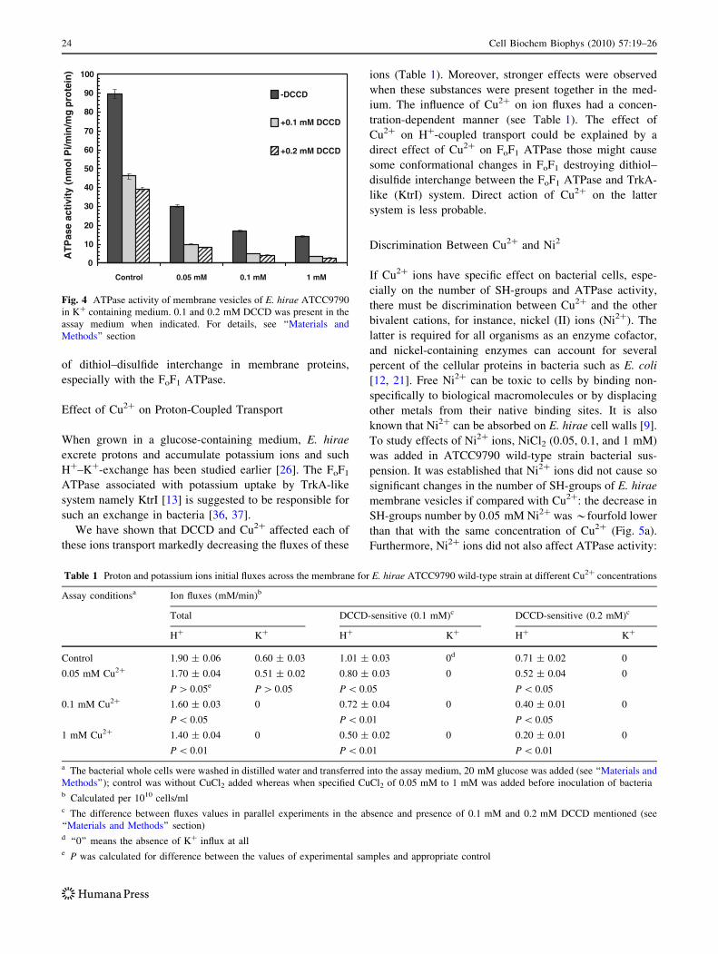
Abrams and Baron [1] had shown many years ago that
when a sample of 0.25 mg dry weight of enterococcal
membranes (membrane preparations were obtained by
osmotic lysis of E. hirae protoplasts) were incubated with
0.2 mM DCCD for 15 min, the inhibition of membrane
ATPase was 89%. This was the maximum inhibition that
can be obtained since some of the membrane ATPase is
resistant to inhibition by DCCD (Fig. 4) and, moreover, the
other ATPase different from the FoF1 ATPase might
express its activity [34]. In our experiments even with
0.2 mM DCCD used, the inhibition of ATPase activity on
*60% was detected (see Fig. 4). This difference might be
due to different growth and assay conditions, the FoF1
ATPase level, DCCD preparations and different treatment
with DCCD applied. In all cases (DCCD ±) even low Cu2?
concentrations had noticeable effect on ATPase activity,
but with less visible concentration-dependent manner.
Thus, the effect of Cu2? on the number of SH-groups and
the FoF1 ATPase activity can be explained by destruction
0
0.2
0.4
0.6
0.8
1
1.2
1.4
No additi
onsATP
0.05m
M
ATP + 0.
05m
M
0.1 m
M
ATP +0.
1 mM
1 mM
ATP+
1m
M
Ch
ang
es in
th
e S
H-g
rou
ps
nu
mb
er
ATCC9790 MS116
Fig. 3 Effects of Cu2? and ATP on the number of accessible SH-
groups in E. hirae ATCC9790 and MS116 membrane vesicles. The
assay medium contains 100 mM K?, 3 mM ATP and/or 0.05 mM to
1 mM of CuCl2 were added into the assay medium when specified.
The levels of SH-groups in these strains were 1; relative changes by
Cu2? and ATP were calculated. For details, see ‘‘Materials and
Methods’’ section
Cell Biochem Biophys (2010) 57:19–26 23
of dithiol–disulfide interchange in membrane proteins,
especially with the FoF1 ATPase.
Effect of Cu2? on Proton-Coupled Transport
When grown in a glucose-containing medium, E. hirae
excrete protons and accumulate potassium ions and such
H?–K?-exchange has been studied earlier [26]. The FoF1
ATPase associated with potassium uptake by TrkA-like
system namely KtrI [13] is suggested to be responsible for
such an exchange in bacteria [36, 37].
We have shown that DCCD and Cu2? affected each of
these ions transport markedly decreasing the fluxes of these
ions (Table 1). Moreover, stronger effects were observed
when these substances were present together in the med-
ium. The influence of Cu2? on ion fluxes had a concen-
tration-dependent manner (see Table 1). The effect of
Cu2? on H?-coupled transport could be explained by a
direct effect of Cu2? on FoF1 ATPase those might cause
some conformational changes in FoF1 destroying dithiol–
disulfide interchange between the FoF1 ATPase and TrkA-
like (KtrI) system. Direct action of Cu2? on the latter
system is less probable.
Discrimination Between Cu2? and Ni2
If Cu2? ions have specific effect on bacterial cells, espe-
cially on the number of SH-groups and ATPase activity,
there must be discrimination between Cu2? and the other
bivalent cations, for instance, nickel (II) ions (Ni2?). The
latter is required for all organisms as an enzyme cofactor,
and nickel-containing enzymes can account for several
percent of the cellular proteins in bacteria such as E. coli
[12, 21]. Free Ni2? can be toxic to cells by binding non-
specifically to biological macromolecules or by displacing
other metals from their native binding sites. It is also
known that Ni2? can be absorbed on E. hirae cell walls [9].
To study effects of Ni2? ions, NiCl2 (0.05, 0.1, and 1 mM)
was added in ATCC9790 wild-type strain bacterial sus-
pension. It was established that Ni2? ions did not cause so
significant changes in the number of SH-groups of E. hirae
membrane vesicles if compared with Cu2?: the decrease in
SH-groups number by 0.05 mM Ni2? was *fourfold lower
than that with the same concentration of Cu2? (Fig. 5a).
Furthermore, Ni2? ions did not also affect ATPase activity:
0
10
20
30
40
50
60
70
80
90
100
Control 0.05 mM 0.1 mM 1 mM
AT
Pas
e ac
tivi
ty (
nm
ol P
i/min
/mg
pro
tein
)
-DCCD
+0.1 mM DCCD
+0.2 mM DCCD
Fig. 4 ATPase activity of membrane vesicles of E. hirae ATCC9790
in K? containing medium. 0.1 and 0.2 mM DCCD was present in the
assay medium when indicated. For details, see ‘‘Materials and
Methods’’ section
Table 1 Proton and potassium ions initial fluxes across the membrane for E. hirae ATCC9790 wild-type strain at different Cu2? concentrations
Assay conditionsa Ion fluxes (mM/min)b
Total DCCD-sensitive (0.1 mM)c DCCD-sensitive (0.2 mM)c
H? K? H? K? H? K?
Control 1.90 ± 0.06 0.60 ± 0.03 1.01 ± 0.03 0d 0.71 ± 0.02 0
0.05 mM Cu2? 1.70 ± 0.04
P [ 0.05e
0.51 ± 0.02
P [ 0.05
0.80 ± 0.03
P \ 0.05
0 0.52 ± 0.04
P \ 0.05
0
0.1 mM Cu2? 1.60 ± 0.03
P \ 0.05
0 0.72 ± 0.04
P \ 0.01
0 0.40 ± 0.01
P \ 0.05
0
1 mM Cu2? 1.40 ± 0.04
P \ 0.01
0 0.50 ± 0.02
P \ 0.01
0 0.20 ± 0.01
P \ 0.01
0
a The bacterial whole cells were washed in distilled water and transferred into the assay medium, 20 mM glucose was added (see ‘‘Materials and
Methods’’); control was without CuCl2 added whereas when specified CuCl2 of 0.05 mM to 1 mM was added before inoculation of bacteriab Calculated per 1010 cells/mlc The difference between fluxes values in parallel experiments in the absence and presence of 0.1 mM and 0.2 mM DCCD mentioned (see
‘‘Materials and Methods’’ section)d ‘‘0’’ means the absence of K? influx at alle P was calculated for difference between the values of experimental samples and appropriate control
24 Cell Biochem Biophys (2010) 57:19–26

if even low Cu2? ions concentrations (0.05 mM CuCl2)
had noticeable effect on ATPase activity (P \ 0.01), much
higher Ni2? concentrations (1 mM NiCl2) did not have
marked influence on ATPase activity (Fig. 5b).
Thus, the effect of Cu2? is specific among bivalent
metal ions, not for one protein, as we have shown that the
same concentration of Ni2? ions has lower effect on SH-
groups number but has no marked effect on ATPase
activity (comp. data on Fig. 5).
Conclusions
In this study, Cu2? ions as oxidizers are shown to inhibit E.
hirae cell growth at alkaline pH affecting lag phase dura-
tion and decreasing specific growth rate. These effects have
a concentration-dependent manner, those are less visible in
atpD mutant MS116 (absence of the b subunit of F1) and
are suggested to be intermediated through Eh (see Fig. 2).
In E. hirae ATCC9790 wild-type strain membrane
vesicles from anaerobically grown cells, ATP increases the
level of accessible SH-groups (see Fig. 3) confirming data
reported previously [27]. The addition of Cu2? ions
decreases the number of SH-groups in a concentration-
dependent manner and these ions block the ATP-stimulated
increase in the number of these groups (see Fig. 3). The
obtained effects on ATPase activity and SH-groups number
are specific for Cu2? (see Fig. 5). These results novel for E.
hirae are in accordance with those obtained with E. coli
[14] except opposite effects on the number of SH-groups.
Therefore, it is most likely to propose action mechanisms
for E. hirae.
Such Cu2? ions influence may be resulted by action of
these ions on Eh (see Fig. 2a) or by their direct effect on
membrane proteins. Cu2? ions are defined to affect directly
the FoF1 ATPase modulating its activity (see Fig. 4) and to
change H?-efflux and H?-coupled transport (see Table 1).
Cu2? is suggested can cause some conformational changes
in the FoF1 ATPase. Revealing action mechanisms with E.
hirae, in contrast to E. coli, oxidizing Cu2? might partic-
ipate in formation a disulfide [19], changing interaction of
the FoF1 ATPase with Trk-like (KtrI) system [26, 36, 37];
decrease in SH-groups level is observed (see Figs. 3, 5).
The breakage of disulfides in membrane proteins when
Cu2? ions are reduced on cell surface [18] is less probable.
So Cu2? might affect SH-groups of the FoF1 ATPase in
E. hirae through disturbing a disulfide–dithiol interchange
between this ATPase and the other membrane proteins that
is installed by ATP and changing activity of this ATPase.
The effects of oxidizing Cu2? ions on E. hirae cell
growth and activity of the FoF1 ATPase have a significance
to regulate bacteria in environment and their application in
biotechnology.
Acknowledgments We thank Prof. H. Kobayashi for supplying E.hirae strains and valuable advices as well as Drs. Anna Poladyan and
Gayane Kirakosyan for help in some experiments and useful com-
ments. The study was supported by the Grant (#1012-2008) from the
Ministry of Education and Science of the Republic of Armenia.
References
1. Abrams, A., & Baron, C. (1970). Inhibitory action of carbodi-
imide on bacterial membrane ATPase. Biochemical and Bio-physical Research Communications, 41, 858–861.
2. Akopyan, K., & Trchounian, A. (2005). Membrane proton con-
ductivity and energy-dependent proton fluxes in Enterococushirae in media with different pH. Biophysics (Moscow), 50, 595–
598.
3. Arikado, E., Ishihara, H., Ehara, T., Shibata, C., Saito, H.,
Kakegawa, T., et al. (1999). Enzyme level of enterococcal FoF1-
ATPase is regulated by pH at the step of assembly. EuropeanJournal of Biochemistry, 259, 262–268.
4. Bagramyan, K., Galstyan, A., & Trchounian, A. (2000). Redox
potential is a determinant in the Escherichia coli anaerobic
growth and survival: Effects of impermeable oxidant. Bioelec-trochemistry, 51, 151–156.
A
0
50
100
150
200
250
300
350
400
Noadditi
onsATP
0.05
mM
ATP +0.
05m
M
0.1 m
M
ATP+
0.1 m
M1
mM
ATP +1
mM
SH
-gro
up
s n
um
ber
(nm
ol/m
g p
rote
in)
Ni2+
Cu2+
B
0102030405060708090
100
Control 0.05 mM 0.1 mM 1 mM
AT
Pas
e ac
tivity
(nm
ol P
i/min
/mg
pro
tein
)
Ni2+
Cu2+
Fig. 5 Changes in the number of SH-groups (a) and ATPase activity
(b) of membrane vesicles of E. hirae ATCC9790 in K? containing
medium in the presence of Ni2? ions. NiCl2 was added into the assay
medium when indicated. For details, see the legends in Figs. 3 and 4,
and ‘‘Materials and Methods’’ section
Cell Biochem Biophys (2010) 57:19–26 25

5. Bagramyan, K. A., & Martirosov, S. M. (1989). Formation of an
ion transport supercomplex in Escherichia coli. An experimental
model of direct transduction of energy. FEBS Letters, 246, 149–
152.
6. Bagramyan, K., Mnatsakanyan, N., Poladyan, A., Vassilian, A.,
& Trchounian, A. (2002). The role of hydrogenases 3 and 4, and
the FoF1-ATP synthase in H2 production by Escherichia coli at
alkaline pH. FEBS Letters, 516, 172–178.
7. Bagramyan, K. A., & Trchounian, A. A. (1997). Decrease of
redox potential in the anaerobic growing Escherichia coli sus-
pension and proton-potassium exchange. Bioelectrochemistry andBioenergetics, 43, 129–134.
8. Bald, D., Noji, H., Yoshida, M., Hirono-Hara, Y., & Hisabori, T.
(2001). Redox regulation of the rotation of F1-ATP synthase.
Journal of Biological Chemistry, 276, 39505–39507.
9. Bossrez, S., Remacle, J., & Coyette, J. (1999). Adsorption of
nickel on Enterococcus hirae cell walls. Journal of ChemicalTechnology and Biotechnology, 70, 45–50.
10. Breznak, J. A., & Costilow, R. H. (1994). Physicochemical fac-
tors in growth. In P. Gerhardt, R. G. Nurrey, W. A. Wood, & N.
R. Krieg (Eds.), Methods for general and molecular bacteriology(pp. 137–155). Washington, DC: ASM Press.
11. Cooksey, D. A. (1993). Copper uptake and resistance in bacteria.
Molecular Microbiology, 7, 1–5.
12. Ermler, U., Grabarse, W., Shima, S., Goubeaud, M., & Thauer, R.
K. (1998). Active sites of transition-metal enzymes with a focus
on nickel. Current Opinion on Structural Biology, 8, 749–758.
13. Kawano, M., Igarashi, K., & Kakinuma, Y. (2002). Isolation of
Enterococcus hirae mutant deficient in low-affinity potassium
uptake at alkaline pH. Bioscience, Biotechnology, Biochemistry,66, 1597–1600.
14. Kirakosyan, G., Bagramyan, K., & Trchounian, A. (2004). Redox
sensing by Escherichia coli: effects of dithiothreitol, a redox
reagent reducing disulphides, on bacterial growth. Biochemicaland Biophysical Research Communication, 325, 803–806.
15. Kirakosyan, G., & Trchounian, A. (2007). Redox sensing by
Escherichia coli: Effects of copper ions as oxidizers on proton-
coupled membrane transport. Bioelectrochemistry, 70, 58–63.
16. Kirakosyan, G., Trchounian, K., Vardanyan, Z., & Trchounian,
A. (2008). Copper (II) ions affect Escherichia coli membrane
vesicles’ SH-groups and a disulfide-dithiol interchange between
membrane proteins. Cell Biochemistry and Biophysics, 51, 45–
50.
17. Kobayashi, H., Suzuki, T., Kinoshita, N., & Unemoto, T. (1984).
Amplification of the Streptococcus faecalis proton-translocating
ATPase by a decrease in cytoplasmatic pH. Journal of Bacteri-ology, 158, 1157–1160.
18. Lebedev, V. S., Volodina, L. A., EYu, Deinega., & YuI, Fedorov.
(2005). Structural modifications of the surface of Escherichia colibacteria and copper induced permeability of plasma membrane.
Biofizika, 50, 107–113. (in Russian).
19. Letelier, M. E., Lepe, A. M., Faundez, M., Salazar, J., Martin, R.,
Aracena, P., et al. (2005). Possible mechanisms underlaying
copper-induced damage in biological membranes leading to cel-
lular toxicity. Chemico-Biological Interactions, 151, 71–82.
20. Lowry, N. O., Rosenbrough, N. J., Farr, A. C., & Randall, R. J.
(1951). Protein measurement with the Folin phenol reagent.
Journal of Biological Chemistry, 193, 263–275.
21. Maroney, M. J. (1999). Structure/function relationships in nickel
metallo-biochemistry. Current Opinion in Chemistry and Biol-ogy, 3, 188–199.
22. Mnatsakanyan, N., Bagramyan, K., Vassilian, A., Nakamoto, R.,
& Trchounian, A. (2002). FO cysteine, bCys21, in the Escherichiacoli ATP synthase is involved in regulation of potassium uptake
and molecular hydrogen production in anaerobic conditions.
Bioscience Reports, 22, 421–430.
23. Mnatsakanyan, N., Poladian, A., Bagramyan, K., & Trchounian,
A. (2003). The number of accessible SH-groups in Escherichiacoli membrane vesicles is increased by ATP and by formate.
Biochemical and Biophysical Research Communications, 308,
655–659.
24. Mugikura, S., Nishikawa, M., Igarashi, K., & Kobayashi, H.
(1990). Maintenance of a neutral cytoplasmic pH is not obliga-
tory for growth of Escherichia coli and Streptococcus faecalis at
an alkaline pH. Journal of Biochemistry, 108, 86–91.
25. Murata, T., Yamato, I., & Kakinuma, Y. (2005). Structure and
mechanism of vacuolar Na?-transporting ATPase from Entero-coccus hirae. Journal of Bioenergetics and Biomembranes, 37,
411–413.
26. Poladyan, A., & Trchounian, A. (1999). Stoichiometry of the
proton-potassium exchange in Enterococcus hirae grown at high
pH values. Biophysics, 44, 472–474.
27. Poladyan, A., & Trchounian, A. (2006). The increase in the
number of accessible SH-groups in the Enterococcal membrane
vesicles by ATP and nicotinamide adenine dinucleotides. CurrentMicrobiology, 52, 300–304.
28. Poladyan, A., Trchounian, R., Tadevosyan, L., & Trchounian, A.
(2008). Effects of Ellman’s and the other thiol reagents on ion
transport and ATPase activity in anaerobically grown Escherichiacoli. Biochemistry (Moscow): A Membrane and Cell Biology, 2, 1–7.
29. Rensing, C., & Grass, G. (2003). Escherichia coli mechanisms of
copper homeostasis in a changing environment. FEMS Microbi-ology Reviews, 27, 197–213.
30. Riddles, P., Blakeley, R., & Zerner, B. (1983). Reassessment of
Ellman’s reagent. Methods of Enzymology, 91, 49–60.
31. Riondet, C., Cachon, R., Wache, Y., Alcarez, G., & Divies, C.
(1999). Changes in the proton-motive force in Escherichia coli in
response to external oxidoreduction potential. European Journalof Biochemistry, 262, 595–599.
32. Rosen, B. P. (2002). Transport and detoxication systems for
transition metals, heavy metals and metalloids in eukaryotic and
prokaryotic microbes. Comparative Biochemistry and Physiol-ogy. A Molecular and Integrative Physiology, 133, 689–693.
33. Shibata, C., Ehara, T., Tomura, K., Igarashi, K., & Kobayashi, H.
(1992). Gene structure of Enterococcus hirae (Streptococcusfaecalis) FoF1-ATPase, which functions as a regulator of cyto-
plasmic pH. Journal of Bacteriology, 174, 6117–6124.
34. Takase, K., Yamato, I., & Kakinuma, Y. (1993). Cloning and
sequencing of the genes coding for the A and B subunits of
vacuolar-type Na?-ATPase from Enterococcus hirae. Coexis-
tence of vacuolar- and FoF1 ATPases in one bacterial cell.
Journal of Biological Chemistry, 268, 11610–11616.
35. Taussky, H., & Shorr, E. (1953). A microcolorimetric method for
the determination of inorganic phosphorus. Journal of BiologicalChemistry, 202, 675–685.
36. Trchounian, A. (2004). Escherichia coli proton-translocating
FoF1 ATP synthase and its association with solute secondary
transporters and/or enzymes of anaerobic oxidation-reduction
under fermentation. Biochemical and Biophysical ResearchCommunications, 315, 1051–1057.
37. Trchounian, A., & Kobayashi, H. (1998). Relationship of K?-
uptaking system with H?-translocating ATPase in Enterococcushirae, growth at a high or low alkaline pH. Current Microbiology,36, 114–118.
38. Vassilian, A., & Trchounian, A. (2009). Environment oxidation-
reduction potential and redox sensing by bacteria. In A. Trc-
hounian (Ed.), Bacterial membranes (pp. 163–195). Kerala
(India): Research Signpost.
39. Volodina, L. A., Zhigach, A. N., Leypunsky, I. O., YuI, Fedorov.,
& Glushenko, N. N. (2009). On the mechanism of toxic effect of
copper nanoparticles on bacteria Escherichia coli. Biofizika, 54,
1060–1065. (in Russian).
26 Cell Biochem Biophys (2010) 57:19–26