Effects of Epigallocatechin gallate against Enterococcus faecalis biofilm and virulence
7
Effects of Epigallocatechin gallate against Enterococcus faecalis biofilm and virulence Pei Lee, Kai Soo Tan * Faculty of Dentistry, National University of Singapore, Singapore 1. Introduction Enterococcus faecalis is an opportunistic pathogen implicated in urinary tract infections, surgical wound infections, bacterial endocarditis and persistent root canal infection 1–3 . This organism is frequently isolated in persistent root canal treatment with prevalence ranging from 24% to 77% 4 . In the root canal environment, virulence factors such as gelatinase, cytolysin, serine proteases and adhesins play important roles to modulate bacterial pathogenesis, biofilm formation and host immune responses 5 . Biofilms are surface attached microbial communities enveloped by extracellular polymeric substances (EPS) a r c h i v e s o f o r a l b i o l o g y 6 0 ( 2 0 1 5 ) 3 9 3 – 3 9 9 a r t i c l e i n f o Article history: Accepted 23 November 2014 Keywords: Enterococcus faecalis Biofilm Epigallocatechin gallate Virulence Hydroxyl radical a b s t r a c t Objective: Epigallocatechin-3-gallate (EGCG), the most abundant polyphenol in green tea (Camellia sinesis) has been shown to exert antimicrobial effects on numerous bacterial pathogens. However its efficacy against Enterococcus faecalis biofilm, which is associated with persistent root canal infection is unknown. The aims of this study were to investigate the effects of EGCG against E. faecalis biofilm and virulence. Design: Minimum inhibitory concentration (MIC) and minimum bactericidal concentration (MBC) of EGCG on E. faecalis were determined. The efficacy of EGCG on E. faecalis biofilms was tested by exposing 7-day old E. faecalis biofilm to EGCG. Flow cytometry analysis of hydro- xyphenyl fluorescein (HPF) labelled E. faecalis was used to determine if EGCG induced intracellular hydroxyl radical formation. Co-treatment of EGCG with the iron chelator 2,2-dipyridyl (DIP) was carried out to determine if hydroxyl radical generated through Fenton reaction played a role in EGCG-mediated killing of E. faecalis. Furthermore, the effects of EGCG on the expression of virulence genes in E. faecalis were evaluated by quantitative polymerase chain reaction. Results: EGCG exhibited a MIC and MBC of 5 mg/mL and 20 mg/mL respectively and effectively eradicated E. faecalis biofilms. EGCG induced the formation of hydroxyl radicals in E. faecalis. The addition of DIP protected E. faecalis against EGCG-mediated antibacterial effects. At sub- MIC, EGCG induced significant down-regulation of E. faecalis virulence genes. Conclusions: EGCG is an effective antimicrobial agent against both the planktonic and biofilm forms of E. faecalis, inhibiting bacterial growth and suppressing the expression of specific genes related to virulence and biofilm formation. The antimicrobial action of EGCG on E. faecalis occurred through the generation of hydroxyl radical. # 2014 Elsevier Ltd. All rights reserved. * Corresponding author. Tel.: +65 6516 4635; fax: +65 6774 5701. E-mail address: [email protected] (K.S. Tan). Available online at www.sciencedirect.com ScienceDirect journal homepage: http://www.elsevier.com/locate/aob http://dx.doi.org/10.1016/j.archoralbio.2014.11.014 0003–9969/# 2014 Elsevier Ltd. All rights reserved.
-
Upload
independent -
Category
Documents
-
view
0 -
download
0
Transcript of Effects of Epigallocatechin gallate against Enterococcus faecalis biofilm and virulence

Effects of Epigallocatechin gallate againstEnterococcus faecalis biofilm and virulence
Pei Lee, Kai Soo Tan *
Faculty of Dentistry, National University of Singapore, Singapore
4
a r c h i v e s o f o r a l b i o l o g y 6 0 ( 2 0 1 5 ) 3 9 3 – 3 9 9
a r t i c l e i n f o
Article history:
Accepted 23 November 2014
Keywords:
Enterococcus faecalis
Biofilm
Epigallocatechin gallate
Virulence
Hydroxyl radical
a b s t r a c t
Objective: Epigallocatechin-3-gallate (EGCG), the most abundant polyphenol in green tea
(Camellia sinesis) has been shown to exert antimicrobial effects on numerous bacterial
pathogens. However its efficacy against Enterococcus faecalis biofilm, which is associated
with persistent root canal infection is unknown. The aims of this study were to investigate
the effects of EGCG against E. faecalis biofilm and virulence.
Design: Minimum inhibitory concentration (MIC) and minimum bactericidal concentration
(MBC) of EGCG on E. faecalis were determined. The efficacy of EGCG on E. faecalis biofilms was
tested by exposing 7-day old E. faecalis biofilm to EGCG. Flow cytometry analysis of hydro-
xyphenyl fluorescein (HPF) labelled E. faecalis was used to determine if EGCG induced
intracellular hydroxyl radical formation. Co-treatment of EGCG with the iron chelator
2,2-dipyridyl (DIP) was carried out to determine if hydroxyl radical generated through
Fenton reaction played a role in EGCG-mediated killing of E. faecalis. Furthermore, the
effects of EGCG on the expression of virulence genes in E. faecalis were evaluated by
quantitative polymerase chain reaction.
Results: EGCG exhibited a MIC and MBC of 5 mg/mL and 20 mg/mL respectively and effectively
eradicated E. faecalis biofilms. EGCG induced the formation of hydroxyl radicals in E. faecalis.
The addition of DIP protected E. faecalis against EGCG-mediated antibacterial effects. At sub-
MIC, EGCG induced significant down-regulation of E. faecalis virulence genes.
Conclusions: EGCG is an effective antimicrobial agent against both the planktonic and
biofilm forms of E. faecalis, inhibiting bacterial growth and suppressing the expression of
specific genes related to virulence and biofilm formation. The antimicrobial action of EGCG
on E. faecalis occurred through the generation of hydroxyl radical.
# 2014 Elsevier Ltd. All rights reserved.
Available online at www.sciencedirect.com
ScienceDirect
journal homepage: http://www.elsevier.com/locate/aob
1. Introduction
Enterococcus faecalis is an opportunistic pathogen implicated in
urinary tract infections, surgical wound infections, bacterial
endocarditis and persistent root canal infection1–3. This
organism is frequently isolated in persistent root canal
* Corresponding author. Tel.: +65 6516 4635; fax: +65 6774 5701.E-mail address: [email protected] (K.S. Tan).
http://dx.doi.org/10.1016/j.archoralbio.2014.11.0140003–9969/# 2014 Elsevier Ltd. All rights reserved.
treatment with prevalence ranging from 24% to 77% . In the
root canal environment, virulence factors such as gelatinase,
cytolysin, serine proteases and adhesins play important roles
to modulate bacterial pathogenesis, biofilm formation and
host immune responses5.
Biofilms are surface attached microbial communities
enveloped by extracellular polymeric substances (EPS)
a r c h i v e s o f o r a l b i o l o g y 6 0 ( 2 0 1 5 ) 3 9 3 – 3 9 9394
including proteins, polysaccharides and nucleic acids6. The
ability of E. faecalis to form biofilms confers this bacterial
species with survival advantages that are not observed in their
free-floating (planktonic) cells, including protection against
host defences and antimicrobial agents, environmental
stresses and enhanced virulence. The inhibition of E. faecalis
biofilms requires very high concentrations of antibiotics such
as ampicillin, vancomycin and linezolid7. It has been reported
that 1% and 3% sodium hypochlorite, and 2% chlorhexidine
solutions are unable to completely disrupt and kill E. faecalis in
biofilms8. In addition, calcium hydroxide, the most commonly
used intra-canal medicament in root canal treatment has been
demonstrated to be ineffective in killing E. faecalis9,10. Reduc-
tion in the penetration of antimicrobials or modulation of the
expression of biofilm-specific antimicrobial resistance mech-
anisms likely contributes to enhanced resistance11. Thus,
there is an increasing need to investigate new antimicrobials
which are effective against E. faecalis biofilms.
(-)-epigallocatechin-3-gallate (EGCG), the main constituent
of green tea (Camellia sinesis) polyphenol exhibits anti-oxida-
tive, anti-tumour and anti-bacterial properties. EGCG is
effective against both Gram-positive and Gram-negative
bacteria12. Most studies show EGCG to exert its antibacterial
effects through eliciting oxidative stress13,14. Additionally,
EGCG has been shown to bind directly to the peptidoglycan
layers of Gram positive bacteria consequently exposing their
peptidoglycan layers leading to cell death15. Amongst the oral
pathogens, EGCG has been demonstrated to disrupt biofilm of
Porphyromonas gingivalis and Streptococcus mutans16,17. However
the effects of EGCG on E. faecalis biofilm eradication and
virulence have not been studied. The aims of this study were,
(i) to determine the efficacy of EGCG on planktonic and biofilm
modes of E. faecalis, and (ii) to determine the effects of EGCG on
expression of virulence genes of E. faecalis.
2. Materials and methods
2.1. Bacterial culture
Enterococcus faecalis ATCC 29212 purchased from the American
Type Culture Collection (ATCC, Manassas, VA, USA) was
cultured in RPMI 1640 medium (Hyclone, Logan, Utah) at 37 8C
with shaking at 100 rpm.
Determination of minimum inhibitory concentrations
(MIC) and minimum bactericidal concentrations (MBC) of
EGCG on E. faecalis
(-)-epigallocatechin-3-gallate (EGCG) (� 95% purity by HPLC)
was purchased from Sigma, St Louis, MO, USA. A stock
solution of 2 mg/mL EGCG in RPMI 1640 was prepared and
sterilized by passing through a 0.2 mM syringe filter (Pall, Port
Washington, NY). MIC of EGCG was determined using broth
micro-dilution assay as described previously18. A 96-well
microtiter plate (Nunc, Rochester, NY) containing 100 ml of
serially diluted EGCG was inoculated with 105 colony forming
units E. faecalis per well. Bacterial growth after incubation for
24 h at 37 8C without shaking in a DG250 anaerobic worksta-
tion (Don Whitley Scientific, West Yorkshire, UK) was
determined by taking optical density measurements at
600 nm using a 96-well plate reader (Biotek, Winooski, VT).
MBC was determined by serial dilution and plating of E. faecalis
on BHI agar (Acumedia, Lansing, MI, USA).
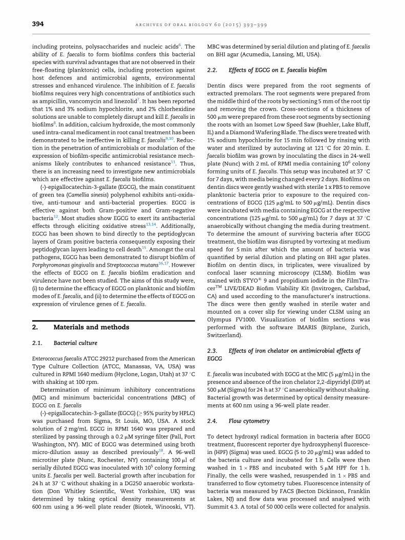
2.2. Effects of EGCG on E. faecalis biofilm
Dentin discs were prepared from the root segments of
extracted premolars. The root segments were prepared from
the middle third of the roots by sectioning 5 mm of the root tip
and removing the crown. Cross-sections of a thickness of
500 mm were prepared from these root segments by sectioning
the roots with an Isomet Low Speed Saw (Buehler, Lake Bluff,
IL) and a Diamond Wafering Blade. The discs were treated with
1% sodium hypochlorite for 15 min followed by rinsing with
water and sterilized by autoclaving at 121 8C for 20 min. E.
faecalis biofilm was grown by inoculating the discs in 24-well
plate (Nunc) with 2 mL of RPMI media containing 106 colony
forming units of E. faecalis. This setup was incubated at 37 8C
for 7 days, with media being changed every 2 days. Biofilms on
dentin discs were gently washed with sterile 1 x PBS to remove
planktonic bacteria prior to exposure to the required con-
centrations of EGCG (125 mg/mL to 500 mg/mL). Dentin discs
were incubated with media containing EGCG at the respective
concentrations (125 mg/mL to 500 mg/mL) for 7 days at 37 8C
anaerobically without changing the media during treatment.
To determine the amount of surviving bacteria after EGCG
treatment, the biofilm was disrupted by vortexing at medium
speed for 5 min after which the amount of bacteria was
quantified by serial dilution and plating on BHI agar plates.
Biofilm on dentin discs, in triplicates, were visualized by
confocal laser scanning microscopy (CLSM). Biofilm was
stained with STYO1 9 and propidium iodide in the FilmTra-
cerTM LIVE/DEAD Biofim Viability Kit (Invitrogen, Carlsbad,
CA) and used according to the manufacturer’s instructions.
The discs were then gently washed in sterile water and
mounted on a cover slip for viewing under CLSM using an
Olympus FV1000. Visualization of biofilm sections was
performed with the software IMARIS (Bitplane, Zurich,
Switzerland).
2.3. Effects of iron chelator on antimicrobial effects ofEGCG
E. faecalis was incubated with EGCG at the MIC (5 mg/mL) in the
presence and absence of the iron chelator 2,2-dipyridyl (DIP) at
500 mM (Sigma) for 24 h at 37 8C anaerobically without shaking.
Bacterial growth was determined by optical density measure-
ments at 600 nm using a 96-well plate reader.
2.4. Flow cytometry
To detect hydroxyl radical formation in bacteria after EGCG
treatment, fluorescent reporter dye hydroxyphenyl fluoresce-
in (HPF) (Sigma) was used. EGCG (5 to 20 mg/mL) was added to
the bacteria culture and incubated for 1 h. Cells were then
washed in 1 � PBS and incubated with 5 mM HPF for 1 h.
Finally, the cells were washed, resuspended in 1 � PBS and
transferred to flow cytometry tubes. Fluorescence intensity of
bacteria was measured by FACS (Becton Dickinson, Franklin
Lakes, NJ) and flow data was processed and analysed with
Summit 4.3. A total of 50 000 cells were collected for analysis.

Fig. 1 – MIC and MBC of EGCG on E. faecalis. (A) MIC of EGCG
on E. faecalis was determined by using the broth dilution
method. Serial dilution of EGCG were inoculated with E.
faecalis and incubated for 24 h. (B) MBC of EGCG on E.
faecalis were carried out by serial dilution and plating on
BHI agar plates. Experiments were carried out 3 times and
each time in triplicate. Data shown are the average results
obtained from 3 independent experiments. OD, optical
density; cfu, colony forming units. **P <0.01; ***P < 0.001
compared to the control group.
a r c h i v e s o f o r a l b i o l o g y 6 0 ( 2 0 1 5 ) 3 9 3 – 3 9 9 395
2.5. RNA extraction and quantitative polymerase chainreaction (qPCR)
2 mL of overnight culture of E. faecalis was treated with sub-
MIC of EGCG (2.5 mg/mL) and incubated at 37 8C anaerobically
for 3 h prior to RNA extraction using the GeneAll RNA
Extraction Kit (Seoul, Korea) according to the manufacturer’s
protocol. Reverse transcription was performed using iScriptTM
Reverse transcription Supermix (BioRad, Hercules, CA, USA).
mRNA expression of gelE, ace, cylA and sprE were determined
by qPCR using iTaq Universal SYBR Green Supermix (BioRad)
in an Applied Biosystem StepOne Plus. Sequences of the
primers used were gelatinase (gelE) forward 50- GGGGCAATA-
CAGGGAAAAAT and reverse 50-TCCTTCCCCAGTTTCCTTTT,
cytolysins activator (cylA) forward 50-GACTCGGGGATTGA-
TAGGC and reverse 50-TTTCCATCTGTCCCATCCAT, serine
protease (sprE) forward 50-GCGTCAATCGGAAGAATCAT and
reverse 50- CATCTTTGGCATTCGGATTT and collagen adhesin
Enteroccocus (ace) forward 50- GGAATGACCGAGAACGATGGCT
and reverse 50–ATTCGGTTGCGAACTATTGG. Expression of the
respective mRNAs was normalized to the relative abundance
of 16S ribosomal RNA gene (16S rRNA) forward 5’–CCGAGT
GCTTGCACTCAATTGG and reverse 50–CCTTGGTGAGCCGT-
TACCT19.
2.6. Statistical analysis
All experiments were carried out in biological triplicate and
repeated 3 independent times. Results were presented as
mean � SD. Statistical significance between multiple groups
was determined by the non-parametric Kruskal-Wallis one-
way ANOVA with Dunn’s multiple comparisons test using
Graphpad Prism software 6.0 (San Diego, CA, USA). Differences
were considered significant if p < 0.05.
3. Results
3.1. EGCG inhibits growth of Enterococcus faecalis
The minimum inhibitory concentration (MIC) and minimum
bactericidal concentration (MBC) were defined as the lowest
concentrations of EGCG required to inhibit growth and kill E.
faecalis respectively following 24 h of exposure. The MIC and
MBC of EGCG were determined to be 5 mg/mL and 20 mg/mL
respectively (Fig. 1A and B).
3.2. EGCG eradicates E. faecalis biofilm
E. faecalis biofilms were cultured on dentin discs. Control
biofilms exposed to media alone were robust with a majority of
cells alive (Fig. 2A). In contrast, viable bacterial cells decreased
significantly following EGCG exposure for 7-days (Fig. 2B–D).
To further corroborate these results, we tested the viability of
these biofilms after exposure to EGCG by bacteriological
culture. Exposure to EGCG significantly reduced bacterial
counts compared to the untreated biofilm (Fig. 2E). Complete
eradication of E. faecalis biofilm was achieved with 500 mg/mL
EGCG.
3.3. Bactericidal action of EGCG involves intracellular iron
Hydroxyphenyl fluorescein (HPF) dye is commonly used to
detect intracellular hydroxyl radicals. In the presence of 5, 10
or 20 mg/mL EGCG, there was a significant increase in HPF
fluorescence (Fig. 3A). Application of iron chelators such as
DIP, a highly membrane permeable iron chelator, is an
established method of blocking Fenton reaction-mediated
hydroxyl radical formation by sequestering unbound intracel-
lular iron20. As shown in Fig. 3B, the addition of DIP protected
E. faecalis against the antibacterial effects of EGCG.
3.4. EGCG inhibits the expression of virulence genes of E.faecalis
The effects of EGCG on the major virulence genes expression
of E. faecalis examined were ace, cylA, gelE and sprE. Treatment
of E. faecalis with sub-MIC of EGCG (2.5 mg/mL) significantly
inhibited the expression of these genes by >75% compared to
the untreated control (Fig. 4).
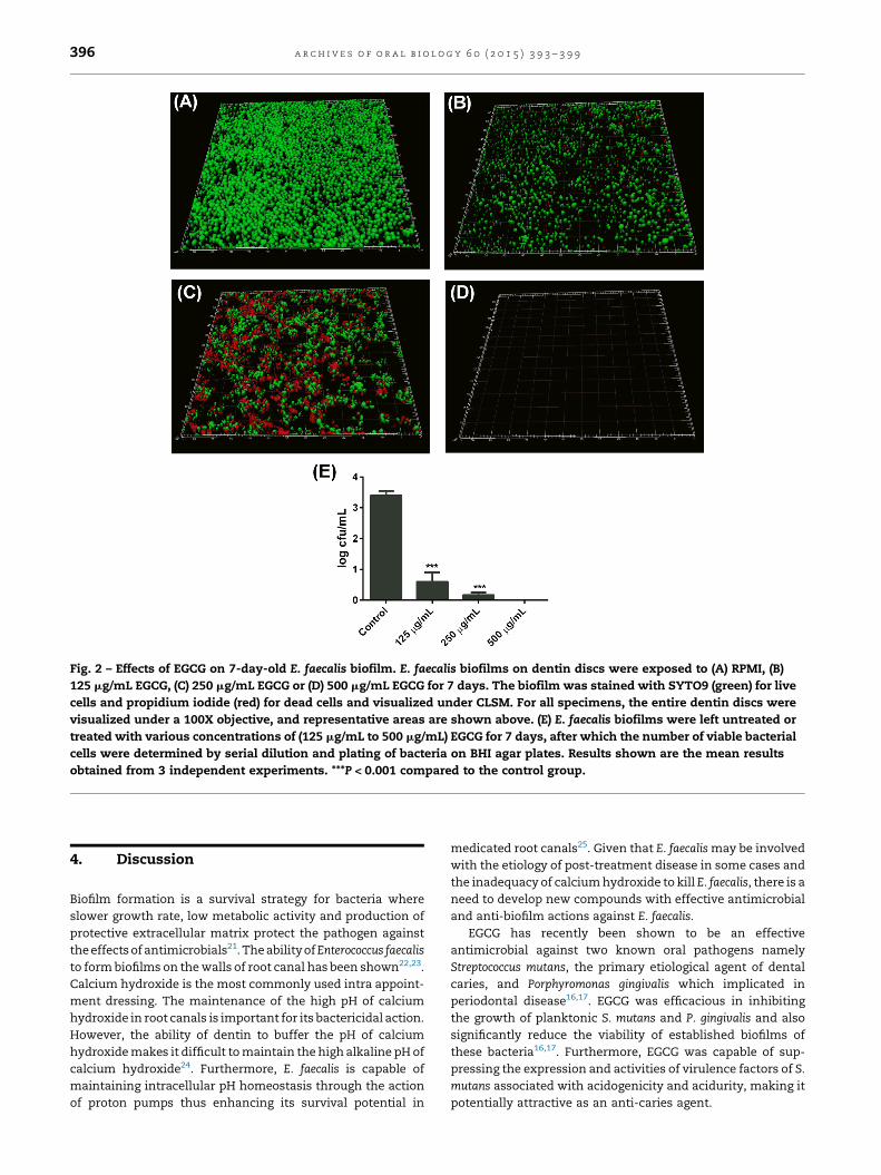
Fig. 2 – Effects of EGCG on 7-day-old E. faecalis biofilm. E. faecalis biofilms on dentin discs were exposed to (A) RPMI, (B)
125 mg/mL EGCG, (C) 250 mg/mL EGCG or (D) 500 mg/mL EGCG for 7 days. The biofilm was stained with SYTO9 (green) for live
cells and propidium iodide (red) for dead cells and visualized under CLSM. For all specimens, the entire dentin discs were
visualized under a 100X objective, and representative areas are shown above. (E) E. faecalis biofilms were left untreated or
treated with various concentrations of (125 mg/mL to 500 mg/mL) EGCG for 7 days, after which the number of viable bacterial
cells were determined by serial dilution and plating of bacteria on BHI agar plates. Results shown are the mean results
obtained from 3 independent experiments. ***P < 0.001 compared to the control group.
a r c h i v e s o f o r a l b i o l o g y 6 0 ( 2 0 1 5 ) 3 9 3 – 3 9 9396
4. Discussion
Biofilm formation is a survival strategy for bacteria where
slower growth rate, low metabolic activity and production of
protective extracellular matrix protect the pathogen against
the effects of antimicrobials21. The ability of Enterococcus faecalis
to form biofilms on the walls of root canal has been shown22,23.
Calcium hydroxide is the most commonly used intra appoint-
ment dressing. The maintenance of the high pH of calcium
hydroxide in root canals is important for its bactericidal action.
However, the ability of dentin to buffer the pH of calcium
hydroxide makes it difficult to maintain the high alkaline pH of
calcium hydroxide24. Furthermore, E. faecalis is capable of
maintaining intracellular pH homeostasis through the action
of proton pumps thus enhancing its survival potential in
medicated root canals25. Given that E. faecalis may be involved
with the etiology of post-treatment disease in some cases and
the inadequacy of calcium hydroxide to kill E. faecalis, there is a
need to develop new compounds with effective antimicrobial
and anti-biofilm actions against E. faecalis.
EGCG has recently been shown to be an effective
antimicrobial against two known oral pathogens namely
Streptococcus mutans, the primary etiological agent of dental
caries, and Porphyromonas gingivalis which implicated in
periodontal disease16,17. EGCG was efficacious in inhibiting
the growth of planktonic S. mutans and P. gingivalis and also
significantly reduce the viability of established biofilms of
these bacteria16,17. Furthermore, EGCG was capable of sup-
pressing the expression and activities of virulence factors of S.
mutans associated with acidogenicity and acidurity, making it
potentially attractive as an anti-caries agent.

Fig. 3 – EGCG induces hydroxyl radicals formation in
E. faecalis. (A) E. faecalis were exposed to EGCG treatment at
5 mg/mL (green), 10 mg/mL (blue) or 20 mg/mL (red)
for 1 h and labelled with 5 mM hydroxyphenyl
fluorescein dye for another hour. A significant increase in
HPF fluorescence was observed as compared to control
(black). (B) The addition of 500 mM DIP protected
E. faecalis against the antibacterial effects of EGCG.
*P < 0.05.
Fig. 4 – Effects of EGCG on E. faecalis virulence genes. E.
faecalis were exposed to sub-MIC of EGCG (2.5 mg/mL) for
3 h. Total RNA was extracted and converted into cDNA,
and qPCR was carried out to determine changes in the
expressions of ace, cylA, gelE, and sprE compared to the
control. mRNA levels were normalized to 16S rRNA and
results shown are the mean results obtained from 3
independent experiments.
a r c h i v e s o f o r a l b i o l o g y 6 0 ( 2 0 1 5 ) 3 9 3 – 3 9 9 397
The efficacy of EGCG on E. faecalis biofilm is currently
unknown. Our results showed EGCG to be effective in
eradicating both planktonic and biofilm forms of E. faecalis.
In this study, 7-day E. faecalis biofilms were cultured on dentin
discs as a solid medium to provide an environment closely
resembling the growth of biofilm in root canals. Since
environmental stresses have been shown to increase resis-
tance of biofilms to antimicrobial treatments 7, minimal media
instead of nutrient rich media was used to culture E. faecalis
biofilms. This serves to mimic starvation conditions typically
encountered by bacteria in root canals. Thus, our results
suggest that EGCG may be a potentially useful natural agent to
eradicate E. faecalis.
We found that EGCG at sub-MIC significantly decrease the
expression of virulence determinants of E. faecalis. Virulence
factors such as gelatinase, collagen-binding antigen, cytoly-
sins and proteases enhance colonization, survival and
persistence of E. faecalis in the root canal. In E. faecalis, two
secreted proteases, mainly gelatinase and serine protease are
auto-regulated through the fsr system. Gelatinase, an extra-
cellular zinc-containing metalloproteinase, hydrolyzes gela-
tin, collagen, casein, hemoglobin, and other bioactive
compounds to provide peptide nutrients for its survival26. In
addition, gelatinase contributes to bone resorption and
degradation of dentin organic matrix, thus playing an
important role in pathogenesis of periapical inflammation.
Serine protease cleaves peptide bonds, together with collagen
binding protein, help E. faecalis to bind to collagen in dentin.
Cytolysins lyse erythrocytes, polymorphonuclear neutrophils
and macrophages, kills bacterial cells, thereby reducing
phagocytosis. Thus, these virulence factors contribute to the
survival and colonization of E. faecalis in the root canal.
The precise antimicrobial action of EGCG is still not fully
understood and appears to differ with different bacterial
species. For instance, EGCG has been suggested to disrupt DNA
synthesis in Stenotrophomonas maltophilia through its anti-
folate activity27. The antibacterial effects of EGCG have also
been attributed to its ability to bind and damage bacterial
membranes, and induction of oxidative stress through
hydrogen peroxide production13,14. Since production of hy-
droxyl radicals is a common mechanism of bactericidal but
not bacteriostatic antibiotics28, we hypothesized that EGCG
may kill E. faecalis through the production of hydroxyl radical.
Indeed, treatment of E. faecalis with EGCG induced the
production of hydroxyl radical (Fig. 3A). Hydroxyl radical is
formed through Fenton reaction through the reduction of
hydrogen peroxide by ferrous iron and is highly deleterious to
cellular components where it can damage DNA, proteins and
lipids20. In the presence of DIP, a highly membrane-permeable
iron chelator, the survival of E. faecalis in the presence of EGCG
was enhanced significantly (Fig. 3B). DIP likely reduces the
cytotoxic effects of hydroxyl radical by chelating ferrous iron.
Sources of ferrous iron for Fenton reaction can come from
extracellular sources through iron import or intracellular
sources through iron storage proteins or iron–sulfur clusters.
In addition to its anti-bacterial properties, EGCG also
exhibit anti-inflammatory effects by suppressing the activa-
tion of NFkB, the main transcription factor controlling the
expression of inflammatory genes29. EGCG is also known for
its antioxidant and free radical scavenger properties30. This

a r c h i v e s o f o r a l b i o l o g y 6 0 ( 2 0 1 5 ) 3 9 3 – 3 9 9398
compound has been shown to suppress bone resorption31,32.
In fact, EGCG significantly reduces the severity of periapical
lesions in a rat model of induced apical periodontitis and may
thus serve as an adjunct in root canal treatment to accelerate
the healing process33. Furthermore, toxicity studies have
shown EGCG to be well tolerated even at high concentrations
in humans34,35. Although EGCG was effective in eradicating E.
faecalis biofilm, a limitation in this study which warrants
further investigations will be to determine the antibiofilm
effects of EGCG in comparison with disinfectants currently
used in endodotic therapy, for example, calcium hydroxide
and sodium hypochloride.
In summary, our results showed EGCG to be effective in
disrupting E. faecalis biofilm and reducing the expression of
various virulence genes. These results support the potential of
EGCG to as a natural agent for the treatment of E. faecalis
related infections.
5. Conclusions
This study is the first to evaluate the effects of EGCG on E.
faecalis virulence and biofilm. Our results showed EGCG to be
effective in eradicating E. faecalis biofilm. Furthermore, this
compound at sub-MIC significantly attenuates the expression
of major virulence genes of this bacterial species. We further
show EGCG to exert its anti-bacterial effects through the
production of hydroxyl radical via Fenton pathway. Thus,
EGCG represents a promising natural antimicrobial compound
for endodontic treatment.
Acknowledgements
The authors deny any conflicts of interests related to this
study. This study was funded by a grant R221-000-051-112
from the Ministry of Education Singapore and R221-000-054-
101 from the Faculty of Dentistry, National University of
Singapore.
r e f e r e n c e s
1. Molander A, Reit C, Dahlen. Kvist T. Microbiological status ofroot-filled teeth with apical periodontitis. Int Endod J1998;31(January (1)):1–7.
2. Murray BE. The life and times of the Enterococcus. ClinMicrobiol Rev 1990;3(January (1)):46–65.
3. Siqueira Jr JF, Ro cas IN. Polymerase chain reaction-basedanalysis of microorganisms associated with failedendodontic treatment. Oral Surg Oral Med Oral Pathol OralRadiol Endod 2004;97(January (1)):85–94.
4. Stuart CH, Schwartz SA, Beeson TJ, Owatz CB.Enterococcus faecalis: its role in root canal treatment failureand current concepts in retreatment. J Endod2006;32(February (2)):93–8.
5. Kayaoglu G, Ørstavik D. Virulence factors of Enterococcusfaecalis: relationship to endodontic disease. Crit Rev Oral BiolMed 2004;15(5):308–20.
6. Costerton JW. Cystic fibrosis pathogenesis and the role ofbiofilms in persistent infection. Trends Microbiol2001;9(Februrary (2)):50–2.
7. Sandoe JA, Wysome J, West AP, Heritage J, Wilcox MH.Measurement of ampicillin, vancomycin, linezolid andgentamicin activity against enterococcal biofilms. JAntimicrob Chemother 2006;57(April (4)):767–70.
8. Clegg MS, Vertucci FJ, Walker C, Belanger M, Britto LR. Theeffect of exposure to irrigant solutions on apical dentinbiofilms in vitro. J Endod 2006;32(May (5)):434–7.
9. McHugh CP, Zhang P, Michalek S, Eleazer PD. pH requiredto kill Enterococcus faecalis in vitro. J Endod 2004;30(April(4)):218–9.
10. Tronstad L, Andreasen JO, Hasselgren G, Kristerson L. Riis I.pH changes in dental tissues after root canal filling withcalcium hydroxide. J Endod 1981;7(January (1)):17–21.
11. Ehrlich GD, Arciola CR. From Koch’s postulates to biofilmtheory. The lesson of Bill Costerton. Int J Artif Organs2012;35(October (10)):695–9.
12. Steinmann J, Buer J, Pietschmann T, Steinmann E. Anti-infective properties of epigallocatechin-3-gallate (EGCG), acomponent of green tea. Br J Pharmacol 2013;168(March(5)):1059–73.
13. Arakawa H, Maeda M, Okubo S, Shimamura T. Role ofhydrogen peroxide in bactericidal action of catechin. BiolPharm Bull 2004;27(March (3)):277–81.
14. Cui Y, Oh YJ, Lim J, Youn M, Lee I, Pak HK, et al. AFM study ofthe differential inhibitory effects of the green teapolyphenol (-)-epigallocatechin-3-gallate (EGCG) againstGram-positive and Gram-negative bacteria. Food Microbiol2012;29(February (1)):80–7.
15. Yoda Y, Hu ZQ, Zhao WH, Shimamura T. Differentsusceptibilities of Staphylococcus and Gram-negative rods toepigallocatechin gallate. J Infect Chemother 2004;10(February(1)):55–8.
16. Asahi Y, Noiri Y, Miura J, Maezono H, Yamaguchi M,Yamamoto R, et al. Effects of the tea catechinepigallocatechin gallate on Porphyromonas gingivalis biofilms.J Appl Microbiol 2014;116(May (5)):1164–71.
17. Xu X, Zhou XD, Wu CD. The tea catechin epigallocatechingallate suppresses cariogenic virulence factors ofStreptococcus mutans. Antimicrob Agents Chemother2011;55(March (3)):1229–36.
18. Andrews JM. Determination of minimum inhibitoryconcentrations. J Antimicrob Chemother 2001 July;48(Suppl1):5–16.
19. Livak KJ, Schmittgen TD. Analysis of relative geneexpression data using real-time quantitative PCR and the2(-Delta Delta C(T)) Method. Methods 2001;25(December(4)):402–8.
20. Imlay JA, Chin SM, Linn S. Toxic DNA damage by hydrogenperoxide through the Fenton reaction in vivo and in vitro.Science 1988 Apr 29;240(April 4852):640–2.
21. Costerton JW, Stewart PS, Greenberg EP. Bacterial biofilms: acommon cause of persistent infections. Science1999;284(May 21 5418):1318–22.
22. Distel JW, Hatton JF, Gillespie MJ. Biofilm formation inmedicated root canals. J Endod 2002;28(October (10)):689–93.
23. Kishen A, George S, Kumar R. Enterococcus faecalis-mediated biomineralized biofilm formation on rootcanal dentine in vitro. J Biomed Mater Res A 2006;77(May(2)):406–15.
24. Portenier I, Haapasalo H, Rye A, Waltimo T, Ørstavik D,Haapasalo M. Inactivation of root canal medicaments bydentine, hydroxylapatite and bovine serum albumin. IntEndod J 2001;34(April (3)):184–8.
25. Evans M, Davies JK, Sundqvist G, Figdor D. Mechanismsinvolved in the resistance of Enterococcus faecalis to calciumhydroxide. Int Endod J 2002;35(March (3)):221–8.
26. Vergis EN, Shankar N, Chow JW, Hayden MK, Snydman DR,Zervos MJ, et al. Association between the presence ofenterococcal virulence factors gelatinase, hemolysin, and
a r c h i v e s o f o r a l b i o l o g y 6 0 ( 2 0 1 5 ) 3 9 3 – 3 9 9 399
enterococcal surface protein and mortality among patientswith bacteremia due to Enterococcus faecalis. Clin Infect Dis2002;35(September 1 5):570–5.
27. Navarro-Martinez MD, Navarro-Peran E, Cabezas-Herrera J,Ruiz-Gomez J, Garcia-Canovas F, Rodriguez-Lopez JN.Antifolate activity of epigallocatechin gallate againstStenotrophomonas maltophilia. Antimicrob Agents Chemother2005;49(July (7)):2914–20.
28. Kohanski MA, Dwyer DJ, Hayete B, Lawrence CA, Collins JJ. Acommon mechanism of cellular death induced bybactericidal antibiotics. Cell 2007;130(September 7 (5)):797–810.
29. Yang F, de Villiers WJ, McClain CJ, Varilek GW. Green teapolyphenols block endotoxin-induced tumour necrosisfactor-production and lethality in a murine model. J Nutr1998;128(December (12)):2334–40.
30. Mitscher LA, Jung M, Shankel D, Dou JH, Steele L, Pillai SP.Chemoprotection: a review of the potential therapeuticantioxidant properties of green tea (Camellia sinensis) andcertain of its constituents. Med Res Rev 1997;17(July (4)):327–65.
31. Yun JH, Kim CS, Cho KS, Chai JK, Kim CK, Choi SH.(-)-Epigallocatechin gallate induces apoptosis, via caspaseactivation, in osteoclasts differentiated from RAW 264.7cells. J Periodontal Res 2007;42(June (3)):212–8.
32. Yun JH, Pang EK, Kim CS, Yoo YJ, Cho KS, Chai JK, et al.Inhibitory effects of green tea polyphenol (-)-epigallocatechin gallate on the expression of matrixmetalloproteinase-9 and on the formation of osteoclasts.J Periodontal Res 2004;39(October (5)):300–7.
33. Lee YL, Hong CY, Kok SH, Hou KL, Lin YT, Chen MH, et al. Anextract of green tea, epigallocatechin-3-gallate, reducesperiapical lesions by inhibiting cysteine-rich 61 expressionin osteoblasts. J Endod 2009 Feb;35(February (2)):206–11.
34. Isbrucker RA, Bausch J, Edwards JA, Wolz E. Safety studieson epigallocatechin gallate (EGCG) preparations Part 1:genotoxicity. Food Chem Toxicol 2006;44(May (5)):626–35.
35. Isbrucker RA, Edwards JA, Wolz E, Davidovich A, Bausch J.Safety studies on epigallocatechin gallate (EGCG)preparations. Part 2: dermal, acute and short-term toxicitystudies. Food Chem Toxicol 2006 May;44(May (5)):636–50.