Crystal structure and biochemical properties of putrescine carbamoyltransferase from Enterococcus...
14
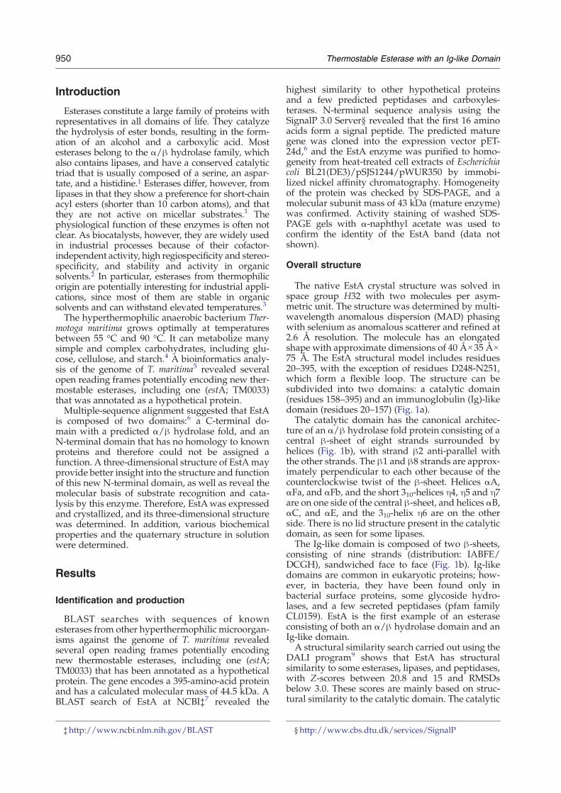
Crystal Structure and Biochemical Properties of a Novel Thermostable Esterase Containing an Immunoglobulin-Like Domain Mark Levisson 1 ⁎†, Lei Sun 1,2 †, Sjon Hendriks 1 , Peter Swinkels 1 , Twan Akveld 1 , Jelle B. Bultema 2 , Arjan Barendregt 3 , Robert H. H. van den Heuvel 3 , Bauke W. Dijkstra 2 , John van der Oost 1 and Servé W. M. Kengen 1 1 Laboratory of Microbiology, Department of Agrotechnology and Food Sciences, Wageningen University, Dreijenplein 10, 6703 HB Wageningen, The Netherlands 2 Laboratory of Biophysical Chemistry, University of Groningen, Nijenborgh 4, 9747 AG Groningen, The Netherlands 3 Biomolecular Mass Spectrometry and Proteomics Group, Bijvoet Center for Biomolecular Research and Utrecht Institute for Pharmaceutical Sciences, Utrecht University, Sorbonnelaan 16, 3584 CA Utrecht, The Netherlands Received 18 July 2008; received in revised form 24 October 2008; accepted 27 October 2008 Available online 5 November 2008 Comparative analysis of the genome of the hyperthermophilic bacterium Thermotoga maritima revealed a hypothetical protein (EstA) with typical esterase features. The EstA protein was functionally produced in Escherichia coli and purified to homogeneity. It indeed displayed esterase activity with optima at or above 95 °C and at pH 8.5, with a preference for esters with short acyl chains (C2–C10). Its 2.6-Å-resolution crystal structure revealed a classical α/β hydrolase domain with a catalytic triad consisting of a serine, an aspartate, and a histidine. EstA is irreversibly inhibited by the orga- nophosphate paraoxon. A 3.0-Å-resolution structure confirmed that this inhibitor binds covalently to the catalytic serine residue of EstA. Remark- ably, the structure also revealed the presence of an N-terminal immunoglo- bulin (Ig)-like domain, which is unprecedented among esterases. EstA forms a hexamer both in the crystal and in solution. Electron microscopy showed that the hexamer in solution is identical with the hexamer in the crystal, which is formed by two trimers, with the N-terminal domains facing each other. Mutational studies confirmed that residues Phe89, Phe112, Phe116, Phe246, and Trp377 affect enzyme activity. A truncated mutant of EstA, in which the Ig-like domain was removed, showed only 5% of wild- type activity, had lower thermostability, and failed to form hexamers. These data suggest that the Ig-like domain plays an important role in the enzyme multimerization and activity of EstA. © 2008 Elsevier Ltd. All rights reserved. Edited by M. Guss Keywords: esterase; Thermotoga maritima; α/β hydrolase; immunoglobulin fold; paraoxon *Corresponding author. E-mail address: [email protected]. † M.L. and L.S. contributed equally to this work. Present address: R.H.H. van den Heuvel, Biotech Analytical Development, Schering-Plough Corporation, Postbus 20, 5340 BH Oss, The Netherlands. Abbreviations used: MAD, multiwavelength anomalous dispersion; PDB, Protein Data Bank; DEP, diethyl phosphate; NCS, noncrystallographic symmetry; DLS, dynamic light scattering; EM, electron microscopy; 7-ACA, 7-aminocephalosporanic acid; SeMet, selenomethionine; CAPS, 3-(cyclohexylamino) 1-propanesulphonic acid. doi:10.1016/j.jmb.2008.10.075 J. Mol. Biol. (2009) 385, 949–962 Available online at www.sciencedirect.com 0022-2836/$ - see front matter © 2008 Elsevier Ltd. All rights reserved.
-
Upload
independent -
Category
Documents
-
view
1 -
download
0
Transcript of Crystal structure and biochemical properties of putrescine carbamoyltransferase from Enterococcus...
doi:10.1016/j.jmb.2008.10.075 J. Mol. Biol. (2009) 385, 949–962
Available online at www.sciencedirect.com
Crystal Structure and Biochemical Properties of aNovel Thermostable Esterase Containing anImmunoglobulin-Like Domain
Mark Levisson1⁎†, Lei Sun1,2†, Sjon Hendriks1, Peter Swinkels1,Twan Akveld1, Jelle B. Bultema2, Arjan Barendregt3,Robert H. H. van den Heuvel3, Bauke W. Dijkstra2,John van der Oost1 and Servé W. M. Kengen1
1Laboratory of Microbiology,Department of Agrotechnologyand Food Sciences, WageningenUniversity, Dreijenplein 10,6703 HB Wageningen,The Netherlands2Laboratory of BiophysicalChemistry, University ofGroningen, Nijenborgh 4,9747 AG Groningen,The Netherlands3Biomolecular MassSpectrometry and ProteomicsGroup, Bijvoet Center forBiomolecular Research andUtrecht Institute forPharmaceutical Sciences,Utrecht University,Sorbonnelaan 16, 3584 CAUtrecht, The Netherlands
Received 18 July 2008;received in revised form24 October 2008;accepted 27 October 2008Available online5 November 2008
*Corresponding author. E-mail addr† M.L. and L.S. contributed equalPresent address: R.H.H. van den H
5340 BH Oss, The Netherlands.Abbreviations used: MAD, multiw
NCS, noncrystallographic symmetry7-aminocephalosporanic acid; SeMe
0022-2836/$ - see front matter © 2008 E
Comparative analysis of the genome of the hyperthermophilic bacteriumThermotoga maritima revealed a hypothetical protein (EstA) with typicalesterase features. The EstA protein was functionally produced in Escherichiacoli and purified to homogeneity. It indeed displayed esterase activity withoptima at or above 95 °C and at pH 8.5, with a preference for esters withshort acyl chains (C2–C10). Its 2.6-Å-resolution crystal structure revealed aclassical α/β hydrolase domain with a catalytic triad consisting of a serine,an aspartate, and a histidine. EstA is irreversibly inhibited by the orga-nophosphate paraoxon. A 3.0-Å-resolution structure confirmed that thisinhibitor binds covalently to the catalytic serine residue of EstA. Remark-ably, the structure also revealed the presence of an N-terminal immunoglo-bulin (Ig)-like domain, which is unprecedented among esterases. EstAforms a hexamer both in the crystal and in solution. Electron microscopyshowed that the hexamer in solution is identical with the hexamer in thecrystal, which is formed by two trimers, with the N-terminal domains facingeach other. Mutational studies confirmed that residues Phe89, Phe112,Phe116, Phe246, and Trp377 affect enzyme activity. A truncated mutant ofEstA, in which the Ig-like domain was removed, showed only 5% of wild-type activity, had lower thermostability, and failed to form hexamers. Thesedata suggest that the Ig-like domain plays an important role in the enzymemultimerization and activity of EstA.
© 2008 Elsevier Ltd. All rights reserved.
Keywords: esterase; Thermotoga maritima; α/β hydrolase; immunoglobulinfold; paraoxon
Edited by M. Gussess: [email protected] to this work.euvel, Biotech Analytical Development, Schering-Plough Corporation, Postbus 20,
avelength anomalous dispersion; PDB, Protein Data Bank; DEP, diethyl phosphate;; DLS, dynamic light scattering; EM, electron microscopy; 7-ACA,t, selenomethionine; CAPS, 3-(cyclohexylamino) 1-propanesulphonic acid.
lsevier Ltd. All rights reserved.
950 Thermostable Esterase with an Ig-like Domain
Introduction
Esterases constitute a large family of proteins withrepresentatives in all domains of life. They catalyzethe hydrolysis of ester bonds, resulting in the form-ation of an alcohol and a carboxylic acid. Mostesterases belong to the α/β hydrolase family, whichalso contains lipases, and have a conserved catalytictriad that is usually composed of a serine, an aspar-tate, and a histidine.1 Esterases differ, however, fromlipases in that they show a preference for short-chainacyl esters (shorter than 10 carbon atoms), and thatthey are not active on micellar substrates.1 Thephysiological function of these enzymes is often notclear. As biocatalysts, however, they are widely usedin industrial processes because of their cofactor-independent activity, high regiospecificity and stereo-specificity, and stability and activity in organicsolvents.2 In particular, esterases from thermophilicorigin are potentially interesting for industrial appli-cations, since most of them are stable in organicsolvents and can withstand elevated temperatures.3
The hyperthermophilic anaerobic bacterium Ther-motoga maritima grows optimally at temperaturesbetween 55 °C and 90 °C. It can metabolize manysimple and complex carbohydrates, including glu-cose, cellulose, and starch.4 A bioinformatics analy-sis of the genome of T. maritima5 revealed severalopen reading frames potentially encoding new ther-mostable esterases, including one (estA; TM0033)that was annotated as a hypothetical protein.Multiple-sequence alignment suggested that EstA
is composed of two domains:6 a C-terminal do-main with a predicted α/β hydrolase fold, and anN-terminal domain that has no homology to knownproteins and therefore could not be assigned afunction. A three-dimensional structure of EstA mayprovide better insight into the structure and functionof this new N-terminal domain, as well as reveal themolecular basis of substrate recognition and cata-lysis by this enzyme. Therefore, EstAwas expressedand crystallized, and its three-dimensional structurewas determined. In addition, various biochemicalproperties and the quaternary structure in solutionwere determined.
Results
Identification and production
BLAST searches with sequences of knownesterases from other hyperthermophilic microorgan-isms against the genome of T. maritima revealedseveral open reading frames potentially encodingnew thermostable esterases, including one (estA;TM0033) that has been annotated as a hypotheticalprotein. The gene encodes a 395-amino-acid proteinand has a calculated molecular mass of 44.5 kDa. ABLAST search of EstA at NCBI‡7 revealed the
‡http://www.ncbi.nlm.nih.gov/BLAST
highest similarity to other hypothetical proteinsand a few predicted peptidases and carboxyles-terases. N-terminal sequence analysis using theSignalP 3.0 Server§ revealed that the first 16 aminoacids form a signal peptide. The predicted maturegene was cloned into the expression vector pET-24d,6 and the EstA enzyme was purified to homo-geneity from heat-treated cell extracts of Escherichiacoli BL21(DE3)/pSJS1244/pWUR350 by immobi-lized nickel affinity chromatography. Homogeneityof the protein was checked by SDS-PAGE, and amolecular subunit mass of 43 kDa (mature enzyme)was confirmed. Activity staining of washed SDS-PAGE gels with α-naphthyl acetate was used toconfirm the identity of the EstA band (data notshown).
Overall structure
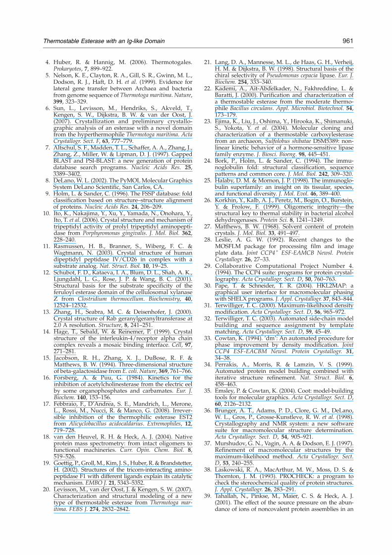
The native EstA crystal structure was solved inspace group H32 with two molecules per asym-metric unit. The structure was determined by multi-wavelength anomalous dispersion (MAD) phasingwith selenium as anomalous scatterer and refined at2.6 Å resolution. The molecule has an elongatedshape with approximate dimensions of 40 Å×35 Å×75 Å. The EstA structural model includes residues20–395, with the exception of residues D248-N251,which form a flexible loop. The structure can besubdivided into two domains: a catalytic domain(residues 158–395) and an immunoglobulin (Ig)-likedomain (residues 20–157) (Fig. 1a).The catalytic domain has the canonical architec-
ture of an α/β hydrolase fold protein consisting of acentral β-sheet of eight strands surrounded byhelices (Fig. 1b), with strand β2 anti-parallel withthe other strands. The β1 and β8 strands are approx-imately perpendicular to each other because of thecounterclockwise twist of the β-sheet. Helices αA,αFa, and αFb, and the short 310-helices η4, η5 and η7are on one side of the central β-sheet, and helices αB,αC, and αE, and the 310-helix η6 are on the otherside. There is no lid structure present in the catalyticdomain, as seen for some lipases.The Ig-like domain is composed of two β-sheets,
consisting of nine strands (distribution: IABFE/DCGH), sandwiched face to face (Fig. 1b). Ig-likedomains are common in eukaryotic proteins; how-ever, in bacteria, they have been found only inbacterial surface proteins, some glycoside hydro-lases, and a few secreted peptidases (pfam familyCL0159). EstA is the first example of an esteraseconsisting of both an α/β hydrolase domain and anIg-like domain.A structural similarity search carried out using the
DALI program9 shows that EstA has structuralsimilarity to some esterases, lipases, and peptidases,with Z-scores between 20.8 and 15 and RMSDsbelow 3.0. These scores are mainly based on struc-tural similarity to the catalytic domain. The catalytic
§http://www.cbs.dtu.dk/services/SignalP
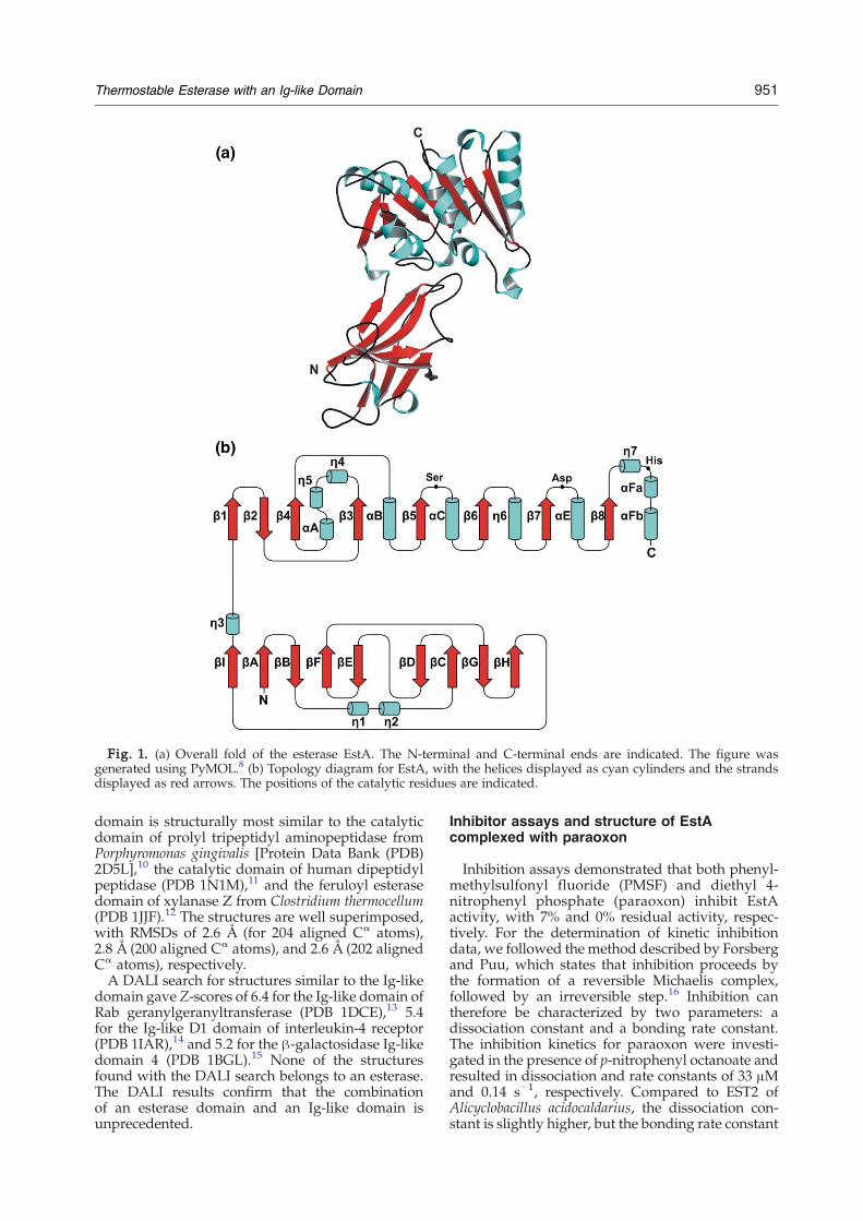
Fig. 1. (a) Overall fold of the esterase EstA. The N-terminal and C-terminal ends are indicated. The figure wasgenerated using PyMOL.8 (b) Topology diagram for EstA, with the helices displayed as cyan cylinders and the strandsdisplayed as red arrows. The positions of the catalytic residues are indicated.
951Thermostable Esterase with an Ig-like Domain
domain is structurally most similar to the catalyticdomain of prolyl tripeptidyl aminopeptidase fromPorphyromonas gingivalis [Protein Data Bank (PDB)2D5L],10 the catalytic domain of human dipeptidylpeptidase (PDB 1N1M),11 and the feruloyl esterasedomain of xylanase Z from Clostridium thermocellum(PDB 1JJF).12 The structures are well superimposed,with RMSDs of 2.6 Å (for 204 aligned Cα atoms),2.8 Å (200 aligned Cα atoms), and 2.6 Å (202 alignedCα atoms), respectively.A DALI search for structures similar to the Ig-like
domain gave Z-scores of 6.4 for the Ig-like domain ofRab geranylgeranyltransferase (PDB 1DCE),13 5.4for the Ig-like D1 domain of interleukin-4 receptor(PDB 1IAR),14 and 5.2 for the β-galactosidase Ig-likedomain 4 (PDB 1BGL).15 None of the structuresfound with the DALI search belongs to an esterase.The DALI results confirm that the combinationof an esterase domain and an Ig-like domain isunprecedented.
Inhibitor assays and structure of EstAcomplexed with paraoxon
Inhibition assays demonstrated that both phenyl-methylsulfonyl fluoride (PMSF) and diethyl 4-nitrophenyl phosphate (paraoxon) inhibit EstAactivity, with 7% and 0% residual activity, respec-tively. For the determination of kinetic inhibitiondata, we followed the method described by Forsbergand Puu, which states that inhibition proceeds bythe formation of a reversible Michaelis complex,followed by an irreversible step.16 Inhibition cantherefore be characterized by two parameters: adissociation constant and a bonding rate constant.The inhibition kinetics for paraoxon were investi-gated in the presence of p-nitrophenyl octanoate andresulted in dissociation and rate constants of 33 μMand 0.14 s−1, respectively. Compared to EST2 ofAlicyclobacillus acidocaldarius, the dissociation con-stant is slightly higher, but the bonding rate constant
952 Thermostable Esterase with an Ig-like Domain
is comparable.17 Inhibition kinetics for PMSF werenot measurable in the presence of substrate. This ispossibly a result of a high dissociation rate constantbecause inhibition was observed when EstA waspreincubated with PMSF in the absence of substrate.Other chemical agents such as diethyl pyrocarbo-nate, dithiothreitol, divalent metal ions, and ethyle-nediaminetetraacetic acid did not influence EstAactivity. EstA was cocrystallized with PMSF andparaoxon, but only crystals for the latter wereobtained. Electron density maps for the paraoxoncocrystallized crystals displayed clear density for adiethyl phosphate (DEP) moiety covalently boundto the side chain of Ser286. The density revealed thatthe p-nitrophenol-leaving group of paraoxon hadbeen cleaved off during cocrystallization, therebyleaving a tetrahedral product resembling the firsttransition state formed during ester hydrolysis. Thenative and paraoxon-bound structures superimposewith an RMSD of 0.4 Å. There are no significantdifferences between the two structures.
Quaternary structure
There are two molecules (protomers A and B) inthe asymmetric unit that are related by noncrystallo-graphic symmetry (NCS). Protomers A and B areessentially similar, with an RMSD of 0.3 over all Cα
atoms. The refined model reveals that the two mole-cules form an interface of 280 Å2 in each monomer, avalue suggesting a low association constant. Theinterface involves four β-strands (βC, βD, βG,and βH) from the N-terminal Ig-like domain ofboth molecules. An intermolecular hydrogen bond(AspA56-LysB61) stabilizes the dimer. With the3-fold crystallographic symmetry of space groupH32, three dimers form a tightly packed hexamer,burying a total surface area of 3585 Å2. The inter-dimer interfaces involve residues from αA, αE, β7,β8, and the loop between β2 and β3, as well as theN-termini of βF, βE, and the loop between them. Saltbridges and hydrogen bonds are formed betweenneighboring protomers such as ArgA213-GluA′362,ArgA222-GluA′360, GluA99-LysA′363, ArgA187-GluA′351, GlnA384-TyrA′358, and AspA182-LysA′347. On average, a surface area of 1454 Å2 permonomer is buried upon hexamerization. Multiple-sequence alignment shows that the residues in-volved in the dimer and trimer interfaces are barelyconserved, suggesting a novel mode of hexamerformation.Native PAGE and size exclusion chromatography
showed that multiple quaternary structures of EstAare present in solution. Therefore, native mass spec-trometry—in which EstAwas measured under non-denaturing conditions in ammonium acetate(pH 6.8)18—and dynamic light scattering (DLS)were performed and revealed that the purifiedEstA protein was present predominantly (N50%) asa hexamer in solution. However, to a minor extent,monomeric, dimeric, trimeric, and highermultimericforms of EstA were also detected using massspectrometry.
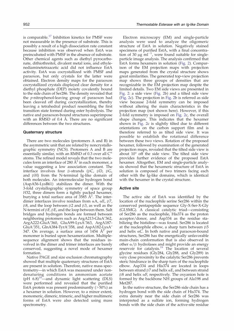
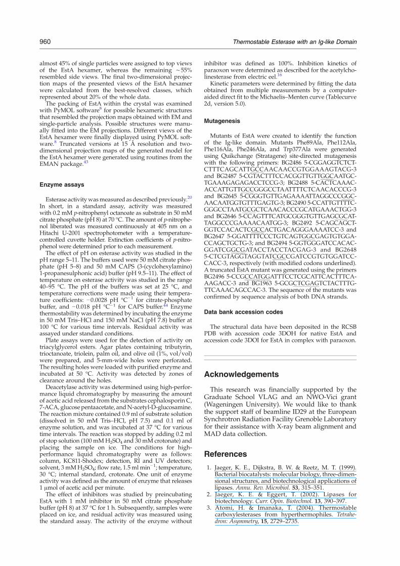
Electron microscopy (EM) and single-particleanalysis were used to analyze the oligomericstructure of EstA in solution. Negatively stainedspecimens of purified EstA, with a final concentra-tion of 30 μg ml−1, were found suitable for single-particle image analysis. The analysis confirmed thatEstA forms hexamers in solution (Fig. 2). Compar-ison of the EM projection maps with projectionmaps generated from the crystal structure showsgreat similarities. The generated top-view projectionmap shows three groups of densities that arerecognizable in the EM projection map despite thelimited details. Two EM side views are presented inFig. 2: a side view (Fig. 2b) and a tilted side view(Fig. 2c). The projection in Fig. 2b is referred as sideview because 2-fold symmetry can be imposedwithout altering the main characteristics in theprojection map (not shown here). However, if this2-fold symmetry is imposed on Fig. 2c, the overallshape changes. This indicates that the hexamershown in Fig. 2c is slightly tilted due to differentorientations on the carbon support film and istherefore referred to as tilted side view. It waspossible to establish the rotational differencebetween these two views. Rotation of the proposedhexamer, followed by examination of the generatedprojection maps, revealed that the tilted side view isabout 10° off the side view. The tilted side viewprovides further evidence of the proposed EstAhexamer. Altogether, EM and single-particle analy-sis showed that the hexameric structure of EstA insolution is composed of two trimers facing eachother with the Ig-like domains, which is identicalwith the hexamer in the crystal structure.
Active site
The active site of EstA was identified by thelocation of the nucleophile serine Ser286 within theconserved pentapeptide sequence Gly-X-Ser-X-Gly(GLSMG). A classical catalytic triad—consistingof Ser286 as the nucleophile, His374 as the protonacceptor/donor, and Asp334 as the residue sta-bilizing the histidine—was found. Ser286 is locatedat the nucleophile elbow, a sharp turn between β5and helix αC. In both native and paraoxon-boundstructures, Ser286 has the energetically unfavorablemain-chain conformation that is also observed inother α/β hydrolases and might provide an energyreservoir for catalysis.19 The location of severalglycine residues (Gly284, Gly288, and Gly289) invery close proximity to the catalytic Ser286 preventssteric hindrance in the sharp turn of the nucleophileelbow. Asp334 and His374 are located in loopsbetween strand β7 and helix αE, and between strandβ8 and helix αF, respectively. The oxyanion hole isformed by the backbone NH groups of Ala198 andMet287.In the native structure, the Ser286 side chain has a
hydrogen bond with the side chain of His374. Theextra density near the side chain of Ser286 wasinterpreted as a sulfate ion, forming hydrogenbonds with the side chain of the active-site residue
Fig. 2. Comparison of the EM projection maps of the EstA hexamer. Top view (a), side view (b), and 10° off side view(c). Two-dimensional projection maps obtained by statistical analysis and classification (a–c); the comparable two-dimensional projection maps with 15 Å resolution (d–f) generated from the proposed EstA hexameric structure (g–i).8 Thescale bar represents 50 Å.
953Thermostable Esterase with an Ig-like Domain
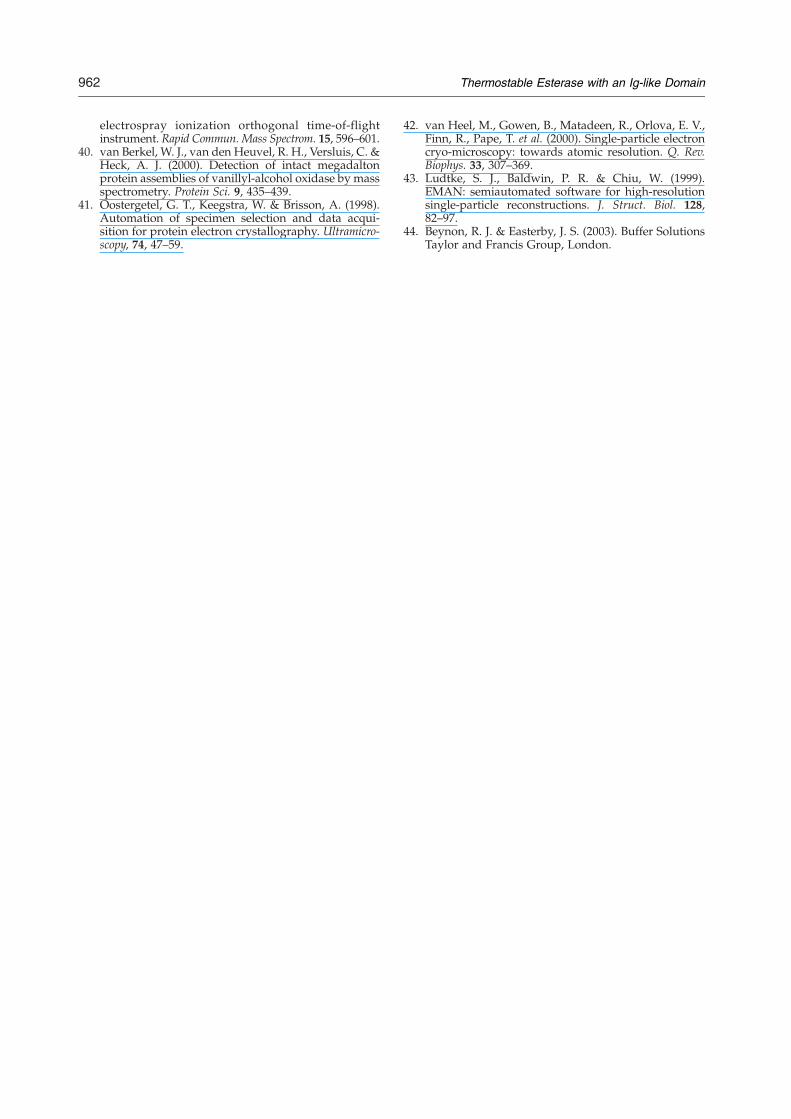
Ser286 and the main-chain nitrogen atoms of Ala198and Met287, and mimicking the oxyanion transitionstate (Fig. 3). Sulfate is present in the crystallizationbuffer and is commonly found as an adduct in otherstructures.In the paraoxon-bound structure, paraoxon is
stabilized by the covalent bond with Ser286, hydro-gen-bonding interactions with the oxyanion hole,and a hydrogen bond to the side chain of His374(Fig. 4). One of the two ethyl arms of boundparaoxon points toward the surface of EstA, whilethe other follows the groove of the acyl-bindingpocket.The catalytic triad and oxyanion hole are located
at the end of a surface depression, characteristicof many α/β hydrolases (Fig. 5a). This groove ex-tends by approximately 15 Å from the catalyticserine into the enzyme where the gorge is closedby Glu33 (S1 in Fig. 5b). The volume of the grooveis ∼790 Å3. The active-site serine residue is coveredby a valine residue (Val336) and a phenylalanine
residue (Phe116; of the Ig-like domain), resulting ina tunnel. The covered gorge extends to the othersite of the catalytic serine by approximately 5 Å.The active-site gorge is slightly curved and isformed by the hydrophobic side chains of Ala198,Gly199, Leu285, Trp377, and Phe112. The tunnel isformed by the hydrophobic side chains of Val336,Val337, Phe116, Leu245, Phe246, and Phe89 (of theN-terminal domain of subunit B), and by thenonpolar side chain of Tyr290. A second side gorgewith a volume of ∼210 Å3 also provides access tothe active site (S2 in Fig. 5b). The∼7-Å-wide openingis lined by residues Leu245, Asp248, Arg249,Pro252, Phe253, Tyr290, Val336, Val337, Pro338,and Asn341.
Substrate specificity and kinetics
Kinetic parameters for the hydrolysis of p-nitro-phenyl esters with varying acyl-chain lengths aregiven in Table 1. Catalytic activity (Kcat) decreases
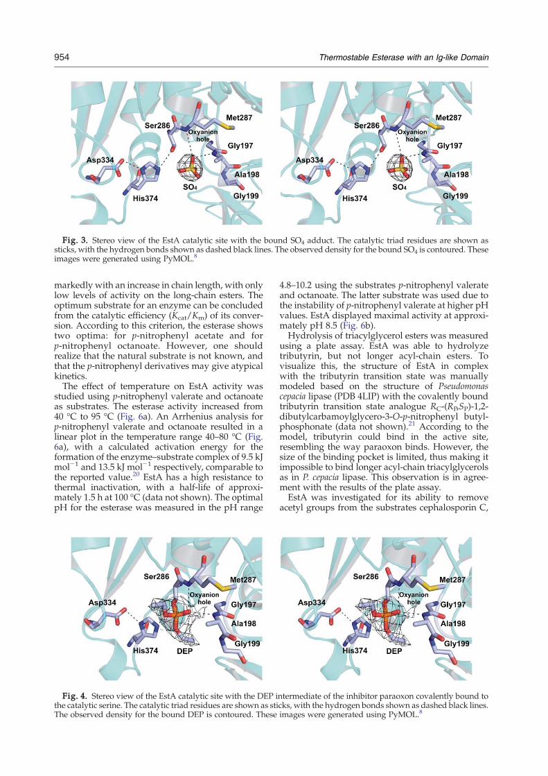
Fig. 3. Stereo view of the EstA catalytic site with the bound SO4 adduct. The catalytic triad residues are shown assticks, with the hydrogen bonds shown as dashed black lines. The observed density for the bound SO4 is contoured. Theseimages were generated using PyMOL.8
954 Thermostable Esterase with an Ig-like Domain
markedly with an increase in chain length, with onlylow levels of activity on the long-chain esters. Theoptimum substrate for an enzyme can be concludedfrom the catalytic efficiency (Kcat/Km) of its conver-sion. According to this criterion, the esterase showstwo optima: for p-nitrophenyl acetate and forp-nitrophenyl octanoate. However, one shouldrealize that the natural substrate is not known, andthat the p-nitrophenyl derivatives may give atypicalkinetics.The effect of temperature on EstA activity was
studied using p-nitrophenyl valerate and octanoateas substrates. The esterase activity increased from40 °C to 95 °C (Fig. 6a). An Arrhenius analysis forp-nitrophenyl valerate and octanoate resulted in alinear plot in the temperature range 40–80 °C (Fig.6a), with a calculated activation energy for theformation of the enzyme–substrate complex of 9.5 kJmol−1 and 13.5 kJ mol−1 respectively, comparable tothe reported value.20 EstA has a high resistance tothermal inactivation, with a half-life of approxi-mately 1.5 h at 100 °C (data not shown). The optimalpH for the esterase was measured in the pH range
Fig. 4. Stereo view of the EstA catalytic site with the DEP ithe catalytic serine. The catalytic triad residues are shown as stiThe observed density for the bound DEP is contoured. These
4.8–10.2 using the substrates p-nitrophenyl valerateand octanoate. The latter substrate was used due tothe instability of p-nitrophenyl valerate at higher pHvalues. EstA displayed maximal activity at approxi-mately pH 8.5 (Fig. 6b).Hydrolysis of triacylglycerol esters was measured
using a plate assay. EstA was able to hydrolyzetributyrin, but not longer acyl-chain esters. Tovisualize this, the structure of EstA in complexwith the tributyrin transition state was manuallymodeled based on the structure of Pseudomonascepacia lipase (PDB 4LIP) with the covalently boundtributyrin transition state analogue RC-(RP,SP)-1,2-dibutylcarbamoylglycero-3-O-p-nitrophenyl butyl-phosphonate (data not shown).21 According to themodel, tributyrin could bind in the active site,resembling the way paraoxon binds. However, thesize of the binding pocket is limited, thus making itimpossible to bind longer acyl-chain triacylglycerolsas in P. cepacia lipase. This observation is in agree-ment with the results of the plate assay.EstA was investigated for its ability to remove
acetyl groups from the substrates cephalosporin C,
ntermediate of the inhibitor paraoxon covalently bound tocks, with the hydrogen bonds shown as dashed black lines.images were generated using PyMOL.8

Fig. 5. Substrate-binding gorge. (a) Stereo view of the active site with the bound DEP intermediate covalently bound tothe catalytic serine. Key residues of the catalytic gorge are shown as sticks, with the hydrogen bonds shown in dashedlines. (b) Surface representation of the EstA catalytic gorge, with some of the key residues and the intermediate DEPshown in stick mode. These images were generated using PyMOL.8
955Thermostable Esterase with an Ig-like Domain
7-aminocephalosporanic acid (7-ACA), glucose pen-taacetate, and N-acetyl-D-glucosamine. EstA dis-played an activity of 20 U mg−1 on glucose penta-acetate. This activity is relatively low, suggesting
Table 1. Kinetic parameters for the hydrolysis of variousp-nitrophenyl esters
p-Nitrophenylesters
Km(μM)
Kcat(s−1)
Kcat/Km(s−1 mM−1)
Acetate (C2) 105±10 115±4 1095±111Butyrate (C4) 414±37 99±3 239±23Valerate (C5) 183±17 85±2 464±43Hexanoate (C6) 89±14 79±4 888±147Octanoate (C8) 27±6 37±3 1370±324Decanoate (C10) 8±1 10±0.4 1250±164Dodecanoate (C12) 6±0.6 1.6±0.03 267±27
that EstA is not an oligosaccharide deacetylase. EstAwas also able to hydrolyze acetyl groups from bothcephalosporin C and 7-ACAwith an activity of 80 Umg−1 for both substrates. Cephalosporin C and 7-ACA are not stable at high temperatures and are,therefore, not considered natural substrates. EstAwas not able to remove the acetyl group from N-acetyl-D-glucosamine, indicating that it is specific forester bonds and unable to cleave amide bonds.
Mutational studies
Five residues, all near the active site or part of theactive-site gorge, were changed to alanines by site-directed mutagenesis in order to analyze theirimportance for EstA activity. The residues selectedfor mutagenesis were three phenylalanines (Phe89,
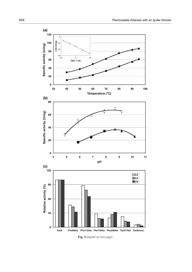
Fig. 6 (legend on next page)
956 Thermostable Esterase with an Ig-like Domain
957Thermostable Esterase with an Ig-like Domain
Phe112, and Phe116) located on loops of the Ig-likedomain and two conserved residues (Phe246 andTrp377) of the catalytic domain. Phe89 is located ona loop (Tyr77-Tyr85) coming from the Ig-like domainof a subunit B (EstA multimer) and is a part of theside gorge. Phe116 is located at the top of a very longloop (Leu100-Ile126), leading all the way back to theactive site and covering the active site. Phe112 is onthe same long loop and is part of the groove. Theconserved Phe246 is part of the tunnel, and Trp377 ispart of the active-site gorge.The mutants Phe89Ala, Phe112Ala, Phe116Ala,
Phe246Ala, and Trp377Ala were expressed andpurified. The mutants were analyzed by mass spec-trometry, which revealed that all mutants werepresent as hexamer in solution. Esterase activity wasdetermined using substrates with different acyl-chain lengths (pNP-C2, pNP-C5, and pNP-C8)(Fig. 6c). A significant reduction in activity wasobserved for all five mutants, confirming their im-portance for the activity of EstA. Their activityrelative to EstA with the substrate pNP-C5 was,respectively, approximately 30% for Phe89Ala, 80%for Phe112Ala, 20% for Phe116Ala, 25% forPhe246Ala, and 15% for Trp377Ala. As can be seenin Fig. 6c, the effect was more pronounced for longeracyl-chain substrates.In addition, a truncated mutant of EstA coding
only for the catalytic domain (EstAtrunc: Asp158-Arg395) was constructed. EstAtrunc was expressed,purified, and found to have a relative activity of only5% compared to EstA (Fig. 6c). Furthermore, it had amuch lower temperature optimum (60 °C), hadlower thermostability (a half-life at 90 °C of 15 min),and was present as a monomeric structure (analyzedby mass spectrometry).
Discussion
A hypothetical protein with esterase features fromthe hyperthermophilic bacterium T. maritima wasproduced heterologously and proved to exhibitester-hydrolyzing activity. The highest activitieswere found on p-nitrophenol derivatives withshort acyl chains (C2 and C4). In accordance, theenzyme also showed activity with tributyrin, but notwith triacylesters with longer chains. However,because of a high catalytic efficiency for the acetate,as well as for the octanoate-p-nitrophenol derivative,we cannot exclude that the physiological substratemay contain acyl chains up to C8. Nevertheless,these data indicate that EstA should be classified asan esterase (bC10), not as a lipase.
Fig. 6. Effect of temperature, pH, and mutations on esteraswas studied using p-nitrophenyl-valerate (■) and p-nitropheny40 °C to 95 °C. Inset: The temperature dependence for p-nitropesterase activity was studied using p-nitrophenyl-valeratenitrophenyl-octanoate (■, citrate phosphate buffer; ▴, CAPS bThe effect of mutations on esterase activity was studied using pp-nitrophenyl-octanoate (C8) at pH 8 and 70 °C.
The three-dimensional structure of the proteinwas determined at 2.6 Å resolution. Analysis of theEstA structure confirms that it is a member of theα/β hydrolase family, with a conserved Ser-Asp-Hiscatalytic triad comprising Ser286, Asp334, andHis374. The active site can be accessed via a gorgeflanked with predominantly hydrophobic residues.The structure was found to be composed of twoclearly distinguishable domains: a C-terminal do-main containing the active site and an unusualN-terminal domain resembling Igs. Such a combina-tion of an esterase domain or an α/β hydrolasedomain with an Ig-like domain is new and has not,as such, yet been described.Analysis of the quaternary structure by gel filtra-
tion, mass spectrometry, and DLS revealed that EstApredominantly exists as a hexamer in solution. Thecrystal structure also shows a hexameric arrange-ment composed of two trimers. EM demonstratedthat the hexamer in solution is identical with thehexamer in the crystal and is constructed as a dimerof trimers, with the N-terminal Ig-like domainsfacing each other. Esterases often have a trimericstructure, as was, for example, described for thethermostable esterase from Bacillus circulans22 and thethermostable esterase from Sulfolobus shibatae.23 Ahexameric structure, however, is rather unusual.Being at the interface of the two trimers, the Ig-likedomain apparently has a function inmultimerization.The function of the Ig-like domain in other bacte-
rial enzymes has been proposed to be substratebinding, directing a substrate to the catalytic grooveor cell adhesion.24,25 The latter option seemsunlikely for EstA, with the Ig-like domains facingeach other. To elucidate the function of the Ig-likedomain of EstA, a truncated mutant composed onlyof the C-terminal catalytic domain was constructed.The resulting EstAtrunc was still active, but had lost95% of its activity and was no longer able to formhexamers. This again points to a role of the Ig-likedomain in multimerization. The inability to formmultimers may also be the reason for the reducedstability observed at higher temperatures. Apolarresidues at the interface become exposed to thesolvent and may contribute to the observed loss ofstability. Multimerization is a phenomenon oftendescribed for enzymes from (hyper)thermophilesand is regarded as one of different mechanisms toincrease thermostability.26
The active site is accessible via a gorge, typical ofmany α/β hydrolases. Unusual, however, is that theactive site is also accessible via a second side gorge.This side gorge could possibly provide an access forthe substrate or an exit for one of the reaction pro-
e activity. (a) The effect of temperature on esterase activityl-octanoate (O) as substrates at temperatures ranging fromhenyl-valerate as an Arrhenius plot. (b) The effect of pH on(□, citrate phosphate buffer; Δ, CAPS buffer) and p-uffer) as substrates at pH values in the range 4.8–10.2. (c)-nitrophenyl-acetate (C2), p-nitrophenyl-valerate (C5), and
958 Thermostable Esterase with an Ig-like Domain
ducts following a nucleophilic attack and formationof the intermediate. On the other hand, compared toother esterases and lipases, the EstA active-site poc-ket is unique in its closure by Val336 and Phe116.Considering that Phe116 is located in a flexible loop,as indicated by a high B-factor for the fragmentPhe112-Leu117, it is possible that, upon substratebinding, Phe116 could change its conformation andopen the tunnel, making it more accessible.Besides Phe116, the active site is surrounded by a
set of aromatic residues. To disclose their function,five residues (Phe89, Phe112, Phe116, Phe246, andTrp377) in proximity to the active site were mutatedto alanines. The specific activity of each mutant wasdecreased, indicating that they all are important foractivity. Least affected was mutant Phe112Ala;however, this residue is located farthest from theactive site. The most pronounced inhibiting effectswere seen with the more hydrophobic longer-chainesters. This observation suggests that the hydro-phobic residues facilitate the entrance of the subs-trate along the gorge, which would hold most for thelonger acyl chains. On the other hand, a moregeneral role of the residues in the stabilization ofthe active site, however, cannot be excluded. One ofthe mutated residues, Phe89, is located on a loopcoming from the Ig-like domain of a subunit B.This interaction of the Ig-like domain of one subunitwith the active site of another subunit also supportsthe view that multimerization is important for—although not essential to—the activity of EstA, sincePhe89Ala still has 30% activity.In conclusion, the structural and biochemical
characterization of EstA showed that it is an unusualesterase composed of a conserved C-terminalcatalytic domain and an unprecedented N-terminalIg-like domain. The Ig-like domain presumablyplays a role in the multimerization of EstA into anunusual hexameric structure. Additionally, it mayalso participate in the catalysis of EstA by guidingthe substrate to the active site. Further mutagenesisand biochemical studies are needed for a betterunderstanding of the role of the N-terminal domain.
Materials and Methods
Protein production and crystallization
The T. maritima estA gene (locus tag TM0033) was clonedinto the expression vector pET24d without the predictedsignal peptide (the first 16 amino acids). EstA wasexpressed and purified as described.6 The purified nativeand selenomethionine (SeMet) EstA proteins were dia-lyzed against 10 mM potassium phosphate buffer (pH 7.5)and concentrated to 15 mg ml−1. EstAwas crystallized byhanging-drop vapor-diffusion at room temperature. Crys-tals of EstA were obtained using a reservoir solutionconsisting of 1.0 M lithium sulphate monohydrate and 2%wt/vol polyethylene glycol 8000. Drops consisting ofequal volumes (1 μl) of protein and reservoir solution wereequilibrated over 500-μl reservoirs. Crystals suitable forX-ray diffraction were obtained within 1 week and2 weeks, respectively, for the native and SeMet-EstA
proteins. A crystal of EstA in complex with its inhibitorparaoxon was obtained by incubating the enzyme (15 mgml−1) supplemented with 0.2 mM paraoxon for 1 h atroom temperature and then by setting up crystallization asdescribed above. Crystals were obtained within 2 weeks.
Data collection
For cryoprotection, crystals were soaked for a fewseconds in a reservoir solution containing 20% (wt/vol)glycerol. The crystals were mounted in a cryoloop andsubsequently flash-frozen in liquid nitrogen. X-ray datawere collected at 100 K on beamline ID29 at the EuropeanSynchrotron Radiation Facility (Grenoble, France). Anative data set was collected to 2.6 Å resolution. Thecrystal belongs to space group H32, with unit cellparameters a=130.2 Å, b=130.2 Å, and c=306.2 Å. Thereare two molecules in the asymmetric unit that have a VMof 2.9 Å3 Da−1 and a solvent content of 58%.27 Crystals ofSeMet-EstA showed a well-defined Se K absorption edgeby fluorescence scanning. A single SeMet-EstA crystal wasused for MAD data collection at the peak (0.9791 Å),inflection (0.9793 Å), and remote (0.9557 Å) wavelengthsup to 2.6 Å resolution. Data were indexed and integratedwith MOSFLM28 and scaled using SCALA.29
Structure determination and refinement
The EstA structure was solved by MAD phasing withMAD data from the SeMet-EstA crystal and the nativedata set, using HKL2MAP.30 Eight selenium sites in theasymmetric unit of the crystal were found and used tocalculate phases to 2.6 Å resolution (Table 2). However,initial density maps were of generally poor quality and notsuitable for tracing the structure. Phases were improvedusing RESOLVE,31,32 allowing the identification of NCS.NCS averaging and solvent flattening were performedusing the program DM33 of the CCP4 suite, giving anelectron density map of better quality. Autobuilt modelsfrom RESOLVE and ARP/wARP34 were combined to givea starting model comprising 170 residues from a total of380, with only 20 residues assigned into sequenceproperly. The structure was then manually rebuilt inCoot35 and refined using CNS36 and REFMAC.37 StrictNCS restraints were applied during the earlier stages ofthe refinement and released at later stages. In the finalstages of refinement, solvent molecules were added usingARP-wARP34 and manually inspected in Coot.35 A sulfateion from the reservoir solution was clearly visible at highcontour level in the omit map. The final refinement Rworkis 19.7%, and Rfree is 26.7%.The structure of the EstA–paraoxon complex was
determined by molecular replacement, which was per-formed with MOLREP using the native structure asmodel. The complex structure was rebuilt in Coot35 andrefined using REFMAC.37 The Fo−Fc electron densitymap and omit density map displayed clear density forparaoxon and were used to assign the head of theparaoxon molecule. No water molecules were picked forthis low-resolution data set. The model with paraoxonwas refined to a final Rwork of 22.0% and an Rfree of26.2%.In the final models (native and paraoxon-complexed
crystal structures), residues 16–20 of both molecules andresidues 249–250 of chain B were missing because of poordensity. The geometry of both models was monitoredusing PROCHECK,38 with the native and paraoxoncomplex models having 85.2% and 82.2% of their residues
Table 2. Data collection, phasing, and refinement statistics
Native SeMet peak SeMet inflection SeMet remote EstA paraoxon complex
Data collectionWavelength (Å) 1.0000 0.9791 0.9793 0.9757 1.0000Resolution range (Å) 50–2.6 (2.74–2.6)a 50–2.6 (2.74–2.6) 50–2.6 (2.74–2.6) 50–2.6 (2.74–2.6) 50–3.0 (3.16–3.0)Space group H32 H32 H32 H32 H32Unit cell parameters (Å)
a 130.2 131.0 131.0 131.0 130.5b 130.2 131.0 131.0 131.0 130.5c 306.2 306.8 306.8 306.8 304.5
Observed reflections 173,066 353,388 358,408 358,161 160,768Unique reflections 31,079 31,295 31,587 31,457 20,405Completeness (%) 100.0 (100.0) 100.0 (100.0) 100.0 (100.0) 100.0 (100.0) 100.0 (100.0)Rmerge
b 0.087 (0.430) 0.086 (0.498) 0.082 (0.412) 0.075 (0.437) 0.152 (0.643)⟨I/σ(I)⟩ 14.2 (3.6) 22.3 (4.6) 22.1 (4.4) 24.8 (5.0) 13.4 (3.0)Redundancy 5.6 (5.7) 11.4 (11.7) 11.3 (11.4) 11.4 (11.3) 7.9 (8.1)
MAD phasingNumber of Se sites 8Figure of merit 0.5
RefinementResolution range (Å) 20–2.6 (2.66–2.6) 35–3.0 (3.08–3.0)Number of reflections 28,373 19,311Rwork (%) 19.7 (31.2) 22.0 (30.7)Rfree (%) 26.7 (35.5) 26.2 (35.6)Average B-factors
Protein 36.8 45.8Water 33.2 —Ligand 49.5 39.6
RMSDBond lengths (Å) 0.018 0.016Bond angles (°) 1.88 1.77a Values in parentheses correspond to the highest-resolution shell.b Rmerge=∑h∑l|Ihl− ⟨Ih⟩|/∑h∑l⟨Ih⟩, where Il is the lth observation of reflection h, and ⟨Ih⟩ is the weighted average intensity for all
observations l of reflection h.
959Thermostable Esterase with an Ig-like Domain
in the most favored regions of the Ramachandran plot,respectively. Cartoon representations were generatedusing PyMOL.8
Native mass spectrometry
Native mass spectrometry measurements were per-formed in positive ion mode using an ElectrosprayIonization Time-of-Flight instrument (LC-T; Micromass,Manchester, UK) equipped with a Z-spray nanoelectro-spray ionization source. Needles were made from boro-silicate glass capillaries (Kwik-Fil; World PrecisionInstruments, Sarasota, FL) on a P-97 puller (SutterInstruments, Novato, CA), coated with a thin gold layerusing an Edwards Scancoat (Edwards Laboratories,Milpitas, CA) six Pirani 501 sputter coater. To produceintact ions in vacuo from EstA in solution, the ions werecooled by increasing the pressure in the first vacuum stagesof the mass spectrometer. In addition, efficient desolva-tion was needed to sharpen the ion signals in order towithdraw the oligomeric states of EstA from the massspectrum. Therefore, source pressure conditions wereraised to values ranging from 7.0 mbar to 7.3 mbar, andnanoelectrospray voltages were optimized for transmis-sion of large protein complexes. The pressure in theinterface region was adjusted by reducing the pumpingcapacity of the rotary pump by closing the speed valve.39,40The applied voltages on the needle and sample cone were1300 V and 150 V, respectively. All spectra were mass-calibrated using an aqueous solution of cesium iodide(20 mg ml−1). Buffer exchange of EstA samples with100 mM ammonium acetate (pH 6.8) was performed using
ultrafiltration units with a cutoff of 5000 Da (Millipore,Bedford, MA). EstAwas diluted to 5 μM and measured atroom temperature.
Dynamic light scattering
DLS was performed at room temperature using theEstA sample that was ready for crystallization. The EstAparticles were monodisperse with a molecular mass ofapproximately 267 kDa, suggesting that the 43-kDasubunits assemble into a hexamer in solution.
EM and single-particle analysis
Samples of purified EstA were negatively stained with2% uranyl acetate on glow-discharged carbon-coatedcopper grids. Images were recorded with a Gatan 4 Kslow-scan charge-coupled device camera on a PhilipsCM12 electron microscope (Fei, Eindhoven, The Nether-lands) operated at 120 kV, using GRACE software forsemiautomated specimen selection and data acquisition.41
The final magnification was 100,000×, with a pixel size(after binning the images) of 3.0 Å at the specimen level.About 8700 single particles were selected and extractedfrom 600 electron micrographs. Single-particle analysiswas performed with the GRoningen Image Processing(GRIP) software package on a PC cluster. The single-particle projections (96×96 pixel frame) were subjected tomultireference alignment and reference-free alignmentprocedure, multivariate statistical analysis, and hierar-chical ascendant classification.42 From the whole data set,
960 Thermostable Esterase with an Ig-like Domain
almost 45% of single particles were assigned to top viewsof the EstA hexamer, whereas the remaining ∼55%resembled side views. The final two-dimensional projec-tion maps of the presented views of the EstA hexamerwere calculated from the best-resolved classes, whichrepresented about 20% of the whole data.The packing of EstA within the crystal was examined
with PyMOL software8 for possible hexameric structuresthat resembled the projection maps obtained with EM andsingle-particle analysis. Possible structures were manu-ally fitted into the EM projections. Different views of theEstA hexamer were finally displayed using PyMOL soft-ware.8 Truncated versions at 15 Å resolution and two-dimensional projection maps of the generated model forthe EstA hexamer were generated using routines from theEMAN package.43
Enzyme assays
Esterase activitywasmeasured asdescribedpreviously.20
In short, in a standard assay, activity was measuredwith 0.2 mM p-nitrophenyl octanoate as substrate in 50 mMcitrate phosphate (pH 8) at 70 °C. The amount of p-nitrophe-nol liberated was measured continuously at 405 nm on aHitachi U-2001 spectrophotometer with a temperature-controlled cuvette holder. Extinction coefficients of p-nitro-phenol were determined prior to each measurement.The effect of pH on esterase activity was studied in the
pH range 5–11. The buffers used were 50 mM citrate phos-phate (pH 5–8) and 50 mM CAPS (3-(cyclohexylamino)1-propanesulphonic acid) buffer (pH 9.5–11). The effect oftemperature on esterase activity was studied in the range40–95 °C. The pH of the buffers was set at 25 °C, andtemperature corrections were made using their tempera-ture coefficients: −0.0028 pH °C−1 for citrate-phosphatebuffer, and −0.018 pH °C−1 for CAPS buffer.44 Enzymethermostability was determined by incubating the enzymein 50 mM Tris–HCl and 150 mM NaCl (pH 7.8) buffer at100 °C for various time intervals. Residual activity wasassayed under standard conditions.Plate assays were used for the detection of activity on
triacylglycerol esters. Agar plates containing tributyrin,trioctanoate, triolein, palm oil, and olive oil (1%, vol/vol)were prepared, and 5-mm-wide holes were perforated.The resulting holes were loadedwith purified enzyme andincubated at 50 °C. Activity was detected by zones ofclearance around the holes.Deacetylase activity was determined using high-perfor-
mance liquid chromatography by measuring the amountof acetic acid released from the substrates cephalosporin C,7-ACA, glucose pentaacetate, andN-acetyl-D-glucosamine.The reaction mixture contained 0.9 ml of substrate solution(dissolved in 50 mM Tris–HCl, pH 7.5) and 0.1 ml ofenzyme solution, and was incubated at 37 °C for varioustime intervals. The reaction was stopped by adding 0.2 mlof stop solution (100 mMH2SO4 and 30mM crotonate) andplacing the sample on ice. The conditions for high-performance liquid chromatography were as follows:column, KC811-Shodex; detection, RI and UV detectors;solvent, 3 mMH2SO4; flow rate, 1.5mlmin−1; temperature,30 °C; internal standard, crotonate. One unit of enzymeactivity was defined as the amount of enzyme that releases1 μmol of acetic acid per minute.The effect of inhibitors was studied by preincubating
EstA with 1 mM inhibitor in 50 mM citrate phosphatebuffer (pH 8) at 37 °C for 1 h. Subsequently, samples wereplaced on ice, and residual activity was measured usingthe standard assay. The activity of the enzyme without
inhibitor was defined as 100%. Inhibition kinetics ofparaoxon were determined as described for the acetylcho-linesterase from electric eel.16
Kinetic parameters were determined by fitting the dataobtained from multiple measurements by a computer-aided direct fit to the Michaelis–Menten curve (Tablecurve2d, version 5.0).
Mutagenesis
Mutants of EstA were created to identify the functionof the Ig-like domain. Mutants Phe89Ala, Phe112Ala,Phe116Ala, Phe246Ala, and Trp377Ala were generatedusing Quikchange (Stratagene) site-directed mutagenesiswith the following primers: BG2486 5-CGGAGGTCTCT-CTTTCAGCATTGCCAACAACCGTGGAAAGTACG-3and BG2487 5-CGTACTTTCCACGGTTGTTGGCAATGC-TGAAAGAGAGACCTCCG-3; BG2488 5-CACTCAAAC-ACCATTGTTGCCGGGCCTAATTTTCTCAACACCCG-3and BG2645 5-CGGGTGTTGAGAAAATTAGGCCCGGC-AACAATGGTGTTTGAGTG-3; BG2490 5-CCATTGTTTTC-GGGCCTAATGCGCTCAACACCCGCATGAAACTGG-3and BG2646 5-CCAGTTTCATGCGGGTGTTGAGCGCAT-TAGGCCCGAAAACAATGG-3; BG2492 5-CAGCAGCT-GGTCCACACTCGCCACTGACAGGGAAAATCC-3 andBG2647 5-GGATTTTCCCTGTCAGTGGCGAGTGTGGA-CCAGCTGCTG-3; and BG2494 5-GGTGGGATCCACAC-GGATCGGCGATACCTACCTACGAG-3 and BG26485-CTCGTAGGTAGGTATCGCCGATCCGTGTGGATCC-CACC-3, respectively (with modified codons underlined).A truncated EstA mutant was generated using the primersBG2496 5-CCGCCATGGATTTCCTCGCATTCACTTTCA-AAGACC-3 and BG1963 5-GCGCTCGAGTCTACTTTG-TTCAAACAGCCAC-3. The sequence of the mutants wasconfirmed by sequence analysis of both DNA strands.
Data bank accession codes
The structural data have been deposited in the RCSBPDB with accession code 3DOH for native EstA andaccession code 3DOI for EstA in complex with paraoxon.
Acknowledgements
This research was financially supported by theGraduate School VLAG and an NWO-Vici grant(Wageningen University). We would like to thankthe support staff of beamline ID29 at the EuropeanSynchrotron Radiation Facility Grenoble Laboratoryfor their assistance with X-ray beam alignment andMAD data collection.
References
1. Jaeger, K. E., Dijkstra, B. W. & Reetz, M. T. (1999).Bacterial biocatalysts: molecular biology, three-dimen-sional structures, and biotechnological applications oflipases. Annu. Rev. Microbiol. 53, 315–351.
2. Jaeger, K. E. & Eggert, T. (2002). Lipases forbiotechnology. Curr. Opin. Biotechnol. 13, 390–397.
3. Atomi, H. & Imanaka, T. (2004). Thermostablecarboxylesterases from hyperthermophiles. Tetrahe-dron: Asymmetry, 15, 2729–2735.
961Thermostable Esterase with an Ig-like Domain
4. Huber, R. & Hannig, M. (2006). Thermotogales.Prokaryotes, 7, 899–922.
5. Nelson, K. E., Clayton, R. A., Gill, S. R., Gwinn, M. L.,Dodson, R. J., Haft, D. H. et al. (1999). Evidence forlateral gene transfer between Archaea and bacteriafrom genome sequence of Thermotoga maritima.Nature,399, 323–329.
6. Sun, L., Levisson, M., Hendriks, S., Akveld, T.,Kengen, S. W., Dijkstra, B. W. & van der Oost, J.(2007). Crystallization and preliminary crystallo-graphic analysis of an esterase with a novel domainfrom the hyperthermophile Thermotoga maritima. ActaCrystallogr. Sect. F, 63, 777–779.
7. Altschul, S. F., Madden, T. L., Schaffer, A. A., Zhang, J.,Zhang, Z., Miller, W. & Lipman, D. J. (1997). GappedBLAST and PSI-BLAST: a new generation of proteindatabase search programs. Nucleic Acids Res. 25,3389–3402.
8. DeLano,W. L. (2002). The PyMOLMolecular GraphicsSystem DeLano Scientific, San Carlos, CA.
9. Holm, L. & Sander, C. (1996). The FSSP database: foldclassification based on structure–structure alignmentof proteins. Nucleic Acids Res. 24, 206–209.
10. Ito, K., Nakajima, Y., Xu, Y., Yamada, N., Onohara, Y.,Ito, T. et al. (2006). Crystal structure and mechanism oftripeptidyl activity of prolyl tripeptidyl aminopepti-dase from Porphyromonas gingivalis. J. Mol. Biol. 362,228–240.
11. Rasmussen, H. B., Branner, S., Wiberg, F. C. &Wagtmann, N. (2003). Crystal structure of humandipeptidyl peptidase IV/CD26 in complex with asubstrate analog. Nat. Struct. Biol. 10, 19–25.
12. Schubot, F. D., Kataeva, I. A., Blum, D. L., Shah, A. K.,Ljungdahl, L. G., Rose, J. P. & Wang, B. C. (2001).Structural basis for the substrate specificity of theferuloyl esterase domain of the cellulosomal xylanaseZ from Clostridium thermocellum. Biochemistry, 40,12524–12532.
13. Zhang, H., Seabra, M. C. & Deisenhofer, J. (2000).Crystal structure of Rab geranylgeranyltransferase at2.0 A resolution. Structure, 8, 241–251.
14. Hage, T., Sebald, W. & Reinemer, P. (1999). Crystalstructure of the interleukin-4/receptor alpha chaincomplex reveals a mosaic binding interface. Cell, 97,271–281.
15. Jacobson, R. H., Zhang, X. J., DuBose, R. F. &Matthews, B. W. (1994). Three-dimensional structureof beta-galactosidase from E. coli.Nature, 369, 761–766.
16. Forsberg, A. & Puu, G. (1984). Kinetics for theinhibition of acetylcholinesterase from the electric eelby some organophosphates and carbamates. Eur. J.Biochem. 140, 153–156.
17. Febbraio, F., D'Andrea, S. E., Mandrich, L., Merone,L., Rossi, M., Nucci, R. & Manco, G. (2008). Irrever-sible inhibition of the thermophilic esterase EST2from Alicyclobacillus acidocaldarius. Extremophiles, 12,719–728.
18. van den Heuvel, R. H. & Heck, A. J. (2004). Nativeprotein mass spectrometry: from intact oligomers tofunctional machineries. Curr. Opin. Chem. Biol. 8,519–526.
19. Goettig, P., Groll,M., Kim, J. S.,Huber, R.&Brandstetter,H. (2002). Structures of the tricorn-interacting amino-peptidase F1 with different ligands explain its catalyticmechanism. EMBO J. 21, 5343–5352.
20. Levisson, M., van der Oost, J. & Kengen, S. W. (2007).Characterization and structural modeling of a newtype of thermostable esterase from Thermotoga mar-itima. FEBS J. 274, 2832–2842.
21. Lang, D. A., Mannesse, M. L., de Haas, G. H., Verheij,H. M. & Dijkstra, B. W. (1998). Structural basis of thechiral selectivity of Pseudomonas cepacia lipase. Eur. J.Biochem. 254, 333–340.
22. Kademi, A., Ait-Abdelkader, N., Fakhreddine, L. &Baratti, J. (2000). Purification and characterization ofa thermostable esterase from the moderate thermo-phile Bacillus circulans. Appl. Microbiol. Biotechnol. 54,173–179.
23. Ejima, K., Liu, J., Oshima, Y., Hirooka, K., Shimanuki,S., Yokota, Y. et al. (2004). Molecular cloning andcharacterization of a thermostable carboxylesterasefrom an archaeon, Sulfolobus shibatae DSM5389: non-linear kinetic behavior of a hormone-sensitive lipasefamily enzyme. J. Biosci. Bioeng. 98, 445–451.
24. Bork, P., Holm, L. & Sander, C. (1994). The immu-noglobulin fold: structural classification, sequencepatterns and common core. J. Mol. Biol. 242, 309–320.
25. Halaby, D. M. & Mornon, J. P. (1998). The immunoglo-bulin superfamily: an insight on its tissular, species,and functional diversity. J. Mol. Evol. 46, 389–400.
26. Korkhin, Y., Kalb, A. J., Peretz, M., Bogin, O., Burstein,Y. & Frolow, F. (1999). Oligomeric integrity—thestructural key to thermal stability in bacterial alcoholdehydrogenases. Protein Sci. 8, 1241–1249.
27. Matthews, B. W. (1968). Solvent content of proteincrystals. J. Mol. Biol. 33, 491–497.
28. Leslie, A. G. W. (1992). Recent changes to theMOSFLM package for processing film and imageplate data. Joint CCP4+ ESF-EAMCB Newsl. ProteinCrystallogr. 26, 27–33.
29. Collaborative Computational Project Number 4.(1994). The CCP4 suite: programs for protein crystal-lography. Acta Crystallogr. Sect. D, 50, 760–763.
30. Pape, T. & Schneider, T. R. (2004). HKL2MAP: agraphical user interface for macromolecular phasingwith SHELX programs. J. Appl. Crystallogr. 37, 843–844.
31. Terwilliger, T. C. (2000). Maximum-likelihood densitymodification. Acta Crystallogr. Sect. D, 56, 965–972.
32. Terwilliger, T. C. (2003). Automated side-chain modelbuilding and sequence assignment by templatematching. Acta Crystallogr. Sect. D, 59, 45–49.
33. Cowtan, K. (1994). ‘dm’: An automated procedure forphase improvement by density modification. JointCCP4 ESF-EACBM Newsl. Protein Crystallogr. 31,34–38.
34. Perrakis, A., Morris, R. & Lamzin, V. S. (1999).Automated protein model building combined withiterative structure refinement. Nat. Struct. Biol. 6,458–463.
35. Emsley, P. & Cowtan, K. (2004). Coot: model-buildingtools for molecular graphics. Acta Crystallogr. Sect. D,60, 2126–2132.
36. Brunger, A. T., Adams, P. D., Clore, G. M., DeLano,W. L., Gros, P., Grosse-Kunstleve, R. W. et al. (1998).Crystallography and NMR system: a new softwaresuite for macromolecular structure determination.Acta Crystallogr. Sect. D, 54, 905–921.
37. Murshudov, G. N., Vagin, A. A. & Dodson, E. J. (1997).Refinement of macromolecular structures by themaximum-likelihood method. Acta Crystallogr. Sect.D, 53, 240–255.
38. Laskowski, R. A., MacArthur, M. W., Moss, D. S. &Thornton, J. M. (1993). PROCHECK: a program tocheck the stereochemical quality of protein structures.J. Appl. Crystallogr. 26, 283–291.
39. Tahallah, N., Pinkse, M., Maier, C. S. & Heck, A. J.(2001). The effect of the source pressure on the abun-dance of ions of noncovalent protein assemblies in an
962 Thermostable Esterase with an Ig-like Domain
electrospray ionization orthogonal time-of-flightinstrument. Rapid Commun. Mass Spectrom. 15, 596–601.
40. van Berkel, W. J., van den Heuvel, R. H., Versluis, C. &Heck, A. J. (2000). Detection of intact megadaltonprotein assemblies of vanillyl-alcohol oxidase by massspectrometry. Protein Sci. 9, 435–439.
41. Oostergetel, G. T., Keegstra, W. & Brisson, A. (1998).Automation of specimen selection and data acqui-sition for protein electron crystallography. Ultramicro-scopy, 74, 47–59.
42. van Heel, M., Gowen, B., Matadeen, R., Orlova, E. V.,Finn, R., Pape, T. et al. (2000). Single-particle electroncryo-microscopy: towards atomic resolution. Q. Rev.Biophys. 33, 307–369.
43. Ludtke, S. J., Baldwin, P. R. & Chiu, W. (1999).EMAN: semiautomated software for high-resolutionsingle-particle reconstructions. J. Struct. Biol. 128,82–97.
44. Beynon, R. J. & Easterby, J. S. (2003). Buffer SolutionsTaylor and Francis Group, London.