
The Gene Cluster for Agmatine Catabolism of Enterococcus faecalis: Study of Recombinant Putrescine...
12
JOURNAL OF BACTERIOLOGY, Feb. 2007, p. 1254–1265 Vol. 189, No. 4 0021-9193/07/$08.000 doi:10.1128/JB.01216-06 Copyright © 2007, American Society for Microbiology. All Rights Reserved. The Gene Cluster for Agmatine Catabolism of Enterococcus faecalis: Study of Recombinant Putrescine Transcarbamylase and Agmatine Deiminase and a Snapshot of Agmatine Deiminase Catalyzing Its Reaction Jose ´ L. Lla ´cer,† Luis Mariano Polo,† Sandra Tava ´rez,† Benito Alarco ´n, Rebeca Hilario, and Vicente Rubio* Instituto de Biomedicina de Valencia (IBV-CSIC), C/Jaime Roig 11, 46010 Valencia, Spain Received 3 August 2006/Accepted 29 November 2006 Enterococcus faecalis makes ATP from agmatine in three steps catalyzed by agmatine deiminase (AgDI), pu- trescine transcarbamylase (PTC), and carbamate kinase (CK). An antiporter exchanges putrescine for agmatine. We have cloned the E. faecalis ef0732 and ef0734 genes of the reported gene cluster for agmatine catabolism, overexpressed them in Escherichia coli, purified the products, characterized them functionally as PTC and AgDI, and crystallized and X-ray diffracted them. The 1.65-Å-resolution structure of AgDI forming a covalent adduct with an agmatine-derived amidine reactional intermediate is described. We provide definitive identification of the gene cluster for agmatine catabolism and confirm that ornithine is a genuine but poor PTC substrate, suggesting that PTC (found here to be trimeric) evolved from ornithine transcarbamylase. N-(Phosphonoacetyl)-putrescine was prepared and shown to strongly (K i 10 nM) and selectively inhibit PTC and to improve PTC crystallization. We find that E. faecalis AgDI, which is committed to ATP generation, closely resembles the AgDIs involved in making polyamines, suggesting the recruitment of a polyamine-synthesizing AgDI into the AgDI pathway. The arginine deiminase (ADI) pathway of arginine catabolism probably supplied the genes for PTC and CK but not those for the agmatine/putrescine antiporter, and thus the AgDI and ADI pathways are not related by a single “en bloc” duplication event. The AgDI crystal structure reveals a tetramer with a five-blade propeller subunit fold, proves that AgDI closely resembles ADI despite a lack of sequence identity, and explains substrate affinity, selectivity, and Cys357-mediated-covalent catalysis. A three-tongued agmatine-triggered gating opens or blocks access to the active center. In addition to the fermentation of carbohydrates, Enterococcus faecalis (formerly Streptococcus faecalis) is able to use arginine and its decarboxylated derivative agmatine as energy sources for growth (8, 10, 45, 48, 49). Arginine and agmatine are metabolized via the arginine deiminase (ADI) and agmatine deiminase (AgDI) pathways, respectively. The two metabolic routes are very similar and include the sequential action of three enzymes (48, 49) and one antiporter (11), which are analogous in the two pathways. Arginine and agmatine are deiminated by ADI (EC 3.5.3.6) and AgDI (EC 3.5.3.12), respectively, yielding citrulline and carbamoyl putrescine, which are phosphorolyzed by ornithine transcarbamylase (OTC) (EC 2.1.3.3) and putrescine transcar- bamylase (PTC) (EC 2.1.3.6). This generates carbamoyl phos- phate for use in ADP phosphorylation by pathway-specific car- bamate kinase (CK) (EC 2.7.2.2) isozymes, producing one ATP molecule (48, 49). The resulting ornithine and putrescine are exchanged with external arginine or agmatine by an arginine/ ornithine antiporter in one pathway and by an agmatine/pu- trescine antiporter in the other pathway (11). Possibly no microbial species has been more important for the biochemical characterization of the ADI and AgDI path- ways than E. faecalis. It was with this microorganism that both pathways were originally demonstrated (20, 24, 45, 50), the corresponding enzymatic steps were characterized and shown to be coordinately induced by arginine or agmatine (48, 49), the enzymes (except AgDI) were purified (31, 32, 42, 56), and CK (the ADI pathway isozyme) was crystallized and its struc- ture determined at atomic resolution (29, 30). Despite the abundance of biochemical information, there was little genetic information on these routes in E. faecalis until we sequenced the genes and determined the gene structure, organization, and some regulatory features for the components of the ADI pathway (3). However, in the case of the AgDI pathway, for very long time there was no other genetic information than the observation that three mutant strains of E. faecalis that were unable to use agmatine were devoid of either AgDI activity, PTC activity, or both (48). The loss of the two enzymes in one mutant and the triggering by agmatine of coordinated in- creases in the levels of AgDI and PTC appeared to be consis- tent with the physical association of the genes for these two enzymes within the same operon, as is the case for the genes for the ADI pathway (3, 48, 49). Only recently, after the iden- tification in Pseudomonas aeruginosa of the gene aguA (38), encoding the AgDI that is involved in putrescine and poly- amine biosynthesis in plants and microorganisms that decar- boxylate arginine (2) (not the case for E. faecalis), a putative aguA gene was identified in the cariogenic organism Strepto- coccus mutans (17) and, by sequence similarity, in E. faecalis * Corresponding author. Mailing address: Instituto de Biomedicina de Valencia (IBV-CSIC), C/Jaime Roig 11, 46010 Valencia, Spain. Phone: 34 96 339 17 72. Fax: 34 96 369 08 00. E-mail: [email protected]. † J.L.L., L.M.P., and S.T. contributed equally to this work. Published ahead of print on 6 October 2006. 1254 on November 25, 2014 by guest http://jb.asm.org/ Downloaded from
-
Upload
independent -
Category
Documents
-
view
1 -
download
0
Transcript of The Gene Cluster for Agmatine Catabolism of Enterococcus faecalis: Study of Recombinant Putrescine...

JOURNAL OF BACTERIOLOGY, Feb. 2007, p. 1254–1265 Vol. 189, No. 40021-9193/07/$08.00�0 doi:10.1128/JB.01216-06Copyright © 2007, American Society for Microbiology. All Rights Reserved.
The Gene Cluster for Agmatine Catabolism of Enterococcus faecalis:Study of Recombinant Putrescine Transcarbamylase and Agmatine
Deiminase and a Snapshot of Agmatine DeiminaseCatalyzing Its Reaction�
Jose L. Llacer,† Luis Mariano Polo,† Sandra Tavarez,† Benito Alarcon,Rebeca Hilario, and Vicente Rubio*
Instituto de Biomedicina de Valencia (IBV-CSIC), C/Jaime Roig 11, 46010 Valencia, Spain
Received 3 August 2006/Accepted 29 November 2006
Enterococcus faecalis makes ATP from agmatine in three steps catalyzed by agmatine deiminase (AgDI), pu-trescine transcarbamylase (PTC), and carbamate kinase (CK). An antiporter exchanges putrescine for agmatine.We have cloned the E. faecalis ef0732 and ef0734 genes of the reported gene cluster for agmatine catabolism,overexpressed them in Escherichia coli, purified the products, characterized them functionally as PTC and AgDI,and crystallized and X-ray diffracted them. The 1.65-Å-resolution structure of AgDI forming a covalent adduct withan agmatine-derived amidine reactional intermediate is described. We provide definitive identification of the genecluster for agmatine catabolism and confirm that ornithine is a genuine but poor PTC substrate, suggesting thatPTC (found here to be trimeric) evolved from ornithine transcarbamylase. N-(Phosphonoacetyl)-putrescine wasprepared and shown to strongly (Ki � 10 nM) and selectively inhibit PTC and to improve PTC crystallization. Wefind that E. faecalis AgDI, which is committed to ATP generation, closely resembles the AgDIs involved in makingpolyamines, suggesting the recruitment of a polyamine-synthesizing AgDI into the AgDI pathway. The argininedeiminase (ADI) pathway of arginine catabolism probably supplied the genes for PTC and CK but not those for theagmatine/putrescine antiporter, and thus the AgDI and ADI pathways are not related by a single “en bloc”duplication event. The AgDI crystal structure reveals a tetramer with a five-blade propeller subunit fold, proves thatAgDI closely resembles ADI despite a lack of sequence identity, and explains substrate affinity, selectivity, andCys357-mediated-covalent catalysis. A three-tongued agmatine-triggered gating opens or blocks access to the activecenter.
In addition to the fermentation of carbohydrates, Enterococcusfaecalis (formerly Streptococcus faecalis) is able to use arginineand its decarboxylated derivative agmatine as energy sources forgrowth (8, 10, 45, 48, 49). Arginine and agmatine are metabolizedvia the arginine deiminase (ADI) and agmatine deiminase(AgDI) pathways, respectively. The two metabolic routes are verysimilar and include the sequential action of three enzymes (48,49) and one antiporter (11), which are analogous in the twopathways. Arginine and agmatine are deiminated by ADI (EC3.5.3.6) and AgDI (EC 3.5.3.12), respectively, yielding citrullineand carbamoyl putrescine, which are phosphorolyzed by ornithinetranscarbamylase (OTC) (EC 2.1.3.3) and putrescine transcar-bamylase (PTC) (EC 2.1.3.6). This generates carbamoyl phos-phate for use in ADP phosphorylation by pathway-specific car-bamate kinase (CK) (EC 2.7.2.2) isozymes, producing one ATPmolecule (48, 49). The resulting ornithine and putrescine areexchanged with external arginine or agmatine by an arginine/ornithine antiporter in one pathway and by an agmatine/pu-trescine antiporter in the other pathway (11).
Possibly no microbial species has been more important forthe biochemical characterization of the ADI and AgDI path-
ways than E. faecalis. It was with this microorganism that bothpathways were originally demonstrated (20, 24, 45, 50), thecorresponding enzymatic steps were characterized and shownto be coordinately induced by arginine or agmatine (48, 49),the enzymes (except AgDI) were purified (31, 32, 42, 56), andCK (the ADI pathway isozyme) was crystallized and its struc-ture determined at atomic resolution (29, 30). Despite theabundance of biochemical information, there was little geneticinformation on these routes in E. faecalis until we sequencedthe genes and determined the gene structure, organization,and some regulatory features for the components of the ADIpathway (3). However, in the case of the AgDI pathway, forvery long time there was no other genetic information than theobservation that three mutant strains of E. faecalis that wereunable to use agmatine were devoid of either AgDI activity,PTC activity, or both (48). The loss of the two enzymes in onemutant and the triggering by agmatine of coordinated in-creases in the levels of AgDI and PTC appeared to be consis-tent with the physical association of the genes for these twoenzymes within the same operon, as is the case for the genesfor the ADI pathway (3, 48, 49). Only recently, after the iden-tification in Pseudomonas aeruginosa of the gene aguA (38),encoding the AgDI that is involved in putrescine and poly-amine biosynthesis in plants and microorganisms that decar-boxylate arginine (2) (not the case for E. faecalis), a putativeaguA gene was identified in the cariogenic organism Strepto-coccus mutans (17) and, by sequence similarity, in E. faecalis
* Corresponding author. Mailing address: Instituto de Biomedicina deValencia (IBV-CSIC), C/Jaime Roig 11, 46010 Valencia, Spain. Phone:34 96 339 17 72. Fax: 34 96 369 08 00. E-mail: [email protected].
† J.L.L., L.M.P., and S.T. contributed equally to this work.� Published ahead of print on 6 October 2006.
1254
on Novem
ber 25, 2014 by guesthttp://jb.asm
.org/D
ownloaded from
(gene ef0734 of E. faecalis V583 genome [TIGR database;http://www.tigr.org]). In both species this gene is preceded bythe genes for a putative antiporter and for a transcarbamylase(in E. faecalis V583, genes ef0733 and ef0732, respectively) andis followed by a putative gene for carbamate kinase (ef0735)(Fig. 1A). Thus, this gene cluster would contain the genes forall the catalysts required for operation of the AgDI pathway,and indeed, a polar disruption of the first gene in this cluster ofS. mutans strongly decreased the AgDI activity measured inpermeabilized cells, as expected for the AgDI operon (17).Further, the amino acid sequence predicted to be encoded byef0732 coincides with the N-terminal sequence reported longago for E. faecalis PTC (39, 54).
Nevertheless, the ultimate test for attributing specific func-tions to genes, i.e., the cloning of the gene, its expression, andthe purification and functional characterization of the corre-sponding gene product, has not been published for the genesfor the AgDI pathway. As a consequence of an independenteffort to identify and characterize the AgDI pathway genes, wedescribe here the cloning of the E. faecalis ef0732 and ef0734
genes, their overexpression in Escherichia coli, the purificationof the corresponding protein products, the enzymatic charac-terization of these products as PTC and AgDI, and their crys-tallization and X-ray analysis. We also report the crystal struc-ture at high resolution of E. faecalis AgDI containing acovalently bound derivative of agmatine at the active center.Our results not only confirm conclusively the nature of theoperon but also are the first that functionally characterize anAgDI committed to fermentative ATP production (all previ-ously well characterized examples are involved in polyaminebiosynthesis, and only one is bacterial, from Pseudomonasaeruginosa [23, 37, 59]), revealing also the structure of thisenzyme during catalysis. For the other gene product studiedhere, PTC, which was previously characterized from a singlesource (E. faecalis) (56), we demonstrate that the bisubstrateanalog for this enzyme, N-(phosphonoacetyl)-putrescine(PAPU), is a highly selective and very powerful PTC compet-itive inhibitor (Ki � 10 nM) relative to carbamoyl phosphate,clarifying the substrate binding order in this enzyme. Thisinhibitor is proven here to be crucial for obtaining good dif-
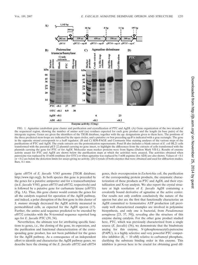
FIG. 1. Agmatine catabolism gene cluster and purification and crystallization of PTC and AgDI. (A) Gene organization of the two strands ofthe sequenced region, showing the number of amino acid (aa) residues expected for each gene product and the length (in base pairs) of theintergenic regions. Genes are given the identifiers of the TIGR database, together with the agc designations given to them here. The positions ofthe three predicted stem-loops are indicated by the open circles, and a putative cre box preceding agcB is indicated with a gray rectangle. The genein the opposite strand corresponds to a luxR regulator. (B and C) SDS-PAGE and Coomassie blue staining analyses of the various steps of thepurifications of PTC and AgDI. The crude extracts are the postsonication supernatants. Panel B also includes a blank extract of E. coli BL21 cellstransformed with the parental pET-22 plasmid carrying no gene insert, to highlight the differences from the extracts of cells transformed with theplasmids carrying the gene for PTC or for AgDI. Molecular mass marker proteins were from Sigma (Dalton Mark VII-L). Results of enzymeactivity assays for PTC and AgDI are shown below the purification steps at which the activities were assayed. The activities obtained whenputrescine was replaced by 10 mM ornithine (for OTC) or when agmatine was replaced by 5 mM arginine (for ADI) are also shown. Values of �0.1or �0.2 are below the detection limits for assays giving no activity. (D) Crystals of both enzymes that were obtained and used for diffraction studies.Bars, 0.1 mm.
VOL. 189, 2007 E. FAECALIS AGMATINE DEIMINASE OPERON AND STRUCTURE 1255
on Novem
ber 25, 2014 by guesthttp://jb.asm
.org/D
ownloaded from

fracting crystals of PTC, opening the way for crystal structuredetermination and for clarification of the structural bases forPTC specificity for putrescine. The finding of clear structuralsimilarities between the ADI and AgDI folds indicates thatthese enzymes, which do not exhibit significant sequence sim-ilarity, are homologous. On the basis of this finding, we pro-pose a potential mechanism for the evolutionary relationshipsbetween the ADI and AgDI operons.
MATERIALS AND METHODS
Bacterial growth and characteristics. E. faecalis SD10 was grown overnight at37°C, without shaking, in medium A (49) supplemented with 25 mM glucose.This strain is highly similar to E. faecalis V583 as judged from previous studies onthe ADI operon (3) and also from the comparison of the sequences determinedhere for genes ef0732 and ef0734, which have revealed, of a total of 2,130 bases,only nine base differences relative to the corresponding sequence of the E.faecalis V583 genome, with only one of them causing an amino acid change(ef0734, H325R). Genomic DNA was isolated according to a standard procedurefor bacteria (57).
Cloning and expression in E. coli of ef0732 and ef0734. ef0732 and ef0734 werePCR amplified from genomic DNA from E. faecalis SD10 by utilizing a high-fidelity thermostable DNA polymerase (Deep Vent; New England Biolabs) andthe primer pairs 5�689512AGGAGGAACACCATATGAAAAGAGATTAC689540
and 5�690565AATCAGTGGAAGCTTGGCCGTTAAATGC690538 (for ef0732)and 5�691969GAACGAAAGCATATGGCTAAACGAATTG691996 and 5�693106ATCACTATTTTTGAATTCTGTTTCCCTCC693078 (for ef0734), where the firstprimer in each pair corresponds to the coding strand and the second to thecomplementary strand, the superscript numbers are the coordinates in the TIGRdatabase for the E. faecalis genome, underlining indicates mutated bases, andboldface identifies nucleotides belonging to the open reading frame to be am-plified. These primers were designed to introduce an NdeI site at the initiatorATG codon and HindIII and EcoRI sites 6 or 11 nucleotides downstream of thestop codon of ef0732 or ef0734, respectively. The PCR products, digested withNdeI-HindIII or NdeI-EcoRI, were inserted directionally in the correspondingsites of plasmid pET-22b behind the promoter recognized by T7 DNA polymer-ase. The resulting plasmids, isolated from transformed E. coli DH5� cells grownin Luria-Bertani (LB) medium containing 0.1 mg ml�1 of ampicillin, were mu-tated at the translation termination codon by using the QuikChange site-directedmutagenesis kit (from Stratagene) and the oligonucleotide pair 5�CAAAGCATTTCAGCGGCCAAGCTTG3� and 5�CTTGGCCGCTGAAATGCTTTGAGTG3� for ef0732, to replace the translation termination codon by serine and tointroduce an extra G after this mutated codon, and the pair 5�GAACCAAAGCGCGTAGGAGGGAAACAGAATTCG3� and 5�CTGTTTCCCTCCTACGCGCTTTGGTTCTTGTTG3� for ef0734, to introduce a G before the translationtermination codon. These mutations abolish termination at the normal stopcodon and place in frame the plasmid sequence for incorporating at the clonedprotein C terminus a linker and a His6 sequence. In this way, the ef0732 andef0734 gene products include, respectively, the 16- and 24-amino acid C-terminalextensions SAAKLAAALEH6 and VGGKQNSSSVDKLAAALEH6. The mu-tant plasmids, isolated from transformed DH5� cells and confirmed by sequenc-ing to carry the correct constructions, were used to transform E. coli BL21(DE3)cells. After growth of the cells at 37°C (ef0732) or 30°C (ef0734) in liquid LBmedium supplemented with 0.1 mg ml�1 of ampicillin until a turbidity at 600 nmof 0.6 to 0.7 was attained, 0.1 mM isopropyl �-D-thiogalactopyranoside (IPTG)was added, and the culture was continued for 3 to 4.5 additional hours before thecells were harvested by centrifugation. All subsequent purification steps werecarried out at 0 to 4°C.
Purification of the product of the cloned ef0732 gene. The cells were sus-pended in 1/100 of the original culture volume of 20 mM K-phosphate, pH 7.4,containing 10 mM putrescine and 20 mM imidazole; they were then broken bysonication (four pulses of 30 s each; MSE Soniprep 150 fitted with the standardprobe), and the sonicate was centrifuged at 15,800 � g for 10 min. The super-natant was loaded onto a 5-ml His-trap Ni affinity column (Amersham Bio-sciences) mounted on an AKTA fast protein liquid chromatography (FPLC)system (Amersham Biosciences), equilibrated, and run at 1 ml min�1 with 50mM K-phosphate, pH 7.0, containing 20 mM imidazole. The column was washedwith the same buffer until the optical absorption of the effluent returned tobaseline, and then a 100-ml linear gradient of 20 to 500 mM imidazole in 50 mMK-phosphate, pH 7, was applied and 3-ml fractions were collected. Fractionscontaining the essentially pure protein (monitored by sodium dodecyl sulfate-
polyacrylamide gel electrophoresis [SDS-PAGE] and Coomassie blue staining)were pooled and concentrated to 20 mg ml�1 by centrifugal ultrafiltration(Amicon Ultra 30K device from Millipore), 20% (vol/vol) glycerol was added,and the protein was stored at �20°C.
Purification of the product of the cloned ef0734 gene. The purification of theproduct of the cloned ef0734 gene was as for the product of the ef0732 geneexcept for (i) the utilization as cell suspension buffer of 50 mM K-phosphate, pH7, containing 1 mM dithiothreitol (DTT) and 0.5 mM phenylmethylsulfonylfluoride; (ii) the inclusion of 1 mM DTT in all the solutions; (iii) the use with theHis-trap step of a 25-ml gradient; and (iv) the incorporation of two additionalpurification steps as follows. The fractions (2 ml each) of the first His-trapcolumn step containing the purer protein (by SDS-PAGE monitoring) werepooled, concentrated, and placed in 50 mM K-phosphate (pH 7.0)–1 mM DTT–0.5 M NaCl by repeated centrifugal ultrafiltration and then were subjected torepurification through the 5-ml His-trap column as in the first step, except for theinclusion in all the solutions of 0.5 M NaCl. The fractions containing the purerprotein were concentrated again, freed from imidazole by centrifugal ultrafiltra-tion, and subjected to size exclusion chromatography (10 mg per injection tothe column) on a Superdex 200HR 10/30 column (Amersham Biosciences)mounted on an AKTA FPLC system equilibrated and run at 0.25 ml min�1, usinga solution of 50 mM K-phosphate (pH 7.0), 1 mM DTT, and 0.5 M NaCl. Thefractions containing the essentially pure protein were pooled, concentrated to20 mg ml�1, and placed in 50 mM Tris-HCl (pH 7.4)–0.5 M NaCl–1 mM DTTby centrifugal ultrafiltration and were then supplemented with 10% (vol/vol)glycerol and stored at �20°C.
Enzyme activity assays. AgDI and PTC activities were assayed at 37°C by theproduction of carbamoyl putrescine, determined colorimetrically at 465 nm in anassay for ureido groups (40) based on the Archibald procedure (1). The coloryield of carbamoyl putrescine in this color reaction (24,320 M�1 cm�1) wasestimated after complete conversion of agmatine to carbamoyl putrescine byusing a large excess of AgDI and was found to be 25% higher than the color yieldof citrulline. The AgDI assay mixture (33) contained 50 mM EDTA brought topH 7.8 with NaOH, 1 mg ml�1 bovine serum albumin (at the high dilutions used,the enzyme was unstable unless 1 mg ml�1 bovine serum albumin was added),and 5 mM agmatine (unless varied) or the compounds tested to replace agmatine(L-arginine, L-argininamide, or arcaine). The PTC assay mixture contained 50mM Tris-HCl (pH 7), 0.1 mg ml�1 bovine serum albumin, and 10 mM of bothcarbamoyl phosphate and putrescine (unless otherwise indicated). When varyingthe concentration of one substrate, the other was fixed at 10 mM. In both assaysthe amount of the enzyme was adjusted to ensure that there was no consumptionof 20% of any substrate, even at the low substrate concentrations used in theinvestigation of Km values. The reactions were terminated after 5 to 15 min with7% cold trichloroacetic acid, and the amount of carbamoyl putrescine was de-termined. Results at variable substrate concentrations were fitted to hyperbolaeby using the program GraphPad Prism (GraphPad Software, San Diego, CA).One enzyme unit corresponds to the production of 1 �mol carbamoyl putrescinemin�1.
Analytical gel filtration chromatography. Analytical gel filtration chromatog-raphy was done with a Superdex 200HR (10/30) column mounted on an AKTAFPLC and equilibrated and eluted at 24°C, at a flow rate of 0.25 ml min�1, witha solution of 50 mM Tris-HCl, pH 7.5, containing 0.15 M NaCl. The samplecontained 0.1 mg of the protein of interest in 0.25 ml. Protein in the effluent wasmonitored by the optical absorption at 280 nm. A semilogarithmic plot of themolecular masses of marker proteins (from Amersham Biosciences or Sigma orproduced in our laboratory [14, 28, 44]) versus the distribution coefficient (Kd)for each protein was used for estimating the masses of AgDI and PTC. Kd valueswere calculated from the expression Kd � (Ve � V0)/(Vi � V0), where V0, Vi, andVe are the elution volumes of dextran blue, water (estimated by monitoringconductivity), and the protein of interest, respectively.
Growth of protein crystals and data collection by X-ray diffraction. Thesparse-matrix sampling vapor-diffusion method (22) was used for crystallizationtests carried out in hanging drops in multiwell plates with commercial kits(Crystal Screen I and II from Hampton Research). The drops contained equalvolumes (1 to 1.5 �l) of reservoir solution and of a 10-mg ml�1 solution of PTCor AgDI, prepared by repeated centrifugal ultrafiltration of the enzyme in 50mM Tris-HCl, pH 7.45, containing also, in the case of AgDI, 1 mM dithiothreitoland 20 mM NaCl. Crystals of the two enzymes grew in about 1 week at 21°C. Thebest PTC crystals were obtained in the presence of 430 �M PAPU, using acrystallization solution consisting of 125 mM (NH4)2SO4, 17% polyethyleneglycol 3.35K (Hampton Research), and 0.1 M bis-Tris, pH 5.5. The best AgDIcrystals were obtained in the presence of 5 mM agmatine, using as reservoir fluid0.1 M HEPES (pH 7.5), 1.5 M sodium chloride, and 1.6 M ammonium sulfate.The crystals were harvested in the corresponding crystallization solution supple-
1256 LLACER ET AL. J. BACTERIOL.
on Novem
ber 25, 2014 by guesthttp://jb.asm
.org/D
ownloaded from
mented with 15% (vol/vol) glycerol as cryoprotectant, flash-cooled in liquidnitrogen, and diffracted at 100 K (Oxford Cryo-Systems) using synchrotronradiation (ESRF, Grenoble, France; beamline ID 23-2 for PTC and BM-16 forAgDI). The PTC and AgDI data sets, collected to 3 and 1.65-Å resolutions,respectively, were processed and scaled with MOSFLM and SCALA (CCP4 suite[6]). Table 1 gives the results of the data collection as well as the spatial groupand size of the cell for each of the proteins.
Phasing, model building, and refinement with the AgDI crystal data. Molec-ular replacement using MOLREP (55), utilizing as a model the deposited (al-though not yet analyzed or reported) structure at 2.9 Å of the subunit of AgDIfrom Streptococcus mutans (PDB accession number 2EWO), yielded a solutionconsisting of eight subunits in the asymmetric unit. Rigid-body and restrainedrefinements were performed using REFMAC (36), alternating with graphicmodel-building sessions with the program Coot (12). B factors and positionalnoncrystallographic symmetry restraints were used and gradually released asrefinement progressed. TLS (58) was used in the last step of refinement.
All of the diffraction data were used throughout the refinement process, exceptthe 5% randomly selected data for calculating Rfree. Refinement converged to afinal R value of 16.8% (Rfree � 19.2%). The final model, at 1.65-Å resolution,consisted of the chain spanning residues 2 to 367, 2 to 364, 2 to 368, 2 to 373, 1to 368, 2 to 368, 2 to 367, and 2 to 366 for subunits A, B, C, D, E, F, G, and H,respectively. The model includes in all of the subunits one molecule of agmatine(as an amidine derivative [see Results]) covalently bound to Cys357. The stereo-chemistry of the model, checked with PROCHECK (27), is reasonably good.Table 1 summarizes the data on the refinement process and on the final model.
Other methods. Protein was assayed by the method of Bradford (5), using acommercial reagent from Bio-Rad and bovine serum albumin as a standard.SDS-PAGE was carried out as described by Laemmli (26). Sequence alignmentswere carried out with ClustalW (53), using default values. Superposition ofstructures was carried out with the program SSM (25). Buried surface areas werecalculated using NACCESS (http://wolf.bms.umist.ac.uk/naccess). Figures ofprotein structures were generated using BOBSCRIPT (13), Raster3D (34), andPymol (http://pymol.sourceforge.net/).
Materials. Purified recombinant E. faecalis ornithine transcarbamylase (3, 31)(specific activity, 4,021 U mg�1) was a gift of J. Selles, from this laboratory.PAPU was prepared and purified as previously reported (41) and had the ex-pected contents of phosphate (determined after hot-acid digestion [4]) and freeamino groups (assayed with ninhydrin [51] or by reverse-phase high-pressureliquid chromatography after ortho-phtaldialdehyde derivatization [43]; phospho-ethanolamine was used as the standard). It yielded a mass (4700 Proteomicmatrix-assisted laser desorption ionization–time-of-flight analyzer from Applied
Biosystems; CIPF, Valencia, Spain) of 212.05 Da (expected mass of the monopo-sitive ion, 211.2 Da). Agmatine, putrescine, cadaverine, ornithine, carbamoylphosphate, and arcaine were from Sigma.
Atomic coordinate and structure factor accession number. The coordinatesand structure factors have been deposited in the Protein Data Bank (http://www.rcsb.org/) with the accession code 2J2T.
RESULTS
Agmatine catabolism gene cluster of E. faecalis V583. Thepredicted genes ef0732, ef0733, ef0734, and ef0735 (Fig. 1) areon the same DNA strand of the E. faecalis V583 chromosome;separated by proposed intergenic distances of 66, 76, and 11bp; and annotated in the current version of the TIGR databaseas the genes for putative ornithine transcarbamylase, an aminoacid permease, a hypothetical conserved protein, and a puta-tive carbamate kinase, respectively. By analogy with the operonfor arginine catabolism, in which the genes for arginine deimi-nase, ornithine transcarbamylase, carbamate kinase, and thearginine/ornithine antiporter are designated arcABCD (for ar-ginine catabolism), we here designate the ef0734, ef0732,ef0735, and ef0733 genes agcABCD (for agmatine catabolism),respectively (Fig. 1A). No open reading frames have beenidentified on the same DNA strand within the 1,143 basespreceding agcB or within the 197 bases following agcC. Thelatter 197-base region hosts a predicted good, highly stable,protein-independent transcription terminator hairpin (termi-nator 851 in the E. faecalis V583 genome [TransTerm v 2.0Beta program; http://www.cbcb.umd.edu/software/TransTerm])that may limit the span of the transcriptional unit downstream.A less stable terminator hairpin is predicted in the 66-residueintergenic region between agcB and agcD (terminator 850 inthe E. faecalis genome), thus resembling the observation forthe ADI operon of an internal hairpin of suboptimal stability
TABLE 1. X-ray data and structure refinement statistics
ParameterValuea for:
PTC AgDI
Data statisticsSpace group P6322 P21Unit cell a � b � 118.6 Å, c � 227.4 Å,
� � � � 90°, � � 120°a � 107.7 Å, b � 130.2 Å,
c � 126.7 Å, � � � � 90°,� � 93.6°
Resolution range (Å) 76.25–3.00 (3.16–3.00) 45.36–1.65 (1.74–1.65)Rsym (%) overallb 16.3 (37.7) 7.1 (37.1)Completeness (%) 98.3 (98.3) 100 (100)I/ b 21.9 (8.4) 8.4 (2.0)No. of total/unique reflections 313,291/18,766 1,556,987/417,377
Refinement statisticsResolution range (Å) 50.0-1.65No. of polypeptide chains/amino acid residues 8/2934No. of agmatine molecules 8 (as covalent adducts)No. of protein atoms/water molecules 23,227/2,174R factor/Rfree
c 16.8/19.2RMSD bonds (Å)/angles (°) 0.015/1.519Ramachandran plot (%) (fav./all./gen.all./disall.)d 87.6/11.7/0.7/0
a Values in parentheses are data for the highest-resolution shell.b Rsym � �I � �I�/�I, where I is the observed intensity and �I� the average intensity. , standard deviation.c R factor � � h �Fobs� � �Fcalc� /�h �Fobs�, where �Fobs� and �Fcalc� are observed and calculated structure factor amplitudes, respectively, for all reflections (R factor).
Rfree, R based on 5% of the data, which were withheld for the cross-validation test.d Obtained using PROCHECK (27). fav., favored. all., allowed. gen.all., generously allowed. disall., disallowed.
VOL. 189, 2007 E. FAECALIS AGMATINE DEIMINASE OPERON AND STRUCTURE 1257
on Novem
ber 25, 2014 by guesthttp://jb.asm
.org/D
ownloaded from
after the gene for ornithine transcarbamylase, which causedonly partial termination (3).
The identification in the TIGR database of the first codon ofthe open reading gene for agcA is in error: there are two moreupstream in-frame ATG codons, at 12 and 28 triplets from theproposed initiator ATG, of which the most upstream one is thegenuine one because (i) it is the only one that is preceded, 12bases upstream, by a good Shine-Dalgarno ribosomal bindingsequence (AGAAGG [the base differing from the canonical se-quence is underlined]); (ii) the protein expressed from this ATGis a highly active AgDI (see below); (iii) there is correspondencebetween these 28 N-terminal residues and the N-terminal se-quence of the AgDI from P. aeruginosa (38); and (iv) in the crystalstructure of E. faecalis AgDI presented here, all these residuesexcept Met1 are well ordered and integrated into the enzymecrystal structure, as expected for a genuine portion of the naturalenzyme. Therefore, this ATG is eight bases into the precedingagcD gene, and thus agcD and agcA overlap.
The product of the cloned agcB gene is genuine putrescinetranscarbamylase. The amino acid sequence encoded by thefirst gene of the cluster, agcB (open reading frame spanningnucleotides 689526 to 690545 of the E. faecalis genome), hasthe same length (339 amino acids) and exhibits 31% sequenceidentity with the OTC encoded by the arcB gene of the ADIoperon of E. faecalis (3). The identity extends to the carbamoylphosphate and ornithine binding signature sequences 52STRTRand 268HCLP (the amino acid numbering corresponds to theagcB-encoded protein sequence) and to 58 of the 85 residues thatare totally conserved in the anabolic and catabolic OTCs of P.aeruginosa and in the arcB-encoded E. faecalis OTC (3). How-ever, as might be expected if the product of the agcB gene were atranscarbamylase that carbamylates a substrate different from or-nithine (although not much different, given the conservation ofthe ornithine signature), 11 of the 14 residues that are invariant inthese three OTCs but that are not conserved or conservativelyreplaced in the agcB product map in the C-terminal half of theenzyme, corresponding to the putative ornithine domain of OTC.Further, the invariant SMG sequence of OTCs, which belongs toa mobile loop that encircles the substituents around the ornithineC� (47), is not conserved in the putative product of agcB.
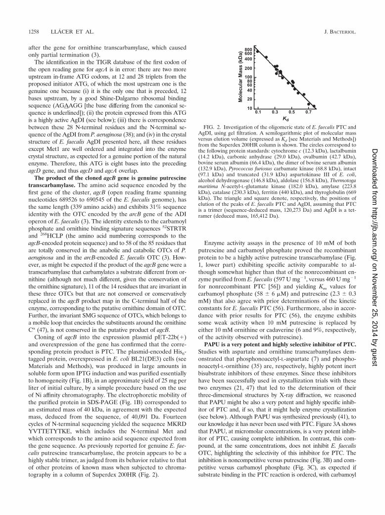
Cloning of agcB into the expression plasmid pET-22b(�)and overexpression of the gene has confirmed that the corre-sponding protein product is PTC. The plasmid-encoded His6-tagged protein, overexpressed in E. coli BL21(DE3) cells (seeMaterials and Methods), was produced in large amounts insoluble form upon IPTG induction and was purified essentiallyto homogeneity (Fig. 1B), in an approximate yield of 25 mg perliter of initial culture, by a simple procedure based on the useof Ni affinity chromatography. The electrophoretic mobility ofthe purified protein in SDS-PAGE (Fig. 1B) corresponded toan estimated mass of 40 kDa, in agreement with the expectedmass, deduced from the sequence, of 40,091 Da. Fourteencycles of N-terminal sequencing yielded the sequence MKRDYVTTETYTKE, which includes the N-terminal Met andwhich corresponds to the amino acid sequence expected fromthe gene sequence. As previously reported for genuine E. fae-calis putrescine transcarbamylase, the protein appears to be ahighly stable trimer, as judged from its behavior relative to thatof other proteins of known mass when subjected to chroma-tography in a column of Superdex 200HR (Fig. 2).
Enzyme activity assays in the presence of 10 mM of bothputrescine and carbamoyl phosphate proved the recombinantprotein to be a highly active putrescine transcarbamylase (Fig.1, lower part) exhibiting specific activity comparable to al-though somewhat higher than that of the nonrecombinant en-zyme purified from E. faecalis (597 U mg�1, versus 460 U mg�1
for nonrecombinant PTC [56]) and yielding Km values forcarbamoyl phosphate (58 � 6 �M) and putrescine (2.3 � 0.3mM) that also agree with prior determinations of the kineticconstants for E. faecalis PTC (56). Furthermore, also in accor-dance with prior results for PTC (56), the enzyme exhibitssome weak activity when 10 mM putrescine is replaced byeither 10 mM ornithine or cadaverine (6 and 9%, respectively,of the activity observed with putrescine).
PAPU is a very potent and highly selective inhibitor of PTC.Studies with aspartate and ornithine transcarbamylases dem-onstrated that phosphonoacetyl-L-aspartate (7) and phospho-noacetyl-L-ornithine (35) are, respectively, highly potent inertbisubstrate inhibitors of these enzymes. Since these inhibitorshave been successfully used in crystallization trials with thesetwo enzymes (21, 47) that led to the determination of theirthree-dimensional structures by X-ray diffraction, we reasonedthat PAPU might be also a very potent and highly specific inhib-itor of PTC and, if so, that it might help enzyme crystallization(see below). Although PAPU was synthesized previously (41), toour knowledge it has never been used with PTC. Figure 3A showsthat PAPU, at micromolar concentrations, is a very potent inhib-itor of PTC, causing complete inhibition. In contrast, this com-pound, at the same concentrations, does not inhibit E. faecalisOTC, highlighting the selectivity of this inhibitor for PTC. Theinhibition is noncompetitive versus putrescine (Fig. 3B) and com-petitive versus carbamoyl phosphate (Fig. 3C), as expected ifsubstrate binding in the PTC reaction is ordered, with carbamoyl
FIG. 2. Investigation of the oligomeric state of E. faecalis PTC andAgDI, using gel filtration. A semilogarithmic plot of molecular massversus elution volume (expressed as Kd [see Materials and Methods])from the Superdex 200HR column is shown. The circles correspond tothe following protein standards: cytochrome c (12.3 kDa), lactalbumin(14.2 kDa), carbonic anhydrase (29.0 kDa), ovalbumin (42.7 kDa),bovine serum albumin (66.4 kDa), the dimer of bovine serum albumin(132.9 kDa), Pyrococcus furiosus carbamate kinase (68.8 kDa), intact(97.1 kDa) and truncated (31.9 kDa) aspartokinase III of E. coli,alcohol dehydrogenase (146.8 kDa), aldolase (156.8 kDa), Thermotogamaritima N-acetyl-L-glutamate kinase (182.0 kDa), amylase (223.8kDa), catalase (230.3 kDa), ferritin (440 kDa), and thyroglobulin (669kDa). The triangle and square denote, respectively, the positions ofelution of the peaks of E. faecalis PTC and AgDI, assuming that PTCis a trimer (sequence-deduced mass, 120,273 Da) and AgDI is a tet-ramer (deduced mass, 165,412 Da).
1258 LLACER ET AL. J. BACTERIOL.
on Novem
ber 25, 2014 by guesthttp://jb.asm
.org/D
ownloaded from
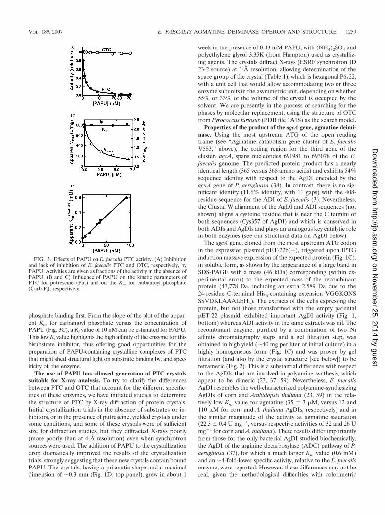
phosphate binding first. From the slope of the plot of the appar-ent Km for carbamoyl phosphate versus the concentration ofPAPU (Fig. 3C), a Ki value of 10 nM can be estimated for PAPU.This low Ki value highlights the high affinity of the enzyme for thisbisubstrate inhibitor, thus offering good opportunities for thepreparation of PAPU-containing crystalline complexes of PTCthat might shed structural light on substrate binding by, and spec-ificity of, the enzyme.
The use of PAPU has allowed generation of PTC crystalssuitable for X-ray analysis. To try to clarify the differencesbetween PTC and OTC that account for the different specific-ities of these enzymes, we have initiated studies to determinethe structure of PTC by X-ray diffraction of protein crystals.Initial crystallization trials in the absence of substrates or in-hibitors, or in the presence of putrescine, yielded crystals undersome conditions, and some of these crystals were of sufficientsize for diffraction studies, but they diffracted X-rays poorly(more poorly than at 4-Å resolution) even when synchrotronsources were used. The addition of PAPU to the crystallizationdrop dramatically improved the results of the crystallizationtrials, strongly suggesting that these new crystals contain boundPAPU. The crystals, having a prismatic shape and a maximaldimension of 0.3 mm (Fig. 1D, top panel), grew in about 1
week in the presence of 0.43 mM PAPU, with (NH4)2SO4 andpolyethylene glycol 3.35K (from Hampton) used as crystalliz-ing agents. The crystals diffract X-rays (ESRF synchrotron ID23-2 source) at 3-Å resolution, allowing determination of thespace group of the crystal (Table 1), which is hexagonal P6322,with a unit cell that would allow accommodating two or threeenzyme subunits in the asymmetric unit, depending on whether55% or 33% of the volume of the crystal is occupied by thesolvent. We are presently in the process of searching for thephases by molecular replacement, using the structure of OTCfrom Pyrococcus furiosus (PDB file 1A1S) as the search model.
Properties of the product of the agcA gene, agmatine deimi-nase. Using the most upstream ATG of the open readingframe (see “Agmatine catabolism gene cluster of E. faecalisV583,” above), the coding region for the third gene of thecluster, agcA, spans nucleotides 691981 to 693078 of the E.faecalis genome. The predicted protein product has a nearlyidentical length (365 versus 368 amino acids) and exhibits 54%sequence identity with respect to the AgDI encoded by theaguA gene of P. aeruginosa (38). In contrast, there is no sig-nificant identity (11.6% identity, with 11 gaps) with the 408-residue sequence for the ADI of E. faecalis (3). Nevertheless,the Clustal W alignment of the AgDI and ADI sequences (notshown) aligns a cysteine residue that is near the C termini ofboth sequences (Cys357 of AgDI) and which is conserved inboth ADIs and AgDIs and plays an analogous key catalytic rolein both enzymes (see our structural data on AgDI below).
The agcA gene, cloned from the most upstream ATG codonin the expression plasmid pET-22b(�), triggered upon IPTGinduction massive expression of the expected protein (Fig. 1C),in soluble form, as shown by the appearance of a large band inSDS-PAGE with a mass (46 kDa) corresponding (within ex-perimental error) to the expected mass of the recombinantprotein (43,778 Da, including an extra 2,589 Da due to the24-residue C-terminal His6-containing extension VGGKQNSSSVDKLAAALEH6). The extracts of the cells expressing theprotein, but not those transformed with the empty parentalpET-22 plasmid, exhibited important AgDI activity (Fig. 1,bottom) whereas ADI activity in the same extracts was nil. Therecombinant enzyme, purified by a combination of two Niaffinity chromatography steps and a gel filtration step, wasobtained in high yield (40 mg per liter of initial culture) in ahighly homogeneous form (Fig. 1C) and was proven by gelfiltration (and also by the crystal structure [see below]) to betetrameric (Fig. 2). This is a substantial difference with respectto the AgDIs that are involved in polyamine synthesis, whichappear to be dimeric (23, 37, 59). Nevertheless, E. faecalisAgDI resembles the well-characterized polyamine-synthesizingAgDIs of corn and Arabidopsis thaliana (23, 59) in the rela-tively low Km value for agmatine (35 � 3 �M, versus 12 and110 �M for corn and A. thaliana AgDIs, respectively) and inthe similar magnitude of the activity at agmatine saturation(22.3 � 0.4 U mg�1, versus respective activities of 32 and 26 Umg�1 for corn and A. thaliana). These results differ importantlyfrom those for the only bacterial AgDI studied biochemically,the AgDI of the arginine decarboxylase (ADC) pathway of P.aeruginosa (37), for which a much larger Km value (0.6 mM)and an 4-fold-lower specific activity, relative to the E. faecalisenzyme, were reported. However, these differences may not bereal, given the methodological difficulties with colorimetric
FIG. 3. Effects of PAPU on E. faecalis PTC activity. (A) Inhibitionand lack of inhibition of E. faecalis PTC and OTC, respectively, byPAPU. Activities are given as fractions of the activity in the absence ofPAPU. (B and C) Influence of PAPU on the kinetic parameters ofPTC for putrescine (Put) and on the Km for carbamoyl phosphate(Carb-Pi), respectively.
VOL. 189, 2007 E. FAECALIS AGMATINE DEIMINASE OPERON AND STRUCTURE 1259
on Novem
ber 25, 2014 by guesthttp://jb.asm
.org/D
ownloaded from

activity assays at low substrate concentrations and given theinstability of AgDI upon large dilution in the assay solution(which was prevented in our case by adding 1 mg ml�1 bovineserum albumin). Similarly to all previous reports with AgDIsfrom other sources (23, 37, 60), the E. faecalis enzyme appearsto be highly specific for agmatine, not using L-arginine (Fig. 1,lower panel), L-argininamide, or arcaine (1,4-diguanidinobu-tane). Arcaine was reported to be a competitive inhibitor (rel-ative to agmatine) of the corn (59) and cucumber (46) en-zymes, with Ki values of 3 and 7 �M, and we have found thiscompound also to be a competitive inhibitor of E. faecalisAgDI, with a Ki value of 28 � 5 �M.
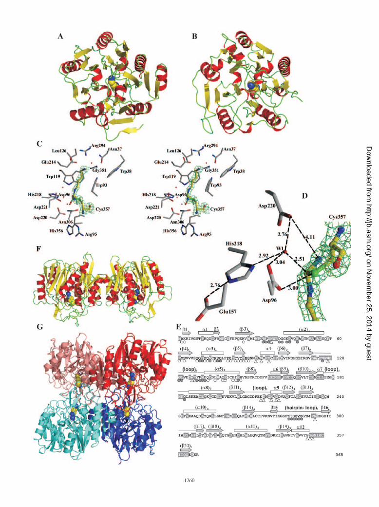
AgDI and ADI share the same basic fold. AgDI monocrys-tals of up to 1 mm in length (Fig. 1D, bottom panel) diffractedX-rays to 1.65 Å, allowing the determination of the crystalstructure of the enzyme at atomic resolution. The asymmetricunit of the AgDI crystals (Table 1) contains eight subunitsorganized as two tetramers having identical structure. Whensuperimposed, the root mean square deviation (RMSD) formonomers is 0.17 Å (for 364 C� atoms). Each of the mono-mers has a globular fold with approximate dimensions of 53 by45 by 40 Å3. The monomer has a tertiary fold similar to that ofthe catalytic domain of ADI (Fig. 4A and B), although it lacks thefive-helix bundle domain of this enzyme (9). Thus, the AgDImonomer has the fan-like structure with five blades that is adistinctive trait of ADI and which results from a fivefold pseudo-symmetric structure in which each repeating element consists of athree-stranded mixed � sheet and a helix in a ���� arrangement.Given the absence in AgDI of the five-helix bundle that in ADIdistorts the fan-like structure, the AgDI monomer is closer tofivefold pseudosymmetry than the catalytic domain of ADI (Fig.4A and B). Nevertheless, the first repeat diverges from the ca-nonical structure of the repeat, since it has two helices and thearrangement �����, and it is flanked near the fivefold pseudo-symmetry axis by the C-terminal strand running antiparallel to theother three strands. The lengths and amino acid sequences, how-ever, vary considerably from one element to another (Fig. 4E)and from those in the catalytic domain of ADI (not shown), andindeed, the superposition of AgDI with the catalytic domain ofADI (from subunit A of the Mycoplasma arginini enzyme [PDBfile 1S9R]) yields a relatively large RMSD (2.77 Å for 238 C�atoms). A distinctive characteristic of the AgDI monomer fold isthe existence of large loops emerging on the side of the fan
corresponding to the C end of the two parallel strands of therepeats (Fig. 4E and bottom part of Fig. 4F). Particularly large isthe loop that emerges from the end of repeat 4, which includes a� hairpin (�15 and �16). This loop folds flat over the other loopsand over a protruding long � helix that emerges from repeat 1(helix 2, Fig. 4E) in the same direction as the loops. The presenceof these loops and of the protruding � helix 2 renders highlydifferent the two faces of the monomer that correspond to oppo-site edges of the repeats � sheets and also serves the purposes offorming the active center and of providing interactions with theother subunits to form the tetramer (see below). Because of thepresence of these loops on one side of the subunit, and also sincethe � helix of each repeat fills the space between adjacent repeatsdiverging from the pseudosymmetry axis, the subunit has a ball-like rounded shape (see each subunit in Fig. 4F and G).
The agmatine binding site explains the high specificity ofAgDI for its substrate. A large mass of electron density notcorresponding to the polypeptide chain and having an elon-gated shape was clearly visible (Fig. 4C), filling an internalcavity of the enzyme and being connected to the density of theS atom of Cys357, the cysteine residue that is close to theenzyme C terminus and that is conserved in both agmatinedeiminase and arginine deiminase. The electron density at1.65-Å resolution fits a completely extended molecule ofagmatine, with its C� atom (the C atom of the guanidinium groupof agmatine) covalently linked with the S atom of Cys357.Agmatine is bound centrally (Fig. 4A), approximately alongthe fivefold pseudosymmetry axis near its exit from the loop-rich side of the fan, in a closed, elongated, and crowded cavity.The central position results in the involvement in the buildingof the site of elements connected to all five repeats of thesubunit. Thus, the site is formed between the long loop ofrepeat 2 and the loops that connect repeats 5 to 1, 1 to 2, and3 to 4. The cavity is closed at its entry by a three-tongued gateformed by the loop of repeat 2 and by the long loops connect-ing repeats 3 to 4 and 4 to 5 (seen laterally in Fig. 4F). In ourstructure the closure is ensured by mutual interactions betweensome residues of these loops, although it is clear that theseloops have to retreat at the beginning and at the end of thecatalytic cycle, to allow substrate binding and carbamoyl pu-trescine release. The extended substrate runs parallel to andmakes extensive Van der Waals contacts with a straight stretchof three glycines (Gly351-Gly352-Gly353), also making a hy-
FIG. 4. Structure of AgDI. (A and B) Ribbon representations of the monomer of E. faecalis AgDI (A) and of the catalytic domain of Mycoplasmaarginini ADI (PDB entry 1S9R without residues 75 to 148, which are not a part of the catalytic domain) (B), both containing the covalently boundsubstrate in space-filling representation. � helices, � sheets, and loops are colored red, yellow, and green, respectively. (C) Stereo view of the active siteof AgDI, showing the density map of 2Fobs � Fcalc, contoured at the 0.9 level, around the covalent adduct. The substrate is colored yellow, and thesurrounding protein residues are colored gray. O, N, and S atoms are colored red, blue, and green, respectively. (D) Interatomic distances (in angstroms)between the catalytic protein residues and the substrate around the reactive carbon center. The interactions with a fixed water molecule (W1) believedto be important in the mechanism are also represented. The density map of 2Fobs � Fcalc, contoured at 0.75 for the covalent amidino complex, is shown.(E) Correspondence between amino acid sequence and secondary structure. Bars, arrows, and lines above the structure denote, respectively, � helices,� strands, and loops (only long loops are depicted), numbered in ascending order from N to C terminus and, when belonging to a repeat, in parenthesesand having a subscript that denotes the repeat number. Open triangles under the sequence denote residues having decreased accessibility upon thebinding of agmatine. Circles denote decreased accessibility upon dimer (open) and tetramer (shaded) formation. The gray sequence backgroundshighlight residues that are invariant in the AgDIs of E. faecalis, Streptococcus mutans, Pseudomonas aeruginosa, and Arabidopsis thaliana (SwissProtaccession numbers Q837U5, Q8DW17, Q9I6J9, and Q8GWW7, respectively). (F) Ribbon diagram of AgDI dimer viewed perpendicularly to themolecular twofold axis. Coloring and substrate representation are as in panel A. (G) Ribbon representation of the AgDI tetramer viewed along one ofthe three twofold molecular axes. The two subunits of the two dimers (as defined in the text) are shown in different shades of red or blue. The covalentlybound substrate is shown in space-filling representation.
VOL. 189, 2007 E. FAECALIS AGMATINE DEIMINASE OPERON AND STRUCTURE 1261
on Novem
ber 25, 2014 by guesthttp://jb.asm
.org/D
ownloaded from

drogen bond between the agmatine amino group and the Oatom of Gly351. These glycines are a part of the conservedsequence (G/A)GGNIHCITQQ(E/Q)P, which includes thecatalytic cysteine (underlined) and which can be considered asignature of AgDI. The four-carbon portion of the molecule ofagmatine is also surrounded by the indolic rings of the invari-ant Trp93 and Trp119 (Fig. 4C), which are like flat tiles thatwall the substrate binding cavity, and by the methyl group ofinvariant Thr215. The agmatine amino group also makes abond with the �-COO� of Glu214 (Fig. 4C), a residue that mayplay a key role in making the enzyme extremely selectiveagainst arginine, since it would not favor placing near it an-other negatively charged group as would be the case for thecarboxylate group of arginine. In any case, the region thatsurrounds carbon 1 of agmatine is packed with predominantlyhydrophobic groups, leaving no room for a carboxyl or for anyother group of substantial size, and less so if the group is polarand charged as in an �-carboxylate. On the opposite end of theagmatine molecule, around the guanidinium group, the invari-ant residues Asp96, His218, and Asp220 surround the boundsubstrate and play catalytic roles (Fig. 4C and D) (see below).
The covalent adduct provides a snapshot of AgDI catalyzingits reaction. A close examination of the electron densityaround the C� atom of agmatine (Fig. 4D) shows that thiscarbon is covalently linked with the S atom of Cys357 (C-Sbond distance, 1.79 Å) and with two nitrogens (Nε and N�2).The C� in our structure is somewhat displaced from the planeformed by its three covalent ligands (S, Nε, and N�2 atoms)towards a water molecule (W1, Fig. 4D) which is located atonly 2.5 Å (a very short distance for nonbonded C and Oatoms). The water molecule is fixed by hydrogen bonds to oneO atom of each of the two side chain carboxylates of Asp220and Asp96 and to the �1N atom of His218. In turn, the ε2N ofHis218 is linked to the �-COO� of Glu157. A similar adductwas reported with ADI within a complex which replicates es-sentially all of the details of the present complex, including thepresence and the interactions of the fixed water (9). This com-plex was interpreted to represent the covalent amidino com-plex proposed long ago to be formed in the mechanism of ADI(9, 16). Therefore, the present complex is highly indicative of acommon, very strictly conserved mechanism of deimination byADI and AgDI. This mechanism (Fig. 5) involves two tetrahe-
FIG. 5. Proposed five-step mechanism for the AgDI reaction. Step1 leads to the formation of the first tetrahedral carbon center inter-mediate as a consequence of the attack by the activated thiol ofCys357. Asp96 and the nonprotonated primary N of the guanidiniumgroup may induce deprotonation of the thiol group. A proton is ex-tracted by His218, which forms a charge relay system with Glu157. Instep 2 the tetrahedral intermediate collapses to the triagonal amidinointermediate, with liberation of ammonia. Asp96, Asp220, and His218help stabilize the leaving ammonia and the positive charge develop-ment in the amidino group. In step 3 ammonia is replaced by waterpositioned for attack on the carbon center, interacting with the samegroups as the ammonia. The intermediate revealed here by X-raycrystallography corresponds to one of the two complexes (either theammonia or the water complex) with the amidino intermediate. Step 4is the formation of the second tetrahedral carbon intermediate. His218helps this step by abstracting one proton from water. The final step isthe collapse of the tetrahedral intermediate to carbamoylputrescineand the regenerated thiol group.
1262 LLACER ET AL. J. BACTERIOL.
on Novem
ber 25, 2014 by guesthttp://jb.asm
.org/D
ownloaded from
dral intermediates but only one amidino adduct. Nevertheless,the amidino adduct must exist first in the presence of theleaving ammonia and later in the presence of the attackingwater. Thus, the W1 molecule could also correspond to am-monia, and the present adduct may represent either theamidino compound with the leaving ammonia or the samecompound with the attacking water.
Architecture of the AgDI tetramer. In accordance with theconclusions derived from gel filtration data, AgDI is organizedas a tetramer. This tetramer has tetrahedral shape, with thefour subunits located in the vertices (Fig. 4G), and can beconsidered to be composed of two identical dimers, each ofthem (Fig. 4F) built by a 180° rotation of the monomer aroundan axis that is approximately parallel to the fivefold pseudo-symmetry axis. Thus, this dimer has the aspect of two fans inbattery. The interactions in this dimer are mediated by theelements of the first and second repeats, with the edge of themore external � strand of the first repeat interacting withthe C-terminal two turns of � helix 4, the helix belonging to thesecond repeat. These interactions are generally hydrophobic inthe core region and polar towards the periphery, and the sur-face involved amounts to an average of 991 Å2 per monomer(determined with a probe radius of 1.4 Å) or 6.9% of thesurface of each monomer. The two subunits in the dimer leavea valley between them (Fig. 4F) in the loop-rich face. It is thisvalley which is used for tetramer formation by having the valleyof one dimer interact in a crossed-over way with the valley inthe other dimer, so that the twofold axes of the two dimers arecoincident and the longest axes of the two dimers run in per-pendicular directions (Fig. 4G). One subunit (called A for thepurpose of this description) of one dimer interacts with the twosubunits of the other dimer (here called C and D). The Ntermini of helices 2 from A and C interact mutually, andresidues 286 to 289 of the long hairpin loop that connectsrepeats 4 and 5 of A interact with the outer surface of helix 3and also with the N-terminal turn of helix 2 of C, and viceversa. The interactions between A and D are restricted tomutual hydrophobic contacts between the long loop of repeat2 of both subunits. The buried surface per monomer is 639 Å2
for the interactions between A and C and only 252 Å2 for thosebetween A and D. Overall, each monomer has in the tetramera buried surface of 1,927 Å2, accounting for 13.5% of its totalaccessible surface area and explaining the stability of the en-zyme tetramer that has been observed in the present studies.
DISCUSSION
By cloning the genes and by studying the expressed proteins,we obtained here the most conclusive proof to date that the E.faecalis agcB and agcA genes encode two key enzymes of ag-matine catabolism, PTC and AgDI, confirming and extendingprevious (17, 18, 39) but more indirect evidence for the iden-tification of these genes. When initially purified from E. fae-calis, PTC exhibited some (although low) activity with orni-thine (56), and this is confirmed here with the recombinant,His tag-purified enzyme, virtually completely excluding OTCcontamination as the cause for this activity. Nevertheless, therelatively low specific activity of PTC compared with the activ-ity of pure OTC (5-fold higher) (the low PTC activity is notdue to the poly-His tail, since wild-type PTC isolated from E.
faecalis has even somewhat less activity [56]) made it desirableto confirm that this enzyme is a genuine PTC, which has beendone here by demonstrating that this enzyme is powerfullyinhibited by very low concentrations (Ki � 10 nM) of thePTC-specific bisubstrate analog inhibitor PAPU, a compoundthat does not inhibit OTC at similar concentrations.
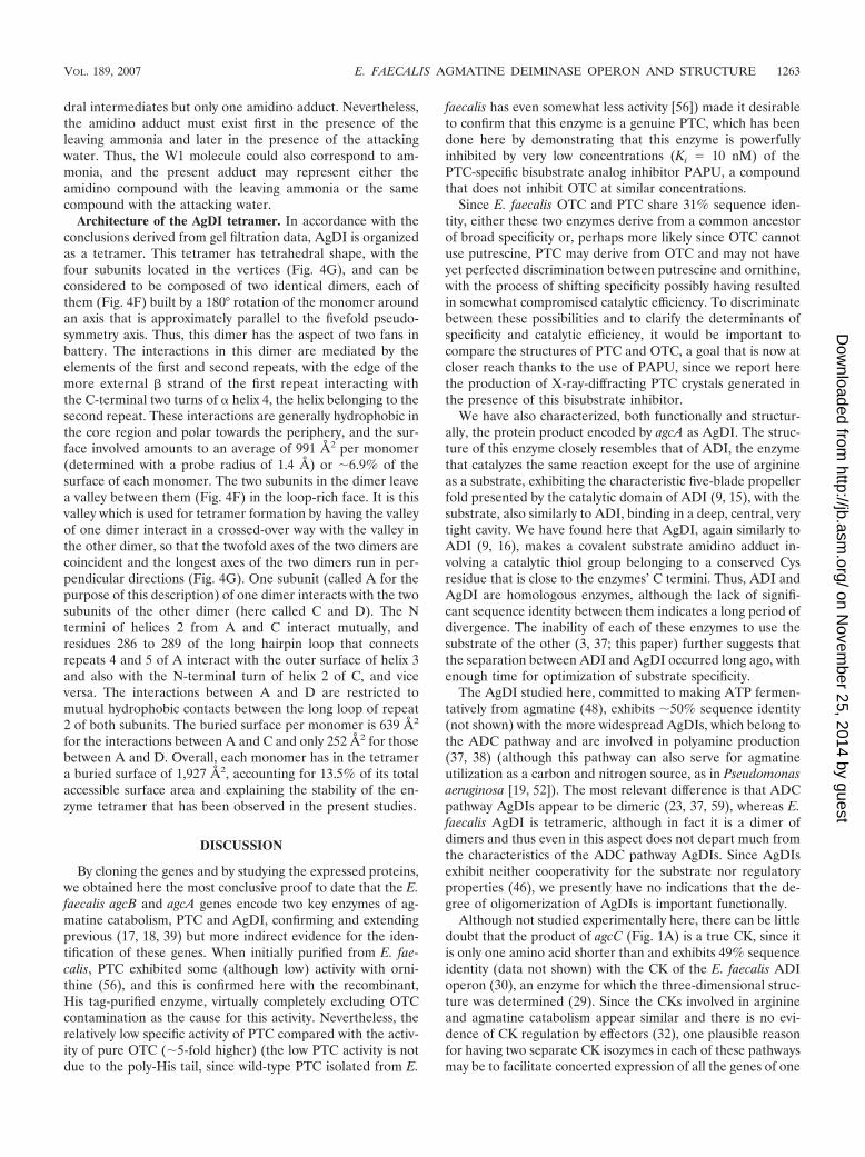
Since E. faecalis OTC and PTC share 31% sequence iden-tity, either these two enzymes derive from a common ancestorof broad specificity or, perhaps more likely since OTC cannotuse putrescine, PTC may derive from OTC and may not haveyet perfected discrimination between putrescine and ornithine,with the process of shifting specificity possibly having resultedin somewhat compromised catalytic efficiency. To discriminatebetween these possibilities and to clarify the determinants ofspecificity and catalytic efficiency, it would be important tocompare the structures of PTC and OTC, a goal that is now atcloser reach thanks to the use of PAPU, since we report herethe production of X-ray-diffracting PTC crystals generated inthe presence of this bisubstrate inhibitor.
We have also characterized, both functionally and structur-ally, the protein product encoded by agcA as AgDI. The struc-ture of this enzyme closely resembles that of ADI, the enzymethat catalyzes the same reaction except for the use of arginineas a substrate, exhibiting the characteristic five-blade propellerfold presented by the catalytic domain of ADI (9, 15), with thesubstrate, also similarly to ADI, binding in a deep, central, verytight cavity. We have found here that AgDI, again similarly toADI (9, 16), makes a covalent substrate amidino adduct in-volving a catalytic thiol group belonging to a conserved Cysresidue that is close to the enzymes’ C termini. Thus, ADI andAgDI are homologous enzymes, although the lack of signifi-cant sequence identity between them indicates a long period ofdivergence. The inability of each of these enzymes to use thesubstrate of the other (3, 37; this paper) further suggests thatthe separation between ADI and AgDI occurred long ago, withenough time for optimization of substrate specificity.
The AgDI studied here, committed to making ATP fermen-tatively from agmatine (48), exhibits 50% sequence identity(not shown) with the more widespread AgDIs, which belong tothe ADC pathway and are involved in polyamine production(37, 38) (although this pathway can also serve for agmatineutilization as a carbon and nitrogen source, as in Pseudomonasaeruginosa [19, 52]). The most relevant difference is that ADCpathway AgDIs appear to be dimeric (23, 37, 59), whereas E.faecalis AgDI is tetrameric, although in fact it is a dimer ofdimers and thus even in this aspect does not depart much fromthe characteristics of the ADC pathway AgDIs. Since AgDIsexhibit neither cooperativity for the substrate nor regulatoryproperties (46), we presently have no indications that the de-gree of oligomerization of AgDIs is important functionally.
Although not studied experimentally here, there can be littledoubt that the product of agcC (Fig. 1A) is a true CK, since itis only one amino acid shorter than and exhibits 49% sequenceidentity (data not shown) with the CK of the E. faecalis ADIoperon (30), an enzyme for which the three-dimensional struc-ture was determined (29). Since the CKs involved in arginineand agmatine catabolism appear similar and there is no evi-dence of CK regulation by effectors (32), one plausible reasonfor having two separate CK isozymes in each of these pathwaysmay be to facilitate concerted expression of all the genes of one
VOL. 189, 2007 E. FAECALIS AGMATINE DEIMINASE OPERON AND STRUCTURE 1263
on Novem
ber 25, 2014 by guesthttp://jb.asm
.org/D
ownloaded from

or the other pathway. The important sequence identity of thesetwo CK isozymes indicates that their separation is not remote.In contrast, the lack of significant sequence identity (14%)between the E. faecalis arcD and agcD gene products (theputative arginine/ornithine and agmatine/putrescine antiport-ers) indicates ancient divergence, the homology of these genesbeing supported by the similarity of the polypeptide lengths(483 and 458 residues, respectively) and transmembrane helixpredictions (11 to 12 helices) and also by the analogous func-tions and substrates of the antiporters.
The comparison of the genes of the ADI and AgDI pathwayscontradicts the naıve view that the two pathways might havearisen by a process of duplication of a complete ancient four-gene cluster. As already indicated, the deiminase and anti-porter components of both pathways have evolved separatelyfor much longer than the transcarbamylase and carbamatekinase components, in contrast with the expectation for a com-mon duplication event for all of the elements of the genecluster, followed by coevolution. Nevertheless, the genes forthe transcarbamylase and for CK may have duplicated simul-taneously, given their similar degree of conservation in onegene cluster relative to the corresponding genes in the othercluster and also since in both clusters the transcarbamylasegene physically precedes the CK gene. Since agmatine utiliza-tion cannot precede agmatine production, the close relation-ship between the AgDIs of the ADC and AgDI pathwayssuggests that the latter may have derived from an ADC path-way gene for AgDI. The evolution of AgDI may have beeninitiated by its divergence from ADI to serve the purpose ofpolyamine synthesis. Much more recently, AgDI may havebecome committed into a novel route of agmatine catabolism,made by recombining elements of the ADI pathway with thearginine decarboxylase pathway element AgDI and with anagmatine antiporter of obscure origin.
An important contribution of the present work is the clari-fication of the structure and, based on the structure, of thereactional and catalytic mechanisms of AgDI. The high affinityof AgDI for agmatine is accounted for by the extension andcloseness of the interactions between substrate and enzyme,since agmatine is buried, fitting tightly a binding site wherethere is no empty space. The high specificity is explained by thenegative charge at the entry of the site provided by Glu214,which would not fit the placement of the �-carboxylate groupof arginine, and also by the very crowded environment whereeven minor volumes around the C1 of agmatine would beexcluded. The relatively low kcat for a hydrolase exhibited byAgDI (17 s�1 at 37°C) may be explained by the deepness of thesite, with the catalytic groups far into the subunit structure, andalso by the existence of a gating mechanism at the entry of thesite that has to open, close, and open again in each catalyticcycle, possibly limiting substrate access or product release.Agmatine binding may trigger site closure, since the amino endof the substrate interacts with elements of the loops that con-tribute to the gating mechanism, particularly with Glu214.Since the formation of the covalent adduct with the thiol groupof Cys357 should shorten somewhat the bound molecule, theclosing mechanism could be described as “pulling the gatefrom the inside” by the covalently bound substrate. The im-portance of substrate binding for gate closure is supported bythe observation of the deposited structure of S. mutans AgDI,
which contains no bound agmatine and where the largest loopinvolved in the gating mechanism is retracted and the site ismore accessible. Our structure, containing the covalentlybound substrate, is closed and would have to be open at theend of the reaction. The simplest triggering mechanism toopen the gate could be defined as “pushing the gate from theinside,” whereby the increase in the volume resulting from thecoexistence of the ureido group in carbamoyl putrescine andthe free thiol in Cys357 may result in some displacement of themolecule of the product towards the gate, particularly giventhe extreme narrowness of the site, which should not allowbending of the bound product.
Puzzlingly, in our crystal structure the enzyme has retainedthe covalent amidino adduct without progressing further alongthe reactional path. The corresponding analog for arginine hasalso been reported for ADI (9). The presence of the trappedintermediate strongly suggests that some component in thecrystallization solution has stabilized the amidino intermedi-ate, in fact resulting in enzyme inhibition. Perhaps ammonia,present in our solution at a concentration of 3.2 M, has re-sulted in the stabilization of the ammonia-containing amidinocomplex (W1 in our structure could equally be ammonia),blocking further reaction with water (Fig. 5). Whatever themechanism, the observation of the complex has had the valueof clarifying substrate binding and catalysis. The catalytic pro-cess involves centrally, as in the case of ADI (9, 15), a chargerelay system consisting of Glu157 and His218, which promotesformation of the tetrahedral intermediates by providing orwithdrawing a proton, and Cys357 with its SH group beingabnormally acidic, perhaps because of the presence of theguanidinium group of the substrate and possibly also by theinducing effect of the nearby (3 Å away) carboxylate of Asp96.We are presently undertaking studies to subject to experimen-tal tests, by site-directed mutagenesis, the roles proposed onthe basis of the structure for catalysis of the reaction by theseresidues.
ACKNOWLEDGMENTS
This work was supported by grant BFU2004-05159 from the SpanishMinistry of Education and Science. L. M. Polo is a fellow of CSIC-Banco de Santander, and J. L. Llacer and S. Tavarez are fellows of theSpanish Ministry of Education and Science. We thank the EU, ESRF,and EMBL Grenoble for financial support for ESRF synchrotronX-ray data collection.
We thank the ESRF personnel for expert help; A. Marina and M.Lopez for diffracting AgDI; J. J. Calvete (IBV-CSIC, Valencia, Spain)for N-terminal sequencing; D. Gigot (Universite Livre de Bruxelles,Belgium) for advice; A. Cantin and M. A. Miranda (ITQ-CSIC, Va-lencia, Spain) for help with the synthesis of PAPU; C. Aguado (CIPF,Valencia, Spain) for matrix-assisted laser desorption ionization–time-of-flight mass spectrometry; and J. Selles, P. Tortosa, and L. Osuna(IBV-CSIC, Valencia, Spain) for technical help.
REFERENCES
1. Archibald, R. 1944. Determination of citrulline and allantoin and demon-stration of citrulline in blood plasma. J. Biol. Chem. 156:121–141.
2. Bagni, N., and A. Tassoni. 2001. Biosynthesis, oxidation and conjugation ofaliphatic polyamines in higher plants. Amino Acids 20:301–317.
3. Barcelona-Andres, B. A. Marina, and V. Rubio. 2002. Gene structure, orga-nization, expression, and potential regulatory mechanisms of arginine catab-olism in Enterococcus faecalis. J. Bacteriol. 184:6289–6300.
4. Bartlett, G. R. 1959. Phosphorus assay in column chromatography. J. Biol.Chem. 234:466–468.
5. Bradford, M. M. 1976. A rapid and sensitive method for the quantitation ofmicrogram quantities of protein utilizing the principle of protein-dye bind-ing. Anal. Biochem. 72:248–254.
1264 LLACER ET AL. J. BACTERIOL.
on Novem
ber 25, 2014 by guesthttp://jb.asm
.org/D
ownloaded from
6. Collaborative Computational Project Number 4. 1994. The CCP4 suite:programs for protein crystallography. Acta Crystallogr. D 50:760–763.
7. Collins, K. D., and G. R. Stark. 1971. Aspartate transcarbamylase. Interac-tion with the transition state analogue N-(phosphonacetyl)-L-aspartate.J. Biol. Chem. 246:6599–6605.
8. Cunin, R., N. Glansdorff, A. Pierard, and V. Stalon. 1986. Biosynthesis andmetabolism of arginine in bacteria. Microbiol. Rev. 50:314–352.
9. Das, K., G. H. Butler, V. Kwiatkowski, A. D. Clark, Jr., P. Yadav, and E.Arnold. 2004. Crystal structures of arginine deiminase with covalent reactionintermediates; implications for catalytic mechanism. Structure 12:657–667.
10. Deibel, R. H. 1964. Utilization of arginine as an energy source for the growthof Streptococcus faecalis. J. Bacteriol. 87:988–992.
11. Driessen, A. J., E. J. Smid, and W. N. Konings. 1988. Transport of diaminesby Enterococcus faecalis is mediated by an agmatine-putrescine antiporter. J.Bacteriol. 170:4522–4527.
12. Emsley, P., and K. Cowtan. 2004. Coot: model-building tools for moleculargraphics. Acta Crystallogr. D 60:2126–2132.
13. Esnouf, R. M. 1999. Further additions to MolScript version 1.4, includingreading and contouring of electron-density maps. Acta Crystallogr. D 55:938–940.
14. Fernandez-Murga, M. L., F. Gil-Ortiz, J. L. Llacer, and V. Rubio. 2004.Arginine biosynthesis in Thermotoga maritima: characterization of the argi-nine-sensitive N-acetyl-L-glutamate kinase. J. Bacteriol. 186:6142–6149.
15. Galkin, A., L. Kulakova, E. Sarikaya, K. Lim, A. Howard, and O. Herzberg.2004. Structural insight into arginine degradation by arginine deiminase, anantibacterial and parasite drug target. J. Biol. Chem. 279:14001–14008.
16. Galkin, A., X. Lu, D. Dunaway-Mariano, and O. Herzberg. 2005. Crystalstructures representing the Michaelis complex and the thiouronium reactionintermediate of Pseudomonas aeruginosa arginine deiminase. J. Biol. Chem.280:34080–34087.
17. Griswold, A. R., Y. Y. Chen, and R. A. Burne. 2004. Analysis of an agmatinedeiminase gene cluster in Streptococcus mutans UA159. J. Bacteriol. 186:1902–1904.
18. Griswold, A. R., M. Jameson-Lee, and R. A. Burne. 2006. Regulation andphysiologic significance of the agmatine deiminase system of Streptococcusmutans UA159. J. Bacteriol. 188:834–841.
19. Haas, D., H. Matsumoto, P. Moretti, V. Stalon, and A. Mercenier. 1984.Arginine degradation in Pseudomonas aeruginosa mutants blocked in twoarginine catabolic pathways. Mol. Gen. Genet. 193:437–444.
20. Hills, G. M. 1940. Ammonia production by pathogenic bacteria. Biochem. J.34:1057–1069.
21. Huang, J., and W. N. Lipscomb. 2004. Aspartate transcarbamylase (ATCase)of Escherichia coli: a new crystalline R-state bound to PALA, or to productanalogues citrate and phosphate. Biochemistry 43:6415–6421.
22. Jancarik, J., and S. H. Kim. 1991. Sparse matrix sampling: a screeningmethod for crystallization of proteins. J. Appl. Crystallogr. 24:409–411.
23. Janowitz, T., H. Kneifel, and M. Piotrowski. 2003. Identification and char-acterization of plant agmatine iminohydrolase, the last missing link in poly-amine biosynthesis of plants. FEBS Lett. 544:258–261.
24. Jones, M. E., and F. Lipmann. 1960. Chemical and enzymatic synthesis ofcarbamyl phosphate. Proc. Natl. Acad. Sci. USA 46:1194–1205.
25. Krissinel, E., and K. Henrick. 2004. Secondary-structure matching (SSM), anew tool for fast protein structure alignment in three dimensions. ActaCrystallogr. D 60:2256–2268.
26. Laemmli, U. K. 1970. Cleavage of structural proteins during the assembly ofthe head of bacteriophage T4. Nature 227:680–685.
27. Laskowski, R. A., M. W. MacArthur, D. S. Moss, and J. M. Thornton. 1993.PROCHECK: a program to check the stereochemical quality of proteinstructures. J. Appl. Crystallogr. 26:283–291.
28. Marco-Marin, C., S. Ramon-Maiques, S. Tavarez, and V. Rubio. 2003. Site-directed mutagenesis of Escherichia coli acetylglutamate kinase and aspar-tokinase III probes the catalytic and substrate-binding mechanisms of theseamino acid kinase family enzymes and allows three-dimensional modelling ofaspartokinase. J. Mol. Biol. 334:459–476.
29. Marina, A., P. M. Alzari, J. Bravo, M. Uriarte, B. Barcelona, I. Fita, and V.Rubio. 1999. Carbamate kinase: new structural machinery for making car-bamoyl phosphate, the common precursor of pyrimidines and arginine. Pro-tein Sci. 8:934–940.
30. Marina, A., M. Uriarte, B. Barcelona, V. Fresquet, J. Cervera, and V. Rubio.1998. Carbamate kinase from Enterococcus faecalis and Enterococcus fae-cium. Cloning of the genes, studies on the enzyme expressed in Escherichiacoli, and sequence similarity with N-acetyl-L-glutamate kinase. Eur. J. Bio-chem. 253:280–291.
31. Marshall, M., and P. P. Cohen. 1972. Ornithine transcarbamylase fromStreptococcus faecalis and bovine liver. I. Isolation and subunit structure.J. Biol. Chem. 247:1641–1653.
32. Marshall, M., and P. P. Cohen. 1966. A kinetic study of the mechanism ofcrystalline carbamate kinase. J. Biol. Chem. 241:4197–4208.
33. Mercenier, A., J. P. Simon, D. Haas, and V. Stalon. 1980. Catabolism ofL-arginine by Pseudomonas aeruginosa. J. Gen. Microbiol. 116:381–389.
34. Merritt, E. A., and M. E. Murphy. 1994. Raster3D version 2.0. A program forphotorealistic molecular graphics. Acta Crystallogr. D 50:869–873.
35. Mori, M., K. Aoyagi, M. Tatibana, T. Ishikawa, and H. Ishii. 1977. N�-(phosphonacetyl)-L-ornithine, a potent transition state analogue inhibitor ofornithine carbamoyltransferase. Biochem. Biophys. Res. Commun. 76:900–904.
36. Murshudov, G. N., A. A. Vagin, and E. J. Dodson. 1997. Refinement ofmacromolecular structures by the maximum-likelihood method. Acta Crys-tallogr. D 53:240–255.
37. Nakada, Y., and Y. Itoh. 2003. Identification of the putrescine biosyntheticgenes in Pseudomonas aeruginosa and characterization of agmatine deiminaseand N-carbamoylputrescine amidohydrolase of the arginine decarboxylase path-way. Microbiology 149:707–714.
38. Nakada, Y., Y. Jiang, T. Nishijyo, Y. Itoh, and C. D. Lu. 2001. Molecularcharacterization and regulation of the aguBA operon, responsible for agma-tine utilization in Pseudomonas aeruginosa PAO1. J. Bacteriol. 183:6517–6524.
39. Naumoff, D. G., Y. Xu, N. Glansdorff, and B. Labedan. 2004. Retrievingsequences of enzymes experimentally characterized but erroneously anno-tated: the case of the putrescine carbamoyltransferase. BMC Genomics 5:52.
40. Nuzum, T. C., and P. J. Snodgrass. 1976. Multiple assays of the five ureacycle enzymes in liver homogenates, p. 325–349. In S. Grisolia, R. Baguena,and F. Mayor (ed.), The urea cycle. John Wiley & Sons, New York, NY.
41. Penninckx, M., and D. Gigot. 1978. Synthesis and interaction with Esche-richia coli L-ornithine carbamolytransferase of two potential transition-stateanalogues. FEBS Lett. 88:94–96.
42. Petrack, B., L. Sullivan, and S. Ratner. 1957. Behavior of purified argininedesiminase from S. faecalis. Arch. Biochem. Biophys. 69:186–197.
43. Portoles, M., and V. Rubio. 1986. High-performance liquid chromatographicassay of argininosuccinate: its application in argininosuccinic aciduria and innormal man. J. Inherit. Metab. Dis. 9:31–38.
44. Ramon-Maiques, S., H. G. Britton, and V. Rubio. 2002. Molecular physiol-ogy of phosphoryl group transfer from carbamoyl phosphate by a hyperther-mophilic enzyme at low temperature. Biochemistry 41:3916–3924.
45. Roon, R. J., and H. A. Barker. 1972. Fermentation of agmatine in Strepto-coccus faecalis: occurrence of putrescine transcarbamoylase. J. Bacteriol.109:44–50.
46. Sakakibara, Y., and H. Yanagisawa. 2003. Agmatine deiminase from cucum-ber seedlings is a mono-specific enzyme: purification and characteristics.Protein Expr. Purif. 30:88–93.
47. Shi, D., H. Morizono, X. Yu, L. Tong, N. M. Allewell, and M. Tuchman. 2001.Human ornithine transcarbamylase: crystallographic insights into substraterecognition and conformational changes. Biochem. J. 354:501–509.
48. Simon, J. P., and V. Stalon. 1982. Enzymes of agmatine degradation and thecontrol of their synthesis in Streptococcus faecalis. J. Bacteriol. 152:676–681.
49. Simon, J. P., B. Wargnies, and V. Stalon. 1982. Control of enzyme synthesisin the arginine deiminase pathway of Streptococcus faecalis. J. Bacteriol.150:1085–1090.
50. Slade, H. D., and W. C. Slamp. 1952. The formation of arginine dihydrolaseby streptococci and some properties of the enzyme system. J. Bacteriol.64:455–466.
51. Spies, J. R. 1957. Colorimetric procedures for amino acids. Methods Enzy-mol. 3:467–477.
52. Stalon, V., and A. Mercenier. 1984. L-Arginine utilization by Pseudomonasspecies. J. Gen. Microbiol. 130:69–76.
53. Thompson, J. D., D. G. Higgins, and T. J. Gibson. 1994. CLUSTAL W:improving the sensitivity of progressive multiple sequence alignment throughsequence weighting, position-specific gap penalties and weight matrix choice.Nucleic Acids Res. 22:4673–4680.
54. Tricot, C., J. L. De Coen, P. Momin, P. Falmagne, and V. Stalon. 1989.Evolutionary relationships among bacterial carbamoyltransferases. J. Gen.Microbiol. 135:2453–2464.
55. Vagin, A., and A. Teplyakov. 1997. MOLREP: an automated program formolecular replacement. J. Appl. Crystallogr. 30:1022–1025.
56. Wargnies, B., N. Lauwers, and V. Stalon. 1979. Structure and properties ofthe putrescine carbamoyltransferase of Streptococcus faecalis. Eur. J. Bio-chem. 101:143–152.
57. Wilson, K. 2001. Preparation of genomic DNA from bacteria, p. 2.4.1–2.4.2.In F. M. Ausubel, R. Brent, R. E. Kingston, D. D. Moore, J. G. Seidman,J. A. Smith, and K. Struhl (ed.), Current protocols in molecular biology. JohnWiley & Sons, New York, NY.
58. Winn, M. D., M. N. Isupov, and G. N. Murshudov. 2001. Use of TLSparameters to model anisotropic displacements in macromolecular refine-ment. Acta Crystallogr. D 57:122–133.
59. Yanagisawa, H. 2001. Agmatine deiminase from maize shoots: purificationand properties. Phytochemistry 56:643–647.
60. Yanagisawa, H., and Y. Suzuki. 1981. Corn agmatine iminohydrolase: puri-fication and properties. Plant Physiol. 67:697–700.
VOL. 189, 2007 E. FAECALIS AGMATINE DEIMINASE OPERON AND STRUCTURE 1265
on Novem
ber 25, 2014 by guesthttp://jb.asm
.org/D
ownloaded from