Recovered changes in the spleen by agmatine treatment after transient cerebral ischemia
Upload
independentCategory
view
0download
0
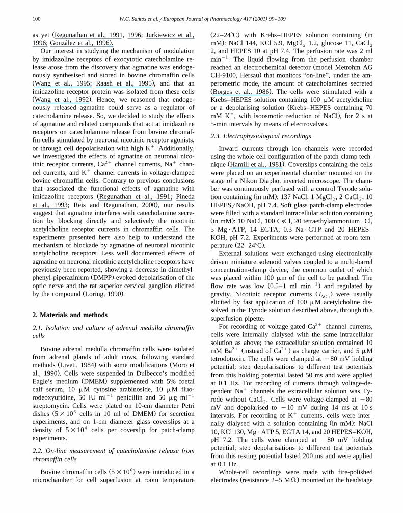
Ž .European Journal of Pharmacology 417 2001 99–109www.elsevier.nlrlocaterejphar
Blockade by agmatine of catecholamine release from chromaffin cells isunrelated to imidazoline receptors
Wilson C. Santosa,b, Jesus M. Hernandez-Guijoc, Ana Ruiz-Nunoc, Roman Olivaresc,´ ´ ˜ ´Aron Jurkiewicza, Luis Gandıac, Antonio G. Garcıac,d,)´ ´
a Departamento de Farmacologıa, Escola Paulista de Medicina, UNIFESP, 04034-970 Sao Paulo, SP, Brazil´b Departamento de Ciencias Fisiologicas, Instituto de Ciencias Biologicas, UniÕersidade do Amazonas, Campus UniÕersitario,ˆ ´ ˆ ´ ´
69.077-000, Manaus, AM, Brazilc Departamento de Farmacologıa, Instituto Teofilo Hernando, Facultad de Medicina, UniÕersidad Autonoma de Madrid, Arzobispo Morcillo,´ ´ ´
4 28029 Madrid, Spaind SerÕicio de Farmacologıa Clınica e Instituto de Gerontologıa, Hospital de la Princesa, Diego de Leon, 62 28006 Madrid, Spain´ ´ ´ ´
Received 23 November 2000; received in revised form 28 February 2001; accepted 2 March 2001
Abstract
The blockade of exocytosis induced by the putative endogenous ligand for imidazoline receptors, agmatine, was studied by usingon-line measurement of catecholamine release in bovine adrenal medullary chromaffin cells. Agmatine inhibited the acetylcholine-evoked
Ž . qrelease of catecholamines in a concentration-dependent manner ICs366 mM ; the K -evoked release of catecholamines was50Ž . Ž .unaffected. Clonidine 100mM and moxonidine 100mM also inhibited by 75% and 50%, respectively, the acetylcholine-evoked
response. In cells voltage-clamped aty80 mV, the intermittent application of acetylcholine pulses elicited whole-cell inward currentsŽ . Ž .I that were blocked 63% by 1 mM agmatine. The onset of blockade was very fastt s31 ms ; the recovery of the current afterACh on
Ž . Ž .washout of agmatine also occurred very rapidlyt s39 ms . Efaroxan 10mM did not affect the inhibition ofI elicited by 1 mMoff ACh
agmatine. I was blocked 90% by 100mM clonidine and 50% by 100mM moxonidine. The concentration–response curve forACh
acetylcholine to elicit inward currents was shifted to the right in a non-parallel manner by 300mM agmatine. The blockade ofIAChŽ .caused by agmatine 100mM was similar at various holding potentials, around 50%. When intracellularly applied, agmatine did not
block I . At 1 mM, agmatine blockedI by 23%, I by 14%, I by 16%, andI by 18%. In conclusion, agmatine blocksACh Na Ba KŽCa. K ŽVD .exocytosis in chromaffin cells by blocking nicotinic acetylcholine receptor currents. In contrast to previous views, these effects seem to beunrelated to imidazoline receptors.q2001 Elsevier Science B.V. All rights reserved.
Keywords: Agmatine; Nicotinic receptor; Ca2q channel; Chromaffin cell
1. Introduction
The idea of the existence of imidazoline receptorsemerged from the observation that clonidine, ana -adren-2
Ž .oceptor agonist Pineda et al., 1993 , also binds to anadditional site in brain membranes, termed the imidazoline
Ž .receptor Bousquet et al., 1984; Ernsberger et al., 1988 . Itis curious that bovine adrenal medulla chromaffin cellslack a -adrenoceptors, but express imidazoline receptors2ŽErnsberger et al., 1989; Regunathan et al., 1991; Wang et
.al., 1992 . Clonidine potently blocks the release of cate-
) Corresponding author. Departamento de Farmacologıa, Instituto´Teofilo Hernando, Facultad de Medicina, Universidad Autonoma de´ ´Madrid, Arzobispo Morcillo, 4 28029 Madrid, Spain. Tel.:q34-91-397-5388; fax:q34-91-397-5397.
Ž .E-mail address: [email protected] A.G. Garcıa .´
Žcholamines from isolated bovine chromaffin cells Powis. Žand Baker, 1986 and the perfused cat adrenal gland Orts
.et al., 1987 by mechanisms unrelated toa -adrenocep-2
tors. Hence, this blockade has been associated with anaction of clonidine on imidazoline receptors of chromaffin
Ž .cells Ohara-Imaizumi and Kumakura, 1992 .Agmatine, a cationic amine, has been proposed to be
Žthe endogenous ligand for imidazoline receptors Li et al.,. Ž1994 . Agmatine has been identified in the brain Feng et
. Žal., 1997 and is synthesised by arginine decarboxylase Li. 2qet al., 1994 , is stored in neurons, is released in a Ca -de-
pendent manner by depolarising stimuli, is metabolisedand degraded to putrescine by agmatine ureohydrolase, and
Žis taken up by synaptosomes for a recent review, see Reis.and Regunathan, 2000 . Hence, agmatine seems to meet all
the criteria of Henry Dale to be considered a brain neuro-transmitter, although with dubious physiological functions
0014-2999r01r$ - see front matterq2001 Elsevier Science B.V. All rights reserved.Ž .PII: S0014-2999 01 00897-4
( )W.C. Santos et al.rEuropean Journal of Pharmacology 417 2001 99–109100
Žas yet Regunathan et al., 1991, 1996; Jurkiewicz et al.,.1996; Gonzalez et al., 1996 .´
Our interest in studying the mechanism of modulationby imidazoline receptors of exocytotic catecholamine re-lease arose from the discovery that agmatine was endoge-nously synthesised and stored in bovine chromaffin cellsŽ .Wang et al., 1995; Raash et al., 1995 , and that animidazoline receptor protein was isolated from these cellsŽ .Wang et al., 1992 . Hence, we reasoned that endoge-nously released agmatine could serve as a regulator ofcatecholamine release. So, we decided to study the effectsof agmatine and related compounds that act at imidazolinereceptors on catecholamine release from bovine chromaf-fin cells stimulated by neuronal nicotinic receptor agonists,or through cell depolarisation with high Kq. Additionally,we investigated the effects of agmatine on neuronal nico-tinic receptor currents, Ca2q channel currents, Naq chan-nel currents, and Kq channel currents in voltage-clampedbovine chromaffin cells. Contrary to previous conclusionsthat associated the functional effects of agmatine with
Žimidazoline receptors Regunathan et al., 1991; Pineda.et al., 1993; Reis and Regunathan, 2000 , our results
suggest that agmatine interferes with catecholamine secre-tion by blocking directly and selectively the nicotinicacetylcholine receptor currents in chromaffin cells. Theexperiments presented here also help to understand themechanism of blockade by agmatine of neuronal nicotinicacetylcholine receptors. Less well documented effects ofagmatine on neuronal nicotinic acetylcholine receptors havepreviously been reported, showing a decrease in dimethyl-
Ž .phenyl-piperazinium DMPP -evoked depolarisation of theoptic nerve and the rat superior cervical ganglion elicited
Ž .by the compound Loring, 1990 .
2. Materials and methods
2.1. Isolation and culture of adrenal medulla chromaffincells
Bovine adrenal medulla chromaffin cells were isolatedfrom adrenal glands of adult cows, following standard
Ž . Žmethods Livett, 1984 with some modifications Moro et.al., 1990 . Cells were suspended in Dulbecco’s modified
Ž .Eagle’s medium DMEM supplemented with 5% foetalcalf serum, 10mM cytosine arabinoside, 10mM fluo-rodeoxyuridine, 50 IU mly1 penicillin and 50mg mly1
streptomycin. Cells were plated on 10-cm diameter PetriŽ 6 .dishes 5=10 cells in 10 ml of DMEM for secretion
experiments, and on 1-cm diameter glass coverslips at adensity of 5=104 cells per coverslip for patch-clampexperiments.
2.2. On-line measurement of catecholamine release fromchromaffin cells
Ž 6.Bovine chromaffin cells 5=10 were introduced in amicrochamber for cell superfusion at room temperature
Ž . Ž22–248C with Krebs–HEPES solution containing in.mM : NaCl 144, KCl 5.9, MgCl 1.2, glucose 11, CaCl2 2
2, and HEPES 10 at pH 7.4. The perfusion rate was 2 mlminy1. The liquid flowing from the perfusion chamber
Žreached an electrochemical detector model Metrohm AG.CH-9100, Hersau that monitorsAon-lineB, under the am-
perometric mode, the amount of catecholamines secretedŽ .Borges et al., 1986 . The cells were stimulated with aKrebs–HEPES solution containing 100mM acetylcholine
Žor a depolarising solution Krebs–HEPES containing 70q .mM K , with isoosmotic reduction of NaCl , for 2 s at
5-min intervals by means of electrovalves.
2.3. Electrophysiological recordings
Inward currents through ion channels were recordedusing the whole-cell configuration of the patch-clamp tech-
Ž .nique Hamill et al., 1981 . Coverslips containing the cellswere placed on an experimental chamber mounted on thestage of a Nikon Diaphot inverted microscope. The cham-ber was continuously perfused with a control Tyrode solu-
Ž .tion containing in mM : 137 NaCl, 1 MgCl , 2 CaCl , 102 2
HEPESrNaOH, pH 7.4. Soft glass patch-clamp electrodeswere filled with a standard intracellular solution containingŽ .in mM : 10 NaCl, 100 CsCl, 20 tetraethylammoniumPCl,5 MgPATP, 14 EGTA, 0.3 NaPGTP and 20 HEPES–KOH, pH 7.2. Experiments were performed at room tem-
Ž .perature 22–248C .External solutions were exchanged using electronically
driven miniature solenoid valves coupled to a multi-barrelconcentration-clamp device, the common outlet of whichwas placed within 100mm of the cell to be patched. The
Ž y1.flow rate was low 0.5–1 ml min and regulated byŽ .gravity. Nicotinic receptor currentsI were usuallyACh
elicited by fast application of 100mM acetylcholine dis-solved in the Tyrode solution described above, through thissuperfusion pipette.
For recording of voltage-gated Ca2q channel currents,cells were internally dialysed with the same intracellularsolution as above; the extracellular solution contained 10
2q Ž 2q.mM Ba instead of Ca as charge carrier, and 5mMtetrodotoxin. The cells were clamped aty80 mV holdingpotential; step depolarisations to different test potentialsfrom this holding potential lasted 50 ms and were appliedat 0.1 Hz. For recording of currents through voltage-de-pendent Naq channels the extracellular solution was Ty-rode without CaCl . Cells were voltage-clamped aty802
mV and depolarised toy10 mV during 14 ms at 10-sintervals. For recording of Kq currents, cells were inter-
Ž .nally dialysed with a solution containing in mM : NaCl10, KCl 130, MgPATP 5, EGTA 14, and 20 HEPES–KOH,pH 7.2. The cells were clamped aty80 mV holdingpotential; step depolarisations to different test potentialsfrom this resting potential lasted 200 ms and were appliedat 0.1 Hz.
Whole-cell recordings were made with fire-polishedŽ .electrodes resistance 2–5 MV mounted on the headstage

( )W.C. Santos et al.rEuropean Journal of Pharmacology 417 2001 99–109 101
of a DAGAN 8900 patch-clamp amplifier, allowing can-cellation of capacitative transients and compensation ofseries resistance. A Labmaster data acquisition and analy-sis board and a 386-based microcomputer with pCLAMP
Ž .software Axon Instruments, Foster City, CA, USA wereused to acquire and analyse the data. Leak and capacitativecurrents were subtracted by using currents elicited bysmall hyperpolarising pulses.
2.4. Materials and solutions
The following materials were used: collagenase fromŽ .Clostridium histolyticum Boehringer-Manheim ; bovine
serum albumin fraction V, cytosine arabinoside, fluo-rodeoxyuridine, acetylcholine, 1,1-dimethyl-4-phenyl-
Ž .piperazinium iodide DMPP , clonidine and ethyleneŽ . X Xglycol-bisb-aminoethyl-ether -N,N,N ,N -tetraacetic acid
Ž . Ž .EGTA Sigma ; foetal calf serum, penicillin and strepto-Ž . Žmycin GIBCO ; moxonidine kindly provided by Ely
. ŽLilly, Brazil and Spain ; and efaroxan kindly provided by.Dr. J.A. Garcıa-Sevilla, Universidad de Baleares, Spain .´
All other chemicals were reagent grade from Sigma, Merckor Bio-Rad.
Agmatine and efaroxan were dissolved in distilled waterat 10y1 M and diluted in saline solution to the desiredconcentrations. Moxonidine was dissolved in 10% DMSOat 10y2 M.
2.5. Statistical analysis
Results are expressed as means"S.E.M. The statisticaldifferences between means of two experimental resultswere assessed by Student’st-test. A value ofp equal to orsmaller than 0.05 was taken as the limit of significance.The IC for blockade of secretion was estimated through50
non-linear regression analysis by ISI software for a PCŽ .computer. To calculate the time constantt for blockade
and recovery of acetylcholine currents, records were fittedto a single exponential curve.
3. Results
3.1. Agmatine blocks the nicotinic receptor-mediated cate-cholamine release response, but not the K q-eÕoked re-sponse
The study of catecholamine release responses can satis-factorily be performed through repeated challenge withsecretagogues of fast-superfused chromaffin cell popula-tions. In the experiment shown in Fig. 1A, cells superfusedwith Krebs–HEPES solution had a basal steady state spon-taneous catecholamine release of about 20 nA. Upon chal-lenge with 5-s pulses of a 70 mM Kq solution, secretionpeaks of around 500–700 nA were obtained; these peakswere very reproducible when the Kq pulses were repeated
at 5-min intervals. In this prototype experiment, the super-Ž .fusion of the cells with agmatine 1 mM , from 5 min
q Ž .before and during the K pulses P4 and P5 , did notappreciably affect the size of the secretory peaks. In fiveexperiments, the release obtained in the presence of 1 mM
Ž .agmatine P5 amounted to 76.7"5.7% of that obtainedŽ .with the pulse P3 immediately preceding the introduction
of the compound, a value similar to that obtained incontrol paired experiments in the absence of agmatineŽ .83.5"8.5% .
With this protocol, but challenging the cells with theŽ .nicotinic agonist dimethylphenylpiperazinium DMPP , se-
cretory peaks of similar magnitude to those of Kq wereŽ .obtained Fig. 1B . Sequential 5-s pulses of 100mM
DMPP, applied at 5-min intervals, produced catecholaminerelease responses that tended to gradually decline, likely
Ždue to desensitisation of nicotinic receptors Schiavone.and Kirpekar, 1982 . Thus, at P7 the size of the secretion
Ž .peak was 72.5"2.4% of that at P1 ns4 experiments .In the prototype experiment shown in Fig. 1B, the intro-duction of 1 mM agmatine from 5 min before and duringthe P4 and P5 pulses drastically reduced the secretoryresponse. In four experiments, the response in P5 was only18.2% of the response obtained in P3 immediately preced-ing the introduction of agmatine. Note also in Fig. 1B that,upon wash out of agmatine, the secretory response recov-ered quickly but partially, likely due to desensitisation ofneuronal nicotinic acetylcholine receptors, as pointed outabove.
In view of the powerful blockade of the DMPP re-sponse, and considering that agmatine is an endogenouscompound with unknown physiological functions, it wasdesirable to test its effects on responses evoked by acetyl-choline, the physiological neurotransmitter at cholinergicsynapses. All subsequent experiments involving neuronalnicotinic acetylcholine receptor activation were, therefore,
Žperformed with acetylcholine 5-s pulses of 100mM.acetylcholine . The effects of increasing concentrations of
agmatine on acetylcholine-evoked secretion were testedŽ .Fig. 1C . The threshold agmatine concentration for block-ade of catecholamine release was 30mM; 1 mM agmatinecaused maximum inhibition of acetylcholine responsesŽ .about 70% . Note also in Fig. 1C the quick recovery ofthe response upon agmatine washout. An IC of 366mM50
was calculated for the agmatine-induced blockade ofŽ .acetylcholine-induced secretion Fig. 1D .
3.2. Effects of clonidine and moxonidine on acetylcholine-eÕoked secretion
Since agmatine, the putative endogenous ligand forimidazoline receptors, blocked the acetylcholine secretoryresponses, it was reasonable to test whether other exoge-
Žnous imidazoline receptor ligands, i.e. clonidine Bousquet.et al., 1984; Ernsberger et al., 1988 and moxonidine
Ž .Ernsberger et al., 1993 , were able to mimic the effects of

( )W.C. Santos et al.rEuropean Journal of Pharmacology 417 2001 99–109102
Fig. 1. Agmatine blocks the catecholamine release responses obtained in fast-superfused cells challenged with pulses of nicotinic receptor agonists, but notthose triggered by Kq stimulation. Cells were superfused with Krebs–HEPES solution containing 2 mM Ca2q and stimulated at 5-min intervals with 5-s
q Ž . Ž .pulses of 70 mM K panel A or with 100mM dimethylphenylpiperazinium DMPP, panel B . Agmatine was present as indicated by the horizontal bars.In panel C, averaged results on the inhibitory effects of increasing concentrations of agmatine on acetylcholine-evoked secretion are shown. Data aremeans"S.E.M. of 6–8 experiments for each agmatine concentration.
agmatine. Hence, the effects of clonidine and moxonidineon catecholamine secretion were investigated in experi-ments with protocols similar to those described in Fig. 1,that is, with the application of eight sequential pulses of
Ž .acetylcholine 100mM, 5-s, 5-min intervals and assess-Ž .ment of the blocking effects of clonidine 100mM .
Clonidine was applied from 5 min before and during theacetylcholine pulses, in P4 and P5. In these experiments,clonidine blocked the acetylcholine response by 79.1"
Ž .5.2% at P4 and by 81.8"5% at P5 ns4 . Washout ofclonidine led to a prompt recovery of the response tonearby its control value. Moxonidine also blocked acetyl-choline-evoked secretion. At 100mM, moxonidine causeda blockade of 42.5"7.4% at P4 and of 63.5"5.3% at P5Ž .ns4 experiments , substantially smaller than that in-
Žduced by 100mM clonidine p-0.05, comparing block-.ade at P4 caused by clonidine and moxonidine . At 1 mM,
moxonidine did not induce a further blockade of theŽ .acetylcholine response not shown . As for clonidine, the
acetylcholine response rapidly recovered to control levelsupon moxonidine washout.
3.3. Blockade by agmatine, clonidine and moxonidine ofnicotinic receptor currents
The blockade by agmatine of acetylcholine-evoked se-cretion, and its lack of action on Kq-evoked secretion,suggested a direct action on neuronal nicotinic acetyl-choline receptors rather than on voltage-dependent Ca2q
channels. To clarify this dilemma with more direct ap-proaches, we measured acetylcholine-evoked inward cur-rents under conditions in which voltage-dependent ioncurrents were excluded, using the whole-cell configurationof the patch-clamp technique.

( )W.C. Santos et al.rEuropean Journal of Pharmacology 417 2001 99–109 103
The experiment shown in Fig. 2A was done with achromaffin cell whose membrane potential was held aty80 mV; the cell was fast-superfused with an extracellu-lar Tyrode solution similar to that used for the secretion
Ž .experiments described above see Section 2 . Acetyl-Ž .choline pulses 100mM, 1 s were applied at 30-s inter-
Ž .vals. This generated an inward currentI that peakedAChŽ .at around 1500 pA 1532"139 pA in 21 cells and
quickly desensitised during the acetylcholine pulse. Theamplitude of this current, however, was fairly constantupon sequential application of 15–20 acetylcholine pulses
Ž .at 30-s intervals not shown . When the acetylcholinepulses were co-applied with 1 mM agmatine, the size ofthe current was reduced in a single-step to 36% of the
Ž .initial current pulses P3, P4 and P5 of Fig. 2A . Thesubsequent application of acetylcholine alone producedagain a current similar to the initialI . This behaviourACh
Žwas reproduced once more in the same cell pulses P11,.P12 and P13 of Fig. 2A .
Using this type of experiment, we decided to studywhether different modes of application of agmatine wouldaffect I . For instance, when 1 mM agmatine was givenACh
only during the 30-s period preceding the acetylcholinepulse, but was absent during the pulse, only 5.8"2.6%
Žblockade of the current was producedns7; Fig. 2A, first.top horizontal bar, and Fig. 2B, column labelledApreB . In
contrast, when agmatine was absent during the 30-s periodpreceding the acetylcholine pulse, but was present onlyduring the 1-s acetylcholine pulse, then 63.4"3.7%
Žblockade of I was induced ns20; Fig. 2B, columnACh.labelled AcoB . Finally, with pre- plus co-application of
Ž .agmatine, 66.9"9.1% ns5 blockade of I wasAChŽelicited ns5; Fig. 2A, second horizontal black bar, and
.Fig. 2B, column labelledAprercoB .
Ž .Fig. 2. Agmatine blocks the inward current through nicotinic acetylcholine receptorsI . The original I traces of panel A were obtained in a cellACh AChŽ .voltage-clamped aty80 mV and continuously superfused with a local superfusion pipette. Acetylcholine pulses 100mM, 1-s duration were regularly
Ž . Ž .applied at 30-s intervals, either in the absence white dots or in the presence of 1 mM agmatine black dots . When indicated by the top horizontal bars,Ž .agmatine 1 mM was superfused in the intervals between the acetylcholine pulses. Panel B shows quantitative averaged data for the % blockade ofIACh
Ž . Ž .elicited by different modes of agmatine 1 mM application, i.e. only during the 30 s preceding the acetylcholine pulseApreB, pre-application mode , onlyŽ . Ž .during the application of the acetylcholine pulseAcoB, co-application mode , or before and during the acetylcholine pulseAprercoB . Panels C and D
show that efaroxan does not prevent the blockade ofI elicited by agmatine in a chromaffin cell voltage-clamped aty80 mV and challenged with 1-sAChŽ .pulses of 100mM acetylcholine, given at 30-s intervals. Acetylcholine pulses were applied in the absence white circles or the presence of 1 mM agmatine
Ž .black circles . Efaroxan was superfused as shown by the top horizontal bar. Panel D shows averaged results of the number of cells shown in parenthesesŽon top of each column. Blockade ofI in each situation efaroxan alone, EF; agmatine alone, Agm; or efaroxanqagmatine given during theACh
. Ž . ))acetylcholine pulse is normalized as % of blockade of the initialI current ordinate . p-0.01 with respect to efaroxan alone.ACh

( )W.C. Santos et al.rEuropean Journal of Pharmacology 417 2001 99–109104
Similar experiments were done to study the blockade byclonidine and moxonidine ofI . In these experiments,ACh
pre-application of 100mM clonidine caused only 11.9"Ž .5.7% ns3 blockade of I . However, its co-applica-ACh
tion with the acetylcholine pulse produced 88.1"1.3%Ž .blockade ns3 ; the combined pre- plus co-application
Ž .provoked 88.6"1.9% inhibition of I ns4 . Compa-ACh
rable results were obtained with 100mM moxonidine: noŽ .blockade with pre-application 4.3"6.1%; ns4 , 57.1"
Ž .7.7% blockade with co-applicationns4 and 50.7"6.4% blockade with combined pre- plus co-applicationŽ .ns4 .
3.4. Efaroxan does not modify the blocking effects ofagmatine on IACh
Ž .Efaroxan, at a concentration 10mM that blocks imida-Ž .zoline receptors Piletz et al., 1996 , did not affectIACh
Ž .Fig. 2C . I amplitude amounted to 1179"119 pAAChŽ .ns8 cells in the absence of efaroxan and to 1052"89
Ž .pA in its presencens8 cells ; thus, blockade ofI byAChŽefaroxan amounted to only 9.7"1.7% ns8 cells; Fig.
.2D . Blockade of I elicited by agmatine amounted,AChŽ .however, to 90.8"2.5% ns3 cells ; in the presence of
Žefaroxan, such blockade reached 85.8"1.3% ns7 cells;.Fig. 2D .
3.5. An extracellular binding site for agmatine on neuronalnicotinic acetylcholine receptors
Ž .In this series of experiments agmatine 1 mM wassequentially given, first intracellularly and subsequentlyextracellularly. After the cell was pierced with a patchpipette filled with 1 mM agmatine,I peaked at 1500ACh
Ž .pA Fig. 3A . This value was similar to that obtained incells dialysed with an intracellular solution free of agma-
Ž .tine Fig. 2A . Thus, it seemed that after 5 min dialysiswith 1 mM agmatine, the current elicited by 1-s acetyl-
Ž .choline pulses 100mM acetylcholine remained fullyunblocked. This finding was corroborated by the resultsobtained with the subsequent application of 1 mM agma-tine to the same cells that were previously dialysed withthe compound. The external co-application of agmatine led
Ž .to a pronounced fast blockade ofI Fig. 3A ; theAChŽ .blockade amounted to 65.2"4.6% ns9 cells; Fig. 3B ,
a figure very similar to that obtained in cells not dialysedŽ .with agmatine Fig. 2A . These experiments strongly sup-
port the idea that agmatine blocks neuronal nicotinicacetylcholine receptors through its binding to an extracel-lular site.
3.6. Onset and offset of I blockade elicited by agma-ACh
tine, clonidine and moxonidine
The co-application experiments described above sug-gested that the blockade by agmatine ofI was veryACh
Ž .Fig. 3. Dialysis of the cells with agmatine 1 mM did not blockI orACh
prevent its blockade by extracellular application of agmatine. After 5 minof dialysis with the standard intracellular solution containing 1 mMagmatine, a chromaffin cell voltage-clamped aty80 mV was stimulatedwith 1-s pulses of 100mM acetylcholine, at 30-s intervals. Acetylcholine
Ž .pulses were applied in the absence white circles or the presence of 1Ž .mM agmatine black circles . Panel B shows quantitative averaged data
on the % blockade ofI elicited by different modes of extracellularAChŽ .agmatine 1 mM application as in Fig. 2. Data are means"S.E.M. of the
number of cells shown on top of each column.)) p-0.01 comparedwith the pre-application mode.
rapid, and was established in less than a second. To studythe rate of blockade and its reversal in more detail, weused a highly efficient device for rapid local superfusion ofthe chromaffin cells. With this device, complete replace-ment of one solution by another is achieved in few mil-
Ž .liseconds Gandıa et al., 1993 . Hence, the introduction´and removal of agmatine in the middle of a short pulse ofacetylcholine was possible. In this manner, the rate ofonset and offset ofI blockade by agmatine could beACh
studied in the millisecond time range.Fig. 4A shows original I traces sequentially ob-ACh
tained in the same voltage-clamped cell. Pulses of 1-sŽ .duration of a low concentration of acetylcholine 30mM
were applied in order to prevent excessive current desensi-tisation. During trace 1,I peaked at 1495 pA andACh
subsequently relaxed to 573 pA at the end of the pulse.Trace 2, obtained 2 min later with a second acetylcholinepulse, perfectly matched the peak and late currents of trace
Ž .1. Agmatine 1 mM , introduced during the period 250–750

( )W.C. Santos et al.rEuropean Journal of Pharmacology 417 2001 99–109 105
Fig. 4. Blockade ofI by agmatine develops in the millisecond timeACh
range. Each pair ofI traces shown in panels A and B were obtained inACh
four separate cells voltage-clamped aty80 mV. In trace 1, acetylcholinewas applied alone for 1 s; in trace 2, 1 mM agmatine was applied withinthe 1-s acetylcholine pulse, during the time period shown by the tophorizontal bars. These are examples of typical experiments; values forton
and t of I blockade elicited by agmatine and other compounds,off ACh
obtained from pooled data of various cells, are given in the text.
ms of this second acetylcholine pulse, caused a fast block-ade of I . The current recovered its control level quicklyACh
after removal of agmatine. In different cells, thet for 1on
mM agmatine blockade was 31.1"3.8 ms, and thetoffŽ .was 38.6"6.3 ms ns18 cells . Blockade induced by
100mM agmatine exhibited at of 51.5"15.9 ms and aonŽ . Žt of 73.7"13.5 ms ns10 . In a second protocol Fig.off
.4B , acetylcholine plus agmatine were initially given to-gether, and during the period 250–750 ms of the 1-sacetylcholine pulse, agmatine was removed. The initialcurrent, which was blocked by 80%, quickly recovered tocontrol levels upon agmatine removal, with at of 34"10.7 ms. The current was again quickly blocked upon
Žagmatine reintroduction, with at of 48.8"14.7 ms ns5.cells .
To define the rate of onset and offset ofI blockadeACh
elicited by clonidine and moxonidine, experiments similarto those of Fig. 4A were performed. In these experiments,100mM clonidine also caused a fast 91% blockade ofIACh
that quickly but partially reversed upon removal of thedrug. In six cells,I blockade by clonidine exhibited aACh
Žt of 13.3"1.5 ms and at of 43.8"1.4 ms noton off. Ž .shown . Blockade by moxonidine 100mM of IACh
showed at of 64.6"8.9 ms and at of 31.3"4.4 mson offŽ .ns14 cells .
3.7. Agmatine interacts with the nicotinic receptor in anon-competitiÕe mode with acetylcholine
To gain insight into the mechanism of interaction ofagmatine at the nicotinic receptor, the following experi-ment was performed. Voltage-clamped cells were stimu-lated with 1-s pulses of increasing concentrations of acetyl-
Ž .choline 0.03–3 mM . At 30mM, acetylcholine elicited aŽ .peak current of 369"14 pA ns13 cells ; the maximum
current was obtained with 1 mM acetylcholine. An EC50
of around 200mM was obtained from the concentration–response curve shown in Fig. 5.
The concentration–response curve for acetylcholine wasrepeated in the presence of a concentration of agmatine,300 mM, close to its IC to block catecholamine release50Ž .see Fig. 1D . Now, the curve was shifted slightly to theright but in a non-parallel manner. In the presence ofagmatine, increasing concentrations of acetylcholine didnot yield the maximal amplitude ofI . This is betterACh
seen when the relative blockade achieved by agmatine ateach acetylcholine concentration is expressed as % of the
Fig. 5. Non-competitive blockade ofI elicited by agmatine in chro-ACh
maffin cells voltage-clamped aty80 mV. In each individual cell, threeconcentrations of acetylcholine were tested, first in the absence of
Ž .agmatine black circles and subsequently in the presence of 300mM ofŽ .this compound white circles . The concentration–response curves for
Ž .I ordinate, in pA are elicited by increasing concentrations of acetyl-AChŽ .choline abscissa . Data are means"S.E.M. of the number of cells shown
in parentheses.

( )W.C. Santos et al.rEuropean Journal of Pharmacology 417 2001 99–109106
current obtained in the absence of agmatine. The currentblockade exerted by agmatine amounted to about 40% atall acetylcholine concentrations tested. This was true withthe co-application and with the combined pre- plus co-ap-plication of agmatine.
3.8. Voltage-independent blockade by agmatine of IACh
Experiments with protocols similar to those described inthe legend to Fig. 4 were performed in cells voltage-clamped at various holding potentials. Acetylcholine pulsesŽ . Ž .100mM each were applied and agmatine 100mM wasgiven for half a second in the middle of the pulse. In theseexperiments a rapid blockade and recovery of the currentwas observed upon agmatine application to and removalfrom cells voltage-clamped aty110,y80, y60 or y40mV. Averaged results from seven cells showed that the
Ž .blockade of I elicited by agmatine around 50–60%AChŽ .was similar at all voltages tested not shown .
3.9. Effects of agmatine on Naq channel currents, Ca2q
channel currents and K q channel currents
Is agmatine a specific blocker of neuronal nicotinicacetylcholine receptors channels or does the compoundalso target other type of voltage-gated channels? To an-swer this question we tested its effects on voltage-depen-dent Naq, Ca2q and Kq channel currents, as well asCa2q-dependent Kq channels. Inward currents through
q Ž .voltage-dependent Na channelsI peaked at test depo-Na
larising pulses toy10 mV, applied from a holding poten-Ž .tial of y80 mV I–V curves not shown . Fig. 6A shows
the time course of peakI in a chromaffin cell dialysedNa
with the standard intracellular solution and superfused with
Fig. 6. Effects of agmatine on Naq, Ca2q and Kq channel currents. Cells were voltage-clamped aty80 mV and dialysed and superfused with appropiateŽ . qintracellular and extracellular solutions see Section 2 in order to isolate each specific current. Panel A shows the time course of the peak inward Na
Ž .current I obtained through the application of 14-ms test depolarising pulses to 0 mV, given at 10-s intervals. Panel B shows the time course of the peakNa2q Ž 2q .inward Ca channel current 10 mM Ba was used as charge carrier;I obtained through the application of 50-ms test depolarising pulses to 0 mV,Ba
given at 10-s intervals. Panel C shows the time course of the peakI and the late plateau outward Kq current I obtained through the applicationKŽC a. K ŽVD .of 200-ms test depolarising pulses to 0 mV, given at 10-s intervals. Insets show typical current traces, taken as indicated by small letters in the timecoursecurve. Agmatine was applied as shown by the top horizontal bar in each panel. Panel D shows the blockade by agmatine of each current. For comparativepurposes, data ofI blockade are also included. Data are means"S.E.M. of the number of cells shown in parentheses.ACh

( )W.C. Santos et al.rEuropean Journal of Pharmacology 417 2001 99–109 107
2q Ž .a Tyrode solution without Ca see Section 2 . In this cellstimulated with 14-ms test depolarising pulses toy10 mVapplied at 10-s intervals, the initial peakI reached aboutNa
Ž .1000 pA. Agmatine 1 mM reversibly reducedI to 700Na
pA. The average blockade ofI amounted to 23.3"1.9%NaŽ .ns13 cells; Fig. 6D . The kinetics of activation orinactivation of the current were not affected by agmatine,as seen in the original current traces shown in the inset toFig. 6A.
To test the effects of agmatine on the Ca2q channelŽ .current I , cells were dialysed with the standard intra-Ba
cellular solution and superfused extracellularly with a Ty-2q Ž 2q.rode solution containing 10 mM Ba instead of Ca .
I was elicited by applying 50-ms test depolarising pulsesBa
to 0 mV, from a holding potential ofy80 mV, applied at10-s intervals. In the cell shown in Fig. 6B, the initialIBa
had an amplitude of near 800 pA. Superfusion of the cellŽ .with agmatine 1 mM reversibly reducedI to about 700Ba
pA; in 17 cells, the blockade amounted to 14.2"3.9%Ž . ŽFig. 6D . I exhibited little inactivation see inset to Fig.Ba
.6B , both in the absence and in the presence of agmatine.Finally, the Kq channel currents were studied in cells
Ždialysed with an intracellular solution containing KCl see.Section 2 and superfused with Tyrode containing 2 mM
Ca2q. Cells were stimulated with 200-ms depolarisingpulses toq20 mV. The outward Kq current was separated
2q Ž .into its Ca -dependent componentI ; Marty, 1981KŽCa.Ž .and its voltage-dependent componentI ; Sala andKŽVD .
.Soria, 1991 .I was estimated by measuring the peakKŽCa.outward current at the beginning of the depolarising pulseand I was estimated by measuring the outward cur-KŽVD .rent at the end of the 200-ms depolarising pulse, asindicated in Fig. 6C. The initial peakI was 1850"264KŽCa.pA and the blockade induced by 1 mM agmatine amounted
Ž .to 16.5"3.4% ns17 cells; Fig. 6D . The controlIKŽVD .Ž .was 1136"136 pA and blockade by agmatine 1 mM
Ž .amounted to 18.2"2.5% ns17 cells; Fig. 6D .
4. Discussion
The pivotal finding in this study is that the endogenousneurotransmitter candidate agmatine blocks the Ca2q-de-pendent exocytotic release of catecholamines from fast-su-perfused bovine chromaffin cells. This finding is not onlyof academic interest—it is of potential physiological im-portance, inasmuch as agmatine selectively blocked thesecretory response to acetylcholine but not that evoked bydepolarisation of chromaffin cells with high Kq concentra-tions. This selectivity of action on the secretory response isconsistent with the findings on current measurements,which indicated the clear blocking effects of agmatine onI and the poor blockade of voltage-gated Naq, Kq andACh
Ca2q-channel currents. These findings prompted the hy-pothesis that, by acting on neuronal nicotinic acetylcholinereceptors, endogenously released agmatine could modulate
the release of catecholamines in chromaffin cells. Attemptswere made to clarify the nature of the interaction ofagmatine with the neuronal nicotinic acetylcholine recep-tors, and the results were interpreted as follows.
The non-competitive nature ofI blockade inducedAChŽ .by agmatine Fig. 5 suggests a binding site for agmatine
other than the acetylcholine binding site on thea subunitŽ .of the receptor Changeux et al., 1992 . This site must be
extracellularly located, since the intracellular application ofagmatine with patch pipettes did not causeI blockadeAChŽ .Fig. 3 . As the blockade was non-competitive, the possi-bility exists that the agmatine binding site might be locateddeep within the pore itself, as proposed for other non-com-petitive blockers such as hexamethonium, amantadine,
Ž .nemantadine and TMB8 Buisson and Bertrand, 1998 .However, the lack of voltage dependence of the agmatine-induced I blockade does not support this hypothesis.ACh
Rather, it seems that agmatine binds to a more superfi-cially located, readily accessible site. The fast blockade of
Ž .I t 30 ms, Fig. 4 exerted by agmatine supports thisACh on
contention.It is most unfortunate that our findings do not fit with
the attractive hypothesis of putative imidazoline receptorsŽ .in chromaffin cells Wang et al., 1992 or in brain synapses
Ž .Feng et al., 1997 . We could not find any effect ofŽefaroxan, an imidazoline receptor blocker Piletz et al.,
.1996 , on theI inhibition induced by agmatine. Hence,ACh
all our findings on the effects of agmatine on exocytosiscan be best explained through its actions on neuronalnicotinic acetylcholine receptors. In chromaffin cells,a b3 4Ž . Ž .Criado et al., 1992 ,a Campos-Caro et al., 1997 and5
Ž .a Garcıa-Guzman et al., 1995 neuronal nicotinic acetyl-´ ´7
choline receptors are likely expressed, and thus agmatinecould act on some or all of these receptors. The exocytoticresponse is mediated at least bya b and a receptors3 4 7Ž .Lopez et al., 1998 , and thus at least these receptor´subtypes must recognise agmatine.
A final question arises on whether agmatine could actas a non-selective blocker of all types of ligand-gated orvoltage-gated ion channels. For instance, agmatine alsoblocks the NMDA subtype of glutamate receptor channel
Ž .of rat hippocampal neurons Yang and Reis, 1998 ; how-Ž .ever, Loring 1990 did not see significant effects of
agmatine on responses induced by glutamate analogues. Itseems that agmatine can also cause some blockade ofvoltage-gated Naq, Ca2q and Kq channels; however, thisblockade was only seen at a high concentration of agma-
Ž . Ž .tine 1 mM and accounted for only 10–20% Fig. 6 .The physiological significance of these findings can be
discussed in the following context. In the adrenal medullaof mammals, chromaffin cells adopt a columnar disposi-tion around a small capillary vessel; the cells in the intact
Žgland are believed to be polarised Carmichael, 1986;.Cuchillo-Ibanez et al., 1999 . Recent experiments with´˜
adrenal medulla slices show that chromaffin cells in situare grouped in clusters with diameters of 80mm; cells in a

( )W.C. Santos et al.rEuropean Journal of Pharmacology 417 2001 99–109108
cluster are excited simultaneously, suggesting that theadrenal medulla is organised into discrete cell complexes
Ž .with a common innervation Kajiwara et al., 1997 . Chro-maffin cell surfaces are exposed to the secretory materials
Žreleased from the cells i.e. ATP, opiates, chromogranin,.and likely agmatine . Under resting conditions, the
splanchnic nerves fire action potentials at a low frequencyŽ .less than 1 Hz . As a result, acetylcholine is released intothe cholinergic-chromaffin cell synapse and activates neu-ronal nicotinic acetylcholine receptors. This causes celldepolarisation, Ca2q channel opening, Ca2q entry, cate-cholamine release and a concomitant elevation of agmatinein the immediate vicinity of the cell secretory surface,leading to partial blockade of neuronal nicotinic acetyl-choline receptors. Thus, fine-tuning of the rate of cate-cholamine release at each moment will be achieved throughthe interplay between the degree of activation of neuronalnicotinic acetylcholine receptors by acetylcholine and theblockade exerted by the endogenously released agmatine.Since agmatine has also been found in the brain of mam-
Ž .mals Feng et al., 1997 , this modulatory mechanism couldalso be exerted at central neuronal nicotinic acetylcholinereceptors. Such modulation seems to be physiologicallyrelevant even for a rapid cholinergic synapse since, as wedemonstrate here, it is exerted in the millisecond timerange, and it reverses also in an ultrafast mode.
In conclusion, we present here the first study showingthat agmatine, the putative endogenous ligand for imidazo-line receptors, blocks exocytosis by selectively modulatingthe activity of neuronal nicotinic acetylcholine receptors.This action could have physiological, physiopathologicaland pharmaco-therapeutic relevance, for instance in neu-
Ž .rodegenerative diseases i.e. Parkinson, Alzheimer whereŽ .cholinergic neurotransmission is altered Schneider, 1996 .
Acknowledgements
This study was supported in part by grants from Funda-Žcion Teofilo Hernando, DGICYT No. PM99-0005 to AGG´ ´
.and PM99-0004 to LG , Comunidad Autonoma de Madrid´ŽNo. 08.5r0007.1r1998 to LG and Programa de Grupos
.Estrategicos del III PRICIT to AGG , Plan Nacional de´Ž .IqD 2FD97-0388-C02-01 to AGG, and from CNPq and
FAPESP to AJ. We thank Mr. Ricardo de Pascual for hisexcellent technical assistance. JMHG and ARN are fellowsof Comunidad Autonoma de Madrid.´
References
Borges, R., Sala, F., Garcıa, A.G., 1986. Continuous monitoring of´catecholamine release from perfused cat adrenal glands. J. Neurosci.Methods 16, 389–400.
Bousquet, P., Feldman, J., Schwartz, J., 1984. Central cardiovasculareffects of alpha adrenergic drugs: differences between catecholaminesand imidazolines. J. Pharmacol. Exp. Ther. 230, 232–236.
Buisson, B., Bertrand, D., 1998. Open-channel blockers at the humana b neuronal nicotinic aceylcholine receptor. Mol. Pharmacol. 53,4 2
555–563.Campos-Caro, A., Smillie, F.I., Domınguez del Toro, E., Rovira, J.C.,´
Vicente-Agullo, F., Chapuli, J., Juiz, J.M., Sala, S., Sala, F., Ballesta,´J.J., Criado, M., 1997. Neuronal nicotinic acetylcholine receptors ofbovine chromaffin cells: cloning, expression, and genomic organiza-tion of receptor subunits. J. Neurochem. 68, 488–497.
Carmichael, S.W., 1986. Morphology and innervation of the adrenalŽ .medulla. In: Rosenheck, K., Lelkes, P. Eds. , Stimulus–Secretion
Coupling, vol. 1, CRC, Boca Raton, FL, pp. 1–29.´Changeux, J.P., Galzi, J.L., Devillers-Thiery, A., Bertrand, D., 1992. The
functional architecture of the acetylcholine nicotinic receptor exploredby affinity labelling and site-directed mutagenesis. Q. Rev. Biophys.25, 395–432.
Criado, M., Alamo, L., Navarro, A., 1992. Primary structure of an agonistbinding subunit to the nicotinic acetylcholine receptor from bovineadrenal chromaffin cells. Neurochem. Res. 17, 281–287.
Cuchillo-Ibanez, I., Michelena, P., Albillos, A., Garcıa, A.G., 1999. A´˜ ´preferential pole for exocytosis in cultured chromaffin cells revealedby confocal microscopy. FEBS Lett. 459, 22–26.
Ernsberger, P., Meeley, M.P., Reis, D.J., 1988. An endogenous substancewith clonidine-like properties: selective binding to imidazoline sites inthe ventrolateral medulla. Brain Res. 441, 309–318.
Ernsberger, P., Feinland, G., Evinger, M.J., Meeley, M.P., Reis, D.J.,1989. Adrenal chromaffin cells express clonidine-specific imidazolinereceptors which bind an endogenous clonidine-displacing substance.Am. J. Hypertens. 2, 93A.
Ernsberger, P., Damon, T.H., Graff, L.M., Schafer, S.G., Christen, M.O.,1993. Moxonidine, a centrally acting antihypertensive agent, is aselective ligand for I1-imidazoline sites. J. Pharmacol. Exp. Ther.264, 172–182.
Feng, Y., Halaris, A.E., Piletz, J.E., 1997. Determination of agmatine inbrain and plasma using high-performance liquid chromatography withfluorescence detection. J. Chromatogr. 691, 277–286.
Gandıa, L., Garcıa, A.G., Morad, M., 1993. ATP modulation of calcium´ ´channels in chromaffin cells. J. Physiol. 470, 55–72.
Garcıa-Guzman, M., Sala, F., Sala, S., Campos-Caro, A., Stuhmer, W.,´ ´ ¨Gutierrez, L.M., Criado, M., 1995.a-Bungarotoxin-sensitive nicotinic´receptors on bovine chromaffin cells: molecular cloning, functionalexpression and alternative splicing of thea7 subunit. Eur. J. Neu-rosci. 7, 647–655.
Gonzalez, C., Regunathan, S., Reis, D.J., Estrada, C., 1996. Agmatine, an´endogenous modulator of noradrenergic neurotransmission in the rattail artery. Br. J. Pharmacol. 119, 677–684.
Hamill, O.P., Marty, A., Neher, E., Sakmann, B., Sigworth, F.J., 1981.Improved patch-clamp techniques for high-resolution recording fromcells and cell-free membrane patches. Pfluegers Arch. 391, 85–100.
Jurkiewicz, N.H., Garcez-Do-Carmo, L., Hirata, H., Santos, W.C., Ju-rkiewicz, A., 1996. Functional properties of agmatine in rat vasdeferens. Eur. J. Pharmacol. 307, 299–304.
Kajiwara, R., Sand, O., Kidokoro, Y., Barish, M.E., Ijima, T., 1997.Functional organization of chromaffin cells and cholinergic synaptictransmission in rat adrenal medulla. Jpn. J. Physiol. 47, 449–464.
Li, G., Regunathan, S., Barrow, C.J., Eshraghi, J., Cooper, R., Reis, D.J.,1994. Agmatine: an endogenous clonidine-displacing substance in thebrain. Science 263, 966–969.
Livett, B.G., 1984. Adrenal medullary chromaffin cells in vitro. Physiol.Rev. 64, 1103–1161.
Lopez, M.G., Montiel, C., Herrero, C.J., Garcıa-Palomero, E., Mayorgas,´ ´I., Hernandez-Guijo, J.M., Villarroya, M., Olivares, R., Gandıa, L.,´ ´McIntosh, J.M., Olivera, B.M., Garcıa, A.G., 1998. Unmasking the´functions of the chromaffin cella nicotinic receptor by using short7
pulses of acetylcholine and novel selective blockers. Proc. Natl. Acad.Sci. U. S. A. 95, 14184–14189.
Loring, R.H., 1990. Agmatine acts as an antagonist of neuronal nicotinicreceptors. Br. J. Pharmacol. 99, 207–211.

( )W.C. Santos et al.rEuropean Journal of Pharmacology 417 2001 99–109 109
Marty, A., 1981. Ca-dependent K channels with large unitary conduc-tance in chromaffin cell membranes. Nature 291, 497–500.
Moro, M.A., Lopez, M.G., Gandıa, L., Michelena, P., Garcıa, A.G.,´ ´ ´1990. Separation and culture of living adrenaline- and noradrenaline-containing cells from bovine adrenal medullae. Anal. Biochem. 185,243–248.
Ohara-Imaizumi, M., Kumakura, K., 1992. Effects of imidazole com-pounds on catecholamine release in adrenal chromaffin cells. Cell.Mol. Neurobiol. 12, 273–283.
Orts, A., Orellana, C., Canto, T., Cena, V., Gonzalez-Garcıa, C., Garcıa,´ ˜ ´ ´ ´A.G., 1987. Inhibition of adrenomedullary catecholamine release bypropranolol isomers and clonidine involving mechanisms unrelated toadrenoceptors. Br. J. Pharmacol. 92, 795–801.
Piletz, J.E., Zhu, H., Chikkala, D.N., 1996. Comparison of ligand bindingaffinities at human I1-imidazoline binding sites and the high affinitystate of alpha-2 adrenoceptor subtypes. J. Pharmacol. Exp. Ther. 279,694–702.
Pineda, J., Ugedo, L., Garcıa-Sevilla, J.A., 1993. Stimulatory effects of´clonidine, cirazoline and rilmenidine on locus coerulous noradrenergicneurones: possible involvement of imidazoline-preferring receptors.Naunyn-Schmiedeberg’s Arch. Pharmacol. 348, 134–140.
Powis, D.A., Baker, P.F., 1986. Alpha 2-adrenoceptors do not regulatecatecholamine secretion by bovine adrenal medullary cells: a studywith clonidine. Mol. Pharmacol. 29, 134–141.
Raash, W., Regunathan, S., Li, G., Reis, D.J., 1995. Agmatine, thebacterial amine, is widely distributed in mammalian tissues. Life Sci.56, 2319–2330.
Regunathan, S., Meeley, M.P., Reis, D.J., 1991. Clonidine-displacingsubstance from bovine brain binds to imidazoline receptors andreleases catecholamines in adrenal chromaffin cells. Mol. Pharmacol.40, 884–888.
Regunathan, S., Youngson, C., Raash, W., Wang, H., Reis, D.J., 1996.Imidazoline receptors and agmatine in blood vessels: a novel systeminhibiting vascular smooth muscle proliferation. J. Pharmacol. Exp.Ther. 276, 1272–1282.
Reis, D.J., Regunathan, S., 2000. Is agmatine a novel neurotransmitter inbrain? Trends Pharmacol. Sci. 21, 187–193.
Sala, S., Soria, B., 1991. Inactivation of delayed potassium current incultured bovine chromaffin cells. Eur. J. Neurosci. 3, 462–472.
Schiavone, M.T., Kirpekar, S.M., 1982. Inactivation of secretory re-sponses to potassium and nicotine in the cat adrenal medulla. J.Pharmacol. Exp. Ther. 223, 743–749.
Schneider, L.S., 1996. New therapeutic approaches to Alzheimer’s dis-Ž .ease. J. Clin. Psychiatry 57 Suppl. 14 , 30–36.
Wang, H., Regunathan, S., Meeley, M.P., Reis, D.J., 1992. Isolation andcharacterization of imidazoline receptor protein from bovine adrenalchromaffin cells. Mol. Pharmacol. 42, 792–801.
Wang, H., Regunathan, S., Youngson, C., Bramwell, S., Reis, D.J., 1995.An antibody to agmatine localizes the amine in bovine adrenalchromaffin cells. Neurosci. Lett. 183, 17–21.
Yang, X.C., Reis, D.J., 1998. Agmatine selectively blocks theN-methyl-D-aspartate subclass of glutamate receptor channels in rat hippocam-pal neurons. J. Pharmacol. Exp. Ther. 288, 544–549.
Copyright © 2022 FDOKUMEN


![3H]Cirazoline as a Tool for the Characterization of Imidazoline Sites](https://static.fdokumen.com/doc/165x107/631ef25b7509c0131f0958a9/3hcirazoline-as-a-tool-for-the-characterization-of-imidazoline-sites.jpg)