Cimipronidine, a Cyclic Guanidine Alkaloid from Cimicifuga r acemosa †
Upload
independentCategory
view
1download
0
Inhibition of monoamine oxidase A and B activities byimidazol(ine)/guanidine drugs, nature of the interaction anddistinction from I2-imidazoline receptors in rat liver
Andre s Ozaita, Gabriel Olmos, M. Assumpcio Boronat, *Jose Miguel Lizcano, *Mercedes Unzeta& 1Jesu s A. Garcõ a-Sevilla
Laboratory of Neuropharmacology, Department of Biology, University of the Balearic Islands, E-07071 Palma deMallorca, Spain,and *Medical Biochemistry Unit, Department of Biochemistry and Molecular Biology, Autonomous University of Barcelona,E-08193 Bellaterra, Catalonia, Spain
1 I2-Imidazoline sites ([3H]-idazoxan binding) have been identi®ed on monoamine oxidase (MAO) and
proposed to modulate the activity of the enzyme through an allosteric inhibitory mechanism (Tesson etal., 1995). The main aim of this study was to assess the inhibitory e�ects and nature of the inhibition ofimidazol(ine)/guanidine drugs on rat liver MAO-A and MAO-B isoforms and to compare theirinhibitory potencies with their a�nities for the sites labelled by [3H]-clonidine in the same tissue.
2 Competition for [3H]-clonidine binding in rat liver mitochondrial fractions by imidazol(ine)/guanidinecompounds revealed that the pharmacological pro®le of the interaction (2 - styryl - 2 - imidazoline, LSL611124idazoxan42 - benzofuranyl - 2 - imidazoline, 2-BFI=cirazoline4guanabenz4oxymetazoline44-clonidine) was typical of that for I2-sites.
3 Clonidine inhibited rat liver MAO-A and MAO-B activities with very low potency (IC50s: 700 mMand 6 mM, respectively) and displayed the typical pattern of competitive enzyme inhibition (Lineweaver-Burk plots: increased Km and unchanged Vmax values). Other imidazol(ine)/guanidine drugs also wereweak MAO inhibitors with the exception of guanabenz, 2-BFI and cirazoline on MAO-A (IC50s: 4 ±11 mM) and 2-benzofuranyl-2-imidazol (LSL 60101) on MAO-B (IC50: 16 mM). Idazoxan was a fullinhibitor, although with rather low potency, on both MAO-A and MAO-B isoenzymes (IC50s: 280 mMand 624 mM, respectively). Kinetic analyses of MAO-A inhibition by these drugs revealed that theinteractions were competitive. For the same drugs acting on MAO-B the interactions were of the mixedtype inhibition (increased Km and decreased Vmax values), although the greater inhibitory e�ects on theapparent value of Vmax/Km than on the Vmax value indicated that the competitive element of the MAO-Binhibition predominated.
4 Competition for [3H]-Ro 41-1049 binding to MAO-A or [3H]-Ro 19-6327 binding to MAO-B in ratliver mitochondrial fractions by imidazol(ine)/guanidine compounds revealed that the drug inhibitionconstants (Ki values) were similar to the IC50 values displayed for the inhibition of MAO-A or MAO-Bactivities. In fact, very good correlations were obtained when the a�nities of drugs at MAO-A or MAO-B catalytic sites were correlated with their potencies in inhibiting MAO-A (r=0.92) or MAO-B (r=0.99)activity. This further suggested a direct drug interaction with the catalytic sites of MAO-A and MAO-Bisoforms.
5 No signi®cant correlations were found when the potencies of imidazol(ine)/guanidine drugs at thehigh a�nity site (pKiH, nanomolar range) or the low-a�nity site (pKiL, micromolar range) of I2-imidazoline receptors labelled with [3H]-clonidine were correlated with the pIC50 values of the same drugsfor inhibition of MAO-A or MAO-B activity. These discrepancies indicated that I2-imidazoline receptorsare not directly related to the site of action of these drugs on MAO activity in rat liver mitochondrialfractions.
6 Although these studies cannot exclude the presence of additional binding sites on MAO that do nota�ect the activity of the enzyme, they would suggest that I2-imidazoline receptors represent molecularspecies that are distinct from MAO.
Keywords: MAO-A and MAO-B isoforms; imidazol(ine)/guanidine drugs; competitive inhibition; mixed inhibition;I2-imidazoline receptors; [
3H]-clonidine
Introduction
The presence of non-adrenoceptor sites for imidazol(ine)/guanidine drugs in various tissues of several species is now wellestablished (for a review see Bousquet, 1995; French, 1995;Reis et al., 1995; Regunathan & Reis, 1996). Based on the rankorder of a�nity for di�erent ligands, imidazoline receptorshave been classi®ed into two main types: the I1- and the I2-
imidazoline receptors (Michel & Insel, 1989; Ernsberger, 1992).The di�erential recognition of I2-imidazoline receptors by theguanidide compound amiloride generated a further subtypingof these sites as amiloride-sensitive (I2A-imidazoline receptors)and amiloride-insensitive (I2B-imidazoline receptors) (Diamantet al., 1992; Miralles et al., 1993; Regunathan et al., 1993;Olmos et al., 1996).
Since the demonstration that I2-imidazoline receptors areenriched in mitochondrial outer membrane fractions (Tesson& Parini, 1991), several studies have provided evidence thatthese receptors are related to the enzyme monoamine oxidase(MAO): (1) the irreversible MAO inhibitor clorgyline displays
1 Author for correspondence at: Lab Neurofarmacologia,Departament de Biologia, Universitat de les Illes Balears, Cra.Valldemossa Km 7.5, E-07071 Palma de Mallorca, Spain.
British Journal of Pharmacology (1997) 121, 901 ± 912 1997 Stockton Press All rights reserved 0007 ± 1188/97 $12.00
high a�nity for I2B-imidazoline receptors and also irreversiblybinds to this subtype but not to the I2A-subtype (Olmos et al.,1993; 1996); (2) chronic treatment with various irreversibleMAO inhibitors down-regulates I2-imidazoline receptor den-sity in the rat brain (Olmos et al., 1993; Alemany et al., 1995b);(3) in rat and human brains the regional distribution of I2-imidazoline receptors correlates well with that of MAO-B, butnot of MAO-A, and the densities of I2- and MAO-B sites, butnot MAO-A sites, increase in human brain during the processof ageing (Sastre & GarcõÂ a-Sevilla, 1993; 1997); (4) I2-imida-zoline receptors and the two MAO isoforms are co-puri®ed byusing di�erent chromatographic procedures and they are co-expressed in yeast after heterologous expression of the cDNAfor both MAO isoforms (Tesson et al., 1995) and (5) photo-labelled I2-imidazoline binding proteins can be immunopreci-pitated with monoclonal antibodies to MAO-A and MAO-Bisoforms (Raddatz et al., 1995).
Moreover, the recent ®nding that some imidazoline drugscan inhibit MAO activity (Carpe ne et al., 1995; Tesson et al.,1995) and that cirazoline appeared to be a noncompetitiveinhibitor of the enzyme (Tesson et al., 1995) led to the proposalthat these drugs interact with a site that is di�erent from theMAO catalytic site and that the I2-imidazoline binding siterepresents a previously unknown regulatory site located on thisenzyme (Tesson et al., 1995; Parini et al., 1996).
In this context, the present study was designed (1) to in-vestigate if [3H]-clonidine, an imidazoline drug able to inhibitbasal MAO activity in rabbit kidney and rat liver (Tesson etal., 1995; Raasch et al., 1996), could also label this enzyme inrat liver mitochondrial-enriched fractions; (2) to assess thee�ects, rank order of potency and, speci®cally, the nature ofthe inhibition of rat liver MAO-A and MAO-B activities byclonidine and a wide variety of imidazol(ine)/guanidine drugs,and (3) to compare the potency of these drugs in inhibitingMAO-A and MAO-B activities with their a�nities for thecatalytic sites of MAO isoforms and for the sites labelled by[3H]-clonidine in the rat liver.
Methods
Preparation of rat liver mitochondrial-enriched fractionsfor radioligand binding experiments
Male Sprague-Dawley rats (200 ± 250 g) were used. The ratsreceived a standard diet with water freely available and werehoused at 20+28C with a 12 h light/dark cycle. The animalswere decapitated and portions of liver were rapidly removedinto ice-cold Tris-sucrose bu�er (50 mM Tris-HCl; 250 mM
sucrose; 1 mM MgCl2; pH 7.4) and frozen at 7808C until re-quired. After thawing, the tissue samples were homogenized(Ultra-Turrax) in 5 ml of ice-cold Tris-sucrose bu�er. Thehomogenates were centrifuged at 1,100 g for 10 min and thesupernatants were then recentrifuged at 11,000 g for 10 min.The resulting pellet containing mitochondria was washed twicewith 2 ml of fresh incubation bu�er (50 mM Tris-HCl, pH 7.4).The ®nal pellet was resuspended in an appropriate volume ofthis bu�er for [3H]-clonidine binding assays or in 50 mM Tris-HCl, 130 mM NaCl, 5 mM KCl, 1 mM MgCl2, 0.5 mM EGTA,pH 7.4 for [3H]-Ro 41-1049 and [3H]-Ro 19-6327 binding as-says. Final protein content was 300 ± 500 mg ml71. Protein wasdetermined by the method of Lowry et al. (1951), with bovineserum albumin as the standard.
[3H]-clonidine, [3H]-Ro 41-1049 and [3H]-Ro 19-6327binding assays
Total [3H]-clonidine binding was measured in 0.5 ml aliquots(50 mM Tris-HCl, pH 7.5) of mitochondrial fractions whichwere incubated with shaking for 30 min at 258C. When 1076
M
(7)-adrenaline was included in the assay, it did not a�ect total[3H]-clonidine binding, suggesting that the radioligand doesnot label a2-adrenoceptors in this tissue, as described for [3H]-
idazoxan (Zonnenschein et al., 1990; Alemany et al., 1995b).Therefore, total [3H]-clonidine binding was determined with-out (7)-adrenaline in the assay. Nonspeci®c binding was de-termined with 1074
M naphazoline (Olmos et al., 1992). Fordrug competition studies, mitochondrial fractions were incu-bated with [3H]-clonidine (1078
M) in the absence or presenceof various concentrations of the competing drugs (10710
M to1073
M; 15 concentrations). Total binding was determined asabove and plotted as a function of the drug concentration.
[3H]-Ro 41-1049 and [3H]-Ro 19-6327 binding to MAO-Aor MAO-B, respectively, were assessed as described previously(Olmos et al., 1993; Sastre & GarcõÂ a-Sevilla, 1993). Mito-chondrial fractions were resuspended in fresh incubation bu�er(see above) and 0.5 ml aliquots were used in drug competitionstudies. Mitochondrial fractions were incubated for 60 min at378C with [3H]-Ro 41-1049 (461079
M) for binding to MAO-A or for 90 min at 208C with [3H]-Ro 19-6327 (261079
M) forbinding to MAO-B and in the absence or presence of variousconcentrations of the competing drugs (10710
M to 1073M; 15
concentrations). Nonspeci®c binding was determined in thepresence of 1076
M clorgyline or 1074M L-deprenyl for [3H]-
Ro 41-1049 and [3H]-Ro 19-6327 binding, respectively.Incubations were terminated by diluting the samples with
5 ml of ice-cold Tris incubation bu�er (48C). Bound and freeradioligands were separated by vacuum ®ltration throughWhatman GF/C glass ®bre ®lters which had been presoakedwith 1% polyethylenimine (Bruns et al., 1983), by use of aBrandel 48R cell harvester (Biomedical Research & Develop-ment Laboratories, U.S.A.). Then the ®lters were rinsed twicewith 5 ml of incubation bu�er, air-dried, transferred to mini-vials containing 5 ml of OptiPhase `HiSafe' II cocktail (LKB,U.K.) and counted for radioactivity by liquid scintillationspectrometry at 57% e�ciency (Packard model 1900 TR).
Analyses of binding data
Analyses of competition experiments (Ki, inhibition constant)as well as the ®tting of data to the appropriate binding modelswere performed by computer-assisted nonlinear regression byuse of the EBDA-LIGAND (Munson & Rodbard, 1980;McPherson, 1985) programmes. All experiments were initiallyanalysed assuming a one-site model of radioligand binding andthen assuming a two-site binding model. The selection betweenthe di�erent binding models was made statistically by the extrasum of squares principle (F test) as outlined by Munson andRodbard (1980). The more complex model was accepted if theP values resulting from the F test was less than 0.05.
Rat liver mitochondrial MAO preparations for inhibitoryand kinetic experiments
In rat liver homogenates, two-thirds of MAO activity is pre-sent in mitochondria. Rat liver mitochondria were preparedfrom Sprague-Dawley rats (200 ± 250 g) fasted overnight. Li-vers were homogenized (1:10, w/v) in 50 mM KH2PO4 bu�er,pH 7.2, containing 0.25 M sucrose, by means of a Douncehomogenizer. Mitochondria were prepared by a standard dif-ferential centrifugation method (Go mez et al., 1988). Brie¯y,the homogenate was ®ltered through gauze and centrifuged at600 g for 10 min. The supernatant was centrifuged again underthe same conditions and the resulting supernatant was centri-fuged at 8,000 g for 10 min. The ®nal pellet containing mito-chondria was washed twice, resuspended to a proteinconcentration of 8 mg ml71 in 50 mM KH2PO4 bu�er, pH 7.2,and stored at 7208C until the assays (Go mez et al., 1988).Protein was determined by the method of Hartree (1972), withbovine serum albumin as the standard.
MAO-A and MAO-B assays
MAO from rat liver mitochondria appears to contain equalamounts of the two known isoforms of the enzyme (Youdim etal., 1988). MAO-A and MAO-B activities were determined
Inhibition of MAO by imidazol(ine)/guanidine drugs902 A. Ozaita et al
radiochemically at 378C as described previously (Fowler &Tipton, 1981; Go mez et al., 1988), with either 100 mM [14C]-5-hydroxytryptamine (5-HT) or 20 mM [14C]-2-b-phenylethyla-mine (PEA) as substrates, respectively. At these concentrations5-HT and PEA have been shown to behave as speci®c sub-strates for the A and B isoforms of MAO, respectively(Johnston, 1968; Fowler & Tipton, 1981). The reaction wascarried out in a ®nal volume of 225 ml of a 50 mM KH2PO4
bu�er, pH 7.2, containing 100 mg of protein (crude enzyme)and 25 ml of radioactivity labelled substrate (®nal concentra-tions: 100 mM for 5-HT and 20 mM for PEA). The incubationtimes were 10 min for both substrates and then the reactionwas stopped by the addition of 100 ml of 2 M citric acid. Thereaction products (i.e., the corresponding aldehydes of 5-HTand PEA after oxidative deamination) were extracted intotoluene/ethyl acetate (1:1, v/v) containing 0.6% (w/v) 2,5-di-phenyloxazole and the radioactivity of the extract in the or-ganic phase was determined by liquid scintillation counting.Both MAO assays were performed over times where productformation was shown to proceed linearly under the conditionsused, and where the initial rate was a linear function of enzymeconcentration.
Inhibition of MAO-A and MAO-B basal activities anddetermination of enzyme kinetic parameters
Inhibition curves for di�erent drugs were assessed againstMAO-A and MAO-B activities. The reaction mixture con-taining the crude enzyme and various drug concentrations(10710
M to 1072M; 8 ± 9 concentrations) was stabilized for
5 min at 378C. Then, the drug inhibitory e�ect was determinedby measuring radiochemically for 10 min the enzyme activityagainst 5-HT or PEA remaining at each drug concentration.Control samples, in which the inhibitor was replaced by anidentical volume of water, were run through the same proce-dure to ensure that no signi®cant loss of activity occurredduring this period of time. The remaining MAO-A and MAO-B activities were expressed as percentages of control basalactivity and plotted as a function of the drug concentration.
For each inhibitor the IC50 value, de®ned as the drug con-centration necessary to give 50% enzyme inhibition, was cal-culated by nonlinear regression by use of the GraFitprogramme (Leatherbarrow, 1990).
Steady-state kinetic constants (Km, Michaelis constant andVmax, maximum velocity) were determined from studies of thee�ects of substrate concentration on the initial velocity ofMAO-A or MAO-B activity in the absence and presence of
120
100
80
60
40
20
0
[3 H]-
clo
nid
ine
bo
un
d (
% t
ota
l)
10–10 10–8 10–6 10–4 10–2
Drug [M]
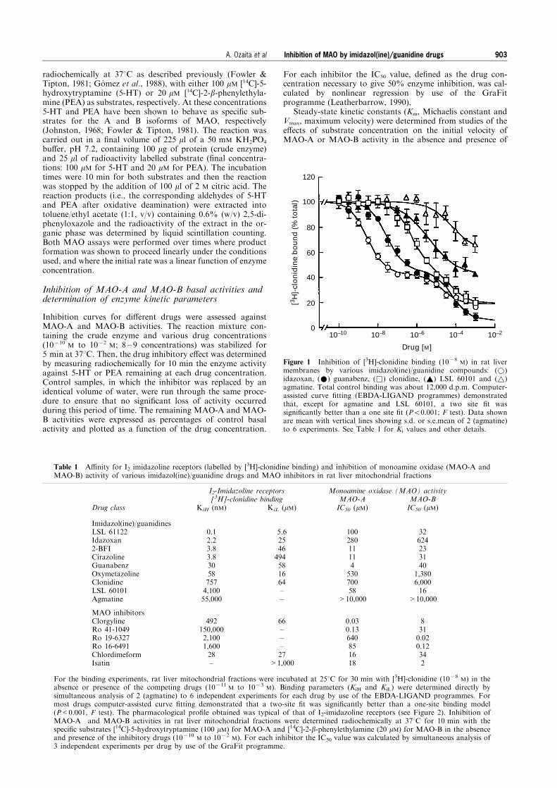
Figure 1 Inhibition of [3H]-clonidine binding (1078M) in rat liver
membranes by various imidazol(ine)/guanidine compounds: (*)idazoxan, (*) guanabenz, (&) clonidine, (~) LSL 60101 and (~)agmatine. Total control binding was about 12,000 d.p.m. Computer-assisted curve ®tting (EBDA-LIGAND programmes) demonstratedthat, except for agmatine and LSL 60101, a two site ®t wassigni®cantly better than a one site ®t (P50.001; F test). Data shownare mean with vertical lines showing s.d. or s.e.mean of 2 (agmatine)to 6 experiments. See Table 1 for Ki values and other details.
Table 1 A�nity for I2 imidazoline receptors (labelled by [3H]-clonidine binding) and inhibition of monoamine oxidase (MAO-A andMAO-B) activity of various imidazol(ine)/guanidine drugs and MAO inhibitors in rat liver mitochondrial fractions
I2-Imidazoline receptors Monoamine oxidase (MAO) activity[3H]-clonidine binding MAO-A MAO-B
Drug class KiH (nM) KiL (mM) IC50 (mM) IC50 (mM)
Imidazol(ine)/guanidinesLSL 61122Idazoxan2-BFICirazolineGuanabenzOxymetazolineClonidineLSL 60101Agmatine
0.12.23.83.830587574,10055,000
5.62546494581664±±
1002801111453070058
>10,000
32624233140
1,3806,00016
>10,000
MAO inhibitorsClorgylineRo 41-1049Ro 19-6327Ro 16-6491ChlordimeformIsatin
492150,0002,1001,60028±
66±±±27
>1,000
0.030.13640851618
8310.020.12342
For the binding experiments, rat liver mitochondrial fractions were incubated at 258C for 30 min with [3H]-clonidine (1078M) in the
absence or presence of the competing drugs (10711M to 1073
M). Binding parameters (KiH and KiL) were determined directly bysimultaneous analysis of 2 (agmatine) to 6 independent experiments for each drug by use of the EBDA-LIGAND programmes. Formost drugs computer-assisted curve ®tting demonstrated that a two-site ®t was signi®cantly better than a one-site binding model(P<0.001, F test). The pharmacological pro®le obtained was typical of that of I2-imidazoline receptors (see Figure 2). Inhibition ofMAO-A and MAO-B activities in rat liver mitochondrial fractions were determined radiochemically at 378C for 10 min with thespeci®c substrates [14C]-5-hydroxytryptamine (100 mM) for MAO-A and [14C]-2-b-phenylethylamine (20 mM) for MAO-B in the absenceand presence of the inhibitory drugs (10710
M to 1072M). For each inhibitor the IC50 value was calculated by simultaneous analysis of
3 independent experiments per drug by use of the GraFit programme.
Inhibition of MAO by imidazol(ine)/guanidine drugs 903A. Ozaita et al
di�erent drug concentrations. Both enzyme isoforms gavegood ®ts to Michaelian type curves by nonlinear regressionanalysis. For each inhibitor, Km (mM) and Vmax (nmolmin71 mg71 protein) values were also estimated, for illustra-tive purposes (Figures 3, 5 and 6), from Lineweaver-Burk plots(substrate concentration71 versus velocity71) which in all casesresulted in straight line plots, with linear regression correlationcoe�cients very close to unity. Dixon plots (inhibitor con-centration versus velocity71) were also used to assess furtherthe nature of the interaction. Kinetic constants for MAO-Awere assessed with ®ve di�erent concentrations of 5-HT (50,100, 200, 300 and 500 mM) after total inhibition of MAO-Bactivity in crude membrane preparations by preincubation for60 min at 378C with 1077
M L-deprenyl. After this incubation,the free inhibitor was removed by centrifugation at 48C and20,000 g for 15 min. The pellet was washed three times moreby resuspension and centrifugation before it was ®nally re-suspended in the incubation bu�er as above. Similarly, kineticconstants for MAO-B were determined with ®ve di�erentconcentrations of PEA (2.5, 5, 10, 25 and 50 mM) after totalinhibition of MAO-A activity in samples that had been pre-incubated with 1077
M clorgyline for 60 min at 378C and wa-shed as above. These concentrations of L-deprenyl andclorgyline were found to inhibit the activity of one isoform ofthe enzyme completely without signi®cantly a�ecting the ac-tivity of the other (Go mez et al., 1986). Under these experi-mental conditions, the ranges of the kinetic parameters ofMAO-A towards 5-HT as substrate (Km: 145 ± 160 mM; Vmax:7.7 ± 8.4 nmol min71 mg71 protein) and of MAO-B with PEAas substrate (Km: 4 ± 7 mM; Vmax: 5.1 ± 5.9 nmol min
71 mg71
protein) were similar to those obtained previously (Go mez etal., 1986).
Statistics
Results are expressed as mean+s.d. or s.e.mean. Pearson'scorrelation coe�cients were calculated by the method of leastsquares and used to test for possible associations betweenvariables. The level of signi®cance was chosen as P=0.05. Alltests were two-tailed.
Drugs
[3H]-clonidine (speci®c activity 65.5 Ci mmol71) was pur-chased from New England Nuclear/Du Pont (U.S.A.). [3H]-Ro41-1049 [N-(2-aminoethyl)-5-(m-¯uorophenyl)-4-thiazole car-boxamide HCl] (speci®c activity, 30.8 Ci mmol71) and [3H]-Ro19-6327 (lazabemide) (N-(2-aminoethyl)-5-chloro-2-pyridinecarboxamide HCl) (speci®c activity 20.2 Ci mmol71) weregenerous gifts from Dr G.J. Richards (Ho�mann-La RocheLtd., Switzerland). For the binding assays, appropriateamounts of the stock solutions were diluted with distilled andpuri®ed water (Milli-Q) containing 2.5 mM HCl and 6%ethanol. [14C]-5-HT (speci®c activity 500 mCi mmol71) and[14C]-PEA (speci®c activity 2.5 mCi mmol71) were purchasedfrom Amersham International plc (U.K.) and New EnglandNuclear/Du Pont, respectively.
Other drugs (and their sources) included: agmatine sulphate(Aldrich Chemical Co., U.S.A.); 2-BFI (2-(2-benzofuranyl)-2-imidazoline) or RX801077 (synthesized by Dr Pla as LSL61103 at S.A. Lasa Laboratories, Spain); chlordimeform HCl(Ciba-Geigy, Switzerland); cirazoline HCl (Synthe labo Re-cherche, France); clonidine HCl (Sigma Chemical Co.,U.S.A.); clorgyline HCl (Sigma); L-deprenyl HCl (ResearchBiochemicals International, U.S.A.); guanabenz base (Sigma);idazoxan HCl (synthesized by Dr F. Geijo at S.A. Lasa La-boratorios); isatin (indole-2,3-dione) base (Sigma); LSL 60101[2-(2-benzofuranyl)imidazole HCl] and LSL 61122 [2-styryl-2-imidazoline HCl, valldemossine] (synthesized by Dr. F. Geijoat S.A. Lasa Laboratorios); oxymetazoline HCl (Sigma); Ro41-1049; Ro 19-6327 and Ro 16-6491 [N-(2-aminoethyl-p-chlorobenzamide] HCl (F. Ho�man-La Roche Ltd.). Otherreagents were obtained from Sigma.
Results
Pharmacological characterization of non-adrenoceptor[3H]-clonidine binding sites in rat liver
[3H]-clonidine binding to rat liver mitochondrial fractions wasessentially to non-adrenoceptor sites because (7)-adrenaline(1076
M) did not displace any signi®cant portion of the bind-ing. Speci®c binding of [3H]-clonidine 1078
M amounted to12,000 d.p.m. Competition experiments for [3H]-clonidinebinding by selected imidazol(ine)/guanidine compounds andMAO inhibitors were performed to assess the pharmacologicalpro®le of the interactions. The compounds LSL 61122, ida-zoxan, 2-BFI, cirazoline, guanabenz, oxymetazoline and clo-nidine displayed biphasic curves (Hill coe�cients lesser thanunity) (Figure 1) and computer analysis resolved the binding of[3H]-clonidine into two components (Figure 1 and Table 1).The high a�nity component for these imidazol(ine)/guanidinedrugs (KiH*0.1 ± 760 nM) represented 60 ± 70% of total [3H]-clonidine binding. The low a�nity component of [3H]-cloni-dine binding displayed inhibition constants in the micromolarrange (KiL*6 ± 500 mM) for these compounds. The imidazoleLSL 60101 and the guanidine agmatine, a proposed endo-genous ligand for imidazoline receptors, displayed moderateand very low a�nity, respectively, for [3H]-clonidine binding,and at 1 mM they only displaced about 60% and 30%, re-spectively, of total radioligand binding and from only one site(Figure 1 and Table 1).
The irreversible and highly selective MAO-A inhibitorclorgyline and chlordimeform, a reversible and non-selectiveMAO inhibitor, fully inhibited [3H]-clonidine binding to ratliver mitochondrial fractions (Table 1). These MAO inhibitorsalso displaced biphasic curves. Chlordimeform was more po-tent in competing for the high a�nity component than clor-
10
9
8
7
6
5
4
33 4 5 6 7 8 9 10
[3H]-clonidine binding pKiH, rat liver
[3 H]-
idaz
oxa
n b
ind
ing
pK
iH, r
at b
rain
1
2
3
4
5
67 8
9
10
11
12
13
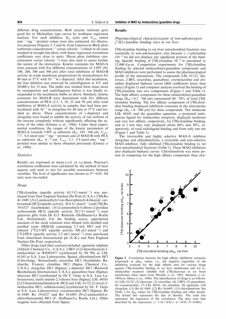
Figure 2 Correlation between the high a�nity inhibition constants(expressed as pKiH values, i.e., the negative logarithm of theinhibition constant for the high a�nity site) for various drugsagainst [3H]-clonidine binding in rat liver membranes and for I2-imidazoline receptors labelled with [3H]-idazoxan in rat brainmembranes (data taken from Miralles et al., 1993; Alemany et al.,1995a,b; Olmos et al., 1996). The identi®cation of drugs is as follows:(1) LSL 61122, (2) idazoxan, (3) cirazoline, (4) 2-BFI, (5) guanabenz,(6) oxymetazoline, (7) LSL 60101, (8) clonidine, (9) agmatine, (10)clorgyline, (11) Ro 41-1049, (12) Ro 16-6491, (13) chlordimeform. SeeTable 1 for KiH values for [3H]-clonidine binding and other details.The dotted line represents the line of identity. The solid linerepresents the regression of the correlation. The data were bestdescribed by the expression: y=1.16+0.826 (r=0.92, P50.001).
Inhibition of MAO by imidazol(ine)/guanidine drugs904 A. Ozaita et al
gyline (Table 1). Other reversible and highly selective MAO-Ainhibitors such as Ro 41-1049 or MAO-B inhibitors such as Ro19-6327 (lazabemide) and Ro 16-6491 displayed monophasiccurves when competing against [3H]-clonidine binding in mi-tochondrial fractions, with Ki values in the micromolar range.The endogenous MAO inhibitor isatin (indole-2,3-dione)(Glover et al., 1988) was ine�ective at displacing [3H]-clonidinebinding to rat liver mitochondrial fractions (7% inhibition at1 mM).
A highly signi®cant correlation was obtained (r=0.92;P50.001) when the potencies of all the drugs tested at the higha�nity site of [3H]-clonidine binding in rat liver mitochondrialfractions were compared with the potencies of the same drugsat the high a�nity site of rat brain I2-imidazoline receptorslabelled with [3H]-idazoxan (Figure 2). This correlation ana-lysis clearly indicated that at least a high proportion (60 ± 70%)of [3H]-clonidine binding in rat liver corresponds to the I2-imidazoline receptors.
Inhibition of rat liver MAO-A and MAO-B activities byclonidine and nature of the interaction
I2-imidazoline binding sites have been proposed to regulate theactivity of the enzyme MAO. To determine if [3H]-clonidinebinding to rat liver I2-imidazoline receptors is related to aninteraction of the radioligand with MAO, the e�ects of cloni-dine on MAO activity (potency and nature of the interaction)in rat liver mitochondria were assessed in comparison withspeci®c MAO inhibitors. As expected, su�ciently high con-centrations of the MAO inhibitors tested fully inhibited theMAO activity and showed the known pattern of selectivity forMAO-A (clorgyline, Ro 41-1049) and MAO-B (Ro 19-6327,Ro 16-6491) or non-selectivity (chlordimeform, isatin) (Table1).
The imidazoline clonidine inhibited rat liver MAO-A ac-tivity in a monophasic manner with a very low potency (IC50:700 mM). It was an even poorer inhibitor of the MAO-B
120
100
80
60
40
20
010–10 10–8 10–6 10–4 10–2
Clonidine [M]
MA
O-A
(%
of
bas
al a
ctiv
ity)
120
100
80
60
40
20
010–10 10–8 10–6 10–4 10–2
Clonidine [M]
MA
O-B
(%
of
bas
al a
ctiv
ity)
–0.2 0 0.2 0.4
1/[PEA], µM–1
3
2
11/V
, (n
et d
.p.m
. x 1
0–3)–1
10
65
0
–0.01 0 0.01 0.02
1/[5-HT], µM–1
1/V
, (n
et d
.p.m
. x 1
0–3)–1
0.8
0.6
0.4
0.2
5
1
0.5
0
a
b
d
c
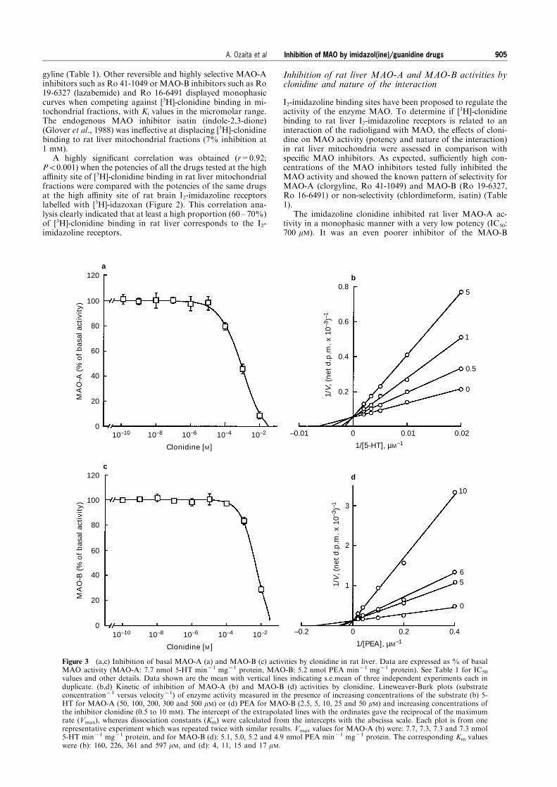
Figure 3 (a,c) Inhibition of basal MAO-A (a) and MAO-B (c) activities by clonidine in rat liver. Data are expressed as % of basalMAO activity (MAO-A: 7.7 nmol 5-HT min71 mg71 protein, MAO-B: 5.2 nmol PEA min71 mg71 protein). See Table 1 for IC50
values and other details. Data shown are the mean with vertical lines indicating s.e.mean of three independent experiments each induplicate. (b,d) Kinetic of inhibition of MAO-A (b) and MAO-B (d) activities by clonidine. Lineweaver-Burk plots (substrateconcentration71 versus velocity71) of enzyme activity measured in the presence of increasing concentrations of the substrate (b) 5-HT for MAO-A (50, 100, 200, 300 and 500 mM) or (d) PEA for MAO-B (2.5, 5, 10, 25 and 50 mM) and increasing concentrations ofthe inhibitor clonidine (0.5 to 10 mM). The intercept of the extrapolated lines with the ordinates gave the reciprocal of the maximumrate (Vmax), whereas dissociation constants (Km) were calculated from the intercepts with the abscissa scale. Each plot is from onerepresentative experiment which was repeated twice with similar results. Vmax values for MAO-A (b) were: 7.7, 7.3, 7.3 and 7.3 nmol5-HT min71 mg71 protein, and for MAO-B (d): 5.1, 5.0, 5.2 and 4.9 nmol PEA min71 mg71 protein. The corresponding Km valueswere (b): 160, 226, 361 and 597 mM, and (d): 4, 11, 15 and 17 mM.
Inhibition of MAO by imidazol(ine)/guanidine drugs 905A. Ozaita et al
activity (IC50: 6 mM) (Table 1). Studies on the e�ects ofclonidine on the initial rates of substrate oxidation by MAO-A and MAO-B, shown as Lineweaver-Burk plots in Figure 3,indicated the inhibition to be competitive with respect to theamine substrate. Dixon plots of the inhibition data (notshown) were also consistent with simple competitive inhibi-tion and gave Ki values of 510+95 mM (n=3) and2.5+0.5 mM (n=3) for MAO-A and MAO-B, respectively.Such competitive inhibition would be consistent with cloni-dine binding to the active sites of the enzymes, although thepossibility that the compound binds to a distinct site andexerts its e�ects through a conformational change cannot beexcluded.
Inhibition of rat liver MAO-A and MAO-B activities byvarious imidazol(ine)/guanidine drugs and nature of theinteractions
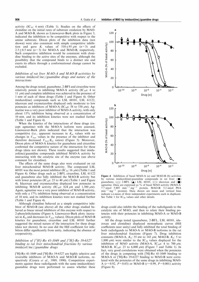
Among the drugs tested, guanabenz, 2-BFI and cirazoline wererelatively potent in inhibiting MAO-A activity (IC50s: 4 to11 mM) and complete inhibition was achieved in the presence of1 mM of each of these drugs (Table 1 and Figure 4). Otherimidazol(ine) compounds such as LSL 60101, LSL 61122,idazoxan and oxymetazoline displayed only moderate to lowpotencies as inhibitors of MAO-A (IC50s: 58 to 530 mM). Ag-matine was a very poor inhibitor of MAO-A activity, with onlyabout 13% inhibition being observed at a concentration of10 mM, and its inhibition kinetics were not studied further(Table 1 and Figure 4).
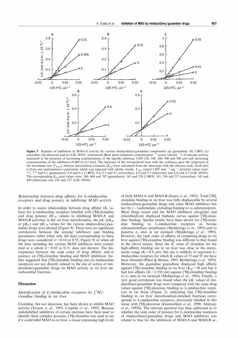
When the kinetics of the interactions of these drugs (ex-cept agmatine) with the MAO-A isoform were assessed,Lineweaver-Burk plots indicated that the interaction wascompetitive (i.e., apparent increases in Km values with nochanges in Vmax values in the presence of the inhibitor andtherefore decreased Vmax/Km ratios) (Figure 5). Moreover,Dixon plots of MAO-A kinetics for guanabenz and cirazolinecon®rmed the competitive nature of the interaction for thesedrugs (data not shown). These results suggested that imida-zol(ine)/guanidine compounds inhibited MAO-A activity byinteracting with the catalytic site of the enzyme (see abovecomment for clonidine).
The e�ects of the same drugs also were evaluated on ratliver mitochondrial MAO-B activity. The compound LSL60101 was the most potent inhibitor (IC50: 16 mM) (Table 1 andFigure 4). Other drugs such as 2-BFI, cirazoline, LSL 61122and guanabenz also fully inhibited the MAO-B activity butwith lower potencies (IC50s: 23 to 40 mM) (Table 1 and Figure4). Idazoxan and oxymetazoline displayed low potencies ininhibiting MAO-B activity (IC50s: 624 mM and 1,380 mM).Again, agmatine was a very poor inhibitor of MAO-B activity,with only a 17% inhibition being observed at a concentrationof 10 mM, and its inhibition kinetics were not studied further(Table 1 and Figure 4).
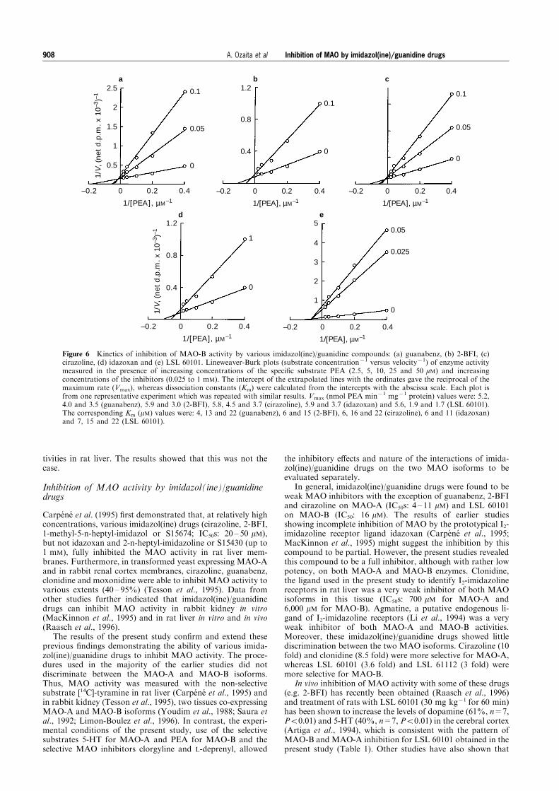
Although clonidine behaved as a simple competitive inhi-bitor of MAO-B (see above) all the other drugs studied be-haved as linear mixed inhibitors of this enzyme with respect to2-phenylethylamine (Figure 6, Lineweaver-Burk plots: increa-ses in Km and decreases in Vmax values). Dixon plots of MAO-Bkinetics for guanabenz, cirazoline and LSL 60101 furthersuggested the mixed nature of the interaction for these drugs(data not shown). In no case did the Hill coe�cient for inhi-bition di�er signi®cantly from unity, indicating the absence ofcooperativity.
Inhibition of [3H]-Ro 41-1049 and [3H]-Ro 19-6327binding to rat liver mitochondrial fractions by variousimidazol(ine)/guanidine drugs
[3H]-Ro 41-1049 and [3H]-Ro 19-6327 are highly selective andreversible inhibitors of MAO-A and MAO-B isoforms, re-spectively (Cesura et al., 1989; 1990). Competition experi-ments against these radioligands with the same imidazol(ine)/guanidine drugs were performed to assess whether these
drugs could also inhibit the binding of the radioligands to thecatalytic site of MAO, and then to relate their binding po-tencies with their potencies in inhibiting MAO-A or MAO-Bactivity.
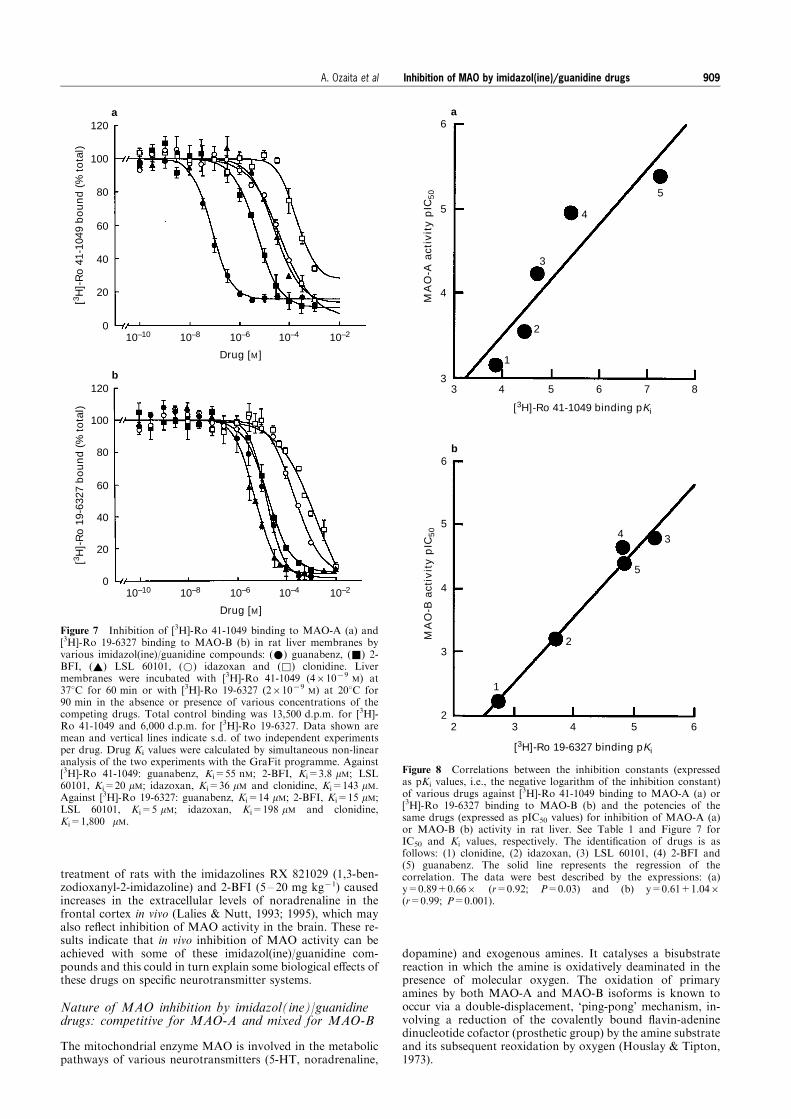
All the drugs tested (guanabenz, 2-BFL, LSL 60101, ida-zoxan and clonidine) displayed monophasic curves (Hillcoe�cients near unity) and fully inhibited the total binding ofboth radioligands to MAO-A or MAO-B isoforms in the ratliver mitochondrial fractions (Figure 7). Drug inhibitionconstants (MAO-A, Kis: 55 nM to 143 mM; MAO-B, Kis: 5 to1,800 mM) were similar to the IC50 values displayed for theinhibition of MAO activity (MAO-A, IC50s: 4 to 700 mM;MAO-B, IC50s: 23 to 6,000 mM) (Figure 7 and Table 1). Infact, very good correlations were obtained when the potenciesof the drugs in competing with [3H]-Ro 41-1049 binding toMAO-A or [3H]-Ro 19-6327 binding to MAO-B were corre-lated with the potencies of the same drugs in inhibiting MAO-A (r=0.92, P=0.03) or MAO-B (r=0.99, P=0.001) activity(Figure 8).
120
100
80
60
40
20
010–10 10–8 10–6 10–4 10–2
Drug [M]
MA
O-A
(%
of
bas
al a
ctiv
ity)
120
100
80
60
40
20
010–10 10–8 10–6 10–4 10–2
Drug [M]
MA
O-B
(%
of
bas
al a
ctiv
ity)
b
a
Figure 4 Inhibition of basal MAO-A (a) and MAO-B (b) activitiesby various imidazol(ine)/guanidine compounds in rat liver: (*)guanabenz, (~) 2-BFI, (~) LSL 60101, (*) idazoxan and (&)agmatine. Data are expressed as % of basal MAO activity (MAO-A:7.7 nmol 5-HT min71 mg71 protein, MAO-B: 5.2 nmol PEAmin71 mg71 protein). Data shown are mean and vertical linesindicate s.e.mean of three independent experiments each in duplicate.See Table 1 for IC50 values and other details.
Inhibition of MAO by imidazol(ine)/guanidine drugs906 A. Ozaita et al
Relationship between drug a�nity for I2-imidazolinereceptors and drug potency in inhibiting MAO activity
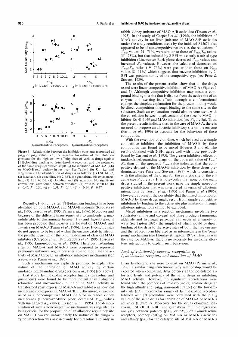
In order to assess relationships between drug a�nity (Ki va-lues) for I2-imidazoline receptors labelled with [3H]-clonidineand drug potency (IC50 values) in inhibiting MAO-A andMAO-B activities in the rat liver mitochondria, the pKi (pKiH
or pKiL) and pIC50 values for the various imidazol(ine)/gua-nidine drugs were plotted (Figure 9). There were no signi®cantcorrelations between the enzyme inhibitory and bindingparameters either when only the data corresponding to thesedrugs were considered (r=0.14 to 0.55; Figure 9) or when allthe data including the various MAO inhibitors were consid-ered as a whole (r=0.03 to 0.11; data not shown). The dis-crepancies between the rank order of drug a�nity and/orpotency on [3H]-clonidine binding and MAO inhibition, fur-ther suggested that [3H]-clonidine binding sites (I2-imidazolinereceptors) are not directly related to the site of action of imi-dazol(ine)/guanidine drugs on MAO activity in rat liver mi-tochondrial fractions.
Discussion
Identi®cation of I2-imidazoline receptors by [3H]-clonidine binding in rat liver
Clonidine, but not idazoxan, has been shown to inhibit MAOactivity (Tesson et al., 1995; Carpe ne et al., 1995). Becauseradiolabelled inhibitors of certain enzymes have been used toidentify these complex proteins, [3H]-clonidine was used to seeif it could label MAO in rat liver, a tissue expressing high levels
of both MAO-A and MAO-B (Saura et al., 1992). Total [3H]-clonidine binding in rat liver was fully displaceable by severalimidazol(ine)/guanidine drugs and some MAO inhibitors butnot by (7)-adrenaline, excluding binding to a2-adrenoceptors.Most drugs tested and the MAO inhibitors clorgyline andchlordimeform displayed biphasic curves against [3H]-cloni-dine binding. Similar results have been shown for [3H]-cloni-dine binding to I1-imidazoline receptors in bovineadrenomedullary membranes (Molderings et al., 1993) and toputative s2 sites in rat stomach (Molderings et al., 1995).However, the rank order of a�nity of competing drugs in ratliver against [3H]-clonidine binding was di�erent to that foundin the above tissues. Since the Ki value of clonidine for thehigh-a�nity binding site in rat liver was close to the micro-molar range (Ki=0.8 mM), this site is unlikely to represent I1-imidazoline receptors for which Ki values of 15 and 55 nM havebeen obtained (Piletz & Sletten, 1993; Molderings et al., 1993).Moreover, the guanidine guanabenz displayed high a�nityagainst [3H]-clonidine binding in rat liver (KiH=30 nM) but ithad low a�nity (Ki=2,538 nM) against [3H]-clonidine bindingto s2 sites in rat stomach (Molderings et al., 1995). Finally, avery good correlation was found when the pKi values of imi-dazol(ine)/guanidine drugs were compared with the same drugvalues against [3H]-idazoxan binding to I2-imidazoline recep-tors in rat brain (Figure 2), indicating that [3H]-clonidinebinding to rat liver mitochondrial-enriched fractions corre-sponds to I2-imidazoline receptors, previously labelled in thistissue with [3H]-idazoxan (Zonneschein et al., 1990; Alemanyet al., 1995b). The relevant question was then addressed as towhether the rank order of potency for I2-imidazoline receptorsof imidazol(ine)/guanidine drugs and MAO inhibitors wasrelated to that for the inhibition of MAO-A and MAO-B ac-
0.8
0.6
0.4
0.2
–0.01 0 0.01 0.02
1/[5-HT], µM–1
1/V
, (n
et d
.p.m
. x 1
0–3)–1
0.01
0.005
0
–0.01 0 0.01 0.02
1/[5-HT], µM–1
0.4
0.3
0.2
0.1
0.01
0
–0.01 0 0.01 0.02
1/[5-HT], µM–1
1
0.8
0.6
0.4
0.2
0.05
0.01
0
–0.01 0 0.01 0.02
1/[5-HT], µM–1
–0.01 0 0.01 0.02
1/[5-HT], µM–1
0.6
0.4
0.2
1/V
, (n
et d
.p.m
. x 1
0–3)–1
0.5
0
0.3
0.2
0.1
0.01
0
ed
cba
Figure 5 Kinetics of inhibition of MAO-A activity by various imidazol(ine)/guanidine compounds: (a) guanabenz, (b) 2-BFI, (c)cirazoline, (d) idazoxan and (e) LSL 60101. Lineweaver-Burk plots (substrate concentration71 versus velocity71) of enzyme activitymeasured in the presence of increasing concentrations of the speci®c substrate 5-HT (50, 100, 200, 300 and 500 mM) and increasingconcentrations of the inhibitors (0.005 to 0.5 mM). The intercept of the extrapolated lines with the ordinates gave the reciprocal ofthe maximum rate (Vmax), whereas dissociation constants (Km) were calculated from the intercepts with the abscissa scale. Each plotis from one representative experiment which was repeated with similar results. Vmax (nmol 5-HT min71 mg71 protein) values were:7.7, 7.7 and 8.1 (guanabenz), 8.4 and 8.2 (2-BFI), 8.4, 8.3 and 8.2 (cirazoline), 8.4 and 8.3 (idazoxan) and 8.4 and 8.3 (LSL 60101).The corresponding Km (mM) values were: 160, 404 and 707 (guanabenz), 145 and 320 (2-BFI), 145, 254 and 237 (cirazoline), 145 and449 (idazoxan) and 145 and 237 (LSL 60101).
Inhibition of MAO by imidazol(ine)/guanidine drugs 907A. Ozaita et al
tivities in rat liver. The results showed that this was not thecase.
Inhibition of MAO activity by imidazol(ine)/guanidinedrugs
Carpe ne et al. (1995) ®rst demonstrated that, at relatively highconcentrations, various imidazol(ine) drugs (cirazoline, 2-BFI,1-methyl-5-n-heptyl-imidazol or S15674; IC50s: 20 ± 50 mM),but not idazoxan and 2-n-heptyl-imidazoline or S15430 (up to1 mM), fully inhibited the MAO activity in rat liver mem-branes. Furthermore, in transformed yeast expressing MAO-Aand in rabbit renal cortex membranes, cirazoline, guanabenz,clonidine and moxonidine were able to inhibit MAO activity tovarious extents (40 ± 95%) (Tesson et al., 1995). Data fromother studies further indicated that imidazol(ine)/guanidinedrugs can inhibit MAO activity in rabbit kidney in vitro(MacKinnon et al., 1995) and in rat liver in vitro and in vivo(Raasch et al., 1996).
The results of the present study con®rm and extend theseprevious ®ndings demonstrating the ability of various imida-zol(ine)/guanidine drugs to inhibit MAO activity. The proce-dures used in the majority of the earlier studies did notdiscriminate between the MAO-A and MAO-B isoforms.Thus, MAO activity was measured with the non-selectivesubstrate [14C]-tyramine in rat liver (Carpe ne et al., 1995) andin rabbit kidney (Tesson et al., 1995), two tissues co-expressingMAO-A and MAO-B isoforms (Youdim et al., 1988; Saura etal., 1992; Limon-Boulez et al., 1996). In contrast, the experi-mental conditions of the present study, use of the selectivesubstrates 5-HT for MAO-A and PEA for MAO-B and theselective MAO inhibitors clorgyline and L-deprenyl, allowed
the inhibitory e�ects and nature of the interactions of imida-zol(ine)/guanidine drugs on the two MAO isoforms to beevaluated separately.
In general, imidazol(ine)/guanidine drugs were found to beweak MAO inhibitors with the exception of guanabenz, 2-BFIand cirazoline on MAO-A (IC50s: 4 ± 11 mM) and LSL 60101on MAO-B (IC50: 16 mM). The results of earlier studiesshowing incomplete inhibition of MAO by the prototypical I2-imidazoline receptor ligand idazoxan (Carpe ne et al., 1995;MacKinnon et al., 1995) might suggest the inhibition by thiscompound to be partial. However, the present studies revealedthis compound to be a full inhibitor, although with rather lowpotency, on both MAO-A and MAO-B enzymes. Clonidine,the ligand used in the present study to identify I2-imidazolinereceptors in rat liver was a very weak inhibitor of both MAOisoforms in this tissue (IC50s: 700 mM for MAO-A and6,000 mM for MAO-B). Agmatine, a putative endogenous li-gand of I2-imidazoline receptors (Li et al., 1994) was a veryweak inhibitor of both MAO-A and MAO-B activities.Moreover, these imidazol(ine)/guanidine drugs showed littlediscrimination between the two MAO isoforms. Cirazoline (10fold) and clonidine (8.5 fold) were more selective for MAO-A,whereas LSL 60101 (3.6 fold) and LSL 61112 (3 fold) weremore selective for MAO-B.
In vivo inhibition of MAO activity with some of these drugs(e.g. 2-BFI) has recently been obtained (Raasch et al., 1996)and treatment of rats with LSL 60101 (30 mg kg71 for 60 min)has been shown to increase the levels of dopamine (61%, n=7,P50.01) and 5-HT (40%, n=7, P50.01) in the cerebral cortex(Artiga et al., 1994), which is consistent with the pattern ofMAO-B and MAO-A inhibition for LSL 60101 obtained in thepresent study (Table 1). Other studies have also shown that
–0.2 0 0.2 0.4
1/[PEA], µM–1
1/V
, (n
et d
.p.m
. x 1
0–3)–1
2.5
2
1.5
1
0.5
0.1
0.05
0
1.2
0.8
0.4
–0.2 0 0.2 0.4
1/[PEA], µM–1
0.1
0
–0.2 0 0.2 0.4
1/[PEA], µM–1
0.1
0.05
0
0.05
0.025
0
–0.2 0 0.2 0.4
1/[PEA], µM–1
5
4
3
2
1
–0.2 0 0.2 0.4
1/[PEA], µM–1
1
0
1.2
0.8
0.4
1/V
, (n
et d
.p.m
. x 1
0–3)–1
d e
a b c
Figure 6 Kinetics of inhibition of MAO-B activity by various imidazol(ine)/guanidine compounds: (a) guanabenz, (b) 2-BFI, (c)cirazoline, (d) idazoxan and (e) LSL 60101. Lineweaver-Burk plots (substrate concentration71 versus velocity71) of enzyme activitymeasured in the presence of increasing concentrations of the speci®c substrate PEA (2.5, 5, 10, 25 and 50 mM) and increasingconcentrations of the inhibitors (0.025 to 1 mM). The intercept of the extrapolated lines with the ordinates gave the reciprocal of themaximum rate (Vmax), whereas dissociation constants (Km) were calculated from the intercepts with the abscissa scale. Each plot isfrom one representative experiment which was repeated with similar results. Vmax (nmol PEA min71 mg71 protein) values were: 5.2,4.0 and 3.5 (guanabenz), 5.9 and 3.0 (2-BFI), 5.8, 4.5 and 3.7 (cirazoline), 5.9 and 3.7 (idazoxan) and 5.6, 1.9 and 1.7 (LSL 60101).The corresponding Km (mM) values were: 4, 13 and 22 (guanabenz), 6 and 15 (2-BFI), 6, 16 and 22 (cirazoline), 6 and 11 (idazoxan)and 7, 15 and 22 (LSL 60101).
Inhibition of MAO by imidazol(ine)/guanidine drugs908 A. Ozaita et al
treatment of rats with the imidazolines RX 821029 (1,3-ben-zodioxanyl-2-imidazoline) and 2-BFI (5 ± 20 mg kg71) causedincreases in the extracellular levels of noradrenaline in thefrontal cortex in vivo (Lalies & Nutt, 1993; 1995), which mayalso re¯ect inhibition of MAO activity in the brain. These re-sults indicate that in vivo inhibition of MAO activity can beachieved with some of these imidazol(ine)/guanidine com-pounds and this could in turn explain some biological e�ects ofthese drugs on speci®c neurotransmitter systems.
Nature of MAO inhibition by imidazol(ine)/guanidinedrugs: competitive for MAO-A and mixed for MAO-B
The mitochondrial enzyme MAO is involved in the metabolicpathways of various neurotransmitters (5-HT, noradrenaline,
dopamine) and exogenous amines. It catalyses a bisubstratereaction in which the amine is oxidatively deaminated in thepresence of molecular oxygen. The oxidation of primaryamines by both MAO-A and MAO-B isoforms is known tooccur via a double-displacement, `ping-pong' mechanism, in-volving a reduction of the covalently bound ¯avin-adeninedinucleotide cofactor (prosthetic group) by the amine substrateand its subsequent reoxidation by oxygen (Houslay & Tipton,1973).
10–10 10–8 10–6 10–4 10–2
Drug [M]
120
100
80
60
40
20
0
[3 H]-
Ro
41-
1049
bo
un
d (
% t
ota
l)
120
100
80
60
40
20
0
b
a
[3 H]-
Ro
19-
6327
bo
un
d (
% t
ota
l)
10–10 10–8 10–6 10–4 10–2
Drug [M]
Figure 7 Inhibition of [3H]-Ro 41-1049 binding to MAO-A (a) and[3H]-Ro 19-6327 binding to MAO-B (b) in rat liver membranes byvarious imidazol(ine)/guanidine compounds: (*) guanabenz, (&) 2-BFI, (~) LSL 60101, (*) idazoxan and (&) clonidine. Livermembranes were incubated with [3H]-Ro 41-1049 (461079
M) at378C for 60 min or with [3H]-Ro 19-6327 (261079
M) at 208C for90 min in the absence or presence of various concentrations of thecompeting drugs. Total control binding was 13,500 d.p.m. for [3H]-Ro 41-1049 and 6,000 d.p.m. for [3H]-Ro 19-6327. Data shown aremean and vertical lines indicate s.d. of two independent experimentsper drug. Drug Ki values were calculated by simultaneous non-linearanalysis of the two experiments with the GraFit programme. Against[3H]-Ro 41-1049: guanabenz, Ki=55 nM; 2-BFI, Ki=3.8 mM; LSL60101, Ki=20 mM; idazoxan, Ki=36 mM and clonidine, Ki=143 mM.Against [3H]-Ro 19-6327: guanabenz, Ki=14 mM; 2-BFI, Ki=15 mM;LSL 60101, Ki=5 mM; idazoxan, Ki=198 mM and clonidine,Ki=1,800 mM.
6
5
4
3 3 4 5 6 7 8
5
4
3
2
1
MA
O-A
act
ivit
y p
IC50
6
5
4
3
2
MA
O-B
act
ivit
y p
IC50
b
2 3 4 5 6
34
5
2
1
a
[3H]-Ro 41-1049 binding pKi
[3H]-Ro 19-6327 binding pKi
Figure 8 Correlations between the inhibition constants (expressedas pKi values, i.e., the negative logarithm of the inhibition constant)of various drugs against [3H]-Ro 41-1049 binding to MAO-A (a) or[3H]-Ro 19-6327 binding to MAO-B (b) and the potencies of thesame drugs (expressed as pIC50 values) for inhibition of MAO-A (a)or MAO-B (b) activity in rat liver. See Table 1 and Figure 7 forIC50 and Ki values, respectively. The identi®cation of drugs is asfollows: (1) clonidine, (2) idazoxan, (3) LSL 60101, (4) 2-BFI and(5) guanabenz. The solid line represents the regression of thecorrelation. The data were best described by the expressions: (a)y=0.89+0.666 (r=0.92; P=0.03) and (b) y=0.61+1.046(r=0.99; P=0.001).
Inhibition of MAO by imidazol(ine)/guanidine drugs 909A. Ozaita et al
Recently, I2-binding sites ([3H]-idazoxan binding) have been
identi®ed on both MAO-A and MAO-B isoforms (Raddatz etal., 1995; Tesson et al., 1995; Parini et al., 1996). Moreover andbecause of the di�erent tissue sensitivity to amiloride, a gua-nidide able to discriminate between I2A- and I2B-subtypes, ithas been proposed that I2A-sites are located on MAO-A andI2B-sites on MAO-B (Parini et al., 1996). These I2-binding sitesdo not appear to be located within the enzyme catalytic site, orthe prosthetic group, or the binding domain of classical MAOinhibitors (Carpe ne et al., 1995; Raddatz et al., 1995; Tesson etal., 1995; Limon-Boulez et al., 1996). Therefore, I2-bindingsites on MAO-A and MAO-B were proposed to representpreviously unknown regulatory sites able to modulate the ac-tivity of MAO through an allosteric inhibitory mechanism (fora review see Parini et al., 1996).
Such a mechanism was explicitly proposed to explain thenature of the inhibition of MAO activity by certainimidazol(ine)/guanidine drugs (Tesson et al., 1995) (see above).In that study I2-imidazoline receptor ligands (cirazoline andguanabenz) were found to be more potent than I1-ligands(clonidine and moxonidine) in inhibiting MAO activity intransformed yeast expressing MAO-A and rabbit renal corticalmembranes co-expressing MAO-A/B. Furthermore, cirazolineacted as a noncompetitive MAO inhibitor in rabbit kidneymembranes (Lineweaver-Burk plots: decreased Vmax valueswith unchanged Km values) (Tesson et al., 1995). The demon-stration of such a noncompetitive mechanism was regarded asbeing crucial for the proposition of an allosteric regulatory siteon MAO. However, unfortunately the nature of the drug/en-zyme interaction was assessed only for cirazoline and only in
rabbit kidney (mixture of MAO-A/B activities) (Tesson et al.,1995). In the study of Carpe ne et al. (1995), the inhibition ofMAO activity in rat liver (mixture of MAO-A/B activitiesunder the assay conditions used) by the imidazol S15674 alsoappeared to be of noncompetitive nature (i.e. the reductions ofVmax values, 24 ± 71%, were similar to those of Vmax/Km ratios,35 ± 73%), but that induced by 2-BFI was clearly a mixed typeinhibition (Lineweaver-Burk plots: decreased Vmax values andincreased Km values). However, the calculated decreases onVmax/Km ratios (19 ± 76%) were greater than those on Vmax
values (8 ± 31%) which suggests that enzyme inhibition by 2-BFI was predominantly of the competitive type (see Price &Stevens, 1989).
The results of the present studies show that all the drugstested were linear competitive inhibitors of MAO-A (Figures 3and 5). Although competitive inhibition may mean a com-pound binding to a site that is distinct from the active site of anenzyme and exerting its e�ects through a conformationalchange, the simplest explanation for the present ®nding wouldbe direct competition through binding to the same site as thesubstrate. Such an explanation would also be consistent withthe correlation between displacement of the speci®c MAO in-hibitor Ro 41-1049 and MAO inhibition (see Figure 8a). Thus,the present results indicate that, in the case of MAO-A, there isno need to propose an allosteric inhibitory site on the enzyme(Parini et al., 1996) to account for the behaviour of thesecompounds.
With the exception of clonidine, which behaved as a simplecompetitive inhibitor, the inhibition of MAO-B by thesecompounds was found to be mixed (Figures 3 and 6). Theresults obtained with 2-BFI agree well with those previouslyfound by Carpe ne et al. (1995). The greater inhibitory e�ects ofimidazol(ine)/guanidine drugs on the apparent value of Vmax/Km than on the apparent Vmax value indicates that the com-petitive element of the MAO-B inhibition by these drugs pre-dominates (see Price and Stevens, 1989), which is consistentwith the a�nities of the drugs for the catalytic site of the en-zyme (see Figure 8b). It is noteworthy that none of the eightdrugs studied in the present work gave the simple non-com-petitive inhibition that was interpreted in terms of allostericinteractions by Tesson et al. (1995) and Parini et al. (1996).However, at present the possibility that the mixed inhibition ofMAO-B by these drugs might result from simple competitiveinhibition by binding to the active site plus inhibition throughallosteric interactions cannot be excluded.
Mixed inhibition in a reaction mechanism involving twosubstrates (amine and oxygen) and three products (ammonia,aldehyde and hydrogen peroxide) can occur in a variety ofways (see Tipton 1996), the simplest of which would be directbinding of the drug to the active sites of both the free enzymeand the reduced form liberated as an intermediate in the `ping-pong' mechanism (see Houslay & Tipton, 1973). Thus, as wasthe case for MAO-A, there is no necessity for invoking allos-teric interactions to explain such behaviour.
Lack of relationship between recognition ofI2-imidazoline receptors and inhibition of MAO
If an I2-allosteric site were to exist on MAO (Parini et al.,1996), a similar drug structure-activity relationship would beexpected when comparing drug potency at the postulated al-losteric I2-site and potency of the same drugs in inhibitingMAO activity. However, no signi®cant correlations werefound when the potencies of imidazol(ine)/guanidine drugs atthe high a�nity site (pKiH, nanomolar range) or the low-a�-nity site (pKiL micromolar range) of I2-imidazoline receptorslabelled with [3H]-clonidine were correlated with the pIC50
values of the same drugs for inhibition of MAO-A or MAO-Bactivities (Figure 9). Moreover, for the drugs clonidine, ida-zoxan, LSL 60101, 2-BFI and guanabenz, multiple regressionanalyses between potency (pKiH or pKiL) on I2-imidazolinereceptors, potency (pIC50) on MAO-A or MAO-B activitiesand a�nity (pKi) for the catalytic site of MAO-A or MAO-B
5
4
3
2
3 4 5 6 7 8 9 10 11
pKiH I2-imidazoline receptors
6
5
4
3
3 4 5 6
pKiL I2-imidazoline receptors
MA
O-A
act
ivit
y p
IC50
5
4
3
2
3 4 5 6 7 8 9 10 11
pKiH I2-imidazoline receptors
MA
O-B
act
ivit
y p
IC50
3 4 5 6
pKiL I2-imidazoline receptors
554
43
3
2
2
1
16
6
7
7
8
89
9
1 1
2 2
33
4 4
5 5
66
88
dc
ba
5
4
3
2
Figure 9 Relationship between the inhibition constants (expressed aspKiH or pKiL values, i.e., the negative logarithm of the inhibitionconstant for the high or low a�nity site) of various drugs against[3H]-clonidine binding to I2-imidazoline receptors and the potenciesof the same drugs (expressed as pIC50) for inhibition of MAO-A (a,b)or MAO-B (c,d) activity in rat liver. See Table 1 for KiH, KiL andIC50 values. The identi®cation of drugs is as follows: (1) LSL 61122,(2) idazoxan, (3) cirazoline, (4) 2-BFI, (5) guanabenz, (6) oxymetazo-line, (7) LSL 60101, (8) clonidine and (9) agmatine. No signi®cantcorrelations were found between variables. (a) r=0.55, P=0.12; (b)r=0.46, P=0.30; (c) r=0.53, P=0.14; (d) r=0.14, P=0.77.
Inhibition of MAO by imidazol(ine)/guanidine drugs910 A. Ozaita et al
also resulted in nonsigni®cant correlations (r=0.06 to 0.59;P50.05), which reinforced the lack of association betweendrug interaction with I2-imidazoline receptors (dependentvariable) and MAO activity (independent variables).
Although these studies cannot exclude the presence of ad-ditional binding sites on MAO that do not a�ect the activity ofenzyme, several proteins with apparent molecular weights inthe range 25 ± 30 kDa, which is very di�erent from those ofeither MAO-A or MAO-B (Bach et al., 1988), have been im-plicated in idazoxan binding (Escriba et al., 1994; 1996) orhave been photolabelled with an imidazoline probe (Lanier etal., 1995) in rat liver. Thus these results and those obtained inthe present study would suggest that the I2-imidazoline re-ceptors represent molecular species that are distinct fromMAO.
Together the results indicate that most imidazol(ine)/gua-nidine drugs are weak inhibitors of MAO-A and MAO-Bisoforms and that the nature of the interactions is mostprobably through competitive/catalytic site related mechan-
isms. Therefore, I2-imidazoline binding sites in rat liver do notappear to represent allosteric inhibitory sites on MAO, assuggested previously (Tesson et al., 1995; Parini et al., 1996).The function, if any, of I2-imidazoline sites on MAO (Tesson etal., 1995) remains to be determined.
This study was supported by DGICYT Grant PB94-0002-Mod C(J.A.G.-S.) from the Ministerio de Educacio n y Cultura, Madrid,and by DGR grant QFN Programme 93-4428 (M.U.) from theComissionat per a Universitats i Recerca of the Generalitat deCatalunya, Barcelona, Spain. A.O. was supported by a fellowshipfrom the Basque Government, Vitoria/Gasteiz, and M.A.B. by afellowship from the Universitat de les Illes Balears. We are gratefulto Dr J.G. Richards (Ho�mann-La Roche Ltd., Switzerland) as wellas the various pharmaceutical ®rms for gifts of drugs. We are alsoindebted to Professor Keith F. Tipton, Department of Biochem-istry, Trinity College, Dublin, for critical comments on themanuscript. J.A.G.-S. is a member of the Institut d'EstudisCatalans.
References
ALEMANY, R., OLMOS, G., ESCRIBAÂ , P.V., MENARGUES, A.,
OBACH, R. & GARCIÂ A-SEVILLA, J.A. (1995a). LSL 60101, aselective ligand for imidazoline I2 receptors, on glial ®brillaryacidic protein concentration. Eur. J. Pharmacol., 280, 205 ± 210.
ALEMANY, R., OLMOS, G. & GARCIÂ A-SEVILLA, J.A. (1995b). Thee�ects of phenelzine and other monoamine oxidase inhibitorsantidepressants on brain and liver I2 imidazoline-preferringreceptors. Br. J. Pharmacol., 114, 837 ± 845.
ARTIGA, O., MENARGUES, A., CEDOÂ , M., OBACH, R. & GARCIÂ A-
SEVILLA, J.A. (1994). E�ects of LSL 60101 on monoamine andmetabolite levels in rat brain. Methods Find. Exp. Clin.Pharmacol., 16 (Suppl. 1), 53.
BACH, A.W., LAN, N.C., JOHNSON, D.L., ABELL, C.W., BEMBENEK,
M.E., KWAN, S.W., SEEBURG, P.H. & SHIH, J.C. (1988). cDNAcloning of human liver monoamine oxidase A and B. Molecularbasis of di�erences in enzymatic properties. Proc. Natl. Acad. Sci.U.S.A., 85, 4934 ± 4938.
BOUSQUET, P. (1995). Imidazoline receptors: from basic concepts torecent developments. J. Cardiovasc. Pharmacol., 26, S1 ± S6.
BRUNS, R.F., LAWSON-WENDLING, K. & PUGSLEY, T.A. (1983). Arapid ®ltration assay for soluble receptors using polyethyleni-mine-treated ®lters. Anal. Biochem., 132, 74 ± 81.
CARPEÂ NEÂ , C., COLLON, P., REMAURY, A., CORDI, A., HUDSON, A.,
NUTT, D. & LAFONTAN, M. (1995). Inhibition of amine oxidaseactivity by derivatives that recognize imidazoline I2 sites. J.Pharmacol. Exp. Ther., 272, 681 ± 688.
CESURA, A.M., BOÈ S, M., GALVA, M.D., IMHOF, R. & DA PRADA, M.
(1990). Characterization of the binding of [3H]Ro 41-1049 to theactive site of human monoamine oxidase-A.Mol. Pharmacol., 37,358 ± 366.
CESURA, A.M., GALVA, M.D., IMHOF, R., KYBURZ, E., PICOTTI,
G.B. & DA PRADA, M. (1989). [3H]Ro 19-6327: a reversible ligandand a�nity labelling probe for monoamine oxidase-B. Eur. J.Pharmacol., 162, 457 ± 465.
DIAMANT, S., ELDAR-GEVA, T. & ATLAS, D. (1992). Imidazolinebinding sites in human placenta: evidence for heterogeneity and asearch for physiological function. Br. J. Pharmacol., 106, 101 ±108.
ERNSBERGER, P. (1992). Heterogeneity of imidazoline binding sites:proposed I1 and I2 subtypes. Fundam. Clin. Pharmacol., 6 (Suppl1), 55s, P18.
ESCRIBAÂ , P.V., ALEMANY, R., SASTRE, M., OLMOS, G., OZAITA, A.
& GARCIÂ A-SEVILLA, J.A. (1996). Pharmacological modulation ofimmunoreactive imidazoline receptor proteins in rat brain:relationship with non-adrenoceptor [3H]-idazoxan binding sites.Br. J. Pharmacol., 118, 2029 ± 2036.
ESCRIBAÂ , P.V., SASTRE, M., WANG, H., REGUNATHAN, S., REIS, D.J.
& GARCIÂ A-SEVILLA, J.A. (1994). Immunodetection of putativeimidazoline receptor proteins in the human and rat brain andother tissues. Neurosci. Lett., 178, 81 ± 84.
FOWLER, C.J. & TIPTON, K.F. (1981). Concentration dependence ofthe oxidation of tyramine by the two forms of rat livermitochondrial monoamine oxidase. Biochem. Pharmacol., 30,3329 ± 3332.
FRENCH, N. (1995). a2-Adrenoceptors and I2 sites in the mammaliancentral nervous system. Pharmacol. Ther., 68, 175 ± 208.
GLOVER, V., HALKET, J.M., WATKINS, P.J., CLOW, A., GOODWIN,
B.L. & SANDLER, M. (1988). Isatin: Identity with the puri®edendogenous monoamine oxidase inhibitor tribulin. J. Neuro-chem., 51, 656 ± 659.
GOÂ MEZ, N., BALSA, D. & UNZETA, M. (1988). A comparative studyof some kinetic and molecular properties of microsomal andmitochondrial monoamine oxidase. Biochem. Pharmacol., 37,3407 ± 3413.
GOÂ MEZ, N., UNZETA, M., TIPTON, K.F., ANDERSON, M.C. &
O'CARROLL, A.M. (1986). Determination of monoamine oxidaseconcentration in rat liver by inhibitor binding. Biochem.Pharmacol., 35, 4467 ± 4472.
HARTREE, E.F. (1972). Determination of protein: a modi®cation ofthe Lowry method that gives linear photometric response. Anal.Biochem., 48, 422 ± 427.
HOUSLAY, M.D. & TIPTON, K.F. (1973). Rat liver mitochondrialmonoamine oxidase. A change in reaction mechanism onsolubilisation. Biochem. J., 145, 311 ± 321.
JOHNSTON, J.P. (1968). Some observations upon a new inhibitor ofmonoamine oxidase in brain tissue. Biochem. Pharmacol., 17,1285 ± 1297.
LALIES, M.D. & NUTT, D.J. (1993). A microdialysis study of theneurochemical e�ects in rat striatum of RX821029, a selectiveligand for non-adrenoceptor binding sites. Br. J. Pharmacol.,109, 85P.
LALIES, M.D. & NUTT, D.J. (1995). The e�ect of a selective I2-siteligand, 2-(-2-benzofuranyl)-2-imidazoline, on in vivo noradrena-line release in rat brain. Br. J. Pharmacol., 114, 413P.
LANIER, B., RADDATZ, R., BAKTHAVACHALAM, V., COUPRY, I.,
NEUMEYER, J.L. & LANIER, S.M. (1995). Structural and ligandrecognition properties of imidazoline binding proteins in tissuesof rat and rabbit. Mol. Pharmacol., 48, 703 ± 710.
LEATHERBARROW, R.J. (1990). GraFit Version 2.0. ErithacusSoftware Ltd., Staines, U.K.
LI, G., REGUNATHAN, S., BARROW, C.J., ESHRAGHI, J., COOPER, R.
& REIS, D.J. (1994). Agmatine - an endogenous clonidine-displacing substance in the brain. Science, 263, 966 ± 969.
LIMON-BOULEZ, I., TESSON, F., GARGALIDIS-MOUDANOS, C. &
PARINI, A. (1996). I2-Imidazoline binding sites: relationship withdi�erent monoamine oxidase domains and identi®cation ofhistidine residues mediating ligand binding regulation by H+1.J. Pharmacol. Exp. Ther., 276, 359 ± 364.
LOWRY, O.H., ROSEBROUGH, N.F., FARR, A.G. & RANDALL, R.J.
(1951). Protein measurement with the Folin phenol reagent. J.Biol. Chem., 193, 265 ± 275.
MACKINNON, A.C., LINTON, C. & BROWN, C.M. (1995). Inhibitionof monoamine oxidase and (I2) imidazoline a�nity in rat kidneyBr. J. Pharmacol., 115, 72P.
MCPHERSON, G.A. (1985). Analysis of radioligand binding experi-ments: a collection of computer programs for IBM PC. J.Pharmacol. Methods, 14, 213 ± 228.
Inhibition of MAO by imidazol(ine)/guanidine drugs 911A. Ozaita et al
MICHEL, M.C. & INSEL, P.A. (1989). Are there multiple imidazolinebinding sites? Trends Pharmacol. Sci., 10, 342 ± 344.
MIRALLES, A., OLMOS, G., SASTRE, M., BARTUREN, F., MARTIN, I.
& GARCIÂ A-SEVILLA, J.A. (1993). Discrimination and pharmaco-logical characterization of I2-imidazoline sites with [
3H]idazoxanand alpha-2 adrenoceptors with [3H]RX821002 (2-methoxyidazoxan) in the human and rat brains. J. Pharmacol. Exp.Ther., 264, 1187 ± 1197.
MOLDERINGS, G.J., MOURA, D., FINK, K., BOÈ NISCH, H. &
GOÈ THERT, M. (1993). Binding of [3H]clonidine to I1-imidazolinesites in bovine adrenal medullary membranes. Naunyn-Schmie-deberg's Arch. Pharmacol., 348, 70 ± 76.
MOLDERINGS, G.J., DONECKER, K. & GOÈ THERT, M. (1995).Characterization of non-adrenergic [3H]clonidine binding sitesin rat stomach: high a�nity of imidazolines, guanidines andsigma ligands. Naunyn-Schmiedeberg's Arch. Pharmacol., 351,561 ± 564.
MUNSON, P.J. & RODBARD, D. (1980). Ligand: a versatilecomputerized approach for the characterization of ligandbinding system. Anal. Biochem., 107, 220 ± 239.
OLMOS, G., ALEMANY, R. & GARCIÂ A-SEVILLA, J.A. (1996).Pharmacological and molecular discrimination of brain I2-imidazoline receptor subtypes. Naunyn-Schmiedeberg's Arch.Pharmacol., 354, 709 ± 716.
OLMOS, G., GABILONDO, A.M., MIRALLES, A., ESCRIBAÂ , P.V. &
GARCIÂ A-SEVILLA, J.A. (1993). Chronic treatment with themonoamine oxidase inhibitors clorgyline and pargyline down-regulates non-adrenoceptor [3H]-idazoxan binding sites in the ratbrain. Br. J. Pharmacol., 108, 597 ± 603.
OLMOS, G., MIRALLES, A., BARTUREN, F. & GARCIÂ A-SEVILLA, J.A.
(1992). Characterization of brain imidazoline receptors innormotensive and hypertensive rats: di�erential regulation bychronic imidazoline drug treatment. J. Pharmacol. Exp. Ther.,260, 1000 ± 1007.
PARINI, A., MOUDANOS, C.G., PIZZINAT, N. & LANIER, S.M. (1996).The elusive family of imidazoline binding sites. TrendsPharmacol. Sci., 17, 13 ± 16.
PILETZ, J.E. & SLETTEN, K. (1993). Nonadrenergic imidazolinebinding sites on human platelets. J. Pharmacol. Exp. Ther., 267,1493 ± 1502.
PRICE, N.C. & STEVENS, L. (1989). Fundamentals of Enzymology, 2ndEdition, pp. 177 ± 179, New York: Oxford University Press.
RAASCH, W., MUHLE, H. & DOMINIAK, P. (1996). I2 speci®c ligandsinhibit monoamine oxidase (MAO) and oxygen utilization invitro and in vivo. Naunyn-Schmiedeberg's Arch. Pharmacol., 353(Suppl.), R86.
RADDATZ, R., PARINI, A. & LANIER, S.M. (1995). Imidazoline/guanidinium binding domains on monoamine oxidases. Rela-tionship to subtypes of imidazoline-binding proteins and tissue-speci®c interaction of imidazoline ligands with monoamineoxidase. J. Biol. Chem., 270, 27961 ± 27968.
REGUNATHAN, S., MEELEY, M.P. & REIS, D.J. (1993). Expression ofnon-adrenergic imidazoline sites in chroma�n cells and mito-chondrial membranes of bovine adrenal medulla. Biochem.Pharmacol., 45, 1667 ± 1675.
REGUNATHAN, S. & REIS, D.J. (1996). Imidazoline receptors andtheir endogenous ligands. Annu. Rev. Pharmacol. Toxicol., 36,511 ± 544.
REIS, D.J., BOUSQUET, P. & PARINI, A. (1995). The imidazolinereceptor: Pharmacology, functions, ligands and relevance tobiology and medicine. Ann. New York. Acad Sci., 763, 1 ± 707.
SASTRE, M. & GARCIÂ A-SEVILLA, J.A. (1993). Opposite age-dependent changes of a2A-adrenoceptors and non-adrenoceptor[3H]idazoxan binding sites (I2-imidazoline sites) in the humanbrain: strong correlation of I2 with MAO-B sites. J. Neurochem.,61, 881 ± 889.
SASTRE, M. & GARCIÂ A-SEVILLA, J.A. (1997). Densities of I2-imidazoline receptors, a2-adrenoceptors and monoamine oxidaseB in brains of suicide victims. Neurochem. Int., 30, 63 ± 72.
SAURA, J., KETTLER, R., DA PRADA, M. & RICHARDS, J.G. (1992).Quantitative enzyme radioautography with [3H]Ro 41-1049 and[3H]Ro 19-6327 in vitro: localization and abundance of MAO-Aand MAO-B in rat CNS, peripheral organs and human brain. J.Neurosci., 12, 1977 ± 1999.
TESSON, F. & PARINI, A. (1991). Identi®cation of an imidazoline-guanidinium receptive site in mitochondria from rabbit cerebralcortex. Eur. J. Pharmacol., 208, 81 ± 83.
TESSON, F., LIMON-BOULEZ, I., URBAN, P., PUYPE, M., VANDER-
KERCKHOVE, J., COUPRY, I., POMPON, D. & PARINI, A. (1995).Localization of I2-imidazoline binding sites on monoamineoxidases. J. Biol. Chem., 270, 1 ± 6.
TIPTON, K.F. (1996). Patterns of enzyme inhibition. In EnzymologyLabFax. ed. Engel, P.C. pp. 115 ± 174. San Diego: Bios Scienti®cPublishers, Oxford University Press & Academic Press.
YOUDIM, M.B.H., FINGBERG, J.P.M. & TIPTON, K.F. (1988).Monoamine Oxidase. In Catecholamines I. ed. Trendelenburg,U. & Weiner, N. pp. 119 ± 191. Berlin: Springer-Verlag.
ZONNENSCHEIN, R., DIAMANT, S. & ATLAS, D. (1990). Imidazolinereceptors in rat liver cells: a novel receptor or a subtype of a2-adrenoceptor? Eur. J. Pharmacol., 190, 203 ± 215.
(Received January 17, 1997Revised March 26, 1997
Accepted March 26, 1997)
Inhibition of MAO by imidazol(ine)/guanidine drugs912 A. Ozaita et al
Copyright © 2022 FDOKUMEN