Correlation between plasma homocyst(e)ine and aortic atherosclerosis
7
Correlation between plasma homocyst(e)ine and aortic atherosclerosis Neal Konecky, MD, a M. Ren6 Malinow, MD, b Paul A. Tunick, MD, a Robin S. Freedberg, MD, a Barry P. Rosenzweig, MD, a Edward S. Katz, MD, a David L. Hess, PhD, b Barbara Upson, BS, b Blanche Leung, BA, a John Perez, MA, a and Itzhak Kronzon, MD a New York, N.Y., and Beaverton, Ore. Plasma homocyst(e)ine [H(e)] levels correlate with the prev- alence of arterial occlusive diseases. Recently, transesoph- ageal echocardiography (TEE) has been used to evaluate patients with atherosclerotic plaques in the thoracic aorta. The purpose of this study was to determine whether H(e) levels correlate with the degree of atherosclerotic plaque in the thoracic aorta (ATH) as seen on TEE. Maximum plaque areas for three locations in the thoracic aorta (arch, proximal descending, and distal descending) were measured with TEE in 156 patients. Maximum plaque areas for these loca- tions were added to yield an estimate of ATH. ATH and H(e) levels, and levels of folic acid, vitamin B12, and pyridoxal 5'- phosphate were measured in a double-blind manner. Univari- ate analysis demonstrated a significant correlation of H(e) with ATH (r= 0.3, p < 0.001). On multivariate analysis, H(e) was independently predictive of ATH (r for the model including H(e) was 0.63, p < 0.0001). Plasma H(e) levels are therefore significantly and independently correlated with the degree of atherosclerosis in the thoracic aorta. (Am Heart J 1997;133:534-40.) Homocystinuria is a rare, autosomal recessive in- born error of metabolism that results in early death from rapidly progressive arteriosclerotic vascular disease. 1 The term H(e) is used to denote the sum of free and bound homocysteine and its oxidized forms (homocysteine-cysteine mixed disulfide and homo- cystine). H(e) levels in patients with homocystinuria have been reported as high as 200 ~mol/L (normal 5 to 15 pmo]/L). 2 The most common genetic defect re- sides in the cystathionine ~-synthase locus and causes decreased transsulfuration of homocysteine. Deficiencies of other enzymes such as methionine synthase and 5,10-methylenetetrahydrofolic acid re- ductase have also been implicated. 3 The heterozy- gous state for the cystathionine ~-synthase gene de- From the aDepartment of Medicine, New York University Medical Center, and the bOregon Regional Primate Center. Reprint requests: Paul A. Tunick, MD, 560 First Ave., New York, NY 10016. E-mail: [email protected] Copyright © 1997 by Mosby-Year Book, Inc. 0002-8703/97/$5.00 + 0 4/2/81013 fect has also been associated with elevated H(e) lev- els. Its prevalence in the general population has been estimated at approximately 1 in 300. 2 Enzyme deficiencies are not the only cause of hy- perhomocyst(e)inemia. Dietary deficiencies of vita- min B6, vitamin B12, or fo]ic acid may also result in elevated plasma H(e) levels because these are essen- tial cofactors for the previously mentioned enzymes. In addition, drugs that inhibit the activity of these enzymes can shift metabolism toward elevated levels of plasma H(e) (e.g., methotrexate, phenytoin, or carbamazepine). 3 Patients with high plasma H(e) levels who do not have homozygous homocystinuria also display a greater likelihood of arterial occlusive disease when compared with the general population. Sound evi- dence exists that this finding is true in the cere- brovascular,4, 5 peripheral vascular, 6, 7 and coronary beds,S, 9, 10 as well as in the extracranial carotid ar- teries.11, 12 H(e) levels have been correlated with the extent of atherosc]erosis,13"16but no significant attempt has been made to quantify atherosclerotic plaque burden and define its relation to H(e). Transesophage{~l ech- ocardiography (TEE) is an excellent method for visualizing the thoracic aorta, 17-1sand this technique has been extensively used to demonstrate the risk of embolic disease caused by aortic atherosclerosis. ~9-21 TEE also has been shown to be a valuable tool for measuring the thickness of atherosclerotic plaque in the thoracic aorta, with pathologic confirmation. 22 The purpose of this study was to quantify the degree of aortic atherosclerotic plaque by TEE and correlate this degree with levels of H(e), vitamins B6 and B12, and folic acid. METHODS Study design. From June 1, 1994, to Nov. 29, 1994, 443 patients were referred to the Charles and Rose Wohlstet- ter Non-Invasive Cardiology Laboratory at New York 534
-
Upload
independent -
Category
Documents
-
view
2 -
download
0
Transcript of Correlation between plasma homocyst(e)ine and aortic atherosclerosis

Correlation between plasma homocyst(e)ine and aortic atherosclerosis
Neal Konecky, MD, a M. Ren6 Malinow, MD, b Paul A. Tunick, MD, a Robin S. Freedberg, MD, a
Barry P. Rosenzweig, MD, a Edward S. Katz, MD, a David L. Hess, PhD, b Barbara Upson, BS, b
Blanche Leung, BA, a John Perez, MA, a and Itzhak Kronzon, MD a New York, N.Y., and Beaverton, Ore.
Plasma homocyst(e)ine [H(e)] levels correlate with the prev- alence of arterial occlusive diseases. Recently, transesoph- ageal echocardiography (TEE) has been used to evaluate patients with atherosclerotic plaques in the thoracic aorta. The purpose of this study was to determine whether H(e) levels correlate with the degree of atherosclerotic plaque in the thoracic aorta (ATH) as seen on TEE. Maximum plaque areas for three locations in the thoracic aorta (arch, proximal descending, and distal descending) were measured with TEE in 156 patients. Maximum plaque areas for these loca- tions were added to yield an estimate of ATH. ATH and H(e) levels, and levels of folic acid, vitamin B12, and pyridoxal 5'- phosphate were measured in a double-blind manner. Univari- ate analysis demonstrated a significant correlation of H(e) with ATH (r= 0.3, p < 0.001). On multivariate analysis, H(e) was independently predictive of ATH (r for the model including H(e) was 0.63, p < 0.0001). Plasma H(e) levels are therefore significantly and independently correlated with the degree of atherosclerosis in the thoracic aorta. (Am Heart J 1997;133:534-40.)
Homocystinuria is a rare, autosomal recessive in- born error of metabolism that results in early death from rapidly progressive arteriosclerotic vascular disease. 1 The term H(e) is used to denote the sum of free and bound homocysteine and its oxidized forms (homocysteine-cysteine mixed disulfide and homo- cystine). H(e) levels in patients with homocystinuria have been reported as high as 200 ~mol/L (normal 5 to 15 pmo]/L). 2 The most common genetic defect re- sides in the cystathionine ~-synthase locus and causes decreased transsulfuration of homocysteine. Deficiencies of other enzymes such as methionine synthase and 5,10-methylenetetrahydrofolic acid re- ductase have also been implicated. 3 The heterozy- gous state for the cystathionine ~-synthase gene de-
From the aDepartment of Medicine, New York University Medical Center, and the bOregon Regional Primate Center.
Reprint requests: Paul A. Tunick, MD, 560 First Ave., New York, NY 10016.
E-mail: [email protected]
Copyright © 1997 by Mosby-Year Book, Inc. 0002-8703/97/$5.00 + 0 4/2/81013
fect has also been associated with elevated H(e) lev- els. Its prevalence in the general population has been estimated at approximately 1 in 300. 2
Enzyme deficiencies are not the only cause of hy- perhomocyst(e)inemia. Dietary deficiencies of vita- min B6, vitamin B12, or fo]ic acid may also result in elevated plasma H(e) levels because these are essen- tial cofactors for the previously mentioned enzymes. In addition, drugs that inhibit the activity of these enzymes can shift metabolism toward elevated levels of plasma H(e) (e.g., methotrexate, phenytoin, or carbamazepine). 3
Patients with high plasma H(e) levels who do not have homozygous homocystinuria also display a greater likelihood of arterial occlusive disease when compared with the general population. Sound evi- dence exists that this finding is true in the cere- brovascular,4, 5 peripheral vascular, 6, 7 and coronary beds,S, 9, 10 as well as in the extracranial carotid ar- teries.11, 12
H(e) levels have been correlated with the extent of atherosc]erosis, 13"16 but no significant attempt has been made to quantify atherosclerotic plaque burden and define its relation to H(e). Transesophage{~l ech- ocardiography (TEE) is an excellent method for visualizing the thoracic aorta, 17-1s and this technique has been extensively used to demonstrate the risk of embolic disease caused by aortic atherosclerosis. ~9-21 TEE also has been shown to be a valuable tool for measuring the thickness of atherosclerotic plaque in the thoracic aorta, with pathologic confirmation. 22 The purpose of this study was to quantify the degree of aortic atherosclerotic plaque by TEE and correlate this degree with levels of H(e), vitamins B6 and B12, and folic acid.
METHODS Study design. From June 1, 1994, to Nov. 29, 1994, 443
patients were referred to the Charles and Rose Wohlstet- ter Non-Invasive Cardiology Laboratory at New York
534

Volume 133, Number 5
American Heart Journal Konecky et al. 535
University Medical Center for TEE for a variety of clinical indications. Emergency or Unconscious patients were ex- cluded, and patients referred late in the day were not in- cluded to facilitate handling of blood samples on the same day. There was no consideration of referring diagnosis in the inclusion criteria. One hundred and fifty-six patients agreed to participate in this study. The study protocol was approved by the Institutional Review Board for Human Subjects, and all subjects gave informed consent. There were no exclusion criteria other than inability to give in- formed consent.
All patients had fasting blood samples drawn for the measurement of plasma H(e), 23, 24 vitamin B6, vitamin BI~, and folic acid levels.
Plasma H(e), or total homocysteine, is the sum of the sulfhydryl-containing amino acid homocysteine plus the homocysteinyl moieties of the disulfides homocystine and cysteine-homocysteine, whether in the free or protein- bound forms, expressed as homocysteine. The normal con- centration of H(e) is about 10 pmol/L. Vitamin B6 was measured as pyridoxal 5'-phosphate.
Measurement of H(e), vitamin B12, folate, and vitamin Bs. Plasma H(e) was measured as previously described 23, 24 by high-power liquid chromatography and electrochemical detection. Plasma concentration of vitamin B12 and folate were simultaneously estimated in a single 200 pl sample from each patient by radioassay with cobalt 57 labeled B12 and iodine 125 labeled folate as tracers (Quantaphase II B~2/folate assay; BioRad Diagnostics, Hercules, Calif.). In- traassay and interassay coefficients of variation were 4.0% and 6.0% (B12) and 4.6% and 9.1% (folate), respectively, measured at two points on the standard curve. Vitamin Be concentrations were estimated in a single 1:20 dilution of patients' plasma by a radioenzymatic assay (Bfihlmann Lab AG, distributed by American Laboratory Products Company, Winham, N.H.). In brief, this assay monitors the amounts of pyridoxal 5'-phosphate by quantitatively de- termining the pyridoxal 5'-phosphate dependent decar- boxylation of hydrogen 3 tyrosine to hydrogen 3 tyramine by the tyrosine apodecarboxylase enzyme. The intraassay and interassay coefficients of variation were 4.9% and 17.6%, respectively, measured in normal and low pools provided by the manufacturer.
TEE. All patients underwent TEE with topical xylocaine oropharyngeal anesthesia and without intravenous seda- tion. Blood pressure, heart rate, and oxygen saturation levels were continuously monitored. All studies were per- formed with a Hewlett-Packard (Andover, Mass.) Sonos 1500 and a 5 MHz biplane or omniplane probe. After com- plete visualization of the heart in multiple views, the tho- racic aorta was visualized. For analysis, the vessel was di- vided into three discrete sections: the proximal descending thoracic aorta (first 10 cm after the aortic arch), the distal descending thoracic aorta (between 11 cm and the level of the diaphragm), and the aortic arch (Fig. 1). The proximal descending and distal descending portions were imaged in the transverse plane and the aortic arch in the longitudi- nal plane to yield representative cross-sections of these three segments. The cross-section of the aorta that re-
Ascending aorta
Aortic w
Proximal descending aorta (2)
Distal descending aorta (3)
. ! / , / °
I
ATH = Plaque area (1) + Plaque area (2) + Plaque area (3)
Fig. 1. Anatomic relation between esophagus and tho- racic aorta. 1, 2 and 3 represent locations for measurement of plaque area.
vealed the largest amount of plaque in each location was selected for measurement. Plaque area in each of these segments was computed by planimetry and added to yield an estimate of the degree of atherosclerosis in the thoracic aorta (ATH). Although visualization of the aorta in the far field was excellent, artifact often interfered with the image quality in the near field of the sector that would not allow accurate measurement of plaque area in this location. We therefore decided to divide the aorta into near- and far-field semicircles and use planimetry to measure aortic and plaque area in only the far-field semicircle. A diame- ter was drawn across the aorta, and the medioadventitial border was traced. The area of this segment was then computed by planimetry, fielding a half-aortic cross- sectional area. The intimal, or plaque surface area, was then similarly traced, yielding a half-luminal cross-sec- tional area (Figs. 2 and 3). The difference between these two measurements was called the "plaque area" for this segment. Similar measurements of the three selected frames (arch, proximal descending, and distal descending aorta) were made, and the sum of these plaque areas yielded an estimate of the degree of ATH for each individ- ual patient. The observer performing these measurements

May 1997 536 Konecky et al. American Heart Journal
Fig. 2. TEE image in horizontal plane of distal descending thoracic aorta of 45-year-old woman. Patient had normal plasma homocysteine level (5.92 ~mol/L) and negligible amount of ATH (0.49 cm2). Dashed line and medioadentitial junction (black arrows) denote "aortic area"; dashed line and intimal surface (white arrows) denote "luminal area." Plaque area for each slice through aorta is aortic area minus the lu- minal area.
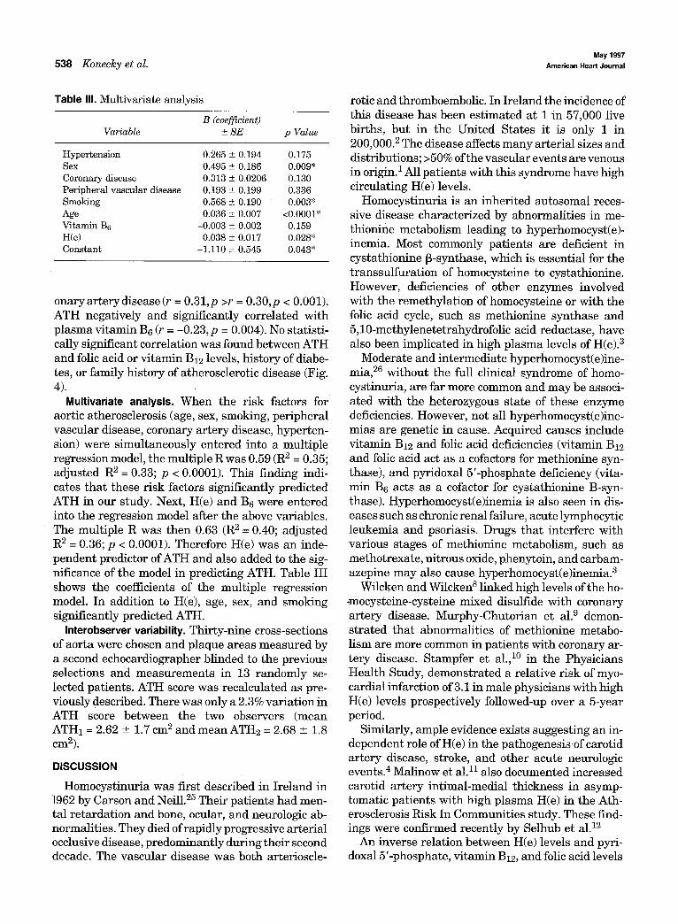
Fig. 3. TEE image in horizontal plane of distal descending thoracic aorta of 72-year-old woman with both elevated homocysteine level (18.65 DmoVL) and ATH (5.3 cm2).
was blinded to the levels of plasma H(e), pyridoxal 5'- phosphate, vitamin B12, and folic acid.
All patients were interviewed and their charts reviewed. Data concerning history of hypertension, diabetes, smok- ing, coronary artery disease (including angina pectoris, myocardial infarction, and prior coronary bypass surgery),
vascular disease (including stroke, transient ischemic at- tack, peripheral arterial disease, and peripheral emboliza- tion), and family history of coronary arter~j disease were obtained for univariate and multivariate analyses.
Statistics. A correlation matrix of the independent vari- ables was computed. We looked for multicoliiiearity, but it

Volume 133, Number 5 American Heart Journal J~onec/cy et al. 537
0.5
A
0.4 i..-
,,, 0 .3 (.J I.J_ ,, 0 .2 L.U 0 L) 0.1 z o - 0
_J " ' -0.1 n--
0 -0.2 (.J
-0.3 H(e) F B-12 B-6 Age Sex DM Tob Vasc CAD Htn Fam
V A R I A B L E
B B . p < 0 . 0 5
Fig. 4. Univariate correlations between ATH and all independent variables. Y axis represents correlation coefficients. CAD, Coronary artery disease; DM, diabetes; F, folic acid; Faro, positive family history of ath- erosclerotic disease; Htn, hypertension; Tob, tobacco use; Vasc, history of vascular disease. Asterisks rep- resent correlations with p value of <0.05.
Table I. Patient characteristics (n = 156) Table II. Results
Age Mean 68.5 yrs (30-89) Sex 73 male (46.8%) Smoking 85 (54.5%) ATH Diabetes 28 (17.9%) H(e) Hypertension 84 (53.8%) Vitamin B6 CAD* 49 (31.4%) Vitamin B12 Vascular disease~ 52 (32.7%) Folic acid
CAD, Coronary artery disease. *Includes angina, myocardial infarction, and history of bypass surgery. ~Includes stroke, transient ischemic attack, peripheral vascular disease, peripheral embolization, or prior peripheral vascular or carotid surgery.
was not found. Another correlation matrix of the indepen- dent variables against the dependent variable (ATH) was computed. From this univariate analysis, any independent variable that significantly correlated (p < 0.05) with ATH was identified. These significant variables were subse- quently included in a multiple regression analysis to assess the independent predictors of the dependent vari- able (ATH). Analysis was done with SPSS personal com- puter version 4 software (Chicago, Ill.).
RESULTS
Of the 156 pa t ien ts enrolled, 73 (47%) were men. The i r m e a n age was 68.5 years (range 30 to 87 years). A his tory of smoking was p resen t in 85 (54.5%), 28 (17.9%) were diabetic, 84 (53.8%) had a h is tory of
Variable Mean (range)
2.53 cm 2 (0.47-8.04) 12.45 pmol/L (3.90-40.30) 49.17 nmol/L (0.72-249.60) 432.44 pmol/L (82.0-1190.0) 34.92 nmol/L (3.39-602.34)
hyper tension, 49 (31.4%) had a h is tory of coronary a r t e ry disease, and 51 (32.7%) had a h is tory of a vas- cular event consis tent wi th embolization. There was a family his tory of coronary a r t e ry disease in 30% (Table I). Table II shows mean plasma H(e), vi tamin B6, vi tamin B12 , folic acid levels, and ATH. Pat ients who were higher than the 80th percentile for H(e) level in this group (>15 pmol/L) had an ATH score of 3.34 c m 2, nearly double tha t of patients in the lowest 20th percentile (H(e) level < 8 pmol/L, ATH = 1.75 cm2).
Univariate analysis. When ATH was selected as the dependen t variable, cer ta in stat is t ical ly significant correlat ions were noted (al lp < 0.05). ATH posit ively and significantly corre la ted wi th H(e) (r = 0.30, p >0.001), age ( r = 0 . 4 4 , p <0.001), male sex (r = 0.22, p = 0.007), h is tory of smoking (r = 0.33, p < 0.001), vascular disease (r = 0,19,p = 0.019), cor-
May 1997 538 Konecky et al. American Heart Journal
Table III. Multivariate analysis
B (coefficient) Variable ± S E p Value
Hypertension 0.265 ± 0.194 0.175 Sex 0.495 ± 0.186 0.009* Coronary disease 0.313 ± 0.0206 0.130 Peripheral vascular disease 0.193 _+ 0.199 0.336 Smoking 0.568 ± 0.190 0.003" Age 0.036 ± 0.007 <0.0001" Vitamin B6 -0.003 ± 0.002 0.159 H(e) 0.038 ± 0.017 0.028* Constant -1.110 ± 0.545 0.043"
onary artery disease (r = 0.31,p >r = 0.30,p < 0.001). ATH negatively and significantly correlated with plasma vitamin Be (r = -0.23, p = 0.004). No statisti- cally significant correlation was found between ATH and folic acid or vitamin B12 levels, history of diabe- tes, or family history of atherosclerotic disease (Fig. 4).
Multivariate analysis. When the risk factors for aortic atherosclerosis (age, sex, smoking, peripheral vascular disease, coronary artery disease, hyperten- sion) were simultaneously entered into a multiple regression model, the multiple R was 0.59 (R 2 = 0 .35;
a d j u s t e d R 2 = 0.33; p < 0.0001). This finding indi- cates that these risk factors significantly predicted ATH in our study. Next, H(e) and Be were entered into the regression model after the above variables. The multiple R was then 0.63 (R 2 = 0 .40; adjusted R 2 = 0.36; p < 0.0001). Therefore H(e) was an inde- pendent predictor of ATH and also added to the sig- nificance of the model in predicting ATH. Table III shows the coefficients of the multiple regression model. In addition to H(e), age, sex, and smoking significantly predicted ATH.
Interobserver variability. Thirty-nine cross-sections of aorta were chosen and plaque areas measured by a second echocardiographer blinded to the previous selections and measurements in 13 randomly se- lected patients. ATH score was recalculated as pre- viously described. There was only a 2.3% variation in ATH score between the two observers (mean ATH1 = 2.62 _+ 1.7 c m 2 and mean ATH2 = 2.68 _+ 1.8 cm2).
DISCUSSION
Homocystinuria was first described in Ireland in 1962 by Carson and Neill. 25 Their patients had men- tal retardation and bone, ocular, and neurologic ab- normalities. They died of rapidly progressive arterial occlusive disease, predominantly during their second decade. The vascular disease was both arterioscle-
rotic and thromboembolic. In Ireland the incidence of this disease has been estimated at 1 in 57,000 live births, but in the United States it is only 1 in 200,000. 2 The disease affects many arterial sizes and distributions; >50% of the vascular events are venous in origin.1 All patients with this syndrome have high circulating H(e) levels.
Homocystinuria is an inherited autosomal reces- sive disease characterized by abnormalities in me- thionine metabolism leading to hyperhomocyst(e)- inemia. Most commonly patients are deficient in cystathionine ~-synthase, which is essential for the transsulfuration of homocysteine to cystathionine. However, deficiencies of other enzymes involved with the remethylation of homocysteine or with the folic acid cycle, such as methionine synthase and 5,10-methylenetetrahydrofolic acid reductase, have also been implicated in high plasma levels of H(e). 3
Moderate and intermediate hyperhomocyst(e)ine- m i a , 26 without the full clinical syndrome of homo- cystinuria, are far more common and may be associ- ated with the heterozygous state of these enzyme deficiencies. However, not all hyperhomocyst(e)ine- mias are genetic in cause. Acquired causes include vitamin B12 and folic acid deficiencies (vitamin B12 and folic acid act as a cofactors for methionine syn- thase), and pyridoxal 5'-phosphate deficiency (vita- min Be acts as a cofactor for cystathionine B-syn- thase). Hyperhomocyst(e)inemia is also seen in dis- eases such as chronic renal failure, acute lymphocytic leukemia and psoriasis. Drugs that interfere with various stages of methionine metabolism, such as methotrexate, nitrous oxide, phenytoin, and carbam- azepine may also cause hyperhomocyst(e)inemia. 3
Wilcken and Wilcken s linked high levels of the ho- -mocysteine-cysteine mixed disulfide with coronary artery disease. Murphy-Chutorian et al. 9 demon- strated that abnormalities Of methionine metabo- lism are more common in patients with coronary ar- tery disease. Stampfer et al., 1° in the Physicians Health Study, demonstrated a relative risk of myo- cardial infarction of 3.1 in male physicians with high H(e) levels prospectively followed-up over a 5-year period.
Similarly, ample evidence exists suggesting an in- dependent role of H(e) in the pathogenesis of carotid artery disease, stroke, and other acute neurologic events. 4 Malinow et al. n also documented increased carotid artery intimal-medial thickness in asymp- tomatic patients with high plasma H(e) in the Ath- erosclerosis Risk In Communities study. These find- ings were confirmed recently by Selhub et al. 12
An inverse relation between H(e) levels and pyri- doxal 5'-phosphate, vitamin B12, and folic acid levels

Volume 133, Number 5
American Heart Journal Konecky et al. 539
has been documented, as well as an inverse relation between evidence of vascular disease and these vita- min levels, but results have been conflicting. Most recently, in 1995 Dalery et al. 27 demonstrated that in healthy patients H(e) levels are partially influenced by levels of vitamins B12 and Be and folic acid, but they found no significant correlation between these levels and the presence of coronary artery disease. Selhub et al. 12 have demonstrated an increased risk for extracranial carotid stenosis in elderly patients with high H(e) and low folic acid and vitamin Be lev- els.
This study shows that hyperhomocyst(e)inemia is an independent risk factor for aortic disease. The significant correlation between H(e) and ATH sug- gests a dose-response relation. The multivariate analysis documents a relation between H(e) and ATH that is clearly independent of other risk factors for atherosclerosis. Furthermore, adding H(e) to the multivariate model, after controlling for the known risk factors (age, sex, smoking, peripheral vascular disease, coronary artery disease, and hypertension) increased the power of the model to predict ATH.
The mean ATH score in our patients was 2.53 cm 2, and it varied from 0.47 to 8.04 cm 2. We have previ- ously shown that an atheroma with a diameter of 0.5 cm (an area of only approximately 0.2 cm 2) is asso- ciated with a high risk of embolic events, whereas smaller atheromas did not have such a risk. 2° The clinical risk of emboli is therefore likely correlated with the ATH score noted in the current patient population. In view of the fact that aortic atheromas have been shown to be associated with coronary ar-
2s tery atherosclerosis, coronary disease is also likely to be more prevalent in those patients with higher ATH scores, although this theory was not specifically evaluated.
Our results also reiterate the well-established positive correlation between H(e) and history of cor- onary artery disease. Significant negative correla- tions between H(e) and plasma levels of vitamins B12 and B6 are again consistent with previous studies, but unlike other previously published data 29, 3o no significant negative correlation was demonstrated between H(e) and folic acid level. The reasons for this
D
discrepancy need further study. The level of vitamin B6 was the only significant negative correlate of ATH among the three vitamins studied.
Limitations. This study is predominantly limited by the technique of measuring plaque area. We selected only cross-sections of the aorta with the largest amount of plaque and performed planimetry of these lesions. Although atherosclerosis is usually a diffuse process, it is not uniform and the present measure-
ments may overestimate plaque burden or skew the results by overemphasizing atherosclerotic burden in focal disease compared with diffuse disease. In addition, because part of the ascending aorta is ob- scured by the tracheal air column and is therefore not visible on TEE, this part of the thoracic aorta was not studied. Moreover, the inability to measure plaques in the near field of the sector because of artifact may affect the estimation of plaque burden. Finally, the ascending aorta was not used because of the inabil- ity to consistently obtain reliable cross-sections on TEE. However, ATH does serve as a marker of the degree of atherosclerosis in three different segments of thoracic aorta, and our evaluation ofinterobserver variability shows that this measurement is highly reproducible.
This study evaluates patients at a single point in time and does not yield information concerning clin- ical follow-up. Additional studies are required to es- tablish the correlations between H(e) levels, ATH score, and the clinical course of these patients.
Conclus ions . Atherosclerosis is a multifactorial pathologic process, and elevated levels of circulating H(e) can be considered one of the risk factors for ar- terial occlusive disease. This study demonstrates an association between H(e) and atherosclerotic plaque in the thoracic aorta. The highly significant correla- tion between H(e) and the estimated degree of ATH suggests a dose-response relation between these two parameters. Since vitamins B12 and B6 are nega- tively correlated with H(e), and vitamin B6 is also a significant negative correlate of ATH, it seems rea- sonable to suspect that t reatment with those vita- mins in patients with atherosclerosis and hyperho- mocyst(e)inemia might slow progression of the dis- ease and decrease the incidence of future vascular even tsY A prospective, randomized, placebo-con- trolled trial to addres~ this issue is warranted.
REFERENCES
1. Mudd SH, Skovby F, Levy HL, Pettigrew RD, Wilken B, Pyeritz RE, et al. The natural history of homocystinuria due to cystathionine beta- synthase deficiency. Am J Hum Genet 1985:37:1-12.
2. Mudd SH, Levy HL, Skovby F. Disorders of transsulfuration. In: Scriver CR, Beaudet AL, Sly WS, Valle D, editors. The metabolic basis of inherited disease, 7th ed. New York: McGraw-Hill; 1995. P. 1279- 327.
3. Robinson K, Mayer E, Jacobsen DW. Homocysteine and coronary artery disease. Cleve Clin J Med 1994:61:438-50.
4. Boers GHJ, Smals AGH, Trijbels FJM, Fowler B, Bakkeren JAJM, Schoonderwalt HC, et al. Heterozygosity for homocystinuia in prema- ture peripheral and cerebral occlusive arterial disease. N Engl J Med 1985;313:709-15.
5. Coull BM, Malinow MR, Beamer N, Sexton G, Nordt F, de Garmo P. Elevated plasma homocyst(e)ine concentraton as a possible indepen- dent risk factor for stroke. Stroke 1990;21:572-6.
6. Malinow MR, Kang SS, Taylor LM, Wong PWK, Coull B, Inahara T, et
May 1997 540 Konecky et al. American Heart Journal
al. Prevalance of hyperhomocyst(e)inemia in patients with peripheral arterial occlusive disease. Circulation 1989;79:1180-8.
7. Taylor LM Jr, DeFrang RD, Haris J Jr, Porter JM. The association of elevated plasma homocyst(e)ine with progression of symptomatic peripheral arterial disease. J Vasc Surg 1991;13:108-36.
8. Wilcken DEL, Wilcken B. The pathogenesis of coronary artery disease: a possible role for methionine metabolism. J Clin Invest 1976;57:1079- 82.
9. Murphy-Chutorian DR, Wexman MP, Grieco AJ, Heininger JA, Glass- man E, Gaull GE, et al. Methionine intolerance: a possible risk factor for coronary artery disease. J Am Coll Cardiol 1985;6:725-30.
10. Stampfer MJ, Malinow MR, Willett WC, Newcomer LM, Upson B, Ull- man D, et al. A prospective study of plasma homocyst(e)ine and risk of myocardial infarction in US physicians. JAMA 1992;268:877-81.
11. Malinow MR, Nieto FJ, Szklo M, Chambless LE, Bond G. Carotid ar- tery intimal-medical wall thickening and plasma homocysteine in as- ymptomatic adults. The Atherosclerosis Risk in Communities study. Circulation 1993:87:1107-13.
12. Selhub J, Jacques PF, Bostom AG, D'Agostino RB, Wilson PWF, Belanger AJ, et al. Association between plasma homocysteine concen- trations and extracranial carotid artery stenosis. N Engl J Med 1995; 332:286-91.
13. Ubbink JB, Vermaak WJH, Bennett JM, Becket PJ, Van Staden AA, Bissbort S. The prevalence of homocystinemia and hyperchotester- olemia in angiographically defined coronary heart disease. Klin Wochenschr 1991;69:529~34.
14. Rubba P, Faccenda F, Pauciullo P, Carbone L, Mancini M, Strisciuglio P, et al. Early signs of vascular disease in homocystinuria: a noninva- sive study by ultrasound methods in eight families with cystationinuria B-synthase deficiency. Metabolism 1990;39:1191-5.
15. yon Eckardstein A, Malinow MR, Upson B, Heinricb J, Schulte H, SchSnfeld R, et al. Effects of age, lipoproteins, and hemostatic param- eters on the role of homocyst(e)inemia as a cardiovascular risk factor in men. Arterioscler Thromb 1994;14:460-4.
16. Herzlick BC, Lichstein E, SchulhoffN, Weinstock M, Pagala M, Ravin- dran K, et al. Relationship among homocyst(e)ine, vitamin B12 and cardiac disease in the elderly: association between vitamin B12 deficiency and decreased left ventricular ejection fraction. J Nutr 1996;126 Suppl 4:12495-535.
17. Kronzon I, Tunick PA. Transesophageal echocardiography as a tool in the evaluation of patients with embolic disorders. Pr Cardiovasc Dis 1993;36:39-60.
18. Blanchard DG, Kimura B J, Dittrick HC, Demaria AN. Transesoph- ageal echocardiography of the aorta. JAMA 1994;272:546-51.
19. Tunick PA, Perez JL, Kronzon I. Protruding atheromas in the thoracic aorta and systemic embolization. Ann Intern Med 1991;115:423-7.
20. Tunick PA, Rosenzweig BP, Katz ES, Freedberg RS, Perez JL, Kronzon I. High risk for vascular events in patients with protruding aortic atheromas: a prospective study. J Am Coll Cardiol 1994;23:1085- 90.
21. Amarenco P, Cohen A, Tzourio C, Bertrand B, Hommel M, Besson G et al. Atherosclerotic disease of the aortic arch and the risk ofischemic stroke. N Engl J IVied 1994;331:1474-9.
22. Nishino M, Masugata H, Yamada Y, Abe H, Hori M, Kamada T. Eval- uation of thoracic aortic atherosclerosis by TEE. Am Heart J 1994; 127:336-44.
23. Malinow MR, Kang SS, Taylor LM, Wang PWK, Coull B, Inahara T, et al. Prevalence of hyperhomocyst(e)inemia in patients with peripheral arterial occlusive disease. Circulation 1989;79:1180-8.
24. Malinow MR, Sexton G, Averbuch M, Grossman M, Wilson D, Upson B. Homocyst(e)inemia in daily practice: levels in coronary disease. Coron Art Dis 1990;1:215-20.
25. Carson NAJ, Neill DW. Metabolic abnormalities detected in a survey of mentally backward individuals in northern Ireland. Arch Dis Child 1962;37:505-13.
26. Kang S-S, Wong PW, Malinow MR. Hyperhomocyst(e)inemia as a risk factor form occlusive vascular disease. Annu Rev Nutr 1992;12:279-98.
27. Dalery K, Lussier-Caoan S, Selhub J, Davignon J, Latour Y, Genest J Jr. ttomocysteine and coronary artery disease in French Canadian subjects: relation with vitamin B~2, B6, pyridoxal phosphate and folic acid. Am J Cardiol 1995;75:1107-11.
28. Fazio GP, Redberg RF, Winslow T, Schiller NB. Transesophageal echocardiographically detected atherosclerotic aortic plaque is a marker for coronary artery disease. J Am Coll Cardiol 1993;21:144-50.
29. Stabler SP, Marcell PD, Podell ER, Allan RH, Savage DG, Lindenbaum J. Elevation of total homocysteine in the serum of patients with cobal- amin or folic acid deficiency detected by capillary gas chromatography- mass spectrometry. J Clin Invest 1988;81:466-74.
30. Pancharuniti N, Lewis CA, Sauerlich HE, Perkins LL, Go RCP, Alva- rez JO, et al. Plasma homocysteine, folic acid and vitamin B12 concen- trations and.the risk of early-onset coronary artery disease. Am J Clin Nutr 1991;59:940-8.
31. Stampfer MJ, MalinowMR. Can lowering homocysteine levels reduce cardiovascular risk [editorial]? N Engl J Med 1995;332:328-9.
BOUND VOLUMES AVAILABLE TO SUBSCRIBERS
Bound volumes of the AMERICAN HEART JOURNAL are available only to subscribers for the 1997 issues from the Publisher at a cost of $92.50 for domestic, $120.38 for Canadian, and $112.50 for international subscribers for Vol. 133 (January-June) and Vol. 134 (July-December). Shipping charges are included. Each bound volume contains a subject and author index, and all advertising is removed. Copies are shipped within 60 days after publication of the last issue in the volume. The binding is durable buckram with the journal name, volume number, and year stamped in gold on the spine. Payment must accompany all orders. Contact Mosby-Year Book, Inc., Subscription Ser- vices, 11830 Westline Industrial Dr., St. Louis, MO 63146, USA; phone (800)453-4351 or (314)453- 4351.
Subscriptions must be in force to qualify. Bound volumes are not available in place of a regular Journal subscription.