Mesenteric stenosis, collaterals, and compensatory blood flow

Agmatine Induced NO Dependent Rat Mesenteric ArteryRelaxation and its Impairment in Salt-Sensitive Hypertension
Tushar V. Gadkari1, Natalie Cortes1, Kumpal Madrasi1, Nikolaos M. Tsoukias1, and MaheshS. Joshi1,*
1Department of Biomedical Engineering, Florida International University, Miami, FL
AbstractL-arginine and its decarboxylated product, agmatine are important mediators of NO productionand vascular relaxation. However, the underlying mechanisms of their action are not understood.We have investigated the role of arginine and agmatine in resistance vessel relaxation of Sprague-Dawley (SD) and Dahl salt-sensitive hypertensive rats. Second or 3rd-order mesenteric arterioleswere cannulated in an organ chamber, pressurized and equilibrated before perfusing intraluminallywith agonists. The vessel diameters were measured after mounting on the stage of a microscopefitted with a video camera. The gene expression in Dahl rat vessel homogenates was ascertainedby real-time PCR. L-arginine initiated relaxations (EC50, 5.8 ± 0.7 mM; n = 9) were inhibited byarginine decarboxylase (ADC) inhibitor, difluoromethylarginine (DFMA) (EC50, 18.3 ± 1.3 mM;n = 5) suggesting that arginine-induced vessel relaxation was mediated by agmatine formation.Agmatine relaxed the SD rat vessels at significantly lower concentrations (EC50, 138.7 ± 12.1 μM;n = 22), which was compromised by L-NAME (L-NG-Nitroarginine methyl ester, an eNOSinhibitor), RX821002 (α-2 AR antagonist) and pertussis toxin (G-protein inhibitor). The agmatine-mediated vessel relaxation from high salt Dahl rats was abolished as compared to that from normalsalt rats (EC50, 143.9 ± 23.4 μM; n = 5). The α-2A AR, α-2B AR and eNOS mRNA expressionwas downregulated in mesenteric arterioles of high-salt treated Dahl hypertensive rats. Thesefindings demonstrate that agmatine facilitated the relaxation via activation of α-2 adrenergic G-protein coupled receptor and NO synthesis, and this pathway is compromised in salt-sensitivehypertension.
Keywordsagmatine; arginine decarboxylase; L-arginine; nitric oxide; α-2 adreno receptor; hypertension
1. IntroductionOver the past 2 decades there has been a significant interest in understanding the role of NOin vascular relaxation. L-arginine serves as a substrate for different nitric oxide synthases(NOS). The potential use of exogenous L-arginine as a therapeutic agent has been widelysuggested in various physiological and pathophysiological processes such as wound healing
© 2013 Elsevier Inc. All rights reserved.*Correspondence: Department of Biomedical Engineering, Florida International University, Miami, FL 33174 504-343-6881 (phone)305-348-6954 (fax) [email protected].
Publisher's Disclaimer: This is a PDF file of an unedited manuscript that has been accepted for publication. As a service to ourcustomers we are providing this early version of the manuscript. The manuscript will undergo copyediting, typesetting, and review ofthe resulting proof before it is published in its final citable form. Please note that during the production process errors may bediscovered which could affect the content, and all legal disclaimers that apply to the journal pertain.
NIH Public AccessAuthor ManuscriptNitric Oxide. Author manuscript; available in PMC 2014 November 30.
Published in final edited form as:Nitric Oxide. 2013 November 30; 0: . doi:10.1016/j.niox.2013.08.005.
NIH
-PA Author Manuscript
NIH
-PA Author Manuscript
NIH
-PA Author Manuscript

[1; 2; 3], protein synthesis and muscle building [4], endocrine metabolism [5], erectiledysfunction [6], and a variety of cardiovascular functions [7; 8; 9]. Supplying arginine to theendothelial cells and vessels contributes to the enhanced NO synthesis and vessel relaxationdespite its saturating cellular levels. Different mechanisms are hypothesized to explain thisarginine paradox including endogenous NOS inhibitors and compartmentalization ofintracellular arginine [10; 11; 12; 13]. However, there still exists an ambiguity inunderstanding how exogenous L-arginine mediates NO-dependent relaxation [14]. Theapparent benefits of L-arginine supplementation are difficult to reconcile with a purelysubstrate based mechanism for NO synthesis. An alternative proposed by us explainsarginine’s NOS substrate independent actions via the activation of α-2 adrenergic receptor(α-2 AR) as demonstrated in cultured endothelial cells [15]. Agmatine [4-(aminobutyl)guanidine] is produced endogenously via decarboxylation of L-arginine by the endothelialarginine decarboxylase (ADC) [16; 17] and it does not act as a substrate for NOS. Abiological function for agmatine was suggested based on the observation that ADC activitytransiently increased 7 fold during cerebral ischemia [18]. In addition, the importance ofagmatine has been highlighted by its discovery as a novel neurotransmitter [19; 20; 21]demonstrating its potential to affect multiple biological targets. The presence of agmatine inserum [22] suggests a physiological role in the vasculature. Agmatine was shown to serve asa ligand for imidazoline and/or α-2 AR [23] and α-2 AR agonists mediate endothelium-dependent relaxation in mouse and rat aorta [24]. α-2 ARs (G-protein coupled receptors)play a pivotal role in the cardiovascular system and influence vascular tone at multiplepoints. These receptors are targets for antihypertensive therapy and their stimulationproduces long lasting drop in systemic blood pressure [25]. However, the signalingmechanisms participating in agmatine-initiated NO synthesis [15; 26] and regulation ofvascular tone is little understood, and the contribution of α-2 ARs is implicated in thisprocess [15]. Compromised NO synthesis and endothelial dysfunction has been reported inthe hypertensive vasculature including salt-sensitive hypertension [27; 28]. However, thefactors that are responsible for its impaired synthesis are varied and not clearly understood.Impaired α-2 AR function has been documented in several models of hypertension [29; 30;31]. However, whether this impairment is a cause or an effect of hypertension remains to beelucidated. Here we show that arginine-mediated arteriolar relaxations are due to agmatineproduced by the actions of ADC and signaling via GPCR in rat microcirculation. Evidenceis also presented documenting attenuated agmatine-mediated relaxation in Dahl salt-sensitive hypertensive rat mesenteric resistant arterioles, which correlates with reduced α-2AR gene expression.
2. Material and Methods2.1 Isolated mesenteric arteriole preparation
Resistance mesenteric arterioles of the 2nd or 3rd order (resting diameter ≤ 150 μm) wereisolated from male Sprague-Dawley (SD) and Dahl rats (250-300 g), cleaned of thesurrounding tissue and cannulated at both ends on glass cannula. The organ chamber wasmaintained constant at 37°C by superfusion with a modified Krebs-Ringer solutioncontaining (mM): NaCl 145, KCl 5, CaCl2 2.5, MgSO4 1.2, KH2PO4 1.2, HEPES 20, andGlucose 10.1, pH 7.4 [32; 33]. Cannulated vessels were axially pre-stretched to remove anybents and to simulate physiological stretch conditions and were pressurized at 50 mmHg andallowed to equilibrate for 60 min before initiating the experiment. To establish aconcentration that gave submaximal constriction, we constructed a dose-response curve tonorepinephrine (NE). The vessels were preconstricted with continuous superfusion of NEand those that retained a constant pressure and a consistent constriction to NE (Fig. S1.a andb), and fully responded to the acetylcholine (Fig. S3.a) were included in the study. Thepresence of functional endothelium was assessed by the ability of acetylcholine (10 μM) to
Gadkari et al. Page 2
Nitric Oxide. Author manuscript; available in PMC 2014 November 30.
NIH
-PA Author Manuscript
NIH
-PA Author Manuscript
NIH
-PA Author Manuscript

induce more than 90% relaxation. The vessel reactivity study was carried out byintraluminal perfusion with various agonist concentrations. This was achieved by anautomated solenoid valve controlled pressure driven perfusion system. Diametermeasurements were tracked in real time by mounting the perfusion chamber on the stage ofan inverted microscope (Olympus, Center Valley, PA) fitted with a CCD camera (QImaging,Surrey, Canada). Post analysis was performed with IPLAB (BioVision Technologies, Exton,PA) and MATLAB (MathWorks, Natick, MA) software. Chemicals NE, L-NAME (L-NG-Nitroarginine methyl ester), RX821002, agmatine, L-arginine were obtained from Sigma-Aldrich Co. (St. Louis, MO) and Pertussis toxin (PTx) were obtained from TocrisBioscience (Ellisville, MO) and SPERNO from Cayman Chemicals (Ann Arbor, MI).
2.2 Animal ModelMale SD and Dahl salt-sensitive (SS/JrHsd) rats and their diet were purchased from HarlanLaboratories (Madison, WI). Sprague Dawley rats were maintained on standard pellet chow(2018 Teklad Global) rodent diet whereas the Dahl salt-sensitive rats were fed 0.49% NaCldiet (Harlan Cat. #TD 96208) or 4% NaCl diet (Harlan Cat. #TD 92034). The animals werehoused in temperature and humidity controlled rooms with 12 hour on/off light cycle at theanimal care facility. All animal studies were performed following Institutional Animal Careand Use approved procedures.
After acclimatization for 1 week, 6-weeks old Dahl salt-sensitive rats were separated into 2diet groups; normal salt (NS), fed 0.49% NaCl diet and high salt (HS), fed 4% NaCl diet for5 weeks. Systolic blood pressure was measured weekly by the tail-cuff method [34] and HSrats consistently demonstrated sustained hypertension (BP > 200 mmHg) while NS ratsremained normotensive (BP < 160 mmHg) (Fig. S2). The rats were euthanized by CO2inhalation and vascular reactivity assessed on isolated, cannulated and pressurizedmesenteric arterioles as described above.
2.3 Determination of plasma nitriteBlood derived from rats was centrifuged immediately at 5,000g for 5 min and plasmacollected. The nitrite analysis was carried out using iodine/iodide in glacial acetic acidsupplemented with 1% v/v antifoam SE-15 (Sigma Aldrich) using an ozone basedchemiluminescence analyzer (Sievers, model 280i) as described [35].
2.4 Real Time-Polymerase Chain Reaction (RT-PCR)RT-PCR was carried out on mesenteric tissue from Dahl rats [36], cleaned of fat andstabilized with RNAlater (Qiagen, Valencia, CA). The tissue was homogenized (~30 mg)with a sonicator in RLT buffer (Qiagen), the lysate centrifuged (10,000g) and total RNApurified with reagents from RNeasy® Fibrous Tissue Mini Kit (Qiagen). A first strandcDNA synthesis was performed using purified mRNA by Superscript III RT (Invitrogen,Grand Island, NY) in a thermocycler (MJ Research). The new cDNA strand was purifiedwith QIAquick® PCR Purification Kit (Qiagen). Pure cDNA (~10 ng) was reacted withPower SYBR Green PCR Master Mix reagent (Applied Biosystems, Mountain View, CA) inRNase-free water in a StepOne RT-PCR system (Applied Biosystems). The relativeexpression of α-2A, α-2B AR and eNOS was determined using β-actin as a housekeepinggene. The primers used were: α-2A; TTT GCA CGT CGT CCA TAG TG (forward) andCAG TGA CAA TGA TGG CCT TG (reverse). α-2B; AAA CAC TGC CAG CAT CTC CT(forward) and CTG GCA ACT CCC ACA TTC TT (reverse). eNOS; CAA CGCTAC CACGAG GAC ATT (forward) and CTC CTG CAA AGA AAA GCT CTG G (reverse). β-actin;TCC TAG CAC CAT GAA GAT C (forward) and AAA CGC AGCTCA GTA ACA G(reverse). Standard curves (initial amount of cDNA versus Ct values) were tested for eachset of primers, demonstrating that for the similar range of total cDNA amplification the
Gadkari et al. Page 3
Nitric Oxide. Author manuscript; available in PMC 2014 November 30.
NIH
-PA Author Manuscript
NIH
-PA Author Manuscript
NIH
-PA Author Manuscript

efficiency of target genes and housekeeping gene (β-actin) were equal. ‘No reversetranscription control’ was used where the PCR reaction was run in the absence of reversetranscriptase. Expression of the gene of interest was divided by the housekeeping gene andexpressed as fold-change compared with the corresponding normal-salt rat group.
2.5 Data analysisRelaxations were expressed as percentage of NE (2 μM) induced contraction. Thevasodilation was studied by obtaining the maximal response and EC50 values were thencalculated by fitting the concentration-response relationship to a logistic function. Amplifiedtranscripts from RT-PCR were quantified using the comparative threshold cycle method(2−ΔΔCt) with β-actin as a normalizer and the corresponding sample from the normal salt fedrat mesentery as internal control.
All data were expressed as mean ± SEM with n representing independent rat experiments.Statistical significance was tested using a paired t-test with P<0.05 considered significant.
3. Results3.1 L-arginine-mediated relaxation is dependent upon ADC activity
As shown in Fig. 1a, L-arginine dose-dependently relaxed the vessel with an EC50 value of5.8 ± 0.7 mM (n = 9). The requirement of mM levels of arginine prompted us to hypothesizethat the actions of arginine may be mediated via its metabolism to agmatine by ADC, whichis shown to be localized in the endothelium. The relaxations to arginine were significantlyinhibited in the presence of ADC inhibitor, DFMA (Fig. 1a: EC50, 18.3 ± 1.3 mM; n = 5).The EC50 value for L-arginine increased several fold in the presence of ADC inhibitor.DFMA is a specific inhibitor of ADC [37] and its specificity in our system was verified bythe absence of any effect on agmatine-mediated vessel relaxation (Fig. 1b). Thus, theseexperiments demonstrated that the arginine’s actions are mediated at least in part by theformation of agmatine.
3.2 Agmatine-induced vessel relaxationTo examine the effect of agmatine treatment on vessel tone, the isolated mesentericarterioles were subjected to increasing agmatine concentrations by intraluminal perfusion.Agmatine dose-dependently relaxed the vessel with an EC50 of 138.7 ± 12.1 μM (Fig.1b; n= 22). Thus, significantly less agmatine was required as compared to arginine for arteriolarrelaxation. As illustrated in Fig 1b, the agmatine-mediated relaxation was partially NOdependent as eNOS inhibitor, L-NAME (0.5 mM) did not completely attenuate therelaxation (EC50, 346.0 ± 19.4 μM; n = 4).
3.3 α-2 AR activity in agmatine-mediated relaxationIt has been previously reported that agmatine acts as an α-2 AR ligand [23]. To validate ifthe agmatine-induced relaxation is mediated via α-2 AR, we treated the vessels withagmatine in the presence and absence of RX821002, a specific antagonist of α-2 AR [38;39]. As shown in Fig 1c, it partly attenuated agmatine-mediated relaxation (EC50, 1498.0 ±294.0 μM; n = 6) indicating that α-2 AR may be participating in the relaxation process.These data corroborate our earlier observation in cultured HUVECs using another selectiveα-2 AR antagonist, rauwolscine, where it attenuated arginine-induced cellular NO synthesis[15].
Gadkari et al. Page 4
Nitric Oxide. Author manuscript; available in PMC 2014 November 30.
NIH
-PA Author Manuscript
NIH
-PA Author Manuscript
NIH
-PA Author Manuscript

3.4 Inhibition of agmatine-mediated relaxation by pertussis toxinTo narrow down the downstream mechanisms that may be participating in the agmatine-induced relaxation, we examined the possible role of G-proteins. We inhibited the G-proteins by pretreating the vessel with PTx (100 nM) for 60 min at 37°C. As shown in Fig1d, we observed a significantly reduced relaxation as opposed to that due to agmatine alonewhile the EC50 value increased to 1300 ± 91 μM (n = 4). These data indicated that G-proteins may mediate agmatine-induced vessel relaxation. Similar inhibition with PTx wasobserved in our experiments with cultured HUVECs [15].
3.5 Agmatine and arginine-mediated vessel relaxation attenuated in DS ratsAccumulating evidence indicates that endothelium-dependent relaxation is impaired in salt-sensitive hypertension [40; 41; 42]. However, the underlying mechanisms have not beenclearly delineated. We investigated if the agmatine-induced arterial relaxation is affected inDahl salt-sensitive rats. 6-week old rats were placed on a diet containing either 0.49% NaCl(normal salt) or 4% NaCl (high salt) for 5 weeks and isolated mesenteric arterial relaxationswere recorded in response to agmatine dose. The agmatine-induced relaxation was greatlyinhibited in high-salt as compared to normal salt conditions (Fig 2a; % max. relaxation, 90.4± 1.7% (NS); 19.8 ± 2.4% (HS)) with normal salt rats exhibiting relaxation to physiologicalagmatine concentration (EC50, 143.9 ± 23.4 μM; n = 5). Similarly, L-arginine-mediatedrelaxation was impaired in high-salt diet rats (Fig. 2b). Ach mediated relaxation was alsoimpaired in vessels from high salt Dahl rats (Fig. S3.b) and thus verifying the presence ofendothelial dysfunction. However, relaxation due to an NO donor, SPER-NO was notaffected, thereby indicating an undiminished signaling pathway downstream of NOsynthesis in Dahl hypertensive rats (Fig. S4). These data illustrate that agmatine-mediatedsignal transduction pathway eliciting vascular relaxation is severely impaired in salt-inducedhypertension.
3.6 Attenuated NO synthesis in Dahl ratsPlasma nitrite levels, as a measure of endogenous NO synthesis, were analyzed by ozonebased chemiluminescence analyzer. The results indicated that NO synthesis wassignificantly attenuated in high salt rats as compared to normal salt rats (Fig. 3a). These dataare supported by the similar observations made by Fujii et al [43].
3.7 Down-regulation of mesenteric artery α-2 AR mRNA expression in Dahl ratsSince agmatine is proposed to act via α-2 AR binding, we carried out mRNA expressionanalysis of α-2 AR and eNOS in Dahl rats by real time RT-PCR. The mRNA expression ofα-2A, α-2B AR and eNOS were attenuated in high-salt Dahl rat mesenteric arteries ascompared to normal-salt arterioles (Fig. 3b) indicating that mRNA expression levels of thesegenes were reversed during the development of salt-sensitive hypertension. Among the 3genes analyzed, there was a 10 fold under-expression of α-2A AR mRNA.
4. DiscussionAlthough the beneficial effects of arginine in vascular relaxation are well documented andappreciated, the mechanism of its action is not clearly understood. Arginine serves a majorrole by acting as a substrate for NOS and synthesizing NO, which in turn participates insignal transduction of vascular relaxation. Since millimolar amounts of arginine are requiredto produce measurable NO levels [44], it was speculated that arginine’s metabolic productscould be contributing to its observed effects including vasodilation [15]. We have proposedthat some of the observed effects of arginine could be attributed to its conversion toagmatine by ADC.
Gadkari et al. Page 5
Nitric Oxide. Author manuscript; available in PMC 2014 November 30.
NIH
-PA Author Manuscript
NIH
-PA Author Manuscript
NIH
-PA Author Manuscript

Our important finding that the inhibition of ADC with DFMA significantly attenuatedarginine-initiated mesenteric arterial dilation, thereby pointing towards an arginine-toagmatine conversion for the observed relaxation. The endothelium is known to possessADC activity [16], which was documented to mediate an increased vasodilation andglomerular filtration rate following arginine infusion into animals [45; 46]. In macrophagesunder inflammatory conditions, ADC was shown to modulate agmatine levels, which in turndown-regulated iNOS catalyzed NO synthesis [47]. Higher levels of ADC are present in thebrain [48]and hence it can be speculated that agmatine synthesized may have biological rolein the distant organs in particular the vascular function. We observed that agmatine initiatedthe relaxation at a significantly lower concentration (EC50, 138.7 ± 12.1 μM; n = 22) ascompared to arginine. The increased vessel relaxation with μM agmatine is physiologicallyrelevant since the plasma agmatine levels have been reported in the μM range [49; 50].Agmatine was shown to activate NO synthesis in endothelial cells but the mechanism ofactivation was not elucidated [26; 51]. The partial attenuation of agmatine-initiatedrelaxation by L-NAME indicated that agmatine partly triggered the relaxation via NOindependent, possibly EDHF mediated pathway. Binding of agmatine to imidazoline or α-2AR is well documented in the mammalian system. Our hypothesis that agmatine acts byreceptor binding, possibly via α-2 AR (Fig. 4), was examined by using an α-2 ARantagonist. The observation that RX821002 did not completely attenuate agmatine-triggeredrelaxation implied that agmatine affecting relaxation partly via α-AR independent pathway.Recently, agmatine’s relaxing effects in rat aorta are documented to be via smallconductance Ca2+-activated K+ channel and ATP-sensitive inward rectifying K+ channels,and idazoxan failed to inhibit these relaxations [52]. It appears that rat aorta is devoid of I-receptors, as Musgrave et al [53] have reported the absence of I-receptor antagonist effects,and thus it is likely that agmatine is using alternative pathways of aortic relaxation.
Since α-2 AR belongs to GPCR family, we tested the effects of G-protein inhibition. PTx, apotent G-protein inhibitor, considerably attenuated agmatine-initiated relaxation. Theinhibitory effects of α-2 AR antagonist (RX821002) and G-protein inhibitor (PTx) togetherprovide stronger evidence for the possible participation of GPCR in the vasodilatory actionsof agmatine. Using 2 knockout mouse models, Shafaroudi et al. [24] have demonstrated themediation of endothelial α-2A AR in vasodilation via NO-dependent mechanisms. Thesereceptors may play important physiological functions as they’re activated by the endogenousligand NE. In addition, there is evidence for their participation in various pathophysiologicalconditions. For example, blood pressure was elevated in transgenic α-2A AR knockout miceon a normal salt diet [54], showing that α-2A AR activation with specific agonists may serveto alleviate hypertensive conditions. Dahl hypertensive rats are reported to exhibitendothelial dysfunction [55] and agmatine-mediated vascular relaxation could be impairedin this model. Small size arterioles (100-500 μm diameter) are proposed to contributesignificantly to the vascular resistance [56]. Our measurements of agmatine-initiated vesselrelaxation showed that relaxation was severely impaired in high salt as compared to normalsalt diet fed Dahl rats. This observation supports our postulation that α-2A AR activity andassociated signaling pathway is down-regulated in high salt treated rats leading toimpairment of agmatine-mediated relaxation. Secondly, mRNA analysis by real time PCRshowed suppression of α-2A and α-2B AR expression in the mesenteric artery isolated fromhigh salt fed Dahl rats as compared those from normal salt fed rats. There was noticeably arobust 10 fold down-regulation of α-2A AR, which could be the predominant factorresponsible for the observed abolishment of vessel relaxation in HS rats. Hirano et al [57]have also observed attenuated pre- and post-synaptic α-2A AR reactivity in Dahl rats fedhigh salt diet. The attenuated L-arginine and agmatine-mediated relaxation could be also dueto the observed down-regulation of eNOS expression. Similarly, down-regulation of eNOSprotein expression was observed in kidney and aorta from Dahl salt-sensitive rats [58]. Ourfinding that substantial depletion in plasma nitrite levels in hypertensive rats could be the
Gadkari et al. Page 6
Nitric Oxide. Author manuscript; available in PMC 2014 November 30.
NIH
-PA Author Manuscript
NIH
-PA Author Manuscript
NIH
-PA Author Manuscript
result of either decreased L-arginine/agmatine levels and/or reduced α-2A AR and eNOSexpression. Further investigations are warranted to delineate the impaired agmatine pathwayas the underlying cause of endothelial dysfunction in hypertension. In summary, we havedemonstrated the mediation of ADC in the arginine-initiated stimulation of rat mesentericartery relaxation. Physiological concentrations of agmatine activated vessel relaxation viaα-2 AR and G-proteins and this was partially dependent on vascular NO synthesis.Agmatine relaxed the vessels at 100 times lower concentration as compared to arginine andthus making it a physiologically relevant agonist at α-2 AR binding site. Most importantly,our investigations have provided a novel mechanistic explanation for the observed beneficialeffects of L-arginine-initiated vessel relaxation via agmatine formation. Lastly, α-2 ARactivity, mRNA expression, and agmatine-mediated relaxations are impaired in vesselsisolated from high salt loaded Dahl hypertensive rats. These findings may help definefundamental aspects of cardiovascular physiology and pathophysiology by demonstratingthat many effects of arginine may occur due to its metabolism to agmatine.
Supplementary MaterialRefer to Web version on PubMed Central for supplementary material.
AcknowledgmentsThis work was supported by the National Institutes of Health grant (NMT). We wish to thank DeepakBalasubramanian and Kalai Mathee for their valuable help in carrying out RT-PCR experiments and analysis ofdata.
Abbreviations
ADC arginine decarboxylase
DFMA difluoromethylarginine
eNOS endothelial nitric oxide synthases
L-NAME L-NG-Nitroarginine methyl ester
AR adrenergic receptor
PTx Pertussis toxin
References[1]. Stechmiller JK, Childress B, Cowan L. Arginine Supplementation and Wound Healing. Nutrition
in Clinical Practice. 2005; 20:52–61. [PubMed: 16207646]
[2]. Angele MK, Nitsch SM, Hatz RA, Angele P, Hernandez-Richter T, Wichmann MW, Chaudry IH,Schildberg FW. L-arginine: a unique amino acid for improving depressed wound immunefunction following hemorrhage. Eur Surg Res. 2002; 34:53–60. [PubMed: 11867902]
[3]. Molderings GJ, Kribben B, Heinen A, Schröder D, Brüss M, Göthert M. Intestinal tumor andagmatine (decarboxylated arginine). Cancer. 2004; 101:858–868. [PubMed: 15305420]
[4]. Morris SM. Arginine: beyond protein. The American Journal of Clinical Nutrition. 2006;83:508S–512S. [PubMed: 16470022]
[5]. Mocchegiani E, Nistico G, Santarelli L, Fabris N. Effect of L-arginine on thymic function.Possible role of L-arginine: Nitric oxide (no) pathway. Archives of Gerontology and Geriatrics.1994; 19:163–170. [PubMed: 18649856]
[6]. Carson CC 3rd. Erectile dysfunction: diagnosis and management with newer oral agents. Proc(Bayl Univ Med Cent). 2000; 13:356–60. [PubMed: 16389341]
Gadkari et al. Page 7
Nitric Oxide. Author manuscript; available in PMC 2014 November 30.
NIH
-PA Author Manuscript
NIH
-PA Author Manuscript
NIH
-PA Author Manuscript
[7]. Wu G, Bazer FW, Davis TA, Kim SW, Li P, Marc Rhoads J, Carey Satterfield M, Smith SB,Spencer TE, Yin Y. Arginine metabolism and nutrition in growth, health and disease. AminoAcids. 2009; 37:153–68. [PubMed: 19030957]
[8]. Appleton J. Arginine: clinical potential of a semi-essential amino acid. (Arginine). AlternativeMedicine Review. 2002; 7:512–11. [PubMed: 12495375]
[9]. Bode-Böger SM, Scalera F, Ignarro LJ. The l-arginine paradox: Importance of the larginine/asymmetrical dimethylarginine ratio. Pharmacology & Therapeutics. 2007; 114:295–306.[PubMed: 17482266]
[10]. Wei LH, Jacobs AT, Morris SM Jr. Ignarro LJ. IL-4 and IL-13 upregulate arginase I expressionby cAMP and JAK/STAT6 pathways in vascular smooth muscle cells. Am J Physiol CellPhysiol. 2000; 279:C248–56. [PubMed: 10898736]
[11]. García-Cardeña G, Oh P, Liu J, Schnitzer JE, Sessa WC. Targeting of nitric oxide synthase toendothelial cell caveolae via palmitoylation: implications for nitric oxide signaling. Proceedingsof the National Academy of Sciences of the United States of America. 1996; 93:6448–6453.[PubMed: 8692835]
[12]. McDonald KK, Zharikov S, Block ER, Kilberg MS. A Caveolar Complex between the CationicAmino Acid Transporter 1 and Endothelial Nitric-oxide Synthase May Explain the “ArginineParadox”. Journal of Biological Chemistry. 1997; 272:31213–31216. [PubMed: 9395443]
[13]. Nagaraja S, Kapela A, Tran CH, Welsh DG, Tsoukias NM. Role of microprojections inmyoendothelial feedback: a theoretical study. The Journal of Physiology. 2013
[14]. Kapela A, Bezerianos A, Tsoukias NM. A mathematical model of Ca2+ dynamics in ratmesenteric smooth muscle cell: Agonist and NO stimulation. Journal of Theoretical Biology.2008; 253:238–260. [PubMed: 18423672]
[15]. Joshi MS, Ferguson TB Jr. Johnson FK, Johnson RA, Parthasarathy S, Lancaster JR Jr. Receptor-mediated activation of nitric oxide synthesis by arginine in endothelial cells. Proc Natl Acad SciU S A. 2007; 104:9982–7. [PubMed: 17535904]
[16]. Regunathan S, Youngson C, Raasch W, Wang H, Reis DJ. Imidazoline receptors and agmatine inblood vessels: a novel system inhibiting vascular smooth muscle proliferation. Journal ofPharmacology & Experimental Therapeutics. 1996; 276:1272–1282. [PubMed: 8786560]
[17]. Zhu MY, Iyo A, Piletz JE, Regunathan S. Expression of human arginine decarboxylase, thebiosynthetic enzyme for agmatine. Biochim Biophys Acta. 2004; 1670:156–64. [PubMed:14738999]
[18]. Gilad GM, Gilad VH, Rabey JM. Arginine and ornithine decarboxylation in rodent brain:coincidental changes during development and after ischemia. Neurosci Lett. 1996; 216:33–6.[PubMed: 8892385]
[19]. Reis DJ, Regunathan S. Agmatine: An Endogenous Ligand at Imidazoline Receptors Is a NovelNeurotransmittera. Annals of the New York Academy of Sciences. 1999; 881:65–80. [PubMed:10415899]
[20]. Donald J Reis SR. Is agmatine a novel neurotransmitter in brain? Trends in PharmacologicalSciences. 2000; 21:187–193. [PubMed: 10785653]
[21]. Regunathan S, Reis DJ. Characterization of Arginine Decarboxylase in Rat Brain and Liver.Journal of Neurochemistry. 2000; 74:2201–2208. [PubMed: 10800966]
[22]. Raasch W, Regunathan S, Li G, Reis DJ. Agmatine, the bacterial amine, is widely distributed inmammalian tissues. Life Sciences. 1995; 56:2319–2330. [PubMed: 7791519]
[23]. Li G, Regunathan S, Barrow CJ, Eshraghi J, Cooper R, Reis DJ. Agmatine: an endogenousclonidine-displacing substance in the brain. Science. 1994; 263:966–969. [PubMed: 7906055]
[24]. Shafaroudi MM, McBride M, Deighan C, Wokoma A, Macmillan J, Daly CJ, McGrath JC. Two“knockout” mouse models demonstrate that aortic vasodilatation is mediated viaalpha2aadrenoceptors located on the endothelium. J Pharmacol Exp Ther. 2005; 314:804–10.[PubMed: 15878998]
[25]. Hieble JP, Kolpak DC. Mediation of the hypotensive action of systemic clonidine in the rat byalpha 2-adrenoceptors. Br J Pharmacol. 1993; 110:1635–9. [PubMed: 8306110]
[26]. Morrissey JJ, Klahr S. Agmatine activation of nitric oxide synthase in endothelial cells.Proceedings of the Association of American Physicians. 1997; 109:51–57. [PubMed: 9010916]
Gadkari et al. Page 8
Nitric Oxide. Author manuscript; available in PMC 2014 November 30.
NIH
-PA Author Manuscript
NIH
-PA Author Manuscript
NIH
-PA Author Manuscript

[27]. Satoh M, Haruna Y, Fujimoto S, Sasaki T, Kashihara N. Telmisartan improves endothelialdysfunction and renal autoregulation in Dahl salt-sensitive rats. Hypertens Res. 2010; 33:135–42.[PubMed: 19927153]
[28]. Zhou M-S, Jaimes EA, Raij L. Atorvastatin Prevents End-Organ Injury in Salt-SensitiveHypertension: Role of eNOS and Oxidant Stress. Hypertension. 2004; 44:186–190. [PubMed:15238570]
[29]. Moura E, Pinto CE, Serrao MP, Afonso J, Vieira-Coelho MA. Adrenal alpha(2)-adrenergicreceptors in the aging normotensive and spontaneously hypertensive rat. Neurobiol Aging. 2012;33:969–78. [PubMed: 20691504]
[30]. Park J, Galligan JJ, Fink GD, Swain GM. Alterations in sympathetic neuroeffector transmissionto mesenteric arteries but not veins in DOCA-salt hypertension. Auton Neurosci. 2010; 152:11–20. [PubMed: 19914150]
[31]. Dohi Y, Thiel MA, Buhler FR, Luscher TF. Activation of endothelial L-arginine pathway inresistance arteries. Effect of age and hypertension, Hypertension. 1990; 16:170–9.
[32]. Duling BR, Gore RW, Dacey RG Jr. Damon DN. Methods for isolation, cannulation, and in vitrostudy of single microvessels. Am J Physiol. 1981; 241:H108–116. [PubMed: 7195654]
[33]. Knot HJ, Lounsbury KM, Brayden JE, Nelson MT. Gender differences in coronary arterydiameter reflect changes in both endothelial Ca2+ and ecNOS activity. Am J Physiol. 1999;276:H961–9. [PubMed: 10070080]
[34]. Feng M, Whitesall S, Zhang Y, Beibel M, D’Alecy L, DiPetrillo K. Validation of volume-pressure recording tail-cuff blood pressure measurements. Am J Hypertens. 2008; 21:1288–91.[PubMed: 18846043]
[35]. Pelletier MM, Kleinbongard P, Ringwood L, Hito R, Hunter CJ, Schechter AN, Gladwin MT,Dejam A. The measurement of blood and plasma nitrite by chemiluminescence: pitfalls andsolutions. Free Radic Biol Med. 2006; 41:541–8. [PubMed: 16863986]
[36]. Taylor S, Wakem M, Dijkman G, Alsarraj M, Nguyen M. A practical approach to RT-qPCR—Publishing data that conform to the MIQE guidelines. Methods. 2010; 50:S1–S5. [PubMed:20215014]
[37]. Yarlett N, Waters WR, Harp JA, Wannemuehler MJ, Morada M, Bellcastro J, Upton SJ, MartonLJ, Frydman BJ. Activities of dl-α-Difluoromethylarginine and Polyamine Analogues againstCryptosporidium parvum Infection in a T-Cell Receptor Alpha-Deficient Mouse Model.Antimicrobial Agents and Chemotherapy. 2007; 51:1234–1239. [PubMed: 17242149]
[38]. Callado LF, Gabilondo AM, Meana JJ. [3H]RX821002 (2-methoxyidazoxan) binds to α2-adrenoceptor subtypes and a non-adrenoceptor imidazoline binding site in rat kidney. EuropeanJournal of Pharmacology. 1996; 316:359–368. [PubMed: 8982708]
[39]. O’Rourke MF, Blaxall HS, Iversen LJ, Bylund DB. Characterization of [3H]RX821002 bindingto alpha-2 adrenergic receptor subtypes. J Pharmacol Exp Ther. 1994; 268:1362–7. [PubMed:7908054]
[40]. Ozawa Y, Hayashi K, Kanda T, Homma K, Takamatsu I, Tatematsu S, Yoshioka K, Kumagai H,Wakino S, Saruta T. Impaired nitric oxide- and endothelium-derived hyperpolarizing factor-dependent dilation of renal afferent arteriole in Dahl salt-sensitive rats. Nephrology (Carlton).2004; 9:272–7. [PubMed: 15504139]
[41]. Hermann M, Camici G, Fratton A, Hurlimann D, Tanner FC, Hellermann JP, Fiedler M, Thiery J,Neidhart M, Gay RE, Gay S, Luscher TF, Ruschitzka F. Differential effects of selectivecyclooxygenase-2 inhibitors on endothelial function in salt-induced hypertension. Circulation.2003; 108:2308–11. [PubMed: 14597594]
[42]. Payne JA, Alexander BT, Khalil RA. Decreased endothelium-dependent NO-cGMP vascularrelaxation and hypertension in growth-restricted rats on a high-salt diet. Hypertension. 2004;43:420–7. [PubMed: 14707161]
[43]. Fujii S, Zhang L, Igarashi J, Kosaka H. L-arginine reverses p47phox and gp91phox expressioninduced by high salt in Dahl rats. Hypertension. 2003; 42:1014–20. [PubMed: 14504257]
[44]. Hall CN, Garthwaite J. What is the real physiological NO concentration in vivo? Nitric Oxide.2009; 21:92–103. [PubMed: 19602444]
Gadkari et al. Page 9
Nitric Oxide. Author manuscript; available in PMC 2014 November 30.
NIH
-PA Author Manuscript
NIH
-PA Author Manuscript
NIH
-PA Author Manuscript

[45]. Blantz RC, Satriano J, Gabbai F, Kelly C. Biological effects of arginine metabolites. ActaPhysiol Scand. 2000; 168:21–5. [PubMed: 10691775]
[46]. Thomson SC, Deng A, Bao D, Satriano J, Blantz RC, Vallon V. Ornithine decarboxylase, kidneysize, and the tubular hypothesis of glomerular hyperfiltration in experimental diabetes. TheJournal of Clinical Investigation. 2001; 107:217–224. [PubMed: 11160138]
[47]. Regunathan S, Piletz JE. Regulation of inducible nitric oxide synthase and agmatine synthesis inmacrophages and astrocytes. Ann N Y Acad Sci. 2003; 1009:20–9. [PubMed: 15028566]
[48]. Li G, Regunathan S, Barrow C, Eshraghi J, Cooper R, Reis D. Agmatine: an endogenousclonidine-displacing substance in the brain. Science. 1994; 263:966–969. [PubMed: 7906055]
[49]. Lortie MJ, Novotny WF, Peterson OW, Vallon V, Malvey K, Mendonca M, Satriano J, Insel P,Thomson SC, Blantz RC. Agmatine, a bioactive metabolite of arginine. Production, degradation,and functional effects in the kidney of the rat. Journal of Clinical Investigation. 1996; 97:413–420. [PubMed: 8567962]
[50]. Cox TT, Boeker EA. Analysis of enzyme kinetics by using integrated rate equations. Argininedecarboxylase. Biochem J. 1987; 245:59–65. [PubMed: 3117044]
[51]. Coleman CS, Hu G, Pegg AE. Putrescine biosynthesis in mammalian tissues. Biochem J. 2004;379:849–55. [PubMed: 14763899]
[52]. Santhanam AVR, Viswanathan S, Dikshit M. Activation of protein kinase B/Akt and endothelialnitric oxide synthase mediates agmatine-induced endothelium-dependent relaxation. EuropeanJournal of Pharmacology. 2007; 572:189–196. [PubMed: 17640632]
[53]. Musgrave IF, Van Der Zypp A, Grigg M, Barrow CJ. Endogenous imidazoline receptor ligandsrelax rat aorta by an endothelium-dependent mechanism. Ann N Y Acad Sci. 2003; 1009:222–7.[PubMed: 15028591]
[54]. Ledent C, Vaugeois JM, Schiffmann SN, Pedrazzini T, El Yacoubi M, Vanderhaeghen JJ,Costentin J, Heath JK, Vassart G, Parmentier M. Aggressiveness, hypoalgesia and high bloodpressure in mice lacking the adenosine A2a receptor. Nature. 1997; 388:674–8. [PubMed:9262401]
[55]. Johnson FK, Johnson RA, Peyton KJ, Durante W. Arginase inhibition restores arteriolarendothelial function in Dahl rats with salt-induced hypertension. Am J Physiol Regul IntegrComp Physiol. 2005; 288:R1057–62. [PubMed: 15591155]
[56]. Christensen KL, Mulvany MJ. Perindopril changes the mesenteric pressure profile of conscioushypertensive and normotensive rats. Hypertension. 1994; 23:325–8. [PubMed: 8125558]
[57]. Hirano Y, Tsunoda M, Shimosawa T, Matsui H, Fujita T, Funatsu T. Suppression of catechol-O-methyltransferase activity through blunting of alpha2-adrenoceptor can explain hypertension inDahl salt-sensitive rats. Hypertens Res. 2007; 30:269–78. [PubMed: 17510509]
[58]. Ni Z, Oveisi F, Vaziri ND. Nitric oxide synthase isotype expression in salt-sensitive and salt-resistant Dahl rats. Hypertension. 1999; 34:552–7. [PubMed: 10523325]
Gadkari et al. Page 10
Nitric Oxide. Author manuscript; available in PMC 2014 November 30.
NIH
-PA Author Manuscript
NIH
-PA Author Manuscript
NIH
-PA Author Manuscript

Highlights
▶ Agmatine induced relaxation in isolated rat mesenteric arterioles.
▶The relaxations were mediated by G-proteins, α-2 AR and eNOS.
▶ L-arginine relaxed at much higher concentrations via arginine decarboxylase.
▶The relaxation to agmatine was abolished in high salt treated Dahl rats.
▶ α-2A AR, α-2B AR and eNOS mRNA expressions were down regulated in highsalt Dahl rats.
Gadkari et al. Page 11
Nitric Oxide. Author manuscript; available in PMC 2014 November 30.
NIH
-PA Author Manuscript
NIH
-PA Author Manuscript
NIH
-PA Author Manuscript

Figure 1. Concentration dependant relaxation responses to L-arginine and agmatine(a). Dose-response to intraluminal perfusion of L-arginine (n = 9) in SD rat mesentericarterioles and after pre-treatment with ADC blocker, DFMA (0.5 mM)(n = 5); #P < 0.05 vs.L-arginine. (b). Concentration dependent dose response curve to intraluminal perfusion ofagmatine in rat mesenteric arterioles in the presence and absence of an eNOS blocker, L-NAME (0.5 mM)(n = 4) and ADC blocker, DFMA (0.5 mM)(n = 3). Values are mean ± SEwith; *P < 0.05 vs. agmatine; **P > 0.05 vs. agmatine. (c). Dose response to agmatine in SDrat vessels was obtained in the presence and absence of an α-2 AR antagonist, RX821002(50 nM) (n = 6); *P < 0.05 vs. agmatine. (d). Agmatine relaxation response in the absenceand presence of G-protein inhibitor, PTx (100 nM). Values are mean ± SE (n = 4); *P < 0.05vs. agmatine.
Gadkari et al. Page 12
Nitric Oxide. Author manuscript; available in PMC 2014 November 30.
NIH
-PA Author Manuscript
NIH
-PA Author Manuscript
NIH
-PA Author Manuscript

Figure 2. Relaxation response to agmatine and L-arginine in Dahl salt-sensitive hypertension(a) Agmatine and (b) L-arginine-mediated relaxation observed in salt-sensitive Dahl ratsmaintained on high salt, HS (4% NaCl) and normal salt, NS (0.49% NaCl) for 5 weeks, (a: n= 5, b: n = 3). Values are mean ± SE, #P < 0.05 vs. L-arginine, *P < 0.05 vs. agmatine.
Gadkari et al. Page 13
Nitric Oxide. Author manuscript; available in PMC 2014 November 30.
NIH
-PA Author Manuscript
NIH
-PA Author Manuscript
NIH
-PA Author Manuscript
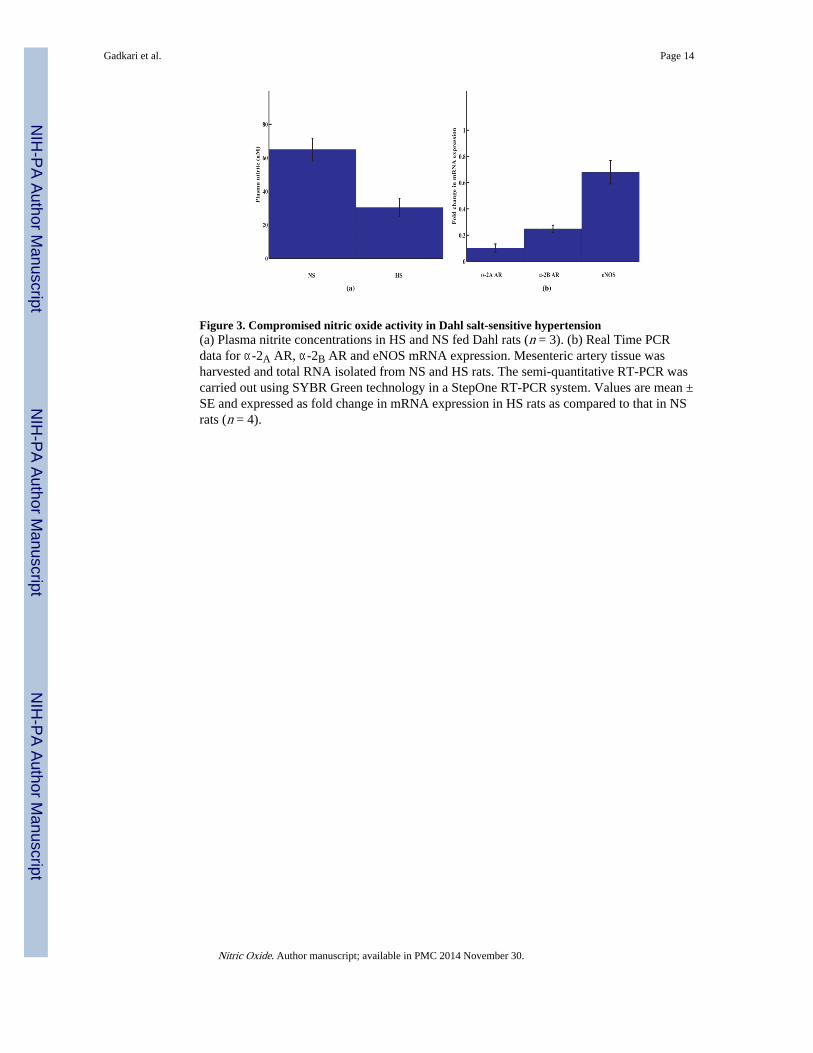
Figure 3. Compromised nitric oxide activity in Dahl salt-sensitive hypertension(a) Plasma nitrite concentrations in HS and NS fed Dahl rats (n = 3). (b) Real Time PCRdata for α-2A AR, α-2B AR and eNOS mRNA expression. Mesenteric artery tissue washarvested and total RNA isolated from NS and HS rats. The semi-quantitative RT-PCR wascarried out using SYBR Green technology in a StepOne RT-PCR system. Values are mean ±SE and expressed as fold change in mRNA expression in HS rats as compared to that in NSrats (n = 4).
Gadkari et al. Page 14
Nitric Oxide. Author manuscript; available in PMC 2014 November 30.
NIH
-PA Author Manuscript
NIH
-PA Author Manuscript
NIH
-PA Author Manuscript

Figure 4.Schematic representation of the proposed receptor-mediated hypothesis for arginine/agmatine stimulation of vascular NO synthesis.
Gadkari et al. Page 15
Nitric Oxide. Author manuscript; available in PMC 2014 November 30.
NIH
-PA Author Manuscript
NIH
-PA Author Manuscript
NIH
-PA Author Manuscript
Copyright © 2022 FDOKUMEN