Carboxypeptidase B and other kininases of the rat coronary and mesenteric arterial bed perfusates
32
1 Carboxypeptidase B and other kininases of the rat coronary and mesenteric arterial bed perfusates Eduardo B. Oliveira 1 , Laura L. Souza 1 , Disney O. Sivieri Jr 2 , Luiz B. Bispo-da-Silva 2 , Hugo J. V. Pereira 1 , Claudio M. Costa-Neto 1 , Marcelo V. Sousa 3 , Maria Cristina O. Salgado 2 Departamentos de Bioquímica e Imunologia 1 e de Farmacologia 2 da Faculdade de Medicina de Ribeirão Preto, Universidade de São Paulo, Brazil, and Departamento de Biologia Celular 3 , ICB, Universidade de Brasília, Brazil Running head: Kininase activity of rat carboxypeptidase B Corresponding Author: Dr. Maria Cristina O. Salgado Departamento de Farmacologia Faculdade de Medicina de Ribeirão Preto-USP 14049-900 Ribeirão Preto, SP Brazil Phone: 55-16-36023046 Fax: 55-16-3633-2301 e-mail: [email protected] Page 1 of 32 Copyright Information Articles in PresS. Am J Physiol Heart Circ Physiol (September 28, 2007). doi:10.1152/ajpheart.00784.2007 Copyright © 2007 by the American Physiological Society.
-
Upload
independent -
Category
Documents
-
view
0 -
download
0
Transcript of Carboxypeptidase B and other kininases of the rat coronary and mesenteric arterial bed perfusates

1
Carboxypeptidase B and other kininases of the rat coronary and
mesenteric arterial bed perfusates
Eduardo B. Oliveira1, Laura L. Souza1, Disney O. Sivieri Jr2, Luiz B. Bispo-da-Silva2,
Hugo J. V. Pereira1, Claudio M. Costa-Neto1, Marcelo V. Sousa3, Maria Cristina O.
Salgado2
Departamentos de Bioquímica e Imunologia1 e de Farmacologia2 da Faculdade de
Medicina de Ribeirão Preto, Universidade de São Paulo, Brazil, and Departamento
de Biologia Celular3, ICB, Universidade de Brasília, Brazil
Running head: Kininase activity of rat carboxypeptidase B
Corresponding Author:
Dr. Maria Cristina O. Salgado
Departamento de Farmacologia Faculdade de Medicina de Ribeirão Preto-USP
14049-900 Ribeirão Preto, SP Brazil
Phone: 55-16-36023046 Fax: 55-16-3633-2301
e-mail: [email protected]
Page 1 of 32
Copyright Information
Articles in PresS. Am J Physiol Heart Circ Physiol (September 28, 2007). doi:10.1152/ajpheart.00784.2007
Copyright © 2007 by the American Physiological Society.

2
Abstract
We describe the enzymes that constitute the major bradykinin (BK)-processing
pathways in the perfusates of mesenteric arterial bed (MAB) and coronary vessels
isolated from Wistar normotensive (WNR) and spontaneously hypertensive (SHR) rats.
The contribution of particular proteases to BK degradation was revealed by the
combined analysis of fragments generated during incubation of BK with representative
perfusate samples and the effect of selective inhibitors on the respective reactions.
Marked differences were seen among the perfusates studied; MAB secretes, per minute
of perfusion, kininase activity capable of hydrolyzing about 300 pmol of BK/min, which is
about 250-fold larger amount on a per unit time basis than that of its coronary
counterpart. BK degradation in the coronary perfusate seems to be mediated by ACE,
NEP-like enzyme and a MGTA-sensitive basic carboxypeptidase; coronary perfusate of
WNR contains an additional BK-degrading enzyme whose specificity resembles that of
neurolysin or thimet oligopeptidase. Diversely, a des-Arg9-BK-forming enzyme,
responsible for nearly all of the kininase activity of MAB perfusates of WNR and SHR,
could be purified by a procedure that involved affinity chromatography over potato
carboxypeptidase inhibitor-Sepharose column and shown to be structurally identical with
rat pancreatic CPB. Comparable levels of CPB mRNA expression were observed in
pancreas, liver, mesentery and kidney, but very low levels were detected in lung, heart,
aorta and carotid artery. In conclusion, distinct BK-processing pathways operate in the
perfusates of rat MAB and coronary bed, with a substantial participation of a des-Arg9-
BK-forming enzyme identical with pancreatic CPB.
Key words: bradykinin, angiotensin converting enzyme, neutral endopeptidase,
carboxypeptidase N, basic carboxypeptidase.
Page 2 of 32
Copyright Information

3
Introduction
The nonapeptide bradykinin (BK) has important pharmacological effects on blood
vessels, heart and kidney; among these, the most conspicuous is the transient
hypotensive effect elicited by the administration of BK into the systemic circulation of all
species studied (3). BK circulates in the low nanomolar range, a concentration two
orders of magnitude lower than that needed to decrease blood pressure (27), indicating
that BK must act as an autocrine or paracrine hormone as a modulator of the
cardiovascular function (2). Consequently, the concentration of BK at its site of action is
a critical determinant for BK-mediated effects which may be markedly influenced by the
presence of kininases in the surrounding tissue and blood. Physiologically relevant
kininases in most tissues or blood are angiotensin I-converting enzyme (kininase II;
ACE), carboxypeptidase N (kininase I; CPN), carboxypeptidase M (CPM), and neutral
endopeptidase 24.11 (NEP). ACE is a metallopeptidase that removes the C-terminal
dipeptide from BK and eventually cleaves its primary metabolite BK(1-7) further into the
shorter fragment BK(1-5); soluble plasma CPN and membrane bound CPM are basic
carboxypeptidases capable of generating des-Arg9-BK from BK, a reaction that may
correspond either to inactivation of BK or alteration of its receptor specificity depending
on the responsiveness of the surrounding tissues; and NEP inactivates BK by cleaving
the Gly4-Phe5 or the Pro7-Phe8 bond of the nonapeptide (10, 11, 35,36). The contribution
of individual kininases to the metabolism of BK in different tissues or blood has been
assessed by the effects of selective protease inhibitors on the pharmacological
properties of the peptide or on its cleavage profile. Accordingly, in human and rat
plasma, BK is degraded mainly by the actions of ACE and CPN (13,15,31). In human
heart membranes ACE alone was found to play a major role in BK metabolism (1), while
Page 3 of 32
Copyright Information

4
in rat heart membranes both ACE and NEP participate in BK degradation (25), the same
two enzymes that inactivate BK during a single passage of the peptide through the
isolated rat coronary bed (7). On the other hand, we have recently shown that a basic
carboxypeptidase is the most important kininase involved in the metabolism of BK that
was allowed to recirculate through the rat isolated mesenteric arterial bed (MAB) (33).
We have previously described that isolated and perfused rat MAB secretes endo-
and exopeptidases capable of metabolizing vasoactive peptides, among which a
carboxypeptidase, referred to as CPN-like enzyme for its ability to generate des-Arg9-
BK from BK, which accounted for nearly all of the kininase activity of the perfusate (23).
Since it is well recognized that local kinins have beneficial effects in renal and
cardiovascular diseases besides their role in lowering blood pressure (18), it is possible
that locally secreted kininases influence BK activity in the vascular wall or in specific
compartments of a particular tissue, including the heart and kidneys, and might have a
role, together with membrane kininases, in pathophysiological conditions. In the present
work we describe the major soluble enzymes responsible for BK degradation in the
perfusates obtained from MAB and heart of normotensive and spontaneously
hypertensive (SHR) rats. Additionally, since a soluble kininase I-type carboxypeptidase
constitutes the major BK-processing pathway in the MAB perfusates and considering
the potential importance of product of this peptidase, the active agonist des-Arg9-BK
that specifically binds to the B1 kinin receptor (17), we also present some essential
biochemical and structural features of this basic carboxypeptidase.
Material and Methods
Page 4 of 32
Copyright Information

5
Animals: The experiments were conducted employing 11-12 weeks old male Wistar
normotensive rats (WNR, n=16) and SHR (n=12) that were maintained in a controlled
environment with a 12-hour light: 12-hour dark lighting cycle and provided food and
water ad libitum. All experimental protocols used in this study were reviewed and
approved by the Animal Care and Use Committee of the Faculdade de Medicina de
Ribeirão Preto da Universidade de São Paulo.
Arterial pressure measurement: The day before the experiments, rats were anesthetized
with tribromoethanol (250 mg/Kg, ip), and a polyethylene catheter was inserted into the
abdominal aorta through the right femoral artery; the distal end was exteriorized through
the animal’s back. On the day of the experiment, after individually recording the arterial
pressure of the conscious rats (Hewlett Packard 7754 A recorder, Palo Alto, CA, USA),
each animal was anesthetized and the mesentery or the heart was rapidly removed and
handled as described below.
Perfused isolated mesenteric bed preparation: The mesentery was removed with a
polyethylene cannula inserted into the superior mesenteric artery and placed ready for
perfusion in a water-jacketed organ bath maintained at 37ºC, as previously described
(28,33). Briefly, the mesenteric arterial bed perfusion was carried out by infusing a
modified Krebs' solution (in mmol/L: NaCl 118; KCl 4.7; CaCl2 2.5; MgSO4 1.64; KH2PO4
1.18; NaHCO3 24.9; glucose 11.1) equilibrated with a 95%O2-5%CO2 mixture (pH 7.4)
through the mesenteric artery at a constant flow rate of 4 mL/min. After 20-min period of
perfusion with input of fresh Krebs' solution to ensure thorough removal of blood
substances from the preparation, the piping connections of the perfusion setup were
Page 5 of 32
Copyright Information

6
altered so to keep 10 mL of the perfusing solution recirculating through the mesenteric
arterial bed for 2 h. The perfusate was then recovered and its protein content was10-fold
concentrated using an Amicon apparatus fitted with a YM-10 membrane, and kept at
4oC until use.
Perfused isolated heart preparation: The heart was rapidly removed and mounted on a
modified Langendorff apparatus, and perfused through a cannula inserted into the aorta
with 100 mL of the above mentioned Krebs' solution containing 2 mmol/L sodium
pyruvate. The perfusion was carried out at a flow rate of 10 or 8 mL/min in hearts
isolated from WNR or SHR, respectively. Under these conditions, the isolated hearts
showed stable heart rate and ventricular contractility for at least 2 h. After a 10-min
period of stabilization with input of fresh Krebs' solution to ensure thorough removal of
blood substances from the preparation, the piping connections of the perfusion setup
were altered so to keep approximately 125 mL of perfusing solution recirculating through
the coronary bed during 2 h. The perfusate was then recovered and its volume reduced
to 0.5 mL, corresponding to a 250-fold concentration of its protein content, by using an
Amicon apparatus fitted with a YM-10 membrane, and kept at 4oC until use.
Determination of proteolytic activities in the cardiac and mesenteric perfusates, and
fractions thereof: The proteolytic activities of the rat mesenteric and cardiac perfusates
toward BK were investigated by determining the HPLC profiles of BK fragments
generated in the presence or absence of protease inhibitors. Assays were carried out in
150 µL of 30 mmol/L Tris buffered saline (TBS, 30 mmol/L Tris-HCl, pH 7.4, containing
150 mmol/L NaCl), by incubating 30 nmol of BK with 10 µL of concentrated mesenteric
Page 6 of 32
Copyright Information
7
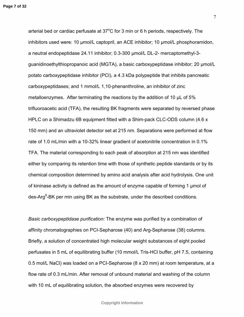
arterial bed or cardiac perfusate at 37oC for 3 min or 6 h periods, respectively. The
inhibitors used were: 10 µmol/L captopril, an ACE inhibitor; 10 µmol/L phosphoramidon,
a neutral endopeptidase 24.11 inhibitor; 0.3-300 µmol/L DL-2- mercaptomethyl-3-
guanidinoethylthiopropanoic acid (MGTA), a basic carboxypeptidase inhibitor; 20 µmol/L
potato carboxypeptidase inhibitor (PCI), a 4.3 kDa polypeptide that inhibits pancreatic
carboxypeptidases; and 1 mmol/L 1,10-phenanthroline, an inhibitor of zinc
metalloenzymes. After terminating the reactions by the addition of 10 µL of 5%
trifluoroacetic acid (TFA), the resulting BK fragments were separated by reversed phase
HPLC on a Shimadzu 6B equipment fitted with a Shim-pack CLC-ODS column (4.6 x
150 mm) and an ultraviolet detector set at 215 nm. Separations were performed at flow
rate of 1.0 mL/min with a 10-32% linear gradient of acetonitrile concentration in 0.1%
TFA. The material corresponding to each peak of absorption at 215 nm was identified
either by comparing its retention time with those of synthetic peptide standards or by its
chemical composition determined by amino acid analysis after acid hydrolysis. One unit
of kininase activity is defined as the amount of enzyme capable of forming 1 µmol of
des-Arg9-BK per min using BK as the substrate, under the described conditions.
Basic carboxypeptidase purification: The enzyme was purified by a combination of
affinity chromatographies on PCI-Sepharose (40) and Arg-Sepharose (38) columns.
Briefly, a solution of concentrated high molecular weight substances of eight pooled
perfusates in 5 mL of equilibrating buffer (10 mmol/L Tris-HCl buffer, pH 7.5, containing
0.5 mol/L NaCl) was loaded on a PCI-Sepharose (8 x 20 mm) at room temperature, at a
flow rate of 0.3 mL/min. After removal of unbound material and washing of the column
with 10 mL of equilibrating solution, the absorbed enzymes were recovered by
Page 7 of 32
Copyright Information

8
percolating a 100 mmol/L Na2CO3 solution, pH 11.4, through the column. Samples of 0.7
mL were collected throughout the experiment, whose pHs were lowered to about 7.5 by
addition of 1.0 mol/L Tris-HCl buffer, pH 7.0, as required. The fractions that had des-
Arg9-BK-forming activity using BK as substrate were pooled and applied on an Arg-
Sepharose column (8 x 100 mm) equilibrated and developed with 20 mmol/L Tris-HCl
buffer, pH 8.1, containing 1.0 mol/L NaCl, conditions under which some of the known
basic carboxypeptidases are retarded relative to other proteins (38). The active fractions
were pooled, concentrated by ultrafiltration under N2 pressure and stored at 40C until
use.
Peptide mass fingerprint: A sample of the affinity-purified basic carboxypeptidase from
the mesenteric arterial bed perfusate, containing 1.6 units of kininase activity, was
subjected to SDS-PAGE on a 12% gel under reducing conditions and stained with
amido black. The gel portion containing the single clearly stained protein band was
excised from the gel slab and treated with trypsin (32). The tryptic peptides formed were
extracted twice with 40 µL of acetonitrile /water/TFA (66:33:0.1) solution for 20 min with
the aid of a sonicator apparatus, the extract dried in a vacuum centrifuge and the
product stored at -20oC prior to use. For mass spectrometric analysis the product was
solubilized in 4 µL 0.1% TFA, followed by microscale concentration and desalting using
C18 Zip-Tips. (Millipore, Bedford, MA, USA). Peptides were eluted directly onto a
MALDI-TOF probe using 1 µL of 50% ACN in 0.1% TFA solution containing matrix (α-
CHCA 20 µg/µL). Mass spectra were determined using a Reflex IV (Bruker Daltonics,
Karlsruhe, Germany) mass spectrometer in positive reflector mode, and processed
using XMASS and Biotools software (Bruker Daltonics). Spectra were internally
Page 8 of 32
Copyright Information

9
calibrated using trypsin autolysis products (m/z 842.509 and m/z 2211.104). Protein
identification was performed using MASCOT (24) at 50 ppm mass tolerance, which
screened the fingerprint of the protein provided by the peptide mass information against
NCBI (nonredundant) and Swiss-Prot databases.
PCR amplification of reverse transcribed mRNA (RT-PCR): Total RNA was extracted
from rat mesentery, pancreas, kidney, liver, lung, heart, aorta and carotid using the
Trizol reagent, following the manufacturer instructions (Invitrogen, Carlsbad, CA, USA).
RNA integrity was confirmed by agarose gel electrophoresis. Four micrograms of total
RNA were used to perform reverse transcription of mRNAs into cDNAs using oligo-d(T)
and SuperScript II protocols (Invitrogen). cDNAs for carboxypeptidase B (CPB) and β-
actin were amplified by PCR using oligonucleotide primers (sense 5’-
gggaatccatgttgctgctactggcc-3’ and antisense 5'-ggctgcagtcaatatagatgttctcggac-3' for
CPB, and sense 5’- ctaaggcaaaccgtgaaaaga-3’ and antisense 5’-
attgccgatagtgatgacctg-3’ for β-actin). The sequences for the CPB primers were based
on the full-lenght nucleotide sequence of the rat pancreatic pre-proCPB, available
through the NCBI's GenBank CoreNucleotide database under identification 6978696
(http://www.ncbi.nlm.nih.gov/entrez/). PCR amplification was performed using Taq DNA
polymerase (Invitrogen). The process of thermal cycling consisted of initial denaturation
for 2 min at 94 ºC followed by 43 cycles of amplification of the cDNA, each comprising 1
min of denaturation at 94ºC, 1 min of annealing carried out at 60 ºC, and 1.5 min of
extension at 72ºC. Samples were incubated for additional 30 min period at 72ºC
(terminal elongation) after completion of the 43 cycles process. Similar amplification
protocol was used for β-actin, except for the annealing temperature at 45ºC. For each
Page 9 of 32
Copyright Information

10
set of primers, RT-PCR was performed on sterile water to check for contamination.
Aliquots of 10 µL of each PCR product were run on a 1% agarose gel, stained with
ethidium bromide and subjected to densitometric scanning by ImageJ software
(http://rsb.info.nih.gov/ij/); the intensity of each particular cDNA was normalized to the
respective β-actin PCR product.
Statistical analysis: The results were expressed as mean±SEM. Blood pressure values
were compared by Student t-test and the amount of fragment generated by the
perfusates was compared by one-way ANOVA followed by Newman-Keuls test. The
statistical analyses were performed using GraphPad Prism Software, and differences
were considered significant when P<0.05.
Results
Blood pressure: Mean arterial pressures in SHR were significantly higher than those in
WNR (156±3 vs. 104±2 mmHg, P<0.0001).
Degradation of BK by mesenteric arterial bed and cardiac perfusates: When BK was
incubated with mesenteric arterial bed perfusate from WNR only two fragments were
generated, corresponding to the major degradation product des-Arg9-BK (>94%) and the
fragment BK(1-7), as shown in Figure 1A. These data also indicate that, on the average,
an individual mesenteric arterial bed released into the perfusate, per minute, an amount
of kininase activity capable of hydrolyzing about 300 pmol of BK/min, under the
described in vitro assay conditions. The results shown in Figure 1B indicate that
Page 10 of 32
Copyright Information

11
perfusates of MAB from WNR and SHR are nearly identical with regard to their
proteolytic specificities and potencies toward BK. On the other hand, an individual
isolated and perfused coronary bed from a WNR, on the average, released an amount
of kininase activity into the perfusate, per min, that was about 250-fold less potent, and
of a strikingly diverse proteolytic specificity, compared with that of its MAB counterpart
as judged by the HPLC analysis of the fragments formed during the cardiac perfusate-
catalyzed BK cleavage reaction (Figure 2A). Moreover, although BK(1-5), BK(1-7) and
des-Arg9-BK were the major degradation products formed during incubation of BK with
both coronary perfusates, the generation of BK(1-5) was significantly greater in the
reaction catalyzed by the perfusate from WNR as compared with that obtained with
perfusate from SHR (Figure 2B).
Effects of protease inhibitors on the kininase activities: In order to investigate the
contribution of enzymes potentially involved in the BK cleavage by the MAB and
coronary perfusates, as suggested by the fragmentation profiles depicted in Figures 1A
and 2A, the effects of some particular enzyme inhibitors were monitored on the
corresponding reactions. Thus, the cleavage at the Phe8-Arg9 bond of BK, which
accounts for nearly all of the kininase activity in samples of MAB perfusates and
generates des-Arg9-BK, was almost fully inhibited by 30 µM MGTA (Figure 1C) when BK
was incubated with perfusates from both WNR (n=3) and SHR (n=3), under the
conditions described in Figure 1; similarly, the formation of des-Arg9-BK upon incubation
of BK with WNR perfusate was blocked by the presence of either 1 mmol/L 1,10-
phenanthroline or 20 µmol/L PCI. Taken together, the inhibitory effects of these
compounds on the formation of des-Arg9-BK indicate that a basic
Page 11 of 32
Copyright Information

12
metallocarboxypeptidase is the most conspicuous kininase of the MAB perfusate. At
least three proteases contribute to the kininase activity of the rat coronary perfusates, as
judged by the effects of captopril, MGTA and phosphoramidon on the degradation of BK
by samples of perfusates (n=5 each) from WNR and SHR (Figure 3). The formation of
des-Arg9-BK by these perfusates was inhibited by MGTA, suggesting the participation of
a basic carboxypeptidase. The generation of BK(1-7) by incubation of BK with samples
of perfusates from WNR and SHR was apparently carried out by two distinct enzymes,
assuming non-overlapping inhibitory effects of captopril and phosphoramidon on the
kininases present in the perfusates; under the conditions described in Figure 2, either of
these compounds decreased the formation of BK(1-7) by 30-60%. Captopril also
inhibited the formation of BK(1-5) catalyzed by coronary perfusate from SHR but not
from WNR, revealing a distinction between the two perfusates concerning BK
degradation.
Isolation and identification of the rat MAB perfusate basic carboxypeptidase: Affinity
chromatography over PCI-Sepharose column proved an efficient means to isolate the
BK-degrading enzyme from rat MAB perfusate (Figure 4). It should be noted that the
PCI-Sepharose resin absorbed all the basic carboxypeptidase activity loaded on the
column, wherefrom the corresponding enzyme was recovered (70% yield) in a single
peak that co-eluted with a carboxypeptidase A-like activity revealed with Cbz-Val-Phe as
the substrate (not shown). The basic carboxypeptidase could be separated from
contaminating proteins by percolating the pooled active fractions from the PCI-
Sepharose chromatography through an Arg-Sepharose column, from which the enzyme
was recovered (39% yield) in a broad peak that was retarded by the resin, in a fashion
Page 12 of 32
Copyright Information

13
reminiscent of pancreatic carboxypeptidase B (CPB) but not CPN (21). This purified
basic carboxypeptidase released solely des-Arg9-BK upon incubation with BK (Figure 5-
A), even after prolonged incubation times, and was inhibited by MGTA and PCI (Figure
5-B and C). Also, this protein migrated essentially as a single band on SDS-PAGE under
reducing conditions, from which protein fragments were prepared by tryptic digestion
and analyzed by mass spectrometry. Seven peptides of precisely determined masses
were screened against a tryptic fragment database derived from over 50,000 proteins by
MASCOT software and recognized as the following fragments of rat pancreatic CPB
(pre-proenzyme numbering; GenBank Protein database accession number P19233;
http://www.ncbi.nlm.nih.gov/entrez/): Pro162-Arg176; Glu177-Arg189; Glu190-Lys202; Ala283-
Arg289; Tyr371-Arg378; Asp379-Arg393; and Tyr405-Arg411. Thus, these results strongly
suggest that the basic carboxypeptidase isolated from the rat MAB perfusate is identical
with the pancreatic CPB. Moreover, none of these fragments were found in the rat TAFI,
also known as rat plasma CPB, sequence (GenBank Protein database accession
number NP_446069).
Tissue distribution of mRNA encoding rat CPB: The expression of CPB mRNA was
investigated in some of the rat tissues using RT-PCR (Figure 6). This procedure, using
the specific CPB oligonucleotides described in Methods, amplified DNA fragments from
total RNA extracts from the indicated tissues whose sizes matched the predicted value
of 1248 bp previously described for rat pancreas pre-proCPB (16). No PCR products
were detected when sterile water was a substitute for the respective cDNA in the
reaction (not shown). CPB mRNA was highly expressed in mesentery, pancreas, liver,
Page 13 of 32
Copyright Information

14
and kidney but its expression was below detection level in lung, heart, aorta and carotid
artery.
Discussion
Comparison between the kininase activities detected in the rat MAB and coronary
perfusates revealed two prominent differences concerning their proteolytic capacities
and specificities. Firstly, it was shown that an individual rat MAB secretes, on the
average, 250-fold more kininase activity per min than its coronary counterpart; secondly,
BK was cleaved almost exclusively at the Phe8-Arg9 bond upon incubation with MAB
perfusates from both normotensive and hypertensive rats, releasing des-Arg9-BK, while
incubation of BK with coronary vessels perfusates generated des-Arg9-BK, BK(1-7) and,
particularly, BK(1-5). Also, it was noted that SHR had a relatively low kininase activity in
their cardiac but not MAB perfusates, as compared with their WNR analogues. In spite
of these conspicuous differences in the overall kinin processing between the coronary
and MAB perfusates, one can only surmise the physiological significance of the
contributing enzymes besides that they are likely to play a role in the local tissue control
mechanisms of kinin action.
The analyses of the fragmentation profiles that resulted from treating BK with rat
MAB (Figure 1) and coronary (Figure 2) perfusates, and the corresponding alterations
effected by some selective protease inhibitors, allowed the identification of some of the
soluble kininases in each perfusate. ACE and phospohoramidon-sensitive
endopeptidase, like NEP 24.11 or endothelin converting enzyme (11, 12), are present in
small quantities in the coronary perfusates of WNR and SHR, the former having also a
so far unidentified BK(1-5)-forming enzyme whose specificity resembles that of
Page 14 of 32
Copyright Information

15
neurolysin or thimet oligopeptidase (21). The specificities of these BK-destroying
proteases present in the rat coronary perfusate are consistent with the products formed
in previously described pathways of BK degradation studied in different isolated rat heart
preparations (1,7,25); the pentapeptide BK(1-5), a major fragment formed by the action
of coronary perfusate on BK, has also been shown to be a stable end-product of BK
degradation in human plasma (19). Although we did not characterize the kininases
responsible for the release of BK(1-5), our results indicate that this activity had a major
role in BK hydrolysis in coronary perfusate from normotensive rats since ACE inhibitor
did not affect the generation of the most abundant product BK(1-5); in contrast, this
endopeptidase activity was greatly reduced in perfusates from SHR in view of the fact
that BK(1-5) released from BK was greatly reduced by ACE inhibition. In effect, a
soluble form of thimet oligopeptidase was reported by Chappell et al (4) as the
predominant protease present in hindlimb perfusates of normotensive rat responsible for
the conversion of angiotensin I to angiotensin(1-7). The significantly lower kininase
activity observed in cardiac perfusate of SHR compared with that of WNR (Figure 2)
may represent a compensatory mechanism associated with hypertension that endows
the heart with protection against ischemic damage, owing to an enhanced preservation
of the cardioprotective actions of BK (18).
Both coronary and MAB perfusates contain a MGTA-sensitive basic
metallocarboxypeptidase reminiscent of CPN, particularly for being a soluble enzyme
capable of generating the active kinin B1-receptor agonist des-Arg9-BK (17). The MAB
perfusates of both WNR and SHR are undistinguishable by their basic carboxypeptidase
contents (Figure 1B), just as the plasma of these animals with regard to CPN (5),
indicating that these BK-cleaving enzymes do not significantly contribute to the SHR
Page 15 of 32
Copyright Information

16
pathophysiology. Interestingly, the average concentration of immunoreactive des-Arg9-
BK was found to be much higher than that of BK in venous blood from normal humans
as well as from patients with essential hypertension (22), indicating the effectiveness of
human plasma basic carboxypeptidases in converting BK into des-Arg9-BK. A typical
isolated and perfused rat MAB releases into the perfusate, per minute, an amount of
basic carboxypeptidase capable of hydrolyzing about 300 pmol of BK/min, which is over
two orders of magnitude larger than that produced by an individual coronary bed; thus,
any attempt to ascribe function to this enzymatic activity must take into consideration the
large difference of its production between different tissues, which may reflect the
contribution of this basic carboxypeptidase to the overall BK metabolism in a particular
tissue. It is worth mentioning that a full dose-vasodilation response curve was obtained
with bolus injection of BK in the range of 1-160 pmol in the rat isolated MAB (33).
However, a role for basic carboxypeptidase in modulating the vasodilator effect of BK
induced by a single passage through the isolated and Krebs perfused MAB could not be
demonstrated in previous study (33), indicating that membrane bound
carboxypeptidases, like CPM, did not have an important role in processing BK in this
vascular bed.
The des-Arg9-BK-forming enzyme of the MAB perfusate was first described as a
CPN-like after two of its readily observable features, namely, its occurrence as a soluble
protease in the perfusate and its proteolytic activity toward the substrates hippuryl-Lys
and BK (23). In the present work we extended the enzymological characterization of the
des-Arg9-BK-forming enzyme of the MAB perfusate by using some inhibitors for basic
carboxypeptidases, among which MGTA and PCI. Whereas CPN is known to be
inhibited by MGTA (30) but refractory to CPI treatment (34), the basic carboxypeptidase
Page 16 of 32
Copyright Information
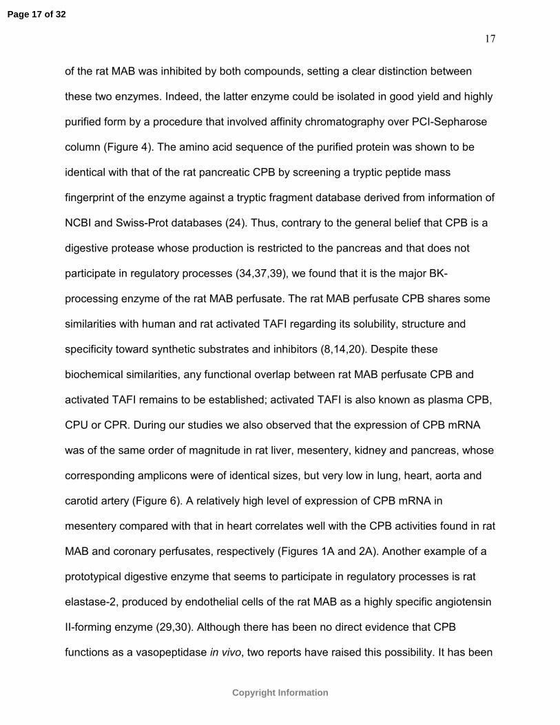
17
of the rat MAB was inhibited by both compounds, setting a clear distinction between
these two enzymes. Indeed, the latter enzyme could be isolated in good yield and highly
purified form by a procedure that involved affinity chromatography over PCI-Sepharose
column (Figure 4). The amino acid sequence of the purified protein was shown to be
identical with that of the rat pancreatic CPB by screening a tryptic peptide mass
fingerprint of the enzyme against a tryptic fragment database derived from information of
NCBI and Swiss-Prot databases (24). Thus, contrary to the general belief that CPB is a
digestive protease whose production is restricted to the pancreas and that does not
participate in regulatory processes (34,37,39), we found that it is the major BK-
processing enzyme of the rat MAB perfusate. The rat MAB perfusate CPB shares some
similarities with human and rat activated TAFI regarding its solubility, structure and
specificity toward synthetic substrates and inhibitors (8,14,20). Despite these
biochemical similarities, any functional overlap between rat MAB perfusate CPB and
activated TAFI remains to be established; activated TAFI is also known as plasma CPB,
CPU or CPR. During our studies we also observed that the expression of CPB mRNA
was of the same order of magnitude in rat liver, mesentery, kidney and pancreas, whose
corresponding amplicons were of identical sizes, but very low in lung, heart, aorta and
carotid artery (Figure 6). A relatively high level of expression of CPB mRNA in
mesentery compared with that in heart correlates well with the CPB activities found in rat
MAB and coronary perfusates, respectively (Figures 1A and 2A). Another example of a
prototypical digestive enzyme that seems to participate in regulatory processes is rat
elastase-2, produced by endothelial cells of the rat MAB as a highly specific angiotensin
II-forming enzyme (29,30). Although there has been no direct evidence that CPB
functions as a vasopeptidase in vivo, two reports have raised this possibility. It has been
Page 17 of 32
Copyright Information

18
proposed that human serum would contain either small quantities of a CPB-like enzyme
or a co-factor capable of augmenting about five times the activity of plasma CPN to
account for the actual serum des-Arg9-BK-forming activity (31); indeed, CPB is known to
remove the C-terminal Arg residue of BK far more readily than CPN (9). A second report
that suggests that CPB might also function as a regulatory enzyme is the one that
describes a significant linkage between the locus of CPB on chromosome 2 in the Lyon
hypertensive rat strain and the pulse pressure component of blood pressure regulation
(6); despite this genetic linkage, experimental evidences demonstrating the involvement
of CPB in this process remains to be established. Although the precise physiological
roles for the soluble basic carboxypeptidases described here are not certain at present,
a reasonable hypothesis is that they play a role in the processing of bioactive peptides to
meet the metabolic requirements of different tissues, as suggested by the wide
difference in the distribution of these enzymes in rat MAB and coronary perfusates.
Acknowledgements: We thank Osmar Vettore and Orlando Mesquita Jr. for their
technical assistance.
Grants: This research was supported by the Fundação de Amparo à Pesquisa do
Estado de São Paulo (FAPESP).
References
1. Blais C Jr, Drapeau G, Raymond P, Lamontagne D, Gervais N, Venneman I,
Adam A. Contribution of angiotensin-converting enzyme to the cardiac metabolism of
bradykinin: an interspecies study. Am J Physiol Heart Circ Physiol 273: H2263-
H2271, 1997.
Page 18 of 32
Copyright Information

19
2. Carretero OA, Scicli AG. Kinins paracrine hormone. Kidney Int 34(Suppl. 26): S52-
S59, 1988.
3. Carretero OA, Yang X-P, Rhaleb N-E. The kallikrein-kinin system as a regulator of
cardiovascular and renal function. In: Hypertension: a companion to Brenner and
Rector’s The Kidney, edited by Oparil S, Weber M, Philadelphia, Penn: Elsevier;
2005.
4. Chappell MC, Gomez MN, Pirro NT, Ferrario CM. Release of angiotensin-(1–7)
from the rat hindlimb: influence of angiotensin-converting enzyme inhibition.
Hypertension 35: 348-352, 2000.
5. Dendorfer A, Wolfrum S, Wagemann M, Qadri F, Dominiak P. Pathways of
bradykinin degradation in blood and plasma of normotensive and hypertensive rats
Am J Physiol Heart Circ Physiol 280: H2182-H2188, 2001.
6. Dubay C, Vincent M, Samani NJ, Hilbert P, Kaiser MA, Beressi JP, Kotelevtsev
Y, Beckmann JS, Soubrier F, Sassard J, Lathrop GM. Genetic determinants of
diastolic and pulse pressure map to different loci in Lyon hypertensive rats. Nat Genet
3: 354-357, 1993.
7. Dumoulin MJ, Adam A, Rouleau JL, Lamontagne D. Comparison of a
vasopeptidase inhibitor with neutral endopeptidase and angiotensin-converting
enzyme inhibitors on bradykinin metabolism in the rat coronary bed. J Cardiovasc
Pharmacol 37: 359-366, 2001.
8. Eaton DL, Malloy BE, Tsai SP, Henzel W, Drayna D. Isolation, molecular cloning,
and partial characterization of a novel carboxypeptidase B from human plasma. J Biol
Chem 266: 21833-21838, 1991.
Page 19 of 32
Copyright Information

20
9. Erdös EG, Yang HYT. Kininase. In: Handbook of Experimental Pharmacology,
Bradykinin, Kallidin and Kallikrein, edited by Erdös EG, New York: Springer-Verlag,
1970.
10. Erdös EG, Skidgel RA. Metabolism of bradykinin by peptidases in health and
disease. In: The Kinin System, edited by Farmer SG, San Diego, CA: Academic;
1997.
11. Graf K, Grafe M, Bossaller C, Niehus J, Schulz KD, Auch-Schwelk W, Fleck E.
Degradation of bradykinin by neutral endopeptidase (EC 3.4.24.11) in cultured human
endothelial cells. Eur J Clin Chem Clin Biochem 31: 267–272, 1993.
12. Hoang MV, Turner AJ. Novel activity of endothelin-converting enzyme: hydrolysis of
bradykinin. Biochem J 327:23–26,1997.
13. Ishida H, Scicli AG, Carretero OA. Contributions of various rat plasma peptidases
to kinin hydrolysis. J Pharmacol Exp Ther 251: 817-820, 1989.
14. Kato T, Akatsu H, Sato T, Matsuo S, Yamamoto T, Campbell W, Hotta N, Okada
N, Okada H. Molecular cloning and partial characterization of rat
procarboxypeptidase R and carboxypeptidase N. Microbiol Immunol 44: 719-728,
2000.
15. Kuoppala, A, Lindstedt KA, Saarinen J, Kovanen PT, Kokkonen JO. Inactivation
of bradykinin by angiotensin-converting enzyme and by carboxypeptidase N in human
plasma. Am J Physiol Heart Circ Physiol 278: H1069-H1074, 2000.
16. MacDonald RJ, Swift GH, Quinto C, Swain W, Pictet RL, Nikovits W, Rutter WJ.
Primary structure of two distinct rat pancreatic preproelastases determined by
sequence analysis of the complete cloned messenger ribonucleic acid sequences.
Biochemistry 21: 1453-1463, 1982.
Page 20 of 32
Copyright Information

21
17. Maceau F, Hess JF, Bachvarov DR. The B1 receptor for kinins. Pharmacol Rev 50:
357-386, 1998.
18. Madeddu P, Emanueli C, El-Dahr S. Mechanisms of disease: the tissue kallikrein-
kinin system in hypertension and vascular remodeling. Nat Clin Pract Nephrol 3: 208-
21, 2007.
19. Murphey LJ, Hachey DL, Oates JA, Morrow JD, Brown NJ. Metabolism of
bradykinin in vivo in humans: identification of BK1-5 as a stable plasma peptide
metabolite. J Pharmacol Exp Ther 294: 263-269, 2000.
20. Muto Y, Suzuki K, Sato E, Ishii H. Carboxypeptidase B inhibitors reduce tissue
factor-induced renal microthrombi in rats. Eur J Pharmacol 461: 181-189, 2003.
21. Norman MU, Reeve SB, Dive V, Smith AI, Lew RA. Endopeptidases 3 4.24.15 and
24.16 in endothelial cells: potential role in vasoactive peptide metabolism. Am J
Physiol Heart Circ Physiol 284: H1978-H1984, 2003.
22. Odya CE, Wilgis FP, Walker JF, Oparil S. Immunoreactive bradykinin and [des-
Arg9]-bradykinin in low-renin essential hypertension--before and after treatment with
enalapril (MK 421). J Lab Clin Med 102: 714-721, 1983.
23. Oliveira EB, Salgado MCO, Turner AJ. A survey of vasoactive peptide metabolizing
enzymes in the rat mesenteric arterial bed perfusate. Biochem Pharmacol 42: 1897-
1904, 1991.
24. Perkins DN, Pappin DJC, Creasy DM, Cottrell JS. Probability-based protein
identification by searching sequence databases using mass spectrometry data.
Electrophoresis 20: 3551-3567, 1999.
Page 21 of 32
Copyright Information

22
25. Raut R, Rouleau JL, Blais C Jr, Gosselin H, Molinaro G, Sirois MG, Lepage Y,
Crine P, Adam A. Bradykinin metabolism in the postinfarcted rat heart: role of ACE
and neutral endopeptidase 24.11. Am J Physiol 276: H1769-1779, 1999.
26. Ryan CA, Hass GM, Kuhn RW. Purification and properties of a carboxypeptidase
inhibitor from potatoes. J Biol Chem 249: 5495-5499, 1974.
27. Salgado MCO, Rabito SF, Carretero OA. Blood kinin in one-kidney, one clip
hypertensive rats. Hypertension 8(Suppl. I): I110-I113, 1986.
28. Salgado MCO, Caldo H, Rodrigues MCG. Effect of bradykinin on isolated
mesenteric arteries of the rat. Hypertension (suppl II): 251-254, 1992.
29. Santos CF, Oliveira EB, Salgado MCO, Greene AS. Molecular cloning and
sequencing of the cDNA for rat mesenteric arterial bed elastase-2, an angiotensin II-
forming enzyme. J Cardiovasc Pharmacol 39:628-635, 2002.
30. Santos CF, Caprio MAV, Oliveira EB, Salgado MCO, Schippers DN,
Munzenmaier DH, Greene AS. Functional role, cellular source, and tissue
distribution of rat elastase-2, an angiotensin II-forming enzyme. Am J Physiol Heart
Circ Physiol 285: H775-H783, 2003.
31. Sheikh IA, Kaplan AP. Mechanism of digestion of bradykinin and lysylbradykinin
(kallidin) in human serum. Role of carboxypeptidase, angiotensin-converting enzyme
and determination of final degradation products. Biochem Pharmacol 38: 993-1000,
1989.
32. Shevchenko A, Mortensen P, Mann M. A strategy for identifying gel-separated
proteins in sequence databases by MS alone. Biochem Soc Trans 24: 893-896, 1996.
33. Sivieri Jr DO, Bispo-da-Silva LB, Oliveira EB, Resende AC, Salgado MCO.
Potentiation of bradykinin effect by angiotensin-converting enzyme inhibition does not
Page 22 of 32
Copyright Information

23
correlate with angiotensin-converting enzyme activity in the rat mesenteric arteries.
Hypertension 50: 110-115, 2007.
34. Skidgel RA. Basic carboxypeptidases: regulators of peptide hormone activity.
Trends Pharmacol Sci 9: 299-304, 1988.
35. Skidgel RA, Davis RM, Tan F. Human carboxypeptidase M. Purification and
characterization of a membrane-bound carboxypeptidase that cleaves peptide
hormones. J Biol Chem 264: 2236-2241, 1989.
36. Skidgel RA. Bradykinin-degrading enzymes: structure, function, distribution, and
potential roles in cardiovascular pharmacology. J Cardiovasc Pharmacol 20(Suppl9):
S4-S9, 1992.
37. Song L, Fricker LD. Cloning and expression of human carboxypeptidase Z, a novel
metallocarboxypeptidase. J Biol Chem 272: 10543-10550, 1997.
38. Sukenaga Y, Akanuma H, Suekane C, Yamasaki M. Ligand bindings of bovine
carboxypeptidase B. II. Affinity chromatography and cooperative ligations. J Biochem
87: 695-707, 1980.
39. Vendrell J, Querol E, Aviles FX. Metallocarboxypeptidases and their protein
inhibitors. Structure, function and biomedical properties. Biochim Biophys Acta 1477:
284-298, 2000.
40. Zwilling R, Jakob F, Bauer H, Neurath H, Enfield DL. Crayfish carboxypeptidase:
Affinity chromatography, characterization and amino-terminal sequence. Eur J
Biochem 94: 223-229, 1979.
Page 23 of 32
Copyright Information

24
Figure Legends
Figure 1. Proteolytic cleavage of BK catalyzed by the perfusate of isolated MAB of WNR
and SHR. (A) Reversed phase HPLC analysis of BK and its fragments generated by the
incubation of 30 nmol of BK for 3 min at 37oC with a sample of perfusate containing the
amount of proteolytic activity secreted by a single isolated MAB of a WNR during about
1.2 min of perfusion. (B) Comparison of the amounts of the two major BK degradation
products released by incubating 30 nmol of BK with samples of perfusates of individual
WNR (n=8) or SHR (n=6). (C) Inhibition of des-Arg9-BK generation upon incubation of
BK with perfusates of WNR (N=3) or SHR (n=3) by MGTA, under the conditions
described above. Data are expressed as mean ±SEM.
Figure 2. Proteolytic cleavage of BK catalyzed by the perfusate of isolated coronary
beds of WNR and SHR. (A) Reversed phase HPLC analysis of BK and its fragments
generated by the incubation of 30 nmol of BK for 6 h at 37oC with a sample of perfusate
containing the amount of proteolytic activity secreted by a single isolated coronary bed
of a WNR during 2.5 min of perfusion. The peaks eluting with retention times of 3 and 9
min correspond to BK(8-9) and BK(6-9), respectively. (B) Comparison of the amounts of
the major BK degradation products released by incubating 30 nmol of BK with samples
of perfusates of individual WNR (n=8) or SHR (n=6), under the conditions described
above. Data are expressed as mean ±SEM. * P<0.001 compared with peptides in the
same group and §P<0.001 compared with WNR.
Page 24 of 32
Copyright Information

25
Figure 3. Comparison of the amounts of the major BK degradation products released by
incubating 30 nmol of BK with samples of coronary perfusates of individual WNR (n=5)
or SHR (n=5), under the conditions described in Figure 2, in the presence of captopril
(10 µmol/L), MGTA (10 µmol/L), or phosphoramidon (10 µmol/L). Data are expressed as
the percentage mean ±SEM of the control in the absence of inhibitors. *P<0.001
compared to control without inhibitors.
Figure 4. Isolation of basic carboxypeptidase from rat MAB perfusate by affinity
chromatography over PCI-Sepharose column. Sample of 5 mL containing high
molecular weight substances from pooled perfusates of eight MAB preparations was
loaded on the column (8 x 30 mm) at room temperature at a flow rate of 0.3 mL/min.
After removal of unbound material by washing the column with 10 mL of 10 mmol/L Tris-
HCl buffer, pH 7.5, containing 0.5 mol/L NaCl, bound enzymes were eluted with a 100
mmol/L Na2CO3 solution, pH 11.4. Absorbance at 280 nm and kininase activity were
determined in each fraction, and plotted against effluent volume, as indicated.
Figure 5. Kininase activity and inhibition of the affinity-purified CPB from rat MAB
perfusate. Thirty nmol of BK were individually incubated with samples of purified CPB,
equivalent to 50 µL of perfusate, in the absence (A) or presence of 30 µmol/L MGTA (B)
or 20 µmol/L PCI (C) for 40 min at 37oC in 150 µL of Tris-buffered saline, pH 7.4. BK
and its fragments were determined by reversed phase HPLC analyses on a Shim-Pack
CLC-ODS column (4.6 x 150 mm) using a linear gradient of acetonitrile concentration
Page 25 of 32
Copyright Information

26
(10-32%) in 0.1% TFA at a flow rate of 1 mL/min. A small peak eluting with retention
time of 6-7 min (B) is also seen in a control run of MGTA alone.
Figure 6. Detection of mRNAs for rat CPB in different tissues using RT-PCR. Each
lane of the ethidium bromide-stained agarose gels shows the RT-PCR products derived
from total RNA for the indicated rat tissues using specific primers for CPB (1248 bp) and
β-actin (351 bp), the latter a control for matching the RT-PCR processes for the different
total mRNA preparations (top panel). The expression level of mRNAs for CPB in the
various tissues was estimated by densitometric scanning of the gels, normalized to the
corresponding β-actin product, and expressed as CPA/ β-actin ratio in the column chart
(bottom panel). Tissues are indicated as Ki (kidney), Pa (pancreas), Li (liver), Lu (lung),
Me (mesentery), He (heart), Ao (aorta) and Ca (carotid artery).
Page 26 of 32
Copyright Information

BK
BK(1-7)
des-Arg9-BK
0.4
0.2
0
Abs
orba
nce
215
nm 0.6
Retention time (min)250 20105 15
0.8 A
Injection
BK
BK(1-7)
des-Arg9-BK
0.4
0.2
0
Abs
orba
nce
215
nm 0.6
Retention time (min)250 20105 15
0.8 A
Injection
-7 -6 -5 -4 -30
20
40
60
80
100
MGTA (mol/L)
Inhi
bitio
n(%
)
C
NWRSHR
0
3
6
9
12
des-Arg9-BK BK(1-7)
nmol
ofpr
oduc
t
B NWRSHR
-7 -6 -5 -4 -30
20
40
60
80
100
MGTA (mol/L)
Inhi
bitio
n(%
)
C
NWRSHR
-7 -6 -5 -4 -30
20
40
60
80
100
MGTA (mol/L)
Inhi
bitio
n(%
)
C
NWRSHR
0
3
6
9
12
des-Arg9-BK BK(1-7)
nmol
ofpr
oduc
t
B NWRSHR
0
3
6
9
12
des-Arg9-BK BK(1-7)
nmol
ofpr
oduc
t
B NWRSHR
Figure 1
Page 27 of 32
Copyright Information

Figure 2
des-Arg9-BK
BK
BK(1-7)BK(1-5)
0.4
0.2
0
Ab
sorb
ance
215
nm
0.6
Retention time (min)250 20105 15
A
Injection
BK(8-9)BK(6-9)
des-Arg9-BK
BK
BK(1-7)BK(1-5)
0.4
0.2
0
Ab
sorb
ance
215
nm
0.6
Retention time (min)250 20105 15
A
Injection
BK(8-9)BK(6-9)
0
3
6
9
12
15
18
des-Arg9-BK BK(1-7) BK(1-5)
§
*
*
* *
nmol
of p
rodu
ct
B NWRSHR
0
3
6
9
12
15
18
des-Arg9-BK BK(1-7) BK(1-5)
§
*
*
* *
nmol
of p
rodu
ct
B NWRSHR
Page 28 of 32
Copyright Information

Figure 3
0
25
50
75
100
125 NW RSHR
**
Des-A
rg9 -B
K
0
25
50
75
100
125
**
* *
BK
(1-7
)
0
25
50
75
100
125
*
BK
(1-5
)
Captopril MGTA Phosphoramidon
0
25
50
75
100
125 NW RSHR
**
Des-A
rg9 -B
K
0
25
50
75
100
125
**
* *
BK
(1-7
)
0
25
50
75
100
125
*
BK
(1-5
)
Captopril MGTA Phosphoramidon
Page 29 of 32
Copyright Information

Figure 4
0 7 14 21
0
0.5
1.0
1.5
2.0
2.5
Ab
sorb
ance
280
nm
0
0.5
1.0
1.5
2.0
Effluent Volume (mL)
En
zym
e U
nit
s
Page 30 of 32
Copyright Information

Figure 5
Ab
sorb
ance
215
nm
0
0.4
0.8
1.2
1.6B
Retention time (min)
0
0.4
0.8
1.2
1.6
0 5 10 15 20
C
des-Arg9-BK
BK0
0.4
0.8
1.2
1.6 A InjectionA
bso
rban
ce 2
15 n
m
0
0.4
0.8
1.2
1.6B
Ab
sorb
ance
215
nm
0
0.4
0.8
1.2
1.6B
Retention time (min)
0
0.4
0.8
1.2
1.6
0 5 10 15 20
C
Retention time (min)
0
0.4
0.8
1.2
1.6
0 5 10 15 20
C
des-Arg9-BK
BK0
0.4
0.8
1.2
1.6 A Injection
des-Arg9-BK
BK0
0.4
0.8
1.2
1.6 A Injection
Page 31 of 32
Copyright Information

Figure 6
CPB
β-actin
Ki Pa Li Lu Me He Ao Ca
Ki Pa Li Lu Me He Ao Ca0.0
0.5
1.0
1.5
2.0
2.5
CPB
/ β-a
ctin
Rat
io
CPB
β-actin
Ki Pa Li Lu Me He Ao Ca
Ki Pa Li Lu Me He Ao Ca0.0
0.5
1.0
1.5
2.0
2.5
CPB
/ β-a
ctin
Rat
io
Page 32 of 32
Copyright Information