Traumatic rupture of the thoracic aorta: Ten years of delayed management
Upload
independentCategory
view
1download
0
RESEARCH PAPER
The vascular effects of different arginase inhibitorsin rat isolated aorta and mesenteric arteries
NN Huynh, EE Harris, JFP Chin-Dusting and KL Andrews
Vascular Pharmacology Laboratory, Baker IDI Heart and Diabetes Institute, Melbourne, Vic., Australia
Background and purpose: Arginase and nitric oxide (NO) synthase share the common substrate L-arginine, and arginaseinhibition is proposed to increase NO production by increasing intracellular levels of L-arginine. Many different inhibitors areused, and here we have examined the effects of these inhibitors on vascular tissue.Experimental approach: Each arginase inhibitor was assessed by its effects on isolated rings of aorta and mesenteric arteriesfrom rats by: (i) their ability to preserve the tolerance to repeated applications of the endothelium-dependent agonistacetylcholine (ACh); and (ii) their direct vasorelaxant effect.Key results: In both vessel types, tolerance (defined as a reduced response upon second application) to ACh was reversed withaddition of L-arginine, (S)-(2-boronethyl)-L-cysteine HCl (BEC) or NG-Hydroxy-L-arginine (L-NOHA). On the other hand,Nw-hydroxy-nor-L-arginine (nor-NOHA) significantly augmented the response to ACh, an effect that was partially reversed withL-arginine. No effect on tolerance to ACh was observed with L-valine, nor-valine or D,L, a-difluoromethylornithine (DFMO).BEC, L-NOHA and nor-NOHA elicited endothelium-independent vasorelaxation in both endothelium intact and denuded aortawhile L-valine, DFMO and nor-valine did not.Conclusions and implications: BEC and L-NOHA, but not nor-NOHA, L-valine, DFMO or nor-valine, significantly reversedtolerance to ACh possibly conserving L-arginine levels and therefore increasing NO bioavailability. However, both BEC andL-NOHA caused endothelium-independent vasorelaxation in rat aorta, suggesting that these inhibitors have a role beyondarginase inhibition alone. Our data thus questions the interpretation of many studies using these antagonists as specificarginase inhibitors in the vasculature, without verification with other methods.British Journal of Pharmacology (2009) 156, 84–93; doi:10.1111/j.1476-5381.2008.00036.x
Keywords: arginase; L-arginine; aorta; mesenteric arteries; nitric oxide; vasorelaxation
Abbreviations: BEC, (S)-(2-boronethyl)-L-cysteine HCl; DFMO, D,L, a-difluoromethylornithine; L-NAME, NG-nitro-L-arginine-methyl ester; L-NOHA, NG-hydroxy-L-arginine; nor-NOHA, Nw-hydroxy-nor-L-arginine; ODQ, 1H-[1,2,4]-oxadiazolol[4,3-1]quinoxaline-1-one
Introduction
Nitric oxide (NO) bioavailability is compromised in manycardiovascular disease states. As L-arginine is rate limiting inNO production, this amino acid precursor has been postu-lated as a factor contributing to NO availability. Also, becausethe enzyme arginase catalyses the catabolism of L-arginine toform ornithine and urea, many argue that arginase can bemanipulated to influence NO bioavailability. In this context,increased expression and/or activity of arginase has beendemonstrated in many vascular pathologies such as pulmo-nary hypertension (associated with sickle cell disease) (Morriset al., 2003), primary pulmonary arterial hypertension (Xu
et al., 2004), ischaemia-reperfusion (Hein et al., 2003),uraemia (Thuraisingham et al., 2002) as well as in animalmodels of arterial hypertension (Johnson et al., 2005), aging(Berkowitz et al., 2003; Santhanam et al., 2007), sexual arousal(Berkowitz et al., 2003; Cama et al., 2003a), diabetes (Romeroet al., 2008) and atherosclerosis (Ming et al., 2004; Ryoo et al.,2006; 2008). Furthermore, inhibition of arginase has beenshown to stimulate NO production (Chicoine et al., 2004),while over-expression of arginase I or II decreases intracellularL-arginine concentrations and suppresses NO synthesis(Li et al., 2001). However, because NG-hydroxy-L-arginine(L-NOHA), a reaction intermediate of NO synthase (NOS), isalso a potent intracellular inhibitor of arginase (Boucher et al.,1994; Daghigh et al., 1994) some of the effects observed maynot confined to substrate competition alone.
Both vascular endothelial and smooth muscle cells expressarginase I and II, but their distribution appears to be vessel-and species-dependent (Buga et al., 1996; Zhang et al., 2001;
Correspondence: Dr Jaye Chin-Dusting, Baker IDI Heart and Diabetes Institute,PO Box 6492 St Kilda Rd Central, Melbourne, Vic. 8008, Australia. E-mail:[email protected] 29 July 2008; revised 27 August 2008; accepted 10 September 2008
British Journal of Pharmacology (2009), 156, 84–93© 2008 The AuthorsJournal compilation © 2008 The British Pharmacological Society All rights reserved 0007-1188/08www.brjpharmacol.org

Bachetti et al., 2004; Ming et al., 2004). In addition to theproduction of urea, arginase is also involved in the biosyn-thesis of polyamines and the amino acids, ornithine, prolineand glutamate (Cederbaum et al., 2004). As such it is notsurprising that amino acids such as ornithine, leucine, valine,lysine, isoleucine and nor-valine inhibit arginase (Hunter andDowns, 1945). Among them, ornithine is the most potentof the competitive amino acids, with nor-valine, a non-competitive inhibitor, demonstrating similar potency (Hunterand Downs, 1945). Another commonly used inhibitor is theindirectly acting, irreversible inhibitor of ornithine decar-boxylase, D,L, a-difluoromethylornithine (DFMO), whichincreases ornithine levels endogenously (Selamnia et al.,1998). Despite the relatively high concentrations required toinhibit arginase, the use of nor-valine, L-valine and DFMOhave recently been reported in the context of studying NOfunction (Ming et al., 2004; Santhanam et al., 2007; Lewiset al., 2008).
As L-NOHA also acts as a substrate for NO production, itsutility as a specific arginase inhibitor is limited, although it isstill used as a specific arginase inhibitor (Sakai et al., 2004;Holan et al., 2006). Due to the complications with the use ofL-NOHA, Nw-hydroxy-nor-L-arginine (nor-NOHA) was syn-thesized and reported to be more potent than L-NOHA, butnot a substrate for NO (Tenu et al., 1999). Aside from theseinhibitors, several boron-based inhibitors have beendesigned. Among these are (S)-(2-boronethyl)-L-cysteine HCl(BEC) and 2(S)-amino-6-boronohexanoic acid (ABH) (Colle-luori and Ash, 2001), both of which have been shown toeffectively inhibit arginase (Khangulov et al., 1995).
Due to the potential for manipulating intracellular L-arginine stores, and possibly increasing NO bioavailability indisease states, the interest in this field of research has esca-lated, resulting in the recent use of many of these inhibitors(Santhanam et al., 2007; Bagnost et al., 2008; Lewis et al.,2008). To address which of the arginase inhibitors is mostspecific and appropriate for studying functional NO effects inthe vasculature, a comparative assessment of these arginaseinhibitors was performed.
Methods
AnimalsAll animal procedures and the study protocol was approvedby the Alfred Medical Research and Education Precinct(AMREP) Animal Ethics Committee (applications E/0323/2003/B, E/0238/2004/2004B and E/0352/2004/B) that adheresto the National Health and Medical Research Council(NHMRC) of Australia Code of Practice for the Care and Use ofAnimals for Scientific Purposes. Male Sprague-Dawley ratswere housed under standard laboratory conditions withaccess to food and water ad libitum. Animals were killed bydecapitation and exsanguination following overexposure to80% CO2 and 20% O2.
Vascular reactivityThe thoracic aorta and mesenteric arteries were excised andplaced into ice-cold modified Krebs solution (composition
in mmol·L-1: NaCl 119, KCl 4.7, MgSO4·7H20 1.17, NaHCO3
25, KH2PO4 1.18, CaCl2 2.5, glucose 11 and EDTA 0.03). Theadipose and connective tissue were removed. Rat aorta wassectioned into eight rings of 3 mm length and mesentericarteries into eight rings of 2 mm length with the aid of adissecting microscope (Olympus, Tokyo). In some of thevessels, endothelium denudation of thoracic aortic rings wasperformed by gently rubbing the lumen of the aorta againsta wire. For mesenteric arteries, this procedure was achievedby pulling a strand of human hair backwards and forwardsthrough the lumen of the vessel.
Aortic rings and mesenteric arteries were mounted inorgan baths and on a wire-myograph as previously described(Lewis et al., 1997; Kimura et al., 2002). Once the vessels weremounted and incubated for an equilibration period of 30 min,all vessels were subjected to an oxygenated and pre-warmed(37°C) high potassium physiological salt solution (KPSS inmmol·L-1; KCl 123, MgSO4·7H20 1.17, NaHCO3 25, KH2PO4
1.18, CaCl2 2.5, glucose 6.05 and EDTA 0.03) until a plateaucontractile response was observed. The vessel was rinsed threetimes with oxygenated and pre-warmed (37°C) modified Krebs’solution. Endothelium integrity or successful denudation wasconfirmed by pre-constricting the vessel with a constrictoragonist followed by addition of acetylcholine (ACh)(1 mmol·L-1). Rat aorta was pre-constricted with noradrenaline(10 nmol·L-1), while for mesenteric arteries, the modified Krebssolution was replaced with 40 mmol·L-1 potassium salt solu-tion (40 mmol·L-1 KCl, composition in mmol·L-1; NaCl 84, KCl40, MgSO4·7H20 1.17, NaHCO3 25, KH2PO4 1.18, CaCl2 2.5,glucose 11 and EDTA 0.03). Potassium was chosen as theconstrictor for mesenteric arteries since at the concentrationused, any effect of endothelial-derived hyperpolarizing factor(EDHF) would be negated, unmasking the contribution of NOto vasodilatation (Chen and Suzuki, 1990). Where applicable,for intact (i.e. non-denuded) vessels, a relaxation response of>80% in rat aorta and >60% in mesenteric arteries was set as theinclusion criteria upon addition of 1 mmol·L-1 ACh. Vesselswere deemed denuded when relaxation was less than 10% ineither aorta or mesenteric arteries.
Experimental designEffect of L-arginine supplementation and arginase inhibitors on NOfunctionCumulative full concentration–response curves in half logincrements to the endothelium- and NO-dependent dilator,ACh, (1 nmol·L-1–10 mmol·L-1), were obtained and repeated30 min apart. Between each curve, vessels were washed, and atotal of three concentration–response curves to ACh were ob-tained successively, 30 min apart, in the absence of any treat-ment and used as the comparative time control experiment.
In separate baths and myograph chambers, vessels wereincubated with L-arginine (1 mmol·L-1 or 10 mmol·L-1) orthe arginase inhibitors: BEC (100 mmol·L-1), nor-NOHA(10 mmol·L-1), L-NOHA (10 mmol·L-1), DFMO (10 mmol·L-1),L-valine (10 mmol·L-1) or nor-valine (10 mmol·L-1) for 30 minbefore and during the repeat concentration–response curve toACh. Therefore, matched controls were obtained for eachcompound, such that (-) and (+) denotes the AChconcentration–response curve performed before and after
Arginase inhibitors in the vasculatureNN Huynh et al 85
British Journal of Pharmacology (2009) 156 84–93
addition of L-arginine or the arginase inhibitor as indicated.The concentration of the arginase inhibitor used during theincubation period was determined from pilot studies. Vasore-laxant responses in rat aorta were performed on vessels pre-constricted with noradrenaline and in mesenteric arteries with40 mmol·L-1 KCl.
The role of the endothelium on the direct vasodilatory effect ofarginase inhibitorsFull cumulative concentration–response curves in half logincrements to the arginase inhibitors: BEC (aorta and mesen-teric arteries: 0.1 mmol·L-1–3 mmol·L-1), nor-NOHA (aorta andmesenteric arteries: 0.1 mmol·L-1–3 mmol·L-1), L-NOHA (aorta:1 mmol·L-1–1 mmol·L-1, mesenteric arteries: 0.1 mmol·L-1–3 mmol·L-1), DFMO, L-valine or nor-valine (aorta: 1 mmol·L-1–3 mmol·L-1, mesenteric arteries: 10 mmol·L-1–3 mmol·L-1) wereobtained in endothelium intact and denuded vessels. Experi-ments were performed in rat aortic rings pre-constricted withnoradrenaline and in mesenteric arteries pre-constricted with40 mmol·L-1 KCl. Concentration–response curves were alsoobtained to L-NOHA, nor-NOHA and BEC in the presence ofthe soluble guanlylyl cyclase (sGC) inhibitor, 1H-[1,2,4]-oxadiazolol[4,3-1]quinoxaline-1-one (ODQ, 10 mmol·L-1) orthe NOS inhibitor, NG-nitro-L-arginine-methyl ester (L-NAME,100 mmol·L-1).
Data analysis and statisticsAll vasorelaxation responses were expressed as per-centage relaxation from the pre-constriction response tonoradrenaline in aortic rings, or to 40 mmol·L-1 KCl in mesen-teric arteries. Rmax depicts the maximum relaxation responseobtained. Variable slope sigmoidal concentration–responsecurves to each agonist were fitted and graphed, and thepotency [-log EC50 (M) i.e. the concentration giving 50% ofthe maximum response] calculated for individual curves byusing GraphPad Prism (V. 4.01, USA). Results were analysedby Student’s t-test (paired or unpaired, as appropriate). Wherethree curves were compared, comparisons were made by usinga one-way ANOVA. Statistical analysis was performed by usingGraphPad Prism where P < 0.05 was considered statisticallysignificant. All data are presented as mean � SEM.
Drugs and reagentsArginase inhibitors, L-NOHA (NG-hydroxy-L-arginine mono-acetate salt), nor-NOHA (Nw-hydroxy-nor-L-arginine diacetatesalt) and BEC, were purchased from Calbiochem, USA. DFMO,L-NAME, L-valine, nor-valine, ODQ, noradrenaline bitartrate,ACh chloride, sodium nitroprusside dihydrate and L-argininewere purchased from Sigma-Aldrich, St. Louis, MO, USA. Drugstock solutions were made in milliQ water and stored at-20°C. All drugs were diluted in modified Krebs solution, withthe exception of ODQ that was made up in 100% ethanol onthe day of experiment and stored on ice until ready for use.Salts used in modified KPSS, modified Krebs solution and40 mmol·L-1 KCl were all purchased from Merck P/L, Kilsyth,Victoria, Australia. Drug/molecular target nomenclature con-forms to the British Journal of Pharmacology Guide to Recep-tors and Channels (Alexander et al., 2008).
Results
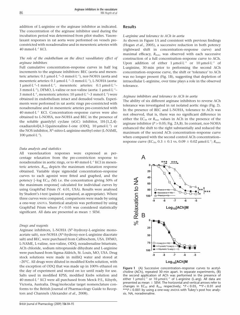
L-arginine and tolerance to ACh in aortaAs shown in Figure 1A and consistent with previous findings(Hogan et al., 2005), a successive reduction in both potency(rightward shift in concentration–response curve) andmaximal efficacy, Rmax, was observed with each successiveconstruction of a full concentration–response curve to ACh.Upon addition of either 1 mmol·L-1 or 10 mmol·L-1 ofL-arginine, 30 min prior to performing the second AChconcentration–response curve, the shift or ‘tolerance’ to AChwas no longer present (Fig. 1B), suggesting that depletion ofintracellular L-arginine, over time plays a role in the observedtolerance.
Arginase inhibitors and tolerance to ACh in aortaThe ability of six different arginase inhibitors to reverse AChtolerance was investigated in rat isolated aortic rings (Fig. 2).In the presence of BEC and L-NOHA, tolerance to ACh wasnot observed, that is, there was no significant difference ineither the EC50 or Rmax values in ACh in the presence of thearginase inhibitor (P > 0.05; Fig. 2A,B). In contrast, nor-NOHAenhanced the shift to the right substantially and reduced themaximum of the second ACh concentration–response curvewhen compared with the second control ACh concentration–response curve (EC50, 0.3 � 0.1 vs. 0.09 � 0.02 mmol·L-1; Rmax,
***
******
-9 -8 -7 -6 -5
0
20
40
60
80
100
First (n = 19)
Second (n = 19)
Third (n = 5)***
Log10[ACh]M
-9 -8 -7 -6 -5
0
25
50
75
100
- L-arg (n = 16)
+ 1 μmol·L–1 L-arg (n = 8)
+ 10μmol·L–1 L-arg (n = 8)
Log10[ACh]M
A
B
% R
ela
xatio
n to N
A
Figure 1 (A) Successive concentration–response curves to acetyl-choline (ACh), repeated 30 min apart. In separate experiments, (B)the second application of ACh was performed in the presence ofeither 1 mmol·L-1 or 10 mmol·L-1 of L-arginine (L-arg). All data arepresented as mean � SEM. The horizontal and vertical arrows refer tochanges in EC50 and Rmax respectively; *P < 0.05, **P < 0.01 and***P < 0.001 by using a one-way ANOVA with Tukey’s post hoc analy-sis. NA, noradrenaline.
Arginase inhibitors in the vasculature86 NN Huynh et al
British Journal of Pharmacology (2009) 156 84–93

34 � 9 vs. 85 � 2%; n = 15–19; P < 0.05; Fig. 2C), an effectthat was partially restored by L-arginine. DFMO and theequipotent (to DFMO) competitive and non-competitivearginase inhibitors, L-valine and nor-valine had no signi-ficant effect on the EC50 of the ACh-induced tolerance,albeit in the presence of L-valine and nor-valine there was nolonger a significant difference in the maximal response(Fig. 2D–F).
Arginase inhibitors as vasodilators in aortaSince arginase expression has been reported in both endothe-lial and vascular smooth muscle cells (Berkowitz et al., 2003;Buchwalow et al., 2004; Johnson et al., 2005), the effects of thearginase inhibitors were examined in both endothelium-intact
and denuded vessels. As shown in Figure 3A, BEC elicitedconcentration-dependent vasorelaxation which was non-endothelium-dependent (EC50 and Rmax, intact vs. denudedrings: 13 � 4 mmol·L-1 and 80 � 6% vs. 62 � 26 mmol·L-1
and 67 � 8%, n = 11–15; P > 0.05). Similarly, L-NOHA andnor-NOHA induced vasorelaxation in both intact anddenuded vessels with comparable potencies (Fig. 3B,C). Inendothelium-denuded aorta, vasorelaxation to BEC, L-NOHAand nor-NOHA, was significantly attenuated in the presence ofthe sGC inhibitor, ODQ (10 mmol·L-1) suggesting a cGMP-dependent mechanism. Responses to L-NOHA were attenuatedby the NOS inhibitor L-NAME (100 mmol·L-1) in both intactand denuded aorta (P < 0.05) while those to BEC were unaf-fected. DFMO, L-valine and nor-valine did not induce signifi-cant vasorelaxation (see Fig. 3D–F) when compared with their
-9 -8 -7 -6 -5
0
25
50
75
100- DFMO (n = 8)
+ DFMO (n = 8) *
*
Log10[ACh]M
-9 -8 -7 -6 -5
0
25
50
75
100- BEC (n = 10)
+ BEC (n = 10)
Log10[ACh]M
-9 -8 -7 -6 -5
0
25
50
75
100
- nor -NOHA (n = 15)
+ nor - NOHA (n = 15)
+ nor-NOHA + L-arg (n = 5)
*
***
Log10[ACh]M
-9 -8 -7 -6 -5
0
25
50
75
100- L-NOHA (n = 10)
+ L-NOHA (n = 10)
Log10[ACh]M
-9 -8 -7 -6 -5
0
25
50
75
100- nor-valine (n = 3)
+ nor-valine (n = 3)
*
Log10[ACh]M-9 -8 -7 -6 -5
0
25
50
75
100- L-valine (n = 10)
+ L-valine (n = 10)
**
Log10[ACh]M
B
C D
E F
A
% R
ela
xatio
n to N
A
Figure 2 Concentration–response curves to ACh were repeated 30 min after the addition of either (A) 100 mmol·L-1 BEC, (B) 10 mmol·L-1
L-NOHA, (C) 10 mmol·L-1 nor-NOHA, (D) 10 mmol·L-1 DFMO, (E) 10 mmol·L-1 L-valine or (F) 10 mmol·L-1 nor-valine. All data are presented asmean � SEM where *P < 0.05 and **P < 0.01 by using a paired Student’s t-test comparison of the EC50 (horizontal arrows) or Rmax (verticalarrows) before and after the addition of an arginase inhibitor. ACh, acetylcholine; BEC, (S)-(2-boronethyl)-L-cysteine HCl; DFMO, D,L,a-difluoromethylornithine; L-NOHA, NG-hydroxy-L-arginine; NA, noradrenaline; nor-NOHA, Nw-hydroxy-nor-arginine.
Arginase inhibitors in the vasculatureNN Huynh et al 87
British Journal of Pharmacology (2009) 156 84–93

time controls (data not shown), which coincided with theirreduced ability to reverse tolerance to ACh.
L-arginine and tolerance to ACh in mesenteric arteriesWhile NO is thought to play a significant role in the vasodila-tory profile in conduit vessels, it has been frequently reportedto play only a minor role in resistance vessels such as mesen-teric arteries, where a larger contribution by other vasodila-tors such as EDHF is reported (Wu et al., 1993; Shimokawaet al., 1996; Chataigneau et al., 1999). Therefore, to negate theeffects of EDHF, which dilates via K+ channels, all experimentswere performed by using a high K+ solution (40 mmol·L-1) asthe vasoconstrictor. Hence, under these conditions, ACh-induced relaxation is NOS-dependent and is abolished in the
presence of L-NAME (data not shown). When similar experi-ments to those described above, but this time in mesentericarteries were performed, tolerance to ACh was also observed(EC50 values, first vs. second control concentration–responsecurves, 0.1 � 0.01 vs. 0.4 � 0.2 mmol·L-1; n = 13; P < 0.05;Fig. 4A) but without an effect on the maximal response toACh (P > 0.05). As observed in aortic rings, supplementationwith either 1 mmol·L-1 or 10 mmol·L-1 L-arginine abolished therightward shift in the concentration–response curve to ACh(P > 0.05; Fig. 4B).
Arginase inhibitors and tachyphylaxis to ACh inmesenteric arteriesAs we had observed in aortic rings, there was no tolerance toACh in rings of mesenteric arteries in the presence of BEC or
-6 -5 -4 -3
0
25
50
75
100Intact (n = 4)
Denuded (n = 3)
Log10[DFMO]M
-7 -6 -5 -4 -3
0
25
50
75
100
Denuded (n = 18)
+ ODQ ( n = 14)
***Intact (n = 11)
Intact + L-NAME (n = 6)Denuded + L-NAME (n =5)
Log10[BEC]M
-7 -6 -5 -4 -3
0
25
50
75
100
Intact (n = 10)
Denuded (n = 23)
+ ODQ (n = 6)
***
Log10[nor-NOHA]M
-6 -5 -4 -3
0
25
50
75
100
Intact (n = 17)
Denuded (n = 14)
+ ODQ (n = 10)
***
Intact + L-NAME (n = 9)
Denuded + L-NAME (n = 6)
*
Log10[L-NOHA]M
-6 -5 -4 -3
0
25
50
75
100Intact (n = 5)
Denuded (n = 5)
Log10[nor-valine]M-6 -5 -4 -3
0
25
50
75
100Intact (n = 4)
Denuded (n = 3)
Log10[L-valine]M
B
C D
E F
A
% R
ela
xatio
n to N
A
Figure 3 Concentration–response curves to the arginase inhibitors: (A) BEC, (B) L-NOHA, (C) nor-NOHA, (D) DFMO, (E) L-valine and (F)nor-valine were performed in endothelium-intact and denuded aortic rings pre-constricted with NA. Responses to L-NOHA, nor-NOHA andBEC were also performed in the presence of the cGMP inhibitor, ODQ (10 mmol·L-1) in endothelium-denuded vessels or the NOS inhibitorL-NAME (100 mmol·L-1) in endothelium-intact and denuded vessels. All responses are expressed as % relaxation to NA and as mean � SEMwhere *P < 0.05 (-nor-NOHA vs. +nor-NOHA) and ***P < 0.001 by using an unpaired Student’s t-test. BEC, (S)-(2-boronethyl)-L-cysteine HCl;DFMO, D,L, a-difluoromethylornithine; L-NAME, NG-nitro-L-arginine-methyl ester; L-NOHA, NG-hydroxy-L-arginine; NA, noradrenaline; nor-NOHA, Nw-hydroxy-nor-arginine; ODQ, 1H-[1,2,4]-oxadiazolol[4,3-1]quinoxaline-1-one.
Arginase inhibitors in the vasculature88 NN Huynh et al
British Journal of Pharmacology (2009) 156 84–93

L-NOHA (Fig. 5A,B). Again, nor-NOHA significantly enhancedthe tolerance to ACh, a finding, which again was partiallyreversed with addition of 100 mmol·L-1 of L-arginine (Fig. 5C).DFMO, L-valine and nor-valine, likewise, did not reducetolerance to Ach (Fig. 5D–F).
The endothelium and arginase inhibitors in mesenteric arteriesIn contrast to the finding in aortic rings, BEC did not inducesignificant vasorelaxation in mesenteric artery rings (Fig. 6A).In mesenteric arteries, L-NOHA and nor-NOHA inducedconcentration-dependent vasorelaxation, similar to thoseobserved in the aorta, and both were abolished in the pres-ence of the sGC inhibitor, ODQ (see Fig. 6B,C). There were novasorelaxant responses to L-valine, DFMO or nor-valine inintact or denuded mesenteric arteries (P > 0.05; two-wayANOVA, when compared with time control; Fig. 6D–F).
Discussion
Several studies have reported that arginase inhibition restoresendothelial function in various animal models of diseaseincluding hypertension (Rodriguez et al., 2004; Zhang et al.,2004; Demougeot et al., 2005; Johnson et al., 2005; Bagnost
et al., 2008), diabetes (Romero et al., 2008), atherosclerosis(Ming et al., 2004; Ryoo et al., 2008) and aging (Berkowitzet al., 2003; Santhanam et al., 2007). This data are consistentwith the hypothesis that arginase competes with NOS for thecatabolism of L-arginine and that the inhibition of arginaseallows increased production of NO. Many of these papers usethe commonly available arginase inhibitors with the assump-tion of arginase and endothelial specificity. The current paper,however, shows that these arginase inhibitors each have dif-fering effects in the vasculature and that some caution shouldbe exercised with their use and in the subsequent interpreta-tion of the data obtained particularly in diseased models.
An in vitro method of inducing decreased NO bioavailabilityby repeated applications of ACh so that a significant shift tothe right in the concentration–response curve to ACh indi-cated decreased potency and a reduction in the response to10 mmol·L-1 ACh indicated decreased maximal effect, in therat aorta. In the mesenteric arteries, a modest decrease inpotency was observed without changes to the maximal effect.ACh is a well-known endothelium-dependent dilator inconduit vessels, where it releases NO via a NOS-dependentmechanism (Furchgott and Zawadzki, 1980). However, inresistance vessels, such as the mesenteric arteries utilized inthis study, the majority of the relaxation to ACh is mediatedvia EDHF, rather than NO, as we and others (Hogan et al.,2005) have shown relaxation is only partially reduced by NOSinhibitors. In both the current study and in bovine intrapul-monary arterial vessels (Gold et al., 1989), reduced responsesto ACh suggest that depletion of intracellular L-arginine overtime plays a role in tolerance, as L-arginine supplementationprevented this loss of response. To examine functionallywhether arginase inhibition increased intracellular L-arginineand could therefore prevent tolerance to ACh, several argin-ase inhibitors were examined by using a protocol, similar tothat used for L-arginine in both the aorta and the mesentericarteries. Interestingly, only two of the arginase inhibitorsexamined, BEC and L-NOHA, effectively prevented ACh tol-erance in aortic and mesenteric artery preparations. This sug-gests that indeed some but not all arginase inhibitors canconserve intracellular L-arginine stores to the level required toallow the preservation of ACh responses, mediated by NO.
Based on the published inhibitory constants of the six com-mercially available arginase inhibitors utilized in this study, itwas anticipated that BEC would be the most potent, followedby nor-NOHA, L-NOHA and collectively least potent: DFMO,L-valine and nor-valine (Hunter and Downs, 1945; Daghighet al., 1994; Custot et al., 1997; Cama et al., 2003b). WhileBEC and L-NOHA were able to prevent tolerance to ACh,DFMO and the valine amino acids failed to inhibit the ACh-induced shift to the right in the concentration–responsecurves or reductions in the maximum response. These find-ings fit with the notion that these compounds are compara-tively poorer inhibitors of arginase and as such are notas effective in the vasculature, in relation to improvingL-arginine levels to a level where a functional advantage isobserved. It has been reported that the synthetic derivative ofthe intermediate L-NOHA, nor-NOHA, is not a substrate for orinhibitor of NOS unlike L-NOHA itself (Daghigh et al., 1994)and as such it has been proposed to be a more specific inhibi-tor of arginase than L-NOHA (Tenu et al., 1999). However, in
-9 -8 -7 -6 -5
0
25
50
75First (n = 13)
Second (n = 13)
**
Log10[ACh]M
-9 -8 -7 -6 -5
0
25
50
75
- L-arg (n = 15)
+ 1 μmol·L–1 L-arg (n = 8)
+ 10 µmol·L–1 L-arg (n = 7)
Log10[ACh]M
A
B
% R
ela
xatio
n to 4
0 m
mol·L
–1 K
Cl
Figure 4 (A) Concentration–response curves to acetylcholine (ACh)were repeated 30 min apart in mesenteric artery rings pre-constrictedwith 40 mmol·L-1 KCl. The second application of ACh was also per-formed (B) in the presence of either 1 mmol·L-1 or 10 mmol·L-1 ofL-arginine (L-arg). All data are presented as mean � SEM where**P < 0.01 by using a paired Student’s t-test.
Arginase inhibitors in the vasculatureNN Huynh et al 89
British Journal of Pharmacology (2009) 156 84–93

both aorta and mesenteric arteries, nor-NOHA caused furtherinhibition of responses to ACh, an effect that was partiallyrestored by L-arginine supplementation. This result was unex-pected because nor-NOHA is 40 times more potent thanL-NOHA as an arginase inhibitor and, unlike L-NOHA, isneither a substrate nor inhibitor of NOS (Tenu et al., 1999). AsL-arginine supplementation in the presence of nor-NOHAimproved the response to ACh, it is possible that nor-NOHAmay in fact inhibit or compete with NOS in contrast to pre-vious findings (Tenu et al., 1999). When used at the sameconcentration as L-NOHA, DFMO that has been reported toinhibit arginase with a similar potency (Selamnia et al., 1998)or the competitive and non-competitive arginase inhibitors,L-valine and nor-valine, did not prevent tolerance to ACh.Taken together, our results suggest that in the vasculature,caution should be used when assessing the effect of arginaseby the use of these inhibitors alone, and that other methodsshould be used to verify the results obtained.
As arginase has been identified in both endothelial andvascular smooth muscle cells (Berkowitz et al., 2003; Buch-walow et al., 2004; Johnson et al., 2005), we examined thedirect effect of the inhibitors as vascular relaxants. L-NOHA,nor-NOHA and BEC all caused concentration-dependentvasorelaxation in the aorta and L-NOHA and nor-NOHA insmall mesenteric arteries, which was not endothelium-dependent. The relaxant effect of all three of these com-pounds is cGMP-dependent, because treatment with the sGCinhibitor, ODQ, in denuded aortic vessels abolished theresponses. The guanididium groups of L-NOHA and nor-NOHA are thought to bind to arginase by displacing themetal-bridging hydroxide ion of the native enzyme and asym-metrically joining to the binuclear manganese cluster (Coxet al., 2001). Thus, it is possible that rather than binding tothe hydroxyl group of arginase, the guanididium group ofL-NOHA and nor-NOHA may bind to other active enzymes.Certainly, as an intermediate of NO production, this is not the
-9 -8 -7 -6 -5
0
25
50
75- DFMO (n = 8)
+ DFMO (n = 8)
*
ns
Log10[ACh]M
-9 -8 -7 -6 -5
0
25
50
75- BEC (n = 6)
+ BEC ( n = 6)
Log10[ACh]M
-9 -8 -7 -6 -5
0
25
50
75
- nor-NOHA (n = 12)
+ nor-NOHA (n = 12)
+ nor-NOHA + L-arg (n = 7)
ns
**ns
Log10[ACh]M
-9 -8 -7 -6 -5
0
25
50
75- L-NOHA (n = 7)
+ L - NOHA (n = 7)
Log10[ACh]M
-9 -8 -7 -6 -5
0
25
50
75- nor-valine (n = 7)
+ nor-valine (n = 7)
*
**
Log10[ACh]M
-9 -8 -7 -6 -5
0
25
50
75- L-valine (n = 6)
+ L-valine (n = 6)
*
Log10[ACh]M
% R
ela
xatio
n to 4
0 m
mol·L
–1 K
Cl
A B
C D
E F
Figure 5 The second concentration–response curve to ACh was repeated in mesenteric artery rings after a 30 min incubation with either (A)100 mmol·L-1 BEC, (B) 10 mmol·L-1 L-NOHA, (C) 10 mmol·L-1 nor-NOHA, (D) 10 mmol·L-1 DFMO, (E) 10 mmol·L-1 L-valine or (F) 10 mmol·L-1
nor-valine. All data are presented as mean � SEM where *P < 0.05 and **P < 0.001 by using a paired t-test comparison of the EC50 and Rmax
before and after the addition of an arginase inhibitor. ACh, acetylcholine; BEC, (S)-(2-boronethyl)-L-cysteine HCl; DFMO, D,L,a-difluoromethylornithine; L-NOHA, NG-hydroxy-L-arginine; nor-NOHA, Nw-hydroxy-nor-arginine.
Arginase inhibitors in the vasculature90 NN Huynh et al
British Journal of Pharmacology (2009) 156 84–93

first time that L-NOHA has been demonstrated to cause bothendothelium-dependent (NOS- and sGC-dependent) andindependent relaxation (proposed to be via an NO-dependentbut NOS-independent mechanism) (Wallace et al., 1991;Abdul-Hussain et al., 1996; Vetrovsky et al., 2002). However,this is the first report of BEC inducing endothelium-independent vasorelaxation, as a previous report (Berkowitzet al., 2003) suggested it was endothelium-dependent (NOS-and sGC-dependent) in aortas from Wistar Kyoto rats. Thevasorelaxation response was sensitive to ODQ but notL-NAME, suggesting BEC may be able to directly activate sGCin an NOS-independent manner. Future studies utilizing puri-fied sGC may be able to identify its mechanism of action.While arginase activity was not measured in the currentstudy, BEC has been shown to effectively decrease arginaseactivity (Kim et al., 2001; Berkowitz et al., 2003; Steppan et al.,
2006). Interestingly, the NOS inhibitor L-NAME blunted thedilatory responses of L-NOHA in both intact and denudedvessels suggesting that the effects of L-NOHA are, in part,dependent on smooth muscle NOS.
In summary, the current study demonstrates that thecommonly used arginase inhibitors differ in their potency inthe vasculature when assessed by their ability to prevent tol-erance to ACh. BEC and L-NOHA appear to be effectiveinhibitors of endothelial arginase in the aorta but also havedirect, non-endothelium-dependent, cGMP-sensitive andvasorelaxant actions in aorta. In both aorta and mesentericarteries, nor-NOHA may in fact compete for NOS. Theamino acids, DFMO, L-valine and nor-valine were ineffectiveat preventing tolerance to ACh, suggesting they did not suf-ficiently increase L-arginine levels to an adequate levelto have an effect functionally. Caution should thus be
-7 -6 -5 -4 -3
0
25
50
75Intact (n = 4)
Denuded (n = 5)
Log10[BEC]M
-6 -5 -4 -3
0
25
50
75Intact (n = 7)
Denuded (n = 16)
+ ODQ (n = 8)
***
**
Log10[nor-NOHA]M
-7 -6 -5 -4 -3
0
25
50
75
Intact (n = 4)
Denuded ( n = 7)
+ ODQ (n = 5)
***
*
Log10[L-NOHA]M
-5 -4 -3
0
25
50
75Intact (n = 5)
Denuded (n = 4)
Log10[L-valine]M
-5 -4 -3 -2
0
25
50
75 Denuded (n = 6)
Intact (n = 4)
Log10[DFMO]M
A B
C D
FE
-5 -4 -3
0
25
50
75
Intact (n = 3)Denuded (n = 3)
Log10[nor-valine]M
% R
ela
xation t
o 4
0 m
mo
l·L
–1 K
Cl
Figure 6 Concentration–response curves to the arginase inhibitors: (A) BEC, (B) L-NOHA, (C) nor-NOHA, (D) L-valine, (E) DFMO and (F)nor-valine were performed in endothelium-intact and denuded mesenteric arteries pre-constricted with 40 mmol·L-1 KCl. Concentration–response curves to L-NOHA and nor-NOHA were also performed in the presence of sGC inhibitor, ODQ (10 mmol·L-1), in endothelium-denudedvessels. All responses are presented as mean � SEM, where *P < **P < 0.01 and ***P < 0.001 by using an unpaired Student’s t-test. BEC,(S)-(2-boronethyl)-L-cysteine HCl; DFMO, D,L, a-difluoromethylornithine; L-NOHA, NG-hydroxy-L-arginine; nor-NOHA, Nw-hydroxy-nor-arginine; ODQ, 1H-[1,2,4]-oxadiazolol[4,3-1]quinoxaline-1-one; sGC, soluble guanlylyl cyclase.
Arginase inhibitors in the vasculatureNN Huynh et al 91
British Journal of Pharmacology (2009) 156 84–93

exercised in the interpretation and use of these antagonistsin vascular tissue, without verification with other methods.
Acknowledgements
This study was supported by a National Health and MedicalResearch Council of Australia Program grant (JFP Chin-Dusting). NN Huynh is a recipient of a Monash GraduateScholarship, and KL Andrews is a NHMRC Peter DohertyFellow.
Conflict of interest
None.
References
Abdul-Hussain MN, Jia YL, Hussain SNA (1996). Mechanisms mediat-ing the vasodilatory effects of N-hydroxy-arginine in coronaryarteries. Eur J Pharmacol 305: 155–161.
Alexander SPH, Mathie A, Peters JA (2008). Guide to Receptors andChannels (GRAC), 3rd edition (2008 revision). Br J Pharmacol 153(Suppl. 2): S1–S209.
Bachetti T, Comini L, Francolini G, Bastianon D, Valetti B, Cadei Met al. (2004). Arginase pathway in human endothelial cells in patho-physiological conditions. J Mol Cell Cardiol 37: 515–523.
Bagnost T, Berthelot A, Bouhaddi M, Laurant P, Andre C, Guillaume Yet al. (2008). Treatment with the arginase inhibitor N(omega)-hydroxy-nor-L-arginine improves vascular function and lowersblood pressure in adult spontaneously hypertensive rat. J Hypertens26: 1110–1118.
Berkowitz DE, White R, Li D, Minhas KM, Cernetich A, Kim S et al.(2003). Arginase reciprocally regulates nitric oxide synthase activityand contributes to endothelial dysfunction in aging blood vessels.Circulation 108: 2000–2006.
Boucher JL, Custot J, Vadon S, Delaforge M, Lepoivre M, Tenu JP et al.(1994). N[omega]-Hydroxy-L-Arginine, an intermediate in theL-arginine to Nitric Oxide pathway, is a strong inhibitor of liver andmacrophage arginase. Biochem Biophys Res Commun 203: 1614–1621.
Buchwalow IB, Podzuweit T, Samoilova VE, Wellner M, Haller H,Grote S et al. (2004). An in situ evidence for autocrine function ofNO in the vasculature. Nitric Oxide 10: 203–212.
Buga GM, Singh R, Pervin S, Rogers NE, Schmitz DA, Jenkinson CPet al. (1996). Arginase activity in endothelial cells: inhibition byNG-hydroxy-L-arginine during high-output NO production. Am JPhysiol 271: H1988–98.
Cama E, Colleluori DM, Emig FA, Shin H, Kim SW, Kim NN et al.(2003a). Human arginase II: crystal structure and physiological rolein male and female sexual arousal. Biochemistry 42: 8445–8451.
Cama E, Emig FA, Ash DE, Christianson DW (2003b). Structural andfunctional importance of first-shell metal ligands in the binuclearmanganese cluster of arginase I. Biochemistry 42: 7748–7758.
Cederbaum SD, Yu H, Grody WW, Kern RM, Yoo P, Iyer RK (2004).Arginases I and II: do their functions overlap? Mol Genet Metab 81:38–44.
Chataigneau T, Feletou M, Huang PL, Fishman MC, Duhault J, Van-houtte PM (1999). Acetylcholine-induced relaxation in bloodvessels from endothelial nitric oxide synthase knockout mice. Br JPharmacol 126: 219–226.
Chen GF, Suzuki H (1990). Calcium dependency of the endothelium-
dependent hyperpolarization in smooth muscle cells of the rabbitcarotid artery. J Physiol 421: 521–534.
Chicoine LG, Paffett ML, Young TL, Nelin LD (2004). Arginaseinhibition increases nitric oxide production in bovine pulmonaryarterial endothelial cells. Am J Physiol Lung Cell Mol Physiol 287:L60–L68.
Colleluori DM, Ash DE (2001). Classical and slow-binding inhibitorsof human type II arginase. Biochemistry 40: 9356–9362.
Cox JD, Cama E, Colleluori DM, Pethe S, Boucher JL, Mansuy D et al.(2001). Mechanistic and metabolic inferences from the binding ofsubstrate analogues and products to arginase. Biochemistry 40:2689–2701.
Custot J, Moali C, Brollo M, Boucher JL, Delaforge M, Mansuy D et al.(1997). The new alpha-amino acid Nw- hydorxy-nor-L-arginine: ahigh -affinity inhibitor of arginase well adapted to bind to itsmanganese cluster. J Am Chem Soc 119: 4086–4087.
Daghigh F, Fukuto JM, Ash DE (1994). Inhibition of rat liver arginaseby an intermediate in NO biosynthesis, NG-hydroxy-L-arginine:implications for the regulation of nitric oxide biosynthesis by argi-nase. Biochem Biophys Res Commun 202: 174–180.
Demougeot C, Prigent-Tessier A, Marie C, Berthelot A (2005). Arginaseinhibition reduces endothelial dysfunction and blood pressurerising in spontaneously hypertensive rats. J Hypertens 23: 971–978.
Furchgott RF, Zawadzki JV (1980). The obligatory role of endothelialcells in the relaxation of arterial smooth muscle by acetylcholine.Nature 288: 373–376.
Gold ME, Bush PA, Ignarro LJ (1989). Depletion of arterial L-argininecauses reversible tolerance to endothelium-dependent relaxation.Biochem Biophys Res Commun 164: 714–721.
Hein TW, Zhang C, Wang W, Chang CI, Thengchaisri N, Kuo L (2003).Ischemia-reperfusion selectively impairs nitric oxide-mediated dila-tion in coronary arterioles: counteracting role of arginase. FASEB J17: 2328–2330.
Hogan M, O’malley KD, Healy J, O’brien S, Bund SJ (2005). Implica-tions for repetitive application of acetylcholine in the determina-tion of the mechanisms of endothelium-dependent relaxation.Vascul Pharmacol 43: 227–233.
Holan V, Pindjakova J, Krulova M, Neuwirth A, Fric J, Zajicova A(2006). Production of nitric oxide during graft rejection is regulatedby the Th1/Th2 balance, the arginase activity, and L-argininemetabolism. Transplantation 81: 1708–1715.
Hunter A, Downs DE (1945). The inhibition of arginase by aminoacids. J Biol Chem 157: 427–446.
Johnson FK, Johnson RA, Peyton KJ, Durante W (2005). Arginaseinhibition restores arteriolar endothelial function in Dahl rats withsalt-induced hypertension. Am J Physiol Regul Integr Comp Physiol288: R1057–R1062.
Khangulov SV, Pessiki PJ, Barynin VV, Ash DE, Dismukes GC (1995).Determination of the metal ion separation and energies of thethree lowest electronic states of dimanganese (II,II) complexes andenzymes: catalase and liver arginase. Biochemistry 34: 2015–2025.
Kim NN, Cox JD, Baggio RF, Emig FA, Mistry SK, Harper SL et al.(2001). Probing erectile function: S-(2-boronoethyl)-L-cysteinebinds to arginase as a transition state analogue and enhancessmooth muscle relaxation in human penile corpus cavernosum.Biochemistry 40: 2678–2688.
Kimura M, Jefferis AM, Watanabe H, Chin-Dusting J (2002). Insulininhibits acetylcholine responses in rat isolated mesenteric arteriesvia a non-nitric oxide nonprostanoid pathway. Hypertension 39:35–40.
Lewis C, Zhu W, Pavkov ML, Kinney CM, Dicorleto PE, Kashyap VS(2008). Arginase blockade lessens endothelial dysfunction afterthrombosis. J Vasc Surg 48: 441–446.
Lewis TV, Dart AM, Chin-Dusting JP (1997). Non-specific inhibitionby human lipoproteins of endothelium dependent relaxation in rataorta may be attributed to lipoprotein phospholipids. CardiovascRes 34: 590–596.
Arginase inhibitors in the vasculature92 NN Huynh et al
British Journal of Pharmacology (2009) 156 84–93

Li H, Meininger CJ, Hawker JR, Jr, Haynes TE, Kepka-Lenhart D, MistrySK et al. (2001). Regulatory role of arginase I and II in nitric oxide,polyamine, and proline syntheses in endothelial cells. Am J PhysiolEndocrinol Metab 280: E75–E82.
Ming XF, Barandier C, Viswambharan H, Kwak BR, Mach F,Mazzolai L et al. (2004). Thrombin stimulates human endothelialarginase enzymatic activity via RhoA/ROCK pathway. Implicationsfor atherosclerotic endothelial dysfunction. Circ 110: 3307–3314.
Morris CR, Vichinsky EP, Van Warmerdam J, Machado L, Kepka-Lenhart D, Morris SM et al. (2003). Hydroxyurea and argininetherapy: impact on nitric oxide production in sickle cell disease.J Pediatr Hematol Oncol 25: 629–634.
Rodriguez S, Schleiffer R, Raul F, Richert L, Berthelot A (2004). Howcould aortic arginase activity enhancement be involved in DOCA-salt hypertension? Clin Exp Hypertens 26: 1–12.
Romero MJ, Platt DH, Tawfik HE, Labazi M, El-Remessy AB, Bartoli Met al. (2008). Diabetes-induced coronary vascular dysfunctioninvolves increased arginase activity. Circ Res 102: 95–102.
Ryoo S, Lemmon CA, Soucy KG, Gupta G, White AR, Nyhan D et al.(2006). Oxidized low-density lipoprotein-dependent endothelialarginase II activation contributes to impaired nitric oxide signaling.Circ Res 99: 951–960.
Ryoo S, Gupta G, Benjo A, Lim HK, Camara A, Sikka G et al. (2008).Endothelial arginase II: a novel target for the treatment of athero-sclerosis. Circ Res 102: 923–932.
Sakai Y, Masuda H, Kihara K, Kurosaki E, Yamauchi Y, Azuma H(2004). Involvement of increased arginase activity in impaired cav-ernous relaxation with aging in the rabbit. J Urol 172: 369–373.
Santhanam L, Lim HK, Miriel V, Brown T, Patel M, Balanson S et al.(2007). Inducible NO synthase dependent S-nitrosylation and acti-vation of arginase1 contribute to age-related endothelial dysfunc-tion. Circ Res 101: 692–702.
Selamnia M, Mayeur C, Robert V, Blachier F (1998). [alpha]-Difluoromethylornithine (DFMO) as a potent arginase activityinhibitor in human colon carcinoma cells. Biochem Pharmacol 55:1241–1245.
Shimokawa H, Yasutake H, Fujii K, Owada MK, Nakaike R, FukumotoY et al. (1996). The importance of the hyperpolarizing mechanism
increases as the vessel size decreases in endothelium-dependentrelaxations in rat mesenteric circulation. J Cardiovasc Pharmacol 28:703–711.
Steppan J, Ryoo S, Schuleri KH, Gregg C, Hasan RK, White AR et al.(2006). Arginase modulates myocardial contractility by a nitricoxide synthase 1-dependent mechanism. Proc Natl Acad Sci USA103: 4759–4764.
Tenu J-P, Lepoivre M, Moali C, Brollo M, Mansuy D, Boucher J-L(1999). Effects of the new arginase inhibitor N[omega]-hydroxy-nor-arginine on NO synthase activity in murine macrophages.Nitric Oxide 3: 427–438.
Thuraisingham RC, Roberts NB, Wilkes M, New DI, Mendes-RibeiroAC, Dodd SM et al. (2002). Altered L-arginine metabolism results inincreased nitric oxide release from uraemic endothelial cells. ClinSci (Lond) 103: 31–41.
Vetrovsky P, Boucher JL, Schott C, Beranova P, Chalupsky K, CallizotN et al. (2002). Involvement of NO in the endothelium-independent relaxing effects of N(omega)-hydroxy-L-arginine andother compounds bearing a C=NOH function in the rat aorta.J Pharmacol Exp Ther 303: 823–830.
Wallace GC, Gulati P, Fukuto JM (1991). N[omega]-Hydroxy-L-arginine: a novel arginine analog capable of causing vasorelaxationin bovine intrapulmonary artery. Biochem Biophys Res Commun 176:528–534.
Wu CC, Chen SJ, Yen MH (1993). Different responses to acetylcholinein the presence of nitric oxide inhibitor in rat aortae and mesentericarteries. Clin Exp Pharmacol Physiol 20: 405–412.
Xu W, Kaneko FT, Zheng S, Comhair SA, Janocha AJ, Goggans T et al.(2004). Increased arginase II and decreased NO synthesis in endot-helial cells of patients with pulmonary arterial hypertension. FASEBJ 18: 1746–1748.
Zhang C, Hein TW, Wang W, Chang CI, Kuo L (2001). Constitutiveexpression of arginase in microvascular endothelial cells counter-acts nitric oxide-mediated vasodilatory function. FASEB J 15: 1264–1266.
Zhang C, Hein TW, Wang W, Miller MW, Fossum TW, Mcdonald MMet al. (2004). Upregulation of vascular arginase in hypertensiondecreases nitric oxide-mediated dilation of coronary arterioles.Hypertension 44: 935–943.
Arginase inhibitors in the vasculatureNN Huynh et al 93
British Journal of Pharmacology (2009) 156 84–93
Copyright © 2022 FDOKUMEN