European guidelines on chronic mesenteric ischaemia
25
Original Article European guidelines on chronic mesenteric ischaemia – joint United European Gastroenterology, European Association for Gastroenterology, Endoscopy and Nutrition, European Society of Gastrointestinal and Abdominal Radiology, Netherlands Association of Hepatogastroenterologists, Hellenic Society of Gastroenterology, Cardiovascular and Interventional Radiological Society of Europe, and Dutch Mesenteric Ischemia Study group clinical guidelines on the diagnosis and treatment of patients with chronic mesenteric ischaemia Luke G Terlouw 1,2 , Adriaan Moelker 2 , Jan Abrahamsen 3 , Stefan Acosta 4,5 , Olaf J Bakker 6,7 , Iris Baumgartner 8 , Louis Boyer 9 , Olivier Corcos 10 , Louisa JD van Dijk 1 , Mansur Duran 11 , Robert H Geelkerken 12,13 , Giulio Illuminati 14 , Ralph W Jackson 15 , Jussi M K€ arkk€ ainen 16,17 , Jeroen J Kolkman 18,19 , Lars L€ onn 20 , Maria A Mazzei 21 , Alexandre Nuzzo 22 , Felice Pecoraro 23 , Jan Raupach 24 , Hence JM Verhagen 25 , Christoph J Zech 26 , Desir ee van Noord 27 and Marco J Bruno 1 1 Department of Gastroenterology and Hepatology, Erasmus MC University Medical Center, Rotterdam, the Netherlands 2 Department of Radiology, Erasmus MC University Medical Center, Rotterdam, the Netherlands 3 Department of Clinical Physiology, Viborg Regional Hospital, Viborg, Denmark 4 Department of Clinical Sciences Malm€ o, Lund University, Lund, Sweden 5 Department of Cardio-Thoracic and Vascular Surgery, Skane University Hospital, Malm€ o, Sweden 6 Department of Vascular Surgery, Sint Antonius hospital, Nieuwegein, the Netherlands 7 Department of Vascular Surgery, University Hospital Leipzig, Leipzig, Germany 8 Division of Angiology, Swiss Cardiovascular Center, Inselspital, Bern University Hospital, University of Bern, Bern, Switzerland 9 Department of Diagnostic and Interventional Radiology, Montpied University Hospital, Clermont-Ferrand, France 10 Department of Gastroenterology, Intestinal Stroke Center, Hopital Beaujon APHP, Clichy, France 11 Department of Vascular and Endovascular Surgery, Marienhospital Gelsenkirchen, Gelsenkirchen, Germany 12 Department of Vascular Surgery, Medisch Spectrum Twente, Enschede, the Netherlands 13 Multi-modality Medical Imaging (M3I) group, Faculty of Science and Technology, Technical Medical Centre, University of Twente, Enschede, the Netherlands 14 Department of Surgical Sciences, University of Rome La Sapienza, Rome, Italy 15 Department of Interventional Radiology, Newcastle upon Tyne Hospitals NHS Foundation Trust, UK 16 Heart Center, Kuopio University Hospital, Kuopio, Finland United European Gastroenterology Journal 0(0) 1–25 ! Author(s) 2020 Article reuse guidelines: sagepub.com/journals-permissions DOI: 10.1177/2050640620916681 journals.sagepub.com/home/ueg
-
Upload
khangminh22 -
Category
Documents
-
view
1 -
download
0
Transcript of European guidelines on chronic mesenteric ischaemia

Original Article
European guidelines on chronic mesentericischaemia – joint United EuropeanGastroenterology, European Associationfor Gastroenterology, Endoscopy andNutrition, European Society ofGastrointestinal and Abdominal Radiology,Netherlands Association ofHepatogastroenterologists, HellenicSociety of Gastroenterology,Cardiovascular and InterventionalRadiological Society of Europe, and DutchMesenteric Ischemia Study group clinicalguidelines on the diagnosis and treatmentof patients with chronic mesentericischaemia
Luke G Terlouw1,2 , Adriaan Moelker2, Jan Abrahamsen3,Stefan Acosta4,5, Olaf J Bakker6,7, Iris Baumgartner8, Louis Boyer9,Olivier Corcos10, Louisa JD van Dijk1 , Mansur Duran11,Robert H Geelkerken12,13 , Giulio Illuminati14,Ralph W Jackson15, Jussi M K€arkk€ainen16,17,Jeroen J Kolkman18,19, Lars L€onn20, Maria A Mazzei21,Alexandre Nuzzo22, Felice Pecoraro23, Jan Raupach24,Hence JM Verhagen25, Christoph J Zech26 , Desir�ee van Noord27
and Marco J Bruno1
1Department of Gastroenterology and Hepatology, Erasmus MCUniversity Medical Center, Rotterdam, the Netherlands2Department of Radiology, Erasmus MC University Medical Center,Rotterdam, the Netherlands3Department of Clinical Physiology, Viborg Regional Hospital, Viborg,Denmark4Department of Clinical Sciences Malm€o, Lund University, Lund,Sweden5Department of Cardio-Thoracic and Vascular Surgery, SkaneUniversity Hospital, Malm€o, Sweden6Department of Vascular Surgery, Sint Antonius hospital, Nieuwegein,the Netherlands7Department of Vascular Surgery, University Hospital Leipzig, Leipzig,Germany8Division of Angiology, Swiss Cardiovascular Center, Inselspital, BernUniversity Hospital, University of Bern, Bern, Switzerland
9Department of Diagnostic and Interventional Radiology, MontpiedUniversity Hospital, Clermont-Ferrand, France10Department of Gastroenterology, Intestinal Stroke Center, HopitalBeaujon APHP, Clichy, France11Department of Vascular and Endovascular Surgery, MarienhospitalGelsenkirchen, Gelsenkirchen, Germany12Department of Vascular Surgery, Medisch Spectrum Twente,Enschede, the Netherlands13Multi-modality Medical Imaging (M3I) group, Faculty of Science andTechnology, Technical Medical Centre, University of Twente, Enschede,the Netherlands14Department of Surgical Sciences, University of Rome La Sapienza,Rome, Italy15Department of Interventional Radiology, Newcastle upon TyneHospitals NHS Foundation Trust, UK16Heart Center, Kuopio University Hospital, Kuopio, Finland
United European GastroenterologyJournal0(0) 1–25! Author(s) 2020Article reuse guidelines:sagepub.com/journals-permissionsDOI: 10.1177/2050640620916681journals.sagepub.com/home/ueg

AbstractChronic mesenteric ischaemia is a severe and incapacitating disease, causing complaints of post-prandial pain, fearof eating and weight loss. Even though chronic mesenteric ischaemia may progress to acute mesenteric ischaemia,chronic mesenteric ischaemia remains an underappreciated and undertreated disease entity. Probable explana-tions are the lack of knowledge and awareness among physicians and the lack of a gold standard diagnostic test.The underappreciation of this disease results in diagnostic delays, underdiagnosis and undertreating of patientswith chronic mesenteric ischaemia, potentially resulting in fatal acute mesenteric ischaemia. This guideline pro-vides a comprehensive overview and repository of the current evidence and multidisciplinary expert agreement onpertinent issues regarding diagnosis and treatment, and provides guidance in the multidisciplinary field of chronicmesenteric ischaemia.
KeywordsMedian arcuate ligament syndrome, atherosclerosis, mesenteric arteries, mesenteric artery stenting, coeliac arteryrelease
Received: 31 January 2020; accepted: 8 March 2020
Introduction
Chronic mesenteric ischaemia is a severe and incapac-itating disease, causing complaints of post-prandialpain, fear of eating and weight loss. Chronic mesentericischaemia can progress to acute mesenteric ischaemia(AMI), a much dreaded and often lethal complication.Nevertheless, chronic mesenteric ischaemia remains anunderappreciated, underdiagnosed and undertreateddisease entity, mainly due to lack of knowledge andawareness among physicians. The increased incidenceof cardiovascular disease in the elderly population andthe rise in the prevalence of obesity and diabetes mel-litus is likely to contribute to an increasing incidence ofchronic mesenteric ischaemia. Although weight loss isstill a consistent finding in patients with chronic mes-enteric ischaemia, modern, faster diagnostic workupcompared to the pre-computed tomography (CT) erahas lowered the proportion of chronic mesentericischaemia patients who are underweight at diagnosis.1,2
Some patients may still be overweight at diagnosis,while others will have a normal body mass index(BMI) at diagnosis but were overweight at the onset
of symptoms, which usually precede diagnosis by at
least six months.3 Hence, the misconception that
patients with chronic mesenteric ischaemia are all
cachectic, as stated in older textbooks, is a diagnostic
pitfall leading to further diagnostic delay and no longer
applies in the current clinical context.Even though criteria and recommendations for mes-
enteric ischaemia have been formulated by radiology,
interventional radiology and vascular surgery societies,
a multidisciplinary guideline covering the full multidis-
ciplinary spectrum of chronic mesenteric ischaemia and
suitable to the needs of all physicians involved in the
care for chronic mesenteric ischaemia patients is
urgently needed.4–6
United European Gastroenterology (UEG)
acknowledged the need for a multidisciplinary guideline
by supporting this guideline with a UEG Activity
Grant. Other organizations including European
Association for Gastroenterology, Endoscopy and
Nutrition (EAGEN), European Society of
Gastrointestinal and Abdominal Radiology
(ESGAR), Cardiovascular and Interventional
Radiological Society of Europe (CIRSE), Dutch
17Department of Vascular Surgery, Mayo Clinic, Rochester, MN, USA18Department of Gastroenterology and Hepatology, Medisch SpectrumTwente, Enschede, the Netherlands19Department of Gastroenterology and Hepatology, University MedicalCenter Groningen, Groningen, the Netherlands20Department of Radiology, University of Copenhagen, Copenhagen,Denmark21Department of Medical, Surgical and Neuro Sciences, DiagnosticImaging, University of Siena, Azienda Ospedaliera UniversitariaSenese, Siena, Italy22Department of Gastroenterology, Hopital Beaujon APHP, Clichy,France23Department of Surgical Oncological and Oral Sciences, University ofPalermo, Vascular Surgery Unit, AOUP `P. Giaccone’ Palermo,Palermo, Italy
24Department of Radiology, University Hospital Hradec Kralove,Hradec Kralove, Czech Republic25Department of Vascular Surgery, Erasmus MC University MedicalCenter, Rotterdam, the Netherlands26Radiology and Nuclear Medicine, University of Basel, Basel,Switzerland27Department of Gastroenterology and Hepatology, FranciscusGasthuis and Vlietland, Rotterdam, the Netherlands
Corresponding author:Luke G Terlouw, Department of Gastroenterology and Hepatology andDepartment of Radiology, Erasmus MC University Medical Center,Dr. Molewaterplein 40, 3015 GD, Rotterdam, The Netherlands.Email: [email protected]
2 United European Gastroenterology Journal 0(0)

Mesenteric Ischemia Study group (DMIS) and nationalsocieties such as Netherlands Association ofHepatogastroenterologists (NVMDL) and Hellenic
Society of Gastroenterology (HSG) also recognisedthe need for a multidisciplinary guideline. We thereforejointly aimed to develop a guideline that provides acomprehensive overview and repository of current evi-dence and expert agreement, and offers guidance to
physicians involved in the multidisciplinary field of gas-trointestinal (GI) diseases.
Methodology
This multidisciplinary clinical consensus guideline con-sists of recommendations for the management ofchronic mesenteric ischaemia. The grading of recom-mendations assessment development and evaluation(GRADE) method was used to assess the quality of
evidence and indicate the strength of recommendation
(Table 1).7 The multidisciplinary European expertpanel was composed of experts publishing on chronicmesenteric ischaemia within the last 10 years andexperts recommended by supporting organizations(CIRSE, EAGEN, ESGAR, DMIS, HSG andNVMDL). The expert panel comprised six gastroenter-ologists, seven radiologists (six interventional, onediagnostic), eight vascular surgeons, a physiologistand an angiologist.
Formulation of draft recommendations was basedon an overview of the evidence – answering predefinedresearch questions – and current clinical practice. Themodified Delphi method was used to improve recom-mendations and to reach consensus.8 A total of threeanonymous voting rounds were held, the first roundduring a plenary expert panel meeting, while thesecond and third rounds consisted of an onlinesurvey. Experts voted by rating their agreement witha recommendation on a nine-point Likert scale, where
Table 1. Explanation of definitions of grading of recommendations assessment development and evaluation (GRADE) scoreused by the GRADE method.
GRADE ExplanationDefinition strength ofrecommendation Definition quality of evidence
1A Strong recommendationHigh quality of evidence
Benefits clearly outweigh risksand burdens, or vice versa
Further research is very unlikelyto change our confidence inthe estimate of effect
1B Strong recommendationModerate quality of evidence
Benefits clearly outweigh risksand burdens, or vice versa
Further research is likely tohave an important impact onour confidence in the esti-mate of effect and maychange the estimate
1C Strong recommendationLow quality of evidence
Benefits clearly outweigh risksand burdens, or vice versa
Further research is very likely tohave an important impact onour confidence in the esti-mate of effect and is likely tochange the estimate
1D Strong recommendationVery low quality of evidence
Benefits clearly outweigh risksand burdens, or vice versa
Any estimate of effect is veryuncertain
2A Weak recommendationHigh quality of evidence
Trade-offs between benefitsand risks and burdens areclosely balanced
Further research is very unlikelyto change our confidence inthe estimate of effect
2B Weak recommendationModerate quality of evidence
Trade-offs between benefitsand risks and burdens areclosely balanced
Further research is likely tohave an important impact onour confidence in the esti-mate of effect and maychange the estimate
2C Weak recommendationLow quality of evidence
Trade-offs between benefitsand risks and burdens areclosely balanced
Further research is very likely tohave an important impact onour confidence in the esti-mate of effect and is likely tochange the estimate
2D Weak recommendationVery low quality of evidence
Trade-offs between benefitsand risks and burdens areclosely balanced
Any estimate of effect is veryuncertain
Terlouw et al. 3

‘1’ meant complete disagreement and ‘9’ meant com-plete agreement. Consensus was reached when �70%scored 7–9 and �15% scored 1–3.9 A recommendationwas excluded when �70% scored 1–3 and �15%scored 7–9 or when consensus was not reached afterthe third voting round. A detailed description of themethods used to develop this guideline and an overviewof the results of the voting process can be found inSupplementary Material Document A.
Results of the systematic literature reviewand modified Delphi method
Arterial anatomy and pathophysiology
The arterial mesenteric circulation is provided by threemajor abdominal aortic branches, from cranial tocaudal: coeliac artery (CA), superior mesentericartery (SMA) and inferior mesenteric artery (IMA)(Figure 1). The CA supplies blood to the liver,spleen, pancreas, stomach, duodenal bulb and thedescending duodenum proximal to the major papilla.10
The SMA provides blood to the duodenum distal to themajor papilla, jejunum, ileum, ascending colon andproximal two-thirds of the transverse colon. TheIMA distributes blood to the distal one-third of thetransverse colon, descending colon, sigmoid andrectum. An extensive collateral network connectingthe CA, SMA and IMA guarantees blood supply andprotects the gut against ischaemia. The mesenteric cir-culation is characterised by a large variety in local anat-omy. The most frequently observed and mostimportant collaterals are discussed below. The CAand SMA are connected by the pancreato-duodenalarcade. The superior part of this arcade is formed bythe gastroduodenal artery – originating from thecommon hepatic branch of the CA – and divides inan anterior and posterior superior pancreatoduodenalartery.11 The connecting anterior and posteriorbranches of the inferior pancreatoduodenal artery orig-inate from the SMA. Two possible collaterals connectthe SMA and IMA. The marginal artery connects themiddle colic artery of the SMA with the ascendingbranch of the left colic artery of the IMA.12 Thearcade of Riolan connects the SMA and IMA with amore medial course and does not run in the proximityof the colon. However, a consistent presence of thiscentrally communicating collateral artery has been dis-puted.13 The IMA and internal iliac arteries are con-nected by anastomoses of the superior and inferiorrectal arteries.
Mesenteric blood flow increases by up to 30–150%after a meal, as oxygen demand increases significantlyduring digestion.14 Patients with earlier stages ofchronic mesenteric ischaemia frequently report
symptoms after eating, symptoms that can be explainedby the increased oxygen demand exceeding the supplyof oxygenated blood after a meal. Patients with moreadvanced stages of chronic mesenteric ischaemia usu-ally experience permanent abdominal symptoms, whichare aggravated by eating, because even pre-prandialblood supply is insufficient.
Prevalence and aetiology
Chronic mesenteric ischaemia is caused by either occlu-sive mesenteric ischaemia or non-occlusive mesentericischaemia (NOMI) (Figure 2). The prevalence of bothocclusive and non-occlusive ischaemia is currentlyunknown, but chronic mesenteric ischaemia may notbe as rare as frequently stated in older literature.Mesenteric artery stenosis is a frequent finding, witha reported prevalence in post-mortem and duplex ultra-sound studies of 6–29% and may be as high as 67% inpersons aged 80 years or older.14–21 Nevertheless, onlya minority of patients with a mesenteric artery stenosisdevelop chronic mesenteric ischaemia, since the gut isprotected against ischaemia by the abundant collateralcirculation.22,23
Occlusive chronic mesenteric ischaemia can becaused by atherosclerosis, median arcuate ligamentsyndrome (MALS), vasculitis or by mesenteric venousthrombosis (MVT). Atherosclerosis is the mostcommon cause of occlusive chronic mesenteric ischae-mia and is most frequently seen in females (65–72%).1,14 The cause of this female predisposition isunknown, but awareness is important to avoid diag-nostic delays in female patients. Risk factors associatedwith atherosclerotic chronic mesenteric ischaemia aresmoking, hypertension, diabetes, hypercholesterolemiaand a (family) history of cardiovascular disease.1,3,24–26
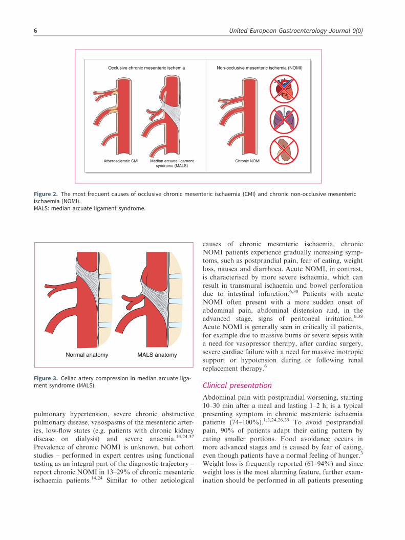
MALS, previously known as Dunbar syndrome orcoeliac artery compression syndrome, is a rare and con-troversial cause of occlusive chronic mesenteric ischae-mia. MALS is defined as a symptomatic eccentriccompression of the CA by the median arcuate ligament(MAL), which is a fibrous arch uniting the diaphrag-matic crura (Figure 3). The exact prevalence of MALSis unknown, but compression of the CA by the MAL ispresent in 3.4–7.3% of asymptomatic patients in whomimaging is performed for other indications.20,27–29
Compression of the CA by the MAL is frequentlyobserved in individuals with a high origin of the CAfrom the aorta or a lower insertion of the MAL. Sincethe position of the diaphragm and thus the MALchanges during respiration, the severity of CA com-pression varies. Compression is most severe duringmaximal expiration. MALS is more prevalent inyoung women (four female: one male) and in patientswith a low BMI.30
4 United European Gastroenterology Journal 0(0)

Vasculitis is a rare cause of occlusive chronic mesen-
teric ischaemia, though it should be considered in chron-
ic mesenteric ischaemia patients because a different
therapeutic approach is needed. Involvement of the mes-
enteric arteries and consequent chronic mesenteric
ischaemia are most frequently seen in patients with pol-
yarteritis nodosa, immunoglobulin (Ig)A vasculitis and
Takayasu arteritis.31 Mesenteric artery involvement is
less frequently reported in rheumatoid arthritis-
associated vasculitis, anti-neutrophil cytoplasmic
antibody (ANCA)-associated vasculitis, systemic lupus
erythematosus, Behcet’s disease and giant cell
arteritis.32,33
MVT can cause AMI due to an outflow obstruction,
thereby compromising the mesenteric circulation.
Chronic mesenteric ischaemia caused by MVT is rare,
since venous collaterals usually form rapidly, but
chronic mesenteric ischaemia can occur in patients
with chronic MVT.34,35 Due to the rarity of MVT as
a cause of chronic mesenteric ischaemia it is not dis-
cussed in detail in this guideline and the reader is
referred to guidelines by Bj€orck et al. and Kearon
et al.6,36
Chronic NOMI was recognised as a disease entity
after the introduction of functional tests; by detecting
mucosal ischaemia in patients with typical symptoms in
the absence of mesenteric artery stenoses or occlusions.
The exact pathophysiology of chronic NOMI is
unknown, although insufficient mesenteric (micro)
circulation seems the most probable explanation.
Chronic NOMI is characterised by low-grade ischae-
mia and is associated with cardiac forward failure,
Pancreato-duodenalarcade
Superiormesenteric artery
Marginal artery
Arcade of riolan
Rectal arteries
Internal iliacartery
Splenic artery
Inferior mesentericartery
Median arcuateligament of diaphragm
Coeliac artery
Figure 1. Anatomy of the arterial mesenteric circulation.
Terlouw et al. 5
pulmonary hypertension, severe chronic obstructivepulmonary disease, vasospasms of the mesenteric arter-ies, low-flow states (e.g. patients with chronic kidneydisease on dialysis) and severe anaemia.14,24,37
Prevalence of chronic NOMI is unknown, but cohortstudies – performed in expert centres using functionaltesting as an integral part of the diagnostic trajectory –report chronic NOMI in 13–29% of chronic mesentericischaemia patients.14,24 Similar to other aetiological
causes of chronic mesenteric ischaemia, chronicNOMI patients experience gradually increasing symp-toms, such as postprandial pain, fear of eating, weightloss, nausea and diarrhoea. Acute NOMI, in contrast,is characterised by more severe ischaemia, which canresult in transmural ischaemia and bowel perforationdue to intestinal infarction.6,38 Patients with acuteNOMI often present with a more sudden onset ofabdominal pain, abdominal distension and, in theadvanced stage, signs of peritoneal irritation.6,38
Acute NOMI is generally seen in critically ill patients,for example due to massive burns or severe sepsis witha need for vasopressor therapy, after cardiac surgery,severe cardiac failure with a need for massive inotropicsupport or hypotension during or following renalreplacement therapy.6
Clinical presentation
Abdominal pain with postprandial worsening, starting10–30 min after a meal and lasting 1–2 h, is a typicalpresenting symptom in chronic mesenteric ischaemiapatients (74–100%).1,3,24,26,39 To avoid postprandialpain, 90% of patients adapt their eating pattern byeating smaller portions. Food avoidance occurs inmore advanced stages and is caused by fear of eating,even though patients have a normal feeling of hunger.3
Weight loss is frequently reported (61–94%) and sinceweight loss is the most alarming feature, further exam-ination should be performed in all patients presenting
Non-occlusive mesenteric ischemia (NOMI)Occlusive chronic mesenteric ischemia
Atherosclerotic CMI Chronic NOMIMedian arcuate ligamentsyndrome (MALS)
Figure 2. The most frequent causes of occlusive chronic mesenteric ischaemia (CMI) and chronic non-occlusive mesentericischaemia (NOMI).MALS: median arcuate ligament syndrome.
Normal anatomy MALS anatomy
Figure 3. Celiac artery compression in median arcuate liga-ment syndrome (MALS).
6 United European Gastroenterology Journal 0(0)

with this symptom.1,3,24,26,39 Other possible complaints
are diarrhoea (19–61%), nausea (5–84%), and worsen-
ing of abdominal pain during exercise (43–
76%).1,3,24,26,39 Physical examination reveals an
abdominal bruit in 17–87% of patients, but can be
completely normal.26,39
The ‘classic triad’ of chronic mesenteric ischaemia,
consisting of postprandial pain, weight loss and an
abdominal bruit, is found in only a minority of chronic
mesenteric ischaemia patients (22%).39 Even when pre-
sent, the predictive value of the classic triad is limited.
Studies have shown a 60% probability of chronic mes-
enteric ischaemia when the classic triad is present, with
an area under the curve of 0.62.3,40
Chronic mesenteric ischaemia patients are at risk of
developing acute-on-chronic mesenteric ischaemia, e.g.
in cases with a ruptured atherosclerotic plaque. Acute-
on-chronic mesenteric ischaemia should be considered
when the abdominal symptoms of a chronic mesenteric
ischaemia patient progress, since urgent revasculariza-
tion might be needed in these patients.6
Diagnostic criteria
Treatment decisions are based on diagnostic criteria.The gold standard diagnosis of chronic mesentericischaemia used in the literature is relief of symptomsafter revascularization. Chronic mesenteric ischaemia,however, remains a diagnostic challenge with a widedifferential diagnosis including chronic pancreatitis,coeliac disease, duodenal ulcers, abdominal
malignancies and irritable bowel syndrome. Exclusionof alternative diagnoses should be performed, prefera-bly by a gastroenterologist. The expert panel recom-mends performing at least an upper GI endoscopyand abdominal imaging (CT scan/magnetic resonanceimaging (MRI) scan). Chronic pancreatitis should beconsidered in all patients, because presenting symp-toms and risk factors (e.g. smoking, hypertriglyceridae-mia) of chronic pancreatitis are similar to presentingsymptoms and risk factors of chronic mesentericischaemia. The diagnostic work-up of chronic pancre-atitis can be found in the chronic pancreatitis guidelineby Dominguez-Munoz et al.41 Coeliac disease shouldbe considered and ruled out in all patients presentingwith weight loss, especially since diagnostic tests arerelatively easy to perform. Colonoscopy is consideredmandatory in patients with diarrhoea – to exclude colo-rectal carcinoma and other ileal and colonic causes ofdiarrhoea – and should be considered in elderlypatients with symptoms indicating colonic disease orischaemia. Colonoscopy seems to be of limited valuein patients with upper GI symptoms.
Patients suspected of having chronic mesentericischaemia should be discussed by a multidisciplinaryexpert panel (consisting of at least a gastroenterologist,(interventional) radiologist and vascular surgeon) toevaluate the compatibility of history, presence orabsence of significant mesenteric artery stenosis onimaging, absence of an alternative diagnosis and,when available, results of a functional test.
Considering the number of stenotic or occludedmesenteric arteries is important when making treat-ment decisions. The SMA is often regarded as themost important mesenteric artery in the framework ofchronic mesenteric ischaemia, since blood flow to theGI tract is predominantly provided by the SMA.14
However, no consistent evidence is available describinghigher clinical success rates after revascularization ofsingle-vessel SMA stenosis compared to single-vesselCA stenosis.42 The IMA is considered the least impor-tant mesenteric artery and single-vessel stenosis of theIMA is deemed clinically trivial, which is supported byresults of endovascular abdominal aortic aneurysmrepairs.14 Occlusion of the IMA during endovascularaneurysm repair rarely results in mesenteric ischaemiawhen the CA and SMA are patent. The probability ofchronic mesenteric ischaemia and clinical success ofrevascularization is higher in patients with multivesseldisease than in patients with single-vessel disease.Clinical success of revascularization ranges from90–100% in patients with involvement of two orthree mesenteric arteries.39,43,44 Hence, the expertpanel considers chronic mesenteric ischaemia likely in
GRADEExpertagreement
Recommendation 1Chronic mesenteric ischaemia should
be considered in patients withunexplained postprandial abdomi-nal pain, weight loss (>5% bodyweight), adapted eating pattern (toavoid abdominal complaints) ordiarrhoea.
1C 100%
Recommendation 2The absence of the classical triad of
chronic mesenteric ischaemia (i.e.postprandial pain, weight loss andabdominal bruit) does not excludea diagnosis of chronic mesentericischaemia.
1C 91%
Terlouw et al. 7

patients with unexplained abdominal symptoms andsignificant stenoses in both CA and SMA. Furtherfunctional testing is not required for the presumptivediagnosis of chronic mesenteric ischaemia in thesepatients.
A more careful approach is warranted in single-vessel disease. Two small prospective cohort studiesconsisting of 37 and 50 patients, respectively, reportedclinical success in 73–76% of symptomatic patientswith single-vessel disease of either CA or SMA, despite
a full workup including a functional test and discussionin a multidisciplinary team.39,42 When a functional testis unavailable, the expert panel suggests that the pres-ence of a compatible history, i.e. postprandial abdom-inal pain and either weight loss or an adapted eatingpattern, is essential for the presumptive diagnosis ofchronic mesenteric ischaemia in patients with single-vessel disease.
A presumptive diagnosis of chronic NOMI is sug-gested by a combination of compatible symptoms andpreferably a positive functional test, in the absence of
significant mesenteric artery stenoses. Very low qualitysingle-centre cohort studies have reported improve-ment of symptoms in 56–63% of patients with a pre-sumptive diagnosis of chronic NOMI treated with avasodilating agent.14,24 Since chronic NOMI is oftenassociated with haemodialysis, severe cardiac orsevere pulmonary disease, treatment of these patientsshould preferably be discussed in amultidisciplinary setting in order to address the under-lying condition.
Diagnostic modalities
Defining haemodynamically significantmesenteric artery stenosis
Imaging is an essential part of the work-up of patientssuspected of having chronic mesenteric ischaemia. Themain purposes are detection of mesenteric artery steno-ses or occlusions and exclusion of alternative diagno-ses. The definition of a significant or clinically relevantmesenteric artery stenosis is still under debate.
GRADEExpertagreement
Recommendation 3To exclude alternative diagnoses at least the following diagnostic tests must be
performed: upper gastrointestinal endoscopy and abdominal imaging (CTscan/MRI scan). Depending on age and symptoms colonoscopy should beconsidered, but is mandatory in patients with diarrhoea.
1D 91%
Recommendation 4A presumptive diagnosis of occlusive chronic mesenteric ischaemia is based on
a combination of compatible history, significant mesenteric artery stenosison radiological imaging and, preferably, a positive functional test. Resultsshould be discussed in an expert multidisciplinary setting by at least a gas-troenterologist, vascular surgeon and (interventional) radiologist.
1C 78%
Recommendation 5In patients with unexplained abdominal symptoms and significant stenoses of
the CA and SMA, the probability of chronic mesenteric ischaemia is high and,consequently, a functional test is not required.
1B 87%
Recommendation 6For the presumptive diagnosis of chronic mesenteric ischaemia in patients with
single-vessel stenosis of CA or SMA, after proper exclusion of alternativediagnoses and no available functional test, the following symptoms shouldbe present: postprandial abdominal pain and either weight loss (>5% bodyweight) or an adapted eating pattern.
2D 91%
Recommendation 7(a) A presumptive diagnosis of chronic NOMI is based on a combination of
compatible symptoms, absence of significant mesenteric artery stenosesand, preferably, a positive functional test.
(b) In presumptive chronic NOMI patients with severe cardiac disease, pulmo-nary disease or in dialysis patients, underlying causes and treatment shouldbe discussed with the respective specialists.
2D 87%
8 United European Gastroenterology Journal 0(0)

A reduction of luminal diameter in the range of 50–75% is used to define a significant stenosis, but com-parative data on the clinical success of treatment invarying degrees of stenoses is not available39,42,45–50
The type and number of affected vessels and conse-quences of overtreatment versus undertreatment areimportant when setting the definitions for mesentericartery stenoses.
In patients with single-vessel disease the probabilityof mistakenly omitting treatment is thought to be low.By contrast, the probability of overtreating patientswithout true chronic mesenteric ischaemia is substan-tial and may expose these patients to complications,potentially without any compensating benefits fromtreatment. Therefore, a �70% stenosis of either CAor SMA could be considered relevant in presumedsymptomatic single-vessel disease. In patients withmore than one stenotic or occluded artery, unsuccessfultreatment is relatively infrequent, but the burden andpossible consequences of mistakenly denying treatmentare substantial (e.g. development of AMI, poor qualityof life, etc.). Comparing the surface area and bloodflow volumes of the SMA and CA, it is apparent thatthe surface area and postprandial flow of the SMA areboth approximately 33% higher.14,51 Therefore, aSMA stenosis of 50% is more likely to become haemo-dynamically relevant, especially when part of the bloodvolume is diverted through the collateral circulation toprovide blood to other stenotic mesenteric arteries. Theexpert panel suggests that a �50% stenosis of the SMAcould be considered relevant in symptomatic patientswith extensive multivessel disease.
Radiological imaging
Computed tomography angiography (CTA) andcontrast-enhanced magnetic resonance angiography(CE-MRA) have replaced conventional angiographyas the gold-standard imaging test for patients suspectedof having chronic mesenteric ischaemia.
Two investigator-blinded cohort studies evaluated theaccuracy of detecting �50% and �70% stenoses ofmesenteric arteries by CTA while using angiographyas a reference.45,52 CTA had a sensitivity of 100%and a specificity of 95–100%. Advantages of CTAinclude its reproducibility, low interobserver variation,guidance of treatment planning and the potential detec-tion of extravascular signs of possible AMI or alterna-tive diagnoses. Signs of AMI or acute-on-chronicmesenteric ischaemia are mostly non-specific and mayinclude bowel lumen dilatation (paralysis), bowel wallthickening and mesenteric fat stranding (oedema) com-bined with an obstruction of mesenteric arteries may bean early sign of on-going intestinal ischaemia, whereasdecreased bowel wall enhancement and pneumatosissuggest more advanced AMI.53 Disadvantages ofCTA are radiation exposure and the risk of contrast-induced nephropathy. A reduction of both radiationexposure and dosage of contrast agents can be achievedby performing a dual energy CTA.54 A CTA afterinjection of an adequate amount of intravenous (i.v.)contrast (1.5–2 ml/kg bodyweight with a concentrationof >300 mg iodine/ml contrast media; flow rate 3.5–4ml/s) with �1 mm acquisition slice thickness in arterialand venous/portal venous phase is recommended inpatients with suspected chronic mesenteric ischaemia,to reliably assess the patency of the mesenteric vascu-lature and possible signs of AMI.
Several studies suggest CE-MRA could be a goodalternative for the detection of CA and SMA stenoses,as it shows 100% sensitivity, 91–100% specificity andalmost perfect interobserver agreement.52,55–58 Smallstudies dating from before 2002 suggest CE-MRAhas difficulties detecting smaller vessels, but majorimprovements in magnetic resonance (MR) imagequality have solved this issue.57–59 Clear benefits ofCE-MRA over other imaging modalities are theabsence of radiation exposure and contrast-inducednephropathy. These factors should be consideredwhen performing imaging in young patients or patientswith impaired renal function. A disadvantage of CE-MRA is the long duration of the examination, whichcould result in a poor image quality due to artefactscaused by breathing or patient movements. The expertpanel considers CE-MRA the test of choice in patientswith contraindications for CTA, for example patientswith a severe allergy for the prescribed contrast agentor impairment of renal function.
Duplex ultrasound (DUS) of the mesenteric arterieshad been extensively studied and used in clinical prac-tice for decades. DUS can image the proximal mesen-teric arteries but is often difficult to perform and maybe negatively affected by abdominal fat and overlyingintestinal gas. A study of the application of DUS in 324patients reported interpretability as good in 66–68% of
GRADEExpertagreement
Recommendation 8In symptomatic patients with single-
vessel disease of either the CA orSMA, a �70% stenosis could beconsidered relevant.
2D 87%
Recommendation 9In symptomatic patients with exten-
sive multivessel mesenteric arterydisease, a �50% stenosis of theSMA could be considered relevant.
2D 78%
Terlouw et al. 9

cases, moderate in 11–18%, and not interpretable in15–23% of cases.60 Various prospective and retrospec-tive studies have determined blood flow velocitycut-offs for significant CA and SMA stenoses by com-paring DUS, as performed by experienced technicians,with angiography. A wide range of cut-offs werereported, with peak systolic velocities (PSVs) rangingbetween 200–320 cm/s in the CA and 205–400 cm/s inthe SMA (sensitivity 58–100% and specificity48–100%).47,49,60–63 Mesenteric artery PSV and enddiastolic velocity (EDV) are strongly influenced byinspiration and expiration.64 This should be consideredwhen interpreting study results, since most studies donot report whether DUS was performed during inspi-ration or expiration. Even though the sensitivity andspecificity of DUS is inferior to the sensitivity and spe-cificity of CTA or CE-MRA, it remains a valuableimaging technique because the costs of DUS are rela-tively low, contrast agents are not needed and radiationexposure is avoided. Hence, the expert panel concludesthat DUS – when performed by an experienced techni-cian – might be used as a screening method to excludesignificant proximal mesenteric artery stenosis. WhenDUS is positive, CTA or CE-MRA is required to con-firm the presence of a significant stenosis and to guidetreatment planning.
Based on the diagnostic performance of non-invasive imaging modalities and the known complica-tions of angiography (e.g. pseudoaneurysm, dissection,bleeding, etc.), the expert panel recommends that angi-ography be reserved for therapeutic purposes only.
Radiological imaging in suspected MALS
In MALS, the severity of CA stenosis is influenced byrespiration. A cohort study, including 78 patients withclinical signs of MALS and previous imaging, per-formed angiography at both maximal inspiration andmaximal expiration.65 The study found that duringexpiration CA compression was present in 100% ofpatients, whereas CA compression was present inonly 56% of patients during inspiration. Standard pro-tocol abdominal CT or MR imaging during inspirationonly is therefore unsuitable when the goal is to excludeMALS.
Features suggesting CA compression on CTA orCE-MRA are an eccentric stenosis of the CA, ahook-shaped appearance of the CA and variation inthe severity of the stenosis between inspiration andexpiration. Evidence supporting the feasibility ofCTA to detect CA compression is limited to very lowquality retrospective studies which reported the pres-ence of CA compression on CTAs performed for otherindications.66,67 No evidence on feasibility of CE-MRAexists for this indication. Two studies, including six and
19 patients with CA compression, respectively, per-formed DUS during both inspiration and expira-tion.68,69 These studies reported a sensitivity of83–95% and specificity of 89–100%.
Based on the lack of comparative studies showingsuperiority of one technique over another, DUS, CTAand CE-MRA are all considered appropriate. MALS ismost often suspected and diagnosed in young patients,in whom harmful effects of radiation should beavoided. We recommend an inspiratory and expiratoryDUS or CE-MRA, with �2 mm slices and 3D recon-structions, in patients of younger age and suspectedMALS. A protocol for radiologists, containing techni-cal details of imaging in chronic mesenteric ischaemiapatients, is provided in Supplementary MaterialDocument B.
GRADEExpertagreement
Recommendation 10In patients with suspected chronic
mesenteric ischaemia, a CTA (�1mm acquisition slice thickness,arterial and venous/portal venousphase) should be performed.
1C 91%
Recommendation 11CE-MRA is the diagnostic test of
choice in case of a contraindicationfor CTA.
1C 87%
Recommendation 12Duplex ultrasound – when performed
by an experienced technician –might be used as a screeningmethod to exclude significantproximal mesenteric artery steno-sis. Additional CTA or MRA imagingis required for patients with a pos-itive duplex ultrasound.
2C 78%
Recommendation 13Angiography should be reserved for
therapeutic purposes.1C 100%
Recommendation 14CA compression in MALS can be
diagnosed by inspiration/expirationduplex ultrasound, CTA or CE-MRA.In patients of younger age, sus-pected of having MALS, both duplexultrasound and CE-MRA (�2 mmslices with 3D reconstructions) ininspiration and expiration are rec-ommended imaging techniques.
1D 74%
10 United European Gastroenterology Journal 0(0)

Functional testing
Functional tests have been developed to characterise
chronic intestinal ischaemia and prove insufficiency of
the collateral circulation.70 The most extensively
described functional tests are tonometry and visible
light spectroscopy (VLS). Tonometry measures the
intraluminal partial pressure of carbon dioxide
(PCO2) in the stomach and small bowel by positioning
two balloons at the end of a nasogastric tube and naso-
jejunal tube. Studies have shown that intraluminal
PCO2 is closely related to local arterial PCO2 and indic-
ative of mucosal ischaemia. Two types of tonometry
have been described and validated: exercise tonometry
and 24-hour tonometry. Exercise tonometry uses a
standardised exercise protocol to elicit ischaemia and
has a 78–97% sensitivity and 69–92% specificity.71,72
During 24-hour tonometry, meals are used to elicit
ischaemia, while PCO2 levels are monitored during a
24-hour period. The sensitivity of this test is 76–92%
and specificity 77–94%.40,70 Major disadvantages of
tonometry are the invasiveness (nasogastric tubes),
long duration and interference of gastric acid and
acids in food. Unfortunately, the disappearance of
this test is expected in the near future, since tonometryequipment is no longer produced.
VLS is performed during upper GI endoscopy and
measures mucosal oxygen saturation in the antrum,
duodenal bulb and descending duodenum. Studies of
VLS in chronic mesenteric ischaemia patients report a
90% sensitivity, 60% specificity and fair to good intra-
observer and interobserver agreement.73,74 VLS is
quicker and less cumbersome than tonometry, but
ischaemia cannot be provoked during VLS. A recent
study that performed VLS measurements both pre-
prandial and 45 min after luminal feeding in the stom-
ach found no additional discriminative value of
postprandial VLS measurements.75 Other disadvan-
tages of VLS are interference of bile and the require-
ment to switch off endoscopic light during
measurements, causing difficulties in maintaining the
position of the probe during peristaltic waves.76
The expert panel acknowledges the value of ischae-
mia detection by a discriminative functional test in
patients with suspected chronic mesenteric ischaemia.
Though of proven value in literature, current function-
al tests have serious limitations and are not widely
available throughout Europe. The expert panel recog-
nises the need for a more potent and widely available
functional test.
Blood tests
Biomarkers for chronic mesenteric ischaemia are not cur-
rently available. Two small prospective cohort studies
using a diagnostic work-up, including a functional test,
have evaluated the diagnostic potential of markers such
as lactate, D-dimer, lactate dehydrogenase, C-reactive
protein, leucocytes, intestinal fatty acid binding protein
and lipopolysaccharide.77,78 None of these markers were
sufficiently discriminative and suitable for clinical prac-
tice. The clinical value of lactate, obtained from a periph-
eral vein, to rule out any form of intestinal ischaemia is
still a widespread misconception. A cohort study mea-
suring lactate before a meal and 60 and 240 min after a
meal revealed the poor sensitivity (34%) of lactate in
chronic mesenteric ischaemia patients.77 Similar sensitiv-
ities of lactate have been shown in a systematic review of
patients presenting with AMI, in whom ischaemia is even
more extensive and severe.79 Though nonspecific, normal
D-dimer levels can exclude AMI (sensitivity 89–100%);
however, the value of D-dimer levels to exclude chronic
mesenteric ischaemia remains unclear.79,80 Based on
these studies the expert panel concludes that normal lac-
tate, lactate dehydrogenase and/or leucocyte levels do
not exclude chronic mesenteric ischaemia.
GI endoscopy
Upper GI endoscopy is essential in the work-up of
patients suspected of having chronic mesenteric ischae-
mia, mainly for exclusion of alternative diagnoses.
Findings reported in chronic mesenteric ischaemia
patients are oedema (35%), erythema (42%), atrophy
of the duodenal mucosa, and gastric and duodenal
ulcers that are not caused by Helicobacter pylori or
non-steroidal anti-inflammatory drugs.14 A prospective
study comparing endoscopic biopsies of 56 chronic
mesenteric ischaemia patients and 26 patients without
chronic mesenteric ischaemia could not demonstrate
discriminative histological changes, and concluded
that biopsies have no value in supporting the diagnosis
of chronic mesenteric ischaemia.81 Based on the rela-
tively low prevalence and often transient nature of
upper GI endoscopic abnormalities in chronic mesen-
teric ischaemia patients, the expert panel states that a
normal GI endoscopy does not exclude chronic mesen-
teric ischaemia.
GRADEExpertagreement
Recommendation 15(a) Normal lactate, lactate dehydro-
genase, and/or leucocytes levels donot exclude chronic mesentericischaemia.
(b) Normal GI endoscopy does notexclude chronic mesentericischaemia.
1C 100%
Terlouw et al. 11

A flowchart containing an overview of the mostimportant steps and criteria of the diagnostic work-up of patients with suspected chronic mesentericischaemia can be found in Figure 4.
Treatment
Over the last decades endovascular revascularization(ER) of the mesenteric arteries has replaced open sur-gical mesenteric artery revascularization (OSMAR). Ameta-analysis of 100 studies reporting on OSMARand/or ER found significantly more in-hospital compli-cations and a trend towards a higher 30-day mortalityafter OSMAR (in-hospital complications relative risk(RR) 2.19 (95% confidence interval (CI) 1.84–2.60); 30-day mortality RR 1.57 (95% CI 0.84–2.93)).1
Nevertheless, long-term results appeared superiorafter OSMAR, with fewer symptom recurrences anda trend towards higher three-year survival (three-yearsymptom recurrence RR 0.47 (95% CI 0.34–1.57);three-year survival RR 0.96 (95% CI 0.86–1.07)).1
However, the quality of the evidence assembled inthis meta-analysis is low, since the design and qualityof the individual studies was flawed. The meta-analysisconsisted of 18,762 patients, but few studies reportedlong-term outcomes.
Durability of revascularization is generallyexpressed by the patency of a vessel after revasculari-zation. Primary patency is defined as the absence of asignificant re-stenosis or occlusion after a primaryrevascularization. Secondary patency is defined as theabsence of a significant re-stenosis or occlusion after asecond revascularization of the target vessel. Twometa-analyses comparing patency rates of OSMARand ER showed significantly higher primary patencyrates for OSMAR (one-year primary patencyOSMAR 91–94% vs ER 69–74%, five-year primarypatency OSMAR 80–81% vs ER 51–52%).82,83 Thenumbers of patients included in the analyses of theone-year primary patency were 465 and 742 (meta-analyses of Gupta et al.83 and Pecoraro et al.,82 respec-tively); 481 and 679 patients were included in theanalyses of the five-year primary patency. OSMARalso exhibited superior five-year secondary patency(OSMAR 96–98% vs ER 79–88%). The quality ofthe evidence provided by these meta-analyses is low,since the design and quality of the individual studieswas flawed and the small sample sizes of the individualstudies could have introduced publication bias.Furthermore, evidence on the cost-effectiveness andgain in quality of life of each treatment strategy wasof low quality, and consisted of a single study showingno long-term differences between OSMAR and ER.84
The expert panel concluded that the reduction inshort-term mortality and morbidity of ER outweighs
the superior long-term results of OSMAR in this gen-erally older population (mean age 69 years) with mul-tiple comorbidities, especially since the long-termpatency of repeated ERs is considered comparable tothat of OSMAR.1 Therefore, the expert panel prefersER and suggests mesenteric bypass procedures mightbe reserved for patients in whom ER is not suitable.
Nutritional status before mesenteric arteryrevascularization
Malnutrition is a common finding in patients withchronic mesenteric ischaemia. A small retrospectivecohort study of chronic mesenteric ischaemia patients,treated by percutaneous mesenteric artery stenting(PMAS) or OSMAR, found an association betweenmalnutrition and increased mortality (30-day mortalitynon-malnourished patients 0%, malnourished patients26%).85 Although pre-operative nutrition is associatedwith improved surgical outcomes in patients withoutchronic mesenteric ischaemia (post-operative complica-tions odds ratio (OR) 0.64 (95% CI 0.53–0.84)),pre-operative oral or enteral nutrition might worsenintestinal ischaemia in patients with chronic mesentericischaemia.86 Tonometry studies confirm that oralintake induces or worsens ischaemia in the stomachand jejunum of chronic mesenteric ischaemiapatients.70 Additional circumstantial evidence can befound in case reports and a cohort study, whichreported AMI in 2% of in-hospital patients receivingenteral tube feeding and even higher rates of AMIamong intensive care unit (ICU) patients, since mostICU patients are haemodynamically unstable andunable to increase mesenteric blood flow.87–89 Thelatter did not use a control group (e.g. total parenteralnutrition (TPN)), and the haemodynamic situation inchronic mesenteric ischaemia patients differs from thatof ICU patients, implying that these results should beinterpreted with caution.
No evidence on TPN in chronic mesenteric ischae-mia patients exists, but a study in patients withoutchronic mesenteric ischaemia has shown that TPNdecreases mesenteric blood flow, an effect that couldinduce AMI in chronic mesenteric ischaemia patients.90
The increased risk of bloodstream infections associatedwith TPN is another disadvantage, especially sincesepsis and subsequent haemodynamic instability willcompromise an already threatened mesentericcirculation.91,92
The expert panel acknowledges that evidence on thistopic is limited to non-existent, yet possible negativeconsequences of attempts to improve nutritionalstatus are considered substantial. Therefore, increasingoral intake, starting enteral tube feeding or startingTPN before revascularization might be
12 United European Gastroenterology Journal 0(0)

Consider CMI in presence of either:
- Postprandial abdominal pain- Weight loss (>5% body weight)- Adapted eating pattern - Diarrhoea
Perform radiological imaging
CTA OR CE-MRA- ≤1mm slices - ≤2 mm slices - Arterial + (portal) venous - 3D reconstructions
Exclude alternative diagnoses
At least perform:- Upper GI endoscopy- Abdominal imaging- Colonoscopy (in case of diarrhoea)
No stenosis
DDx- Chronic NOMI*
Single-vessel stenosisCA or SMA ≥70%
DDx- Atherosclerosis- CA compression- Other (e.g. vasculitis)
Multi-vessel stenosisCA ≥70% / SMA ≥50%
DDx- Atherosclerosis- Other (e.g. vasculitis)
Functional test
If not available etiher:- Postprandial abdominal pain + weight loss- Postprandial abdominal pain + adapted eating pattern
Discussion in expert multidisciplinary settingGastroenterologist + (interventional) radiologist + vascular surgeon
* Discuss underlying cause and treatment with respective specialist
Other causes of CMI
Chronic NOMI
MALS Atherosclerotic CMI
No CMI
Figure 4. Flowchart of the diagnostic work-up of patients with suspected chronic mesenteric ischaemia (CMI).CA: celiac artery; CE-MRA: contrast-enhanced magnetic resonance angiography; CTA: computed tomography angiography; DDx:differential diagnosis; GI: gastrointestinal; NOMI: non-occlusive mesenteric ischaemia; SMA: superior mesenteric artery.
Terlouw et al. 13

contra-indicated. Restoring mesenteric circulation –preferably by PMAS – has priority and should not bedelayed by attempts to improve nutritional status.
PMAS
Two types of ER have been described in literature, per-cutaneous transluminal angioplasty (PTA) and PMAS.PMAS is the current standard approach and the ratio-nale behind this strategy is based on technical successrates. Case series and expert experience suggest that thevast majority of mesenteric artery stenoses occur at thevessel’s origin and that these stenoses are often heavilycalcified.93,94 Studies on revascularization of ostialrenal artery stenosis have shown that elastic recoil ismore likely to occur in ostial stenoses, causing recur-rence of the stenosis when recoil is not prevented bystenting.95,96 Evidence comparing both strategies is ofvery low quality. A systematic review comparing mes-enteric artery PTA and PMAS in 328 patients found asignificantly higher technical success rate for PMAS(PMAS 95% vs PTA 83%), comparable symptomrelief (PMAS 91% vs PTA 89%) and a higher resteno-sis rate (PMAS 35% vs PTA 21%).97 A more recentretrospective cohort study reported a lower reinterven-tion rate after PTA compared to PMAS, although thedifference was not statistically significant.98 Despite thevery low quality of evidence, the expert panel stronglyrecommends PMAS over PTA alone in atheroscleroticmesenteric artery stenosis as technical success, and thusadequate revascularization, is considered the mainobjective.
For decades the preferred route of arterial access forendovascular mesenteric artery revascularization wasvia either the brachial or femoral artery. Some centresadvocate use of (left) brachial artery access, since thecaudal angulation of the mesenteric arteries makes thisapproach more convenient. Manipulation of guide-wires, sheaths and catheters through the
brachiocephalic artery and aortic arch is limited oravoided when using left brachial access instead ofright brachial access. Left brachial access has beenreported to decrease the risk of cerebral embolizationdue to dislodged aortic plaques or thrombus afteradvanced endovascular aortic repair with visceralartery stent grafting.99 However, reported complicationrisks of PMAS using brachial access (16–19%) appearto be higher than the complication risks of femoralaccess (7.5%).50,100 When using brachial access,ultrasound-guided cannulation of the brachial arteryis advised to avoid nerve injury. Introduction ofradial access provides a third possible access route forPMAS.101 Low rates of access-site complication havebeen reported in coronary artery angiographies; how-ever, these results cannot be extrapolated to mesentericartery interventions, considering that longer sheathsare often used for PMAS. The literature on radialaccess for PMAS is limited. A meta-analysis of radialand femoral access in infra-aortic interventions, using amaximum sheath size of 6–8.5 French, reported signif-icantly fewer complications when radial access wasused (OR 0.25, 95% CI 0.07–0.86).102 A meta-analysis of hepatic artery interventions in patientswith primary or secondary liver malignancy reportedno differences in access-site-related outcomes of radialand femoral access, but did show a patient preferencefor radial access.103 Based on extensive experience withfemoral access, the still limited evidence supportingradial access, and the substantial complication rate ofbrachial access, the preferred entry site for PMAS iscurrently femoral, followed by (left) brachial orradial, but is primarily dependent on the expertise ofthe operator.
Retrograde open mesenteric stenting (ROMS) is ahybrid mesenteric artery revascularization strategyusing midline laparotomy to gain direct retrograde vas-cular access to a mesenteric artery.104 This access tech-nique can be used to perform stenting in patients withacute-on-chronic ischaemia undergoing a laparotomyfor resection of necrotic bowel.104,105 ROMS mightalso be used to stent severely stenotic or occludedostia of mesenteric arteries that cannot be cannulatedthrough femoral, brachial or radial access.
Use of bare-metal stents (BMSs) for PMAS is stan-dard practice, even though long-term primary patencyis unsatisfactorily low (five-year primary patency: 51–52%) due to in-stent intimal hyperplasia.82,83 Somecentres have used covered stents (CSs) in an attemptto reduce in-stent intimal hyperplasia. One retrospec-tive study compared use of BMS and CS, suggesting asuperior patency of CS over BMS (one-year primarypatency CS 92% vs BMS 75%, three-year primarypatency CS 92% vs BMS 52%).106 The CS also hadsuperior freedom from symptom recurrence (one-year
GRADEExpertagreement
Recommendation 16Mesenteric bypass procedures might
be reserved for patients in whomendovascular revascularization isnot suitable.
2C 100%
Recommendation 17In patients with chronic mesenteric
ischaemia it might be disadvanta-geous to increase oral intake, startenteral tube feeding or start totalparenteral nutrition beforerevascularization.
2D 96%
14 United European Gastroenterology Journal 0(0)

freedom from symptom recurrence CS 92% vs BMS73%, three-year freedom from symptom recurrenceCS 92% vs BMS 50%). Though promising, superiorityof the CS remains debatable, since the quality of evi-dence is very low and results are biased by a lownumber of patients with a follow-up duration of >1year after PMAS using a CS. Possible disadvantagesof the CS are the higher costs than the BMS andunknown complications of the CS, e.g. stent thrombo-sis with subsequent AMI. Due to current uncertaintiesregarding superiority of the CS, the expert panel couldnot reach consensus on statements recommending useof the CS. The results of a randomised controlled trial(NCT02428582) comparing the CS and BMS areawaited.107 Comparative data on PMAS with a drug-eluting stent (DES) is not available in the literature.
In patients with occlusive disease of both CA andSMA, revascularization of one versus both arteriesremains an ongoing debate. Evidence on partial vscomplete revascularization is limited to low-qualitycohort studies comparing partial and completeOSMAR, and cohort series showing high rates of clin-ical success after revascularization of the SMAalone.46,48,50,108–113 Sample sizes of comparative studieswere too small to show a significant difference in five-year survival, but a trend towards superiority of com-plete revascularization was observed.48,108,110,113
Complete revascularization is associated with superiorfreedom from symptom recurrence at 3, 5 and 10 yearsafter revascularization.48,50 The expert panel acknowl-edges current evidence should be interpreted with cau-tion but does agree that complete revascularizationseems beneficial in the long run. Accordingly, revascu-larization of both CA and SMA might be attemptedwhen feasible. When single-vessel revascularization isperformed, the SMA is considered the preferred targetartery followed by the CA.
Antiplatelet therapy after PMAS
Initiation of dual antiplatelet therapy (DAPT) directlyafter PMAS is common practice. The duration ofDAPT remains uncertain and there is always a trade-off between benefits (reduction of stent thrombosis andreintervention rates) and harm (increased risk of bleed-ing). Due to an absence of evidence on this topicthe expert panel extrapolated evidence from stentingin the coronary arteries, since these are most similarto the mesenteric arteries with their tortuosity andsmall diameter. A one-month DAPT duration wascommon practice in the coronary BMS era. No studieshave compared one-month DAPT with a longer dura-tion of DAPT after coronary artery stenting using aBMS. A longer DAPT duration was introduced dueto the increased risk of stent thrombosis associated
with the DES. Current guidelines on DAPT in coro-
nary artery disease recommend use of the DES and a
consequent 12-month duration of DAPT.114 Meta-
analyses of this antiplatelet strategy report a clear asso-
ciation between long DAPT duration and an increased
risk of bleeding.115,116 In terms of patient preferences, a
study questioning patients on preferences for long
DAPT versus short DAPT reported that patients
place high value on avoidance of the drawbacks asso-
ciated with DAPT.117 Based on the extrapolated liter-
ature regarding DAPT after stenting in the coronary
arteries, increased bleeding rates, patients’ preferences
and use of BMS during PMAS, a one-month duration
of DAPT is suggested after PMAS, followed by lifelong
treatment with an antiplatelet monotherapy.Combining antiplatelet agents with antithrombotic
agents (e.g. direct oral anticoagulant (DOAC), vitamin
K antagonist, low molecular weight heparin (LMWH))
increases the risk of bleeding events. A meta-analysis of
patients after coronary artery stenting compared oral
anticoagulation with the addition of either one (dual
therapy) or two (triple therapy) antiplatelet agents.
Risks of both major bleeding (OR 0.55, 95% CI
0.39–0.78) and minor bleeding (OR 0.43, 95% CI
0.33–0.56) were significantly lower when dual therapy
was used.118 Risks of stent thrombosis, myocardial
infarction and cardiovascular death did not differ sig-
nificantly, though studies included in the meta-analysis
were not powered for these outcomes. Studies compar-
ing use of an antithrombotic agent alone and dual ther-
apy, after coronary artery stenting, were not found.
Based on the substantial risks of bleeding and question-
able benefits of long duration antiplatelet therapy in
patients treated with DOAC, vitamin K antagonists
or LMWH, the expert panel suggests that the prescrip-
tion of an antiplatelet agent be restricted to the first
4 weeks after PMAS.
GRADEExpertagreement
Recommendation 18The preferred entry site for mesen-
teric artery revascularization is thefemoral artery, followed by the leftbrachial or radial artery, and isdependent on expertise.
1D 87%
Recommendation 19In atherosclerotic mesenteric artery
lesions, PTA and stenting is recom-mended over PTA alone.
1D 100%
(continued)
Terlouw et al. 15

OSMAR
OSMAR is most frequently performed by constructing
a mesenteric bypass. Bypass grafts can be positioned in
an antegrade (origin proximal of stenotic artery) or
retrograde (origin distal of stenotic artery) orientation.
Studies comparing both strategies are of very low qual-
ity due to retrospective designs, low numbers of
patients and the large variety in graft material. Four
studies report no differences in survival or graft paten-
cy between antegrade and retrograde graft orienta-
tion.119–122 One study reported improved median
survival when using an antegrade bypass (antegrade
5.7 years vs retrograde 4.0 years), but patients under-
going an antegrade bypass were younger (antegrade 65
years vs retrograde 75 years).110 Nowadays mesenteric
bypass procedures are reserved for patients unsuitable
for PMAS, resulting in a different patient population.
Mesenteric bypass procedures are currently performed
in older patients with more comorbidities and more
extensive calcification, making supracoeliac anastomo-
sis more challenging and increasing the risks of supra-
coeliac clamping and lengthy surgical procedures. The
decision to use antegrade or retrograde graft orienta-
tion should be tailored to the patient’s local anatomy
and comorbidities, while taking the surgeons’ experi-
ence into account.A mesenteric bypass can be constructed using
venous or prosthetic material, e.g. Dacron or polyte-
trafluoroethylene. Very low quality retrospective stud-
ies show similar patency rates for venous and
prosthetic material.119–124 A disadvantage of prostheticgrafts is the potential risk of graft infection, especiallyin patients with acute-on-chronic ischaemia with spillof bowel content into the abdominal cavity. However,veins of sufficient quality are not always available andgraft infection is rare, while patency is comparable toprosthetic grafts. There might not be a clear preferencefor venous or prosthetic grafts and the choice maydepend on the type of procedure, patients’ comorbid-ities, and quality and availability of venous grafts.
Treatment of MALS
The existence of MALS is still questioned by many
physicians who argue that available evidence is incon-clusive and potentially affected by publication bias.
Treatment of MALS is currently by surgical divisionof the MAL. A systematic review reported improve-
ment of symptoms in 83% of 400 patients treated byCA release.125 Eighty per cent experienced sustained
symptom relief, indicating that the placebo effect asan explanation of symptom relief is unlikely. Based
on these substantial rates of symptom improvement,surgical CA release might be considered in patients
with presumptive MALS.Release of the CA can be performed by laparotomy,
laparoscopy and retroperitoneal videoscopy. Evidence
showing superiority of one technique over another isnot available. The previously described systematic
review included 279 patients with a CA release by lap-arotomy and 121 patients by laparoscopy.125 Sustained
symptom improvement was present in 75% of patientsafter laparotomy and 90% after laparoscopy, suggest-
ing laparoscopic CA release is feasible. Laparoscopicand retroperitoneal videoscopic CA release are less
invasive, resulting in smaller scars, and have the advan-tage of lower rates of gastroesophageal reflux disease,
since only one side of the crus is dissected. Not access-ing the abdominal cavity is an additional advantage of
retroperitoneal videoscopic CA release. This approach
Continued
Recommendation 20In patients with occlusive disease of
both the CA and SMA, endovascularrevascularization of both vesselsmight be attempted. The SMA is thepreferred target artery followed bythe CA.
2D 91%
Recommendation 21After endovascular mesenteric artery
stenting, we suggest administeringdual antiplatelet therapy for atleast one month, followed by life-long antiplatelet monotherapy.
2D 91%
Recommendation 22In patients treated with DOAC, vitamin
K antagonists or LMWH, we suggestadding one antiplatelet agent for 4weeks after endovascular mesen-teric artery stenting.
2D 83%
GRADEExpertagreement
Recommendation 23There might be no preference for an
antegrade or retrograde approachwhen performing mesentericbypass.
2D 81%
Recommendation 24There might be no preference for
venous or prosthetic grafts whenperforming mesenteric bypass.
2D 71%
16 United European Gastroenterology Journal 0(0)

avoids the risk of female infertility due to adhesions inthese often young patients. However, experience withendoscopic CA release is limited to only a few expertcentres and little is known on the learning curve tosafely perform these procedures. Due to the very lowquality of evidence and uncertainty regarding the fea-sibility of implementation of laparoscopic and retro-peritoneal videoscopic CA release in centresthroughout Europe, the expert panel did not reach con-sensus on recommendations on this topic.
Evidence on the risks or benefits of PMAS as a pri-mary treatment for MALS does not exist. However,according to expert experience, stenting of the CAwithout preceding CA release will likely result in astent fracture, compromising blood flow and potential-ly resulting in life-threatening AMI. Due to this possi-ble hazard the expert panel considers endovascularstenting of the CA contraindicated in patients with pre-sumptive MALS and no preceding CA release.
Treatment of vasculitis
Vasculitis involving the mesenteric arteries is a rare
cause of chronic mesenteric ischaemia, but a potentially
life-threatening manifestation of systemic vasculitis.
Literature on this topic consists of case reports and
small case series. Still, the expert panel emphasises
the importance of awareness of vasculitis as a potential
cause of chronic mesenteric ischaemia, since a different
clinical approach is needed. CTA seems to be a reason-
able initial non-invasive screening method in suspected
mesenteric vasculitis and can detect aneurysms in ves-
sels as small as 3 mm.31 Nonetheless, angiography
remains an important, and in some cases indispensable,
imaging modality in patients with a high suspicion of
small or medium vessel vasculitis, such as polyarteritis
nodosa, and seems to be the preferred imaging modal-
ity when CTA is inconclusive or negative.31
A retrospective database study, including patients
evaluated for vasculitis over a 24-year period, found
symptoms of mesenteric ischaemia in 120 of 7514
(1.6%) patients.126 Only 15 of these patients required
revascularization. Management with immunosuppres-
sive medication was sufficient in the remaining patients.
Though evidence is of very low quality, referral to an
expert in the treatment of vasculitis is suggested before
proceeding to ER in patients with symptoms and radio-
logical features of vasculitis. An additional argument to
promote early recognition and treatment is to prevent
progression, which might otherwise result in a severe
disease course such as kidney failure.
Asymptomatic mesenteric artery stenosis
Mesenteric artery stenosis is a frequent finding on
abdominal imaging. Experts agree that revasculariza-
tion is not needed in patients with asymptomatic
single-vessel stenosis. Two prospective cohort studies,
following asymptomatic patients for a mean duration
of 2.6 and 6.5 years, reported no occurrences of AMI
or chronic mesenteric ischaemia in patients with single-
vessel disease.22,23 A decision not to intervene is less
clear in asymptomatic patients with stenosis or occlu-
sion of all three mesenteric arteries, since the ability to
compensate and maintain adequate blood flow through
collaterals is limited, creating a potential risk of AMI
when atherosclerosis progresses or an acute occlusion
(e.g. embolus, thrombosis) occurs. Little is known on
the natural course of asymptomatic three-vessel mesen-
teric artery disease. The only study reporting on this
matter dates from 1998 and included 15 patients with a
mean follow-up duration of 2.6 years.22 Four out of 15
(27%) patients developed mesenteric ischaemia, one
presented with AMI and the remaining three patients
became symptomatic and were diagnosed with chronic
mesenteric ischaemia. Given the low level of evidence, a
tailor-made approach is suggested in all asymptomatic
patients with stenosis or occlusion of all three mesen-
teric vessels. Risks and benefits of revascularization
should be carefully assessed, taking age, comorbidities
and patient preferences into account.When planning major abdominal surgery in patients
with known asymptomatic mesenteric artery stenosis,
the surgeon needs to be aware of local anatomy and the
collateral circulation. Especially in patients with steno-
sis or occlusion of two or more mesenteric arteries,
ligation of collaterals could compromise the mesenteric
circulation, resulting in AMI.127,128 ER might be con-
sidered, to prevent AMI, in patients with significant
stenosis or occlusion of two or more mesenteric arteries
undergoing major abdominal surgery with potential
ligation of the collateral circulation.
GRADEExpertagreement
Recommendation 25Patients with MALS might be consid-
ered for surgical coeliac arteryrelease.
2D 96%
Recommendation 26In patients with MALS (and no pre-
ceding adequate coeliac arteryrelease) endovascular stenting ofthe CA is contraindicated.
1D 100%
Terlouw et al. 17

Secondary prevention
Secondary prevention is important when treating
patients with atherosclerotic chronic mesenteric ischae-
mia, considering that atherosclerosis is a systemic dis-
ease and secondary prevention reduces the risk of all
cardiovascular events. Evidence concerning secondary
prevention in patients with atherosclerotic chronic mes-
enteric ischaemia is not currently available; therefore
evidence and guidelines on secondary prevention in
general have been extrapolated to chronic mesenteric
ischaemia patients.129–131
To evaluate whether secondary prevention is indi-
cated, an assessment of the cardiovascular risk profile
is suggested in all symptomatic and asymptomatic
patients with an atherosclerotic mesenteric artery ste-
nosis, because all patients with proven atherosclerosis
are prone to atherosclerosis and cardiovascular events
in other vascular beds. A history of arterial revascular-
ization (e.g. PMAS) is associated with a very high risk
of cardiovascular events. Assessment of cardiovascular
risk factors is essential when adjusting secondary pre-
vention to an individual patient’s situation, especially
since stricter treatment targets are recommended inpatients with a very high risk profile.1,130 The expertpanel suggests that secondary prevention should beginas soon as a diagnosis of atherosclerotic chronic mes-enteric ischaemia is established. Current EuropeanSociety of Cardiology guidelines provide recommenda-tions on treatment target levels for low density lipid-cholesterol (LDL-C), and diastolic and systolic bloodpressure targets.130,131 Recommendations regardingantiplatelet therapy are stated above.
Follow-up after revascularization
Experts agree that clinical follow-up is important
during the first year after mesenteric artery revascular-
ization, especially considering the regularity of symp-
tom recurrence due to in-stent restenosis and the
potentially severe consequences of stent occlusion. A
possible benefit of active surveillance is the ability to
prevent AMI. By monitoring the development and
progress of restenosis, revascularization can be per-
formed before a total occlusion occurs and revascular-
ization becomes more challenging. Possible drawbacks
of active surveillance are the additional costs of imag-
ing and risk of complications, issues that are especially
relevant when performing re-interventions in asymp-
tomatic patients.Evidence on this topic is limited to a single prospec-
tive database of 145 patients, which showed 17%
symptom recurrence and 39% in-stent restenosis on
routine DUS imaging, with a mean follow-up of 12
months after mesenteric artery revascularization.132
Re-intervention was performed when symptoms
GRADEExpertagreement
Recommendation 27In patients with symptoms and
radiological features of vasculitis,referral to an expert in the treat-ment of vasculitis is indicatedbefore proceeding to ER.
1D 83%
Recommendation 28Revascularization to prevent occur-
rence of AMI in asymptomaticpatients with significant stenosis/occlusion of all three mesentericvessels should only be performedafter carefully weighing the risksand benefits of treatment, given thelow level of evidence.
2D 83%
Recommendation 29In asymptomatic patients with signif-
icant stenosis/occlusion of 2 ormore mesenteric vessels who needto undergo major abdominal sur-gery with potential ligation of col-lateral circulation, endovascularintervention may be considered toprevent occurrence of AMI.
2D 74%
GRADEExpertagreement
Recommendation 30In patients with symptomatic athero-
sclerotic chronic mesentericischaemia, we suggest that cardio-vascular secondary preventionshould start as soon as the diag-nosis is made.
2C 100%
Recommendation 31In patients with an asymptomatic
atherosclerotic stenosis of themesenteric arteries, we suggestthat the cardiovascular risk profilebe assessed.
2D 100%
18 United European Gastroenterology Journal 0(0)

recurred in the presence of a significant restenosis or
when restenosis was severe while the collateral network
was poor. Forty-seven percent of patients with in-stent
restenosis remained free from re-intervention, suggest-
ing restenosis alone does not necessitate a
re-intervention. The expert panel weighed the evidence
and advantages and disadvantages but could not reach
agreement on recommendations regarding follow-up
strategies.Imaging is recommended to assess stent patency in
patients with recurrence of symptoms. However, evi-
dence is limited regarding the reliability of in-stent ste-
nosis detection using available imaging techniques. No
literature reports on CTA or MRA were found, but in
clinical practice CTA is commonly used to detect in-
stent stenosis. MRA would appear to be less suitable,
since metal artefacts make assessment of stent patency
challenging. Detection of in-stent stenosis by DUS is
possible, but one should be aware of changes in hae-
modynamics after PMAS as several studies have shown
higher PSV and EDV values in patients treated by
PMAS.132–134 Two retrospective studies assessed the
reliability of DUS after PMAS and both reported a
low specificity (30–39%) when using a PSV cut-off of
>200 cm/s in CA and >275 cm/s in SMA.98,135 A very
low quality retrospective study of 30 patients after
PMAS found 88–100% sensitivity and 89–100% spe-
cificity when using higher DUS cut-offs.135 A recent
prospective study of 51 in-stent SMA stenoses assessed
the performance of DUS, using a transstenotic mean
arterial pressure gradient of �10 mm Hg as a refer-
ence.136 Receiver-operator characteristics showed an
area under the curve of 0.75 for PSV and 0.80 for
EDV. A combination of a PSV >300 cm/s and EDV
>50 cm/s resulted in a sensitivity of 32% and a specif-
icity of 100%. Based on the limitations of current imag-
ing techniques and available evidence, the expert panel
recommends the use of DUS and/or CTA to assess
stent patency in patients with recurrence of symptoms.In patients undergoing CA release to treat MALS,
follow-up is indicated to assess symptom relief. A sys-
tematic review of CA release procedures reported that
17–26% of patients needed an additional arterial
reconstruction or PTA to restore adequate blood flow
after CA release.125 This outcome might be due to
intraluminal narrowing and intimal hyperplasia
caused by the sustained external pressure of the
median arcuate ligament. In order to detect or exclude
residual stenosis in patients without an improvement in
symptoms after CA release, the expert panel recom-
mends performing imaging as specified in recommen-
dation 14.
Future research
As the epidemiology of chronic mesenteric ischaemia isstill poorly understood, the assessment of population-based incidence, prevalence and cause-specific mortali-ty in chronic mesenteric ischaemia should be givenpriority. Further evidence is also required to fullyunderstand the aetiology and pathophysiology ofchronic mesenteric ischaemia. One starting pointwould be an international multicentre registry ofpatients with mesenteric artery disease, which wouldfacilitate a better appreciation of the natural courseof chronic mesenteric ischaemia. Examples of otherresearch topics that could be addressed using thistype of registry are: differences in symptomatology ofearly and end-stage chronic mesenteric ischaemia, thespectrum of radiological features associated withchronic mesenteric ischaemia, the influence of the pres-ence or absence of collaterals on the probability ofdeveloping chronic mesenteric ischaemia, differencesin the clinical manifestations of CA and SMA stenosis,factors associated with and predictive of acute-on-chronic mesenteric ischaemia, and the incidence andprevalence of acute-on-chronic ischaemia. Othertopics that should be researched are the naturalcourse of asymptomatic stenoses of one, two or allmesenteric arteries, and the need for prophylacticrevascularization.
Detecting and characterising intestinal ischaemia isone of the major challenges when diagnosing chronicmesenteric ischaemia and hence a sensitive and specific(functional) test is needed to help detect ischaemia andidentify chronic mesenteric ischaemia patients. A testof this type would help to prevent invasive treatmentsin patients misdiagnosed with chronic mesentericischaemia and facilitate the early diagnosis and appro-priate treatment of chronic mesenteric ischaemia
GRADEExpertagreement
Recommendation 32In a patient with recurrence of symp-
toms, DUS and/or CTA are the rec-ommended diagnostic tools toassess in-stent stenosis.
1D 100%
Recommendation 33In patients without improvements in
symptoms after coeliac arteryrelease, a diagnostic test as speci-fied in recommendation 14 shouldbe performed.
1D 83%
Terlouw et al. 19

patients with atypical symptoms. The discriminativeability of diagnostic methods including preprandialand postprandial measurements of mesenteric bloodflow using MRI, MRI-based detection and quantifica-tion of lactate in the portal vein, and endoscopic meas-urements of mucosal mitochondrial oxygen levels arecurrently being investigated. Development of bio-markers for the identification of patients with chronicmesenteric ischaemia or AMI would be of great clinicalvalue and should be considered an important researchtopic.
As the existence of MALS is still questioned bymany physicians, CA release remains a controversialprocedure. A blinded, randomised controlled trial com-paring a CA release with a sham operation would endthis ongoing debate.
Patency rates of BMSs are known to be disappoint-ingly low, yet evidence on the use of other stent types toimprove patency of PMAS is still inconclusive. A rand-omised controlled trial comparing the BMS and CS iscurrently underway and results are expected in 2021.107
However, questions concerning the role of the DES inthe mesenteric arteries and the optimal revasculariza-tion strategy in patients with an in-stent stenosisremain unanswered. Development of validated soft-ware tools for accurate grading of in-stent stenosis onCTA, using the transstenotic mean arterial pressuregradient under angiography as a reference, is anotherimportant research topic.
Ethics approvalNot applicable.
AcknowledgementsThe authors thank the information specialist of the Erasmus
MC medical library, Wichor Bramer, for his assistance in
defining the literature search for this guideline.
Declaration of conflicting interests
The following authors disclose conflicts of interest for the sub-
mitted work: OC received grant support from MSDAvenir.
The following authors disclose conflicts of interests outside
the submitted work: LB received grant support from
Guerbet and Boston Scientific; MB received grant support
from Boston Scientific, Cook Medical, Pentax Medical, and
3M, honoraria for consulting from Boston Scientific, Cook
Medical, Pentax Medical, 3M, Mylan, MediRisk, and
Medicom, as well as speaker fees from Boston Scientific,
Cook Medical, Pentax Medical, 3M, and GastroUpdate; OC
received grant support from Shire; RJ received grant support
from Abbott Vascular, as well as speaker fees from Gore
Medical; AN received grant support from Fondation de lav-
enir, Soci�ete’Francaise de Gastro-enterologie, and
MSDAvenir; HV received personal fees from Medtronic,
WL Gore, Endologix, Arsenal, Abbott, and Philips; CZ
received honoraria for consulting and speaker fees from
Bayer AG. The remaining authors declare no conflicts of
interests.
Funding
The author(s) disclosed receipt of the following financial sup-
port for the research, authorship, and/or publication of this
article: This guideline was developed with the support of a
UEG Activity Grant.
Informed consentNot applicable.
ORCID iDsLuke G Terlouw https://orcid.org/0000-0002-4983-5741Louisa JD van Dijk https://orcid.org/0000-0002-6507-
5666Robert H Geelkerken https://orcid.org/0000-0003-4640-
8725Christoph J Zech https://orcid.org/0000-0002-4707-4563
Supplemental materialSupplemental material for this article is available online.
References1. Alahdab F, Arwani R, Pasha AK, et al. A systematic
review and meta-analysis of endovascular versus open
surgical revascularization for chronic mesenteric ische-
mia. J Vasc Surg 2018; 67: 1598–1605.2. Bjornsson S, Resch T and Acosta S. Symptomatic mes-
enteric atherosclerotic disease–lessons learned from the
diagnostic workup. J Gastrointest Surg 2013; 17:
973–980.3. ter Steege RW, Sloterdijk HS, Geelkerken RH, et al.
Splanchnic artery stenosis and abdominal complaints:
Clinical history is of limited value in detection of gas-
trointestinal ischemia. World J Surg 2012; 36: 793–799.4. Ginsburg M, Obara P, Lambert DL, et al. ACR appro-
priateness criteria imaging of mesenteric ischemia. J Am
Coll Radiol 2018; 15: S332–S340.5. Pillai AK, Kalva SP, Hsu SL, et al. Quality improve-
ment guidelines for mesenteric angioplasty and stent
placement for the treatment of chronic mesenteric ische-
mia. J Vasc Interv Radiol 2018; 29: 642–647.6. Bj€orck M, Koelemay M, Acosta S, et al. Editor’s choice
– management of the diseases of mesenteric arteries and
veins: Clinical practice guidelines of the European
Society of Vascular Surgery (ESVS). Eur J Vasc
Endovasc Surg 2017; 53: 460–510.7. Schunemann H, Bro_zek J, Guyatt G, et al. (eds)
GRADE handbook for grading quality of evidence and
strength of recommendations. Updated October 2013.
The GRADE Working Group, guidelinedevelopment.
https://gdt.gradepro.org/app/handbook/handbook.
20 United European Gastroenterology Journal 0(0)

html#h.fueh5iz0cor4 (October 2013, accessed 14January 2018)
8. Hsu C, Sandford BA. (2007). The Delphi Technique:Making Sense of Consensus. Practical Assessment
Research & Evaluation, 12(10). Available online: http://pareonline.net/getvn.asp?v=12&n=10
9. Dorman SL, Shelton JA, Stevenson RA, et al.Management of medial humeral epicondylefractures in children: A structured review protocol fora systematic review of the literature and identification ofa core outcome set using a Delphi survey. Trials 2018;19: 119.
10. Drake RL, Vogl AW, Mitchell AWM, et al. Gray’s anat-omy for students. 2nd ed. London, UK: ChurchillLivingstone, 2009.
11. Jaster A, Choudhery S, Ahn R, et al. Anatomic andradiologic review of chronic mesenteric ischemia andits treatment. Clin Imaging 2016; 40: 961–969.
12. Douard R, Chevallier JM, Delmas V, et al. Clinical
interest of digestive arterial trunk anastomoses. Surg
Radiol Anat 2006; 28: 219–227.13. Lange JF, Komen N, Akkerman G, et al. Riolan’s arch:
Confusing, misnomer, and obsolete. A literature surveyof the connection(s) between the superior and inferiormesenteric arteries. Am J Surg 2007; 193: 742–748.
14. Mensink PB, Moons LM and Kuipers EJ. Chronic gas-trointestinal ischaemia: Shifting paradigms. Gut 2011;60: 722–737.
15. Croft RJ, Menon GP and Marston A. Does ‘intestinalangina’ exist? A critical study of obstructed visceralarteries. Br J Surg 1981; 68: 316–318.
16. Derrick JR, Pollard HS and Moore RM. The pattern ofarteriosclerotic narrowing of the celiac and superiormesenteric arteries. Ann Surg 1959; 149: 684–689.
17. Glockner JF. Incidental findings on renal MR angiog-raphy. AJR Am J Roentgenol 2007; 189: 693–700.
18. Hansen KJ, Wilson DB, Craven TE, et al. Mesentericartery disease in the elderly. J Vasc Surg 2004; 40:45–52.
19. Jarvinen O, Laurikka J, Sisto T, et al. Atherosclerosis ofthe visceral arteries. Vasa 1995; 24: 9–14.
20. Park CM, Chung JW, Kim HB, et al. Celiac axis steno-sis: Incidence and etiologies in asymptomatic individu-als. Korean J Radiol 2001; 2: 8–13.
21. Roobottom CA and Dubbins PA. Significant disease ofthe celiac and superior mesenteric arteries in asymptom-atic patients: Predictive value of Doppler sonography.AJR Am J Roentgenol 1993; 161: 985–988.
22. Thomas JH, Blake K, Pierce GE, et al. The clinicalcourse of asymptomatic mesenteric arterial stenosis.J Vasc Surg 1998; 27: 840–844.
23. Wilson DB, Mostafavi K, Craven TE, et al. Clinicalcourse of mesenteric artery stenosis in elderly americans.
Arch Intern Med 2006; 166: 2095–2100.24. Harki J, Vergouwe Y, Spoor JA, et al. Diagnostic accu-
racy of the combination of clinical symptoms and CT orMR angiography in patients with chronic gastrointesti-nal ischemia. J Clin Gastroenterol 2017; 51: e39–e47.
25. Veenstra RP, Ter Steege RWF, Geelkerken RH, et al.The cardiovascular risk profile of atherosclerotic gastro-intestinal ischemia is different from other vascular beds.Am J Med 2012; 125: 394–398.
26. Moawad J and Gewertz BL. Chronic mesenteric ische-mia: Clinical presentation and diagnosis. Surg Clin
North Am 1997; 77: 357–369.27. Petnys A, Puech-Le~ao P, Zerati AE, et al. Prevalence of
signs of celiac axis compression by the median arcuateligament on computed tomography angiography inasymptomatic patients. J Vasc Surg 2018; 68:1782–1787.
28. Kazan V, Qu W, Al-Natour M, Abbas J, et al. Celiacartery compression syndrome: A radiological findingwithout clinical symptoms? Vascular 2013; 21: 293–299.
29. Soman S, Sudhakar SV and Keshava SN. Celiac axiscompression by median arcuate ligament on computedtomography among asymptomatic persons. Indian J
Gastroenterol 2010; 29: 121–123.
30. Kim EN, Lamb K, Relles D, et al. Median arcuate lig-ament syndrome – review of this rare disease. JAMA
Surg 2016; 151: 471–477.31. Koster MJ and Warrington KJ. Vasculitis of the mes-
enteric circulation. Best Pract Res Clin Gastroenterol
2017; 31: 85–96.32. Rits Y, Oderich GS, Bower TC, et al. Interventions for
mesenteric vasculitis. J Vasc Surg 2010; 51: 392–400.e2.33. Ha HK, Lee SH, Rha SE, et al. Radiologic features of
vasculitis involving the gastrointestinal tract.Radiographics 2000; 20: 779–794.
34. Harnik IG and Brandt LJ. Mesenteric venous thrombo-sis. Vasc Med 2010; 15: 407–418.
35. Orr DW, Harrison PM, Devlin J, et al. Chronic mesen-teric venous thrombosis: Evaluation and determinantsof survival during long-term follow-up. Clin
Gastroenterol Hepatol 2007; 5: 80–86.36. Kearon C, Akl EA, Comerota AJ, et al. Antithrombotic
therapy for VTE disease: Antithrombotic therapy andprevention of thrombosis, 9th ed.: American College ofChest Physicians evidence-based clinical practice guide-lines. Chest 2012; 141: e419S–e496S.
37. Wilcox MG, Howard TJ, Plaskon LA, et al.Current theories of pathogenesis and treatment of non-occlusive mesenteric ischemia. Dig Dis Sci 1995; 40:709–716.
38. Corcos O and Nuzzo A. Gastro-intestinal vascularemergencies. Best Pract Res Clin Gastroenterol 2013;27: 709–725.
39. Mensink PB, van Petersen AS, Geelkerken RH, et al.Clinical significance of splanchnic artery stenosis. Br J
Surg 2006; 93: 1377–1382.40. Sana A, Vergouwe Y, van Noord D, et al. Radiological
imaging and gastrointestinal tonometry add value in
diagnosis of chronic gastrointestinal ischemia. Clin
Gastroenterol Hepatol 2011; 9: 234–241.41. Dominguez-Munoz JE, Drewes AM, Lindkvist B, et al.
Recommendations from the United EuropeanGastroenterology evidence-based guidelines for the
Terlouw et al. 21

diagnosis and therapy of chronic pancreatitis.
Pancreatology 2018; 18: 847–854.
42. van Dijk LJD, Moons LMG, van Noord D, et al.
Persistent symptom relief after revascularization in
patients with single-artery chronic mesenteric ischemia.
J Vasc Surg 2018; 68: 779–785.43. Silva JA, White CJ, Collins TJ, et al. Endovascular ther-
apy for chronic mesenteric ischemia. J Am Coll Cardiol
2006; 47: 944–950.44. Char D and Hines G. Chronic mesenteric ischemia:
Diagnosis and treatment. Heart Dis 2001; 3: 231–235.45. Cademartiri F, Palumbo A, Maffei E, et al. Noninvasive
evaluation of the celiac trunk and superior mesenteric
artery with multislice CT in patients with chronic mes-
enteric ischaemia. Radiol Med 2008; 113: 1135–1142.46. Mateo RB, O’Hara PJ, Hertzer NR, et al. Elective sur-
gical treatment of symptomatic chronic mesenteric
occlusive disease: Early results and late outcomes.
J Vasc Surg 1999; 29: 821–832.47. Zwolak RM, Fillinger MF, Walsh DB, et al. Mesenteric
and celiac duplex scanning: A validation study. J Vasc
Surg 1998; 27: 1078–1087; discussion: 1088.48. Lejay A, Georg Y, Tartaglia E, et al. Chronic mesenteric
ischemia: 20 Year experience of open surgical treatment.
Eur J Vasc Endovasc Surg 2015; 49: 587–592.49. Moneta GL, Lee RW, Yeager RA, et al. Mesenteric
duplex scanning: A blinded prospective study. J Vasc
Surg 1993; 17: 79–84; discussion: 85–86.50. Peck MA, Conrad MF, Kwolek CJ, et al. Intermediate-
term outcomes of endovascular treatment for symptom-
atic chronic mesenteric ischemia. J Vasc Surg 2010; 51:
140–7.e2.51. Kolkman JJ and Geelkerken RH. Diagnosis and treat-
ment of chronic mesenteric ischemia: An update. Best
Pract Res Clin Gastroenterol 2017; 31: 49–57.52. Schaefer PJ, Pfarr J, Trentmann J, et al. Comparison of
noninvasive imaging modalities for stenosis grading in
mesenteric arteries. Rofo 2013; 185: 628–634.53. Lehtimaki TT, Karkkainen JM, Saari P, et al. Detecting
acute mesenteric ischemia in CT of the acute abdomen is
dependent on clinical suspicion: Review of 95 consecu-
tive patients. Eur J Radiol 2015; 84: 2444–2453.54. Lourenco PDM, Rawski R, Mohammed MF, et al.
Dual-energy CT iodine mapping and 40-keV monoener-
getic applications in the diagnosis of acute bowel ische-
mia. AJR Am J Roentgenol 2018; 211: 564–570.55. Wasser MN, Geelkerken RH, Kouwenhoven M, et al.
Systolically gated 3D phase contrast MRA of mesenter-
ic arteries in suspected mesenteric ischemia. J Comput
Assist Tomogr 1996; 20: 262–268.56. Holland GA, Dougherty L, Carpenter JP, et al. Breath-
hold ultrafast three-dimensional gadolinium-enhanced
MR angiography of the aorta and the renal and other
visceral abdominal arteries. AJR Am J Roentgenol 1996;
166: 971–981.57. Meaney JF, Prince MR, Nostrant TT and Stanley JC.
Gadolinium-enhanced MR angiography of visceral
arteries in patients with suspected chronic mesenteric
ischemia. J Magn Reson Imaging 1997; 7: 171–176.
58. Carlos RC, Stanley JC, Stafford-Johnson D, et al.
Interobserver variability in the evaluation of chronic
mesenteric ischemia with gadolinium-enhanced MR
angiography. Acad Radiol 2001; 8: 879–887.59. Hany TF, Schmidt M, Schoenenberger AW, et al.
Contrast-enhanced three-dimensional magnetic reso-
nance angiography of the splanchnic vasculature
before and after caloric stimulation. Original investiga-
tion. Invest Radiol 1998; 33: 682–686.60. van Petersen AS, Meerwaldt R, Kolkman JJ, et al. The
influence of respiration on criteria for transabdominal
duplex examination of the splanchnic arteries in patients
with suspected chronic splanchnic ischemia. J Vasc Surg
2013; 57: 1603–1611.61. Bowersox JC, Zwolak RM, Walsh DB, et al. Duplex
ultrasonography in the diagnosis of celiac and mesen-
teric artery occlusive disease. J Vasc Surg 1991; 14:
780–786; discussion 786–788.62. Perko MJ, Just S and Schroeder TV. Importance of
diastolic velocities in the detection of celiac and mesen-
teric artery disease by duplex ultrasound. J Vasc Surg
1997; 26: 288–293.63. AbuRahma AF, Stone PA, Srivastava M, et al.
Mesenteric/celiac duplex ultrasound interpretation crite-
ria revisited. J Vasc Surg 2012; 55: 428–436.64. van Dijk LJ, van Petersen AS and Moelker A. Vascular
imaging of the mesenteric vasculature. Best Pract Res
Clin Gastroenterol 2017; 31: 3–14.65. Osiecki M, Zukowski P, Brzozowski K, et al.
Angiographic evaluation of celiac trunk functional ste-
nosis – own experiences. Chir Pol 2003; 5: 229–234.
66. Patten RM, Coldwell DM and Ben-Menachem Y.
Ligamentous compression of the celiac axis: CT findings
in five patients. AJR Am J Roentgenol 1991; 156:
1101–1103.67. Al-Zoubi NA, Yaghan RJ, Hijazi EM, et al. Prevalence of
incidental celiac artery compression caused by median
arcuate ligament among patients with peripheral arterial
disease Ital J Vasc Endovasc Surg 2017; 24: 47–49.68. Wang XM, Hua XP and Zheng GL. Celiac artery com-
pression syndrome evaluated with 3-D contrast-
enhanced ultrasonography: A new approach.
Ultrasound Med Biol 2018; 44: 243–250.69. Gruber H, Loizides A, Peer S, et al. Ultrasound of the
median arcuate ligament syndrome: A new approach to
diagnosis. Med Ultrason 2012; 14: 5–9.70. van Noord D and Kolkman JJ. Functional testing in the
diagnosis of chronic mesenteric ischemia. Best Pract Res
Clin Gastroenterol 2017; 31: 59–68.
71. Mensink PBF, van Petersen AS, Kolkman JJ, et al.
Gastric exercise tonometry: The key investigation in
patients with suspected celiac artery compression syn-
drome. J Vasc Surg 2006; 44: 277–281.72. Otte JA, Geelkerken RH, Oostveen E, et al. Clinical
impact of gastric exercise tonometry on diagnosis and
management of chronic gastrointestinal ischemia. Clin
Gastroenterol Hepatol 2005; 3: 660–666.73. Van Noord D, Sana A, Benaron DA, et al. Endoscopic
visible light spectroscopy: A new, minimally invasive
22 United European Gastroenterology Journal 0(0)

technique to diagnose chronic GI ischemia. Gastrointest
Endosc 2011; 73: 291–298.
74. van Dijk LJD, van der Wel T, van Noord D, et al.
Intraobserver and interobserver reliability of visible
light spectroscopy during upper gastrointestinal endos-
copy. Expert Rev Med Devices 2018; 15: 605–610.75. van Dijk LJD, Harki J, van Noord D, et al. Detection of
mesenteric ischemia by means of endoscopic visible light
spectroscopy after luminal feeding. Gastrointest Endosc
2019; 89: 94–102.76. Ubbink R, van Dijk LJD, van Noord D, et al.
Evaluation of endoscopic visible light spectroscopy:
Comparison with microvascular oxygen tension meas-
urements in a porcine model. J Transl Med 2019; 17: 65.77. van Noord D, Mensink PB, de Knegt RJ, et al. Serum
markers and intestinal mucosal injury in chronic gastro-
intestinal ischemia. Dig Dis Sci 2011; 56: 506–512.78. Mensink PB, Hol L, Borghuis-Koertshuis N, et al.
Transient postprandial ischemia is associated with
increased intestinal fatty acid binding protein in patients
with chronic gastrointestinal ischemia. Eur J
Gastroenterol Hepatol 2009; 21: 278–282.79. Derikx JP, Schellekens DH and Acosta S. Serological
markers for human intestinal ischemia: A systematic
review. Best Pract Res Clin Gastroenterol 2017; 31:
69–74.80. Acosta S and Nilsson T. Current status on plasma bio-
markers for acute mesenteric ischemia. J Thromb
Thrombolysis 2012; 33: 355–361.81. Van Noord D, Biermann K, Moons LM, et al.
Histological changes in patients with chronic upper gas-
trointestinal ischaemia. Histopathology 2010; 57:
615–621.
82. Pecoraro F, Rancic Z, Lachat M, et al. Chronic mesen-
teric ischemia: Critical review and guidelines for man-
agement. Ann Vasc Surg 2013; 27: 113–122.83. Gupta PK, Horan SM, Turaga KK, et al. Chronic mes-
enteric ischemia: Endovascular versus open revasculari-
zation. J Endovasc Ther 2010; 17: 540–549.84. Hogendoorn W, Hunink MG, Schlosser FJ, et al. A
comparison of open and endovascular revascularization
for chronic mesenteric ischemia in a clinical decision
model. J Vasc Surg 2014; 60: 715–25.e2.85. Allain C, Besch G, Guelle N, et al. Prevalence and
impact of malnutrition in patients surgically treated
for chronic mesenteric ischemia. Ann Vasc Surg 2019;
58: 24–31.86. Burden S, Todd C, Hill J, et al. Pre-operative nutrition
support in patients undergoing gastrointestinal surgery.
Cochrane Database Syst Rev 2012; 11: CD008879.87. Sarap AN, Sarap MD and Childers J. Small bowel
necrosis in association with jejunal tube feeding.
JAAPA 2010; 23: 28, 30–32.88. Melis M, Fichera A and Ferguson MK. Bowel necrosis
associated with early jejunal tube feeding: A complica-
tion of postoperative enteral nutrition. Arch Surg 2006;
141: 701–704.89. Wang K, McIlroy K, Plank LD, et al. , Outcomes, and
management of enteral tube feeding intolerance: A
retrospective cohort study in a tertiary center. JPEN J
Parenter Enteral Nutr 2017; 41: 959–967.90. Gatt M, MacFie J, Anderson AD, et al. Changes in
superior mesenteric artery blood flow after oral, enteral,
and parenteral feeding in humans. Crit Care Med 2009;
37: 171–176.91. Dissanaike S, Shelton M, Warner K, et al. The risk for
bloodstream infections is associated with increased par-
enteral caloric intake in patients receiving parenteral
nutrition. Crit Care 2007; 11: R114.
92. Mueller C, Borriello R and Perlov-Antzis L.
Parenteral nutrition support of a patient with chronic
mesenteric artery occlusive disease. Nutr Clin Pract
1993; 8: 73–77.93. Landis MS, Rajan DK, Simons ME, et al. Percutaneous
management of chronic mesenteric ischemia: Outcomes
after intervention. J Vasc Interv Radiol 2005; 16:
1319–1325.94. Sheeran SR, Murphy TP, Khwaja A, et al. Stent place-
ment for treatment of mesenteric artery stenoses or
occlusions. J Vasc Interv Radiol 1999; 10: 861–867.95. van de Ven PJ, Kaatee R, Beutler JJ, et al. Arterial
stenting and balloon angioplasty in ostial atherosclerot-
ic renovascular disease: A randomised trial. Lancet
1999; 353: 282–286.96. Dorros G, Prince C and Mathiak L. Stenting of a renal
artery stenosis achieves better relief of the obstructive
lesion than balloon angioplasty. Cathet Cardiovasc
Diagn 1993; 29: 191–198.97. Kougias P, El Sayed HF, Zhou W, et al. Management
of chronic mesenteric ischemia. The role of endovascu-
lar therapy. J Endovasc Ther 2007; 14: 395–405.98. Schoch DM, LeSar CJ, Joels CS, et al. Management of
chronic mesenteric vascular insufficiency: An endovas-
cular approach. J Am Coll Surg 2011; 212: 668–675;
discussion 675–677.99. Mirza AK, Oderich GS, Sandri GA, et al. Outcomes of
upper extremity access during fenestrated-branched
endovascular aortic repair. J Vasc Surg 2019; 69:
635–643.100. Oderich GS, Tallarita T, Gloviczki P, et al. Mesenteric
artery complications during angioplasty and stent place-
ment for atherosclerotic chronic mesenteric ischemia.
J Vasc Surg 2012; 55: 1063–1071.101. Neumann FJ, Sousa-Uva M, Ahlsson A, et al. 2018
ESC/EACTS guidelines on myocardial revasculariza-
tion. Eur Heart J 2019; 40: 87–165.102. Meertens MM, Ng E, Loh SEK, et al. Transradial
approach for aortoiliac and femoropopliteal interven-
tions: A systematic review and meta-analysis.
J Endovasc Ther 2018; 25: 599–607.103. Chen YY, Liu P, Wu YS, et al. Transradial vs trans-
femoral access in patients with hepatic malignancy and
undergoing hepatic interventions: A systematic review
and meta-analysis. Medicine (Baltimore) 2018; 97:
e13926.104. Oderich GS, Macedo R, Stone DH, et al.
Multicenter study of retrograde open mesenteric artery
stenting through laparotomy for treatment of acute and
Terlouw et al. 23

chronic mesenteric ischemia. J Vasc Surg 2018; 68:
470–80.e1.105. Blauw JT, Meerwaldt R, Brusse-Keizer M, et al.
Retrograde open mesenteric stenting for acute mesenter-
ic ischemia. J Vasc Surg 2014; 60: 726–734.106. Oderich GS, Erdoes LS, Lesar C, et al. Comparison of
covered stents versus bare metal stents for treatment
of chronic atherosclerotic mesenteric arterial disease.
J Vasc Surg 2013; 58: 1316–1323.107. van Dijk LJD, Harki J, van Noord D, et al. Covered
stents versus bare-metal stents in chronic atherosclerotic
gastrointestinal ischemia (CoBaGI): Study protocol for
a randomized controlled trial. Trials 2019; 20: 519.108. Wagenh€auser MU, Meyer-Janiszewski YK, Dueppers
P, et al. Chronic mesenteric ischemia: Patient outcomes
using open surgical revascularization. Dig Surg 2017; 34:
340–349.109. Flis V, Mrd�za B, �Stirn B, et al. Revascularization of the
superior mesenteric artery alone for treatment of chron-
ic mesenteric ischemia.Wien Klin Wochenschr 2016; 128:
109–113.
110. Park WM, Cherry KJ Jr, Chua HK, et al. Current
results of open revascularization for chronic mesenteric
ischemia: A standard for comparison. J Vasc Surg 2002;
35: 853–858.111. Foley MI, Moneta GL, Abou-Zamzam Jr AM, et al.
Revascularization of the superior mesenteric artery
alone for treatment of intestinal ischemia. J Vasc Surg
2000; 32: 37–47.112. Gentile AT, Moneta GL, Taylor LM Jr, et al. Isolated
bypass to the superior mesenteric artery for intestinal
ischemia. Arch Surg 1994; 129: 926–932.113. McAfee MK, Cherry KJ Jr, Naessens JM, et al.
Influence of complete revascularization on chronic mes-
enteric ischemia. Am J Surg 1992; 164: 220–224.114. Valgimigli M, Bueno H, Byrne RA, et al. 2017 ESC
focused update on dual antiplatelet therapy in coronary
artery disease developed in collaboration with EACTS:
The Task Force for Dual Antiplatelet Therapy in
Coronary Artery Disease of the European Society of
Cardiology (ESC) and of the European Association
for Cardio-Thoracic Surgery (EACTS). Eur Heart J
2018; 39: 213–260.115. Bittl JA, Baber U, Bradley SM, et al. Duration of dual
antiplatelet therapy: A systematic review for the 2016
ACC/AHA guideline focused update on duration of
dual antiplatelet therapy in patients with coronary
artery disease: A report of the American College of
Cardiology/American Heart Association Task Force
on Clinical Practice Guidelines. J Am Coll Cardiol
2016; 68: 1116–1139.116. Giustino G, Baber U, Sartori S, et al. Duration of dual
antiplatelet therapy after drug-eluting stent implanta-
tion: A systematic review and meta-analysis of random-
ized controlled trials. J Am Coll Cardiol 2015; 65:
1298–1310.117. Qintar M, Chhatriwalla AK, Arnold SV, et al. Beyond
restenosis: Patients’ preference for drug eluting or bare
metal stents. Catheter Cardiovasc Interv 2017; 90:
357–363.118. Cavallari I and Patti G. Meta-analysis comparing the
safety and efficacy of dual versus triple antithrombotic
therapy in patients with atrial fibrillation undergoing
percutaneous coronary intervention. Am J Cardiol
2018; 121: 718–724.119. Cho JS, Carr JA, Jacobsen G, et al. Long-term outcome
after mesenteric artery reconstruction: A 37-year expe-
rience. J Vasc Surg 2002; 35: 453–460.120. Cormier JM, Fichelle JM, Vennin J, et al.
Atherosclerotic occlusive disease of the superior mesen-
teric artery: Late results of reconstructive surgery. Ann
Vasc Surg 1991; 5: 510–518.121. McMillan WD, McCarthy WJ, Bresticker MR, et al.
Mesenteric artery bypass: Objective patency determina-
tion. J Vasc Surg 1995; 21: 729–741.122. Rheudasil JM, Stewart MT, Schellack JV, et al. Surgical
treatment of chronic mesenteric arterial insufficiency.
J Vasc Surg 1988; 8: 495–500.123. Kihara TK, Blebea J, Anderson KM, et al. Risk factors
and outcomes following revascularization for chronic
mesenteric ischemia. Ann Vasc Surg 1999; 13: 37–44.124. Geelkerken RH, van Bockel JH, de Roos WK, et al.
Chronic mesenteric vascular syndrome. Results of
reconstructive surgery. Arch Surg 1991; 126: 1101–1106.125. Jimenez JC, Harlander-Locke M and Dutson EP. Open
and laparoscopic treatment of median arcuate ligament
syndrome. J Vasc Surg 2012; 56: 869–873.126. Rits Y, Oderich GS, Bower TC, et al. Interventions for
mesenteric vasculitis. J Vasc Surg 2010; 51: 392–400.e2.127. Gaujoux S, Sauvanet A, Vullierme MP, et al. Ischemic
complications after pancreaticoduodenectomy:
Incidence, prevention, and management. Ann Surg
2009; 249: 111–117.128. Machado MA, Herman P, Montagnini AL, et al. A new
test to avoid arterial complications during pancreatico-
duodenectomy. Hepatogastroenterology 2004; 51:
1671–1673.129. Aboyans V, Ricco JB, Bartelink MEL, et al. 2017 ESC
guidelines on the diagnosis and treatment of peripheral
arterial diseases, in collaboration with the European
Society for Vascular Surgery (ESVS): Document cover-
ing atherosclerotic disease of extracranial carotid and
vertebral, mesenteric, renal, upper and lower extremity
arteries. Endorsed by: The European Stroke
Organization (ESO)The Task Force for the Diagnosis
and Treatment of Peripheral Arterial Diseases of the
European Society of Cardiology (ESC) and of the
European Society for Vascular Surgery (ESVS). Eur
Heart J 2018; 39: 763–816.130. Piepoli MF, Hoes AW, Agewall S, et al. 2016 European
guidelines on cardiovascular disease prevention in clin-
ical practice: The Sixth Joint Task Force of the
European Society of Cardiology and Other Societies
on Cardiovascular Disease Prevention in Clinical
Practice (constituted by representatives of 10 societies
and by invited experts). Developed with the special con-
tribution of the European Association for
24 United European Gastroenterology Journal 0(0)

Cardiovascular Prevention and Rehabilitation(EACPR). Eur Heart J 2016; 37: 2315–2381.
131. Williams B, Mancia G, Spiering W, et al. 2018 ESC/ESH guidelines for the management of arterial hyper-tension. Eur Heart J 2018; 39: 3021–3104.
132. Tallarita T, Oderich GS, Macedo TA, et al.Reinterventions for stent restenosis in patients treatedfor atherosclerotic mesenteric artery disease. J Vasc
Surg 2011; 54: 1422–9.e1.133. Baker AC, Chew V, Li CS, et al. Application of duplex
ultrasound imaging in determining in-stent stenosisduring surveillance after mesenteric artery revasculari-zation. J Vasc Surg 2012; 56: 1364–1371; discussion:1371.
134. Mitchell EL, Chang EY, Landry GJ, et al. Duplex cri-teria for native superior mesenteric artery stenosis over-estimate stenosis in stented superior mesenteric arteries.J Vasc Surg 2009; 50: 335–340.
135. Aburahma AF, Mousa AY, Stone PA, et al.Velocity criteria for native celiac/superior mesentericartery stenosis vs in-stent stenosis. J Vasc Surg 2012;55: 730–738.
136. Acosta S, Bjorgell O and Ekberg O. Prospective studyon diagnostic performance of color doppler ultrasoundusing trans-stenotic mean arterial pressure gradient as areference in stented superior mesenteric artery. Ann
Vasc Surg 2019; 56: 294–302.
Terlouw et al. 25