Effectiveness of Management Strategies for Renal Artery Stenosis: A Systematic Review

FromenEpofofSuofUG
AuthAddRepDA
Thetom
0741Cophttp
Mesenteric stenosis, collaterals, and compensatoryblood flowAndré S. van Petersen, MD,a,f Jeroen J. Kolkman, MD, PhD,b,h Robbert Meerwaldt, MD, PhD,a
Ad B. Huisman, MD, PhD,c Job van der Palen, MSc, PhD,d,e Clark J. Zeebregts, MD, PhD,g andRobert H. Geelkerken, MD, PhD,a Enschede, Oss-Uden-Veghel, and Groningen, The Netherlands
Background: The mesenteric circulation has an extensive collateral network. Therefore, stenosis in one or more mesentericarteries does not necessarily lead to symptoms. The objective of this study was to determine the effect of collateral flow onceliac artery (CA) and superior mesenteric artery (SMA) duplex parameters.Methods: Between 1999 and 2007, a cohort of 228 patients analyzed for suspected chronic mesenteric syndrome wasstudied. Stenosis of the mesenteric vessels and collateral flow patterns were identified on angiography and categorized.The effect of stenosis in one mesenteric vessel and the presence of collaterals from the other unaffected vessel wasexamined in both the CA and SMA.Results: Stenosis of the CA resulted in a significantly higher peak systolic velocity (PSV) and end-diastolic velocity in thenormal SMA without stenosis. This was also found for the CA without stenosis in the presence of a stenosis of the SMA.An incremental effect of the severity of the CA stenosis was found with a mean SMA PSV of 158 cm/s when normal and259 cm/s when occluded. The presence of collaterals had a clear effect on duplex parameters of the angiographicallynormal SMA. In the presence of collaterals and a 70% CA stenosis, the PSV in the normal SMA was significantly higher(P [ .025).Conclusions: This study shows that stenosis in either the CA or SMA increases flow velocities in the other unaffectedmesenteric artery. This increase was correlated with the presence of collaterals. Collaterals and stenoses in one of themesenteric arteries may lead to mimicking or overgrading of stenosis in the other mesenteric artery. (J Vasc Surg2014;60:111-9.)
Although arterial stenosis of the mesenteric circulationis common, ischemic complaints develop in a minority ofpatients. This is illustrated by the low estimated incidenceof symptomatic stenotic disease, or chronic mesentericischemia, of 2.8 per 100,000 persons per year,1 despite ahigh prevalence of chronic mesenteric disease of 3% to18% in the few available large series.2-5 The developmentof ischemic complaints does not depend on vessel stenosisalone. We have demonstrated the presence of ischemia, andsuccessful treatment, in single-vessel involvement.6,7 Incontrast, patients with two diseased vessels may remainasymptomatic for years, which may be explained by the
the Departments of Surgery (Division of Vascular Surgery),a Gastro-terology,b Radiology (Division of Interventional Radiology),c andidemiology,d Medical Spectrum Twente, Enschede; the DepartmentResearch Methodology, Measurement and Data Analysis, UniversityTwente, Enschedee; the Department of Surgery (Division of Vascularrgery), Bernhoven Hospital, Oss-Uden-Veghelf; and the DepartmentsSurgery (Division of Vascular Surgery)g and Gastroenterology,h
niversity Medical Center Groningen, University of Groningen,roningen.or conflict of interest: none.itional material for this article may be found online at www.jvascsurg.org.rint requests: André S. van Petersen, MD, Department of Surgery,ivision of Vascular Surgery, Bernhoven Hospital, PO Box 707, 5400S Uden, The Netherlands (e-mail: [email protected]).editors and reviewers of this article have no relevant financial relationshipsdisclose per the JVS policy that requires reviewers to decline review of anyanuscript for which they may have a conflict of interest.-5214/$36.00yright � 2014 by the Society for Vascular Surgery.://dx.doi.org/10.1016/j.jvs.2014.01.063
extensive collateral vascular pathways of the mesenteric cir-culation.8-12 These mesenteric collaterals, embryonic rem-nants of vessels connecting the celiac artery (CA),superior mesenteric artery (SMA), and inferior mesentericartery (IMA), can develop within one mesenteric arteryoutflow, between two mesenteric arteries, or betweenmesenteric and parietal or body wall vessels. The mostcommon collateral pathways found between the CA andthe SMA are the pancreaticoduodenal arcades and occa-sionally the arc of Buhler. Common connections betweenthe SMA and the IMA include the marginal artery ofDrummond and the more centrally located arc of Riolan.13
Although it is widely assumed that the abundant collateralnetwork prevents development of symptomatic ischemiccomplaints in most subjects,8,12 the relation between ste-nosis and collateral flow changes has not been established.
In the carotid circulation, the role of compensationalblood flow has been well demonstrated in unilateral occlu-sion.14-16 It was shown that the blood flow on the side ofthe internal carotid artery occlusion indeed depends oncollateral vessels. The middle cerebral artery flow territorydepended on increased flow in the vertebrobasilar arteries,and the anterior cerebral artery flow territories dependedon the collateral flow by the unaffected internal carotid ar-tery. Another effect of this increased collateral flow is over-estimation of stenosis severity on the contralateral carotidartery if the standard flow velocity criteria are used.17,18
In the mesenteric circulation, data on collateral flowpatterns and compensational flow are limited. An animalstudy of Boley et al19,20 demonstrated that occlusion of
111
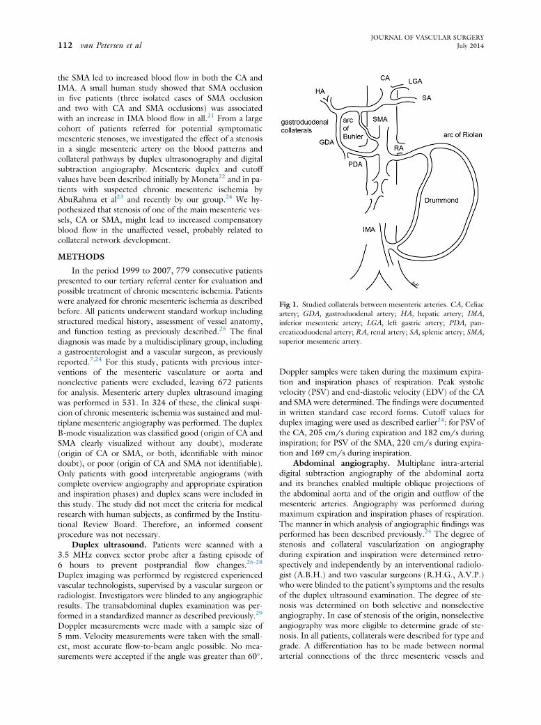
Fig 1. Studied collaterals between mesenteric arteries. CA, Celiacartery; GDA, gastroduodenal artery; HA, hepatic artery; IMA,inferior mesenteric artery; LGA, left gastric artery; PDA, pan-creaticoduodenal artery; RA, renal artery; SA, splenic artery; SMA,superior mesenteric artery.
JOURNAL OF VASCULAR SURGERY112 van Petersen et al July 2014
the SMA led to increased blood flow in both the CA andIMA. A small human study showed that SMA occlusionin five patients (three isolated cases of SMA occlusionand two with CA and SMA occlusions) was associatedwith an increase in IMA blood flow in all.21 From a largecohort of patients referred for potential symptomaticmesenteric stenoses, we investigated the effect of a stenosisin a single mesenteric artery on the blood patterns andcollateral pathways by duplex ultrasonography and digitalsubtraction angiography. Mesenteric duplex and cutoffvalues have been described initially by Moneta22 and in pa-tients with suspected chronic mesenteric ischemia byAbuRahma et al23 and recently by our group.24 We hy-pothesized that stenosis of one of the main mesenteric ves-sels, CA or SMA, might lead to increased compensatoryblood flow in the unaffected vessel, probably related tocollateral network development.
METHODS
In the period 1999 to 2007, 779 consecutive patientspresented to our tertiary referral center for evaluation andpossible treatment of chronic mesenteric ischemia. Patientswere analyzed for chronic mesenteric ischemia as describedbefore. All patients underwent standard workup includingstructured medical history, assessment of vessel anatomy,and function testing as previously described.25 The finaldiagnosis was made by a multidisciplinary group, includinga gastroenterologist and a vascular surgeon, as previouslyreported.7,24 For this study, patients with previous inter-ventions of the mesenteric vasculature or aorta andnonelective patients were excluded, leaving 672 patientsfor analysis. Mesenteric artery duplex ultrasound imagingwas performed in 531. In 324 of these, the clinical suspi-cion of chronic mesenteric ischemia was sustained and mul-tiplane mesenteric angiography was performed. The duplexB-mode visualization was classified good (origin of CA andSMA clearly visualized without any doubt), moderate(origin of CA or SMA, or both, identifiable with minordoubt), or poor (origin of CA and SMA not identifiable).Only patients with good interpretable angiograms (withcomplete overview angiography and appropriate expirationand inspiration phases) and duplex scans were included inthis study. The study did not meet the criteria for medicalresearch with human subjects, as confirmed by the Institu-tional Review Board. Therefore, an informed consentprocedure was not necessary.
Duplex ultrasound. Patients were scanned with a3.5 MHz convex sector probe after a fasting episode of6 hours to prevent postprandial flow changes.26-28
Duplex imaging was performed by registered experiencedvascular technologists, supervised by a vascular surgeon orradiologist. Investigators were blinded to any angiographicresults. The transabdominal duplex examination was per-formed in a standardized manner as described previously.29
Doppler measurements were made with a sample size of5 mm. Velocity measurements were taken with the small-est, most accurate flow-to-beam angle possible. No mea-surements were accepted if the angle was greater than 60�.
Doppler samples were taken during the maximum expira-tion and inspiration phases of respiration. Peak systolicvelocity (PSV) and end-diastolic velocity (EDV) of the CAand SMA were determined. The findings were documentedin written standard case record forms. Cutoff values forduplex imaging were used as described earlier24: for PSV ofthe CA, 205 cm/s during expiration and 182 cm/s duringinspiration; for PSV of the SMA, 220 cm/s during expira-tion and 169 cm/s during inspiration.
Abdominal angiography. Multiplane intra-arterialdigital subtraction angiography of the abdominal aortaand its branches enabled multiple oblique projections ofthe abdominal aorta and of the origin and outflow of themesenteric arteries. Angiography was performed duringmaximum expiration and inspiration phases of respiration.The manner in which analysis of angiographic findings wasperformed has been described previously.24 The degree ofstenosis and collateral vascularization on angiographyduring expiration and inspiration were determined retro-spectively and independently by an interventional radiolo-gist (A.B.H.) and two vascular surgeons (R.H.G., A.V.P.)who were blinded to the patient’s symptoms and the resultsof the duplex ultrasound examination. The degree of ste-nosis was determined on both selective and nonselectiveangiography. In case of stenosis of the origin, nonselectiveangiography was more eligible to determine grade of ste-nosis. In all patients, collaterals were described for type andgrade. A differentiation has to be made between normalarterial connections of the three mesenteric vessels and

Fig 2. A, Collateral visible on selective injection only (grade 1) in a patient with stenosis of the celiac artery (CA).B, Collateral visible on nonselective visceral angiography (grade 2) in a patient with stenosis of the CA. GDA,Gastroduodenal artery; HA, hepatic artery; SA, splenic artery; SMA, superior mesenteric artery.
JOURNAL OF VASCULAR SURGERYVolume 60, Number 1 van Petersen et al 113
pathologic hypertrophic collaterals. We classified onlyinterconnecting vessels as collateral when they were bothclearly visible and hypertrophic. Collaterals found werecategorized in one of the following groups: gastroduo-denal, Buhler, Riolan, and Drummond (Fig 1). For analysisof data, a new definition of collaterals is proposed. Col-laterals were graded as visualized only during selectiveangiography (grade 1) or visualized prominently duringnonselective visceral angiography (grade 2) (Fig 2).
Data management and statistical analysis. Interpret-ability of the duplex and angiography investigations wasdocumented and categorized as good, moderate, andpoor. In cases of occlusion and absent flow signal, datawere excluded from analysis of the mean values. Compar-ison of angiograms and duplex ultrasound scans was per-formed with the same phase of respiration. To study
prevalence of collaterals, five different groups wereanalyzed: patients with normal findings on angiography,patients with <70% stenosis of the CA or SMA, patientswith $70% stenosis of the CA and #70% stenosis of theSMA, patients with $70% stenosis of the SMA and#70% stenosis of the CA, and patients with $70% steno-sis of the CA and SMA. A subanalysis was performed inpatients with an angiographically normal SMA and steno-sis of the CA (normal, <50%, $50%-70%, $70%-99%,occlusion).
Data were expressed as mean and standard error of themean. Independent-samples t test was used for comparisonof duplex parameters between groups. Analysis of variancewith post hoc Tukey test was used for comparison betweenmultiple groups. All data were analyzed with SPSS 20 (SPSS,Chicago, Ill). Statistical significance was assumed for P< .05.

Table. Distinguished cohort of patients suspected to have chronic mesenteric syndrome with good interpretableduplex scans
Vessel stenosis Age (range), years No. of patients
Collateral grade Collateral type
% of N Grade 1 Grade 2 GD B R/D
I Normal 50 (18-81) 46 2 1 0 1II CA and SMA both <70% 51 (14-88) 65 20 11 2 7 1 10III CA $70% and SMA <70% 43 (15-79) 86 53 25 21 41 5 7IV SMA $70% and CA <70% 56 (22-72) 10 50 1 4 2 3V CA and SMA both >70% 61 (44-82) 21 86 3 15 3 1 15Total 49 (14-88) 228 36 52 42 53 7 36
B, Buhler; CA, celiac artery; GD, gastroduodenal; R/D, arcade of Riolan/Drummond; SMA, superior mesenteric artery.Data are shown as mean 6 standard error of the mean.
Fig 3. Effect of stenosis in the celiac artery (CA) on duplex velocity parameters of the CA and superior mesentericartery (SMA) during expiration. Data are shown as mean 6 standard error of the mean. PSV, Peak systolic velocity.*P < .005 comparing stenosis vs no stenosis.
JOURNAL OF VASCULAR SURGERY114 van Petersen et al July 2014
RESULTS
The study cohort consisted of 228 patients suspectedto have chronic mesenteric ischemia and with goodinterpretable duplex ultrasound scans and angiograms.Duplex data for expiration and inspiration were availablein >90% of the patients with an ultrasound assessment ofgood interpretability. Normal findings on angiographyof the CA and SMA were found in 46 patients (20%),stenosis <70% in both CA and SMA in 65 patients(29%), stenosis $70% in the CA and <70% in the SMAin 86 patients (38%), stenosis $70% in the SMAand <70% in the CA in 10 patients (4%), and stenosis$70% of both arteries in 21 patients (9%) (Table).
Group I: Normal inflow of CA and SMA. Com-pletely normal findings on angiography of the CA andthe SMA were found in 46 patients. A grade 1 collateralwas observed in one patient with an IMA occlusion(Table). In this patient, duplex parameters of the CA andSMA were not different from those of patients with normalfindings on angiography.
Group II: <70% stenosis in the CA or SMA. Inpatients with stenosis <70% in either the CA or SMA,collateral circulation was observed in 20%. Grade 1 collat-erals were visualized in 85% of the cases (Table).
No significant changes were found in duplex parame-ters in the angiographically normal CA or SMA in

Fig 4. Patients with an angiographically normal superior mesenteric artery (SMA): effect of grade of celiac artery (CA)stenosis on SMA peak systolic velocity (PSV) during expiration. Data are shown as mean 6 standard error of the mean.P ¼ .009, analysis of variance between groups; *P < .05, post hoc Tukey test compared with normal.
JOURNAL OF VASCULAR SURGERYVolume 60, Number 1 van Petersen et al 115
coexistence with a <70% stenosis in the other mesentericartery (data not shown). Also, the presence of collateralsdid not affect duplex parameters in this group.
Group III: $70% stenosis in the CA. A >70% steno-sis in the CA with an SMA with <70% stenosis was foundin 86 patients. In 53% of these, one or more collateralswere present (Table). In 46% of these patients, this was agrade 2 collateral. The majority (89%) of the observedcollaterals were through the gastroduodenal pathway.
The effect of a$70% stenosis in the CA compared withpatients with <70% stenosis or normal angiography find-ings on duplex parameters in the CA and SMA is shownin Fig 3 (Supplementary Table I, online only). The increasein PSV and EDV in the stenotic CA was as expected. How-ever, in these patients, an increased PSV and EDV in thenot significantly stenotic SMA was seen, during both expi-ration and inspiration (Fig 3 and Supplementary Table I,online only). To further study the effect of CA stenosisseverity on the SMA blood flow, we performed a subanal-ysis of patients with an angiographically normal SMA. Ofthese 178 patients, during expiration 56 had a normalCA, 20 patients had a stenosis of >50% to 70%, 67 patientshad a stenosis of >70%, and 5 patients had an occludedCA. The effect of increasing stenosis of the CA on PSV
and EDV of the normal SMA is shown in Fig 4(Supplementary Table II, online only). Patients with$70% CA stenosis and a grade 2 collateral had a signifi-cantly higher PSV in the SMA compared with thosewithout a collateral (Fig 5 and Supplementary Table III,online only); with grade 1 collaterals, only a trend towardhigher PSV and EDV in the normal SMA was observed(Fig 6 and Supplementary Table IV, online only).
Group IV: $70% stenosis in the SMA. A $70% ste-nosis of only the SMA and not of the CA was found in 10patients. In five patients, collaterals were seen on angiog-raphy, most of them (80%) grade 2.
With a $70% SMA stenosis, the PSV and EDVincreased both in the affected SMA, as expected, and inthe unaffected CA (Fig 7 and Supplementary Table I,online only). The group of patients with an angiographic-ally normal CA was small (n ¼ 55), and just four patientshad a concomitant stenosis of $70% in the SMA. Thesenumbers did not allow further subanalysis in patientswith an angiographically normal CA as performed ingroup III.
Group V:$70% stenosis in both the CA and SMA.Stenosis in both the CA and SMA was observed in 21 pa-tients. A collateral circulation was observed in 86% of these

Fig 5. Patients with an angiographically normal superior mesenteric artery (SMA) with and without$70% celiac artery(CA) stenosis: effect of existence of pathologic collateral pathways on SMA peak systolic velocity (PSV) during expi-ration. Data are shown as mean 6 standard error of the mean. *P < .05 comparing collateral vs no collateral andstenosis vs no stenosis.
JOURNAL OF VASCULAR SURGERY116 van Petersen et al July 2014
patients; 83% of these were grade 2. In all of these patients,an arcade of Riolan was visible. The number of patients wastoo low to reach statistical significance also because of thehigher number of occlusions with consequently no duplexvalues.
DISCUSSION
This study is the first report of mesenteric arterialcollateral pathways and their influence on CA and SMAduplex parameters in relation to mesenteric stenosis in alarge cohort of 228 patients. The data from this study sug-gest that a stenosis grade $70% in the mesenteric arteriesmay be the cutoff for collateral development and increasedcompensatory blood flow. Also, it emerges that not all col-laterals are equal: collaterals observed during nonselectiveangiography always indicated important mesenteric steno-sis, whereas grade 1 collaterals observed during selective in-jection were predominantly seen in normal and minimallystenosed vessels. In this study, a Riolan artery was indica-tive of multivessel disease, whereas gastroduodenal collat-erals were seen frequently in minimally stenotic vessels.
The distinction between collaterals shown on selectiveangiography (grade 1) or nonselective angiography(grade 2) and their clinical implications have, to our knowl-edge, not been reported before. The underlying physics in-volves the relative importance of the blood flow: withselective injection, the bolus may “push away” the normalblood flow, whereas collaterals viewed during nonselective
angiography indicate actual blood flow to the perfusionareas of the stenotic vessel. Indeed, grade 1 was observedpredominantly in patients with less extensive mesentericdisease, whereas grade 2 was observed more in patientswith severe stenosis in the CA and SMA. In patients withgrade 2 collaterals, the flow in the unaffected SMA washigher compared with those with grade 1 collaterals, sup-porting the idea of compensatory blood flow. It seems plau-sible that grade 1 collaterals may develop to grade 2 withincreasing stenosis from progressing mesenteric disease.
Collateral flow in patients with an isolated stenosis ofthe CA was facilitated mainly through the gastroduodenalpathway, in line with previous studies of patients with CAstenosis.12 In the study of Libicher et al,30 balloon occlu-sion of the CA was used as a pretest for endovascularcovering. The most important collateral vessels observedin this study were the pancreaticoduodenal arcades andthe dorsal pancreatic artery. The pancreaticoduodenal col-laterals showed retrograde flow to the gastroduodenal ar-tery and normal antegrade filling of the appropriatehepatic artery. The collaterals via the dorsal pancreatic ar-tery were mainly responsible for filling of the splenic arterybut showed also feeding vessels to the hepatic circulation.In patients with combined CA and SMA stenosis, collateralcirculation was mainly facilitated through the arcade ofRiolan or Drummond. In this group of patients, flow inthe IMA is likely to be higher. In our study, the IMAwas not investigated by standard duplex examination. In

Fig 6. Patients with an angiographically normal superior mesenteric artery (SMA): effect of existence and type ofcollateral pathways on SMA duplex velocity parameters. Data are shown as mean 6 standard error of the mean. PSV,Peak systolic velocity. *P < .05 comparing groups (analysis of variance with post hoc Tukey test) compared with nocollateral.
JOURNAL OF VASCULAR SURGERYVolume 60, Number 1 van Petersen et al 117
the presence of two-vessel disease, a significant rise induplex parameters was found in the CA and SMAcompared with comparable stenosis in single-vessel disease.This suggests that collateral flow from the IMA is not suf-ficient. As the grade of stenosis is comparable, a lower pe-ripheral resistance in the mesenteric arterial circulation canonly explain the increased PSV.
A differentiation of normal arterial connections be-tween the three mesenteric vessels has to be made frompathologic hypertrophic collaterals. We have described col-laterals on the basis of pattern of filling, when clearly visibleand hypertrophic. It is likely that these collaterals havedeveloped or are stimulated by the presence of a stenosis,stimulated by ischemia. We assume that they arise fromsmall and physiologic embryonic remnants of interconnect-ing mesenteric arteries. However, we cannot exclude thatthese were already present before development of stenosis.The fact that these collaterals are not observed in this wayin patients without stenosis, however, makes this unlikely.Most likely, these connections are available in most patientsto be developed when needed.31 In the study of Kornblithet al,9 anatomic specimens of a broad selection of patientswere correlated with angiography findings. An anatomicincidence of 2% was described for the arc of Buhler andwith a visibility index of 2% infrequently visualized on angi-ography. Other collateral pathways were not found ordescribed. The current understanding of collateral network
development involves a complex remodeling of preexistingconduit vessels and is probably driven by inflammatory pro-cesses.32 These inflammatory processes are triggered byischemic events. Because an increase in collateral develop-ment was seen only in patients with >70% stenosis grade,this suggests that this is the minimum stenosis grade fordevelopment of ischemia. Whether the presence of collat-erals is indicative of ischemia development, or conversely,that the lack of collaterals indicates the absence of ischemianeeds to be confirmed.
The measured increased, compensational, blood flow isin line with the small studies available.20,21 Both studiesfound increased blood flow in the other mesenteric arteryin the presence of SMA occlusion. Increased flow is mostlikely to be facilitated by collaterals and consequentlydecreased peripheral resistance. The effect of lower periph-eral resistance on duplex parameters has been documentedin the femoropopliteal bypass. It is known that vasodilatingstimuli (eg, papaverine) are able to increase flow in bypassgrafts.33 In the study of Karacagil et al,34 increase in PSV,mean velocity, and flow volume was found in autografts.33
This effect has also been demonstrated in allograft by-passes, pointing to a decreased peripheral resistance causingan increased flow without change of diameter. In the studyof Boley et al,35 collateral flow was studied in dogs duringexperimental reduction of SMA flow. Flow rose in the CAand IMA after a reduction of flow in the SMA. This rise of

Fig 7. Effect of stenosis in the superior mesenteric artery (SMA) on duplex velocity parameters of the celiac artery(CA) and SMA during expiration. Data are shown as mean 6 standard error of the mean. PSV, Peak systolic velocity.*P < .005 comparing stenosis vs no stenosis.
JOURNAL OF VASCULAR SURGERY118 van Petersen et al July 2014
flow was more prominent in animals with intact collaterals,which evidently shows the function of collaterals. In a studyin dogs after clamping of the SMA, Martin et al36 found anincrease in the rate of flow after the administration ofpapaverine. This effect of papaverine was found only inthe presence of collateral vessels. Our findings are in linewith these observations, although these studies are animalstudies for acute mesenteric ischemia in contrast to our pa-tient group with suspected chronic mesenteric ischemia.
One consequence of the increased blood flow parame-ters in the unaffected mesenteric vessel may be an overesti-mation of the stenosis severity in that vessel. A highervelocity was found in the nonstenosed mesenteric arteryin patients with an isolated stenosis in both the CA andSMA. With use of the currently applicable cutoff valuesfor duplex parameters to define mesenteric stenoses,24
the flow velocities in the nonaffected vessels would havebeen classified as stenosis. If angiography had not been per-formed, this would have resulted in a false-positive diag-nosis (stenosis $70%) in 40% of patients with a singleSMA stenosis and in 7% of patients with a CA stenosis.Therefore, normal values should be viewed in the contextof the other mesenteric vessel assessment. This also empha-sizes the importance of B mode. An increased flow velocitythat is not focal or associated with visualized stenosisshould not necessarily be diagnosed as a stenosis. If flow ve-locities were used without B mode for diagnosis of a 70%
stenosis, the percentage of incorrect diagnoses wouldhave been higher (respectively, 70% and 36%). This couldalso explain the differences in published cutoff values formesenteric duplex parameters23,24 because a higher pro-portion of patients with one-vessel stenosis may lead tomore patients with higher flow in nondiseased vesselswithin the cohort and thus higher normal cutoff values.
The limitation of this study is the retrospective analysisduring a prolonged time. However, because data on duplexultrasound and angiography studies were collected in astandardized, prospective fashion, this probably has littleeffect on the main findings. Another limitation is that weincluded only patients referred for suspected gastrointes-tinal ischemia; no healthy controls were studied. Finally,symptoms have not been analyzed for this study but aresubject to further study.
CONCLUSIONS
This study shows that mesenteric stenosis above 70%leads to more collateral development and higher flowvelocities in the unaffected vessel. Collateral vessels seenon nonselective angiography are indicative of significantmesenteric stenosis. When flow velocities are used as a mea-sure of mesenteric stenosis, the effect of compensatoryincreased blood flow should be taken into account to avoidfalse-positive diagnoses.

JOURNAL OF VASCULAR SURGERYVolume 60, Number 1 van Petersen et al 119
AUTHOR CONTRIBUTIONS
Conception and design: AP, RGAnalysis and interpretation: AP, JK, JP, CZ, RGData collection: AP, JK, RM, AH, RGWriting the article: AP, JK, CZ, RGCritical revision of the article: AP, JK, RM, AH, JP, CZ,
RGFinal approval of the article: AP, JK, RM, AH, JP, CZ, RGStatistical analysis: AP, JPObtained funding: Not applicableOverall responsibility: AP
REFERENCES
1. Kerkhof I, Kolkman JJ, Geelkerken RH, Brusse M. Incidence ofchronic gastrointestinal ischemia: a large Dutch cohort study. Gastro-enterology 2013;144:S902-3.
2. Roobottom CA, Dubbins PA. Significant disease of the celiac and su-perior mesenteric arteries in asymptomatic patients: predictive value ofDoppler sonography. AJR Am J Roentgenol 1993;161:985-8.
3. Thomas JH, Blake K, Pierce GE, Hermreck AS, Seigel E. The clinicalcourse of asymptomatic mesenteric arterial stenosis. J Vasc Surg1998;27:840-4.
4. Hansen KJ, Wilson DB, Craven TE, Pearce JD, English WP,Edwards MS, et al. Mesenteric artery disease in the elderly. J Vasc Surg2004;40:45-52.
5. Fried LP, Borhani NO, Enright P, Furberg CD, Gardin JM,Kronmal RA, et al. The Cardiovascular Health study: design andrationale. Ann Epidemiol 1991;1:263-76.
6. Mensink PB, van Petersen AS, Geelkerken RH, Otte JA, Huisman AB,Kolkman JJ. Clinical significance of splanchnic artery stenosis. Br J Surg2006;93:1377-82.
7. Mensink PB, van Petersen AS, Kolkman JJ, Otte JA, Huisman AB,Geelkerken RH. Gastric exercise tonometry: the key investigation inpatients with suspected celiac artery compression syndrome. J VascSurg 2006;44:277-81.
8. Fisher DF Jr, Fry WJ. Collateral mesenteric circulation. Surg GynecolObstet 1987;164:487-92.
9. Kornblith PL, Boley SJ, Whitehouse BS. Anatomy of the splanchniccirculation. Surg Clin North Am 1992;72:1-30.
10. Rosenblum JD, Boyle CM, Schwartz LB. The mesenteric circulation.Anatomy and physiology. Surg Clin North Am 1997;77:289-306.
11. Walker TG. Mesenteric vasculature and collateral pathways. SeminIntervent Radiol 2009;26:167-74.
12. Song SY, Chung JW, Kwon JW, Joh JH, Shin SJ, Kim HB, et al.Collateral pathways in patients with celiac axis stenosis: angiographic-spiral CT correlation. Radiographics 2002;22:881-93.
13. van Bockel JH, Geelkerken RH, Wasser MN. Chronic splanchnicischaemia. Best Pract Res Clin Gastroenterol 2001;15:99-119.
14. van Everdingen KJ, Klijn CJ, Kappelle LJ, Mali WP, van der Grond J.MRA flow quantification in patients with a symptomatic internal ca-rotid artery occlusion. The Dutch EC-IC Bypass Study Group. Stroke1997;28:1595-600.
15. Fujitani RM, Mills JL, Wang LM, Taylor SM. The effect of unilateralinternal carotid arterial occlusion upon contralateral duplex study:criteria for accurate interpretation. J Vasc Surg 1992;16:459-67;discussion: 467-8.
16. Forconi S, Johnston KW. Effect of contralateral internal carotid stenosison the accuracy of continuous wave Doppler spectral analysis results.J Cardiovasc Surg (Torino) 1987;28:715-8.
17. AbuRahma AF, Richmond BK, Robinson PA, Khan S, Pollack JA,Alberts S. Effect of contralateral severe stenosis or carotid occlusion onduplex criteria of ipsilateral stenoses: comparative study of variousduplex parameters. J Vasc Surg 1995;22:751-61; discussion: 761-2.
18. Kolkert JL, van den Dungen JJ, Loonstra J, Tielliu IF, Verhoeven EL,Beck AW, et al. Overestimation of contralateral internal carotid artery
stenosis before ipsilateral surgical endarterectomy. Eur J Radiol2011;77:68-72.
19. Boley SJ, Regan JA, Tunick PA, Everhard ME, Winslow PR, Veith FJ.Persistent vasoconstrictionda major factor in nonocclusive mesentericischemia. Curr Top Surg Res 1971;3:425-33.
20. Boley SJ, Brandt LJ, Veith FJ. Ischemic disorders of the intestines. CurrProbl Surg 1978;15:1-85.
21. Erden A, Yurdakul M, Cumhur T. Doppler waveforms of the normaland collateralized inferior mesenteric artery. AJR Am J Roentgenol1998;171:619-27.
22. Moneta GL, Lee RW, Yeager RA, Taylor LM Jr, Porter JM. Mesentericduplex scanning: a blinded prospective study. J Vasc Surg 1993;17:79-84; discussion: 85-6.
23. AbuRahma AF, Stone PA, Srivastava M, Dean LS, Keiffer T, Hass SM,et al. Mesenteric/celiac duplex ultrasound interpretation criteriarevisited. J Vasc Surg 2012;55:428-36.e6.
24. van Petersen AS, Meerwaldt R, Kolkman JJ, Huisman AB, van derPalen J, van Bockel JH, et al. The influence of respiration on criteria fortransabdominal duplex examination of the splanchnic arteries inpatients with suspected chronic splanchnic ischemia. J Vasc Surg2013;57:1603-11.
25. Otte JA, Geelkerken RH, Huisman AB, Kolkman JJ. What is the bestdiagnostic approach for chronic gastrointestinal ischemia? Am J Gas-troenterol 2007;102:2005-10.
26. Volteas N, Labropoulos N, Leon M, Kalodiki E, Chan P,Nicolaides AN. Detection of superior mesenteric and coeliac arterystenosis with colour flow Duplex imaging. Eur J Vasc Surg 1993;7:616-20.
27. Gentile AT, Moneta GL, Lee RW, Masser PA, Taylor LM Jr,Porter JM. Usefulness of fasting and postprandial duplex ultrasoundexaminations for predicting high-grade superior mesenteric arterystenosis. Am J Surg 1995;169:476-9.
28. Mitchell EL, Moneta GL. Mesenteric duplex scanning. Perspect VascSurg Endovasc Ther 2006;18:175-83.
29. Delahunt TA, Geelkerken RH, Hermans J, Van Baalen JM,Vaughan AJ, Hajo Van Bockel J. Comparison of trans- and intra-abdominal duplex examinations of the splanchnic circulation. Ultra-sound Med Biol 1996;22:165-71.
30. Libicher M, Reichert V, Aleksic M, Brunkwall J, Lackner KJ,Gawenda M. Balloon occlusion of the celiac artery: a test for evaluationof collateral circulation prior endovascular coverage. Eur J VascEndovasc Surg 2008;36:303-5.
31. Bertelli L, Lorenzini L, Bertelli E. The arterial vascularization of thelarge intestine. Anatomical and radiological study. Surg Radiol Anat1996;18(Suppl 1):A1-6. S1-59.
32. la Sala A, Pontecorvo L, Agresta A, Rosano G, Stabile E. Regulation ofcollateral blood vessel development by the innate and adaptive immunesystem. Trends Mol Med 2012;18:494-501.
33. Karacagil S, Granbo A, Almgren B, Ljungman C, Bergqvist D. Theeffect of postocclusion reactive hyperaemia, papaverine and nifedipineon duplex derived haemodynamic parameters of infrainguinal bypassgrafts. Eur J Vasc Endovasc Surg 1995;9:107-11.
34. Pedersen G, Laxdal E, Amundsen SR, Dregelid E, Jonung T,Nyheim T, et al. Flow measurement before and after papaverine in-jection in above-knee prosthetic femoropopliteal bypass. J Vasc Surg2006;43:729-34.
35. Boley SJ, Treiber W, Winslow PR, Gliedman ML, Veith FJ. Circulatoryresponse to acute reduction of superior mesenteric arterial blood flow.Physiologist 1969;12:180.
36. Martin WB, Laufman H, Tuell SW. Rationale of therapy in acutevascular occlusions based upon micrometric observations. Ann Surg1949;129:476-93.
Submitted Nov 26, 2013; accepted Jan 25, 2014.
Additional material for this article may be found onlineat www.jvascsurg.org.

Supplementary Table I (online only). Effect of stenosis in either the celiac artery (CA) or superior mesenteric artery(SMA) on duplex velocity parameters (inspiratory and expiratory) of both vessels
Stenosis in CA
Pa
Stenosis in SMA
Pa
Normal Stenosis $70%
<70%, $70%, <70%, $70%, CA and SMA, CA and SMA,(n ¼ 121) (n ¼ 107) (n ¼ 197) (n ¼ 31) (n ¼ 46) (n ¼ 21)
PSV, cm/sCA, expiration 218 6 9 326 6 16 <.0005 251 6 9 352 6 40 .001 193 6 16 417 6 57CA, inspiration 219 6 9 334 6 26 <.0005 232 6 9 350 6 45 <.0005 204 6 25 451 6 69SMA, expiration 178 6 8 215 6 11 .006 183 6 6 333 6 42 <.0005 154 6 9 350 6 53SMA, inspiration 157 6 6 199 6 17 .004 156 6 5 296 6 33 <.0005 135 6 10 343 6 50
EDV, cm/sCA, expiration 73 6 5 124 6 10 <.0005 88 6 5 139 6 29 .003 63 6 8 176 6 45CA, inspiration 65 6 4 135 6 17 <.0005 72 6 4 148 6 32 <.0005 56 6 8 209 6 52SMA, expiration 33 6 2 55 6 6 .0005 37 6 2 117 6 29 <.0005 29 6 3 129 6 41SMA, inspiration 31 6 2 53 6 9 <.0005 31 6 2 94 6 17 <.0005 25 6 2 115 6 28
EDV, End-diastolic velocity; PSV, peak systolic velocity.Values are mean 6 standard error of the mean; duplex ultrasonography and angiography compared with same respiration phase.aComparing stenosis vs no stenosis (t test).
Supplementary Table II (online only). Effect of stenosis in the celiac artery (CA) on duplex velocity parameters(inspiratory and expiratory) of the normal superior mesenteric artery (SMA)
CA stenosis
PaNormal <50% 50%-<70% 70%-99% Occlusion
Expiration n ¼ 56 n ¼ 30 n ¼ 20 n ¼ 67 n ¼ 5SMA PSV, cm/s 158 6 9 153 6 13 172 6 13 190 6 10 259 6 61b .009SMA EDV, cm/s 28 6 2 29 6 3 28 6 2 43 6 4b 58 6 15 .001
Inspiration n ¼ 66 n ¼ 51 n ¼ 25 n ¼ 30 n ¼ 5SMA PSV, cm/s 137 6 8 153 6 8 146 6 11 159 6 13 187 6 32 .246SMA EDV, cm/s 26 6 2 30 6 2 23 6 2 35 6 7 41 6 6 .112
EDV, End-diastolic velocity; PSV, peak systolic velocity.Values are mean 6 standard error of the mean; duplex ultrasonography and angiography compared with same respiration phase.aComparing different groups (analysis of variance).bP < .05 (post hoc Tukey test) compared with normal.
Supplementary Table III (online only). Patients with an angiographically normal superior mesenteric artery (SMA)with and without $70% celiac artery (CA) stenosis: Effect of existence of pathologic collateral pathways on SMA peaksystolic velocity (PSV) during expiration
$70% CA stenosis during expiration
Grade 2 collateral
No Yes P Total
No 159 6 6 (96) 180 6 43 (3) .562 160 6 6Yes 182 6 9 (54) 236 6 29 (15) .025 194 6 10P .036 .428 .002Total 167 6 5 227 6 25 .001
Values are mean PSV (cm/s) 6 standard error of the mean, and values in parentheses are number of patients.
JOURNAL OF VASCULAR SURGERY119.e1 van Petersen et al July 2014

Supplementary Table IV (online only). Patients with an angiographically normal superior mesenteric artery (SMA):Effect of existence and type of collateral pathways on SMA duplex velocity parameters
SMA duplex parameters
Collateral
Pa Pb PcNo
n ¼ 128Grade 1n ¼ 31
Grade 2n ¼ 19
PSV, expiration, cm/s 165 6 6 179 6 13 227 6 25 .591 .068 .002PSV, inspiration, cm/s 143 6 5 157 6 9 182 6 21 .493 .366 .034EDV, expiration, cm/s 31 6 2 42 6 7 50 6 8 .080 .472 .005EDV, inspiration, cm/s 27 6 1 30 6 2 43 6 11 .631 .109 .006
EDV, End-diastolic velocity; PSV, peak systolic velocity.Values are mean 6 standard error of the mean. Analysis of variance with post hoc Tukey test.aBetween no collateral and grade 1.bBetween grade 1 and grade 2.cBetween no collateral and grade 2.
JOURNAL OF VASCULAR SURGERYVolume 60, Number 1 van Petersen et al 119.e2
Copyright © 2022 FDOKUMEN