Drug resistance in HIV-1 protease: Flexibility-assisted mechanism of compensatory mutations
10
Drug resistance in HIV-1 protease: Flexibility-assisted mechanism of compensatory mutations STEFANO PIANA, 1 PAOLO CARLONI, 2 AND URSULA ROTHLISBERGER 1 1 Laboratory of Inorganic Chemistry, ETH Hönggerberg–HCI, Zürich, Switzerland 2 Scuola Internazionale Superiore di Studi Avanzati and INFM–DEMOCRITOS National Simulation Center, Trieste, Italy (RECEIVED March 11, 2002; FINAL REVISION July 15, 2002; ACCEPTED July 17, 2002) Abstract The emergence of drug-resistant variants is a serious side effect associated with acquired immune deficiency syndrome therapies based on inhibition of human immunodeficiency virus type 1 protease (HIV-1 PR). In these variants, compensatory mutations, usually located far from the active site, are able to affect the enzymatic activity via molecular mechanisms that have been related to differences in the conformational flexibility, although the detailed mechanistic aspects have not been clarified so far. Here, we perform multinanosecond molecular dynamics simulations on L63P HIV-1 PR, corresponding to the wild type, and one of its most frequently occurring compensatory mutations, M46I, complexed with the substrate and an enzymatic intermediate. The quality of the calculations is established by comparison with the available nuclear magnetic resonance data. Our calculations indicate that the dynamical fluctuations of the mutated enzyme differ from those in the wild type. These differences in the dynamic properties of the adducts with the substrate and with the gem-diol intermediate might be directly related to variations in the enzymatic activity and therefore offer an explanation of the observed changes in catalytic rate between wild type and mutated enzyme. We anticipate that this “flexibility-assisted” mechanism might be effective in the vast majority of compensatory mutations, which do not change the electrostatic properties of the enzyme. Keywords: HIV-1 protease; molecular dynamics; protein flexibility; compensatory mutations; drug resis- tance Supplemental material: See www.proteinscience.org. The aspartyl protease from the human immunodeficiency virus type 1 (HIV-1 PR) is an enzyme essential for the viral metabolism (Wlodawer and Vondrasek 1998). Some of its inhibitors constitute the most powerful anti-acquired im- mune deficiency syndrome (AIDS) drugs and have been used in the clinics for several years. Unfortunately, the long- term efficiency of these antiviral agents is severely limited by the emergence of viral variants that display cross-resis- tance against HIV-1 PR inhibitors (Erickson and Burt 1996; Gulnik et al. 2000; Young and Kuritzkes 2001) (Fig. 1). Apparently, these variants are able to evolve resistance by complementary strategies: several active-site (Fig. 1, red) and nonactive-site (Fig.1, green) mutations reduce the af- finity for both drugs and substrates. This may be achieved by disrupting favorable drug-protease interactions (Chen et al. 1995; Markowitz et al. 1995; Schaffer and Verkhivker 1998; Tawa et al. 1998; Mahalingam et al. 1999,2001; Rosin et al. 1999; Hong et al. 2000; Wang and Kollman 2001). In contrast, “compensatory mutations” (Fig. 1, blue) are able to enhance the protease activity that is decreased by the active-site mutations (Gulnik et al. 2000). The way these variants exert their catalytic role is not fully understood. Compensatory mutations are usually lo- cated away from the active site and do not involve charged residues. Thus, they cannot influence the enzymatic reac- Reprint requests to: Professor Paolo Carloni, International School for Advanced Studies, via Beirut 4,34014, Trieste, Italy; e-mail: [email protected]; fax: +39-040-3787-528 Article and publication are at http://www.proteinscience.org/cgi/doi/ 10.1110/ps.0206702. Protein Science (2002), 11:2393–2402. Published by Cold Spring Harbor Laboratory Press. Copyright © 2002 The Protein Society 2393
-
Upload
independent -
Category
Documents
-
view
0 -
download
0
Transcript of Drug resistance in HIV-1 protease: Flexibility-assisted mechanism of compensatory mutations
Drug resistance in HIV-1 protease: Flexibility-assistedmechanism of compensatory mutations
STEFANO PIANA,1 PAOLO CARLONI,2 AND URSULA ROTHLISBERGER1
1Laboratory of Inorganic Chemistry, ETH Hönggerberg–HCI, Zürich, Switzerland2Scuola Internazionale Superiore di Studi Avanzati and INFM–DEMOCRITOS National Simulation Center,Trieste, Italy
(RECEIVED March 11, 2002; FINAL REVISION July 15, 2002; ACCEPTED July 17, 2002)
Abstract
The emergence of drug-resistant variants is a serious side effect associated with acquired immune deficiencysyndrome therapies based on inhibition of human immunodeficiency virus type 1 protease (HIV-1 PR). Inthese variants, compensatory mutations, usually located far from the active site, are able to affect theenzymatic activity via molecular mechanisms that have been related to differences in the conformationalflexibility, although the detailed mechanistic aspects have not been clarified so far. Here, we performmultinanosecond molecular dynamics simulations on L63P HIV-1 PR, corresponding to the wild type, andone of its most frequently occurring compensatory mutations, M46I, complexed with the substrate and anenzymatic intermediate. The quality of the calculations is established by comparison with the availablenuclear magnetic resonance data. Our calculations indicate that the dynamical fluctuations of the mutatedenzyme differ from those in the wild type. These differences in the dynamic properties of the adducts withthe substrate and with the gem-diol intermediate might be directly related to variations in the enzymaticactivity and therefore offer an explanation of the observed changes in catalytic rate between wild type andmutated enzyme. We anticipate that this “flexibility-assisted” mechanism might be effective in the vastmajority of compensatory mutations, which do not change the electrostatic properties of the enzyme.
Keywords: HIV-1 protease; molecular dynamics; protein flexibility; compensatory mutations; drug resis-tance
Supplemental material: See www.proteinscience.org.
The aspartyl protease from the human immunodeficiencyvirus type 1 (HIV-1 PR) is an enzyme essential for the viralmetabolism (Wlodawer and Vondrasek 1998). Some of itsinhibitors constitute the most powerful anti-acquired im-mune deficiency syndrome (AIDS) drugs and have beenused in the clinics for several years. Unfortunately, the long-term efficiency of these antiviral agents is severely limitedby the emergence of viral variants that display cross-resis-tance against HIV-1 PR inhibitors (Erickson and Burt 1996;
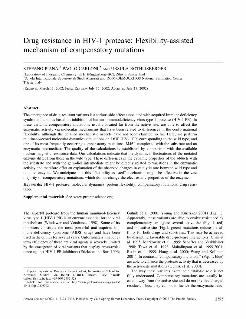
Gulnik et al. 2000; Young and Kuritzkes 2001) (Fig. 1).Apparently, these variants are able to evolve resistance bycomplementary strategies: several active-site (Fig. 1, red)and nonactive-site (Fig.1, green) mutations reduce the af-finity for both drugs and substrates. This may be achievedby disrupting favorable drug-protease interactions (Chen etal. 1995; Markowitz et al. 1995; Schaffer and Verkhivker1998; Tawa et al. 1998; Mahalingam et al. 1999,2001;Rosin et al. 1999; Hong et al. 2000; Wang and Kollman2001). In contrast, “compensatory mutations” (Fig. 1, blue)are able to enhance the protease activity that is decreased bythe active-site mutations (Gulnik et al. 2000).
The way these variants exert their catalytic role is notfully understood. Compensatory mutations are usually lo-cated away from the active site and do not involve chargedresidues. Thus, they cannot influence the enzymatic reac-
Reprint requests to: Professor Paolo Carloni, International School forAdvanced Studies, via Beirut 4,34014, Trieste, Italy; e-mail:[email protected]; fax: +39-040-3787-528
Article and publication are at http://www.proteinscience.org/cgi/doi/10.1110/ps.0206702.
Protein Science (2002), 11:2393–2402. Published by Cold Spring Harbor Laboratory Press. Copyright © 2002 The Protein Society 2393

tivity through long-range electrostatic interactions (Warshel1978) and in fact they appear to have little effect on sub-strate and inhibitor binding (Pazhanisamy et al. 1996;Schock et al. 1996). Intriguingly, they also lead to onlyminor geometric rearrangements that cannot account for themarked increase in catalytic activity (Chen et al. 1995;Schock et al. 1996; Ridky et al. 1998).
In the last years, several laboratories have pointed to thedifference in conformational flexibility between wild typeand mutants as a possible key factor for the catalytic activity(Chen et al. 1995; Gulnik et al. 2000). This proposal iscorroborated by X-ray crystallography and theoretical data.In fact, in the structures solved so far that contain compen-satory mutations (K45I [Mahalingam et al. 2001], M46I[Chen et al. 1995], L90M [Hong et al. 2000]), the mutatedenzymes turn out to be less flexible as indicated by lower Bfactors. The motion of the flap tips in the free HIV-1 PR hasalso been investigated with nuclear magnetic resonance
(NMR) (Ishima et al. 1999; Freedberg et al. 2002) and mo-lecular dynamics simulation (Scott and Schiffer 2000).These studies indicate that the flaps are highly mobile andcan adopt a large number of conformations on the ns to mstimescale, thus suggesting that mutations in the flaps can bevery important for the kinetics of substrate and inhibitorbinding. A classical molecular dynamics (MD) study onwild type and M46I variant free enzyme, arguably the mostcommon and representative compensatory mutation (Ho etal. 1994) (Fig. 1), has indicated that the two systems differin their flexibility and, in particular, that this mutation ap-pears to stabilize the closed conformation of the flaps (Col-lins et al. 1995). However, the M46I mutation appears tohave little effect on the HIV-1 PR affinity for most of theinhibitors (Pazhanisamy et al. 1996; Schock et al. 1996),thus indicating that other factors, besides the binding affin-ity must play a role in this kind of mutation.
Our recent quantum chemical and MD calculations on thecomplex between L63P HIV-1 PR3 and a peptide substrate(SUB) have indicated that the overall flexibility of the en-zyme/substrate complex is an essential ingredient for theenzymatic function (Piana et al. 2002). Specifically, large-scale motions of the enzyme have been found to be essentialto drive the substrate in an appropriate position in which thecatalytic cleavage can occur (Piana et al. 2002). Similarconclusions have been drawn for other enzymatic systems(Lau and Bruice 2000; Radkiewicz and Brooks 2000). Thishas led to the suggestion that conformational flexibilitymight play a role not only in substrate or inhibitor binding,but also for the increase in catalytic efficiency.
Here, we have used multi-ns MD on complexes of L63Penzyme (which can be considered as a representative of thewild type enzyme3) and the double mutant M46I-L63P toshed light on this issue. Specifically, we have prolonged oursimulation of the L63P/SUB complex to 11 ns and per-formed simulations on the same time scale for the complexbetween L63P-M46I HIV-1 PR and SUB as well as with thecorresponding gem-diol intermediate (INT) of the catalyticcycle (Hyland et al. 1991) (Chart 1).
Our calculations suggest that while the MD-averagedstructures of L63P/SUB and M46I-L63P/SUB turn out to be
Fig. 1. Inhibitor binding and drug resistance in human immunodeficiencyvirus type 1 protease (HIV-1 PR). The figure shows the complex betweenthe protease (gray) and the A79285 inhibitor (yellow) (Wlodawer andErickson 1993). The five U.S. Food and Drug Administration (FDA)-approved inhibitors for clinical use (saquinavir, indinavir, ritonavir, nelfi-navir, and amprenavir) bind, like A79285, in the active site cavity (red) soas to compete with peptide substrate binding (Wlodawer and Erickson1993). The figure also displays selected residues undergoing mutationsassociated with viral resistance to the FDA-approved drugs. Active-sitemutations (red spheres) are located in direct contact with the proteaseinhibitors such as to disrupt favorable drug-enzyme interactions. Othermutations (green) are often located in close proximity of the active site andinfluence the energetics of binding through second-order effects. Compen-satory mutations (blue) are located away from the active site and their roleusually cannot be inferred from structural studies. M46I is a very commonmutation associated with compensatory cross-resistance against four of theFDA-approved drugs (Condra et al. 1996; Mammano et al. 2000; Youngand Kuritzkes 2001) and in combination with L63P, is found to increase thecatalytic activity by 110–530% in a large wealth of diverse substrates(Pazhanisamy et al. 1996; Schock et al. 1996; Mahalingam et al. 1999). Asother variants exhibiting compensatory resistance (L63P, I47V, A71T), thismutation is located away from the active site and does not involve chargedresidues.
Chart 1.
Piana et al.
2394 Protein Science, vol. 11

very similar, their flexibility differs distinctly, especially inthe flap and substrate regions. Furthermore, the M46I mu-tation induces a displacement of the reaction intermediatetoward the active site. This demonstrates that relativelysmall local changes in the protein structure can lead to al-terations of the dynamical properties, which are intimatelylinked to the enzymatic activity (Piana et al. 2002). Both theinduced change in substrate flexibility and intermediateconformation may account for the difference in catalyticrates between the two forms of the enzyme.
Results and Discussion
Structural features
The structures of all the complexes investigated here (L63P/SUB, L63P/INT, L63P-M46I/SUB, and L63P-M46I/INT)were stable over the time scale investigated (7–16 ns), asindicated by the small values of the root mean square de-viation (rmsd) of the backbone atoms with respect to thestarting structure (rmsd ∼ 1.0–1.4 Å) (Fig. 1SI of Supple-mental Material). A comparison of the MD-averaged struc-tures of the four complexes (Fig. 2S and 2SI of Supplemen-tal Material) further confirms that the overall folding is wellmaintained in the four systems.
Overall, the four adducts share similar structural features.The largest differences between initial and MD-averagedstructures of the four complexes are observed in the mostflexible protein regions [residues 1(1�)–4(4�), 16(16�)–18(18�) 38(38�)–42(42�) and 68(68�)–71(71�)] and in the
flap tips [residues 48(48�)–51(51�)]. In particular, the MD-averaged structural properties around site 46 are rather simi-lar (see Table 1 and Table 2SI of Supplemental Material).This result is in line with previous MD calculations (Harteet al. 1992; Luo et al. 1998) and with experimental data(Chen et al. 1995).
Most of the hydrogen-bond interactions between SUB (orINT) and HIV-1 PR are maintained during the four simu-lations (Table 2); the few hydrogen bonds that are lost arelocated between SUB (or INT) and the cleavage site region.Interestingly, two different binding modes are observed forthe side chain of Gln P2� (Fig. 2B,D), a residue that isimportant for HIV-1 PR activity (Poorman et al. 1991; Pol-gár et al. 1994; Szeltner and Polgár 1996). In the L63P/INT
Table 1. Molecular dynamics-averaged values (degrees) ofselected dihedral angles relative to residue 46(46�)
Dihedral angle WT/SUB M46I/SUB WT/INT M46I/INT
46 � −127 (16) −134 (10) −128 (19) −132 (12)� 134 (17) 135 (11) 145 (12) 139 (9)
46� � −130 (13) −133 (13) −133 (12) −135 (11)� 139 (15) 135 (13) 143 (11) 141 (9)
C�46-C�46-N46-C45 110 (17) 99 (10) 107 (20) 102 (12)C�46-C�46-C46-N45 −104 (17) −100 (11) −91 (13) −97 (9)C�46�-C�46�-N46�-C45� 107 (13) 99 (13) 104 (13) 99 (11)C�46�-C�46�-C46�-N45� −99 (15) −100 (12) −95 (11) −94 (9)
� � C-N-C�-C; � � N-C�-C-N. Standard deviations are reported inparenthesis.
Table 2. Hydrogen bonds between SUB (or INT) and HIV-1 PR
Substrate-proteinhydrogen bonds
Presence of the interaction (%)
4HVP WT/SUB M46I/SUB WT/INT M46I/INT
N-H1,2,3(P3). . .O�1,2(48) 100 30 100 99 100N-H(29). . .O(P3) 100 27 95 97 100N-H(P2). . .O(48) 100 100 99 93 100O-H(wat_b). . .O(P2) 100 99 100 100 99N-H(P1). . .O(27) 100 1 3 39 27O-H(wat_b). . .O(P1�) 100 98 99 100 99N-H(P2�). . .O(27�) 100 84 89 12 95N�-H�1(P2�). . .O(30�) 0 100 100 99 94N-H(29�). . .O�(P2�) 0 81 90 25 79N-H(P3�). . .O(48�) 100 95 79 99 99N-H(48). . .O1,2(P3�) 0 9 20 100 100
Labeling as in Chart 2. In the Table, the A-H. . .B H-bond is present at each step of thedynamics if d(H. . .A) < 3.0 Å and ∠ (B-H. . .A) < 120°. 4HVP is the crystal structure ofHIV-1 PR complexed with MVT101 (Miller et al. 1989). The overall agreement is fairlygood, as most of the H-bonds present in the crystal structure are maintained in themolecular dynamics simulations. The few exceptions (presence < 50%) that are marked inbold face, are located at the cleavage site region. The largest discrepancy, which involvesN-H(P2�). . .O(27�), could be a consequence of the structural differences between theMVT101 inhibitor (Miller et al. 1989) and SUB/INT (Rose et al. 1996). Other significantdiscrepancies are found for the two hydrogen bonds that restrain the P3 terminal substratein wild type/SUB, indicating a large flexibility of this residue (Fig. 4B) and for the twohydrogen bonds at the P2� site in wild type/INT. In this case, the side chain of GlnP2�adopts a different binding mode and there does not seem to be a large effect on the INTflexibility at the P2� site (Fig. 4C).
Molecular dynamics simulations of HIV-1 protease
www.proteinscience.org 2395

simulation the binding is the same as in the crystal structure;in the other three simulations the O� of Gln P2� is hydrogenbonded to the backbone amide nitrogen of Asp 29�.
Finally, we note that the hydrogen bonds in the loopsregion are mostly preserved, although occasional local un-folding events are observed in the glycine-rich tip of the flapregion (residues 49–52) (Table 1SI of Supplemental Mate-rial).
We conclude that the structural differences between L63Pand the L63P-M46I MD-averaged structures are small. Thisagrees with a structural study on the M46I-L63P-V82T-I84V quadruple mutant (Chen et al. 1995), which is found toexhibit only a minor rearrangement of the flaps relative towild type.
Dynamics: Local fluctuations
We now turn our attention to the flexibility of the protein,which is the main focus of this work. The conformationalfluctuations of the adducts in the sub-ns time scale can bedescribed in terms of generalized order parameters S2 forwhich direct contact with NMR data (Lipari and Szabo1982a) can be made (Lipari and Szabo 1982b; Luo et al.1998). The calculated values (Fig. 3) turn out to be verysimilar in the four systems and are in excellent agreement
with the available experimental data (Nicholson et al. 1995;Tjandra et al. 1996; Freedberg et al. 1998). The only residuethat systematically exhibits large deviations is Gly 68(68�)whose calculated order parameter (0.6) is much lower thanthe experimental values (0.85–0.9). This discrepancy mightarise from the fact that Gly68(68�) is part of the 68(68�)–71(71�) mobile loop. Indeed, this loop encompasses the sol-vent-exposed His69(69�), which is very likely to be affectedby proton exchange at the imidazole ring on time scaleslonger than the one simulated here. This proton exchange,which cannot be accounted for in our MD simulations,could influence the dynamics of the adjacent Gly 68 residuegiving rise to the discrepancy in the order parameters.
Further information about the local mobility of the pro-tein can be obtained by a plot of the rmsd for each residuewith respect to the MD-averaged structure (Fig. 4A–C). Inall four systems, the largest flexibility is observed in theregions characterized by the presence of one or more resi-dues with a low order parameter (residues 16–18, 37–42,68–71, and 78–82 in Fig. 3 and 4A) (Nicholson et al. 1995;Tjandra et al. 1996; Freedberg et al. 1998), which mostlycorrespond to the protein loops. This result is in agreementwith a recent study (Zoete et al. 2002) where 73 crystalstructures of HIV-1 PR are compared; based on the differ-
Fig. 2. MD-averaged structures: Wild type/SUB complex (red); M46I PR/SUB complex (yellow); wild type/INT complex (blue);M46I/INT complex (cyan). (A) Wild type/SUB complexes and (B) active-site region. (C) Wild type/INT complexes and (D) active-siteregion. In A and C, only the backbone C�, C, and N atoms are shown for clarity. Note the different binding modes adopted by GlnP2� in (C).
Piana et al.
2396 Protein Science, vol. 11

ences in the crystal structures in combination with MD re-sults, the authors conclude that these are the regions oflargest flexibility.
The flap regions at the active site [the so-called “flaptips”, corresponding to residues 45(45�)–55(55�)] are moremobile in the complexes with SUB than in those with INT.The difference is not surprising as this region is in closecontact with the ligand and its conformation clearly influ-ences binding (Baca and Kent 1993; Prabu-Jeyabalan et al.2000). Indeed, in this area large differences are observed in
the S2 values for HIV-1 PR bound to different inhibitors(Nicholson et al. 1995; Freedberg et al.1998) and for theresidues of the two HIV-1 PR subunits in presence of anasymmetric inhibitor (Freedberg et al. 1998).
Differences in flexibility are also observed between L63Pand the L63P-M46I mutant in complex with either SUB orINT (Fig. 4A). The flap elbows (residue 38–42) turn out tobe more flexible in the double mutant. However, the restof the flaps (residues 45–49 and 52–57), as well as SUB/INT (Fig. 4B,C), are more flexible in the L63P enzyme.Accordingly, although the average conformation of residue46(46�) is very much the same in the four simulations(Table 1 and 2SI), the standard deviations of the torsionalangles clearly indicate that SUB is more flexible than INTand L63P is more flexible than M46I-L63P. All these find-ings are in line with previous MD calculations of the freeM46I enzyme in vacuum (Collins et al. 1995), which indi-cated that the M46I mutation strongly influences the dy-namics of flap opening.
Thus, we conclude that the M46I mutation inducesmostly a lower flexibility, although the MD-averaged struc-tural properties are similar.
Dynamics: Large-scale motions
The overall flexibility of the system can be further analyzedin terms of correlations between the substrate and the pro-tein motions (Fig. 5a–d) (Harte et al. 1992; Radkiewicz andBrooks 2000; Piana et al. 2002). The corresponding dy-namic cross-correlation matrix indicates that in all four sys-tems the substrate motions are tightly coupled to fluc-
Fig. 3. Calculated generalized-order parameters (S2)(Lipari and Szabo1982a,b) for the backbone amide groups. Wild type/SUB complex (red);M46I/SUB complex (yellow); wild type/INT complex (blue); M46I/INTcomplex (cyan). The experimental values measured for wild type com-plexed with KNI272 (Freedberg et al. 1998), P9941 (Nicholson et al.1995), DMP323 (at 300Mhz)(Nicholson et al. 1995), DMP323 (2) (at600Mhz) (Nicholson et al. 1995; Tjandra et al. 1996) inhibitors are repre-sented as ‘+’, ‘�’, ‘x’, ‘*’, ‘�’, respectively. Only one of the two mono-mers is shown for clarity, the other monomer shares very similar patterns.
Fig. 4. Average root mean square deviation (rmsd) calculated on each residue of the protein (A), peptide substrate (SUB) (B), gem-diolintermediate (INT) (C) relative to the average molecular dynamics structure: Wild type/SUB complex (red); M46I/SUB complex(yellow); wild type/INT complex (blue); M46I/INT complex (cyan). The difference between the value calculated for the wild type andthe M46I enzyme is reported as ‘+’ for the human immunodeficiency virus type 1 protease (HIV-1 PR)/SUB complexes and as ‘x’ forthe HIV-1 PR/INT complexes.
Molecular dynamics simulations of HIV-1 protease
www.proteinscience.org 2397

tuations in the tip of the flaps (residue 48–51) and in thecleavage site (residues 25–30) (Fig. 5a–d). This is consistentwith the H-bond pattern described in Table 2, which showsthat the hydrogen bonds between SUB (or INT) and theloops are fully maintained. To a lesser extent, motions of thesubstrate appear to be correlated with the 78–85 loop, whichis crucial for substrate selectivity (Stebbins et al. 1997) anddrug resistance (Gulnik et al. 2000).
Implications for the enzymatic activityand substrate selectivity
Our MD simulations of the enzyme/substrate and enzyme/intermediate complexes in aqueous solution have providedevidence for the lower conformational flexibility of thedouble mutant. This finding is in line with previous calcu-lations of the free enzyme in vacuo (Collins et al. 1995) andwith X-ray data (Miller et al. 1989).
In this section, we attempt to relate the different dynami-cal properties of the ligand (either SUB or INT, Scheme 1)to the two steps of the enzymatic mechanism, namely the
formation of INT and the subsequent cleavage of the C-Nbond (Scheme 1). Previously, we have shown that the sub-strate/enzyme distance da (Scheme 3) is an appropriate re-action coordinate for the first step of the enzymatic reaction(Piana et al. 2002). In particular, the reactive wild type/SUBconformations have been shown to be characterized by shortda values of da<dmin ∼ 7.5.
A plot of da as a function of time in both L63P andL63P-M46I/SUB complexes is presented in Fig. 6a. TheL63P HIV-PR samples often-reactive conformations (as de-fined by a critical distance da<7.5Å) for short times. How-ever, in the mutant MD simulation da samples once a criticalstate for a long time. Such an event is observed only once inthe simulation and could be an artifact because of the lim-ited statistics. However, analysis of the da distance distri-bution indicates that while the average da in L63P (7.91 Å)and in the double mutant (7.89 Å) are very similar, becauseof the difference in flexibility, the standard deviation of thedouble mutant (0.20 Å) is smaller than that of the L63Penzyme (0.24 Å). The same difference in flexibility is ob-tained if only the first or second half of the simulations is
Fig. 5. Dynamic cross correlation matrix between the protein and peptide substrate (SUB) (or gem-diol intermediate [INT]). (a) Wildtype/SUB; (b) MUT/SUB; (c) wild type/INT; (d) MUT/INT. The maps have been calculated for the C� atoms from the moleculardynamics simulation. Coloring goes from red (+1) to green (+0.4) to blue (<−0.1). The map elements range between ±1. A value >+0.5reflects a high correlation between the motions of a pair of C� atoms.
Fig. 6. Peptide substrate (SUB)- and gem-diol intermediate (INT)-Asp dyad distance (da) plotted as a function of time. (a) da forL63P/SUB (red); M46I-L63P/SUB (yellow). (b) da for L63P/INT (blue); M46I-L63P/INT (cyan).
Piana et al.
2398 Protein Science, vol. 11
considered (Table 3SI). We conclude that within the limitsof the short timescale investigated, which is several ordersof magnitude smaller than that relevant for the biologicalcatalysis, the simulations indicate that substrate flexibilityand sampling of the reactive conformation in the two casescould be different. The high sensitivity of the reaction freeenergy barrier on da (Piana et al. 2002), strongly suggeststhat even small differences in the probability distribution ofda could account for the differences in catalytic activitybetween L63P and the double mutant (Schock et al. 1996).
Figure 6b shows the analogous plot for the adducts withINT. As for the substrate complex, the fluctuations of da arelarger in L63P, consistent with the larger flexibility of L63P(Fig. 4B,C). However, in this case INT is closer to the activesite in the M46I-L63P mutant simulations (average da 7.5Å) with respect to L63P (7.7 Å). The differences in struc-tural and dynamical properties of INT at the active sitemight also be related to its reactivity. However, the directimplication of differences in INT flexibility and conforma-tion on the catalytic activity has not yet firmly been estab-lished through a detailed quantum-mechanical study.
Such a flexibility-assisted catalysis mechanism may alsoprovide a rationale for the strikingly different behavior ofprotease/peptide adducts. Indeed, peptides with similar af-finity have been found to either act as inhibitors (Kotler etal. 1988) or to be hydrolyzed via rate-limiting chemical orphysical steps (Polgár et al. 1994; Szeltner and Polgár1996). This is rather surprising as the chemical character-istics of the peptide bond are expected to be very similar inall cases; therefore, HIV-1 PR-bound peptides might beexpected to be cleaved in a similar way. In terms of theflexibility-assisted mechanism presented here, some pep-tides will form adducts with the protease which might notsample “reactive conformations” because of their particularflexibility properties and thus these ligands will act as in-hibitors. Other peptide/protease adducts will easily achievethe proper conformation for cleavage and the rate-limitingstep in these cases will be imposed by the chemical catalysisreaction itself. Finally, a third class of peptides will sample“reactive conformations” less often and eventually, achiev-ing the proper conformation, will become rate limiting (i.e.,a “physical” step).
Conclusions
We have presented multi-ns MD simulations on adductsbetween HIV-1 PR with the model substrate SUB (10–16ns) and INT (7 ns).
Our calculations suggest that the small chemical changesin going from L63P to M46I-L63P, while not modifying theglobal structure of the complexes, can induce subtle differ-ences in the dynamics of the protein (especially the flaps)and of the substrate itself. Most interestingly for the present
discussion, the M46I mutation strongly affects the overallflexibility of the flaps that induce corresponding changes inthe dynamic properties of SUB/INT.
The changes in conformational flexibility in the substratecomplexes may play an important role for the enzymaticactivity. Indeed, as this motion is crucial for modulating theactivation free energy of the reaction (Piana et al. 2002), wepropose that the difference in substrate mobility on passingfrom wild type to M46I is a crucial ingredient for the ob-served increase of kcat upon mutation (Schock et al. 1996).This ‘flexibility-assisted catalysis’ mechanism might alsobe effective in other compensatory mutations not involvingcharged residues (Fig. 1) and may provide a rationale for thedifferent fate of adducts between the protease and a varietyof peptides with similar affinity.
Materials and methods
The structural model of wild type/SUB (SUB � Thr-Ile-Met-Met-Gln-Arg) was built from the X-ray structure of HIV-1 PR com-plexed with MVT101 (Miller et al. 1989; Hyland et al. 1991)(entry 4HVP in the Protein Data Bank [PDB] [Berman et al.2000]). This structure contains the L63P mutation. This mutationis a common polymorphism of HIV-1 PR and does not affect theviral fit (Martinez-Picado et al. 2000). The choice of SUB is basedon the following facts: (i) its sequence corresponds to a naturalcleavage site for HIV-1 PR (the so-called p2/NC site) (Ratner et al.1985); (ii) kinetic measurements for cleavage of this substrate bythe wild type and the L63P-M46I double mutant are available(Pazhanisamy et al. 1996; Schock et al. 1996). The complexesL63P-M46I/SUB, L63P-M46I/INT, and L63P/INT were con-structed from the wild type/SUB equilibrated structure (see be-low). The M46I mutant was obtained by replacing the Met 46(46�)side chain with Ile. The gem-diol INT (Piana et al. 2002) wasobtained by replacing the carbonyl group of the P1-P1� peptidebond with the gem-diol group −C(OH)2. In the INT complexes, thecatalytic water molecule present in the SUB complexes, was elimi-nated and Asp25 was protonated (Piana et al. 2002). Note that thetotal number of atoms does not change in going from SUB to INTas the atoms that constitute the catalytic water molecule in theSUB complex are part of the gem-diol in the INT complex. Bond,angle, torsion, and Lennard-Johnes parameters for the gem-diolwere derived from the AMBER force field (Cornell et al. 1995) asthey were consistent with the ab-initio MD simulation of the activesite of HIV-1 PR complexed with a gem-diol reaction INT (Pianaet al. 2002). ESP charges for the INT nonstandard atoms werecalculated as average from 10 snapshots of the ab-initio MD simu-lation of the gem-diol INT (Piana et al. 2002). Values of thecharges for C, O, Ho, N, and Hn are 0.490, −0.550, 0.310, −0.463,and 0.252, respectively.
Protein residues belonging to subunit 1 were numbered from 1to 99 and those belonging to subunit 2 from 1� to 99�. Substrateresidues binding to subunit 1 (2) were numbered from P1 (P1�) toP3 (P3�) (Berger and Schechter 1970): Thr (P3)-Ile (P2)-Met (P1)-Met (P1�)-Gln (P2�)-Arg (P3�). Thus, the peptide bond to becleaved by the enzyme belongs to residues P1 and P1� (Met-Met).
The HIV-1 PR/SUB complex was immersed in a66.8 × 55.2 × 43.0 Å3 box containing 4170 water molecules. Thepositive charge of the HIV-1 PR peptide complex (+5) was neu-tralized by adding five chloride ions close to five positively
Molecular dynamics simulations of HIV-1 protease
www.proteinscience.org 2399

charged groups not involved in salt bridges in the crystal structureof the enzyme. The total system was composed of 15,749 atoms.
The classical MD simulations were carried out using the AM-BER suite of programs (Case et al. 1997). The force-field param-eters of the protein, SUB and Cl− were those of the AMBER forcefield (Cornell et al. 1995) whereas for water the TIP3P model wasused.
Electrostatic interactions were calculated with the Ewald par-ticle mesh method, using a 64 × 54 × 45 grid (Darden and York1993; Essman et al. 1995; Weerasinghe et al. 1995; Duan andKillman 1999) and a cubic interpolation between the points. Themotion of the center of mass was removed every 30 ps. Constanttemperature and pressure conditions were achieved by couplingthe system to a Berendsen’s thermostat and barostat (Berendsen etal. 1984) with a relaxation time of 2.0 ps. Bonds involving hydro-gen atoms were constrained to their equilibrium position with theSHAKE algorithm. The time step was 1.5 fs.
The system was heated to 150 K in 15 ps, then to 300 K in 15ps. Subsequently, 1200 ps of equilibration at 300 K were carriedout. Following Liu et al. 1996, we imposed position restraints inthe cleavage site during the first 700 ps of the equilibration phase.Specifically we restrained the position of the Asp dyad with aharmonic restraint of 1.0 kcal Mol−1 Å2 and the HN(Gly27,27�) −O(MetP1,P2�) H-bonds (see Chart 2) with one of 0.5 kcal Mol−1 Å2
centered at 2.3 Å. These H-bonds are conserved in the complexesof HIV-1 PR with peptido-mimetic inhibitors for which the X-raystructure has been determined. On the other hand, previous MDstudies reported difficulties in maintaining them (Liu et al. 1996).After 700 ps, these constraints were released and 500 ps of furtherequilibration were carried out.
The simulations of L63P/SUB were performed for 11.2 ns at300 K and 1 Atm. The last 10 ns were collected for analysis. TheMD structure obtained after 1.2 ns of equilibration was used as astarting point to build L63P/INT, L63P-M46I/SUB, and L63P-M46I/INT. The structures were equilibrated by carrying out 15 psof NPT MD simulation at 150 K and 1 Atm, followed by further100 ps of NPT MD simulation at 300 K. The last 16, 7, and 7 ns,respectively, were collected for analysis.
Generalized order parameters (S2) were calculated according tothe model-free formulation of Lipari and Szabo (Lipari and Szabo1982a,b). The order parameters were calculated as MD averagescalculated on windows of 100 ps taken along the entire trajectory.
The dynamical cross-correlation matrix Cij of the C� atoms iand j was calculated as (Harte et al. 1990):
Cij =��r�i�t��r�j�t��
���r�i�t��2��r�j�t��
2�1�2
where ⟨⟩ denotes an MD-averaged quantity and the instantaneousdisplacement from the average MD position of atom i during agiven time step. Cij varies from −1.0 for completely anticorrelatedmotions to 1.0 for completely correlated motions.
Convergence of the residue rmsd and NMR order parameterswere assessed by calculating these properties on different seg-ments of the L63P-M46I/SUB simulation. The results of thesecontrol calculations are presented in the supplementary informa-tion and indicate that the residue rmsds converge on the 1 ns timescale, while a longer sampling (>5 ns) is required to achieve con-vergence of the NMR order parameters.
The SUB/INT-active site distance (da) was defined as the aver-age distance (measured on the four C�s) between the Asp dyadand the methionine residues of SUB/INT (Piana et al. 2002) (seeChart 3):
da � (d(C�25−C�P1) + d(C�25−C�P1�) + d(C�25�−C�P1)+ d(C�25�−C�P1�))/4.0.
Electronic supplemental material
Supplemental materials are provided as Microsoft Word 97 file.Filename: suppmat.doc. They include: a table of selected distancesin the loop region; a table of the loop dihedral angles; a table of theaverage da calculated for different parts of the simulation; a plot ofthe rmsd as a function of time in the four complexes; a picture ofthe average MD structure of the four complexes; a plot of theresidue rmsd convergence as a function of the simulation time; aplot of the S2 order parameters convergence as a function of time.
Acknowledgments
We thank INFM for financial support.
References
Baca, M. and Kent, S.B.H. 1993. Catalytic contribution of flap-substrate hy-drogen bonds in HIV-1 protease explored by chemical synthesis. Proc. Natl.Acad. Sci. 90: 11638–11642.
Chart 3.
Chart 2.
Piana et al.
2400 Protein Science, vol. 11

Berendsen, H.J.C., Postma, J.P.M., Van Gusteren, W.F., DiNola, A., and Haak,J.R. 1984. Molecular dynamics with coupling to an external bath. J. Chem.Phys. 81: 3684–3690.
Berger, A. and Schechter, I. 1970. Mapping the active site of papain with the aidof peptide substrates and inhibitors. Philos. Trans. R. Soc. Lond. B. Biol. Sci.257: 249–264.
Berman, H.M., Westbrook, J., Feng, Z., Gilliland, G.L., Bhat, T.N., Weissig, H.,Shindyalov, I.N., and Bourne, P.E. 2000. The protein data bank. NucleicAcids Res. 28: 235–242.
Case, D.A., Pearlman, D.A., Caldwell, J.W., Cheatham III, T.E., Ross, W.S.,Simmerling, C.L., Darden, T.A., Merz, K.M., Stanton, R.V., Cheng, A.L.,Vincent, J.J., Crowley, M.F., Ferguson, D.M., Radmer, R.J., Singh, U.C.,Weiner, P.K., and Kollman, P.A. 1997. AMBER 5.0.
Chen, Z., Li, Y., Schock, H.B., Hall, D., Chen, E., and Kuo, L.C. 1995. Three-dimensional structure of a mutant HIV-1 protease displaying cross-resis-tance to all protease inhibitors in clinical trials. J. Biol. Chem. 270: 21433–21436.
Collins, J.R., Burt, S.K., and Erickson, J.W. 1995. Flap opening in HIV-1protease simulated by ‘activated’ molecular dynamics. Nature Struct. Biol.2: 334–338.
Condra, J.H., Holder, D.J., Schleif, W.A., Blahy, O.M., Danovich, R.M., Gabry-elski, L.J., Graham, D.J., Laird, D., Quintero, J.C., Rhodes, A., Robbins,H.L., Roth, E., Shivaprakash, M., Yang, T., Chodakewitz, J.A., Deutsch,P.J., Leavitt, R.Y., Massari, F.E., Mellors, J.W., Squires, K.E., Steigbigel,R.T., Teppler, H., and Emini, E.A. 1996. Genetic correlates of in vivo viralresistance to indinavir, a human immunodeficiency virus type 1 proteaseinhibitor. J. Virol. 70: 8270–8276.
Cornell, W.D., Cieplack, P., Bayly, C.I., Gould, I.R., Merz, K.M., Ferguson,D.M., Spellmeyer, D.C., Fox, T., Caldwell, J.W., and Kollman, P.A. 1995.A second generation force field for the simulation of proteins, nucleic acids,and organic molecules. J. Am. Chem. Soc. 117: 5179–5197.
Darden, T.A. and York, D. 1993. Particle mesh Ewald: an N log(N) method forEwald sums in large systems. J. Chem. Phys. 98: 10089–10094.
Duan, Y. and Kollman, P.A. 1999. Pathways to a protein folding intermediateobserved in a 1-microsecond simulation in aqueous solution. Science 282:740–744.
Erickson, J.W. and Burt, S.K. 1996. Structural mechanisms of HIV drug resis-tance. Annu. Rev. Pharmacol. Toxicol. 36: 545–571.
Essman, U., Perera, L., Berkowitz, M.L., Darden, T.A., Lee, H., and Pedersen,L.G. 1995. A smooth particle mesh Ewald method. J. Chem. Phys. 103:8577–8593.
Freedberg, D.I., Wang, Y.X., Stahl, S.J., Kaufman, J.D., Wingfield, P.T., Kiso,Y., and Torchia, D.A. 1998. Flexibility and function in HIV protease: Dy-namics of the HIV-1 protease bound to the asymmetric inhibitor Kynostatin272 (KNI-272). J. Am. Chem. Soc. 120: 7916–7923.
Freedberg, D.I., Ishima, R., Jacob, J., Wang, Y.X., Kustanovich, I., Louis, J.M.,and Torchia, D.A. 2002. Rapid structural fluctuations of the free HIV pro-tease flaps in solution: Relationship to crystal structures and comparisonwith predictions of dynamics calculations. Protein Sci. 11: 221–232.
Gulnik, S., Erickson, J.W., and Xie, D. 2000. HIV protease: Enzyme functionand drug resistance. Vitam. Horm. 58: 213–256.
Harte, W.E., Swaminathan, S., Mansuri, M.M., Martin, J.C., Rosenberg, I.E.,and Beveridge, D.L. 1990. Domain communication in the dynamical struc-ture of human immunodeficiency virus 1 protease. Proc. Natl. Acad. Sci. 87:8864–8868.
Harte, W.E., Swaminathan, S., and Beveridge, D.L. 1992. Molecular dynamicsof HIV-1 protease. PROTEINS: Str. Funct. Gen. 13: 175–194.
Ho, D.D., Toyoshima, T., Mo, H., Kempf, D.J., Norbeck, D., Chen, C.M.,Wideburg, N.E., Burt, S.K., Erickson, J.W., and Singh, M.K. 1994. Char-acterization of human immunodeficiency virus type 1 variants with in-creased resistance to a C2-symmetric protease inhibitor. J. Virol. 68: 2016–2020.
Hong, L., Zhang, X.C., Hartsuck, J.A., and Tang, J. 2000. Crystal structure ofan in vivo HIV-1 protease mutant in complex with saquinavir: Insights intothe mechanisms of drug resistance. Protein Sci. 9: 1898–1904.
Hyland, L.J., Tomaszek, T.A., Roberts, G.D., Carr, S.A., Maagard, V.W.,Bryan, H.L., Fakhoury, S.A., Moore, M.L., Minnich, M.D., Culp, J.S.,DesJarlais, R.L., and Meek, T.D. 1991. Human immunodeficiency virus-1protease. 1. Initial velocity studies and kinetic characterization of reactionintermediates by 18O isotope exchange. Biochemistry 30: 8441–8453.
Ishima, R., Freedberg, D.I., Wang, Y.X., Louis, J.M., and Torchia, D.A. 1999.Flap opening and dimer-interface flexibility in the free and inhibitor-boundHIV protease, and their implications for function. Structure Fold Des. 7:1047–1055.
Kotler, M., Katz, R.A., Danho, W., Leis, J., and Skalka, A.M. 1988. Synthetic
peptides as substrates and inhibitors of a retroviral protease. Proc. Natl.Acad. Sci. 85: 4185–4189.
Lau, E.Y. and Bruice, T.C. 2000. Comparison of the dynamics for ground-stateand transition-state structures in the active site of catechol O-Methyl trans-ferase. J. Am. Chem. Soc. 122: 7165–7171.
Lipari, G. and Szabo, A. 1982a. Model free approach to the interpretation ofnuclear magnetic resonance relaxation in macromolecules. 2. Analysis ofexperimental results. J. Am. Chem. Soc. 104: 4559–4570.
Lipari, G. and Szabo, A. 1982b. Model free approach to the interpretation ofnuclear magnetic resonance relaxation in macromolecules. 1. Theory andrange of validity. J. Am. Chem. Soc. 104: 4546–4559.
Liu, H., Müller-Plathe, F., and Van Gusteren, W.F. 1996. A combined quantum/classical molecular dynamics study of the catalytic mechanism of HIVprotease. J. Mol. Biol. 261: 454–469.
Luo, X., Kato, R., and Collins, J.R. 1998. Dynamic flexibility of protein-inhibi-tor complexes: A study of the HIV-1 protease/KNI-272 complex. J. Am.Chem. Soc. 120: 12410–12418.
Mahalingam, B., Louis, J.M., Hung, J., Harrison, R.W., and Weber, I.T. 2001.Structural implications of drug-resistant mutants of HIV-1 protease: High-resolution crystal structures of the mutant protease/substrate analogue com-plexes. Proteins 43: 455–464.
Mahalingam, B., Louis, J.M., Reed, C.C., Adomat, J.M., Krouse, J., Wang,Y.F., Harrison, R.W., and Weber, I.T. 1999. Structural and kinetic analysisof drug resistant mutants of HIV-1 protease. Eur. J. Biochem. 263: 238–245.
Mammano, F., Trouplin, V., Zennou, V., and Clavel, F. 2000. Retracing theevolutionary pathways of human immunodeficiency virus type 1 resistanceto protease inhibitors: Virus fitness in the absence and in the presence ofdrug. J. Virol. 74: 8524–8531.
Markowitz, M., Mo, H., Kempf, D.J., Norbeck, D.W., Bhat, T.N., Erickson,J.W., and Ho, D.D. 1995. Selection and analysis of human immunodefi-ciency virus type 1 variants with increased resistance to ABT-538, a novelprotease inhibitor. J. Virol. 69: 701–706.
Martinez-Picado, J., Savara, A.V., Shi, L., Sutton, L., and D’Aquila, R.T. 2000.Fitness of human immunodeficiency virus type 1 protease inhibitor-selectedsingle mutants. Virology 275: 318–322.
Miller, M., Schneider, J., Sathyanarayana, B.K., Toth, M.V., Marshall, G.R.,Clawson, L., Selk, L.M., Kent, S.B.H., and Wlodawer, A. 1989. Structure ofcomplex of synthetic HIV-1 protease with a substrate-based inhibitor at 2.3Å resolution. Science 246: 1149–1152.
Nicholson, L.K., Yamazaki, T., Torchia, D.A., Grzesiek, S., Bax, A., Stahl, S.J.,Kaufman, J.D., Wingfield, P.T., Lam, P.Y., and Jadhav, P.K. 1995. Flex-ibility and function in HIV-1 protease [see comments]. Nat. Struct. Biol. 2:274–280.
Pazhanisamy, S., Stuver, C.M., Cullinan, A.B., Margolin, N., Rao, B.G., andLivingston, D.J. 1996. Kinetic characterization of human immunodeficiencyvirus type-1 protease-resistant variants. J. Biol. Chem. 271: 17979–17985.
Piana, S., Parrinello, M., and Carloni, P. 2002. Role of conformational fluctua-tions in the enzymatic reaction of HIV-1 protease. J. Mol. Biol. 319: 567–583.
Polgár, L., Szeltner, Z., and Boros, I. 1994. Substrate-dependent mechanism inthe catalysis of human immunodeficiency virus protease. Biochemistry 33:9351–9357.
Poorman, R.A., Tomasselli, A.G., Heinrikson, R.L., and Kezdy, F.L. 1991. Acumulative specificity model for proteases form human immunodeficiencyvirus types 1 and 2, inferred from statistical analysis of an extended sub-strate data base. J. Biol. Chem. 266: 14554–14561.
Prabu-Jeyabalan, M., Nalivaika, E., and Schiffer, C.A. 2000. How does a sym-metric dimer recognize an asymmetric substrate? A substrate complex ofHIV-1 protease. J. Mol. Biol. 301: 1207–1220.
Radkiewicz, J.L. and Brooks, C.L. 2000. Protein dynamics in enzymatic ca-talysis: Exploration of dihydrofolate reductase. J. Am. Chem. Soc. 122:225–231.
Ratner, L., Haseltine, W., Patarca, R., Livak, K.J., Starcich, B., Josephs, S.F.,Doran, E.R., Rafalski, J.A., Whitehorn, E.A., Baumeister, K., Ivanoff, L.,Petteway Jr, S.R., Pearson, M.L., Lautenberger, J.A., Papas, T.S., Ghrayeb,J., Chang, N.T., Gallo, R.C., and Wong-Staal, F. 1985. Complete nucleotidesequence of the AIDS virus, HTLV-III. Nature 313: 277–284.
Ridky, T.W., Kikonyogo, A., Leis, J., Gulnik, S., Copeland, T., Erickson, J.W.,Wlodawer, A., Kurinov, I., Harrison, R.W., and Weber, I.T. 1998. Drug-resistant HIV-1 protease identify enzyme residues important for substrateselection and catalytic rate. Biochemistry 37: 13835–13845.
Rose, R., Craik, C.S., Douglas, N.L., and Stroud, R.M. 1996. Three-dimensionalstructures of HIV-1 and SIV protease product complexes. Biochemistry 35:12933–12944.
Rosin, C.D., Belew, R.K., Walker, W.L., Morris, G.M., Olson, A.J., and Good-
Molecular dynamics simulations of HIV-1 protease
www.proteinscience.org 2401

sell, D.S. 1999. Coevolution and subsite decomposition for the design ofresistance-evading HIV-1 protease inhibitors. J. Mol. Biol. 287: 77–92.
Schaffer, L. and Verkhivker, G.M. 1998. Predicting structural effects in HIV-1protease mutant complexes with flexible ligand docking and protein side-chain optimization. Proteins 33: 295–310.
Schock, H.B., Garsky, V.M., and Kuo, L.C. 1996. Mutational anatomy of anHIV-1 protease variant conferring cross-resistance to protease inhibitors inclinical trials. Compensatory modulations of binding and activity. J. Biol.Chem. 271: 31957–31963.
Scott, W.R. and Schiffer, C.A. 2000. Curling of flap tips in HIV-1 protease asa mechanism for substrate entry and tolerance of drug resistance. StructureFold. Des. 8: 1259–1265.
Stebbins, J., Towler, E.M., Tennant, M.G., Deckman, I.C., and Debouck, C.1997. The 80’s loop (residues 78 to 85) is important for the differentialactivity of retroviral proteases. J. Mol. Biol. 267: 467–475.
Szeltner, Z. and Polgár, L. 1996. Rate-determining steps in HIV-1 proteasecatalysis. J. Biol. Chem. 271: 32180–32184.
Tawa, G.J., Topol, I.A., Burt, S.K., and Erickson, J.W. 1998. Calculation of therelative binding free energies of peptidic inhibitors to HIV-1 protease and itsI84V mutant. J. Am. Chem. Soc. 120: 8856–8863.
Tjandra, N., Wingfield, P., Stahl, S.J., and Bax, A. 1996. Anisotropic rotational
diffusion of perdeuterated HIV protease from 15N NMR relaxation mea-surements at two magnetic fields. J. Bio. NMR 8: 273–284.
Wang, W. and Kollman, P.A. 2001. Computational study of protein specificity:The molecular basis of HIV-1 protease drug resistance. Proc. Natl. Acad.Sci. 98: 14937–14942.
Warshel, A. 1978. Energetics of enzyme catalysis. Proc. Natl. Acad. Sci. 75:5250–5254.
Weerasinghe, S., Smith, P.E., Mohan, V., Cheng, Y.-K., and Pettitt, B.M. 1995.Nanosecond dynamics and structure of a model DNA triple helix in salt-water solution. J. Am. Chem. Soc. 117: 2147–2158.
Wlodawer, A. and Erickson, J.W. 1993. Structure-based inhibitors of HIV-1protease. Annu. Rev. Biochem. 62: 543–585.
Wlodawer, A. and Vondrasek, J. 1998. Inhibitors of HIV-1 protease: A majorsuccess of structure-based drug design. Annu. Rev. Biophys. Biomol. Struct.27: 249–284.
Young, B. and Kuritzkes, D.S., 2001. Resistance to HIV-1 inhibitors. In Pro-tease inhibitors in AIDS therapy (eds. R.C. Ogden and C. Flexner). MarcelDekker, New York.
Zoete, V., Michielin, O., and Karplus, M. 2002. Relation between sequence andstructure of HIV-1 protease inhibitor complexes: A model system for theanalysis of protein flexibility. J. Mol. Biol. 315: 21–52.
Piana et al.
2402 Protein Science, vol. 11