Effects of Epigallocatechin gallate against Enterococcus faecalis biofilm and virulence

SummaryPurpose: Ponatinib (P) has been used for the treatment of chronic myeloid leukemia (CML) and it is known that in-hibition of BCR-ABL fusion protein by ponatinib induces apoptosis of CML cells. Epigallocatechin-3-gallate (EGCG), which is a polyphenol in green tea, induces apoptosis in different types of cancer cells. The purpose of this study was to determine the cytotoxic and apoptotic effects of ponati-nib and EGCG combination in K562 CML cell line. This study also aimed to detect alterations of the expression lev-els of cell cycle-regulation related genes after ponatinib and EGCG combination in K562 CML cell line.
Methods: The cytotoxic effects of the compounds on K562 cells were determined in a time-and dose-dependent man-ner by using WST-1 analysis. The combination index (CI) isobologram was used to analyze the data. Apoptotic effects of P-EGCG were defined by flow cytometry and gene expres-
sions were detected by RT-qPCR.
Results: IC50 values of ponatinib and EGCG were 87.13 nM and 50µM, respectively. CI value of the P-EGCG was 0.658 and the combination showed synergistic effect (ED90 value: 28.39 nM ponatinib, 117.12 µg/ml EGCG). Ponati-nib, EGCG and P-EGCG induced apoptosis compared to control cells. CyclinD1 and CDC25A were downregulated by P-EGCG by 2.49 and 2.63-fold, respectively. TGF-β2 was upregulated by 4.57-fold..
Conclusion: EGCG possesses cytotoxic and apoptotic properties and may cooperate with the growth inhibiting activity of ponatinib synergistically against CML cells. P-EGCG mediated apoptosis might be associated with up-regulation of TGF-β2 gene and downregulation of cyclinD1 and CDC25A genes.
Key words: chronic myeloid leukemia, epigallocatechin-3-gal-late, ponatinib, tyrosine kinase inhibitors
Synergistic effect of ponatinib and epigallocatechin-3-gallate induces apoptosis in chronic myeloid leukemia cells through altering expressions of cell cycle regulatory genesBakiye Goker1, Cansu Caliskan1, Hasan Onur Caglar2, Cagla Kayabasi1, Tugce Balci1, Burcu Erbaykent Tepedelen3, Duygu Aygunes1, Sunde Yilmaz Susluer1, Zeynep Mutlu1, Nur Selvi Gunel1, Mehmet Korkmaz4, Guray Saydam5, Cumhur Gunduz1, Cigir Biray Avci1
1Department of Medical Biology, School of Medicine, Ege University, Izmir; 2Ege University, Health Science Institute, Department of Stem Cell, Bornova, Izmir; 3Department of Molecular Biology and Genetics, Faculty of Science and Letters, Avrasya University, Trabzon; 4Celal Bayar University, Faculty of Medicine, Department of Medical Biology, Manisa; 5Department of Hematology, School of Medicine, Ege University, Izmir, Turkey
Correspondence to: Cigir Biray Avci, PhD. Ege University Medical Faculty, Department of Medical Biology, Bornova, 35100, Izmir, Turkey. Cell phone: +90 532 654 55 50, Fax: +90 232 3420542, E-mail: [email protected] Received: 27/03/2014; Accepted: 23/04/2014
Introduction
CML is a malignant myeloproliferative disor-der of adults, originating from hematopoietic stem cells (HSCs), and the median age on presentation is approximately 60 years [1-3]. Because CML is a clonal disorder of HSCs, not only myeloid cells but also other hematopoietic cells in peripheral blood are affected by this condition [1]. Patients with CML are generally diagnosed by the pres-ence of Philadelphia chromosome [4,5]. This chro-
mosome is the result of translocation between chromosome 22 (BCR gene), and chromosome 9 (ABL gene) [6,7]. BCR-ABL fusion protein which has abnormal tyrosine kinase activity results by this reciprocal translocation [8].
BCR-ABL fusion protein can activate different signal pathways such as MAP kinase, PI3 kinase, and Jak-Stat pathways which are associated with cell proliferation and malignant transformation [9]. BCR-ABL induces uncontrolled G1-S phase transition of the cell cycle and reduces the activ-
JBUON 2014; 19(4): 992-998ISSN: 1107-0625, online ISSN: 2241-6293 • www.jbuon.comE-mail: [email protected]
ORIGINAL ARTICLE

Ponatinib and epigallocatechin in leukemia 993
JBUON 2014; 19(4): 993
ity of cell cycle negative regulators such as p21 and p27 [10-12]. Because of these, inhibition of tyrosine kinase activity of BCR-ABL fusion pro-tein is the main approach to cure CML patients. Although tyrosine kinase inhibitors (TKIs) have been used for the treatment of CML patients, re-sistance against TKIs can be observed in CML pa-tients. Ponatinib, a third generation TKI, is used for the CML treatment, especially for patients who have resistance or intolerance to other TKIs [13].
EGCG, which is a polyphenol found in green tea, induces apoptosis and autophagy in human mesothelioma cells [14]. However apoptotic effect of EGCG is observed in Ishikawa cells. In Ishika-wa cell treated with 17β-estradiol, EGCG increas-es the expression of caspase 6 and caspase 10, and decreases the anti-apoptotic gene Bcl-XL [15]. Furthermore, the expression levels of cdk1, cdk2, and cyclin D3 are downregulated in Ishikawa cells despite the proliferative effect of 17β-estradiol on Ishikawa cells [15].
This study aimed to determine the cytotoxic and apoptotic effects of ponatinib and EGCG com-bination in K562 CML cell line. It also aimed to detect alterations of the expression levels of cell cycle-regulation related genes after ponatinib and EGCG combination in K562 CML cell line.
Methods
Cell culture
K562 cells were grown in RPMI 1640 medium, containing 10% fetal bovine serum (FBS) and 1% peni-cillin–streptomycin. Cells were maintained in a stand-ard cell culture incubator at 37 °C in 5% CO2.
Cell treatment
Stock solutions of ponatinib and EGCG at a con-centration of 0.5M were prepared in distilled water and stored at +4 oC until use. Required concentrations (1-100 nM ponatinib; 1-100 μM EGCG, for WST-1 assay) were freshly prepared by diluting the stock solution in culture medium immediately before use. Twenty four hrs after seeding, the cells were treated continuously with appropriate concentrations of ponatinib, EGCG, and combination of ponatinib and EGCG (P-EGCG) for 24, 48 and 72 hrs in culture conditions (37 oC, 5% CO2). Control cells were incubated under same conditions without any treatment.
WST-1 assay
Determination of IC50 doses of ponatinib, EGCG, and P-EGCG in K562 cells was performed by using the WST-1 assay (Roche Diagnostics) as indicated in the
manufacturer’s instruction. Cell culture was carried out in 96-well plates and cells were incubated for 24 hrs without any agent. After addition of ponatinib, EGCG, and P-EGCG, cells were incubated for 24, 48 and 72 hrs, and cell proliferation was assessed by using WST-1 kit. WST-1 solution (20 μl/well) was added to cells in 96-well plates followed by incubation for 2 hrs at 37°C. The plate was read spectrophotometrically at 440 nm using a microplate reader (Bio-Rad, Coda, Richmond, CA). The viability of the cells was calculated as the per-centage of WST-1 reduction. The absorbance of control cells (subjected to the same procedure) was assumed as 100%. The 50% of lethal doses were calculated by the GraphPad Prism 5.0 program (GraphPad Software, California, USA).
Detection of apoptosis by flow cytometry
To evaluate apoptosis in ponatinib, EGCG, and P-EGCG treated cells, a single step staining assay for labelling DNA breaks with FITCdUTP was performed by using APO-DIRECT kit (BD Pharmingen, New Jer-sey, USA). Control and cells of treatment groups were incubated for 24, 48 and 72 hrs. After centrifugation the cell pellet was resuspended in 500 μl PBS. Paraformal-dehyde (1%) in PBS was then added to the cell suspen-sion and incubated for 15 min on ice. After washing twice with PBS, the cell pellet was again resuspend-ed in 70% ethanol and stored at -20°C. Cell samples were then pelleted, washed, and resuspended in 50μL of staining solution containing reaction buffer, termi-nal deoxynucleotidyl transferase enzyme, FITC-labeled deoxyuridine triphosphate nucleotides, and deionized water. After 60- min incubation at 37°C, cells were washed again, pelleted, and resuspended in 250 μl of propidium iodine/RNase solution and incubated for 30 min at room temperature. The protocol was provided by the manufacturer and followed accordingly includ-ing the use of positive and negative controls. Apopto-sis was evaluated using BD Accuri C6 Flow Cytometer (Becton–Dickinson, USA) utilizing a 488 nm argon la-ser light source for excitation and a 530-nm band-pass filter for FITC fluorescence (FL-1H) and a fluorescence detector equipped with a 585/42 band pass filter for FL2A. A total of 20,000 events were acquired for analy-sis using Cell Quest Software.
Total RNA isolation
To evaluate gene expression, a total RNA isolation was conducted to extract RNA from the K562 cell line using RNeasy Mini Kit (Qiagen, Germany) according to the manufacturer’s manual. Total RNA was isolated from untreated and treated K562 cells.
Reverse transcription quantitative RT-PCR
Complementary DNA (cDNA) synthesis was per-formed on the total RNAs obtained from untreated and treated K562 cells. RT2 First Strand Kit (Roche Diag-

Ponatinib and epigallocatechin in leukemia994
JBUON 2014; 19(4): 994
nostics, Germany) was used for the synthesis of cDNA from cell lines as first step of reverse transcription. Custom RT2 PCR Array was used to evaluate the quan-titative gene expression analysis including housekeep-ing genes at LightCycler 480 qRT-PCR platform. The expression analyses of cyclinD1, CDC25A, and TGF-β2 genes were studied on LightCycler 480 qRT-PCR plat-form. The results of expression were proportioned to 18S ribosomal RNA, GAPDH, Hypoxanthine phosphoribo-syltransferase 1, and Glucuronidase genes (housekeeping genes) expressions to calculate the relative expression ratios.
Statistics
Data analysis was evaluated with the ΔΔCT meth-od and quantified with a computer program named “LightCycler 480 Quantification Software” (Roche, Ger-many). Statistical analyses of gene expressions and ac-tivities were performed via the Web-based RT2 Profiler PCR Array Data Analysis, version 3.5. P values were calculated by the Student’s t-test of the replicate ΔΔCT
values for each gene in both groups (control and treat-ment groups). A p value <0.05 was considered as statis-tically significant.
Results
Cytotoxic effects of ponatinib and/or EGCG in K562 cells
Ponatinib and EGCG were treated alone or in combination to the detect the cytotoxic effect on K562 CML cells. IC50 values of ponatinib and EGCG were determined as 87.12 nM and 50 μM, respectively. The CI isobologram was used to ana-lyze the data. The analyses were calculated by Cal-cuSyn software. CI value of the P-EGCG was 0.658 and the combination was accepted as synergistic (ED90 value: 28.39 nM ponatinib, 117.12 μg/ml EGCG). Data of cytotoxic effects of ponatinib and/or EGCG is given in Figure 1.
Figure 1. Dose-effect curves for ponatinib (a), EGCG (b), and EGCG+ponatinib (c) groups. Combination shows synergism (d). IC50 values of ponatinib and EGCG were determined as 87.12 nM and 50 μM, respectively. IC50 doses were analyzed according to control cells which were incubated under the same conditions without any treatment.
Ponatinib and epigallocatechin in leukemia 995
JBUON 2014; 19(4): 995
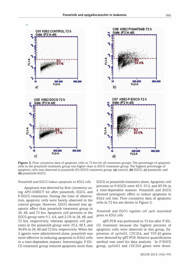
Ponatinib and EGCG induce apoptosis in K562 cells
Apoptosis was detected by flow cytometry us-ing APO-DIRECT kit after ponatinib, EGCG, and P-EGCG treatments. During the time of observa-tion, apoptotic cells were barely observed in the control groups. However, EGCG showed less ap-optotic effect than ponatinib treatment group in 24, 48, and 72 hrs. Apoptotic cell percents in the EGCG group were 9.1, 4.4, and 2.1% in 24, 48, and 72 hrs, respectively, whereas apoptotic cell per-cents in the ponatinib group were 23.2, 48.3, and 34.4% in 24, 48 and 72 hrs, respectively. When the 2 agents were administered alone, ponatinib was more effective in inducing apoptosis in K562 cells in a time-dependent manner. Interestingly, P-EG-CG treatment group induced apoptosis more than
EGCG or ponatinib treatment alone. Apoptotic cell percents in P-EGCG were 43.9, 57.6, and 83.5% in a time-dependent manner. Ponatinib and EGCG showed synergistic effect to induce apoptosis in K562 cell line. Flow cytometry data of apoptotic cells in 72 hrs are shown in Figure 2.
Ponatinib and EGCG regulate cell cycle associated genes in K562 cells
qRT-PCR was performed in 72 hrs after P-EG-CG treatment because the highest percents of apoptotic cells were observed in this group. Ex-pression of cyclinD1, CDC25A, and TGF-β2 genes were detected by qRT-PCR. Relative quantification method was used for data analysis. In P-EGCG group, cyclinD1 and CDC25A genes were down-
Figure 2. Flow cytometry data of apoptotic cells in 72 hrs for all treatment groups. The percentage of apoptotic cells in the ponatinib treatment group was higher than in EGCG treatment group. The highest percentage of apoptotic cells was observed in ponatinib (P)+EGCG treatment group. (a) control, (b) EGCG, (c) ponatinib, and (d) ponatinib+EGCG.
Ponatinib and epigallocatechin in leukemia996
JBUON 2014; 19(4): 996
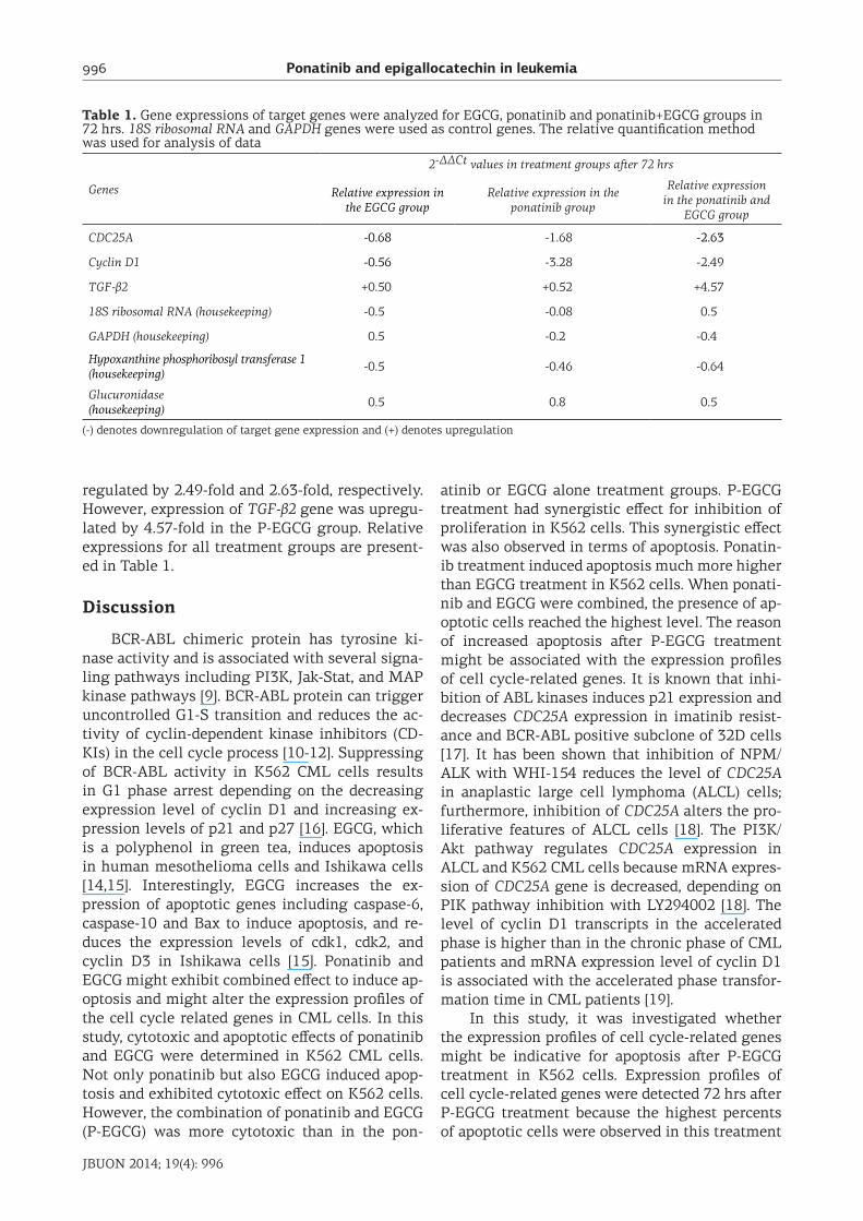
regulated by 2.49-fold and 2.63-fold, respectively. However, expression of TGF-β2 gene was upregu-lated by 4.57-fold in the P-EGCG group. Relative expressions for all treatment groups are present-ed in Table 1.
Discussion
BCR-ABL chimeric protein has tyrosine ki-nase activity and is associated with several signa-ling pathways including PI3K, Jak-Stat, and MAP kinase pathways [9]. BCR-ABL protein can trigger uncontrolled G1-S transition and reduces the ac-tivity of cyclin-dependent kinase inhibitors (CD-KIs) in the cell cycle process [10-12]. Suppressing of BCR-ABL activity in K562 CML cells results in G1 phase arrest depending on the decreasing expression level of cyclin D1 and increasing ex-pression levels of p21 and p27 [16]. EGCG, which is a polyphenol in green tea, induces apoptosis in human mesothelioma cells and Ishikawa cells [14,15]. Interestingly, EGCG increases the ex-pression of apoptotic genes including caspase-6, caspase-10 and Bax to induce apoptosis, and re-duces the expression levels of cdk1, cdk2, and cyclin D3 in Ishikawa cells [15]. Ponatinib and EGCG might exhibit combined effect to induce ap-optosis and might alter the expression profiles of the cell cycle related genes in CML cells. In this study, cytotoxic and apoptotic effects of ponatinib and EGCG were determined in K562 CML cells. Not only ponatinib but also EGCG induced apop-tosis and exhibited cytotoxic effect on K562 cells. However, the combination of ponatinib and EGCG (P-EGCG) was more cytotoxic than in the pon-
atinib or EGCG alone treatment groups. P-EGCG treatment had synergistic effect for inhibition of proliferation in K562 cells. This synergistic effect was also observed in terms of apoptosis. Ponatin-ib treatment induced apoptosis much more higher than EGCG treatment in K562 cells. When ponati-nib and EGCG were combined, the presence of ap-optotic cells reached the highest level. The reason of increased apoptosis after P-EGCG treatment might be associated with the expression profiles of cell cycle-related genes. It is known that inhi-bition of ABL kinases induces p21 expression and decreases CDC25A expression in imatinib resist-ance and BCR-ABL positive subclone of 32D cells [17]. It has been shown that inhibition of NPM/ALK with WHI-154 reduces the level of CDC25A in anaplastic large cell lymphoma (ALCL) cells; furthermore, inhibition of CDC25A alters the pro-liferative features of ALCL cells [18]. The PI3K/Akt pathway regulates CDC25A expression in ALCL and K562 CML cells because mRNA expres-sion of CDC25A gene is decreased, depending on PIK pathway inhibition with LY294002 [18]. The level of cyclin D1 transcripts in the accelerated phase is higher than in the chronic phase of CML patients and mRNA expression level of cyclin D1 is associated with the accelerated phase transfor-mation time in CML patients [19].
In this study, it was investigated whether the expression profiles of cell cycle-related genes might be indicative for apoptosis after P-EGCG treatment in K562 cells. Expression profiles of cell cycle-related genes were detected 72 hrs after P-EGCG treatment because the highest percents of apoptotic cells were observed in this treatment
Table 1. Gene expressions of target genes were analyzed for EGCG, ponatinib and ponatinib+EGCG groups in 72 hrs. 18S ribosomal RNA and GAPDH genes were used as control genes. The relative quantification method was used for analysis of data
Genes
2-ΔΔCt values in treatment groups after 72 hrs
Relative expression in the EGCG group
Relative expression in the ponatinib group
Relative expression in the ponatinib and
EGCG group
CDC25A -0.68 -1.68 -2.63
Cyclin D1 -0.56 -3.28 -2.49
TGF-β2 +0.50 +0.52 +4.57
18S ribosomal RNA (housekeeping) -0.5 -0.08 0.5
GAPDH (housekeeping) 0.5 -0.2 -0.4
Hypoxanthine phosphoribosyl transferase 1 (housekeeping) -0.5 -0.46 -0.64
Glucuronidase(housekeeping) 0.5 0.8 0.5
(-) denotes downregulation of target gene expression and (+) denotes upregulation

Ponatinib and epigallocatechin in leukemia 997
JBUON 2014; 19(4): 997
group. Expressions of cyclinD1 and CDC25A genes were downregulated by P-EGCG in 72 hrs com-pared to control cells. However, expression of TGF-β2 gene was upregulated by P-EGCG in 72 hrs. Downregulation of CDC25A and upregulation of p15 and p21 are associated with TGF-β related growth inhibition [20-23]. TGF-β can prevent G1 to S phase transition in the cell cycle process and induces apoptosis via downregulation of X-linked inhibitor of apoptosis protein (XIAP) which has a role in the PI3K/Akt pathway [24-27]. In hemato-poietic cells, TGF-β induces apoptosis through SHIP gene which is regulated by smad pathway and inhibits Akt and protein kinase B phosphoryl-
ation [28]. TGF-β2 suppresses cell proliferation in bovine corneal epithelium cells and inhibits AKT activation [29].
Conclusion
Ponatinib and EGCG exhibited synergistic effect on the proliferation and apoptosis in CML cells. P-EGCG mediated apoptosis might be as-sociated with upregulation of TGF-β2 gene and downregulation of cyclinD1 and CDC25A genes; furthermore, expression patterns of the defined genes might affect the PI3K/Akt signaling path-way.
References1. Sawyers CL. Chronic myeloid leukemia. N Engl J Med
1999;340:1330-1340.
2. Sant M, Allemani C, Tereanu C et al. Incidence of he-matologic malignancies in Europe by morphologic subtype: results of the HAEMACARE project. Blood 2010;116:3724-3734.
3. Smith A, Howell D, Patmore R, Jack A, Roman E. In-cidence of haematological malignancy by sub-type: a report from the Haematological Malignancy Research Network. Br J Cancer 2011;105:1684-1692.
4. Bernstein R. Cytogenetics of chronic myelogenous leukemia. Semin Hematol 1988;25:20-34.
5. Morris CM. Chronic myeloid leukemia: cytogenetic methods and applications for diagnosis and treat-ment. Methods Mol Biol 2011;730:33-61.
6. Bartram CR, de Klein A, Hagemeijer A et al. Translo-cation of c-ab1 oncogene correlates with the presence of a Philadelphia chromosome in chronic myelocytic leukaemia. Nature 1983;306:277-280.
7. Groffen J, Stephenson JR, Heisterkamp N, de Klein A, Bartram CR, Grosveld G. Philadelphia chromosomal breakpoints are clustered within a limited region, bcr, on chromosome 22. Cell 1984;36:93-99.
8. Lugo TG, Pendergast AM, Muller AJ, Witte ON. Ty-rosine kinase activity and transformation potency of bcr-abl oncogene products. Science 1990;247:1079-1082.
9. Deininger MW, Goldman JM, Melo JV. The molec-ular biology of chronic myeloid leukemia. Blood 2000;96:3343-3356.
10. Cortez D, Reuther G, Pendergast AM. The Bcr-Abl ty-rosine kinase activates mitogenic signaling pathways and stimulates G1-to-S phase transition in hemato-poietic cells. Oncogene 1997;15:2333-2342.
11. Gesbert F, Sellers WR, Signoretti S, Loda M, Griffin JD. BCR/ABL regulates expression of the cyclin-de-pendent kinase inhibitor p27Kip1 through the phos-phatidylinositol 3-Kinase/AKT pathway. J Biol Chem
2000;275:39223-39230.
12. Nimmanapalli R, Fuino L, Bali P et al. Histone deacetylase inhibitor LAQ824 both lowers expression and promotes proteasomal degradation of Bcr-Abl and induces apoptosis of imatinib mesylate-sensitive or -refractory chronic myelogenous leukemia-blast cri-sis cells. Cancer Res 2003;63:5126-5135.
13. Shamroe CL, Comeau JM. Ponatinib: A new tyrosine kinase inhibitor for the treatment of chronic mye-loid leukemia and Philadelphia chromosome-positive acute lymphoblastic leukemia. Ann Pharmacother 2013;47:1540-1546.
14. Satoh M, Takemura Y, Hamada H, Sekido Y, Kubota S. EGCG induces human mesothelioma cell death by inducing reactive oxygen species and autophagy. Can-cer Cell Int 2013;13:19.
15. Park SB, Bae JW, Kim JM, Lee SG, Han M. Anti-proliferative and apoptotic effect of epigallocate-chin-3-gallate on Ishikawa cells is accompanied by sex steroid receptor downregulation. Int J Mol Med 2012;30:1211-1218.
16. Rangatia J, Bonnet D. Transient or long-term silenc-ing of BCR-ABL alone induces cell cycle and prolifer-ation arrest, apoptosis and differentiation. Leukemia 2006;20:68-76.
17. Mancini M, Brusa G, Zuffa E et al. Persistent Cdk2 in-activation drives growth arrest of BCR-ABL-express-ing cells in response to dual inhibitor of SRC and ABL kinases SKI606. Leuk Res 2007;31:979-987.
18. Fernandez-Vidal A, Mazars A, Gautier EF, Prevost G, Payrastre B, Manenti S. Upregulation of the CDC25A phosphatase down-stream of the NPM/ALK oncogene participates to anaplastic large cell lymphoma en-hanced proliferation. Cell Cycle 2009;8:1373-1379.
19. Liu JH, Yen CC, Lin YC et al. Overexpression of cyclin D1 in accelerated-phase chronic myeloid leukemia. Leukemia Lymphoma 2004;45:2419-2425.
20. Massague J. TGFbeta signalling in context. Nat Rev Mol Cell Biol 2012;13:616-630.
Ponatinib and epigallocatechin in leukemia998
JBUON 2014; 19(4): 998
21. Kang SH, Bang YJ, Jong HS, Seo JY, Kim NK, Kim SJ. Rapid induction of p21WAF1 but delayed down-reg-ulation of Cdc25A in the TGF-beta-induced cell cy-cle arrest of gastric carcinoma cells. Br J Cancer 1999;80:1144-1149.
22. Miyazaki M, Ohashi R, Tsuji T, Mihara K, Gohda E, Namba M. Transforming growth factor-beta 1 stim-ulates or inhibits cell growth via down- or up-regu-lation of p21/Waf1. Biochem Biophys Res Commun 1998;246:873-880.
23. Wang X, Sun W, Bai J et al. Growth inhibition induced by transforming growth factor-beta1 in human oral squamous cell carcinoma. Mol Biol Rep 2009;36:861-869.
24. Caron PL, Frechette-Frigon G, Shooner C, Leblanc V, Asselin E. Transforming growth factor beta isoforms regulation of Akt activity and XIAP levels in rat endo-metrium during estrous cycle, in a model of pseudo-pregnancy and in cultured decidual cells. Reprod Biol
Endocrinol 2009;7:80.
25. Gottfried Y, Rotem A, Lotan R, Steller H, Larisch S. The mitochondrial ARTS protein promotes apoptosis through targeting XIAP. EMBO J 2004;23:1627-1635.
26. Bachman KE, Park BH. Duel nature of TGF-beta signa-ling: tumor suppressor vs. tumor promoter. Curr Opin Oncol 2005;17:49-54.
27. Shima Y, Nakao K, Nakashima T et al. Activation of caspase-8 in transforming growth factor-beta-induced apoptosis of human hepatoma cells. Hepatology 1999;30:1215-1222.
28. Valderrama-Carvajal H, Cocolakis E, Lacerte A et al. Activin/TGF-beta induce apoptosis through Smad-de-pendent expression of the lipid phosphatase SHIP. Nat Cell Biol 2002;4:963-969.
29. Lu J, Lu Z, Reinach P et al. TGF-beta2 inhibits AKT activation and FGF-2-induced corneal endothelial cell proliferation. Exp Cell Res 2006;312:3631-3640.
Copyright © 2022 FDOKUMEN