A fratricidal mechanism is responsible for eDNA release and contributes to biofilm development of...
28
A fratricidal mechanism is responsible for eDNA release and contributes to biofilm development of Enterococcus faecalis Vinai Chittezham Thomas 1 , Yasuaki Hiromasa 2 , Nathan Harms 1 , Lance Thurlow 1 , John Tomich 2 , and Lynn Hancock 1,* 1 Division of Biology, Kansas State University, 116 Ackert Hall, Manhattan, KS 2 Department of Biochemistry, Kansas State University, 141 Chalmers Hall, Manhattan, KS Summary Extracellular DNA (eDNA), a by-product of cell lysis was recently established as a critical structural component of the Enterococcus faecalis biofilm matrix. Here, we describe fratricide as the governing principle behind gelatinase (GelE) mediated cell death and eDNA release. GFP reporter assays confirmed that GBAP quorum non-responders (GelE − SprE − ) were a minority subpopulation of prey cells susceptible to the targeted fratricidal action of the quorum responsive predatorial majority (GelE + SprE + ). The killing action is dependent on GelE, and the GelE producer population is protected from self-destruction by the co-production of SprE as an immunity protein. Targeted gene inactivation and protein interaction studies demonstrate that extracellular proteases execute their characteristic effects following downstream interactions with the primary autolysin, AtlA. Finally, we address a mechanism by which GelE and SprE may modify the cell wall affinity of proteolytically processed AtlA resulting in either a pro- or anti- lytic outcome. Keywords Enterococcus faecalis; fratricide; autolysis; AtlA; gelatinase; serine protease Introduction Opportunistic enterococcal infections have traditionally been perceived as serious clinical threats due to rising incidence of antibiotic resistance and lateral transfer of resistance traits (Chang et al., 2003; Noble et al., 1992). More recently, these same threats have been associated with the existence of bacteria as surface adherent communities called biofilms (Fux et al., 2005). The close residential proximity of organisms within biofilms and altered genetic responses primarily driven by density dependent mechanisms may aid the observed heightened tolerance to antibiotics and dissemination of resistance traits (Jayaraman and Wood, 2008). Observations of clinical enterococcal biofilms on endodontic surfaces, bilary stents, urinary catheters, heart valves and tissue surfaces suggest a correlation of this pathogen’s community lifestyle and virulence (Carniol and Gilmore, 2004). Analogous to developmental processes (biofilm development, competence regulation and sporulation) displayed by some bacteria, biofilm formation by enterococci involves quorum signaling (Hancock and Perego, 2004; Mohamed et al., 2004). The quorum signal is derived from the fsr gene cluster of E. faecalis (fsrABDC) and results from the processing and * Corresponding author: Lynn E. Hancock, Division of Biology, Kansas State University, 116 Ackert Hall, Manhattan, KS 66506, Ph: (785) 532-6122, Fax: (785) 532-6653, [email protected]. NIH Public Access Author Manuscript Mol Microbiol. Author manuscript; available in PMC 2010 May 1. Published in final edited form as: Mol Microbiol. 2009 May ; 72(4): 1022–1036. doi:10.1111/j.1365-2958.2009.06703.x. NIH-PA Author Manuscript NIH-PA Author Manuscript NIH-PA Author Manuscript
-
Upload
independent -
Category
Documents
-
view
0 -
download
0
Transcript of A fratricidal mechanism is responsible for eDNA release and contributes to biofilm development of...

A fratricidal mechanism is responsible for eDNA release andcontributes to biofilm development of Enterococcus faecalis
Vinai Chittezham Thomas1, Yasuaki Hiromasa2, Nathan Harms1, Lance Thurlow1, JohnTomich2, and Lynn Hancock1,*
1 Division of Biology, Kansas State University, 116 Ackert Hall, Manhattan, KS2 Department of Biochemistry, Kansas State University, 141 Chalmers Hall, Manhattan, KS
SummaryExtracellular DNA (eDNA), a by-product of cell lysis was recently established as a criticalstructural component of the Enterococcus faecalis biofilm matrix. Here, we describe fratricide asthe governing principle behind gelatinase (GelE) mediated cell death and eDNA release. GFPreporter assays confirmed that GBAP quorum non-responders (GelE−SprE−) were a minoritysubpopulation of prey cells susceptible to the targeted fratricidal action of the quorum responsivepredatorial majority (GelE+SprE+). The killing action is dependent on GelE, and the GelEproducer population is protected from self-destruction by the co-production of SprE as animmunity protein. Targeted gene inactivation and protein interaction studies demonstrate thatextracellular proteases execute their characteristic effects following downstream interactions withthe primary autolysin, AtlA. Finally, we address a mechanism by which GelE and SprE maymodify the cell wall affinity of proteolytically processed AtlA resulting in either a pro- or anti-lytic outcome.
KeywordsEnterococcus faecalis; fratricide; autolysis; AtlA; gelatinase; serine protease
IntroductionOpportunistic enterococcal infections have traditionally been perceived as serious clinicalthreats due to rising incidence of antibiotic resistance and lateral transfer of resistance traits(Chang et al., 2003; Noble et al., 1992). More recently, these same threats have beenassociated with the existence of bacteria as surface adherent communities called biofilms(Fux et al., 2005). The close residential proximity of organisms within biofilms and alteredgenetic responses primarily driven by density dependent mechanisms may aid the observedheightened tolerance to antibiotics and dissemination of resistance traits (Jayaraman andWood, 2008). Observations of clinical enterococcal biofilms on endodontic surfaces, bilarystents, urinary catheters, heart valves and tissue surfaces suggest a correlation of thispathogen’s community lifestyle and virulence (Carniol and Gilmore, 2004).
Analogous to developmental processes (biofilm development, competence regulation andsporulation) displayed by some bacteria, biofilm formation by enterococci involves quorumsignaling (Hancock and Perego, 2004; Mohamed et al., 2004). The quorum signal is derivedfrom the fsr gene cluster of E. faecalis (fsrABDC) and results from the processing and
*Corresponding author: Lynn E. Hancock, Division of Biology, Kansas State University, 116 Ackert Hall, Manhattan, KS 66506, Ph:(785) 532-6122, Fax: (785) 532-6653, [email protected].
NIH Public AccessAuthor ManuscriptMol Microbiol. Author manuscript; available in PMC 2010 May 1.
Published in final edited form as:Mol Microbiol. 2009 May ; 72(4): 1022–1036. doi:10.1111/j.1365-2958.2009.06703.x.
NIH
-PA Author Manuscript
NIH
-PA Author Manuscript
NIH
-PA Author Manuscript

secretion of FsrD by FsrB to yield an 11-amino acid cyclized peptide lactone termedgelatinase biosynthesis-activating pheromone (GBAP) capable of density dependent signaltransduction via the FsrAC two-component histidine kinase-response regulator pathway(Nakayama et al., 2006). Unlike the peptide pheromones involved in conjugal mating andplasmid transfer in Enterococcus, the formation of a cyclized lactone is thought to protectGBAP from proteolytic turnover by enterococcal proteases (Nakayama et al., 2006).Transcriptional profiling has indicated strong expression of two co-transcribed secretedextracellular proteases, gelatinase (GelE) and serine protease (SprE) following Fsr signalactivation (Bourgogne et al., 2006; Qin et al., 2000). A number of independent studies haveobserved a critical role for gelatinase in enterococcal biofilm development (Hancock andPerego, 2004; Kristich et al., 2004; Mohamed et al., 2004). Gelatinase is proposed toactivate lysis of a sub-population of bacteria and thereby catalyze the release of genomicDNA (eDNA), a critical component of the biofilm matrix (Thomas et al., 2008). Intriguinglythe phenotypic effects of SprE opposed those of GelE with higher rates of lysis, eDNArelease and increased biofilm development observed upon its inactivation (Thomas et al.,2008). Earlier observations by Shockman and others suggested the cellular target of GelE tobe an autolysin (Shockman and Cheney, 1969; Waters et al., 2003). Although a directinteraction of SprE with a cell surface autolysin is yet to be established, evidence suggests itcan modify rates of autolysis (Thomas et al., 2008). At least three autolysins (AtlA, AtlBand AtlC) have been identified to be secreted by E. faecalis of which AtlA is thought to becrucial for biofilm development (Kristich et al., 2008; Mesnage et al., 2008). Based onsequence similarity, AtlA was proposed to be made up of three domains. The centralcatalytic domain is responsible for the glucosaminidase activity (Eckert et al., 2006). The C-terminal domain is composed of six LysM modules that afford peptidoglycan affinity andpossibly target autolysins to the division septum and poles (Steen et al., 2008). No knownfunction yet exists for the T/E rich N-terminal domain (Eckert et al., 2006).
The existence of a bimodal bacterial population was hypothesized to explain the nature ofenterococcal cell death leading to biofilm development (Thomas et al., 2008). The first sub-population would be a predatory population that produces GelE as an effector of cell deathand is immune to self-inflicted GelE toxicity by virtue of the co-transcribed SprE proteasewhile the second, a lysis susceptible prey population that does not produce eitherextracellular protease. Such bimodality in an otherwise isogenic bacterial population duringgrowth is plausible given that cell density dictates those cells that respond to the GBAPquorum peptide and activate protease expression. Bacteria that do not respond to quorumsignaling would then be susceptible to GelE mediated cell death due to their inability toproduce the immunity factor, SprE. Although the above hypothesis is consistent withbacterial fratricide (Claverys and Havarstein, 2007), wherein a specific cell is responsiblefor predation of its clonal neighbor, an alternate hypothesis of altruistic suicide orprogrammed cell death of the quorum responder remains a possibility (Bayles, 2007; Rice etal., 2007).
In the current study, we show that extracellular proteases regulate enterococcal fratricideleading to eDNA release and biofilm development. Further AtlA is demonstrated to be thetarget of both GelE and SprE and this interaction is critical to the regulation of enterococcalfratricide and biofilm development.
ResultsSprE prevents altruistic suicide in enterococcal populations
Programmed cell death was recently hypothesized to be critical for S. aureus biofilmdevelopment (Bayles, 2007; Rice et al., 2007). To investigate whether extracellularproteases (GelE and SprE) controlled an altruistic nature of cell death and lysis in E. faecalis
Thomas et al. Page 2
Mol Microbiol. Author manuscript; available in PMC 2010 May 1.
NIH
-PA Author Manuscript
NIH
-PA Author Manuscript
NIH
-PA Author Manuscript

populations, a GBAP responsive GFP-reporter construct (pVT31) was introduced into V583,VT01(ΔgelE), VT02 (ΔsprE) and VT03 (ΔgelE sprE) isogenic protease deletion mutantbackgrounds resulting in strains VT15, VT16 (ΔgelE), VT17 (ΔsprE) and VT18(ΔgelEsprE) respectively. The plasmid pVT31 contains a 1055-bp fragment composed of agelE promoter-gfp gene fusion, in a high copy vector (pAT28) background. As theexpression of GelE in native circumstances is dependent on GBAP levels, the coupling ofthe gelE promoter to the gfp gene in pVT31 would enable the expression of GFP to bedependent on the extracellular concentration of GBAP. Hence enterococci (bearing plasmidpVT31) that respond to the quorum peptide (GBAP) would switch on the expression ofGFP. However cells that do not respond to GBAP would retain a GFP− phenotype. Thisphenotypic segregation in GFP expression effectively facilitated tracking of cells thatresponded to the quorum peptide within the wild type and each of the mutant populations byflow cytometry. The stringency of pVT31 as a reporter construct was determined using E.faecalis VT14, a derivative of V583 harboring pVT30 (reporter construct lacking functionalpromoter fusion) and VT13, an fsrA insertion mutant that contains pVT31. The absence of afunctional FsrA would prevent VT13 from responding to the accumulation of quorumpeptide (GBAP). Accordingly figure 1A depicts a clear shift in fluorescence intensities ofVT15, VT16, VT17 and VT18 relative to VT14. Both the above controls (VT13 and VT14)exhibited a signal noise less than ~6% relative to VT15 (V583 pVT31) (Fig 1B). Fluorescentsignal generated by VT13 likely arises from a basal level of expression from the gelEpromoter, which is known to occur independent of FsrA (Singh et al., 2005). The relativelyweak signal observed in strain VT14 is likely attributed to use of a weak promoter in thevector sequence, as plasmid pVT30 lacks the sequence for the gelE promoter.
Quantitative estimates of GFP+ cells (GBAP responders) in wild type and extracellularprotease mutant backgrounds were achieved by flow cytometry. The settings were adjustedsuch that GFP− cells were within the first log decade while cells with higher intensities wereconsidered GFP+ (Fig 1A). Estimation of GFP+ cells (GBAP responders) by flow cytometryindicate (Fig 1B) that although the majority of the wild-type (VT15) E. faecalis populationunder the present assay conditions responded to quorum signaling at the stationary phase ofgrowth, approximately 15% correspond to GBAP non-responders. The independent effectsof GelE and SprE on GBAP responders were characterized with respect to relative numbersof GFP+ responders within isogenic extracellular protease mutant populations and theirgeometric mean fluorescence intensities (GMFI). Relative numbers of GFP+ cells (Fig 1B)in VT16 (85.68 ± 7.02%) and VT18 (87.09 ± 6.5%) were similar to those found in E.faecalis VT15 (87.26 ± 4.37%). Interestingly, the absence of SprE expression in VT17resulted in a significant decrease in GFP+ responder cells to 74.66 ± 5.63% as compared toparental strain (P< 0.05, Bonferronis test). This suggested that GBAP responders within apopulation underwent significant lysis only in the absence of SprE. Consistent with thisobservation the geometric mean fluorescence intensity (GMFI) of GFP+ responder cells ofthe VT17 (ΔsprE) population was significantly lower (59.23 ± 2.64) compared to that ofV583 and mutants deficient in GelE expression (V583, 74.64 ± 13.11, VT16, 81.61 ± 6.97and VT18, 83.22 ± 3.55). These observations are also consistent with earlier reportssuggesting a pro-lytic activity for GelE and an anti-lytic activity for SprE (Thomas et al.,2008).
Interplay of GelE and SprE in enterococcal fratricideIn order to assess the role of extracellular proteases of Enterococcus faecalis in fratricide,predator-prey co-culture lysis assays were performed. Parental and isogenic proteasedeletion mutants (predators) – V583, VT01 (ΔgelE), VT02 (ΔsprE) and VT03 (ΔgelEsprE)were co-cultivated in the presence of VT12, a double protease mutant strain thatconstitutively expressed GFP from plasmid pMV158GFP (Nieto and Espinosa, 2003). This
Thomas et al. Page 3
Mol Microbiol. Author manuscript; available in PMC 2010 May 1.
NIH
-PA Author Manuscript
NIH
-PA Author Manuscript
NIH
-PA Author Manuscript

previously characterized prey strain is deficient in lysis due to its inability to produce GelE(Thomas et al., 2008). However as the downstream cellular target of GelE that affectsautolysis is still intact within these cells; their lysis may be activated when they are exposedto diffusible GelE. Consequently we reasoned that fratricidal control of the GFP+ preypopulation by diffusible extracellular proteases produced by the co-cultivated predatorstrains may be assayed by tracking the levels of GFP released into the culture supernatant.Neither of the purified extracellular proteases (GelE or SprE) exhibited an effect on GFPturnover at the concentrations assayed (data not shown) and hence any signal detected inculture supernatants should be a true reflection of bacterial lysis. Western blot analysis ofculture supernatants, using anti-GFP antibody clearly showed that the lysis of the prey(VT12) population is dependent on the diffusible gelatinase (GelE) produced by the predator(Fig 2A; compare lanes 1 and 3 to lanes 2 and 4). Whereas co-cultivation of the parentalV583 (GelE+SprE+) predators resulted in wild type levels of target lysis, GelE mutantpredators (VT01 and VT03) demonstrated no detectable lysis of VT12 (prey). Interestingly,the use of VT02 (SprE−) as the predator resulted in an increased lysis of prey (Fig 2A,compare lanes 1 and 3), suggesting that diffusible SprE was able to modulate GelEdependent fratricide. As expected, spectrofluorometric measures of GFP fluorescenceemanating from the prey population that survived the fratricidal action mediated by thepredators showed a significant reduction in cell fluorescence when co-cultivated with VT02(ΔsprE) compared to V583, VT01 and VT03 (data not shown).
Consistent with these results, we also observed that the addition of purified extracellularproteases (GelE and SprE) in physiological concentrations (~30nM) (Hancock and Perego,2004; Makinen et al., 1989) resulted in the differential lysis of VT03 (ΔgelEsprE) (Fig 2B).Incubation with purified GelE alone caused a rapid increase in the lysis-rate of VT03(ΔgelEsprE). While SprE by itself did not affect VT03 (ΔgelEsprE) rates of lysis, itprevented the initial increase in optical density observed in the VT03 strain withoutexogenous proteases. Furthermore, co-incubation of SprE with an equal concentration ofpurified GelE reduced the rate of GelE mediated lysis. These findings not only point to theability of GelE to initiate fratricide but also of SprE to modulate GelE activity.
Comparison of autolysin profiles among enterococcal strainsBased on previously reported autolytic rates of protease mutants (Thomas et al., 2008) andon observations by Shockman reflecting the role of GelE in activating an autolysin(Shockman and Cheney, 1969), we hypothesized enterococcal cell death to be a directconsequence of extracellular proteases manipulating cell surface associated autolysin(s). Todetermine potential autolysins regulated by extracellular proteases, we subjected cell wallprotein extracts of GelE mutants from E. faecalis V583 and OG1RF strain backgrounds tozymography (Fig 3). Surface autolysin profiles of V583 and OG1RF exhibited significantdifferences. While a minor high molecular weight autolysin (~66kDa) was apparent inprotein extracts derived from OG1RF, this activity was not visualized in VT20(OG1RFΔgelE) or V583 cell wall extracts (Fig 3; compare lane 1 to lanes 2, 3 and 4).However a novel 38kDa autolytic band that seemed to be immune to the proteolytic activityof GelE was uniquely found in the V583 strain background and not in OG1RF (Figure 3;compare lane 1 and 2 to lanes 3 and 4). Similarly, a 50kDa autolytic band could only bedetected in VT01 cell wall extracts, suggesting sensitivity toward GelE activity (Figure 3;compare lanes 3 and 4). In light of a recent study by Bourgogne et al. (Bourgogne et al.,2008) the absence of the 38kDa and 50kDa autolytic bands from OG1RF is not surprising asthis strain lacks the prophage-associated endolysins present in V583 (Paulsen et al., 2003).
Interestingly, only two autolytic bands (71.6 kDa, found in VT01 (V583ΔgelE) and VT20(OG1RFΔgelE); 61.1 kDa in V583 and OG1RF) remained common among the two strains.Consistent with previous reports (Eckert et al., 2006; Mesnage et al., 2008), we identified
Thomas et al. Page 4
Mol Microbiol. Author manuscript; available in PMC 2010 May 1.
NIH
-PA Author Manuscript
NIH
-PA Author Manuscript
NIH
-PA Author Manuscript

and mapped the 71.6 kDa activity to AtlA by MALDI-TOF mass spectrometry. Targetedmutagenesis of atlA by insertion inactivation (see Materials and Methods) in V583 andOG1RF strain backgrounds (VT19 and VT21, respectively) demonstrated the 61.1 kDaactivity to correspond to the proteolytically processed form of AtlA (see supplementary FigS1). Interestingly, autolysin profiles of VT02 (ΔsprE) when compared with V583 appearedto display a considerable increase in the intensity of the 61.1 kDa band (see supplementaryFig S2). These results collectively suggest that in parental strains, GelE and SprE arecapable of differentially regulating the turnover of AtlA.
AtlA is critical for eDNA release and biofilm developmentRecent studies have observed that inactivation of AtlA was detrimental to biofilmdevelopment of E. faecalis OG1RF (Kristich et al., 2008). Given that, AtlA was the soleautolysin whose activity was demonstrated to be regulated by both proteases, we tested fordifferences in eDNA release and biofilm forming abilities of VT19 (V583::atlA) and VT21(OG1RF::atlA) against parental strains. Culture supernatants of overnight grown E. faecalisV583 and OG1RF parental strains and their corresponding AtlA mutants were assayed foreDNA using a DNA specific dye, SYTOX green (Molecular Probes). As expected andconfirming our previous observations (Thomas et al., 2008) deletion of gelE in V583(VT01) and OG1RF (VT20) backgrounds resulted in a significant 4- and 12-fold decrease ineDNA release as compared to their respective parental strains (Fig. 4A, P< 0.0001, t-test).More importantly, the decrease in eDNA release due to the inactivation of atlA paralleledGelE mutants from both strain backgrounds. This strongly suggested that AtlA may be themajor target of extracellular proteases on the cell surface, that mediates fratricide and eDNArelease.
Because AtlA mutants are deficient in eDNA release, we further hypothesized these strainsto be defective in biofilm formation. Quantitative analysis of biofilms grown on polystyrenesurfaces revealed that AtlA mutants, similar to GelE mutants formed significantly lessbiofilm biomass as compared to parental strains (Fig. 4B, P< 0.0001, t-test). The observeddeficiencies in biofilm formation were not a result of growth defects as all mutants displayedsimilar growth kinetics compared to the parental strains (data not shown). Theseobservations are consistent with a critical role for AtlA in eDNA release and biofilmformation.
Role of AtlA in fratricideGiven that extracellular proteases and AtlA contribute to similar biological pathways in E.faecalis, we hypothesized that fratricidal effector- modulator functions of GelE and SprE,are channeled through their downstream interactions with AtlA. However, prey-predator co-culture lysis assays using VT23 (an atlA mutant derivative of VT12 (ΔgelE-sprE, GFP+) asprey showed no abrogation of V583 and VT02 (ΔsprE) predator-mediated fratricide (Fig5A; lanes 1 and 3), although maintaining GelE dependency (Fig 5A; lanes 2 and 4).
In an attempt to address these seemingly confounding observations, we tested the autolyticrates of the atlA mutant derivative of VT03, designated VT22, in the presence of purifiedenterococcal extracellular proteases. Addition of physiological concentrations of GelE orSprE (~30 nM) individually or together did not lead to any significant differences in lysis ofVT22 as compared to protease negative controls (Fig 5B). This suggested that GelEdependent fratricide of VT23 observed earlier was possibly due to predator specific factors(in addition to GelE) that interacted with the prey population. As AtlA has previously beenshown to be a diffusible enzyme capable of hydrolyzing the cell walls of Micrococcuslysodeikticus (Qin et al., 1998) and E. faecalis (Mesnage et al., 2008), we hypothesized thatAtlA may fulfill the role of a predator factor responsible for the lysis of prey cells in the
Thomas et al. Page 5
Mol Microbiol. Author manuscript; available in PMC 2010 May 1.
NIH
-PA Author Manuscript
NIH
-PA Author Manuscript
NIH
-PA Author Manuscript

population. Such diffusible AtlA may possibly be released from a subpopulation of predatorcells (~15%, refer Fig 1B) that have not responded to the quorum signal and hence aresusceptible to the effector functions of GelE. To test this hypothesis we co-cultured VT19(V583::atlA) as a predator population against VT23 (ΔgelEsprE,::atlA, GFP+) preypopulation. Figure 5A (lane 5) clearly indicates the absence of any detectable fratricide inthe prey population upon inactivation of the predator AtlA, suggesting a crucial role forsoluble (diffusible) forms of AtlA in fratricide.
Characterizing the effects of GelE and SprE on recombinant AtlATo further investigate the interaction between extracellular proteases and AtlA, we subjectedrecombinant histidine tagged AtlA (rAtlA, Fig 6A) expressed and purified from E. colicultures to GelE and SprE treatment. GelE treated rAtlA was multiply processed into minorforms within 30 minutes (Fig 6B; lane 2) and was completely turned over by 5 hours (datanot shown). Zymogram analysis showed that most processed forms of rAtlA were active onV583 cell wall (Fig 6C; lane 2) suggesting that GelE preferentially cleaves initially at non-catalytic sites within AtlA. The full-length 72.1 kDa autolytic band seemed to show activityonly after extended periods of incubation (data not shown), suggesting an enhancement inactivity upon proteolytic cleavage. Interestingly SprE action on rAtlA resulted in a singlemajor 62kDa form and a minor higher molecular weight form (Fig 6B and 6C, lane 3). Fromthe current experimental conditions, although it would seem that rAtlA has a faint auto-catalytic activity and processes itself into the 62kDa form, the action of SprE clearlyaugments the rate of this processing while that of gelatinase seemed to affect the turnover ofrAtlA.
Comparison of the molecular weights of GelE and SprE processed recombinant AtlAfragments (Fig 6B) to their corresponding spectrograms (generated from MALDI TOF MS/MS peptide mass mapping) empirically identified regions within rAtlA that were susceptibleto protease attack. As compared to the full length recombinant AtlA (72.3 kDa, includes6XHis tag, but devoid of signal peptide), the 62kDa (Fig 6B, lanes 2 and 3) form maintainedan intact C-terminal domain inclusive of the histidine tag, suggesting that both GelE andSprE specifically cleaved within the N-terminal T/E rich domain of rAtlA. Additionally,GelE also displayed cleavage specificities within the C- terminal domain. This was evidentas the smaller rAtlA forms (55.3, 49.2, and 43.7kDa; Fig 6B, lane 2) shared a common N-terminus but exhibited clear differences in their C- terminal peptide fingerprints (data notshown).
Given that the C-terminal domain of AtlA is composed of multiple units of LysM modules(Fig 6A) it seemed plausible that the action of GelE on the C-terminal domain of AtlA maycatalyze its release from the cell surface. To test this hypothesis and to investigate any effectSprE may have toward rAtlA’s affinity to the cell surface, we assayed the cell wall affinitiesof full-length and proteolytically processed forms of rAtlA. Recombinant AtlA was initiallyallowed to bind E. faecalis V583 cell walls, followed by individual treatments with GelEand SprE proteases. Relative affinities of rAtlA forms processed by GelE and SprE to thecell wall was determined as a function of their ability to remain in the supernatant fractionon centrifugation as opposed to the pelleted cell wall bound fraction. Although it was clearlyevident from the analysis that cell wall bound rAtlA displayed altered processing by GelEcompared to the unbound form (compare proteolytic pattern from Fig 7, lane 5 and Fig 6B,lane 2), the supernatant fractions revealed that GelE treatment resulted in processed solubleforms of rAtlA (Fig 7, lane 2). Notably, SprE treatment resulted in the absence of anydetectable soluble forms of AtlA in the supernatant fraction. Complementing theseobservations, comparison of the GelE and SprE processed cell wall bound forms of rAtlAdisplayed significant differences (Fig 7; compare lanes 5 and 6). Cleavage of rAtlA by SprEresulted in processed forms with molecular masses approximately equal to and greater than
Thomas et al. Page 6
Mol Microbiol. Author manuscript; available in PMC 2010 May 1.
NIH
-PA Author Manuscript
NIH
-PA Author Manuscript
NIH
-PA Author Manuscript

62kDa (Fig 7, lane 6) that showed significantly higher affinity to peptidoglycan compared tothe ~ 62kDa band resulting from GelE treatment (Fig 7, lane 5). Given that cleavage ofrAtlA by SprE did not result in the loss of LysM modules (a known peptidoglycan bindingand localization domain) it stands to reason that AtlA may be shuttled to the septumoptimally after SprE processing.
We reasoned that optimal proteolytic activity of SprE toward rAtlA may prevent GelE fromsubsequently mediating its (rAtlA’s) release from the cell surface, thus arguing for theobserved resistance of attacker subpopulations (SprE+) to GelE mediated lysis. To test thishypothesis, we pretreated rAtlA bound to peptidoglycan with physiological concentrationsof SprE and then subjected it to the proteolytic action of an equimolar concentration ofGelE. Supplemental figure S3 clearly demonstrates the inability of GelE to release SprEprocessed rAtlA from the peptidoglycan fraction into the supernatant. Further, SprE wasunable to prevent the release of peptidoglycan bound rAtlA into the supernatant if GelEacted on the autolysin first. This suggests that SprE prevents lysis of attacker populations byefficiently competing with GelE for the primary autolysin, AtlA. However, the exactmechanism of competition by the extracellular proteases toward AtlA remains to beelucidated. Collectively these results suggest that in wild-type populations the role of SprEmay be to prevent altruistic suicide of quorum responders by limiting the release of AtlAfrom the cell surface.
DiscussionThe results presented in this study demonstrate extracellular protease mediated fratricide tobe responsible for governing eDNA release and biofilm development of E. faecalis. Death ofsibling cells (fratricide) mediated by isogenic cells within the same population haspreviously been implicated in developmental processes (competence and sporulation) ofStreptococcus pnuemoniae and Bacillus subtilis (Ellermeier et al., 2006; Gilmore and Haas,2005; Gonzalez-Pastor et al., 2003; Guiral et al., 2005; Havarstein et al., 2006). Cell-cellsignaling during these developmental processes results in the coordinated production ofspecific killing factors and immunity proteins within a subpopulation of cells (predators).Killing factors which include bacteriocins and murein hydrolases target the death of a smallsusceptible isogenic population (prey) (Claverys and Havarstein, 2007). Susceptibility ofprey to the killing factors is largely due to the absence of immunity proteins in thispopulation, a cost these cells possibly pay for not participating in the signaling process.Death of the prey ultimately benefits the surviving population either in the form of nutrientsin nutritionally stressed B. subtilis or as released genomic DNA for naturally competent S.pneumonia (Claverys and Havarstein, 2007). In analogy with these model systems, we haveidentified two prominent driving forces of cell death (killing factors) responsible for biofilmdevelopment in enterococci; secreted gelatinase (GelE) and the soluble autolysin, AtlA (Fig8). Although cell wall peptidoglycan is refractory toward GelE’s ability to affect turnover ofcell surface localized AtlA as compared to a relatively quick turnover in solution, itnevertheless allows limited cleavage potentially resulting in its release from the cell surface.We propose that released AtlA may result in bystander cell death especially in high celldensity biofilms, although arguably the extent of such bystander effects will be limited byGelE to small sub-populations within the biofilm. Consistent with this hypothesis, we earlierreported the development of pockets filled with dead/lysed cells within the primary biofilmmatt (Thomas et al., 2008).
Biofilm development in Staphylococcus aureus was recently demonstrated to be dependenton cell death and eDNA release (Rice et al., 2007). However, the nature of cell deathobserved in this case was proposed to be due to altruistic suicide or programmed cell death(PCD) (Bayles, 2007). Altruistic suicide of GelE+ enterococci would be conceptually
Thomas et al. Page 7
Mol Microbiol. Author manuscript; available in PMC 2010 May 1.
NIH
-PA Author Manuscript
NIH
-PA Author Manuscript
NIH
-PA Author Manuscript

improbable as over 85% of the population in stationary phase show evidence of theirparticipation in Fsr signaling (Fig 1A) and the Fsr quorum response is known todifferentially regulate over 300 genes involved in secondary regulatory cascades, virulenceand metabolism (Bourgogne et al., 2006).
How do cells producing GelE ensure their own safety against self-inflicted lysis? Severallines of evidence suggest the co-transcribed serine protease, SprE, to be an immunityprotein. First, GFP reporter assays confirm a significant increase in death of predatorpopulations in an isogenic SprE mutated strain compared to the parental strain. Consistentwith this observation, co-culture lysis assays which detect solubilized GFP from lysed cellsalso indicate that secreted SprE has significant trans-protective activities toward prey cells.Second, purified SprE was able to significantly reduce the rate of GelE induced cell lysis.Finally, pretreatment of peptidoglycan bound AtlA with purified SprE significantly reducedits GelE mediated release (Figure S3), a necessity for cell death. Consequently, it may bereasoned that SprE protects predator cells from lysis by modifying surface localized AtlAagainst further proteolysis by GelE.
Autolysins have consistently been described as regulators of cell death and biofilmdevelopment in different gram-positive species, although mechanistic details at a molecularlevel remain vague. Inactivation of the primary autolysins of Streptococcus mutans,Staphylococcus aureus and Staphylococcus epidermidis have been shown to decrease theirabilities to form biofilms, presumably due to a defect in eDNA release (Ahn and Burne,2006; Biswas et al., 2006; Heilmann et al., 1997; Qin et al., 2007; Shibata et al., 2005).GelE has previously been implicated in the proteolytic processing of a latent high molecularweight E. hirae muramidase 1 (137 kDa) to the active 87 kDa form (Shockman and Cheney,1969). Although E. faecalis muramidase activities have been characterized for AtlB andAtlC (Mesnage et al., 2008), their prophage origins and existence in the active state as lowmolecular weight lysins with predicted molecular masses (50 kDa and 47 kDa respectively)suggest that GelE is not required for their activation from latency. On the contrary,zymogram analysis suggests that GelE targets a ~ 50 kDa autolysin of V583 (Fig 3, comparelanes 3 and 4). Whether AtlB identified by Mesnage et al. (Mesnage et al., 2008)corresponds to the ~ 50 kDa band that is only present in the absence of GelE in strain V583awaits further characterization. However it is tempting to speculate based on protein size, aswell as enzymatic activity that GelE may be turning over AtlB. Furthermore, the affect ofprotease processing on AtlB and AtlC by GelE or SprE would not have been observed in thestudy by Mesnage et al. (Mesnage et al., 2008) as this study used strain JH2-2, known to bedeficient in protease production because of the absence of the fsr locus (Zeng et al., 2005).In spite of a recent study that suggested AtlA, the primary N-actyl glucosaminidase of E.faecalis as not being prone to proteolytic activation (Eckert et al., 2006), we clearlydemonstrate that purified GelE activates cellular autolysis in an AtlA dependent manner(compare Fig 2B and Fig 5B). We propose that the observed activation of cellular autolysismay possibly be due to the altered affinity of AtlA to the cell wall of E. faecalis in thepresence of GelE and SprE, rather than the activation of latent AtlA. In agreement with thishypothesis, our observations suggest that soluble forms of AtlA are critical to the process ofenterococcal fratricide. Of the three domains within AtlA, the C-terminal LysM domain iscritical for cell wall localization. Consistent with this observation, we noted that all C-terminal truncated forms of AtlA (that lost one or more LysM modules) resulting from GelEproteolysis also displayed reduced affinity to cell wall peptidoglycan (data not shown). It isnoteworthy that the C-terminal truncated forms of AtlA still appear to retain at least fourfunctional LysM modules, based on detectable tryptic fragments from MS analysis.Intriguingly, we also observed a 62 kDa N-terminal form of AtlA with an intact C-terminusthat resulted from GelE proteolysis, but displayed significantly decreased peptidoglycanaffinity. Although at physiological concentrations, SprE was capable of generating the
Thomas et al. Page 8
Mol Microbiol. Author manuscript; available in PMC 2010 May 1.
NIH
-PA Author Manuscript
NIH
-PA Author Manuscript
NIH
-PA Author Manuscript

62kDa form of AtlA, the presence of cell wall seemed to significantly alter this processinginto higher molecular weight truncations (> 62kDa). This is consistent with the predictedcleavage specificity of SprE, given the high glutamic acid content of the N-terminal TE-richdomain.
In light of the present findings, it is likely that in E. faecalis the N-terminal domain of AtlAis also required for efficient adherence to peptidoglycan and that GelE mediates the releaseof active AtlA from prey cell surfaces by proteolytically processing the N- and C-terminusof AtlA. Furthermore, processing of AtlA by GelE to generate soluble forms of the autolysinmay allow localization to cell regions other than the septum leading to the lysis of prey cells.The ability of SprE to process AtlA on predator cell walls may alter its structuralconformation potentially targeting AtlA to the septum where its activity would govern celldivision rather than lysis. The affinity of SprE-processed AtlA for the cell wall would alsoprevent GelE from further processing AtlA. (Fig 8). Future studies will address whetherproteolytic processing affects AtlA localization on the cell wall and this may provideadditional clues as to the role of SprE and GelE in triggering a pro or anti-lytic response. Itmay also be noted that the relative concentration of GelE in a biofilm would be highest inthe vicinity of the predators, and solubilized forms of AtlA have been shown to be muchmore susceptible to GelE turnover, which may provide an additional control point to ensurethat the GelE producer population is not killed by soluble forms of AtlA.
The fsr-dependent quorum sensing may be considered a coordinate cooperative behaviorthat enterococci employ to produce costly public goods (e.g. proteases) that benefit thewhole population. However as with any population that employs cooperative strategies, athreat in the emergence of cheaters that does not contribute to the cost of public goods, butbenefit from them is very high (Diggle et al., 2007; West et al., 2006). The occurrences ofquorum sensing cheaters have previously been reported among different bacterialcommunities (Diggle et al., 2007; Sandoz et al., 2007). Within the limits of our experimentalconditions (see Materials and Methods) we have estimated approximately 15% of thepopulation in the stationary phase of growth to be comprised of cheaters (Fig 1A) that do notrespond to GBAP (as these quorum non-responders potentially benefit from nutrientsgenerated from proteolytic activity of the remaining majority). This relatively highpercentage of GBAP non responders is surprising considering the fact that by the timeenterococci enter into the log phase, they have already secreted nano-molar concentrationsof GBAP enough to activate fsr signaling within every cell of the population (Nakayama etal., 2001). Although, previous studies have implied the absence of a 23.9 kB region withinthe fsr locus (Eaton and Gasson, 2001; Nakayama et al., 2002)as a possible contributor tothe rise of cheaters, this is unlikely to be the case in our experimental set up and hence moreinvestigations are necessary to further characterize the mechanisms of cheater developmentamong enterococcal populations. Although arguably cheaters may enjoy a fitness advantageover their cooperative siblings (as they do not share the same metabolic burden), ourobservations suggest the contrary wherein co-culturing the wild-type V583 with its isogenicFsrA quorum sensing mutant (cheaters) or a double protease mutant, led to ~12% decreasein the cheater population after overnight growth (Table S1). Based on these observations, itis tempting to speculate that fratricide may have evolved among cooperative enterococci as away to police cheaters and prevent them from taking over the entire population.
Experimental proceduresBacterial strains, plasmids and growth conditions
Relevant bacterial strains and plasmids used in the present study are listed in Table 1.Strains were cultured in Todd-Hewitt Broth (THB) or M17 media and grown as standingcultures at 37°C unless otherwise indicated. Escherichia coli Electro-Ten Blue was used for
Thomas et al. Page 9
Mol Microbiol. Author manuscript; available in PMC 2010 May 1.
NIH
-PA Author Manuscript
NIH
-PA Author Manuscript
NIH
-PA Author Manuscript
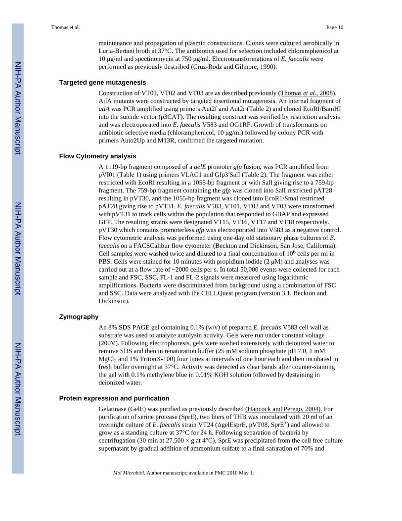
maintenance and propagation of plasmid constructions. Clones were cultured aerobically inLuria-Bertani broth at 37°C. The antibiotics used for selection included chloramphenicol at10 μg/ml and spectinomycin at 750 μg/ml. Electrotransformations of E. faecalis wereperformed as previously described (Cruz-Rodz and Gilmore, 1990).
Targeted gene mutagenesisConstruction of VT01, VT02 and VT03 are as described previously (Thomas et al., 2008).AtlA mutants were constructed by targeted insertional mutagenesis. An internal fragment ofatlA was PCR amplified using primers Aut2f and Aut2r (Table 2) and cloned EcoRI/BamHIinto the suicide vector (p3CAT). The resulting construct was verified by restriction analysisand was electroporated into E. faecalis V583 and OG1RF. Growth of transformants onantibiotic selective media (chloramphenicol, 10 μg/ml) followed by colony PCR withprimers Auto2Up and M13R, confirmed the targeted mutation.
Flow Cytometry analysisA 1119-bp fragment composed of a gelE promoter gfp fusion, was PCR amplified frompVI01 (Table 1) using primers VLAC1 and Gfp3′SalI (Table 2). The fragment was eitherrestricted with EcoRI resulting in a 1055-bp fragment or with SalI giving rise to a 759-bpfragment. The 759-bp fragment containing the gfp was cloned into SalI restricted pAT28resulting in pVT30, and the 1055-bp fragment was cloned into EcoR1/SmaI restrictedpAT28 giving rise to pVT31. E. faecalis V583, VT01, VT02 and VT03 were transformedwith pVT31 to track cells within the population that responded to GBAP and expressedGFP. The resulting strains were designated VT15, VT16, VT17 and VT18 respectively.pVT30 which contains promoterless gfp was electroporated into V583 as a negative control.Flow cytometric analysis was performed using one-day old stationary phase cultures of E.faecalis on a FACSCalibur flow cytometer (Beckton and Dickinson, San Jose, California).Cell samples were washed twice and diluted to a final concentration of 106 cells per ml inPBS. Cells were stained for 10 minutes with propidium iodide (2 μM) and analyses wascarried out at a flow rate of ~2000 cells per s. In total 50,000 events were collected for eachsample and FSC, SSC, FL-1 and FL-2 signals were measured using logarithmicamplifications. Bacteria were discriminated from background using a combination of FSCand SSC. Data were analyzed with the CELLQuest program (version 3.1, Beckton andDickinson).
ZymographyAn 8% SDS PAGE gel containing 0.1% (w/v) of prepared E. faecalis V583 cell wall assubstrate was used to analyze autolysin activity. Gels were run under constant voltage(200V). Following electrophoresis, gels were washed extensively with deionized water toremove SDS and then in renaturation buffer (25 mM sodium phosphate pH 7.0, 1 mMMgCl2 and 1% TritonX-100) four times at intervals of one hour each and then incubated infresh buffer overnight at 37°C. Activity was detected as clear bands after counter-stainingthe gel with 0.1% methylene blue in 0.01% KOH solution followed by destaining indeionized water.
Protein expression and purificationGelatinase (GelE) was purified as previously described (Hancock and Perego, 2004). Forpurification of serine protease (SprE), two liters of THB was inoculated with 20 ml of anovernight culture of E. faecalis strain VT24 (ΔgelEsprE, pVT08, SprE+) and allowed togrow as a standing culture at 37°C for 24 h. Following separation of bacteria bycentrifugation (30 min at 27,500 × g at 4°C), SprE was precipitated from the cell free culturesupernatant by gradual addition of ammonium sulfate to a final saturation of 70% and
Thomas et al. Page 10
Mol Microbiol. Author manuscript; available in PMC 2010 May 1.
NIH
-PA Author Manuscript
NIH
-PA Author Manuscript
NIH
-PA Author Manuscript

incubated overnight at 4°C with constant agitation. The precipitants were removed bycentrifugation (30 min at 27,500 × g at 4°C) and resuspended in 200 ml of 200 mM Tris-HCl and 5 mM CaCl2, pH 7.6. The 200 ml sample was filter-sterilized (0.2μm) andextensively dialyzed (2 weeks) against dialysis buffer (50 mM Tris-HCl and 5 mM CaCl2,pH 7.6). The sample was concentrated (Pierce ICON™ concentrator) down to 2 ml and wasresolved at room temperature on a TSK G3000SW gel filtration column (7.8 mm × 300 mm;Tosoh Bioscience, Montgomeryville, PA) at a flow rate of 1 ml/min, using TSK buffer (0.06M sodium phosphate, 0.1 M sodium sulfate, pH 7.0) and stored at 4°C.
Histidine tagged AtlA (devoid of signal sequence) was purified from Escherichia coli BL21(DE3) harboring the expression construct pVT21 and grown in 1 L LB medium containingampicillin (100μg/ml). Expression of recombinant protein was induced by the addition of0.5 mM isopropyl-β-D-thiogalactopyranoside (IPTG) to the E. coli cultures at an opticaldensity (at 600 nm) of 0.6. Following IPTG treatment cultures were incubated for anadditional 3 hours at 37°C after which bacterial cells were harvested by centrifugation andresuspended in 40 ml lysis buffer (20 mM Tris-HCl, 0.5 M NaCl, 10 mM imidazole, 20mM-mercaptoethanol (pH 7.9). Cells were disrupted by ultra sonicaton (5 cycles ofintermittent 20 sec pulse and 20 sec cooling) and the supernatant retrieved afterultracentrifugation at 45,000 rpm for 1 h at 4°C to remove cell debris. The supernatant waspassed through a 0.2μm filter prior to being loaded onto a nickel nitrilotriacetic acid-agarosecolumn (Qiagen). The His-tagged AtlA was eluted with a step-wise gradient of imidazole(20, 40, 100, and 250 mM) in lysis buffer and optimal purity obtained at 100 mMconcentration of imidazole in lysis buffer. Purified AtlA was concentrated and bufferexchange (25 mM sodium phosphate buffer, pH 7) carried out using an ICON™ concentrator(Pierce). Purified protein was stored at 4°C for short term or −80°C for long term storage.Protein purity was analyzed by sodium dodecyl sulfate (SDS)-acrylamide gelelectrophoresis (>95% for all purified proteins) and tryptic digests of the proteins followedby MALDI-TOF mass spectrometry confirmed their identities.
MS and MS/MS analysisAfter staining gels with Coomassie R-250, selected gel bands were excised manually as 1–2mm slices and transferred to 1.5 ml Eppendorf tubes. An equivalent slice from the protein-free region of the gel was excised as a background control. The control and test gel sectionswere then destained using three 30 min washes of 60 μl 1:1 acetonitrile: water at 30°C. Gelpieces were then dried for 10 min under vacuum. The gels were subjected to reduction andalkylation using 50 mM Tris (2-carboxyethyl) phosphine (TCEP) at 55°C for 10 minfollowed by 100 mM iodoacetamide in the dark at 30°C for 60 min. The carboxymethylatedsamples were thoroughly washed and redried in vacuo, then incubated with sequencinggrade trypsin (Trypsin Gold, Promega, Madison, WI), 20 ng/μl in 40 mM ammoniumbicarbonate, in 20 μl. Upon rehydration of the gels, an additional 15 μl of 40 mMammonium bicarbonate and 10% acetonitrile was added, and gel sections incubated at 30°Cfor 17 h in sealed Eppendorf tubes. The aqueous digestion solutions were transferred to 1.5ml clean Eppendorf tubes, and those tryptic fragments remaining within the gel sectionswere recovered by a single extraction with 50 μl of 50% acetonitrile in water containing 2%trifluoracetic acid (TFA) at 30°C for 1 h. The acetonitrile-water fractions were combinedwith the previous aqueous fractions and the liquid removed by speed vacuum concentration.The dried samples were resuspended in 10 μl of 30 mg/ml 2,5-dihydroxylbenzonic acid(DHB) (Sigma, St. Louis, MO) dissolved in 33% acetonitrile/0.1% TFA and 2 μl of peptide/matrix solution was applied to a Bruker aluminum target plate for MALDI-TOF and TOF/TOF analysis.
Mass spectra and tandem mass spectra were obtained on a Bluker Ultraflex II TOF/TOFmass spectrometer. Positively charged ions were analyzed in the reflector mode. MS and
Thomas et al. Page 11
Mol Microbiol. Author manuscript; available in PMC 2010 May 1.
NIH
-PA Author Manuscript
NIH
-PA Author Manuscript
NIH
-PA Author Manuscript

MS/MS spectra were analyzed with Flex analysis 3.0 and Bio Tools 3.0 software (BrukerDaltonics). Measurements were externally calibrated with 8 different peptides ranging from757.39 to 3147.47 (Peptide Calibration Standard I, Bruker Daltonics) and internally re-calibrated with peptides from the autoproteolysis of trypsin.
Peptide ion search was performed using MASCOT software (Matrix Science) against theexpected recombinant AtlA protein sequence. The following parameters were used for thedatabase search: MS and MS/MS accuracies were set to < 0.6 Da. Trypsin as an enzyme,missed cleavages 1, carbamidomethylation of cysteine as fixed modification and oxidationof methionine as a variable modification.
GelE and SprE mediated proteolysis of AtlAGelE or SprE (30nM) were incubated with 10μg of AtlA separately in 100 μl of reactionbuffer (50 mM Tris-HCl and 5 mM CaCl2, pH 7.6). After 10, 30, 60 and 300 mins ofincubation at 37°C, aliquots (25μl) were withdrawn and the reaction was stopped by theaddition of SDS-PAGE sample loading buffer and boiling for 10 minutes. Samples wereanalyzed by sodium dodecyl sulfate (SDS)-acrylamide gel electrophoresis and silverstaining. MALDI-TOF MS and MS/MS analysis was carried out as previously described, toempirically map regions of AtlA that were susceptible to proteolytic attack.
Biofilm assayBiofilm assays were carried out as described previously (Thomas et al., 2008).
eDNA release assayMeasurements of eDNA release were carried out on stationary phase cultures using thenucleic acid stain SYTOX green (Invitrogen). Overnight grown cultures of E. faecalis(37°C) were centrifuged (13,000 × g; 3 minutes) to pellet cells and 200 μl of the culturesupernatants were transferred to microtiter plate wells in triplicate. SYTOX green wassupplemented to these wells to a final concentration of 1 μM and incubated for 10 minutesbefore being spectrofluorometrically measured with excitation at 485 nm and emission at535 nm on a Perkin Elmer Victor 3 fluorescent plate reader.
Fratricide assayCo-culture lysis assays were carried out using V583, VT01 (ΔgelE), VT02 (ΔsprE), VT03(ΔgelEsprE) and VT19 (V583::atlA) as attacker strains and either VT12 (ΔgelEsprE, GFP+)or VT23 (ΔgelEsprE,::atlA, GFP+) as targets. Attacker strains grown overnight were diluted1:1000 in fresh Todd Hewitt broth (THB) media and were co-inoculated with target strainsdiluted 1:20 from an overnight culture grown for an equivalent period at 37°C in THBG(THB + 2% glycine). Following 24 hours of co-culture at 37°C, GFP (released as a result oflysis of target) from 250 μL of supernatant was precipitated using 4 volumes of cold acetone(−20°C) and resolved by SDS-PAGE. Detection of GFP in the supernatant was carried outby western blot analysis using anti-GFP antibodies.
Lysis assayLysis assays were carried out as described previously with the following modifications(Thomas et al., 2008). Briefly, pre-cultures of VT03 (ΔgelEsprE)or VT22(ΔgelEsprE,::atlA) were grown overnight at 37°C in 2.5 ml THB and diluted 100-fold inSM17 media supplemented with 3% glycine. Following overnight growth at 37°C, 1.5 mLcultures were centrifuged at 13,000 rpm for 3 minutes and washed thoroughly, thrice in ice-cold sterile distilled water. After the third wash, the cells were resuspended in 10 mMsodium phosphate buffer (pH 6.8) and supplemented with 1 mM CaCl2. Two hundred
Thomas et al. Page 12
Mol Microbiol. Author manuscript; available in PMC 2010 May 1.
NIH
-PA Author Manuscript
NIH
-PA Author Manuscript
NIH
-PA Author Manuscript

microliters of the suspended cells were dispensed into a 96 well plate and the optical densityat 600 nm was monitored for 9 h at 30-min intervals either in the presence or absence ofprotease treatments (30 nM of either GelE or SprE or both).
Peptidoglycan affinity assayPurified recombinant AtlA (10 μg) was allowed to bind with 20 mg wet-weight of isolatedE. faecalis V583 cell wall for 15 minutes at 37°C in 100 μL of buffer (10 mM Tris-HCl and5 mM CaCl2, pH 7.6). Approximately, 30 nM of either GelE or SprE was supplementedseparately into each sample mix. For co-protease treatments recombinant AtlA bound toV583 peptidoglycan (20mg wet-weight) were either pretreated with 30 nM GelE or SprE for10 minutes. This was followed by the immediate addition of the second complementaryprotease (30 nM; SprE or GelE) for 30 minutes. Samples (25 μL) were withdrawn atintervals of 10, 30, 60 and 300 minutes. Supernatants containing unbound proteolyticallyprocessed AtlA derivatives were retrieved after centrifugation (13,000 × g; 10 minutes). Thepellet was washed twice in fresh buffer to remove loosely adherent AtlA from pellet and thecell wall bound fraction eluted in SDS PAGE sample loading buffer. Unbound and boundAtlA derivatives were resolved by SDS PAGE and visualized by silver staining.
AcknowledgmentsWe are very grateful to Tammy Koopman, Melinda J Wilkerson and Sherry Fleming for support with flowcytometry. We also thank John M Pfeffer and Anthony J Clarke for help with zymography. Finally we are indebtedto Yu Xue and Jian Ren for help with using the protein domain visualization software DOG 1.0. This study wassupported by a Heartland Affiliate Beginning Grant-in-Aid 0660072Z and 0860084Z from the American HeartAssociation (to L.E.H), a grant-in-aid from the Terry C. Johnson Cancer Center at Kansas State University (toV.C.T), NSF-MRI grant-0521587 (to J.T) and the Targeted Excellence Program at K-State, Functional GenomicsConsortium (to J.T), and a training award (to N.H.) from NIH Grant # P20 RR16475 from the INBRE program ofthe National Center for Research Resources.
ReferencesAhn SJ, Burne RA. The atlA operon of Streptococcus mutans: role in autolysin maturation and cell
surface biogenesis. J Bacteriol. 2006; 188:6877–6888. [PubMed: 16980491]Bayles KW. The biological role of death and lysis in biofilm development. Nat Rev Microbiol. 2007;
5:721–726. [PubMed: 17694072]Biswas R, Voggu L, Simon UK, Hentschel P, Thumm G, Gotz F. Activity of the major staphylococcal
autolysin Atl. FEMS Microbiol Lett. 2006; 259:260–268. [PubMed: 16734789]Bourgogne A, Hilsenbeck SG, Dunny GM, Murray BE. Comparison of OG1RF and an isogenic fsrB
deletion mutant by transcriptional analysis: the Fsr system of Enterococcus faecalis is more than theactivator of gelatinase and serine protease. J Bacteriol. 2006; 188:2875–2884. [PubMed: 16585749]
Bourgogne A, Garsin DA, Qin X, Singh KV, Sillanpaa J, Yerrapragada S, Ding Y, Dugan-Rocha S,Buhay C, Shen H, Chen G, Williams G, Muzny D, Maadani A, Fox KA, Gioia J, Chen L, Shang Y,Arias CA, Nallapareddy SR, Zhao M, Prakash VP, Chowdhury S, Jiang H, Gibbs RA, Murray BE,Highlander SK, Weinstock GM. Large scale variation in Enterococcus faecalis illustrated by thegenome analysis of strain OG1RF. Genome Biol. 2008; 9:R110. [PubMed: 18611278]
Carniol K, Gilmore MS. Signal transduction, quorum-sensing, and extracellular protease activity inEnterococcus faecalis biofilm formation. J Bacteriol. 2004; 186:8161–8163. [PubMed: 15576763]
Chang S, Sievert DM, Hageman JC, Boulton ML, Tenover FC, Downes FP, Shah S, Rudrik JT, PuppGR, Brown WJ, Cardo D, Fridkin SK. Infection with vancomycin-resistant Staphylococcus aureuscontaining the vanA resistance gene. N Engl J Med. 2003; 348:1342–1347. [PubMed: 12672861]
Claverys JP, Havarstein LS. Cannibalism and fratricide: mechanisms and raisons d’etre. Nat RevMicrobiol. 2007; 5:219–229. [PubMed: 17277796]
Cruz-Rodz AL, Gilmore MS. High efficiency introduction of plasmid DNA into glycine treatedEnterococcus faecalis by electroporation. Mol Gen Genet. 1990; 224:152–154. [PubMed: 2126058]
Thomas et al. Page 13
Mol Microbiol. Author manuscript; available in PMC 2010 May 1.
NIH
-PA Author Manuscript
NIH
-PA Author Manuscript
NIH
-PA Author Manuscript

Diggle SP, Griffin AS, Campbell GS, West SA. Cooperation and conflict in quorum-sensing bacterialpopulations. Nature. 2007; 450:411–414. [PubMed: 18004383]
Eaton TJ, Gasson MJ. Molecular screening of Enterococcus virulence determinants and potential forgenetic exchange between food and medical isolates. Appl Environ Microbiol. 2001; 67:1628–1635. [PubMed: 11282615]
Eckert C, Lecerf M, Dubost L, Arthur M, Mesnage S. Functional analysis of AtlA, the major N-acetylglucosaminidase of Enterococcus faecalis. J Bacteriol. 2006; 188:8513–8519. [PubMed:17041059]
Ellermeier CD, Hobbs EC, Gonzalez-Pastor JE, Losick R. A three-protein signaling pathwaygoverning immunity to a bacterial cannibalism toxin. Cell. 2006; 124:549–559. [PubMed:16469701]
Fux CA, Costerton JW, Stewart PS, Stoodley P. Survival strategies of infectious biofilms. TrendsMicrobiol. 2005; 13:34–40. [PubMed: 15639630]
Gilmore MS, Haas W. The selective advantage of microbial fratricide. Proc Natl Acad Sci U S A.2005; 102:8401–8402. [PubMed: 15939890]
Gonzalez-Pastor JE, Hobbs EC, Losick R. Cannibalism by sporulating bacteria. Science. 2003;301:510–513. [PubMed: 12817086]
Guiral S, Mitchell TJ, Martin B, Claverys JP. Competence-programmed predation of noncompetentcells in the human pathogen Streptococcus pneumoniae: genetic requirements. Proc Natl Acad SciU S A. 2005; 102:8710–8715. [PubMed: 15928084]
Hancock LE, Perego M. The Enterococcus faecalis fsr two-component system controls biofilmdevelopment through production of gelatinase. J Bacteriol. 2004; 186:5629–5639. [PubMed:15317767]
Hancock LE, Perego M. Systematic inactivation and phenotypic characterization of two-componentsignal transduction systems in Enterococcus faecalis V583. J Bacteriol. 2004; 186:7951–7958.[PubMed: 15547267]
Havarstein LS, Martin B, Johnsborg O, Granadel C, Claverys JP. New insights into the pneumococcalfratricide: relationship to clumping and identification of a novel immunity factor. Mol Microbiol.2006; 59:1297–1307. [PubMed: 16430701]
Heilmann C, Hussain M, Peters G, Gotz F. Evidence for autolysin-mediated primary attachment ofStaphylococcus epidermidis to a polystyrene surface. Mol Microbiol. 1997; 24:1013–1024.[PubMed: 9220008]
Jayaraman A, Wood TK. Bacterial quorum sensing: signals, circuits, and implications for biofilms anddisease. Annu Rev Biomed Eng. 2008; 10:145–167. [PubMed: 18647113]
Kristich CJ, Li YH, Cvitkovitch DG, Dunny GM. Esp-independent biofilm formation by Enterococcusfaecalis. J Bacteriol. 2004; 186:154–163. [PubMed: 14679235]
Kristich CJ, Nguyen VT, Le T, Barnes AM, Grindle S, Dunny GM. Development and use of anefficient system for random mariner transposon mutagenesis to identify novel genetic determinantsof biofilm formation in the core Enterococcus faecalis genome. Appl Environ Microbiol. 2008;74:3377–3386. [PubMed: 18408066]
Makinen PL, Clewell DB, An F, Makinen KK. Purification and substrate specificity of a stronglyhydrophobic extracellular metalloendopeptidase (“gelatinase”) from Streptococcus faecalis (strain0G1-10). J Biol Chem. 1989; 264:3325–3334. [PubMed: 2536744]
Mesnage S, Chau F, Dubost L, Arthur M. Role of N-acetylglucosaminidase and N-acetylmuramidaseactivities in Enterococcus faecalis peptidoglycan metabolism. J Biol Chem. 2008; 283:19845–19853. [PubMed: 18490448]
Mohamed JA, Huang W, Nallapareddy SR, Teng F, Murray BE. Influence of origin of isolates,especially endocarditis isolates, and various genes on biofilm formation by Enterococcus faecalis.Infect Immun. 2004; 72:3658–3663. [PubMed: 15155680]
Murray BE, Singh KV, Ross RP, Heath JD, Dunny GM, Weinstock GM. Generation of restriction mapof Enterococcus faecalis OG1 and investigation of growth requirements and regions encodingbiosynthetic function. J Bacteriol. 1993; 175:5216–5223. [PubMed: 8349561]
Thomas et al. Page 14
Mol Microbiol. Author manuscript; available in PMC 2010 May 1.
NIH
-PA Author Manuscript
NIH
-PA Author Manuscript
NIH
-PA Author Manuscript

Nakayama J, Cao Y, Horii T, Sakuda S, Akkermans AD, de Vos WM, Nagasawa H. Gelatinasebiosynthesis-activating pheromone: a peptide lactone that mediates a quorum sensing inEnterococcus faecalis. Mol Microbiol. 2001; 41:145–154. [PubMed: 11454207]
Nakayama J, Kariyama R, Kumon H. Description of a 23.9-kilobase chromosomal deletion containinga region encoding fsr genes which mainly determines the gelatinase-negative phenotype of clinicalisolates of Enterococcus faecalis in urine. Appl Environ Microbiol. 2002; 68:3152–3155.[PubMed: 12039782]
Nakayama J, Chen S, Oyama N, Nishiguchi K, Azab EA, Tanaka E, Kariyama R, Sonomoto K.Revised model for Enterococcus faecalis fsr quorum-sensing system: the small open reading framefsrD encodes the gelatinase biosynthesis-activating pheromone propeptide corresponding tostaphylococcal agrD. J Bacteriol. 2006; 188:8321–8326. [PubMed: 16980448]
Nieto C, Espinosa M. Construction of the mobilizable plasmid pMV158GFP, a derivative of pMV158that carries the gene encoding the green fluorescent protein. Plasmid. 2003; 49:281–285.[PubMed: 12749839]
Noble WC, Virani Z, Cree RG. Co-transfer of vancomycin and other resistance genes fromEnterococcus faecalis NCTC 12201 to Staphylococcus aureus. FEMS Microbiol Lett. 1992;72:195–198. [PubMed: 1505742]
Paulsen IT, Banerjei L, Myers GS, Nelson KE, Seshadri R, Read TD, Fouts DE, Eisen JA, Gill SR,Heidelberg JF, Tettelin H, Dodson RJ, Umayam L, Brinkac L, Beanan M, Daugherty S, DeBoyRT, Durkin S, Kolonay J, Madupu R, Nelson W, Vamathevan J, Tran B, Upton J, Hansen T,Shetty J, Khouri H, Utterback T, Radune D, Ketchum KA, Dougherty BA, Fraser CM. Role ofmobile DNA in the evolution of vancomycin-resistant Enterococcus faecalis. Science. 2003;299:2071–2074. [PubMed: 12663927]
Qin X, Singh KV, Xu Y, Weinstock GM, Murray BE. Effect of disruption of a gene encoding anautolysin of Enterococcus faecalis OG1RF. Antimicrob Agents Chemother. 1998; 42:2883–2888.[PubMed: 9797220]
Qin X, Singh KV, Weinstock GM, Murray BE. Effects of Enterococcus faecalis fsr genes onproduction of gelatinase and a serine protease and virulence. Infect Immun. 2000; 68:2579–2586.[PubMed: 10768947]
Qin Z, Ou Y, Yang L, Zhu Y, Tolker-Nielsen T, Molin S, Qu D. Role of autolysin-mediated DNArelease in biofilm formation of Staphylococcus epidermidis. Microbiology. 2007; 153:2083–2092.[PubMed: 17600053]
Ren J, Wen L, Gao X, Jin C, Xue Y, Yao X. DOG 1.0: illustrator of protein domain structures. CellRes. 2009; 19:271–273. [PubMed: 19153597]
Rice KC, Mann EE, Endres JL, Weiss EC, Cassat JE, Smeltzer MS, Bayles KW. The cidA mureinhydrolase regulator contributes to DNA release and biofilm development in Staphylococcusaureus. Proc Natl Acad Sci U S A. 2007; 104:8113–8118. [PubMed: 17452642]
Sandoz KM, Mitzimberg SM, Schuster M. Social cheating in Pseudomonas aeruginosa quorumsensing. Proc Natl Acad Sci U S A. 2007; 104:15876–15881. [PubMed: 17898171]
Shibata Y, Kawada M, Nakano Y, Toyoshima K, Yamashita Y. Identification and characterization ofan autolysin-encoding gene of Streptococcus mutans. Infect Immun. 2005; 73:3512–3520.[PubMed: 15908380]
Shockman GD, Cheney MC. Autolytic enzyme system of Streptococcus faecalis. V. Nature of theautolysin-cell wall complex and its relationship to properties of the autolytic enzyme ofStreptococcus faecalis. J Bacteriol. 1969; 98:1199–1207. [PubMed: 4977984]
Singh KV, Nallapareddy SR, Nannini EC, Murray BE. Fsr-independent production of protease(s) mayexplain the lack of attenuation of an Enterococcus faecalis fsr mutant versus a gelE-sprE mutant ininduction of endocarditis. Infect Immun. 2005; 73:4888–4894. [PubMed: 16041002]
Steen A, Buist G, Kramer NE, Jalving R, Benus GF, Venema G, Kuipers OP, Kok J. Reduced lysisupon growth of Lactococcus lactis on galactose is a consequence of decreased binding of theautolysin AcmA. Appl Environ Microbiol. 2008; 74:4671–4679. [PubMed: 18539791]
Thomas VC, Thurlow LR, Boyle D, Hancock LE. Regulation of autolysis-dependent extracellularDNA release by Enterococcus faecalis extracellular proteases influences biofilm development. JBacteriol. 2008; 190:5690–5698. [PubMed: 18556793]
Thomas et al. Page 15
Mol Microbiol. Author manuscript; available in PMC 2010 May 1.
NIH
-PA Author Manuscript
NIH
-PA Author Manuscript
NIH
-PA Author Manuscript

Trieu-Cuot P, Carlier C, Poyart-Salmeron C, Courvalin P. A pair of mobilizable shuttle vectorsconferring resistance to spectinomycin for molecular cloning in Escherichia coli and in gram-positive bacteria. Nucleic Acids Res. 1990; 18:4296. [PubMed: 2143017]
Waters CM, Antiporta MH, Murray BE, Dunny GM. Role of the Enterococcus faecalis GelE proteasein determination of cellular chain length, supernatant pheromone levels, and degradation of fibrinand misfolded surface proteins. J Bacteriol. 2003; 185:3613–3623. [PubMed: 12775699]
West SA, Griffin AS, Gardner A, Diggle SP. Social evolution theory for microorganisms. Nat RevMicrobiol. 2006; 4:597–607. [PubMed: 16845430]
Zeng J, Teng F, Murray BE. Gelatinase is important for translocation of Enterococcus faecalis acrosspolarized human enterocyte-like T84 cells. Infect Immun. 2005; 73:1606–1612. [PubMed:15731060]
Thomas et al. Page 16
Mol Microbiol. Author manuscript; available in PMC 2010 May 1.
NIH
-PA Author Manuscript
NIH
-PA Author Manuscript
NIH
-PA Author Manuscript

Figure 1.Estimation of altruistic suicide in enterococcal populations by flow cytometry. A.Representative flow histograms of GBAP responders from wild type VT15, and isogenicprotease mutants- VT16 (ΔgelE), VT17 (ΔsprE) and VT18 (ΔgelEsprE) cultured tostationary phase. The pVT31 reporter plasmid in these isogenic strains activates GFPexpression in response to GBAP quorum signaling. GFP fluorescence (FL-1H)corresponding to VT15, VT16, VT17 and VT18 are plotted (thick solid lines) and arecontrasted in each instance with autofluorescence (thin solid lines) from VT14 (V583pVT30, reporter construct lacking functional gelE promoter fusion). B. Percent GFP+ cells(GBAP responders) following overnight growth of VT13 (V583::fsrA), VT14, VT15, VT16,
Thomas et al. Page 17
Mol Microbiol. Author manuscript; available in PMC 2010 May 1.
NIH
-PA Author Manuscript
NIH
-PA Author Manuscript
NIH
-PA Author Manuscript

VT17 and VT18. The percent estimates were derived from histograms of seven independenttrials ± SE using the CELLQuest program (version 3.1, Beckton and Dickinson).
Thomas et al. Page 18
Mol Microbiol. Author manuscript; available in PMC 2010 May 1.
NIH
-PA Author Manuscript
NIH
-PA Author Manuscript
NIH
-PA Author Manuscript

Figure 2.Extracellular proteases regulate enterococcal fratricide. A. Detection of GFPimmunoreactivity in stationary phase predator-prey co-culture supernatants. Predator (V583,GelE+SprE+; VT01, GelE−SprE+; VT02, GelE+SprE−; and VT03, GelE−SprE−) and prey(VT12, GelE−SprE− GFP+) were co-inoculated at a 20:1 ratio of prey: predator and grownovernight at 37°C prior to immunodetection of GFP. B. Effect of extracellular proteases onlysis of VT03 (GelE−SprE−). Differences in lysis rates of VT03 in the absence or presenceof extracellular proteases (GelE and SprE) are exhibited as percent values of initial opticaldensity at 600 nm. VT03 was incubated alone (●) or in the presence of 100 ng of GelE (■),SprE (▲) or GelE and SprE (▼;) together over a period of 12 hours at 37°C. Data are mean± SE of three independent trials with each performed in triplicate.
Thomas et al. Page 19
Mol Microbiol. Author manuscript; available in PMC 2010 May 1.
NIH
-PA Author Manuscript
NIH
-PA Author Manuscript
NIH
-PA Author Manuscript

Figure 3.Enterococcus faecalis cell surface autolysin profiles. Surface proteins were extracted fromovernight grown cultures by boiling bacterial pellets in SDS-PAGE sample loading buffer.Samples were resolved on an 8% SDS polyacrylamide gel containing 0.1% purified OG1RFcell wall as substrate. Lanes 1 through 4 represents cell wall protein extracts from OG1RF(GelE+SprE+), VT20 (OG1RFΔgelE; GelE−SprE+), V583 (GelE+SprE+) and VT01(V583ΔgelE; GelE−SprE+), respectively. Calculated molecular weights are indicated on theleft.
Thomas et al. Page 20
Mol Microbiol. Author manuscript; available in PMC 2010 May 1.
NIH
-PA Author Manuscript
NIH
-PA Author Manuscript
NIH
-PA Author Manuscript

Figure 4.AtlA is critical for eDNA release and biofilm formation. A. Detection of extracellular DNAin culture supernatants. Cell free culture supernatants of V583 (GelE+), VT01 (V583ΔgelE),VT19 (V583::atlA), OG1RF (GelE+), VT20 (OG1RF ΔgelE) and VT03 (OG1RF::atlA) weresupplemented with the DNA specific dye, SYTOX green to a final concentration of 1 μMbefore being assayed spectrofluorometrically as described. Fluorescence intensity data arerepresented as arbitrary units (AU). B. Biofilm formation of E. faecalis strains onpolystyrene. Biofilm formation was assayed as a function of crystal violet stain (measured at550 nm) retained by the biofilm biomass grown for 24 hours. Data are mean ± SE of threeindependent trials. The strain designations shown in panel B correlate with panel A.
Thomas et al. Page 21
Mol Microbiol. Author manuscript; available in PMC 2010 May 1.
NIH
-PA Author Manuscript
NIH
-PA Author Manuscript
NIH
-PA Author Manuscript
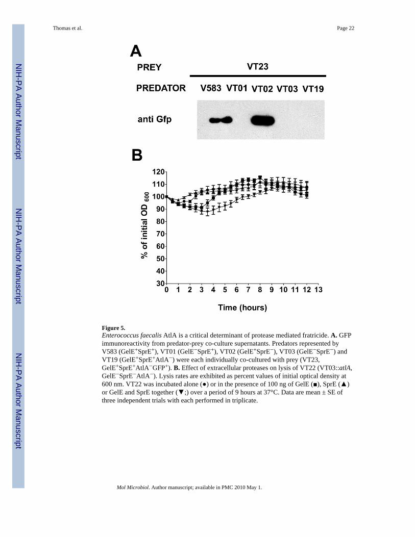
Figure 5.Enterococcus faecalis AtlA is a critical determinant of protease mediated fratricide. A. GFPimmunoreactivity from predator-prey co-culture supernatants. Predators represented byV583 (GelE+SprE+), VT01 (GelE−SprE+), VT02 (GelE+SprE−), VT03 (GelE−SprE−) andVT19 (GelE+SprE+AtlA−) were each individually co-cultured with prey (VT23,GelE+SprE+AtlA−GFP+). B. Effect of extracellular proteases on lysis of VT22 (VT03::atlA,GelE−SprE−AtlA−). Lysis rates are exhibited as percent values of initial optical density at600 nm. VT22 was incubated alone (●) or in the presence of 100 ng of GelE (■), SprE (▲)or GelE and SprE together (▼;) over a period of 9 hours at 37°C. Data are mean ± SE ofthree independent trials with each performed in triplicate.
Thomas et al. Page 22
Mol Microbiol. Author manuscript; available in PMC 2010 May 1.
NIH
-PA Author Manuscript
NIH
-PA Author Manuscript
NIH
-PA Author Manuscript
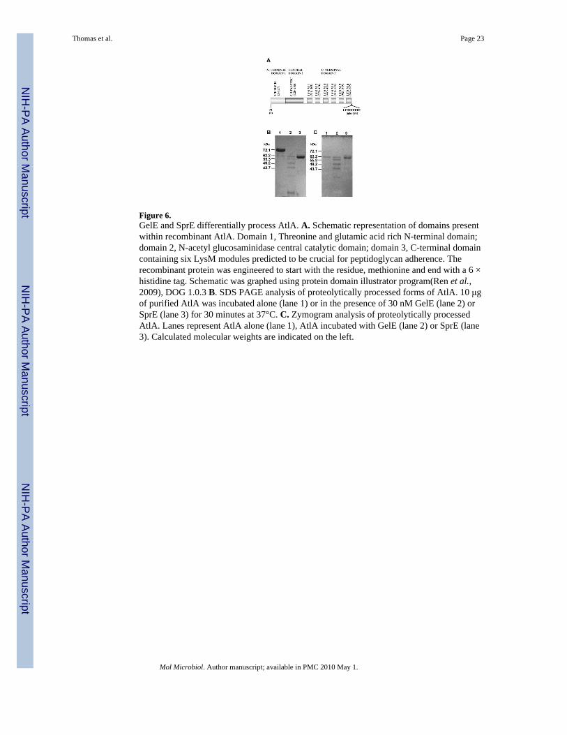
Figure 6.GelE and SprE differentially process AtlA. A. Schematic representation of domains presentwithin recombinant AtlA. Domain 1, Threonine and glutamic acid rich N-terminal domain;domain 2, N-acetyl glucosaminidase central catalytic domain; domain 3, C-terminal domaincontaining six LysM modules predicted to be crucial for peptidoglycan adherence. Therecombinant protein was engineered to start with the residue, methionine and end with a 6 ×histidine tag. Schematic was graphed using protein domain illustrator program(Ren et al.,2009), DOG 1.0.3 B. SDS PAGE analysis of proteolytically processed forms of AtlA. 10 μgof purified AtlA was incubated alone (lane 1) or in the presence of 30 nM GelE (lane 2) orSprE (lane 3) for 30 minutes at 37°C. C. Zymogram analysis of proteolytically processedAtlA. Lanes represent AtlA alone (lane 1), AtlA incubated with GelE (lane 2) or SprE (lane3). Calculated molecular weights are indicated on the left.
Thomas et al. Page 23
Mol Microbiol. Author manuscript; available in PMC 2010 May 1.
NIH
-PA Author Manuscript
NIH
-PA Author Manuscript
NIH
-PA Author Manuscript

Figure 7.Differential affinities of GelE and SprE processed recombinant AtlA forms to cell wallpeptidoglycan. Following incubation with purified cell wall peptidoglycan, rAtlA wastreated with GelE (lanes 2 and 5) and SprE (lanes 3 and 6). Lanes 1 and 4 contained rAtlAthat was not treated with either GelE or SprE. Lanes 1–3 represent the soluble fraction ofrAtlA after cell wall recovery, and lanes 4–6 represents the cell wall bound forms of rAtlA.
Thomas et al. Page 24
Mol Microbiol. Author manuscript; available in PMC 2010 May 1.
NIH
-PA Author Manuscript
NIH
-PA Author Manuscript
NIH
-PA Author Manuscript

Figure 8.Model of GelE mediated fratricide in Enterococcus faecalis. In response to GBAP quorumsensing, predators (A) co-secrete GelE ( ) and SprE ( ) extracellular proteases into theimmediate environment. GelE and SprE diffuse from the producer cell and if GelE hits thetarget prior to SprE this event may result in lysis of sibling cells that have not responded tothe quorum peptide, prey (B) resulting in the release of AtlA ( ) from their surface.Alternatively, unbound, but enzymatically active AtlA could bind and lyse neighboring cells(C). The extent of such bystander cell lysis may be directly dependent on the rate of AtlAturnover by GelE. SprE primarily acts as an immunity proteins in predator (protected cellsdepicted as solid lines) cells by increasing affinity of AtlA to localized subcellular locationson the cell surface (septum) to prevent GelE from activating autolysis. Cells that aredamaged by AtlA are depicted with discontinuous lines. Cells that are protected against thelytic action of AtlA are depicted with solid lines.
Thomas et al. Page 25
Mol Microbiol. Author manuscript; available in PMC 2010 May 1.
NIH
-PA Author Manuscript
NIH
-PA Author Manuscript
NIH
-PA Author Manuscript

NIH
-PA Author Manuscript
NIH
-PA Author Manuscript
NIH
-PA Author Manuscript
Thomas et al. Page 26
Table 1
Bacterial strains and plasmids
Strains Genotype/relevant phenotype Origin
E. faecalis
OG1RF Clinical isolate (Murray et al., 1993)
V583 Clinical isolate, TIGR sequence strain; VnR (Thomas et al., 2008)
VT01 V583ΔgelE; SprE+ (Thomas et al., 2008)
VT02 V583ΔsprE; GelE+ (Thomas et al., 2008)
VT03 V583ΔgelE-sprE (Thomas et al., 2008)
VT12 VT03 (pMV158GFP); GFP+ TetR (Thomas et al., 2008)
VT13 V583::fsrA (pVT31); CmR, SpcR This study
VT14 V583 (pVT30); SpcR This study
VT15 V583 (pVT31); GFP+ SpcR This study
VT16 VT01 (pVT31); GFP+ SpcR This study
VT17 VT02 (pVT31); GFP+ SpcR This study
VT18 VT03 (pVT31); GFP+ SpcR This study
VT19 V583:: atlA; CmR This study
VT20 OG1RFΔgelE; SprE+ This study
VT21 OG1RF:: atlA; CmR This study
VT22 VT03::atlA; CmR This study
VT23 VT12::atlA (pMV158GFP); GFP+, CmR, TetR This study
VT24 VT03 (pVT08); SprE+ This study
KS10 V583ΔfsrA Gorman, M. and Hancock, L.E.,(unpublished)
E. coli
Electro Ten Blue General plasmid maintenance strain Stratagene
BL21 (DE3) Protein expression strain Stratagene
VT25 BL21(DE3) (pVT21; AtlA+ * AmpR This study
Plasmids
pAT28 Broad host range shuttle vector; SpcR (Trieu-Cuot et al., 1990)
p3CAT p3TET derivative (Hancock and Perego, 2004), suicide vector; CmR, usedfor targeted gene inactivation
Hancock, LE (unpublished)
pMV158GFP Gram-positive replicative vector expressing a GFP reporter (Nieto and Espinosa, 2003)
pML115 p3CAT derivative carrying an internal fragment of fsrA; generates fsrA::cmmutation in E. faecalis
Hancock, L.E (unpublished)
pVI01 Low copy gelE promoter-gfp fusion reporter construct Iyer, V and Hancock, L.E (unpublished)
pVT08 pAT28 derivative carrying a 2123-bpEcoRI/BamHI fragment (native gelEpromoter and full-length sprE); leads to the over expression of SprE
(Thomas et al., 2008)
pVT10 p3CAT derivative carrying a 1006-bp EcoR1/BamH1 internal fragment ofatlA; generates atlA::cm mutation in E. faecalis
This study
pVT21 pET21b derivative carrying a 2056-bp Nde1/Xho1 atlA fragment devoid ofsignal peptide; used for the expression of AtlA in E. coli
This study
Mol Microbiol. Author manuscript; available in PMC 2010 May 1.

NIH
-PA Author Manuscript
NIH
-PA Author Manuscript
NIH
-PA Author Manuscript
Thomas et al. Page 27
Strains Genotype/relevant phenotype Origin
pVT30 pAT28 derivative carrying a 759-bp SalI/SalI gfp fragment; promoterlessgfp
This study
pVT31 pAT28 derivative carrying a gelE promoter-gfp fusion; reporter constructthat allows discrimination of predators within E. faecalis population
This study
RResistant
Vn, vancomycin; Cm, chloramphenicol; Spc, spectinomycin; Tet, tetracycline; Amp, ampicllin
*AtlA devoid of signal peptide (53 amino acid residues from the N-terminus)
Mol Microbiol. Author manuscript; available in PMC 2010 May 1.

NIH
-PA Author Manuscript
NIH
-PA Author Manuscript
NIH
-PA Author Manuscript
Thomas et al. Page 28
Table 2
Primers used in this study
Primer name Sequence (5′ →3′)
M13R CAGCTATGACCATGATTACG
Aut2-f GAGAGAATTCTGGGGAGCAAGTACGCTATC
Aut2-r CTCTGGATCCCCACCGGTGTTACCTGAAGT
Auto2Up GAGACACCAACAACAGAA
Ef0799-SP-NdeI GAGACATATGACAGAAGAGCAGCCAACAAATGC
Ef0799XhoI GAGACTCGAGACCAACTTTTAAAGTTTGAC
VLAC1 GTTGAATAACACTTATTCCTATC
Gfp3′SalI CTCTGTCGACTACGAATGCTATTTGTATAG
Mol Microbiol. Author manuscript; available in PMC 2010 May 1.