Comparative genomics of Enterococcus faecalis from healthy Norwegian infants
11
BioMed Central Page 1 of 11 (page number not for citation purposes) BMC Genomics Open Access Research article Comparative genomics of Enterococcus faecalis from healthy Norwegian infants Margrete Solheim* 1 , Ågot Aakra 1 , Lars G Snipen 2 , Dag A Brede 1 and Ingolf F Nes 1 Address: 1 Laboratory of Microbial Gene Technology and Food Microbiology, Department of Chemistry, Biotechnology and Food Science, The Norwegian University of Life Sciences, N-1432 Ås, Norway and 2 Section for Biostatistics, Department of Chemistry, Biotechnology and Food Science, The Norwegian University of Life Sciences, N-1432 Ås, Norway Email: Margrete Solheim* - [email protected]; Ågot Aakra - [email protected]; Lars G Snipen - [email protected]; Dag A Brede - [email protected]; Ingolf F Nes - [email protected] * Corresponding author Abstract Background: Enterococcus faecalis, traditionally considered a harmless commensal of the intestinal tract, is now ranked among the leading causes of nosocomial infections. In an attempt to gain insight into the genetic make-up of commensal E. faecalis, we have studied genomic variation in a collection of community-derived E. faecalis isolated from the feces of Norwegian infants. Results: The E. faecalis isolates were first sequence typed by multilocus sequence typing (MLST) and characterized with respect to antibiotic resistance and properties associated with virulence. A subset of the isolates was compared to the vancomycin resistant strain E. faecalis V583 (V583) by whole genome microarray comparison (comparative genomic hybridization (CGH)). Several of the putative enterococcal virulence factors were found to be highly prevalent among the commensal baby isolates. The genomic variation as observed by CGH was less between isolates displaying the same MLST sequence type than between isolates belonging to different evolutionary lineages. Conclusion: The variations in gene content observed among the investigated commensal E. faecalis is comparable to the genetic variation previously reported among strains of various origins thought to be representative of the major E. faecalis lineages. Previous MLST analysis of E. faecalis have identified so-called high-risk enterococcal clonal complexes (HiRECC), defined as genetically distinct subpopulations, epidemiologically associated with enterococcal infections. The observed correlation between CGH and MLST presented here, may offer a method for the identification of lineage-specific genes, and may therefore add clues on how to distinguish pathogenic from commensal E. faecalis. In this work, information on the core genome of E. faecalis is also substantially extended. Background Enterococci are Gram-positive facultative anaerobic cocci with a low GC-content. They are natural inhabitants of the mammalian gastrointestinal (GI) tract and among the first lactic acid bacteria to colonize the intestines of a new- born [1]. During the last three decades, enterococci have emerged as important pathogens and as a major cause of nosocomial infections. The majority of hospital-acquired, Published: 24 April 2009 BMC Genomics 2009, 10:194 doi:10.1186/1471-2164-10-194 Received: 12 November 2008 Accepted: 24 April 2009 This article is available from: http://www.biomedcentral.com/1471-2164/10/194 © 2009 Solheim et al; licensee BioMed Central Ltd. This is an Open Access article distributed under the terms of the Creative Commons Attribution License (http://creativecommons.org/licenses/by/2.0 ), which permits unrestricted use, distribution, and reproduction in any medium, provided the original work is properly cited.
-
Upload
independent -
Category
Documents
-
view
0 -
download
0
Transcript of Comparative genomics of Enterococcus faecalis from healthy Norwegian infants

BioMed CentralBMC Genomics
ss
Open AcceResearch articleComparative genomics of Enterococcus faecalis from healthy Norwegian infantsMargrete Solheim*1, Ågot Aakra1, Lars G Snipen2, Dag A Brede1 and Ingolf F Nes1Address: 1Laboratory of Microbial Gene Technology and Food Microbiology, Department of Chemistry, Biotechnology and Food Science, The Norwegian University of Life Sciences, N-1432 Ås, Norway and 2Section for Biostatistics, Department of Chemistry, Biotechnology and Food Science, The Norwegian University of Life Sciences, N-1432 Ås, Norway
Email: Margrete Solheim* - [email protected]; Ågot Aakra - [email protected]; Lars G Snipen - [email protected]; Dag A Brede - [email protected]; Ingolf F Nes - [email protected]
* Corresponding author
AbstractBackground: Enterococcus faecalis, traditionally considered a harmless commensal of the intestinaltract, is now ranked among the leading causes of nosocomial infections. In an attempt to gain insightinto the genetic make-up of commensal E. faecalis, we have studied genomic variation in a collectionof community-derived E. faecalis isolated from the feces of Norwegian infants.
Results: The E. faecalis isolates were first sequence typed by multilocus sequence typing (MLST)and characterized with respect to antibiotic resistance and properties associated with virulence. Asubset of the isolates was compared to the vancomycin resistant strain E. faecalis V583 (V583) bywhole genome microarray comparison (comparative genomic hybridization (CGH)). Several of theputative enterococcal virulence factors were found to be highly prevalent among the commensalbaby isolates. The genomic variation as observed by CGH was less between isolates displaying thesame MLST sequence type than between isolates belonging to different evolutionary lineages.
Conclusion: The variations in gene content observed among the investigated commensal E.faecalis is comparable to the genetic variation previously reported among strains of various originsthought to be representative of the major E. faecalis lineages. Previous MLST analysis of E. faecalishave identified so-called high-risk enterococcal clonal complexes (HiRECC), defined as geneticallydistinct subpopulations, epidemiologically associated with enterococcal infections. The observedcorrelation between CGH and MLST presented here, may offer a method for the identification oflineage-specific genes, and may therefore add clues on how to distinguish pathogenic fromcommensal E. faecalis. In this work, information on the core genome of E. faecalis is alsosubstantially extended.
BackgroundEnterococci are Gram-positive facultative anaerobic cocciwith a low GC-content. They are natural inhabitants of themammalian gastrointestinal (GI) tract and among the
first lactic acid bacteria to colonize the intestines of a new-born [1]. During the last three decades, enterococci haveemerged as important pathogens and as a major cause ofnosocomial infections. The majority of hospital-acquired,
Published: 24 April 2009
BMC Genomics 2009, 10:194 doi:10.1186/1471-2164-10-194
Received: 12 November 2008Accepted: 24 April 2009
This article is available from: http://www.biomedcentral.com/1471-2164/10/194
© 2009 Solheim et al; licensee BioMed Central Ltd. This is an Open Access article distributed under the terms of the Creative Commons Attribution License (http://creativecommons.org/licenses/by/2.0), which permits unrestricted use, distribution, and reproduction in any medium, provided the original work is properly cited.
Page 1 of 11(page number not for citation purposes)

BMC Genomics 2009, 10:194 http://www.biomedcentral.com/1471-2164/10/194
enterococcal infections is caused by Enterococcus faecalis[2]. Several putative virulence factors have been character-ized in E. faecalis (reviewed in [2]), and their roles in path-ogenicity have been established in various animal models[3-6] and cultured cell lines [7,8]. A large number ofreports on enterococcal pathogenicity has focused on thepresence or absence of these virulence determinants inenterococcal isolates from different origins [9-14]. Theresults have shown that several of the putative virulencetraits are detected in enterococcal isolates independent oftheir origin, suggesting that these factors may not be cru-cial for enterococcal pathogenicity. However, a higherincidence of some of the virulence determinants in clini-cal isolates may indicate that these genes enhance the abil-ity of E. faecalis to cause disease, as suggested by virulencestudies on bacterial mutants in animal models [3].
The sequencing of the E. faecalis V583 genome (V583)[15] made global analyses of whole genome diversitywithin this species possible [16-18], by the use of micro-array-based comparative genomic hybridization (CGH).The approximate size and composition of the E. faecaliscore genome have been investigated on clinical, food andenvironmental isolates [17,18]. The CGH-approach hasalso been used to evaluate the dissemination of variabletraits from the V583 genome within diverse lineages of thespecies [17]. Previous analyses have shown that the maingenomic variation between the strains correspond to pre-viously identified mobile genetic elements (MGEs) inV583 [17,18]. However, an effort to explore the gene con-tent of commensal E. faecalis by CGH has not previouslybeen made and little is known about genetic determinantsthat may explain the differences in life style between path-ogenic and non-pathogenic E. faecalis strains.
The aim of this study was to investigate the genomic diver-sity among fecal E. faecalis isolated from healthy Norwe-gian infants by means of CGH. In an attempt to studygenetic variability of commensal E. faecalis isolates, weused genome-wide DNA arrays to probe the presence of3219 open reading frames (ORFs) from V583 and 10ORFs representing a 17-kb deletion in the V583 patho-genicity island in our collection of community-derivedNorwegian fecal E. faecalis baby isolates. The isolates werealso characterized with respect to antibiotic resistance andproperties associated with virulence, by PCR and pheno-typic assays.
MethodsStool samplesThis study included 11 healthy Norwegian infants (7 maleand 4 female) born in the Eastern part of Norway. Thebabies were all born in Oslo and Akershus Counties.Informed consent had been obtained from their parents.From infants A-C, stool samples were collected once each
month during the first six months and once after 12months from October 2004 to September 2005, whilefrom infants D-K, samples were collected once during thefirst six months of life during 2002. All the samples werecollected after mother and child had left the obstetricward. All the infants were born by vaginal delivery and allwere breast-fed during the period of sampling. None ofthe infants were treated with antibiotics during the sam-pling period. A total of 29 stool samples were collected.
Identification of enterococcal isolates and growth conditionsFrom each stool sample, two cultures were prepared: 1 gfecal material was homogenized in 1) 10 mL deMan-Rog-osa-Sharpe (MRS; Oxoid) broth and 2) 10 mL Arroyo,Martin and Cotton broth (AMC; [19]), by vortexing. Serialdilutions were made and 100 μl of the 10-5 – 10-7 dilutionswere plated on MRS- and AMC agar plates, respectively.MRS plates were incubated aerobically over night (ON) at37°C, while AMC plates were incubated anaerobicallyON at 37°C. Plates were then examined for growth, andcolonies with different morphology and from differentplates were picked and inoculated in 5 ml MRS- or AMC-broth, depending on which plates they were isolatedfrom. The cultures were incubated as described above.Genomic DNA from each sample was isolated, and foridentification, the 16S rRNA gene from each isolate wasamplified and sequenced using general 16S rDNA primers(Additional file 1). PCR was accomplished usingDyNAzyme™ II DNA Polymerase (Finnzymes). Thermocy-cling conditions were as follows: 2 min at 94°C; followedby 30 cycles of 30 s at 94°C, 30 s at 56°C, and 1.5 min at72°C; followed by 10 min at 72°C. A total of 31 differentE. faecalis-isolates were identified, and further analyzed inthis study (Table 1). The 31 enterococcal isolates were iso-lated as part of two surveys of the content of lactic acidbacteria (LAB) in baby feces, hence, the culture mediaused were rich media, not particularly chosen to select forenterococci. In the present study, E. faecalis were grownaerobically ON in brain heart infusion broth (BHI;Oxoid) at 37°C.
MLST analysisMLST was performed according to the scheme presentedby Ruiz-Garbajosa et al. [20], using the ABI Prism Big dyeCycle Sequencing Ready Reaction kit (Applied Biosys-tems) in an ABI PrismTM 3100 Genetic Analyzer.Sequence types were defined by the allelic variation at theseven loci aroE, gdh, gki, gyd, pstS, xpt and yqiL. Isolateswith the same sequence type are thought to be membersof a single clone or lineage. Clonal complexes weredefined as groups of isolates that differed in no more thantwo of the seven loci analyzed, by use of eBURST http://www.mlst.net[21]. Each clonal complex was designatedafter its ancestor sequence type (ST) or the representative
Page 2 of 11(page number not for citation purposes)
BMC Genomics 2009, 10:194 http://www.biomedcentral.com/1471-2164/10/194
ST that appeared with the highest frequency. All the MLSTdata from this study has been deposited into the E. faecalisMLST database http://efaecalis.mlst.net/.
Phenotypic assaysCytolysin assayHemolytic activity was qualitatively detected by the use ofblood agar plates supplemented with 5% (v/v) defibri-nated horse blood, 1% (w/v) glucose and 0.03% (w/v) L-arginine (Sigma) [22]. Overnight cultures were diluted1:100, spotted onto fresh plates and incubated at 37°C for24 h. Zones of clearing around colonies indicated produc-tion of cytolysin.
Gelatinase assayDetection of gelatinase activity was performed by the useof Todd-Hewitt (Oxoid) agar plates containing 3% gelatin[23]. Overnight cultures were diluted 1:100, spotted onto
fresh plates and incubated at 37°C overnight, before theywere placed at 4°C for 5 h. Zones of turbidity around col-onies indicated hydrolysis of gelatin.
Antimicrobial susceptibility testingBHI agar plates supplemented with 4 μg/ml ampicillin, 20μg/ml chloramphenicol, 50 μg/ml erythromycin, 500 μg/ml gentamicin, 10 μg/ml tetracycline or 4 μg/ml vanco-mycin were used. The plates were inoculated by spotting5 μl (106–107 CFU) overnight culture (1: 200) and incu-bated at 37°C overnight. Growth was interpreted as resist-ance to the antibiotic added to the medium.
Detection of genes encoding virulence factors and bacteriocin genesThe presence of ace, agg, esp, cylL and gelE were detected bymeans of polymerase chain reactions (PCR) as previouslydescribed [9,24]. Isolates were also tested for the presenceof iolE, iolR and genes coding for the following bacterioc-
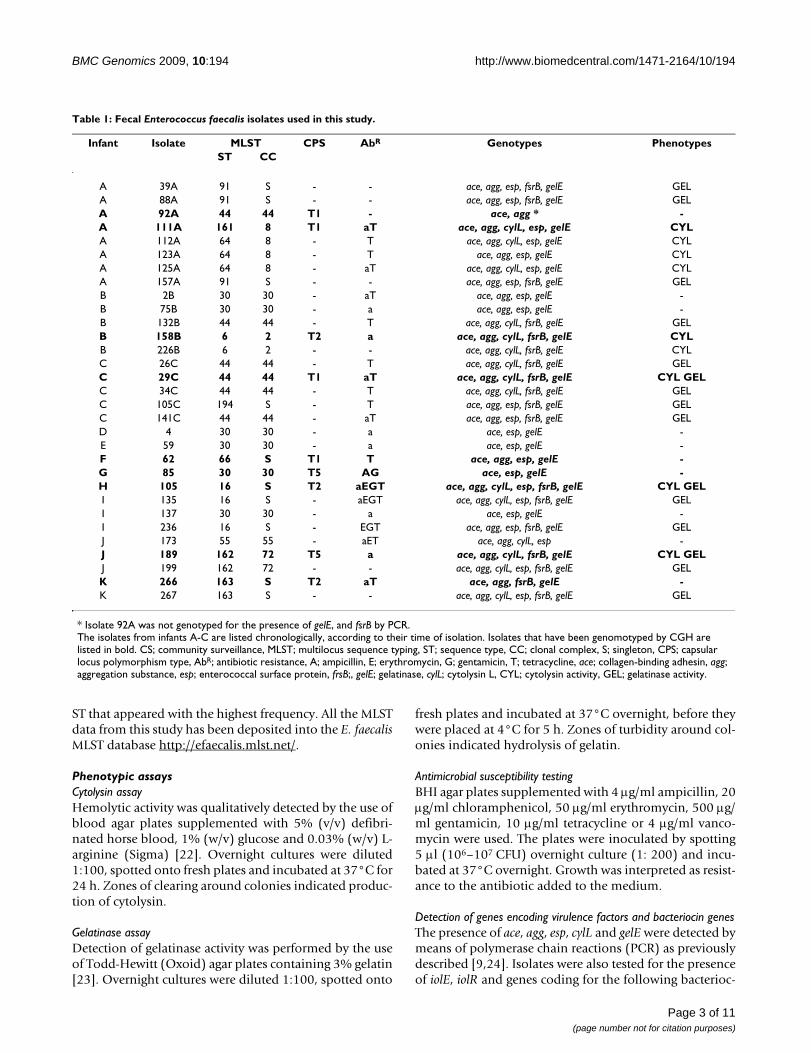
Table 1: Fecal Enterococcus faecalis isolates used in this study.
Infant Isolate MLST CPS AbR Genotypes PhenotypesST CC
A 39A 91 S - - ace, agg, esp, fsrB, gelE GELA 88A 91 S - - ace, agg, esp, fsrB, gelE GELA 92A 44 44 T1 - ace, agg * -A 111A 161 8 T1 aT ace, agg, cylL, esp, gelE CYLA 112A 64 8 - T ace, agg, cylL, esp, gelE CYLA 123A 64 8 - T ace, agg, esp, gelE CYLA 125A 64 8 - aT ace, agg, cylL, esp, gelE CYLA 157A 91 S - - ace, agg, esp, fsrB, gelE GELB 2B 30 30 - aT ace, agg, esp, gelE -B 75B 30 30 - a ace, agg, esp, gelE -B 132B 44 44 - T ace, agg, cylL, fsrB, gelE GELB 158B 6 2 T2 a ace, agg, cylL, fsrB, gelE CYLB 226B 6 2 - - ace, agg, cylL, fsrB, gelE CYLC 26C 44 44 - T ace, agg, cylL, fsrB, gelE GELC 29C 44 44 T1 aT ace, agg, cylL, fsrB, gelE CYL GELC 34C 44 44 - T ace, agg, cylL, fsrB, gelE GELC 105C 194 S - T ace, agg, esp, fsrB, gelE GELC 141C 44 44 - aT ace, agg, esp, fsrB, gelE GELD 4 30 30 - a ace, esp, gelE -E 59 30 30 - a ace, esp, gelE -F 62 66 S T1 T ace, agg, esp, gelE -G 85 30 30 T5 AG ace, esp, gelE -H 105 16 S T2 aEGT ace, agg, cylL, esp, fsrB, gelE CYL GELI 135 16 S - aEGT ace, agg, cylL, esp, fsrB, gelE GELI 137 30 30 - a ace, esp, gelE -I 236 16 S - EGT ace, agg, esp, fsrB, gelE GELJ 173 55 55 - aET ace, agg, cylL, esp -J 189 162 72 T5 a ace, agg, cylL, fsrB, gelE CYL GELJ 199 162 72 - - ace, agg, cylL, esp, fsrB, gelE GELK 266 163 S T2 aT ace, agg, fsrB, gelE -K 267 163 S - - ace, agg, cylL, esp, fsrB, gelE GEL
* Isolate 92A was not genotyped for the presence of gelE, and fsrB by PCR.The isolates from infants A-C are listed chronologically, according to their time of isolation. Isolates that have been genomotyped by CGH are listed in bold. CS; community surveillance, MLST; multilocus sequence typing, ST; sequence type, CC; clonal complex, S; singleton, CPS; capsular locus polymorphism type, AbR; antibiotic resistance, A; ampicillin, E; erythromycin, G; gentamicin, T; tetracycline, ace; collagen-binding adhesin, agg; aggregation substance, esp; enterococcal surface protein, frsB;, gelE; gelatinase, cylL; cytolysin L, CYL; cytolysin activity, GEL; gelatinase activity.
Page 3 of 11(page number not for citation purposes)

BMC Genomics 2009, 10:194 http://www.biomedcentral.com/1471-2164/10/194
ins by PCR: enterocin A (EA), enterocin B (EB), enterocinP (EP), enterolysin A (EN), enterocin L50 (EL50) andenterocin 1071A and B (E1071A&B). Thermocycling con-ditions were as follows: 2 min at 94°C; followed by 30cycles of 30 s at 94°C, 30 s at 50 ± 10°C, and 30 s at 72°C;followed by 10 min at 72°C. Primers are listed in Addi-tional file 1.
API 50 CH for determination of fermentation patternsCarbohydrate fermentation patterns were obtained forselected isolates with API 50 CH kits (BioMerieux) accord-ing to the manufacturer's instructions.
Comparative genomic hybridizationMicroarraysThe microarrays used in this work contained 3219 openreading frames from the genome of Enterococcus faecalisV583 [15] represented by oligonucleotides (70-mers;probes). Of these 3219 ORFs, 3093 were chromosomalORFs and 126 were located on plasmids. In addition, tengenes from the pathogenicity island (PAI) of E. faecalisMMH594 (deleted in the PAI of V583) were represented[25]. The 70-mer oligos were supplied by Invitrogen. Theoligos were spotted in triplicates onto epoxy-coated glassslides (Corning). In order to reduce biases due to posi-tional effects, the replicate spots were spotted at randompositions within a subarray on the array. Alien reportersequences (SpotReport®Alien® Oligo Array Validation Sys-tem, Stratagene), without homology to any knownnucleic acid sequences in public databases, were spottedas negative controls on the array. The microarray designhas been deposited in the ArrayExpress database with theaccession number A-MEXP-1069.
DNA isolationFor CGH, 9 isolates were chosen based on their represen-tation of MLST sequence type diversity across the babiesand of novel sequence types. Genomic DNA was isolatedby using the FP120 FastPrep bead-beater (BIO101/Savent) and the QiaPrep MiniPrep kit (Qiagen) as fol-lows: 10 mL overnight cultures were centrifuged for 5min. at 6000 rpm in an Eppendorf 5804R tabletop centri-fuge at 4°C, and pellets were resuspended in 250 μl RNa-seA-containing Buffer P1 (100 μg/mL RNaseA). The cellsuspensions were transferred to 2 mL screw cap FastPreptubes (Qbiogene) containing 0.5 g acid-washed glassbeads (< 106 μm) (Sigma). Cells were lysed by shakingthe tubes for 20 s at 6 m/s in the FastPrep bead-beater.After a short spin, lysed cells were transferred to Eppen-dorf tubes and 250 μl Buffer P2 and 350 μl Buffer N3 wasadded to each tube. Then, the suspensions were centri-fuged for 10 min. at 13000 rpm in a tabletop centrifuge(Biofuge Pico, Heraeus) at room temperature, before thesupernatant fluids were loaded on to Qiaprep spin col-umns. The columns were washed again and genomic DNAwas eluted according to the Qiaprep Spin MiniPrep kit
protocol. Concentration and purity of the genomic DNAwas measured using the NanoDrop spectrophotometer(NanoDrop Technologies). 5 μg genomic DNA was usedfor each labeling reaction.
Fluorescent labeling and hybridizationGenomic DNA was labeled and purified with the Bio-Prime Array CGH Genomic labeling System (Invitrogen)and Cyanine Smart Pack dUTP (PerkinElmer Life Sci-ences), according to the manufacturer's protocol.
Purified samples were then dried, prior to resuspension in140 μl hybridization solution (5 × SSC, 0.1% (w/v) SDS,1.0% (w/v) bovine serum albumin, 50% (v/v) formamideand 0.01% (w/v) single-stranded salmon sperm DNA)and hybridized for 16 h at 42°C to the E. faecalis oligonu-cleotide array in a Tecan HS 400 pro hybridization station(Tecan). Arrays were washed twice at 42°C with 2 × SSC +0.2% SDS, and twice at 23°C with 2 × SSC, followed bywashes at 23°C with 1) 0.2 × SSC and 2) H2O. Two repli-cate hybridizations (dye-swap) were performed for eachtest strain. Hybridized arrays were scanned at wavelengthsof 532 nm (Cy3) and 635 nm (Cy5) with a Tecan scannerLS (Tecan). Fluorescent intensities and spot morphologieswere analyzed using GenePix Pro 6.0 (Molecular Devices),and spots were excluded based on slide or morphologyabnormalities. All water used for the various steps of thehybridization and for preparation of solutions was fil-tered (0.2 μM) MilliQ dH20.
Data analysisStandard methods in the LIMMA package [26] in R http://www.r-project.org/, available from the Bioconductorhttp://www.bioconductor.org were employed for pre-processing and normalization. Within-array normaliza-tion was first conducted by subtracting the median fromthe log-ratios for each array. A standard loess-normaliza-tion was then performed, where smoothing was basedonly on spots with abs(log-ratio) < 2.0 to avoid biases dueto extreme skewness in the log-ratio distribution. For thedetermination of present and divergent genes, a newmethod that predicts sequence identity based on array sig-nals was used, as described by Snipen et al. [27]. A briefdescription of the method follows: Initially, all arrayprobe sequences were queried against the fully sequencedV583 reference genome (R-genome) in a BLAST search.The value Rb for each probe was defined as the number ofidentical bases in the best local alignment found byblastn, divided by the probe length. Given the sequenceidentity Rb for each probe, the corresponding array signalRa will in general correlate in a positive way, i.e. strongersequence identity yields stronger array signal. This postu-lation also holds for the unsequenced sample genome (S-genome), where Sa denote the array signal and Sb theunknown sequence identity. The basic idea is that Sb canbe predicted from Sa based on how sequence identity Rb
Page 4 of 11(page number not for citation purposes)
BMC Genomics 2009, 10:194 http://www.biomedcentral.com/1471-2164/10/194
and array signal Ra relate to each other. A threshold of0.75 was assigned to the Sb-values in order to obtain a cat-egorical response of presence or divergence, i. e. geneswith Sb-value > 0.75 were classified as present, whilegenes with Sb-value < 0.75 were classified as divergent. Ina comparison with other methods for analyzing CGH dataas reviewed by Carter et al. [28], the proposed methodgave significantly better classification of present/diver-gent. Based on the fully sequenced genomes of V583 andOG1RF [15,29], tests gave sensitivity of 0.99 and specifi-city of 0.95 for detecting present probes based on CGHdata.
Comparative phylogenomicsThe relationship of the reference strain and the test strainsbased on the presence and divergence of genes was deter-mined with Bayesian-based algorithms implementedthrough MR BAYES 3.1 [30], as previously described [31].With samples and saves from every 40th tree, 1.1 × 10⁶ gen-erations of four incrementally heated Markov chainMonte Carlos (MCMC) were performed on the CGH databy using an annealing temperature of 0.5, a burn-in of100 000 MCMC generations and an 8-category distribu-tion. Consensus trees and clade credibility values were vis-ualized by using TreeView version 1.6.6 http://taxonomy.zoology.gla.ac.uk/rod/treeview.html.
Microarray data accession numberThe microarray data have been deposited in the ArrayEx-press database with the series accession number E-TABM-466.
ResultsMLST – allelic variation in community-derived fecal E. faecalis baby isolatesThe MLST analysis was performed on 31 E. faecalis-iso-lates, obtained from 11 healthy Norwegian infants duringtheir first year of life. These isolates were considered aslegitimate representatives of commensal E. faecalis as theyhave been resident in the gut without causing any appar-ent negative effect to the health of the host. Infants A-Cwere sampled once each month during the first sixmonths and once after 12 months, and a total of 8, 5 and5 different isolates were recovered from the respectiveinfants over the period of sampling. Infants D-K weresampled once during the first six months of life and 1–3different isolates were obtained from these infants. Fromthe 31 isolates, 12 different sequence types (STs) wereidentified, of which four were novel STs (ST161, ST162,ST163 and ST194; Table 1). Of these novel STs, ST161 andST162, were single-locus variants (SLV; differing from theancestor ST in one allele) of ST64 and ST72, respectively.ST64 and ST161 belong to the previously defined clonalcomplex CC8. In addition to the ST161 (n = 1) isolate thatwas detected in one of the infants, three different isolates
displaying ST64 (n = 3) were obtained from the sameinfant (A) during the same month. Other clonal com-plexes that were represented within our collection of iso-lates were CC2 (n = 2), CC30 (n = 6), CC44 (n = 6) andCC55 (n = 1). The remaining isolates were singletons.
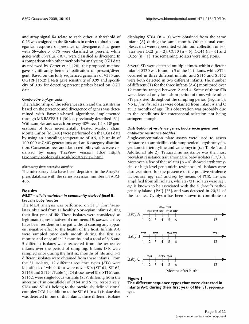
Several STs were detected multiple times, within differentinfants: ST30 was found in 5 of the 11 infants, while ST44occurred in three different infants, and ST16 and ST162were both detected in two different infants. The numberof different STs for the three infants (A-C) monitored over12 months, ranged between 2 and 4. Some of these STswere detected only for a short period of time, while otherSTs persisted throughout the sampling period (Figure 1).No E. faecalis isolates were obtained from infant A and Cat 12 months of age. This observation was probably dueto the conditions for enterococcal selection not beingstringent enough.
Distribution of virulence genes, bacteriocin genes and antibiotic resistance profilesSingle-concentration plate assays were used to assessresistance to ampicillin, chloramphenicol, erythromycin,gentamicin, tetracyline and vancomycin (see Table 1 andAdditional file 2). Tetracycline resistance was the mostprevalent resistance trait among the baby isolates (17/31).Moreover, a few of the isolates (n = 4) showed erythromy-cin- or high-level gentamicin resistance. All isolates werealso examined for the presence of the putative virulencefactors ace, agg, cylL and esp by means of PCR. ace wasamplified from all isolates, while 27/31 isolates were agg+.esp is known to be associated with the E. faecalis patho-genicity island (PAI) [25], and was detected in 20/31 ofthe isolates. Cytolysin has been shown to contribute to
The different sequence types that were detected in infants A-C during their first year of lifeFigure 1The different sequence types that were detected in infants A-C during their first year of life. ST; sequence type.
Page 5 of 11(page number not for citation purposes)
BMC Genomics 2009, 10:194 http://www.biomedcentral.com/1471-2164/10/194
virulence in animal models of enterococcal infections,and cylL, encoding one of the structural subunits of ente-rococcal cytolysin was present in 16/31 of the isolates byPCR. Cytolysin production was detected in 9/31 of theisolates on blood agar plates. The isolates were also exam-ined for gelatinase activity, which have been associatedwith virulence (reviewed in [2]). 15/31 isolates were gela-tinase positive (GelE+). Since the absence of the regulator(fsrB) can cause a lack of the gelatinase phenotype despitethe presence of a positive gelE genotype [23], the isolateswere also tested for the presence of gelE and fsrB by PCR.29 of the isolates were gelE+ and 18 were fsrB+ (see Table 1and Additional file 2). PCR-screening for content of bac-teriocin genes among the test strains further discriminatedbetween isolates with matching resistance profiles and vir-ulence characteristics (see Additional file 2).
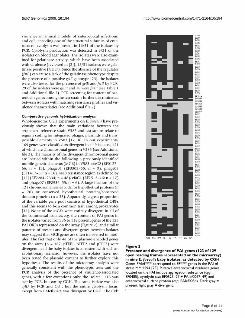
Comparative genomic hybridization analysisWhole-genome CGH experiments on E. faecalis have pre-viously shown that the main variations between thesequenced reference strain V583 and test strains relate toregions coding for integrated phages, plasmids and trans-posable elements in V583 [17,18]. In our experiments,169 genes were classified as divergent in all 9 isolates, 121of which are chromosomal genes in V583 (see Additionalfile 3). The majority of the divergent chromosomal genesare located within the following 6 previously identifiedmobile genetic elements (MGE) in V583: efaC2 (EF0127–66; n = 19), phage01 (EF0303–55; n = 9), phage03(EF1417–89; n = 16), vanB resistance region as defined by[17] (EF2284–2334; n = 48), efaC1 (EF2512–46; n = 17)and phage07 (EF2936–55; n = 6). A large fraction of the121 chromosomal genes code for hypothetical proteins (n= 70) or conserved hypothetical proteins/conserveddomain proteins (n = 35). Apparently, a great proportionof the variable gene pool consists of hypothetical ORFsand this seems to be a common trait among prokaryotes[32]. None of the MGEs were entirely divergent in all ofthe commensal isolates, e.g. the content of PAI genes inthe isolates varied from 36 to 118 present genes of the 123PAI ORFs represented on the array (Figure 2), and similarpatterns of present and divergent genes between isolatesmay suggest that MGE genes are often transferred in mod-ules. The fact that only 48 of the plasmid-encoded geneson the array (n = 167; pTEF1, pTEF2 and pTEF3) weredivergent in all the baby isolates is consistent with such anevolutionary scenario; however, the isolates have notbeen tested for plasmid content to further explore thishypothesis. The results of the microarray analysis weregenerally consistent with the phenotypic tests and thePCR analysis of the presence of virulence-associatedgenes, with a few exceptions only: the isolate 111A wasesp+ by PCR, but esp- by CGH. The same isolate was alsocylL+ by PCR and Cyl+, but the entire cytolysin locus,except from PAIef0049, was divergent by CGH. The Cyl+
Presence and divergence of PAI genes (123 of 129 open reading frames represented on the microarray) in nine E. fae-calis baby isolates, as detected by CGHFigure 2Presence and divergence of PAI genes (123 of 129 open reading frames represented on the microarray) in nine E. faecalis baby isolates, as detected by CGH. Genes PAIef**** correspond to EF**** genes in the PAI of strain MMH594 [25]. Putative enterococcal virulence genes located on the PAI include aggregation substance (agg; EF0485), cytolysin (cyl; EF0523–27 + PAIef0047–49) and enterococcal surface protein (esp; PAIef0056). Dark gray = present, light gray = divergent.
Page 6 of 11(page number not for citation purposes)

BMC Genomics 2009, 10:194 http://www.biomedcentral.com/1471-2164/10/194
phenotypes of the isolates 158B and 189 were also incon-sistent with the CGH data for some of the additional PAIgenes on the array (PAIef0047–49). The observed incon-sistencies between phenotypes/PCR and CGH may be dueto sequence variations in the microarray-probe targetsequences in the test stains.
Each of the fecal baby isolates showed a minimum of76.5% presence of probes represented on the array(76.6% of the V583 genome). The CGH analysis classified2092 of the 3093 chromosomal V583 ORFs as present inall 9 isolates. This set of shared genes is slightly higherthan the core genomes that were previously reported for E.faecalis [17,18], probably due to the constricted environ-ment and the limited geographical area, from which theisolates were obtained. The observed genetic variationamong the investigated commensal E. faecalis shows thatthe genetic diversity is comparable to that among strainsfrom other sources (food, clinical, environmental, animalhusbandry etc) [17,18]. Our data confirmed the establish-ment of phage02 as a part of the E. faecalis core genome.
The E. faecalis V583 genome contains 35 probable phos-phoenolpyruvate-dependent sugar phosphotransferase(PTS) systems and pathways for exploitation of 15 differ-ent sugars have been predicted [15]. Carbohydrate fer-mentation patterns obtained with API 50 CH kits forselected baby isolates whose gene content were also ana-lyzed by CGH (see Additional file 4), showed only smalldifferences in the metabolic capabilities of the test strains.Also, compared to API 50 CH results previously obtainedfor V583 [18], only minor variations were observed (seeAdditional file 4). These observations were supported bythe high degree of conservation of genes encoding PTScomponents revealed by the CGH data. The recent publi-cation of the E. faecalis OG1RF genome sequence revealedan iol operon that is not found in the V583 genome [29].Interestingly, two of the baby isolates (111A and 105)were, according to the API assay, able to ferment myo-inositol. All the baby isolates were therefore surveyed forthe presence of iolE and iolR by PCR. A total of 13 isolates,including 111A and 105, were both iolE+ and iolR+ (seeAdditional file 2). The presence of a partial iol operon inisolates 189 and 266 which were unable to ferment myo-inositol by the API assay is consistent with previous find-ings [29].
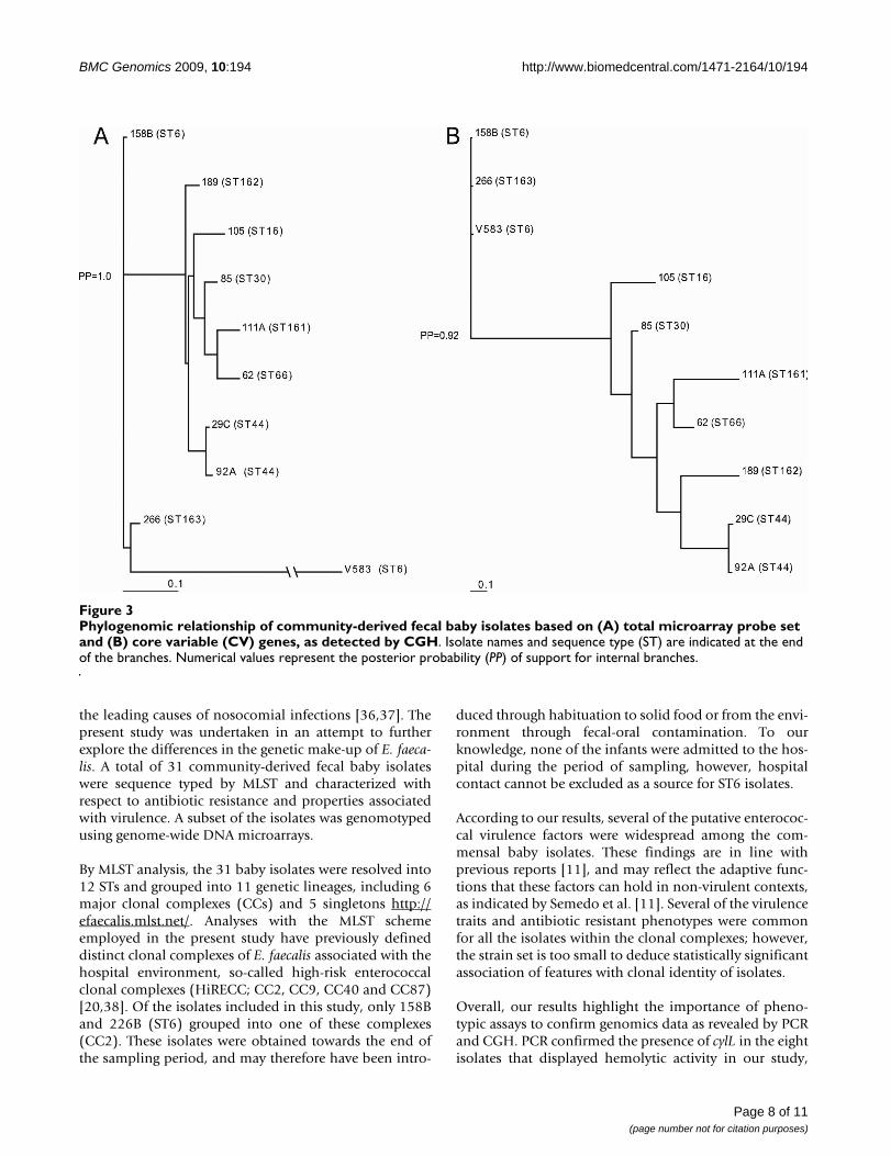
Phylogenomic analyses of the CGH data using Bayesian-based algorithms revealed a distinct clade containingseven of the nine community-derived baby isolates (Baye-sian posterior probabilities [PP] = 1.0; Figure 3A). Initialbranching within this clade was also fairly reliable (PP >0.80). The remaining three isolates seemed to be moredivergent, and V583 formed a distinct out-group. Basedon this analysis, isolate 266 (ST163) appeared moreclosely related to V583 (ST6) than isolate 158B, which
also displays ST6. Nevertheless, the Bayesian phylogenysuggested that the lineages defined by CGH generally cor-related with those identified by MLST: Within the latterclade, the isolates 92A and 29C (ST44) formed one sepa-rate clade (Figure 3A). To further examine the correlationbetween MLST and CGH, additional CGH data obtainedfrom the literature, in addition to unpublished data fromfive additional non-baby isolates of E. faecalis wereincluded in the analysis. In this extended analysis, onlygenes (n = 3043) represented on all three arrays that wereused in the different studies were considered. The cluster-ing of identical STs was here further supported (PP > 0.55;see Additional file 5). Previous studies have suggested thatgenomotyping of E. faecalis by CGH is heavily influencedby extensive horizontal transfer of MGEs in E. faecalis [17].To further analyze the effect of MGEs in our data set, theCGH data were reanalyzed after all core- and MGE geneshad been removed from the gene list, as previouslydescribed [33]. The MGE genes comprised putative MGEspredicted in V583 [15], as well as an additional phage-related region identified by McBride et al. [17]. This revi-sion left a list of 370 genes (see Additional file 6). Lindsayand coworkers [33,34] previously denominated suchgenes core variable (CV; all genes minus core genes minusMGE genes). The phylogenetic tree based on the contentof CV genes in the isolates recovered a clade containingthe same seven baby isolates as in the analysis with theentire probe set (PP = 0.92; Figure 3B).
Of the 370 CV genes, 145 genes code for hypothetical pro-teins. Among the remaining functionally annotated CVgenes, many genes are predicted to code for surface-exposed proteins in E. faecalis, e.g. the cps locus codingfor the capsule in E. faecalis [35]. The cps locus of E. faec-alis consists of 11 genes (cpsABCDEFGHIJK) and inser-tional inactivation of genes in this locus have resulted inmutants with enhanced susceptibility to phagocytic kill-ing [35]. Three different cps polymorphisms have beenidentified in E. faecalis so far: 1) type 2 (cps2) whichincludes all 11 genes, 2) type 5 (cps5) which includes allgenes except cpsF and 3) type 1 (cps1) with only cpsA andcpsB present. All three polymorphisms were detectedamong our test strains, by CGH (Table 1). The cps type haspreviously been found to be invariant within MLSTsequence types in E. faecalis [17], and our data is consist-ent with this finding. Analysis of the CGH data withrespect to the cps type suggested that the E. faecalis PAI, orsegments thereof, may be enriched among cps2-isolates.These observations support the hypothesis of convergenceof enterococcal virulence determinants and cps2 by inde-pendent selection in E. faecalis [17].
DiscussionEnterococci are among the first bacteria to colonize theneonatal GI tract [1]. Though originally considered asharmless commensals, the enterococci now rank among
Page 7 of 11(page number not for citation purposes)
BMC Genomics 2009, 10:194 http://www.biomedcentral.com/1471-2164/10/194
the leading causes of nosocomial infections [36,37]. Thepresent study was undertaken in an attempt to furtherexplore the differences in the genetic make-up of E. faeca-lis. A total of 31 community-derived fecal baby isolateswere sequence typed by MLST and characterized withrespect to antibiotic resistance and properties associatedwith virulence. A subset of the isolates was genomotypedusing genome-wide DNA microarrays.
By MLST analysis, the 31 baby isolates were resolved into12 STs and grouped into 11 genetic lineages, including 6major clonal complexes (CCs) and 5 singletons http://efaecalis.mlst.net/. Analyses with the MLST schemeemployed in the present study have previously defineddistinct clonal complexes of E. faecalis associated with thehospital environment, so-called high-risk enterococcalclonal complexes (HiRECC; CC2, CC9, CC40 and CC87)[20,38]. Of the isolates included in this study, only 158Band 226B (ST6) grouped into one of these complexes(CC2). These isolates were obtained towards the end ofthe sampling period, and may therefore have been intro-
duced through habituation to solid food or from the envi-ronment through fecal-oral contamination. To ourknowledge, none of the infants were admitted to the hos-pital during the period of sampling, however, hospitalcontact cannot be excluded as a source for ST6 isolates.
According to our results, several of the putative enterococ-cal virulence factors were widespread among the com-mensal baby isolates. These findings are in line withprevious reports [11], and may reflect the adaptive func-tions that these factors can hold in non-virulent contexts,as indicated by Semedo et al. [11]. Several of the virulencetraits and antibiotic resistant phenotypes were commonfor all the isolates within the clonal complexes; however,the strain set is too small to deduce statistically significantassociation of features with clonal identity of isolates.
Overall, our results highlight the importance of pheno-typic assays to confirm genomics data as revealed by PCRand CGH. PCR confirmed the presence of cylL in the eightisolates that displayed hemolytic activity in our study,
Phylogenomic relationship of community-derived fecal baby isolates based on (A) total microarray probe set and (B) core vari-able (CV) genes, as detected by CGHFigure 3Phylogenomic relationship of community-derived fecal baby isolates based on (A) total microarray probe set and (B) core variable (CV) genes, as detected by CGH. Isolate names and sequence type (ST) are indicated at the end of the branches. Numerical values represent the posterior probability (PP) of support for internal branches.
Page 8 of 11(page number not for citation purposes)

BMC Genomics 2009, 10:194 http://www.biomedcentral.com/1471-2164/10/194
however, cylL was also found to be present in an addi-tional eight Cyl--isolates. A similar discrepancy betweenfermentation capabilities and the presence of iolE and iolRwas also observed. Moreover, three isolates carrying bothgelE and fsrB came out as GelE- in the plate assay. The con-fined genotype-phenotype correlation that is herereported, visualizes the need for phenotypic confirmationof genotypes.
esp which is known to be associated with the E. faecalispathogenicity island (PAI) [25], was detected in two thirdsof the commensal isolates by PCR. According to the CGHdata, none of the baby isolates contained complete PAIs.These findings were as expected, considering that the PAIhas been shown to be enriched among infection-derivedenterococcal isolates [25]. More surprisingly, but consist-ent with a previous report [39], all the isolates studiedcontained some PAI genes. Several of the baby isolatesshowed similar patterns of present and divergent PAIgenes (Figure 2). This suggests that the evolution of theenterococcal PAI may be driven by insertion and deletionof larger modules, as hypothesized in [39]. Shankar et al.also suggested that parts of the enterococcal PAI originatefrom pheromone-responsive plasmids, with subsequentindels of transposable elements driving the evolution ofthe PAI [39]. Indeed, conjugal transfer of a segment of theE. faecalis PAI has been demonstrated [40]. The CGHrevealed a high degree of plasticity within all the MGEsrepresented on the microarray. These "mosaic structures"may reflect a complex evolutionary history of elementsthat have been frequently rearranged by horizontal genetransfer (HGT) and homologous recombination.
According to the CGH data presented here, a preliminaryE. faecalis core genome consisting of 2092 (out of the3093 chromosomal V583 ORFs) can be delineated. Com-piled analysis of the data from Aakra et al. and McBride etal. [17,18] with the data from the present study produceda core genome estimate of 1722 genes. An additional 62genes were only represented on one or two of the threedifferent arrays used, but were defined as core genes inthese experiments. Although the size of the core genomemay fluctuate due to the stringency of the statistical meth-ods used in the different studies, our data do add substan-tial information on the E. faecalis core genome.
In general, the genomic variation between isolates that areevolutionary -linked, e.g. isolates with the same ST, wasexpected to be lower than the variation between isolatesthat belong to different evolutionary lineages. Bayesian-based phylogenetic analysis confirmed these expectations(Figure 3A). McBride et al. previously reported genomo-typing by CGH to be biased by the activity of MGEs in E.faecalis [17], and we therefore repeated the Bayesian anal-ysis, using the CV genes, only. The phylogenetic analysis
based on CV genes recovered the same patterns of related-ness as the analysis comprising all genes, with slight inter-nal rearrangements of branches (Figure 3B). Theserearrangements supported the hypothesis on the distribu-tion of mobile elements as a source of genomic diversityin E. faecalis. Moreover, our data suggest that within line-ages, most of the variation detected by CGH is due toMGEs. However, the conserved clade identified by theanalyses based both on the CV genes and the completegene-set, indicates that also other and more complex dis-criminatory factors contribute to the genomic diversity inE. faecalis. Since an overall correlation between CGH andMLST was revealed, it is reasonable to believe that genescontributing to the formation of clades, i.e. lineage-spe-cific genes may be identified. In the 7 baby isolates thatformed a clade in the phylogenetic analysis, we were ableto recognize 137 genes that were divergent, but present inthe remaining three isolates (including the referencestrain). The majority of these genes were MGE geneslocated in phage03 (n = 39), phage06 (n = 28) and aphage-related region identified by McBride et al. [17](EF2240–82/EF2335–51; n = 44). Lepage et al. have pre-viously reported phage03 to be absent from several foodisolates [16]. Since ST6 is part of CC2, which has beenfound to be significantly enriched among nosocomial iso-lates, phage03 may potentially represent an element asso-ciated with increased fitness in the hospital environment.The latter report also identified eight genes as potentialmarkers for the V583/MMH594-lineage [16]. V583 andMMH594 both display ST6 [17], and five of the eightgenes (EF2250, EF2253, EF2254, EF3155 and EF3252)were also present in the ST6-isolates (158B andLMGT3303; results not shown) analyzed by CGH in ourstudy.
Comparative genome analyses have revealed that patho-gen evolution often progresses through gene acquisitionvia HGT [32]. The 169 genes that were characterized asdivergent in all the community-derived baby isolates byCGH may be potential pathogen-specific genes in E. faec-alis, or genes that are specific to V583. However, addi-tional CGH data from both pathogenic and non-pathogenic isolates are needed to address this issue. Van-comycin-resistant E. faecalis (VREfs) isolates appear to bewidely spread in hospital environments, while isolationof VREfs from healthy volunteers rarely occurs [41-44].Accordingly, the vanB operon was divergent in all the iso-lates studied by CGH in our lab (altogether 21 strains;[18]and results not shown). In addition to gene acquisition,pathoadaptive mutations via gene loss also plays animportant role in evolution of bacteria [45]. A disadvan-tage of the comparative genomic analyses presented here,is that the comparison of gene content is based on a singlereference strain (V583), only. The E. faecalis OG1RFgenome showed that, in addition to a shared core of 2474
Page 9 of 11(page number not for citation purposes)
BMC Genomics 2009, 10:194 http://www.biomedcentral.com/1471-2164/10/194
ORFs [29], both the V583 and the OG1RF genome carryunique genes, suggesting that the E. faecalis pan-genomewill be further extended as more strains will be sequenced.The availability of additional E. faecalis genome sequencesand the construction of a pan-species array would furtherincrease the sensitivity of such approaches.
ConclusionThe data presented here suggest that the genetic variationamong the investigated commensal E. faecalis is compara-ble to the genetic variation previously detected in a strainset thought to be representative of the major E. faecalis lin-eages. The widespread distribution of putative virulencedeterminants in the fecal baby isolates in this study sup-ports the conception of enterococcal virulence, not as aresult of any single virulence factor, but as a more complexprocess. Population structure studies of E. faecalis byMLST have identified so-called high-risk enterococcalclonal complexes (HiRECCs), defined as distinct geneticcomplexes that predominate among nosocomial infec-tions. The failure to identify a shared set of pathogen-spe-cific genes in E. faecalis so far opens up the possibility thatthe fitness and virulence of different HIRECCs may be dueto genes that are unique within a lineage, but that thecombined effects of the different gene-sets result in thesame phenotype, i.e. infection. The observed correlationbetween CGH and MLST presented here, may offer amethod for the identification of lineage-specific genes,and may therefore add clues on how to distinguish path-ogenic from commensal E. faecalis.
Authors' contributionsMS participated in the design of the study, carried out theexperimental work and drafted the manuscript. ÅAa par-ticipated in the design of the study and helped to draft themanuscript. LS proposed the statistical analysis and didthe programming and statistical analysis in R. DAB andIFN participated in the design of the study and assisted incritical review of the manuscript. All authors read andapproved the final manuscript.
Additional material
AcknowledgementsThis work was financially supported by the European Union 6th Framework Programme "Approaches to Control multi-resistant Enterococci: Studies on molecular ecology, horizontal gene transfer, fitness and prevention". Cathrine Herrera and Rønnaug Volden are acknowledged for the collection of the fecal baby isolates. We also acknowledge Zhian Salehian for technical assistance and Aksel Flack, The Norwegian Microarray Consortium, Oslo, for printing of the microarray slides.
References1. Fanaro S, Chierici R, Guerrini P, Vigi V: Intestinal microflora in
early infancy: composition and development. Acta PaediatrSuppl 2003, 91(441):48-55.
2. Hancock LE, Gilmore MS: Pathogenicity of enterococci. In Gram-positive pathogens Edited by: Fischetti VA, Novick RP, Ferretti JJ, Port-noy DA, Rood JI. Washington DC: ASM Press; 2006:299-311.
3. Shankar N, Lockatell CV, Baghdayan AS, Drachenberg C, Gilmore MS,Johnson DE: Role of Enterococcus faecalis surface protein Espin the pathogenesis of ascending urinary tract infection. InfectImmun 2001, 69(7):4366-4372.
4. Chow JW, Thal LA, Perri MB, Vazquez JA, Donabedian SM, ClewellDB, Zervos MJ: Plasmid-associated hemolysin and aggregationsubstance production contribute to virulence in experimen-tal enterococcal endocarditis. Antimicrob Agents Chemother 1993,37(11):2474-2477.
5. Jett BD, Jensen HG, Nordquist RE, Gilmore MS: Contribution ofthe pAD1-encoded cytolysin to the severity of experimentalEnterococcus faecalis endophthalmitis. Infect Immun 1992,60(6):2445-2452.
6. Schlievert PM, Gahr PJ, Assimacopoulos AP, Dinges MM, Stoehr JA,Harmala JW, Hirt H, Dunny GM: Aggregation and binding sub-stances enhance pathogenicity in rabbit models of Enterococ-cus faecalis endocarditis. Infect Immun 1998, 66(1):218-223.
Additional File 1Primers used in this study. A table of primers used in this study.Click here for file[http://www.biomedcentral.com/content/supplementary/1471-2164-10-194-S1.xls]
Additional File 2Genotypic and phenotypic characteristics of E. faecalis baby isolates. A table of the results from the phenotypic- and genotypic assays conducted in this study.Click here for file[http://www.biomedcentral.com/content/supplementary/1471-2164-10-194-S2.xls]
Additional File 3Genes divergent in all the baby isolates. A table of genes that were clas-sified as divergent in all the baby isolates analyzed by CGH.Click here for file[http://www.biomedcentral.com/content/supplementary/1471-2164-10-194-S3.doc]
Additional File 4Fermentation patterns from API 50 CH assays. A table of results from fermentation profiling of selected E. faecalis baby isolates by API 50 CH.Click here for file[http://www.biomedcentral.com/content/supplementary/1471-2164-10-194-S4.xls]
Additional File 5The phylogenomic relationship of E. faecalis isolates based on gene content, as detected by CGH. A phylogenetic tree based on CGH data from the present study, in addition to previously published CGH data from the literature.Click here for file[http://www.biomedcentral.com/content/supplementary/1471-2164-10-194-S5.png]
Additional File 6Core variable genes. A table of genes that were classified as core variable by CGH.Click here for file[http://www.biomedcentral.com/content/supplementary/1471-2164-10-194-S6.doc]
Page 10 of 11(page number not for citation purposes)
http://www.ncbi.nlm.nih.gov/entrez/query.fcgi?cmd=Retrieve&db=PubMed&dopt=Abstract&list_uids=8285637
http://www.ncbi.nlm.nih.gov/entrez/query.fcgi?cmd=Retrieve&db=PubMed&dopt=Abstract&list_uids=8285637
http://www.ncbi.nlm.nih.gov/entrez/query.fcgi?cmd=Retrieve&db=PubMed&dopt=Abstract&list_uids=8285637
BMC Genomics 2009, 10:194 http://www.biomedcentral.com/1471-2164/10/194
7. Kreft B, Marre R, Schramm U, Wirth R: Aggregation substance ofEnterococcus faecalis mediates adhesion to cultured renaltubular cells. Infect Immun 1992, 60(1):25-30.
8. Olmsted SB, Dunny GM, Erlandsen SL, Wells CL: A plasmid-encoded surface protein on Enterococcus faecalis augmentsits internalization by cultured intestinal epithelial cells. JInfect Dis 1994, 170(6):1549-1556.
9. Eaton TJ, Gasson MJ: Molecular screening of Enterococcus viru-lence determinants and potential for genetic exchangebetween food and medical isolates. Appl Environ Microbiol 2001,67(4):1628-1635.
10. Lempiainen H, Kinnunen K, Mertanen A, von Wright A: Occurrenceof virulence factors among human intestinal enterococcalisolates. Lett Appl Microbiol 2005, 41(4):341-344.
11. Semedo T, Santos MA, Lopes MF, Figueiredo Marques JJ, Barreto Cre-spo MT, Tenreiro R: Virulence factors in food, clinical and ref-erence Enterococci: A common trait in the genus? Syst ApplMicrobiol 2003, 26(1):13-22.
12. Creti R, Imperi M, Bertuccini L, Fabretti F, Orefici G, Di Rosa R, Bal-dassarri L: Survey for virulence determinants among Entero-coccus faecalis isolated from different sources. J Med Microbiol2004, 53(Pt 1):13-20.
13. Franz CM, Muscholl-Silberhorn AB, Yousif NM, Vancanneyt M, SwingsJ, Holzapfel WH: Incidence of virulence factors and antibioticresistance among Enterococci isolated from food. Appl Envi-ron Microbiol 2001, 67(9):4385-4389.
14. Mannu L, Paba A, Daga E, Comunian R, Zanetti S, Dupre I, Sechi LA:Comparison of the incidence of virulence determinants andantibiotic resistance between Enterococcus faecium strains ofdairy, animal and clinical origin. Int J Food Microbiol 2003, 88(2–3):291-304.
15. Paulsen IT, Banerjei L, Myers GS, Nelson KE, Seshadri R, Read TD,Fouts DE, Eisen JA, Gill SR, Heidelberg JF, et al.: Role of mobileDNA in the evolution of vancomycin-resistant Enterococcusfaecalis. Science 2003, 299(5615):2071-2074.
16. Lepage E, Brinster S, Caron C, Ducroix-Crepy C, Rigottier-Gois L,Dunny G, Hennequet-Antier C, Serror P: Comparative genomichybridization analysis of Enterococcus faecalis: identificationof genes absent from food strains. J Bacteriol 2006,188(19):6858-6868.
17. McBride SM, Fischetti VA, Leblanc DJ, Moellering RC Jr, Gilmore MS:Genetic diversity among Enterococcus faecalis. PLoS ONE 2007,2(7):e582.
18. Aakra A, Nyquist OL, Snipen L, Reiersen TS, Nes IF: Survey ofgenomic diversity among Enterococcus faecalis strains bymicroarray-based comparative genomic hybridization. ApplEnviron Microbiol 2007, 73(7):2207-2217.
19. Arroyo L, Cotton LN, Martin JH: AMC Agar–A compositemedium for selective enumeration of Bifidobacteriumlongum. Cultured Dairy Products Journal 1995, 30:12-15.
20. Ruiz-Garbajosa P, Bonten MJ, Robinson DA, Top J, Nallapareddy SR,Torres C, Coque TM, Canton R, Baquero F, Murray BE, et al.: Multi-locus sequence typing scheme for Enterococcus faecalisreveals hospital-adapted genetic complexes in a backgroundof high rates of recombination. J Clin Microbiol 2006,44(6):2220-2228.
21. Feil EJ, Li BC, Aanensen DM, Hanage WP, Spratt BG: eBURST:inferring patterns of evolutionary descent among clusters ofrelated bacterial genotypes from multilocus sequence typingdata. J Bacteriol 2004, 186(5):1518-1530.
22. Booth MC, Bogie CP, Sahl HG, Siezen RJ, Hatter KL, Gilmore MS:Structural analysis and proteolytic activation of Enterococcusfaecalis cytolysin, a novel lantibiotic. Mol Microbiol 1996,21(6):1175-1184.
23. Qin X, Singh KV, Weinstock GM, Murray BE: Effects of Enterococ-cus faecalis fsr genes on production of gelatinase and a serineprotease and virulence. Infect Immun 2000, 68(5):2579-2586.
24. Rich RL, Kreikemeyer B, Owens RT, LaBrenz S, Narayana SV, Wein-stock GM, Murray BE, Hook M: Ace is a collagen-bindingMSCRAMM from Enterococcus faecalis. J Biol Chem 1999,274(38):26939-26945.
25. Shankar N, Baghdayan AS, Gilmore MS: Modulation of virulencewithin a pathogenicity island in vancomycin-resistant Entero-coccus faecalis. Nature 2002, 417(6890):746-750.
26. Smyth GK, Speed T: Normalization of cDNA microarray data.Methods 2003, 31(4):265-273.
27. Snipen L, Nyquist OL, Solheim M, Aakra A, Nes IF: Improved anal-ysis of bacterial CGH data beyond the log-ratio paradigm.BMC Bioinformatics 2009, 10(1):91.
28. Carter B, Wu G, Woodward M, Anjum M: A process for analysisof microarray comparative genomics hybridisation studiesfor bacterial genomes. BMC Genomics 2008, 9(1):53.
29. Bourgogne A, Garsin DA, Qin X, Singh KV, Sillanpaa J, YerrapragadaS, Ding Y, Dugan-Rocha S, Buhay C, Shen H, et al.: Large scale var-iation in Enterococcus faecalis illustrated by the genome anal-ysis of strain OG1RF. Genome Biol 2008, 9(7):R110.
30. Ronquist F, Huelsenbeck JP: MrBayes 3: Bayesian phylogeneticinference under mixed models. Bioinformatics 2003,19(12):1572-1574.
31. Champion OL, Gaunt MW, Gundogdu O, Elmi A, Witney AA, HindsJ, Dorrell N, Wren BW: Comparative phylogenomics of thefood-borne pathogen Campylobacter jejuni reveals geneticmarkers predictive of infection source. Proc Natl Acad Sci USA2005, 102(44):16043-16048.
32. Dobrindt U, Hacker J: Whole genome plasticity in pathogenicbacteria. Curr Opin Microbiol 2001, 4(5):550-557.
33. Lindsay JA, Moore CE, Day NP, Peacock SJ, Witney AA, Stabler RA,Husain SE, Butcher PD, Hinds J: Microarrays reveal that each ofthe ten dominant lineages of Staphylococcus aureus has aunique combination of surface-associated and regulatorygenes. J Bacteriol 2006, 188(2):669-676.
34. Waldron DE, Lindsay JA: Sau1: a novel lineage-specific type Irestriction-modification system that blocks horizontal genetransfer into Staphylococcus aureus and between S. aureusisolates of different lineages. J Bacteriol 2006,188(15):5578-5585.
35. Hancock LE, Gilmore MS: The capsular polysaccharide of Ente-rococcus faecalis and its relationship to other polysaccharidesin the cell wall. Proc Natl Acad Sci USA 2002, 99(3):1574-1579.
36. Richards MJ, Edwards JR, Culver DH, Gaynes RP: Nosocomialinfections in combined medical-surgical intensive care unitsin the United States. Infect Control Hosp Epidemiol 2000,21(8):510-515.
37. Wisplinghoff H, Bischoff T, Tallent SM, Seifert H, Wenzel RP, EdmondMB: Nosocomial bloodstream infections in US hospitals:analysis of 24,179 cases from a prospective nationwide sur-veillance study. Clin Infect Dis 2004, 39(3):309-317.
38. Kawalec M, Pietras Z, Danilowicz E, Jakubczak A, Gniadkowski M,Hryniewicz W, Willems RJ: Clonal structure of Enterococcus fae-calis isolated from Polish hospitals: characterization of epi-demic clones. J Clin Microbiol 2007, 45(1):147-153.
39. Shankar N, Baghdayan AS, Willems R, Hammerum AM, Jensen LB:Presence of pathogenicity island genes in Enterococcus faec-alis isolates from pigs in Denmark. J Clin Microbiol 2006,44(11):4200-4203.
40. Coburn PS, Baghdayan AS, Dolan GT, Shankar N: Horizontal trans-fer of virulence genes encoded on the Enterococcus faecalispathogenicity island. Mol Microbiol 2007, 63(2):530-544.
41. Coque TM, Tomayko JF, Ricke SC, Okhyusen PC, Murray BE: Van-comycin-resistant enterococci from nosocomial, commu-nity, and animal sources in the United States. Antimicrob AgentsChemother 1996, 40(11):2605-2609.
42. Endtz HP, Braak N van den, van Belkum A, Kluytmans JA, KoelemanJG, Spanjaard L, Voss A, Weersink AJ, Vandenbroucke-Grauls CM,Buiting AG, et al.: Fecal carriage of vancomycin-resistant ente-rococci in hospitalized patients and those living in the com-munity in The Netherlands. J Clin Microbiol 1997,35(12):3026-3031.
43. Gambarotto K, Ploy MC, Turlure P, Grelaud C, Martin C, Bordes-soule D, Denis F: Prevalence of vancomycin-resistant entero-cocci in fecal samples from hospitalized patients andnonhospitalized controls in a cattle-rearing area of France. JClin Microbiol 2000, 38(2):620-624.
44. Torell E, Cars O, Olsson-Liljequist B, Hoffman BM, Lindback J, Bur-man LG: Near absence of vancomycin-resistant enterococcibut high carriage rates of quinolone-resistant ampicillin-resistant enterococci among hospitalized patients and non-hospitalized individuals in Sweden. J Clin Microbiol 1999,37(11):3509-3513.
45. Maurelli AT: Black holes, antivirulence genes, and gene inacti-vation in the evolution of bacterial pathogens. FEMS MicrobiolLett 2007, 267(1):1-8.
Page 11 of 11(page number not for citation purposes)
http://www.ncbi.nlm.nih.gov/entrez/query.fcgi?cmd=Retrieve&db=PubMed&dopt=Abstract&list_uids=1729187
http://www.ncbi.nlm.nih.gov/entrez/query.fcgi?cmd=Retrieve&db=PubMed&dopt=Abstract&list_uids=1729187
http://www.ncbi.nlm.nih.gov/entrez/query.fcgi?cmd=Retrieve&db=PubMed&dopt=Abstract&list_uids=7995995
http://www.ncbi.nlm.nih.gov/entrez/query.fcgi?cmd=Retrieve&db=PubMed&dopt=Abstract&list_uids=7995995
http://www.ncbi.nlm.nih.gov/entrez/query.fcgi?cmd=Retrieve&db=PubMed&dopt=Abstract&list_uids=8898386
http://www.ncbi.nlm.nih.gov/entrez/query.fcgi?cmd=Retrieve&db=PubMed&dopt=Abstract&list_uids=8913473
http://www.ncbi.nlm.nih.gov/entrez/query.fcgi?cmd=Retrieve&db=PubMed&dopt=Abstract&list_uids=8913473
http://www.ncbi.nlm.nih.gov/entrez/query.fcgi?cmd=Retrieve&db=PubMed&dopt=Abstract&list_uids=8913473
http://www.ncbi.nlm.nih.gov/entrez/query.fcgi?cmd=Retrieve&db=PubMed&dopt=Abstract&list_uids=9399488
http://www.ncbi.nlm.nih.gov/entrez/query.fcgi?cmd=Retrieve&db=PubMed&dopt=Abstract&list_uids=9399488