Mutations associated with neutropenia in dogs and humans disrupt intracellular transport of...
7
LETTERS Cyclic hematopoiesis is a stem cell disease in which the number of neutrophils and other blood cells oscillates in weekly phases. Autosomal dominant mutations of ELA2, encoding the protease neutrophil elastase 1 , found in lysosome- like granules, cause cyclic hematopoiesis 2 and most cases of the pre-leukemic disorder severe congenital neutropenia (SCN; ref. 3) in humans. Over 20 different mutations of neutrophil elastase have been identified, but their consequences are elusive, because they confer no consistent effects on enzymatic activity 4 . The similar autosomal recessive disease of dogs, canine cyclic hematopoiesis 5 , is not caused by mutations in ELA2 (data not shown). Here we show that homozygous mutation of the gene encoding the dog adaptor protein complex 3 (AP3) β-subunit, directing trans-Golgi export of transmembrane cargo proteins to lysosomes, causes canine cyclic hematopoiesis. C-terminal processing of neutrophil elastase exposes an AP3 interaction signal responsible for redirecting neutrophil elastase trafficking from membranes to granules. Disruption of either neutrophil elastase or AP3 perturbs the intracellular trafficking of neutrophil elastase. Most mutations in ELA2 that cause human cyclic hematopoiesis prevent membrane localization of neutrophil elastase, whereas most mutations in ELA2 that cause SCN lead to exclusive membrane localization. Canine cyclic hematopoiesis (Fig. 1a) is known as gray collie syndrome, because it arose in this breed and affected dogs have hypopigmented coats. Canine cyclic hematopoiesis resembles Hermansky–Pudlak syn- drome, type 2 (HPS2), a human disorder of partial albinism and neu- tropenia caused by homozygous mutation of AP3B1, encoding the AP3 β-subunit (β3a; refs. 6,7). After excluding canine ELA2 (data not shown), we tested AP3B1 as a candidate gene for canine cyclic hematopoiesis. We cloned the dog homolog of AP3B1 cDNA, compris- ing 26 exons and 3,964 bp, and mapped the gene to autosome 3. Flanking polymorphic microsatellite markers indicate linkage (lod > 9) and linkage disequilibrium in unrelated pedigrees (Fig. 1b). Genomic sequencing identified one coding alteration: insertion of an adenine in a tract of nine adenine residues in exon 20 (Fig. 1c), causing frameshift and premature termination. Northern blotting (Fig. 1d) indicated that the resultant mRNA is subject to nonsense-mediated decay, with 50% less mRNA in carriers and no mRNA in affected dogs, confirming mutation in AP3B1 as the cause of canine cyclic hematopoiesis. AP3 is one of four tetrameric adaptor protein complexes of animal cells that coat the cytoplasmic surfaces of membrane-bound vesicles and direct protein trafficking 8 . AP3 specifically shuttles transmem- brane ‘cargo’ proteins from the trans-Golgi to lysosomes 9 . Luminal cargo proteins extrude a short C-terminal tail through the lipid bilayer of vesicles and associate with µ-subunits in the cytosol through tyro- sine-based sorting signals 10 or with β-subunits through dileucine sort- ing signals 11 . The µ-subunits become degraded when β-subunits are deficient in humans and mice 6,12 with mutations in AP3B1. We found that the mutation in AP3B1 that causes canine cyclic hematopoiesis also leads to absence of both β3a and µ3a (data not shown). Because mutations in AP3B1 and ELA2 cause similar illnesses in humans, we reasoned that AP3 recognizes neutrophil elastase as a cargo protein and normally traffics it to granules. We therefore deter- mined whether human neutrophil elastase interacts with either of the two AP3 subunits, µ3a or β3a, that contact cargo proteins and are defi- cient in canine cyclic hematopoiesis, using a yeast two-hybrid assay established for this purpose 13 . In addition to N-terminal pre-pro sequences, neutrophil elastase contains a proteolytically processed 20- residue C-terminal extension 14 of previously unknown function but not required for proteolytic activity or granule localization 4,14 . To avoid complexities related to the N-terminal processing of neutrophil elastase and its antifungal antibiotic properties, we developed baits consisting of the distal half of human neutrophil elastase, extending from Pro110 and comprising one of its two cylindrical domains, with (NE-P110) or without (NE-P110∆C) the C-terminal extension. Lysosomal membrane protein LAMP-2 served as a positive control 13 . 1 Division of Medical Genetics / Department of Medicine and 2 Department of Pathology, University of Washington School of Medicine, Box 357720, 1705 NE Pacific Street, HSB-K236B, Seattle, Washington 98195, USA. 3 College of Veterinary Medicine, Cornell University, Ithaca, New York 14853, USA. 4 College of Veterinary Medicine, Auburn University, Auburn, Alabama 36849, USA. 5 Present addresses: Cabinet Beau de Loménie, 59800 Lille, France (D.A.); Rocky Mountain Laboratories, National Institute of Allergy and Infectious Diseases, Hamilton, Montana 59840, USA (K.M.-W.); University of Miami School of Medicine, Miami, Florida 33101, USA (J.W.). Correspondence should be addressed to M.H. ([email protected]). Published online 3 August 2003; doi:10.1038/ng1224 Mutations associated with neutropenia in dogs and humans disrupt intracellular transport of neutrophil elastase Kathleen F Benson 1 , Feng-Qian Li 1 , Richard E Person 2 , Dalila Albani 1,5 , Zhijun Duan 1 , Jeremy Wechsler 1,5 , Kimberly Meade-White 1,5 , Kayleen Williams 1 , Gregory M Acland 3, Glenn Niemeyer 4 , Clinton D Lothrop 4 & Marshall Horwitz 1 NATURE GENETICS VOLUME 35 | NUMBER 1 | SEPTEMBER 2003 90 © 2003 Nature Publishing Group http://www.nature.com/naturegenetics
-
Upload
independent -
Category
Documents
-
view
2 -
download
0
Transcript of Mutations associated with neutropenia in dogs and humans disrupt intracellular transport of...
L E T T E R S
Cyclic hematopoiesis is a stem cell disease in which thenumber of neutrophils and other blood cells oscillates inweekly phases. Autosomal dominant mutations of ELA2,encoding the protease neutrophil elastase1, found in lysosome-like granules, cause cyclic hematopoiesis2 and most cases ofthe pre-leukemic disorder severe congenital neutropenia (SCN;ref. 3) in humans. Over 20 different mutations of neutrophilelastase have been identified, but their consequences areelusive, because they confer no consistent effects onenzymatic activity4. The similar autosomal recessive disease ofdogs, canine cyclic hematopoiesis5, is not caused by mutationsin ELA2 (data not shown). Here we show that homozygousmutation of the gene encoding the dog adaptor proteincomplex 3 (AP3) β-subunit, directing trans-Golgi export oftransmembrane cargo proteins to lysosomes, causes caninecyclic hematopoiesis. C-terminal processing of neutrophilelastase exposes an AP3 interaction signal responsible forredirecting neutrophil elastase trafficking from membranes togranules. Disruption of either neutrophil elastase or AP3perturbs the intracellular trafficking of neutrophil elastase.Most mutations in ELA2 that cause human cyclichematopoiesis prevent membrane localization of neutrophilelastase, whereas most mutations in ELA2 that cause SCN leadto exclusive membrane localization.
Canine cyclic hematopoiesis (Fig. 1a) is known as gray collie syndrome,because it arose in this breed and affected dogs have hypopigmentedcoats. Canine cyclic hematopoiesis resembles Hermansky–Pudlak syn-drome, type 2 (HPS2), a human disorder of partial albinism and neu-tropenia caused by homozygous mutation of AP3B1, encoding the AP3β-subunit (β3a; refs. 6,7). After excluding canine ELA2 (data notshown), we tested AP3B1 as a candidate gene for canine cyclichematopoiesis. We cloned the dog homolog of AP3B1 cDNA, compris-ing 26 exons and 3,964 bp, and mapped the gene to autosome 3.Flanking polymorphic microsatellite markers indicate linkage (lod > 9)
and linkage disequilibrium in unrelated pedigrees (Fig. 1b). Genomicsequencing identified one coding alteration: insertion of an adeninein a tract of nine adenine residues in exon 20 (Fig. 1c), causingframeshift and premature termination. Northern blotting (Fig. 1d)indicated that the resultant mRNA is subject to nonsense-mediateddecay, with 50% less mRNA in carriers and no mRNA in affecteddogs, confirming mutation in AP3B1 as the cause of canine cyclichematopoiesis.
AP3 is one of four tetrameric adaptor protein complexes of animalcells that coat the cytoplasmic surfaces of membrane-bound vesiclesand direct protein trafficking8. AP3 specifically shuttles transmem-brane ‘cargo’ proteins from the trans-Golgi to lysosomes9. Luminalcargo proteins extrude a short C-terminal tail through the lipid bilayerof vesicles and associate with µ-subunits in the cytosol through tyro-sine-based sorting signals10 or with β-subunits through dileucine sort-ing signals11. The µ-subunits become degraded when β-subunits aredeficient in humans and mice6,12 with mutations in AP3B1. We foundthat the mutation in AP3B1 that causes canine cyclic hematopoiesisalso leads to absence of both β3a and µ3a (data not shown).
Because mutations in AP3B1 and ELA2 cause similar illnesses inhumans, we reasoned that AP3 recognizes neutrophil elastase as acargo protein and normally traffics it to granules. We therefore deter-mined whether human neutrophil elastase interacts with either of thetwo AP3 subunits, µ3a or β3a, that contact cargo proteins and are defi-cient in canine cyclic hematopoiesis, using a yeast two-hybrid assayestablished for this purpose13. In addition to N-terminal pre-prosequences, neutrophil elastase contains a proteolytically processed 20-residue C-terminal extension14 of previously unknown function butnot required for proteolytic activity or granule localization4,14. Toavoid complexities related to the N-terminal processing of neutrophilelastase and its antifungal antibiotic properties, we developed baitsconsisting of the distal half of human neutrophil elastase, extendingfrom Pro110 and comprising one of its two cylindrical domains, with(NE-P110) or without (NE-P110∆C) the C-terminal extension.Lysosomal membrane protein LAMP-2 served as a positive control13.
1Division of Medical Genetics / Department of Medicine and 2Department of Pathology, University of Washington School of Medicine, Box 357720, 1705 NE PacificStreet, HSB-K236B, Seattle, Washington 98195, USA. 3College of Veterinary Medicine, Cornell University, Ithaca, New York 14853, USA. 4College of VeterinaryMedicine, Auburn University, Auburn, Alabama 36849, USA. 5Present addresses: Cabinet Beau de Loménie, 59800 Lille, France (D.A.); Rocky MountainLaboratories, National Institute of Allergy and Infectious Diseases, Hamilton, Montana 59840, USA (K.M.-W.); University of Miami School of Medicine, Miami,Florida 33101, USA (J.W.). Correspondence should be addressed to M.H. ([email protected]).
Published online 3 August 2003; doi:10.1038/ng1224
Mutations associated with neutropenia in dogs and humansdisrupt intracellular transport of neutrophil elastaseKathleen F Benson1, Feng-Qian Li1, Richard E Person2, Dalila Albani1,5, Zhijun Duan1, Jeremy Wechsler1,5,Kimberly Meade-White1,5, Kayleen Williams1, Gregory M Acland3, Glenn Niemeyer4, Clinton D Lothrop4
& Marshall Horwitz1
NATURE GENETICS VOLUME 35 | NUMBER 1 | SEPTEMBER 2003 90
©20
03 N
atu
re P
ub
lish
ing
Gro
up
h
ttp
://w
ww
.nat
ure
.co
m/n
atu
reg
enet
ics

L E T T E R S
NATURE GENETICS VOLUME 35 | NUMBER 1 | SEPTEMBER 2003 91
NE-P110∆C interacted with µ3a but not β3a (Fig. 2a), whereas NE-P110 did not interact with either subunit. As there is only one tyrosineresidue in the interacting neutrophil elastase fragment, the sequenceLYPDA starting at position 198 is probably a µ3a sorting signal.
To confirm this as an interaction signal, we carried out two exper-iments. First, we shortened the bait to begin at Pro176 or Ser196 andfound that NE-P176∆C (Fig. 2a) and NE-S196∆C (data not shown)continued to specifically interact, indicating that the signal must be
present in the final 23 residues of C-terminally processed neutrophilelastase. Second, we introduced a Y199A substitution and foundthat it abolished the interaction of either bait, NE-P176∆C-Y199A(Fig. 2a) or NE-S196∆C-Y199A (data not shown). These resultsindicate that the processed form of neutrophil elastase interacts invitro with the µ3a subunit of AP3 through a tyrosine-based sortingsignal in neutrophil elastase and that the C-terminal extensionseems to block interaction with AP3.
Cycle days
Neu
tro
ph
ils
Mo
no
cyte
s
1
2
3
4
5
123456789
10
1 7 14 21 28
a
1
2
3
4
5
123456789
10
1 7 14 21 28
1
2
3
4
5
123456789
10
1 7 14 21 28
c
No
rmal
Car
rier
Aff
ecte
dd
AP3B1
ACTB
6 kb -
4 kb -
3 kb -
2 kb -
-
-
-
-
-
-
-
-
FH2137C03.629C00802FH2541
162210166151
162206166147
b
CarrierAAAG AAAAAAAAANNAANG N AN NNN
AffectedAAA G AAAAAAAAAA CAA AG G AAG ATA
NormalAAAG AAAAAAAAACAA AG G AAG ATA
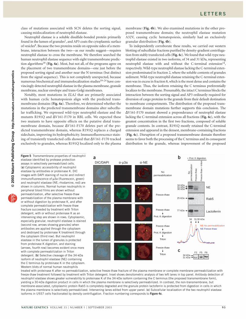
Figure 1 Mutation of AP3B1 causes canine cyclic hematopoiesis. (a) Phenotype of canine cyclic hematopoiesis5, showing 2-week reciprocal cycles ofneutrophil and monocyte production (number of cells × 10–3 per µl) and gray coat. (b) Canine cyclic hematopoiesis pedigrees. Dots indicate carriers(identified by affected offspring). Each color represents a unique haplotype. Arrow shows region of linkage disequilibrium. Numbers refer to sizes (innucleotides) of alleles for each marker. (c) Genomic DNA electropherogram. Arrows indicate A-insertion mutation. (d) Northern blot of total RNA from blood,showing reduced AP3B1 transcript compared to ACTB control.
0
0.2
0.4
0.6
0 20 40 60 80 100
NE (ng)
Rel
ativ
e en
zym
atic
act
ivit
y
Affected dog
Normal dog
0
0.2
0.4
0.6
0 20 40 60 80 100
Neutrophil elastase (ng)
Rel
ativ
e en
zym
atic
act
ivit
y
Affected dog
Normal dog
CarrierNormal Affected
c
a
–His
µ3a
NE-P176
NE-P176∆C
β3a
LAMP-2
NE-P176∆C/Y199A
NE-P110
NE-P110∆C
b
Figure 2 Interaction of AP3 and neutrophilelastase. (a) Yeast two-hybrid analysis onselective (–His) medium, showing µ3ainteraction with the tyrosine-based sortingsignal in neutrophil elastase. (b) Nearabsence of neutrophil elastase enzymaticactivity in affected dog (red) compared tonormal (green) on a standard curve (blue).(c) Absence of neutrophil elastase in dog blood films. Restoration three-dimensional microscopy of indirectimmunofluorescence detection ofneutrophil elastase (fluorescein, green),with counterstaining of granules andnuclei by LAMP-1 (phycoerythrin, red) and DAPI (blue), respectively. (Uniformstaining of erythrocytes results from cross-reaction with the phycoerythrin-conjugated secondary antibody.)
©20
03 N
atu
re P
ub
lish
ing
Gro
up
h
ttp
://w
ww
.nat
ure
.co
m/n
atu
reg
enet
ics

L E T T E R S
92 VOLUME 35 | NUMBER 1 | SEPTEMBER 2003 NATURE GENETICS
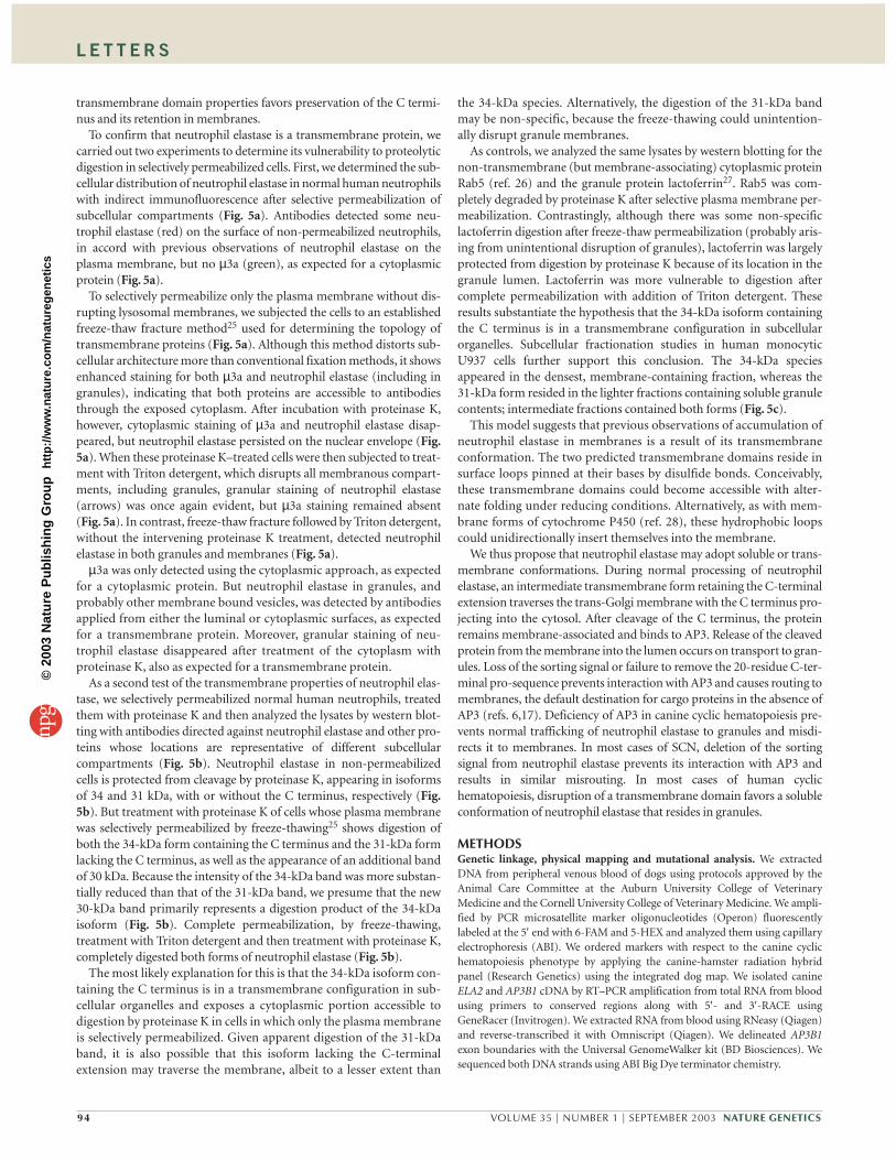
Figure 4 Mutations of neutrophil elastasethat cause cyclic hematopoiesis generallyaccentuate granule accumulation,whereas those that cause SCN generallyaccentuate membrane accumulation. (a) Primary neutrophil elastase sequence,showing general alignment of mutationsthat cause cyclic hematopoiesis and SCN (in black and pink, respectively) withtransmembrane domains and with the µ3asorting signal. Dots and lines representamino acid missense substitutions anddeletions, respectively. Each markerrepresents the independent occurrence ofthe mutation. Predicted transmembranesegments are aligned with correspondingalgorithm. (b) Subcellular distribution ofproteins with transmembrane domainmutations showing reduced oraccentuated granule accumulationdepending on the effect of the proposedtransmembrane domain. Restorationthree-dimensional microscopy oftransiently transfected constructs, imagedby indirect immunofluorescence ofneutrophil elastase (red) and LAMP-1(green), with DAPI (blue) counterstainingof nuclei. (c) Retention of C terminus ofneutrophil elastase in mutants localizingto membranes and preferential processingof C terminus in mutants localizing togranules. Subcellular localization ofmutant neutrophil elastase detected bywestern blotting of stably transfected RBL cells fractioned by density-gradient centrifugation. Negative control (Neg) was transfected with wild-type ELA2(WT) but uninduced. Inset shows density increasing across fractions with cellular compartment identification.
To determine if neutrophil elastase and µ3aassociate intracellularly, we tested for defi-ciency of neutrophil elastase in neutrophilsfrom dogs with canine cyclic hematopoiesis.We measured neutrophil elastase activity on aspecific peptide substrate (Fig. 2b) and foundthat neutrophils from affected dogs have only8% of the enzymatic activity of normal dogs,despite having a normal ELA2 genotype.Immunofluorescence staining (Fig. 2c) ofneutrophils from dogs with canine cyclichematopoiesis showed a correspondingpaucity of neutrophil elastase (green) andreduced granular staining for lysosomalmembrane protein LAMP-1 (red). (LAMP-1is also an AP3 cargo protein6, but granulescontaining LAMP-1 are distinct from gran-ules containing neutrophil elastase15.) These observations suggest thatAP3 influences the subcellular trafficking of neutrophil elastase.
The most common class of mutations in ELA2 associated with SCNprematurely truncate the protein near the C terminus just ahead ofthe proposed tyrosine-based µ3a sorting signal16, suggesting that theycould disrupt interactions with AP3. To examine the effect of thesemutations, we used immunofluorescence to detect neutrophil elastasewith intact N and C termini in RBL cells (rat basophils containinggranules lacking endogenous neutrophil elastase4) transiently trans-fected with ELA2 cDNA. We stained neutrophil elastase (red) andcounterstained LAMP-1 (green) and nuclei (with DAPI, blue). Wild-type neutrophil elastase appeared predominately in granules and also
in a perinuclear distribution (Fig. 3). We also tested two mutant con-structs: the Y199X mutation removes the proposed sorting signal andC-terminal extension, and the engineered mutation 205∆C, corre-sponding to a neutropenic frameshift mutation, retains the predictedsorting signal but lacks the C-terminal extension. Y199X was absentin granules and appeared instead in the plasma membrane andnuclear envelope, whereas 205∆C had an accentuated granular pat-tern (Fig. 3). These results are consistent with observations in AP3-deficient human and mouse cells6,17, in which lysosomal cargoproteins are incorrectly routed to the plasma membrane. We con-clude that AP3 directs neutrophil elastase to granules through the µ3atyrosine-based sorting signal and that the most commonly occurring
WTWT Y199XY199X 205C205∆C
Figure 3 Mislocalization of neutrophil elastase in mutants lacking the µ3a sorting signal and C-terminal extension (Y199X) or just the C-terminal signal (205∆C) relative to wild-type (WT).Restoration three-dimensional microscopy of transiently transfected RBL cells, imaged by indirectimmunofluorescence of neutrophil elastase (rhodamine, red) and LAMP-1 (fluorescein, green), withDAPI (blue) counterstaining of nuclei.
R19
1Q∆V
161-
F17
0
WT
Neg
.
34 kDa31 kDa
F1 F2 F3 F4 F5 F6 F7 F8
DensityLDH (cytosol)
β-glucuronidase (granules& microsomes)TGN38 (trans-Golgi)
1 8
Qua
ntity
Fraction
pre pro C-tail
Predicted transmembrane domains
∆V161-F170
µ3A
Y199XC26Y
A28TI31T
A32V C42S V72M93+PQ P110L
∆V145-152
∆V157-F170
L177FG181V
G185RR191Q
P205 -1fsP200 -1fs
S97L G196X
G192X
a
R191Q∆V161-F170 A32VR191QR191Q∆V161-F170∆V161-F170 A32VA32Vb
c
TopPred 2TMpredTMAPHMMTOPMEMSATSOSUI
C122S C194X
G174R
† ‡ § ¶ † ‡ § ¶ † ‡ § ¶ † ‡ § ¶ † ‡ § ¶ † ‡ § ¶ † ‡ § ¶ † ‡ § ¶
©20
03 N
atu
re P
ub
lish
ing
Gro
up
h
ttp
://w
ww
.nat
ure
.co
m/n
atu
reg
enet
ics
L E T T E R S
NATURE GENETICS VOLUME 35 | NUMBER 1 | SEPTEMBER 2003 93
class of mutations associated with SCN deletes the sorting signal,causing mislocalization of neutrophil elastase.
Neutrophil elastase is a soluble disulfide-bonded protein primarilyfound in the lumen of granules1, and AP3 coats the cytoplasmic surfaceof vesicles8. Because the two proteins reside on opposite sides of a mem-brane, interaction between the two—as our results suggest—requiresneutrophil elastase to cross the membrane. We therefore searched thehuman neutrophil elastase sequence with eight transmembrane predic-tion algorithms18 (Fig. 4a). Most, but not all, of the programs agree onthe placement of two transmembrane domains—one just before theproposed sorting signal and another near the N terminus (but distinctfrom the signal sequence). This is not completely unexpected, becausenumerous biochemical and immunolocalization studies19–24 have con-vincingly detected neutrophil elastase in the plasma membrane, granulemembrane, nuclear envelope and trans-Golgi membranes.
Notably, most mutations in ELA2 that are primarily associatedwith human cyclic hematopoiesis align with the predicted trans-membrane domains (Fig. 4a). Therefore, we determined whether themutations in the predicted transmembrane domains alter subcellu-lar trafficking. We expressed wild-type neutrophil elastase and themutants R191Q and ∆V161-F170 in RBL cells. We expected thesetwo mutants to have opposite effects on the putative distal trans-membrane domain, because ∆V161-F170 deletes part of the pre-dicted transmembrane domain, whereas R191Q replaces a chargedsidechain, improving its hydrophobicity. Immunofluorescence stain-ing of transiently transfected cells showed that ∆V161-F170 trackedexclusively to granules, whereas R191Q localized only to the plasma
membrane (Fig. 4b). We also examined mutations in the other pro-posed transmembrane domain; the neutrophil elastase mutationA32V, causing cyclic hematopoiesis, similarly had an exclusivelygranular distribution (Fig. 4b).
To independently corroborate these results, we carried out westernblotting of subcellular fractions purified by density-gradient centrifuga-tion from stably transfected cells (Fig. 4c). We found that wild-type neu-trophil elastase existed in two isoforms, of 34 and 31 kDa, representingneutrophil elastase with and without the C-terminal extension14,respectively. Wild-type neutrophil elastase lacking the C-terminal exten-sion predominated in fraction 2, where the soluble contents of granulessediment. Wild-type neutrophil elastase retaining the C-terminal exten-sion was in excess in fraction 8, which is the most dense and contains themembrane. Thus, the isoform retaining the C terminus preferentiallylocalizes to the membrane. Presumably, the intact C terminus blocks theinteraction between the sorting signal and AP3 ordinarily required fordiversion of cargo proteins to the granule from their default destinationto membrane compartments. The distribution of the proposed trans-membrane domain mutations further supports this conclusion. The∆V161-F170 mutant showed a preponderance of neutrophil elastaselacking the C-terminal extension across all fractions (Fig. 4c), with thegreatest concentration in the first two fractions, composed of solublegranule contents. In contrast, R191Q mostly retained the C-terminalextension and appeared in the densest, membrane-containing fractions(Fig. 4c). Disruption of a proposed transmembrane domain thereforeseems to favor both the processing of the C terminus and its consequentdistribution to the granule, whereas improvement of the proposed
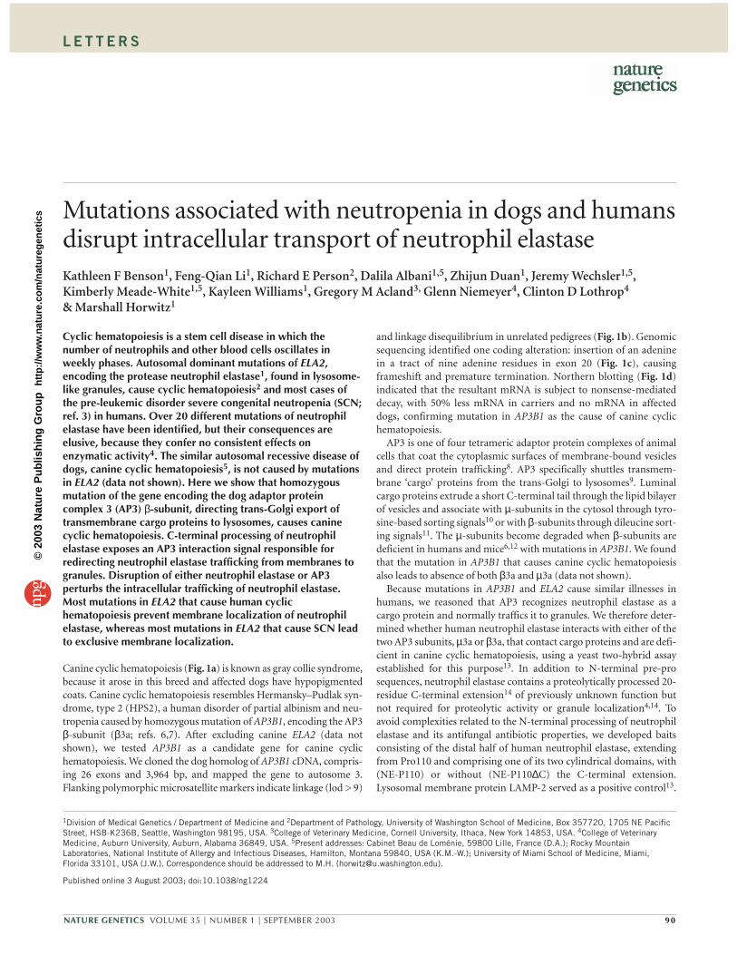
Figure 5 Transmembrane properties of neutrophilelastase identified by protease protection assays in selectively permeabilized cells. (a) Cytoplasmic accessibility of neutrophilelastase by antibodies or proteinase K. DICimages with DAPI staining of nuclei and indirectimmunofluorescence of µ3a (fluorescein, green)and neutrophil elastase (NE; rhodamine, red) areshown in columns. Normal human neutrophils inperipheral blood films are shown withoutpermeabilization, after selective freeze-thawpermeabilization of the plasma membrane withor without digestion by proteinase K, and aftercomplete permeabilization with freeze-thawfracture succeeded by treatment with Tritondetergent, with or without proteinase K as anintervening step are shown in rows. Cytoplasmic,especially granular, neutrophil elastase is stained(second row, arrows showing granules) whenantibodies are applied through the cytoplasmand destroyed by proteinase K treatment throughthe cytoplasm (third row). But neutrophilelastase in the lumen of granules is protectedfrom proteinase K digestion, and staining(arrows, fourth row) becomes evident once morewith complete permeabilization in Tritondetergent. (b) Selective cleavage of the 34-kDaisoform of neutrophil elastase (NE) containingthe C terminus by proteinase K in the cytoplasm.Western blots of normal human neutrophilstreated with proteinase K after no permeabilization, selective freeze-thaw fracture of the plasma membrane or complete membrane permeabilization withfreeze-thaw treatment followed by treatment with Triton detergent. Inset shows densitometric analysis of two left lanes in top panel. Antibody detection ofneutrophil elastase shows greater vulnerability to proteinase K of the 34-kDa isoform containing the C terminus (the proposed transmembrane form),yielding a 30-kDa digestion product in cells in which the plasma membrane is selectively permeabilized. In contrast, the non-transmembrane, butmembrane-associated, cytoplasmic protein Rab5 is completely degraded and the granule protein lactoferrin is protected from digestion in cells in whichthe plasma membrane is selectively permeabilized. Intervening lanes edited from upper panel. (c) Subcellular localization of the two neutrophil elastaseisoforms in U937 cells fractionated by density centrifugation. Fraction numbering corresponds to Figure 4c.
No
perm
eabi
lizat
ion
Free
ze-t
haw
Free
ze-t
haw
+ tr
iton
Lactoferrin
Rab5
NE
+ Prot. K digestionb
c
34 kDa31 kDa30 kDa
25 kDa
77 kDa
34 31 30 kDa
NE-No permeabilizationNE-Freeze-thaw
DIC/DAPI
Nopermeabilization
Freeze-thaw
Freeze-thaw
Prot. K digestion
Freeze-thaw
Prot. K digestion
Triton
Freeze-thaw
Triton
α-µ3a α-NEa
34 kDa31 kDa
F2 F5 F8
©20
03 N
atu
re P
ub
lish
ing
Gro
up
h
ttp
://w
ww
.nat
ure
.co
m/n
atu
reg
enet
ics
L E T T E R S
94 VOLUME 35 | NUMBER 1 | SEPTEMBER 2003 NATURE GENETICS
transmembrane domain properties favors preservation of the C termi-nus and its retention in membranes.
To confirm that neutrophil elastase is a transmembrane protein, wecarried out two experiments to determine its vulnerability to proteolyticdigestion in selectively permeabilized cells. First, we determined the sub-cellular distribution of neutrophil elastase in normal human neutrophilswith indirect immunofluorescence after selective permeabilization ofsubcellular compartments (Fig. 5a). Antibodies detected some neu-trophil elastase (red) on the surface of non-permeabilized neutrophils,in accord with previous observations of neutrophil elastase on theplasma membrane, but no µ3a (green), as expected for a cytoplasmicprotein (Fig. 5a).
To selectively permeabilize only the plasma membrane without dis-rupting lysosomal membranes, we subjected the cells to an establishedfreeze-thaw fracture method25 used for determining the topology oftransmembrane proteins (Fig. 5a). Although this method distorts sub-cellular architecture more than conventional fixation methods, it showsenhanced staining for both µ3a and neutrophil elastase (including ingranules), indicating that both proteins are accessible to antibodiesthrough the exposed cytoplasm. After incubation with proteinase K,however, cytoplasmic staining of µ3a and neutrophil elastase disap-peared, but neutrophil elastase persisted on the nuclear envelope (Fig.5a). When these proteinase K–treated cells were then subjected to treat-ment with Triton detergent, which disrupts all membranous compart-ments, including granules, granular staining of neutrophil elastase(arrows) was once again evident, but µ3a staining remained absent(Fig. 5a). In contrast, freeze-thaw fracture followed by Triton detergent,without the intervening proteinase K treatment, detected neutrophilelastase in both granules and membranes (Fig. 5a).
µ3a was only detected using the cytoplasmic approach, as expectedfor a cytoplasmic protein. But neutrophil elastase in granules, andprobably other membrane bound vesicles, was detected by antibodiesapplied from either the luminal or cytoplasmic surfaces, as expectedfor a transmembrane protein. Moreover, granular staining of neu-trophil elastase disappeared after treatment of the cytoplasm withproteinase K, also as expected for a transmembrane protein.
As a second test of the transmembrane properties of neutrophil elas-tase, we selectively permeabilized normal human neutrophils, treatedthem with proteinase K and then analyzed the lysates by western blot-ting with antibodies directed against neutrophil elastase and other pro-teins whose locations are representative of different subcellularcompartments (Fig. 5b). Neutrophil elastase in non-permeabilizedcells is protected from cleavage by proteinase K, appearing in isoformsof 34 and 31 kDa, with or without the C terminus, respectively (Fig.5b). But treatment with proteinase K of cells whose plasma membranewas selectively permeabilized by freeze-thawing25 shows digestion ofboth the 34-kDa form containing the C terminus and the 31-kDa formlacking the C terminus, as well as the appearance of an additional bandof 30 kDa. Because the intensity of the 34-kDa band was more substan-tially reduced than that of the 31-kDa band, we presume that the new30-kDa band primarily represents a digestion product of the 34-kDaisoform (Fig. 5b). Complete permeabilization, by freeze-thawing,treatment with Triton detergent and then treatment with proteinase K,completely digested both forms of neutrophil elastase (Fig. 5b).
The most likely explanation for this is that the 34-kDa isoform con-taining the C terminus is in a transmembrane configuration in sub-cellular organelles and exposes a cytoplasmic portion accessible todigestion by proteinase K in cells in which only the plasma membraneis selectively permeabilized. Given apparent digestion of the 31-kDaband, it is also possible that this isoform lacking the C-terminalextension may traverse the membrane, albeit to a lesser extent than
the 34-kDa species. Alternatively, the digestion of the 31-kDa bandmay be non-specific, because the freeze-thawing could unintention-ally disrupt granule membranes.
As controls, we analyzed the same lysates by western blotting for thenon-transmembrane (but membrane-associating) cytoplasmic proteinRab5 (ref. 26) and the granule protein lactoferrin27. Rab5 was com-pletely degraded by proteinase K after selective plasma membrane per-meabilization. Contrastingly, although there was some non-specificlactoferrin digestion after freeze-thaw permeabilization (probably aris-ing from unintentional disruption of granules), lactoferrin was largelyprotected from digestion by proteinase K because of its location in thegranule lumen. Lactoferrin was more vulnerable to digestion aftercomplete permeabilization with addition of Triton detergent. Theseresults substantiate the hypothesis that the 34-kDa isoform containingthe C terminus is in a transmembrane configuration in subcellularorganelles. Subcellular fractionation studies in human monocyticU937 cells further support this conclusion. The 34-kDa speciesappeared in the densest, membrane-containing fraction, whereas the31-kDa form resided in the lighter fractions containing soluble granulecontents; intermediate fractions contained both forms (Fig. 5c).
This model suggests that previous observations of accumulation ofneutrophil elastase in membranes is a result of its transmembraneconformation. The two predicted transmembrane domains reside insurface loops pinned at their bases by disulfide bonds. Conceivably,these transmembrane domains could become accessible with alter-nate folding under reducing conditions. Alternatively, as with mem-brane forms of cytochrome P450 (ref. 28), these hydrophobic loopscould unidirectionally insert themselves into the membrane.
We thus propose that neutrophil elastase may adopt soluble or trans-membrane conformations. During normal processing of neutrophilelastase, an intermediate transmembrane form retaining the C-terminalextension traverses the trans-Golgi membrane with the C terminus pro-jecting into the cytosol. After cleavage of the C terminus, the proteinremains membrane-associated and binds to AP3. Release of the cleavedprotein from the membrane into the lumen occurs on transport to gran-ules. Loss of the sorting signal or failure to remove the 20-residue C-ter-minal pro-sequence prevents interaction with AP3 and causes routing tomembranes, the default destination for cargo proteins in the absence ofAP3 (refs. 6,17). Deficiency of AP3 in canine cyclic hematopoiesis pre-vents normal trafficking of neutrophil elastase to granules and misdi-rects it to membranes. In most cases of SCN, deletion of the sortingsignal from neutrophil elastase prevents its interaction with AP3 andresults in similar misrouting. In most cases of human cyclichematopoiesis, disruption of a transmembrane domain favors a solubleconformation of neutrophil elastase that resides in granules.
METHODSGenetic linkage, physical mapping and mutational analysis. We extractedDNA from peripheral venous blood of dogs using protocols approved by theAnimal Care Committee at the Auburn University College of VeterinaryMedicine and the Cornell University College of Veterinary Medicine. We ampli-fied by PCR microsatellite marker oligonucleotides (Operon) fluorescentlylabeled at the 5′ end with 6-FAM and 5-HEX and analyzed them using capillaryelectrophoresis (ABI). We ordered markers with respect to the canine cyclichematopoiesis phenotype by applying the canine-hamster radiation hybridpanel (Research Genetics) using the integrated dog map. We isolated canineELA2 and AP3B1 cDNA by RT–PCR amplification from total RNA from bloodusing primers to conserved regions along with 5′- and 3′-RACE usingGeneRacer (Invitrogen). We extracted RNA from blood using RNeasy (Qiagen)and reverse-transcribed it with Omniscript (Qiagen). We delineated AP3B1exon boundaries with the Universal GenomeWalker kit (BD Biosciences). Wesequenced both DNA strands using ABI Big Dye terminator chemistry.
©20
03 N
atu
re P
ub
lish
ing
Gro
up
h
ttp
://w
ww
.nat
ure
.co
m/n
atu
reg
enet
ics
L E T T E R S
NATURE GENETICS VOLUME 35 | NUMBER 1 | SEPTEMBER 2003 95
Northern-blot analysis. We isolated total RNA from blood, resolved 5 µg ofeach sample by electrophoresis on a 1.0% agarose formaldehyde-denaturinggel, blot-transferred the RNA to a nylon membrane (BrightStar-Plus,Ambion) and immobilized it by ultraviolet cross-linking. We carried outhybridization in Ultrahyb (Ambion) at 42 °C using an 800-bp 32P-labeledprobe corresponding to canine AP3B1 cDNA nucleotides 3,129–3,927. Werehybridized washed filters with a 32P-labeled probe complementary to ACTBto control for loading.
Yeast two-hybrid analysis. We fused ELA2 cDNA to the Matchmaker(Clontech) Gal4 DNA-binding domain vector pGBKT7. µ3a and β3a Gal4activation domain and LAMP-2 Gal4 DNA binding domain constructs13 weregifts of X. Zhu and J. Bonifacino (National Institutes of Health, Bethesda,Maryland, USA). We screened for double transformants in AH109 S. cere-visiae as described13.
Immunofluorescence. We grew RBL cells on cover slips, transiently trans-fected them with ELA2 cDNA expression vectors, fixed them with methanoland processed them for indirect immunofluorescence staining of neutrophilelastase as described4. We counterstained with LY1C6 mouse monoclonalantibody to rat LAMP-1 (Calbiochem) at 1:250 with secondary detection withgoat antibody to mouse conjugated with fluorescein (1:500; JacksonResearch). We fixed dog peripheral blood films with methanol and processedthem similarly, except we used goat antibody to rabbit conjugated with fluo-rescein and goat antibody to mouse conjugated with phycoerythrin (both1:400; Pharmingen) for secondary detection of neutrophil elastase and LAMP-1, respectively, to minimize background cross-reaction. (No antibody amongthose tested specifically detected canine µ3a.) We used a Deltavision (AppliedPrecision) restoration three-dimensional microscope with a Zeiss 63× infin-ity-corrected plan-apochromat objective for imaging.
Subcellular fractionation. We used the Tet-On system (Clontech) to stablyexpress ELA2 cDNA in RBL cells. We first stably transfected RBL cells with theTet-On vector using G418 selection and then co-transfected G418-resistantcells with ELA2 cDNA pTRE2 expression vectors and pTK-hygro underhygromycin selection. We induced ELA2 expression with 1 µg ml–1 doxycy-cline. We resuspended 106 RBL cells or U937 human monocytic cells in 1.5 mlof 0.34 M sucrose, 10 mM HEPES buffer pH 7.3 and 0.3 mM EDTA, applied 50strokes with a Dounce homogenizer in the presence of 1 mg ml–1 Pefabloc(Roche) and then centrifuged at 700g for 10 min. We layered the supernatanton top of 6 ml of 20% Percoll (Amersham) in 15 mM HEPES buffer pH 7.3and 0.25 M sucrose, centrifuged at 32,000g and 4 °C for 1 h in a Sorvall SM-24rotor and then collected fractions of 0.8 ml from the top. We extracted granulecontents by treatment with 0.3% hexadecyltrimethyl-ammonium bromide(CTAB), 150 mM NaCl and 5 mM HEPES buffer pH 7.4 on ice for 1 h andcleared them by centrifugation at 14,000g and 4 °C for 1 h. We carried outwestern blotting of neutrophil elastase as described4. We measured lactatedehydrogenase activity, to identify cytosolic fractions, using the Sigma LDHassay kit. We measured β-glucuronidase, whose activity has a dual localizationin lysosomes and microsomal membranes29, fluorometrically. To localizetrans-Golgi fractions, we carried out western blotting with mouse monoclonalantibody to TGN38 (Pharmingen).
Selective cell permeabilization assays. The assay is based on a modification ofthe method of Mardones and Gonzalez25. We freshly collected 5 ml peripheralblood from healthy human donors into EDTA preservative tubes by peripheralvenipuncture; red blood cells lysis took place with 1× hemolytic buffer (150mM NH4Cl, 12 mM NaHCO3) and white cells pelleted with centifugation for5 min at 1,000g. We resuspended the pellet in 600 µl 1× phosphate-bufferedsaline and subjected aliquots of 150 µl to one of the following three processes:no treatment, one freeze-thaw cycle (liquid nitrogen for 1 min followed by 37°C until thawed) or 0.2% Triton X (final concentration) for 5 min at 37 °C. Weincubated the cells with proteinase K (100 µg ml–1) for 30 min at 37 °C andthen inactivated the proteinase K with phenylmethylsulfonyl fluoride (1 mM).We lysed the cells by adding an equal volume of 2× lysis buffer (100 mM Tris-Cl pH 8.0, 8% glycerol, 2% Triton X, 1% Nonidet-P40 and 3 µg ml–1 each ofpepstatin and aprotinin) and subjected them to three freeze-thaw cycles andcentrifugation (15,000g for 15 min at 4 °C).
For western blotting, we first denatured samples at 100 °C for 3 min in 1×sodium dodecyl sulfate (SDS) gel loading buffer (50 mM Tris-Cl, 2% SDS,0.1% bromophenol blue, 10% glycerol, 100 mM dithiothreitol), separatedthem by SDS–PAGE on a Novex 12% Tris-glycine gel (Invitrogen) at 160 V for1.5 h, and electroblotted them overnight onto nitrocellulose (Biorad). Allimmunodetections took place in 5% non-fat milk, 1× Tris-buffered saline and0.1% Tween with 1 h of blocking, 1 h of incubation with primary antibody(1:1,000) and 1 h of incubation with secondary antibody (1:10,000) followedby two 20-min washes with 1× Tris-buffered saline and 0.1% Tween and detec-tion with the ECL system (Amersham Biosciences). We used the following pri-mary antibodies: rabbit polyclonal antibody to neutrophil elastase(Calbiochem), rabbit polyclonal antibody to lactoferrin (United StatesBiological) and mouse monoclonal antibodies to µ3A and Rab5 (BDBiosciences Pharmingen). Species-specific secondary antibodies conjugatedwith horseradish peroxidase were from Jackson Research.
We made blood films on glass slides with 1 µl of normal human peripheralblood and air-dried them. We carried out freeze-thaw treatment of slides, fix-ation and immunofluorescence staining as described by Mardones andGonzalez25. We carried out proteinase K digestion on cells after freeze-thawtreatment and fixation. After the final washing with phosphate-bufferedsaline, we mounted samples in Crystal Mount (Biomeda) and imaged themwith epifluorescence and differential interface contrast (DIC) on a ZeissAxioskop microscope with a 63× plan-apochromat objective.
Neutrophil elastase activity assay. We measured neutrophil elastase activity inextracts of 105 mononuclear cells from freshly drawn peripheral dog bloodpurified on Ficoll-Hypaque density gradients spectrophotometrically on thespecific substrate suc-Ala-Ala-Ala-pNa as described4.
Transmembrane domain prediction. Transmembrane prediction algorithms(TopPred, TMpred, TMAP, HMMTOP, MEMSAT, DAS, PRED-TMR2 andSOSUI; ref. 18) used World Wide Web interfaces (see URLs).
GenBank accession numbers. Canine ELA2, AY221639; canine AP3B1,AY221640.
URLs. Integrated Dog Map is available at http://www-recomgen.univ-rennes1.fr/Dogs/maquette-1800.html. Transmembrane prediction algorithmsare available online at the following sites: TopPred 2, http://bioweb.pasteur.fr/seqanal/interfaces/toppred.html; TMpred, http://www.ch.embnet.org/soft-ware/TMPRED_form.html; TMAP, http://workbench.sdsc.edu/, HMMTOPat http://www.enzim.hu/hmmtop/; MEMSAT, http://saier-144-37.ucsd.edu/memsat.html; DAS, http://www.sbc.su.se/∼ miklos/DAS/; PRED-TMR2,http://biophysics.biol.uoa.gr/PRED-TMR2/; SOSUI, http://sosui.proteome.bio.tuat.ac.jp/sosuiframe0.html.
ACKNOWLEDGMENTSWe thank J. Miller for help with microscopy, E. Ostrander for sample conveyance,G. Cuneo for collie photo, X. Zhu and J. Bonifacino for reagents and advice and M. Gelb, E. Davie and the late H. Neurath for critical discussion. This work wassupported by grants from the US National Institutes of Health, Doris DukeFoundation (in vitro studies only) and Burroughs-Wellcome (to M.H.).
COMPETING INTERESTS STATEMENTThe authors declare that they have no competing financial interests.
Received 12 May; accepted 14 July 2003Published online at http://www.nature.com/naturegenetics/
1. Bieth, J.G. Leukocyte elastase. in Handbook of Proteolytic Enzymes (ed. Woessner,J.F.) 54–60 (Academic, San Diego, 1998).
2. Horwitz, M., Benson, K.F., Person, R.E., Aprikyan, A.G. & Dale, D.C. Mutations inELA2, encoding neutrophil elastase, define a 21-day biological clock in cyclichaematopoiesis. Nat. Genet. 23, 433–436 (1999).
3. Dale, D.C. et al. Mutations in the gene encoding neutrophil elastase in congenitaland cyclic neutropenia. Blood 96, 2317–2322 (2000).
4. Li, F.Q. & Horwitz, M. Characterization of mutant neutrophil elastase in severe con-genital neutropenia. J. Biol. Chem. 276, 14230–14241 (2001).
5. Lothrop, C.D.Jr. et al. Cyclic hormonogenesis in gray collie dogs: interactions ofhematopoietic and endocrine systems. Endocrinology 120, 1027–1032 (1987).
6. Dell’Angelica, E.C., Shotelersuk, V., Aguilar, R.C., Gahl, W.A. & Bonifacino, J.S.
©20
03 N
atu
re P
ub
lish
ing
Gro
up
h
ttp
://w
ww
.nat
ure
.co
m/n
atu
reg
enet
ics
L E T T E R S
96 VOLUME 35 | NUMBER 1 | SEPTEMBER 2003 NATURE GENETICS
Altered trafficking of lysosomal proteins in Hermansky–Pudlak syndrome due tomutations in the β3A subunit of the AP-3 adaptor. Mol. Cell 3, 11–21 (1999).
7. Huizing, M. et al. Nonsense mutations in ADTB3A cause complete deficiency of theβ3A subunit of adaptor complex-3 and severe Hermansky–Pudlak syndrome type 2.Pediatr. Res. 51, 150–158 (2002).
8. Boehm, M. & Bonifacino, J.S. Genetic analyses of adaptin function from yeast tomammals. Gene 286, 175–186 (2002).
9. Simpson, F., Peden, A.A., Christopoulou, L. & Robinson, M.S. Characterization ofthe adaptor-related protein complex, AP-3. J. Cell Biol. 137, 835–845 (1997).
10. Bonifacino, J.S. & Dell’Angelica, E.C. Molecular bases for the recognition of tyro-sine-based sorting signals. J. Cell Biol. 145, 923–926 (1999).
11. Rapoport, I., Chen, Y.C., Cupers, P., Shoelson, S.E. & Kirchhausen, T. Dileucine-based sorting signals bind to the beta chain of AP-1 at a site distinct and regulateddifferently from the tyrosine-based motif-binding site. EMBO J. 17, 2148–2155(1998).
12. Feng, L. et al. The β3A subunit gene (Ap3b1) of the AP-3 adaptor complex is alteredin the mouse hypopigmentation mutant pearl, a model for Hermansky–Pudlak syn-drome and night blindness. Hum. Mol. Genet. 8, 323–330 (1999).
13. Aguilar, R.C. et al. Signal-binding specificity of the µ4 subunit of the adaptor pro-tein complex AP-4. J. Biol. Chem. 276, 13145–13152 (2001).
14. Gullberg, U. et al. Carboxyl-terminal prodomain-deleted human leukocyte elastaseand cathepsin G are efficiently targeted to granules and enzymatically activated inthe rat basophilic/mast cell line RBL. J. Biol. Chem. 270, 12912–12918 (1995).
15. Bainton, D.F. Distinct granule populations in human neutrophils and lysosomalorganelles identified by immuno-electron microscopy. J. Immunol. Methods 232,153–168 (1999).
16. Horwitz, M. et al. Role of neutrophil elastase in bone marrow failure syndromes: molec-ular genetic revival of the chalone hypothesis. Curr. Opin. Hematol. 10, 49–54 (2003).
17. Yang, W., Li, C., Ward, D.M., Kaplan, J. & Mansour, S. L. Defective organellar mem-brane protein trafficking in Ap3b1-deficient cells. J. Cell Sci. 113, 4077–4086(2000).
18. Moller, S., Croning, M.D. & Apweiler, R. Evaluation of methods for the prediction ofmembrane spanning regions. Bioinformatics 17, 646–653 (2001).
19. Owen, C.A., Campbell, M.A., Sannes, P.L., Boukedes, S.S. & Campbell, E.J. Cellsurface-bound elastase and cathepsin G on human neutrophils: a novel, non-oxida-tive mechanism by which neutrophils focus and preserve catalytic activity of serineproteinases. J. Cell Biol. 131, 775–789 (1995).
20. Fuks, A., Zucker-Franklin, D. & Franklin, E. C. Identification of elastases associatedwith purified plasma membranes isolated from human monocytes and lymphocytes.Biochim. Biophys. Acta 755, 195–203 (1983).
21. Allen, D.H. & Tracy, P.B. Human coagulation factor V is activated to the functionalcofactor by elastase and cathepsin G expressed at the monocyte surface. J. Biol.Chem. 270, 1408–1415 (1995).
22. Kolkenbrock, H., Zimmermann, J., Burmester, G.R. & Ulbrich, N. Activation of prog-elatinase B by membranes of human polymorphonuclear granulocytes. Biol. Chem.381, 49–55 (2000).
23. Kaup, M. et al. Processing of the human transferrin receptor at distinct positionswithin the stalk region by neutrophil elastase and cathepsin G. Biol. Chem. 383,1011–1020 (2002).
24. Clark, J.M., Vaughan, D.W., Aiken, B.M. & Kagan, H.M. Elastase-like enzymes inhuman neutrophils localized by ultrastructural cytochemistry. J. Cell Biol. 84,102–119 (1980).
25. Mardones, G. & Gonzalez, A. Selective plasma membrane permeabilization byfreeze-thawing and immunofluorescence epitope access to determine the topologyof intracellular membrane proteins. J. Immunol. Methods 275, 169–177 (2003).
26. Vita, F., Soranzo, M.R., Borelli, V., Bertoncin, P. & Zabucchi, G. Subcellular localiza-tion of the small GTPase Rab5a in resting and stimulated human neutrophils. Exp.Cell Res. 227, 367–373 (1996).
27. Kjeldsen, L., Bainton, D.F., Sengelov, H. & Borregaard, N. Structural and functionalheterogeneity among peroxidase-negative granules in human neutrophils: identifica-tion of a distinct gelatinase-containing granule subset by combined immunocyto-chemistry and subcellular fractionation. Blood 82, 3183–3191 (1993).
28. Danielson, P.B. The cytochrome P450 superfamily: biochemistry, evolution and drugmetabolism in humans. Curr. Drug Metab. 3, 561–597 (2002).
29. Paigen, K. Mammalian β-glucuronidase: genetics, molecular biology, and cell biol-ogy. Prog. Nucleic Acid Res. Mol. Biol. 37, 155–205 (1989).
©20
03 N
atu
re P
ub
lish
ing
Gro
up
h
ttp
://w
ww
.nat
ure
.co
m/n
atu
reg
enet
ics