A forced disrupt: “the next normal” for the airline industry - RUN

10.1128/IAI.74.2.839-849.2006.
2006, 74(2):839. DOI:Infect. Immun. RosenshineChen Nadler, Yulia Shifrin, Shani Nov, Simi Kobi and Ilan Attachment to SubstratumDisrupt Host Cell Spreading and
Mutants That Fail ToEscherichia coliCharacterization of Enteropathogenic
http://iai.asm.org/content/74/2/839Updated information and services can be found at:
These include:
REFERENCEShttp://iai.asm.org/content/74/2/839#ref-list-1at:
This article cites 62 articles, 31 of which can be accessed free
CONTENT ALERTS more»articles cite this article),
Receive: RSS Feeds, eTOCs, free email alerts (when new
http://journals.asm.org/site/misc/reprints.xhtmlInformation about commercial reprint orders: http://journals.asm.org/site/subscriptions/To subscribe to to another ASM Journal go to:
on October 26, 2014 by guest
http://iai.asm.org/
Dow
nloaded from
on October 26, 2014 by guest
http://iai.asm.org/
Dow
nloaded from

INFECTION AND IMMUNITY, Feb. 2006, p. 839–849 Vol. 74, No. 20019-9567/06/$08.00�0 doi:10.1128/IAI.74.2.839–849.2006Copyright © 2006, American Society for Microbiology. All Rights Reserved.
Characterization of Enteropathogenic Escherichia coli Mutants ThatFail To Disrupt Host Cell Spreading and Attachment to Substratum
Chen Nadler, Yulia Shifrin, Shani Nov, Simi Kobi, and Ilan Rosenshine*Department of Molecular Genetics and Biotechnology, The Hebrew University, Faculty of Medicine,
POB 12272, Jerusalem 91120, Israel
Received 31 August 2005/Returned for modification 13 October 2005/Accepted 8 November 2005
Upon infection of host cells, enteropathogenic Escherichia coli (EPEC) delivers a set of effector proteins intothe host cell cytoplasm via the type III secretion system (TTSS). The effectors subvert various host cellfunctions. We found that EPEC interferes with the spreading and ultimately with the attachment of suspendedfibroblasts or epithelial cells, and we isolated mini-Tn10kan insertion mutants that failed to similarly affecthost cells. In most mutants, the insertion sites were mapped to genes encoding TTSS components, includingcesD, escC, escJ, escV, espD, sepL, espB, and escF. Other mutants contained insertions in micC or upstream ofbfpP, yehL, or ydeP. The insertion upstream of ydeP was associated with a reduction in TTSS protein productionand was studied further. To determine whether the apparent repression was due to constitutive expression ofthe downstream encoded genes, ydeP and ydeO expression vectors were constructed. Expression of recombinantYdeP, YdeO, or EvgA, a positive regulator of both ydeP and ydeO, repressed TTSS protein production. Ourresults suggest that upon activation of the EvgAS two-component system, EvgA (the response regulator)activates both ydeP and ydeO expression and that YdeP and YdeO act conjointly, directly or indirectlyrepressing expression of the TTSS genes.
Enteropathogenic Escherichia coli (EPEC) causes infantilediarrhea, which results in the death of several hundred thou-sand children each year in developing countries (10). EPECinfection is characterized by intimate binding to intestinal en-terocytes, localized effacement of absorptive microvilli, tran-sient filopodium formation, and accumulation of polymerizedactin beneath the bacteria (26, 46, 54). This histopathology istermed the attaching and effacing lesion (18, 28). The genesresponsible for attaching and effacing lesion formation areclustered in the locus of enterocyte effacement (LEE) (37).The LEE consists of 41 genes encoding a type III secretionsystem (TTSS), as well as several effector proteins. The LEEgenes are organized in five major operons, LEE1 to LEE5 (16,17). Operons LEE2 to LEE5 are positively regulated by Ler,which is encoded by the LEE1 operon (19, 38). Regulation ofthe LEE1 operon is complex and involves many factors, in-cluding H-NS (3, 56), integration host factor (19), Fis (21),PerC (3, 38), BipA (23), GadX (48), GrlA, and GrlR (9), aswell as quorum sensing (27, 49). In the closely related organismenterohemorrhagic E. coli (EHEC), the LEE operons are reg-ulated by EtrA, EivF, RpoS, and ClpX (25, 63). For efficientattachment, EPEC uses the plasmid-encoded bundle-formingpilus (BFP) (18, 46). The BFP is encoded by two operons,perABC (bfpTVW) and bfpA-L, which contains 13 genes encod-ing the pilus structural genes (22, 53).
E. coli K-12 has 32 two-component systems (39), one ofwhich is the EvgAS system, whose physiological role is obscure.EvgS is the sensor histidine kinase, and EvgA is the corre-sponding response regulator (40, 43, 44). The transcriptome of
an E. coli K-12 evgAS mutant has been compared with that ofthe wild type, using DNA array analysis (42). In addition, theattempts to identify the evgAS mutant phenotype have in-cluded a high-throughput phenotype microarray analysis (64),in which nearly 2,000 different phenotypes were examined.However, neither study showed that there was a significantdifference between the evgAS mutant and the wild-type strain.Additionally, an in vitro examination revealed low levels ofEvgS autophosphorylation and EvgA transphosphorylation(59). The results of these studies may imply that the EvgAStwo-component system does not function in E. coli or that theinput signal for evgAS activation has not been found.
Here we show that EPEC prevents spreading and ultimatelyinterferes with the attachment of suspended fibroblasts, orepithelial cells, to the substratum. Furthermore, we isolatedmini-Tn10kan insertion mutants deficient in inhibition of hostcell attachment. These mutants contain insertions in LEEgenes, including cesD, escC, escJ, escV, espD, sepL, espB, andescF, in non-LEE genes, including micC, and in the regionsupstream of bfpP, yehL, and ydeP. Further characterization ofthe insertion upstream of ydeP indicated that it is associatedwith a reduction in TTSS protein production, probably as aresult of constitutive expression of ydeP and ydeO. Our resultssuggest that upon EvgA activation, ydeP and ydeO are ex-pressed and possibly act jointly, directly or indirectly, to repressthe expression of the TTSS genes.
MATERIALS AND METHODS
Bacterial strains, plasmids, and oligonucleotides. The strains and plasmidsused in this study are listed in Table 1, and the oligonucleotides used are listedin Table 2. Bacteria were grown in either LB broth, M9 minimal medium (35),or Dulbecco’s modified Eagle’s medium (DMEM) (Sigma). When indicatedbelow, the medium was supplemented with either streptomycin (100 �g/ml),tetracycline (10 �g/ml), ampicillin (50 �g/ml), kanamycin (50 �g/ml), or chlor-amphenicol (25 �g/ml).
* Corresponding author. Mailing address: Department of MolecularGenetics and Biotechnology, Faculty of Medicine, The Hebrew Uni-versity, POB 12272, Jerusalem 91120, Israel. Phone: 972 2 6758754.Fax: 972 2 6757308. E-mail: [email protected].
839
on October 26, 2014 by guest
http://iai.asm.org/
Dow
nloaded from

Analysis of inhibition of host cell spreading and attachment. Bacterial cul-tures that were grown overnight at 37°C in LB broth without shaking werediluted 1:50 in DMEM and grown without shaking under conditions known tostimulate TTSS expression for 3.5 h in the presence of 5% CO2 at 37°C to themid-logarithmic growth phase (optical density at 600 nm, �0.3), which createdpreactivated cultures (19). Prior to infection, DU17 fibroblasts (47) weretrypsinized, washed with phosphate-buffered saline (PBS), and resuspended inDMEM supplemented with 10% fetal calf serum. A 500-�l suspension of DU17cells (�105 fibroblasts) was infected with 500 �l of a culture of preactivatedbacteria (�5 � 107 bacteria) at a multiplicity of infection (MOI) of �1:500. Theinfected cells were seeded onto coverslips in wells of 24-well plates. After 15, 60,or 180 min of infection, cells were fixed for 10 min using 3.7% paraformaldehydein PBS, washed, permeabilized for 2 min with 0.1% Triton X-100, rewashed, andactin stained using phalloidin-rhodamine (Sigma). Slides were prepared, andmicrographs were obtained by fluorescence microscopy.
Mutagenesis and isolation of mutants deficient in blocking host cell attach-ment. EPEC strain E2348/69 was mutated using a mini-Tn10kan transposon thatwas delivered into EPEC strain E2348/69 (Str) by mating with E. coli SM10�pir(Kanr) containing pLOF/Km (Ampr Kanr), as previously described (8).Transconjugate EPEC colonies were selected on LB medium plates supple-mented with kanamycin and streptomycin and tested for ampicillin sensitivity toconfirm the lack of plasmid integration into the EPEC chromosome. Eachmutated colony was inoculated into LB broth in 96-well tissue plates (masterplates) and grown overnight at 37°C. The cultures were then preactivated, main-taining the 96-well format. Concurrently, DU17 fibroblast cultures weretrypsinized, washed, and resuspended in DMEM supplemented with 10% fetalcalf serum. Using a 96-well plate, 50 �l of suspended DU17 cells (�3.4 � 104
fibroblasts) was inoculated with 50-�l portions of different mutants (�5 � 106
bacteria) (MOI, �1:66). DU17 was infected with each mutant in duplicate.Infection with wild-type EPEC and infection with the escN mutant served aspositive and negative controls, respectively. After 1 h of infection at 37°C, thewells were washed three times with PBS, stained with 100 �l Giemsa stain(Sigma) for 10 min, and then washed with PBS to remove the excess Giemsastain. The wells were then examined. An inability to block cell attachment
resulted in blue staining of a well. Mutants that were unable to block host cellattachment were recovered from the master plates.
Quantitative attachment assay. Using 24-well tissue culture plates, 500 �l ofsuspended DU17 cells (�2.5 � 105 fibroblasts) was infected with 500 �l ofpreactivated cells of different mutants (�5 � 107 bacteria) (MOI, 1:50). Specificwells in each plate in which the cells were infected with wild-type EPEC and withthe escN mutant served as positive and negative controls, respectively. After 1 hof infection at 37°C, the wells were washed three times with DMEM and incu-bated for 20 min with 200 �l lysis buffer (0.1% Triton X-100, 20 mM MgCl2),which specifically lysed the DU17 cells. Next, 180 �l of the lysate was cleared by5 min of centrifugation (174 � g). The protein concentration of the cleared lysatewas directly correlated with the number of attached DU17 cells (data not shown).The protein concentration was measured using a BCA protein assay kit (Sigma).Assays were carried out in triplicate, and standard errors were calculated. Theeffect of each mutant was compared with the effects of the controls; the effect ofwild-type EPEC was defined as 0%, and the effect of the escN mutant wasdefined as 100%.
Determination of the mini-Tn10kan insertion sites. All mutants were analyzedby PCR using primers for various LEE genes (6) and for the mini-Tn10kancassette (C1_Tn10_out_R and C4_Tn10_out_F), followed by sequencing (Table2). DNA fragments containing insertions in non-LEE mutants were cloned. Tothis end, the total DNA of each mutant was digested with either EcoRI, PstI,MfeI, or SalI and subjected to Southern analysis. As a probe, we used a digoxi-genin-labeled kanamycin cassette generated by PCR, using primersC2_Tn10_in_F and C3_Tn10_in_R (Table 2) and a DIG kit (Roche). Digestedfragments smaller than 5 kbp were purified from the agarose gel and ligated intopUC18, using either EcoRI, PstI, MfeI, or SalI depending on the enzyme usedfor genomic DNA fragmentation. Clones containing mini-Tn10kan were selectedusing LB agar plates supplemented with kanamycin. Sequencing using primersM13_F and M13_R revealed the exact location of the mini-Tn10kan insertion.
Construction of mutants and plasmids. Specific genes were deleted with theaid of the � red system, using the DY378 strain or the pKD46 plasmid, aspreviously described (7, 62). We used pKD3, pKD4 (7), and the chromosomalDNA of Salmonella containing Tn10d-Tet (15) as chloramphenicol, kanamycin,
TABLE 1. Strains and plasmids
Strain or plasmid Relevant genotypea Reference or source
StrainsE2348/69 EPEC (strain O127:H6) isolated from an outbreak in Taunton, United Kingdom, Str 3227-3-2(1) EPEC �escN::TnphoA Str Kanr Nals 11DF2 E2348/69 �ler::kan 19SM10�pir E. coli thi-1 thi leu tonA lacY supE recA::RP4-2-Tc::Mu, �pir, Tetr Kanr M. S. DonnenbergDY378 W3110 �cI857 �(cro-bioA) 62SA42 Salmonella enterica serovar Typhimurium SL1344 hemA26::Tn10d-tet 15SN116 E2348/69 with mini-Tn10kan insertion upstream of ydeP This studyCNY1794 E2348/69 �evgA::kan This studyCNY1929 E2348/69 �evgA::cm This studyCNY2092 E2348/69 �ydeP::kan This studyCNY2077 E2348/69 �ydeO::kan This studyCNY2075 E2348/69 �ydeP-b1500-ydeO::kan This studyCNY2288 E2348/69 �evgA::cm �gadX::tet This studyCNY2289 E2348/69 �evgA::kan �gadXW::tet This studyCNY2290 E2348/69 �ydeO::kan �gadX::tet This studyCNY2291 E2348/69 �ydeO::kan �gadXW::tet This study
PlasmidspLOF/Km mini-Tn10kan delivery plasmid, Kanr Ampr 8pUC18 Cloning vector 60pKD46 pBAD promoter 30°C sensitive replicon, Ampr 7pKD4 Template plasmid, Kanr Ampr 7pKD3 Template plasmid, Cmr Ampr 7pSA10 pKK177-3 derivative containing lacIq 45pCNY2441 pSA10 carrying evgA, Ampr This studypCNY1743 pSA10 carrying ydeP, Ampr This studypCNY1696 pSA10 carrying b1500, Ampr This studypCNY1697 pSA10 carrying ydeO, Ampr This studypCNY1698 pSA10 carrying yhiF, Ampr This studypTU14 pACYC184 encoding lacIq and Ptac-ler 2
a Str, streptomycin resistance; Kanr, kanamycin resistance; Nals, nalidixic acid sensitivity; Tetr, tetracycline resistance; Cmr, chloramphenicol resistance; Ampr,ampicillin resistance.
840 NADLER ET AL. INFECT. IMMUN.
on October 26, 2014 by guest
http://iai.asm.org/
Dow
nloaded from

and tetracycline DNA template-resistant cassettes, respectively. The primersused for mutant analysis are shown in Table 2. In some cases, mutations werefirst generated in DY378 and then transferred into wild-type EPEC harboringpKD46. To generate the expression plasmids, genes were amplified with specificprimers (Table 2) and cloned into the pSA10 cloning vector (45) as previouslydescribed (2). The plasmid sequence was verified by sequencing, using analysisprimers pSA10_F and pSA10_R (Table 2).
Analysis of protein production and secretion under conditions stimulatingTTSS and BFP production. One milliliter of preactivated bacteria was centri-fuged, and supernatants containing the secreted proteins, as well as pelletscontaining total proteins, were recovered. Where indicated below, isopropyl-�-
D-thiogalactopyranoside (IPTG) was added 1 h after dilution in DMEM. Theprotein levels in the different samples were adjusted according to the cell density,and the samples were used for immunoblot analysis. Membranes were incubatedwith different primary polyclonal rabbit antibodies (anti-EspB, -Tir, -intimin,-Ler, -EscJ, -BfpA, or -SSB) diluted in Tris-buffered saline (150 mM NaCl, 20mM Tris-HCl; pH 7.5) containing 1% bovine serum albumin. Binding of sec-ondary anti-rabbit immunoglobulin G conjugated to alkaline phosphatase(Sigma) was detected using BCIP (5-bromo-4-chloro-3-indolylphosphate)-ni-troblue tetrazolium (Promega).
Conditions used for comparison of the evgA mutant with wild-type EPEC. Tocreate TTSS-repressing conditions, we compared EspB production in overnight
TABLE 2. Oligonucleotides used in this study
Oligonucleotide Sequencea Restrictionenzyme
Oligonucleotides usedfor mini-Tn10kanmutantcharacterization
C1_Tn10_out_R CGCGAGCCCATTTATACCC2_Tn10_in_F GTCGGGCAATCAGGTGCGC3_Tn10_in_R GGAGAAAACTCACCGAGGCC4_Tn10_out_F GGTATTGATAATCCTGATATGM13_F CGCCAGGGTTTTCCCAGTCACGACM13_R AGCGGATAACAATTTCACACAGGApSA10_F GCTCGTATAATGTGTGGAATTGpSA10_R CGATGGTGTCAACGTAAATGC
Oligonucleotides usedfor cloning andsequencing
evgA_F GCGAATTCATGAACGCAATAATTATTGATG EcoRIevgA_R CGCTGCAGTTAGCCGATTTTGTTACGTT PstIydeP_F GCCAATTGATGAAGAAAAAAATTGAATCC MfeIydeP_R CAGTCGACTTAGTGATGGTGATGGTGATGATTTGATGGTTCTAATTCAACC SalIpre_ydeP_F GCTCCATTCACGAAAATTGCydeP_R4 CTTTACCCACACGGATACTCGydeP_F2 CGACTGCTCCAACATGTGCydeP_R3 GTACATTAGAGTGACCTCGTAGTGGydeP_F3 CCAGAACGTACAGCAACTGGydeP_R2 GGATGCGCTGGTTATAGTCGydeP_F4 CTGTGCTGCCAGAGTTTGCb1500_F GCGAATTCATGCATGCGACCATAGTG EcoRIb1500_R CACTGCAGTCAGTCTTGCAAACTATTGATAATG PstIydeO_F GCGAATTCATGTCGCTCGTTTGTTCTG EcoRIydeO_R CACTGCAGCTAAAGCATTCATCGTGTTGTC PstI
Oligonucleotides usedfor generationand analysis ofgene inactivation
evgA K/O_F CTGTATTACTACAGGGAGAAGGGAAATGCTTCATTGCAAAGGGAATAATCTgtgtaggctggagctgcttcevgA K/O_R CTTATGGTCGACCAAAGACCACAACAGAGAAGAAAAATATAGGGTAAAAAcatatgaatatcctccttaydeP K/O_F ATCATCGCTATTACAAATCCTAATAATTCATTTCCACACAGGATAAGCAGgtgtaggctggagctgcttcydeP K/O_R ATTTGATGGTTCTAATTCAACCGGAATACTTTTATAGCCAGGAATGCCACcatatgaatatcctccttaydeO K/O_F GAAATGTTAAAAAAGTATCGATAAAAACTTTATTGTTTTAAGGAGATAAAAgtgtaggctggagctgcttcydeO K/O_R TACGCAGCGTGTGTGGTTGACTAGTCGTTAGCAAATAATCAAATAGCTAAcatatgaatatcctccttagadX K/O_F CGGCGTGCTACATTAATAAACAGTAATATGTTTATGTAATATTAAGTCAACTgtgtaggctggagctgcttcgadX K/O_R TCCTCTTCCCGGTCCCCTATGCCGGGTTTTTTTTATGTCTGAGTAAAACTcatatgaatatcctccttagadW K/O_F TAGTATACTGACATTGAAATAATCGCAGTAATGAAATATAAGGAATAGTCgtgtaggctggagctgcttcgadW K/O_R TCACATGAAGCAGACGTGAGATCCTGACCAATATTCAAATGCGAAATATGcatatgaatatcctccttapre evgA_F CAATTCTTACGCCTGTAGGATTAGTGpost evgA_R CTTCTCTGTTGTGGTCTTTGGpre ydeP_F GCTCCATTCACGAAAATTGCpost ydeO_R CAGCTTTACATCATCTGCCGpre gadX_F TCCTGTTTTCCCGCTTCGpost gadW_R GAACTAGCCGGTAGCTCGTCK1_pKD_out CAGTCATAGCCGAATAGCCTK2_pKD_out CGGTGCCCTGAATGAACTGC
a Restriction enzyme sites are underlined, a stop codon is indicated by italics, a six-His-encoding sequence is indicated by boldface type, and priming site sequencesof template plasmids used for gene inactivation are indicated by lowercase type.
VOL. 74, 2006 EvgAS DOWNREGULATES LEE EXPRESSION 841
on October 26, 2014 by guest
http://iai.asm.org/
Dow
nloaded from

FIG. 1. EPEC uses a TTSS to inhibit fibroblast attachment. Suspended DU17 fibroblasts were infected with preactivated wild-type EPEC oran escN mutant or were not infected. Immediately after infection, the cells were seeded into 24-well plates. At 15, 60, and 180 min postinfection,cells were fixed and actin filaments were stained. The micrographs in panels A, C, and E were taken at a low magnification (bar � 200 �m). Highermagnifications are shown in panels B, D, and F (bar � 20 �m). The infecting strains, as well as the infection periods, are indicated. Actin pedestalsare indicated by arrows. Filopodia and lamellipodia are indicated by arrowheads.
842
on October 26, 2014 by guest
http://iai.asm.org/
Dow
nloaded from

cultures grown without shaking in LB broth, either at 37°C or at 27°C, or in M9minimal medium. To create bvgAS-repressing conditions (31), 5 mM nicotinicacid or 20 mM MgSO4 was added to the LB broth.
Qualitative and quantitative analysis of actin pedestal formation in infectedHeLa cells. HeLa cells were infected, fixed, stained with phalloidin-rhodamine,and analyzed by fluorescence microscopy as previously described (61). To quan-tify pedestal formation efficacy, the number of HeLa cells with more than fouractin pedestals in several randomly chosen fields was divided by the total numberof HeLa cells, which yielded the percentage of HeLa cells with actin pedestals.About 100 cells were examined for each strain.
RESULTS
EPEC inhibits cell spreading and ultimately interferes withcell attachment by a TTSS-dependent mechanism. EPEC in-duces infected host cells to detach from the substratum, andthe detachment is particularly enhanced in DU17 fibroblasts(1, 47). We tested whether EPEC also prevents the attachmentof suspended host cells to the substratum. Adherent DU17fibroblasts were trypsinized and infected with activated wild-type EPEC or the EPEC escN mutant or not infected. Imme-diately upon infection, infected cells were seeded, and at dif-ferent times the unattached cells were washed off. Theremaining cells were fixed, actin stained, and examined micro-scopically (Fig. 1). At 15 min after seeding, infected and un-infected cells were found to be similarly attached. At 60 minafter seeding, uninfected cells and cells infected with the escNmutant began to spread. Cells infected with wild-type EPECfailed to spread and later (180 min) detached and were washedaway (Fig. 1). In contrast, uninfected cells and cells infected
FIG. 2. Isolation of mutants deficient in inhibiting host cell attach-ment. (A) Flow chart of the screening protocol used for identificationof mutants deficient in inhibition of host cell attachment. (B) Part of a96-well plate showing positive and negative Giemsa staining (wild-typeEPEC and escN mutant, respectively). Three mutants are also shown;one of these mutants, SN116, is deficient in blocking host cell attach-ment, while the others act like the wild type. The strains were tested induplicate.
TABLE 3. LEE mutants
Mutant(s) Genea Role of associatedgeneb
Mini-Tn10kanorientationc
Cell attachment(%)d
Actinpedestale
EspBsecretionf
SN6 cesD Chaperone 100 �/
SN51 cesD Chaperone 100 �/ �/
SN97 escC TTSS structure 90
SN5, SN62, SN76, SN90 escJ TTSS structure 96, 100, 100, 90
SN69, SN98 escJ TTSS structure 100, 97
SN99 escJ TTSS structure 91
SN59 escV TTSS structure 26 � �
SN50 escV TTSS structure 100
SN8, SN9 escV TTSS structure 95, 100
SN4 escV TTSS structure 81
SN46 escV TTSS structure 91
SN67 escD TTSS structure 50 � �
SN23 sepL Translocation 72
SN7 espB Translocon 100
SN17 espB Translocon 100
SN115 escF TTSS structure 91
a Gene in which mini-Tn10kan was inserted.b Known function of the gene in which mini-Tn10kan was inserted.c The orientation of the mini-Tn10kan cassette in the TTSS gene is indicated by an arrowhead. The number indicates the location within the gene, expressed as
number of base pairs from the beginning of the gene.d Percentage of fibroblast reattachment after incubation with mutant bacteria, measured by a protein assay of the remaining fibroblast lysate (100% was the protein
level for fibroblast lysate infected by escN::TnphoA); when there is more than one mutant, the value for each mutant is indicated.e Formation of actin pedestals in HeLa cells infected by the mutant bacteria.f Presence of EspB in bacterial supernatants under TTSS-inducing conditions. �, EspB secretion; , no EspB secretion; �/, partial EspB secretion.
VOL. 74, 2006 EvgAS DOWNREGULATES LEE EXPRESSION 843
on October 26, 2014 by guest
http://iai.asm.org/
Dow
nloaded from

with the escN mutant remained attached and spread. Thesefindings indicate that wild-type EPEC allows initial host cellattachment to the substratum. However, EPEC ultimately in-terferes with host cell attachment and spreading in a TTSS-dependent manner. We called this sequence of events “attach-ment inhibition.”
Isolation of mutants deficient in inhibiting cell attachment.Using Giemsa staining in a 96-well format, cell attachment wasvisible to the naked eye as bluish turbidity at the bottom of awell, whereas a lack of attachment resulted in clear wells (Fig.2B). We used this assay to screen �4,000 mini-Tn10kan mu-tants of EPEC for a deficiency in preventing fibroblast attach-ment (Fig. 2). Mutants that repeatedly failed to prevent hostcell attachment were analyzed further by (i) mapping the in-sertion site, (ii) quantifying the inhibition of cell attachment,(iii) testing EspB secretion, and (iv) microscopically observingthe infected HeLa cells. The latter procedure was used toanalyze the formation of actin pedestals in infected cells and todetermine the pattern of bacterial attachment. We identified26 different mutants; 21 of these mutants contained insertionslocated within the LEE, and 5 mutants contained insertionslocated outside the LEE. The 21 LEE mutants had insertionsin cesD, escC, escJ, escV, espD, sepL, espB, and escF (Table 3).
In most cases, insertion within the LEE genes resulted inmutants deficient in EspB secretion and in inducing actin ped-estal formation. These characteristics were also observed forfive of the escV::mini-Tn10kan mutants but not for SN59,which secreted EspB and only partially inhibited cell attach-ment (26%) (Table 3). The insertion in SN59 was mapped 9 bpupstream of that of SN50, flanking the sequence CGTTATGCG (the boldface C and G are mini-Tn10kan insertion sitesin SN59 and SN50, respectively, and the underlined methio-nine codon represents M79). We speculate that in SN59, themini-Tn10kan sequence incidentally forms a ribosomal bindingsite, which allows translation initiation from M79. This maylead to synthesis of truncated EscV that is sufficient for assem-bly of a partially active TTSS, which may explain the differ-ences in the escV mutant phenotypes.
Another interesting observation was an observation madewith the SN67 mutant, which despite its active TTSS, suggestedby its EspB secretion and its actin pedestal induction, allowedonly partial cell attachment (50%) (Table 3). This may suggesta role for a possible effector; however, the SN67 insertion hasbeen localized to escD, which is known to code for a structuralprotein that together with EscJ forms a ring-like structure inthe TTSS inner membrane (30, 50, 51). The insertion in SN67is located 12 bp upstream of the M14 codon, which may allowtranslation initiation from M14, as described for SN59. Theputative truncated EscD may support the formation of a par-tially active TTSS. This suggests that high translocation effi-ciency may be required to block host cell attachment.
Similar phenotypes were observed in SN6 and SN51 mu-tants, both of which contained insertions in cesD encoding aTTSS chaperone for EspD and EspB. These mutants exhibitedattenuated TTSS activity and allowed cell attachment, like anescN mutant, again suggesting that high translocation efficiencyis required to block host cell attachment. In conclusion, asidefrom mutants with insertions in cesD and escD all LEE mutantsexhibited similar phenotypes, which supports the notion thatultimately EPEC inhibition of host cell spreading and attach-
ment is TTSS dependent. In addition, the results validate ourscreening methodology.
Five non-LEE mutants were isolated, SN61, SN63, SN64,SN66, and SN116 (Table 4). Strain SN66, in which the inser-tion was in the bfpF-P intergenic region, failed to form micro-colonies on infected HeLa cells compared to the wild type(data not shown). Mutants SN61 and SN63 both had an inser-tion in micC encoding a small RNA that negatively regulatedthe adjacent ompC porin (5) (Table 4). In SN64, the mini-Tn10kan was localized between two open reading frames, yehIand yehL, in a truncated gene, similar to the E. coli K-12 yehKgene. This is a variable region in different E. coli isolates, andthe role of the genes flanking it has not been determined. Theinsertion site in SN116 was mapped between ydeP and itspromoter. Among the non-LEE mutants only SN116 was com-pletely deficient in inhibition of host cell attachment and inEspB secretion. In further studies, therefore, we focused onthis mutant.
Overexpression of either evgA, ydeP, or ydeO negatively reg-ulates the production of TTSS and BFP. The mini-Tn10kaninsertion in SN116 introduced a constitutive promoter (the kanpromoter) that can direct expression of three downstreamgenes, ydeP, b1500, and ydeO (Table 4). In E. coli K-12 the ydePgene and the ydeO promoter are activated upon EvgA overex-pression (29, 36). Therefore, we tested the hypothesis that theinsertion in SN116 mimics activation of the evgAS cascade todownregulate LEE gene expression. To this end, we clonedevgA, ydeP, b1500, or ydeO under control of the IPTG-induc-ible promoter in pSA10. The wild-type EPEC was transformedwith the different plasmids, and the production of differentLEE proteins was tested in each strain (Fig. 3A). Overexpres-sion of evgA, ydeP, or ydeO caused decreased production ofEscJ, EspB, Tir, intimin, and Ler. Overexpression of ydeP alsorepressed BfpA production. In contrast, overexpression ofb1500 affected neither LEE nor BFP protein production (Fig.3A).
EvgA-mediated repression of LEE genes is not Ler depen-dent. Ler is a positive regulator of most of the LEE operons(19, 38). Thus, EvgA-mediated ler repression (Fig. 3A) mightbe necessary and sufficient for EvgA-mediated repression ofthe other LEE genes. The other possibility is that EvgA me-diates the repression of LEE promoters, regardless of thepresence of Ler. To distinguish between these two options, thewild-type EPEC was transformed with two compatible plas-mids, pCNY2441 and pTU14, allowing simultaneous evgA andler expression from a tac promoter. Expression of LEE pro-teins was compared in wild-type EPEC, in a strain overexpress-ing evgA, and in a strain overexpressing both evgA and ler. Wefound that overexpression of the recombinant Ler did notrescue the evgA-mediated repression of Tir and EspB produc-tion (Fig. 3B). These results suggest that although EvgA re-presses ler expression, this repression is not required for theEvgA-mediated downregulation of Tir and EspB production.
Search for EvgAS-activating conditions. We hypothesizedthat under EvgA-activating conditions an EPEC evgA mutantshould overexpress the TTSS genes. In spite of extensive ef-forts, we could not define EvgA-activating conditions in EPECor a TTSS-related phenotype in evgA, ydeP, ydeO, or ydeP-ydeOmutants (data not shown). Similarly, neither the environmentalconditions that activate EvgA nor a phenotype for the evgAS
844 NADLER ET AL. INFECT. IMMUN.
on October 26, 2014 by guest
http://iai.asm.org/
Dow
nloaded from

mutants was found in E. coli K-12 (42, 64). The GadXWregulatory cascade was reported to overlap with the regulatorycascade of EvgAS (34) and to indirectly repress the LEE genes(48). Therefore, double mutants, including evgA/gadX, evgA/gadXW, ydeO/gadX, and ydeO/gadXW mutants, were con-structed and tested for differences in expression of TTSS genesunder various conditions. However, all of these mutants exhib-ited a phenotype similar to that of the wild-type strain (datanot shown). In conclusion, the environmental conditions thatstimulate EvgA activation remain elusive.
EvgA-mediated downregulation of the LEE genes is depen-dent on the ydeP-ydeO region. We next tested whether EvgA-mediated repression of LEE genes is dependent on ydeP orydeO. The evgA-expressing vector pCNY2441 was introducedinto wild-type EPEC and into mutants CNY2092, CNY2077,and CNY2075 (�ydeP::kan, �ydeO::kan, and �ydePO::kan, re-spectively). According to an immunoblot analysis, evgA expres-sion resulted in repression of EscJ, EspB, Tir, intimin, Ler, andBfpA production in the wild-type strain but not in the�ydePO::kan mutant. Expression of evgA in the �ydeP::kanmutant or in the �ydeO::kan mutant caused only partial re-pression of the production of LEE proteins (Fig. 4A). As anegative control, we examined the production of single-strand-binding protein (SSB), a housekeeping protein. SSB expressionwas not altered in the various strains, indicating that there wasa specific effect. These results suggest that EvgA activates theexpression of both ydeP and ydeO, which are required for fullefficiency of LEE repression.
To substantiate these results, we tested the abilities of thedifferent strains to induce the formation of actin pedestals ininfected HeLa cells (Fig. 4B to K). In these experiments, weused wild-type EPEC and a ler mutant as positive and negativecontrols, respectively (Fig. 4B, I, and K). Overexpression ofrecombinant evgA, ydeP, or ydeO caused a reduction in theefficiency of actin pedestal formation (Fig. 4C to E and K).However, actin pedestal formation was evident when evgA wasexpressed in the �ydeP::kan and �ydeO::kan mutants, and de-letion of the entire ydeP-ydeO region (in the �ydePO::kan
FIG. 3. Overexpression of either evgA, ydeP, or ydeO resulted indecreased production of TTSS and BFP proteins in a Ler-independentpathway. (A) EPEC strains containing expression vectors (pSA10)encoding either evgA, ydeP, b1500, or ydeO were grown under TTSS-and BFP-stimulating conditions. Protein extracts were used for immu-noblot analysis. The expressed recombinant genes are indicated at thetop. IPTG was added as indicated. Expression of evgA, ydeP, and ydeOresulted in decreased expression of EscJ, EspB, Tir, intimin, Ler, andBfpA, as shown by immunoblotting. The corresponding proteins areindicated by arrows. (B) EPEC strains were transformed withpCNY2441 expressing evgA and with pTU14 expressing ler, as indi-cated. Cultures were grown under TTSS- and BFP-stimulating condi-tions, and IPTG was added as indicated. Protein extracts were ana-lyzed by immunoblot analysis with anti-Tir, anti-EspB, and anti-Lerantibodies. EvgA-mediated repression of Tir and EspB production wasnot affected by ler expression. The positions of the correspondingproteins are indicated by arrows.
TABLE 4. Non-LEE mutants
Mutant(s) Associatedgenea Mini-Tn10kan orientationb Commentc Cell attachment
(%)dActin
pedestaleEspB
secretionf
SN66 bfpP Intergenic region insertion 59 � �
SN61, SN63 micC Insertion in sRNA 26, 29 �/ �/
SN64 Z3290 Unknown function locus 33 � �
SN116 ydeP Intergenic region insertion 100
a Gene closest to the mini-Tn10kan insertion.b The orientation of the mini-Tn10kan cassette in the associated gene is indicated by an arrowhead. The number indicates either the number of base pairs from the
beginning of the gene or the number of base pairs to the ATG of the next downstream gene.c Comment regarding the insertion locus.d Percentage of fibroblast reattachment after incubation with mutant bacteria, measured by a protein assay of the remaining fibroblast lysate (100% was the protein
level for fibroblast lysate infected by escN::TnphoA); when there is more than one mutant, the value for each mutant is indicated.e Formation of actin pedestals in HeLa cells infected by mutant bacteria.f Presence of EspB in bacterial supernatants under TTSS-inducing conditions. �, EspB secretion; , no EspB secretion; �/, partial EspB secretion.
VOL. 74, 2006 EvgAS DOWNREGULATES LEE EXPRESSION 845
on October 26, 2014 by guest
http://iai.asm.org/
Dow
nloaded from

846 NADLER ET AL. INFECT. IMMUN.
on October 26, 2014 by guest
http://iai.asm.org/
Dow
nloaded from
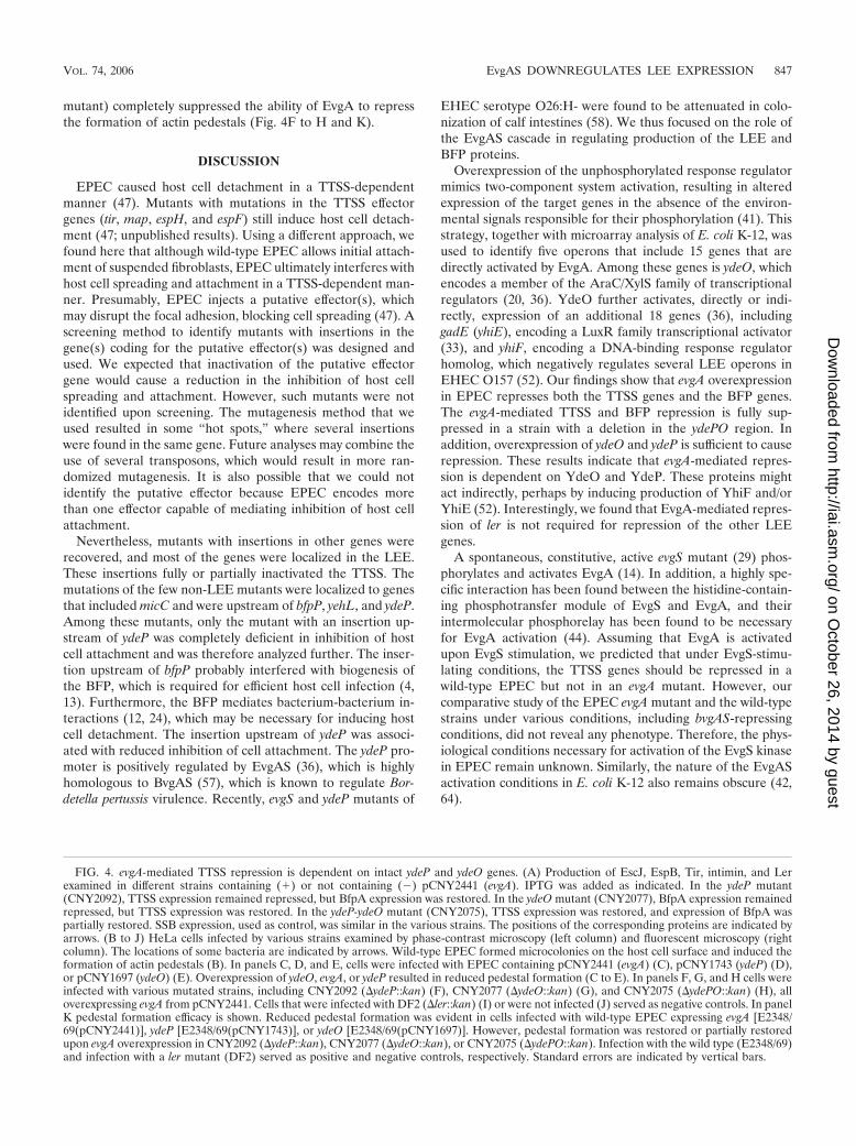
mutant) completely suppressed the ability of EvgA to repressthe formation of actin pedestals (Fig. 4F to H and K).
DISCUSSION
EPEC caused host cell detachment in a TTSS-dependentmanner (47). Mutants with mutations in the TTSS effectorgenes (tir, map, espH, and espF) still induce host cell detach-ment (47; unpublished results). Using a different approach, wefound here that although wild-type EPEC allows initial attach-ment of suspended fibroblasts, EPEC ultimately interferes withhost cell spreading and attachment in a TTSS-dependent man-ner. Presumably, EPEC injects a putative effector(s), whichmay disrupt the focal adhesion, blocking cell spreading (47). Ascreening method to identify mutants with insertions in thegene(s) coding for the putative effector(s) was designed andused. We expected that inactivation of the putative effectorgene would cause a reduction in the inhibition of host cellspreading and attachment. However, such mutants were notidentified upon screening. The mutagenesis method that weused resulted in some “hot spots,” where several insertionswere found in the same gene. Future analyses may combine theuse of several transposons, which would result in more ran-domized mutagenesis. It is also possible that we could notidentify the putative effector because EPEC encodes morethan one effector capable of mediating inhibition of host cellattachment.
Nevertheless, mutants with insertions in other genes wererecovered, and most of the genes were localized in the LEE.These insertions fully or partially inactivated the TTSS. Themutations of the few non-LEE mutants were localized to genesthat included micC and were upstream of bfpP, yehL, and ydeP.Among these mutants, only the mutant with an insertion up-stream of ydeP was completely deficient in inhibition of hostcell attachment and was therefore analyzed further. The inser-tion upstream of bfpP probably interfered with biogenesis ofthe BFP, which is required for efficient host cell infection (4,13). Furthermore, the BFP mediates bacterium-bacterium in-teractions (12, 24), which may be necessary for inducing hostcell detachment. The insertion upstream of ydeP was associ-ated with reduced inhibition of cell attachment. The ydeP pro-moter is positively regulated by EvgAS (36), which is highlyhomologous to BvgAS (57), which is known to regulate Bor-detella pertussis virulence. Recently, evgS and ydeP mutants of
EHEC serotype O26:H- were found to be attenuated in colo-nization of calf intestines (58). We thus focused on the role ofthe EvgAS cascade in regulating production of the LEE andBFP proteins.
Overexpression of the unphosphorylated response regulatormimics two-component system activation, resulting in alteredexpression of the target genes in the absence of the environ-mental signals responsible for their phosphorylation (41). Thisstrategy, together with microarray analysis of E. coli K-12, wasused to identify five operons that include 15 genes that aredirectly activated by EvgA. Among these genes is ydeO, whichencodes a member of the AraC/XylS family of transcriptionalregulators (20, 36). YdeO further activates, directly or indi-rectly, expression of an additional 18 genes (36), includinggadE (yhiE), encoding a LuxR family transcriptional activator(33), and yhiF, encoding a DNA-binding response regulatorhomolog, which negatively regulates several LEE operons inEHEC O157 (52). Our findings show that evgA overexpressionin EPEC represses both the TTSS genes and the BFP genes.The evgA-mediated TTSS and BFP repression is fully sup-pressed in a strain with a deletion in the ydePO region. Inaddition, overexpression of ydeO and ydeP is sufficient to causerepression. These results indicate that evgA-mediated repres-sion is dependent on YdeO and YdeP. These proteins mightact indirectly, perhaps by inducing production of YhiF and/orYhiE (52). Interestingly, we found that EvgA-mediated repres-sion of ler is not required for repression of the other LEEgenes.
A spontaneous, constitutive, active evgS mutant (29) phos-phorylates and activates EvgA (14). In addition, a highly spe-cific interaction has been found between the histidine-contain-ing phosphotransfer module of EvgS and EvgA, and theirintermolecular phosphorelay has been found to be necessaryfor EvgA activation (44). Assuming that EvgA is activatedupon EvgS stimulation, we predicted that under EvgS-stimu-lating conditions, the TTSS genes should be repressed in awild-type EPEC but not in an evgA mutant. However, ourcomparative study of the EPEC evgA mutant and the wild-typestrains under various conditions, including bvgAS-repressingconditions, did not reveal any phenotype. Therefore, the phys-iological conditions necessary for activation of the EvgS kinasein EPEC remain unknown. Similarly, the nature of the EvgASactivation conditions in E. coli K-12 also remains obscure (42,64).
FIG. 4. evgA-mediated TTSS repression is dependent on intact ydeP and ydeO genes. (A) Production of EscJ, EspB, Tir, intimin, and Lerexamined in different strains containing (�) or not containing () pCNY2441 (evgA). IPTG was added as indicated. In the ydeP mutant(CNY2092), TTSS expression remained repressed, but BfpA expression was restored. In the ydeO mutant (CNY2077), BfpA expression remainedrepressed, but TTSS expression was restored. In the ydeP-ydeO mutant (CNY2075), TTSS expression was restored, and expression of BfpA waspartially restored. SSB expression, used as control, was similar in the various strains. The positions of the corresponding proteins are indicated byarrows. (B to J) HeLa cells infected by various strains examined by phase-contrast microscopy (left column) and fluorescent microscopy (rightcolumn). The locations of some bacteria are indicated by arrows. Wild-type EPEC formed microcolonies on the host cell surface and induced theformation of actin pedestals (B). In panels C, D, and E, cells were infected with EPEC containing pCNY2441 (evgA) (C), pCNY1743 (ydeP) (D),or pCNY1697 (ydeO) (E). Overexpression of ydeO, evgA, or ydeP resulted in reduced pedestal formation (C to E). In panels F, G, and H cells wereinfected with various mutated strains, including CNY2092 (�ydeP::kan) (F), CNY2077 (�ydeO::kan) (G), and CNY2075 (�ydePO::kan) (H), alloverexpressing evgA from pCNY2441. Cells that were infected with DF2 (�ler::kan) (I) or were not infected (J) served as negative controls. In panelK pedestal formation efficacy is shown. Reduced pedestal formation was evident in cells infected with wild-type EPEC expressing evgA [E2348/69(pCNY2441)], ydeP [E2348/69(pCNY1743)], or ydeO [E2348/69(pCNY1697)]. However, pedestal formation was restored or partially restoredupon evgA overexpression in CNY2092 (�ydeP::kan), CNY2077 (�ydeO::kan), or CNY2075 (�ydePO::kan). Infection with the wild type (E2348/69)and infection with a ler mutant (DF2) served as positive and negative controls, respectively. Standard errors are indicated by vertical bars.
VOL. 74, 2006 EvgAS DOWNREGULATES LEE EXPRESSION 847
on October 26, 2014 by guest
http://iai.asm.org/
Dow
nloaded from

An alternative explanation for the lack of a phenotype forthe evgA mutant is that the physiological function of the EvgASsystem is redundant with respect to other regulatory cascades.Indeed, previous reports showed that there is a partial overlapbetween the EvgAS and GadXW cascades. This overlap in-cludes activation of YhiE (GadE) and YhiF (36, 55). More-over, GadX overexpression in EPEC represses the expressionof TTSS and BFP genes via expression of the perABC operon(48). The reports mentioned above and the lack of a phenotypein the EPEC evgA mutant led us to construct double mutantswith mutations in evgA or ydeO and in gadX or the gadX-gadWregion. However, the four double mutants examined did notdiffer from the wild type.
In conclusion, many LEE regulators, most of which functionat the transcriptional level, have been discovered. Here wereport that the EvgAS two-component system, apparently ac-tivated by an unknown external signal, represses TTSS expres-sion. Identification of the nature of the elusive EvgS activatingsignal should elucidate the physiological role of EvgAS-medi-ated repression of the TTSS genes in EPEC.
ACKNOWLEDGMENTS
We thank O. Amster-Choder for providing the anti-SSB antibodyand G. Frankel for providing the anti-EspB, -Tir, -Int, -EscJ, and-BfpA antibodies.
This work was supported by grants from The Israel Science Foun-dation founded by The Israel Academy of Science and Humanities andfrom the United States-Israel Binational Science Foundation. I.R. isthe Etta Rosensohn Professor of Bacteriology.
REFERENCES
1. Baldwin, T. J., M. B. Lee-Delaunay, S. Knutton, and P. H. Williams. 1993.Calcium-calmodulin dependence of actin accretion and lethality in culturedHEp-2 cells infected with enteropathogenic Escherichia coli. Infect. Immun.61:760–763.
2. Berdichevsky, T., D. Friedberg, C. Nadler, A. Rokney, A. Oppenheim, and I.Rosenshine. 2005. Ler is a negative autoregulator of the LEE1 operon inenteropathogenic Escherichia coli. J. Bacteriol. 187:349–357.
3. Bustamante, V. H., F. J. Santana, E. Calva, and J. L. Puente. 2001. Tran-scriptional regulation of type III secretion genes in enteropathogenic Esch-erichia coli: Ler antagonizes H-NS-dependent repression. Mol. Microbiol.39:664–678.
4. Chen, H. D., and G. Frankel. 2005. Enteropathogenic Escherichia coli: un-ravelling pathogenesis. FEMS Microbiol. Rev. 29:83–98.
5. Chen, S., A. Zhang, L. B. Blyn, and G. Storz. 2004. MicC, a second small-RNA regulator of Omp protein expression in Escherichia coli. J. Bacteriol.186:6689–6697.
6. Creasey, E. A., R. M. Delahay, S. J. Daniell, and G. Frankel. 2003. Yeasttwo-hybrid system survey of interactions between LEE-encoded proteins ofenteropathogenic Escherichia coli. Microbiology 149:2093–2106.
7. Datsenko, K. A., and B. L. Wanner. 2000. One-step inactivation of chromo-somal genes in Escherichia coli K-12 using PCR products. Proc. Natl. Acad.Sci. USA 97:6640–6645.
8. de Lorenzo, V., and K. N. Timmis. 1994. Analysis and construction of stablephenotypes in gram-negative bacteria with Tn5- and Tn10-derived minitrans-posons. Methods Enzymol. 235:386–405.
9. Deng, W., J. L. Puente, S. Gruenheid, Y. Li, B. A. Vallance, A. Vazquez, J.Barba, J. A. Ibarra, P. O’Donnell, P. Metalnikov, K. Ashman, S. Lee, D.Goode, T. Pawson, and B. B. Finlay. 2004. Dissecting virulence: systematicand functional analyses of a pathogenicity island. Proc. Natl. Acad. Sci. USA101:3597–3602.
10. DeVinney, R., A. Gauthier, A. Abe, and B. B. Finlay. 1999. EnteropathogenicEscherichia coli: a pathogen that inserts its own receptor into host cells. Cell.Mol. Life Sci. 55:961–976.
11. Donnenberg, M. S., S. B. Calderwood, A. Donohue-Rolfe, G. T. Keusch, andJ. B. Kaper. 1990. Construction and analysis of TnphoA mutants of entero-pathogenic Escherichia coli unable to invade HEp-2 cells. Infect. Immun.58:1565–1571.
12. Donnenberg, M. S., J. A. Giron, J. P. Nataro, and J. B. Kaper. 1992. Aplasmid-encoded type IV fimbrial gene of enteropathogenic Escherichia coliassociated with localized adherence. Mol. Microbiol. 6:3427–3437.
13. Donnenberg, M. S., J. B. Kaper, and B. B. Finlay. 1997. Interactions between
enteropathogenic Escherichia coli and host epithelial cells. Trends Microbiol.5:109–114.
14. Eguchi, Y., T. Oshima, H. Mori, R. Aono, K. Yamamoto, A. Ishihama, andR. Utsumi. 2003. Transcriptional regulation of drug efflux genes by EvgAS,a two-component system in Escherichia coli. Microbiology 149:2819–2828.
15. Elgrably-Weiss, M., S. Park, E. Schlosser-Silverman, I. Rosenshine, J. Im-lay, and S. Altuvia. 2002. A Salmonella enterica serovar Typhimurium hemAmutant is highly susceptible to oxidative DNA damage. J. Bacteriol. 184:3774–3784.
16. Elliott, S. J., L. A. Wainwright, T. K. McDaniel, K. G. Jarvis, Y. K. Deng,L. C. Lai, B. P. McNamara, M. S. Donnenberg, and J. B. Kaper. 1998. Thecomplete sequence of the locus of enterocyte effacement (LEE) from en-teropathogenic Escherichia coli E2348/69. Mol. Microbiol. 28:1–4.
17. Elliott, S. J., J. Yu, and J. B. Kaper. 1999. The cloned locus of enterocyteeffacement from enterohemorrhagic Escherichia coli O157:H7 is unable toconfer the attaching and effacing phenotype upon E. coli K-12. Infect. Im-mun. 67:4260–4263.
18. Frankel, G., A. D. Phillips, I. Rosenshine, G. Dougan, J. B. Kaper, and S.Knutton. 1998. Enteropathogenic and enterohaemorrhagic Escherichia coli:more subversive elements. Mol. Microbiol. 30:911–921.
19. Friedberg, D., T. Umanski, Y. Fang, and I. Rosenshine. 1999. Hierarchy inthe expression of the locus of enterocyte effacement genes of enteropatho-genic Escherichia coli. Mol. Microbiol. 34:941–952.
20. Gallegos, M. T., R. Schleif, A. Bairoch, K. Hofmann, and J. L. Ramos. 1997.Arac/XylS family of transcriptional regulators. Microbiol. Mol. Biol. Rev.61:393–410.
21. Goldberg, M. D., M. Johnson, J. C. Hinton, and P. H. Williams. 2001. Roleof the nucleoid-associated protein Fis in the regulation of virulence proper-ties of enteropathogenic Escherichia coli. Mol. Microbiol. 41:549–559.
22. Gomez-Duarte, O. G., and J. B. Kaper. 1995. A plasmid-encoded regulatoryregion activates chromosomal eaeA expression in enteropathogenic Esche-richia coli. Infect. Immun. 63:1767–1776.
23. Grant, A. J., M. Farris, P. Alefounder, P. H. Williams, M. J. Woodward, andC. D. O’Connor. 2003. Co-ordination of pathogenicity island expression bythe BipA GTPase in enteropathogenic Escherichia coli (EPEC). Mol. Mi-crobiol. 48:507–521.
24. Hicks, S., G. Frankel, J. B. Kaper, G. Dougan, and A. D. Phillips. 1998. Roleof intimin and bundle-forming pili in enteropathogenic Escherichia coli ad-hesion to pediatric intestinal tissue in vitro. Infect. Immun. 66:1570–1578.
25. Iyoda, S., and H. Watanabe. 2005. ClpXP protease controls expression of thetype III protein secretion system through regulation of RpoS and GrlR levelsin enterohemorrhagic Escherichia coli. J. Bacteriol. 187:4086–4094.
26. Jepson, M. A., S. Pellegrin, L. Peto, D. N. Banbury, A. D. Leard, H. Mellor,and B. Kenny. 2003. Synergistic roles for the Map and Tir effector moleculesin mediating uptake of enteropathogenic Escherichia coli (EPEC) into non-phagocytic cells. Cell. Microbiol. 5:773–783.
27. Kanamaru, K., K. Kanamaru, I. Tatsuno, T. Tobe, and C. Sasakawa. 2000.SdiA, an Escherichia coli homologue of quorum-sensing regulators, controlsthe expression of virulence factors in enterohaemorrhagic Escherichia coliO157:H7. Mol. Microbiol. 38:805–816.
28. Kaper, J. B. 1998. EPEC delivers the goods. Trends Microbiol. 6:169–172.(Discussion, 6:172–173.)
29. Kato, A., H. Ohnishi, K. Yamamoto, E. Furuta, H. Tanabe, and R. Utsumi.2000. Transcription of emrKY is regulated by the EvgA-EvgS two-componentsystem in Escherichia coli K-12. Biosci. Biotechnol. Biochem. 64:1203–1209.
30. Kimbrough, T. G., and S. I. Miller. 2000. Contribution of Salmonella typhi-murium type III secretion components to needle complex formation. Proc.Natl. Acad. Sci. USA 97:11008–11013.
31. Knapp, S., and J. J. Mekalanos. 1988. Two trans-acting regulatory genes (virand mod) control antigenic modulation in Bordetella pertussis. J. Bacteriol.170:5059–5066.
32. Levine, M. M., E. J. Bergquist, D. R. Nalin, D. H. Waterman, R. B. Hornick,C. R. Young, and S. Sotman. 1978. Escherichia coli strains that cause diar-rhoea but do not produce heat-labile or heat-stable enterotoxins and arenon-invasive. Lancet i:1119–1122.
33. Ma, Z., S. Gong, H. Richard, D. L. Tucker, T. Conway, and J. W. Foster.2003. GadE (YhiE) activates glutamate decarboxylase-dependent acid resis-tance in Escherichia coli K-12. Mol. Microbiol. 49:1309–1320.
34. Ma, Z., N. Masuda, and J. W. Foster. 2004. Characterization of EvgAS-YdeO-GadE branched regulatory circuit governing glutamate-dependentacid resistance in Escherichia coli. J. Bacteriol. 186:7378–7389.
35. Maniatis, T., E. F. Fritsch, and J. Sambrook. 1991. Molecular cloning: alaboratory manual. Cold Spring Harbor Laboratory Press, Cold Spring Har-bor, N.Y.
36. Masuda, N., and G. M. Church. 2003. Regulatory network of acid resistancegenes in Escherichia coli. Mol. Microbiol. 48:699–712.
37. McDaniel, T. K., and J. B. Kaper. 1997. A cloned pathogenicity island fromenteropathogenic Escherichia coli confers the attaching and effacing pheno-type on E. coli K-12. Mol. Microbiol. 23:399–407.
38. Mellies, J. L., S. J. Elliott, V. Sperandio, M. S. Donnenberg, and J. B. Kaper.1999. The Per regulon of enteropathogenic Escherichia coli: identification ofa regulatory cascade and a novel transcriptional activator, the locus of en-
848 NADLER ET AL. INFECT. IMMUN.
on October 26, 2014 by guest
http://iai.asm.org/
Dow
nloaded from

terocyte effacement (LEE)-encoded regulator (Ler). Mol. Microbiol. 33:296–306.
39. Mizuno, T. 1997. Compilation of all genes encoding two-component phos-photransfer signal transducers in the genome of Escherichia coli. DNA Res.4:161–168.
40. Nishino, K., and A. Yamaguchi. 2002. EvgA of the two-component signaltransduction system modulates production of the yhiUV multidrug trans-porter in Escherichia coli. J. Bacteriol. 184:2319–2323.
41. Ogura, M., H. Yamaguchi, K. Yoshida, Y. Fujita, and T. Tanaka. 2001. DNAmicroarray analysis of Bacillus subtilis DegU, ComA and PhoP regulons: anapproach to comprehensive analysis of B. subtilis two-component regulatorysystems. Nucleic Acids Res. 29:3804–3813.
42. Oshima, T., H. Aiba, Y. Masuda, S. Kanaya, M. Sugiura, B. L. Wanner, H.Mori, and T. Mizuno. 2002. Transcriptome analysis of all two-componentregulatory system mutants of Escherichia coli K-12. Mol. Microbiol. 46:281–291.
43. Perraud, A. L., K. Rippe, M. Bantscheff, M. Glocker, M. Lucassen, K. Jung,W. Sebald, V. Weiss, and R. Gross. 2000. Dimerization of signalling modulesof the EvgAS and BvgAS phosphorelay systems. Biochim. Biophys. Acta1478:341–354.
44. Perraud, A. L., V. Weiss, and R. Gross. 1999. Signalling pathways in two-component phosphorelay systems. Trends Microbiol. 7:115–120.
45. Schlosser-Silverman, E., M. Elgrably-Weiss, I. Rosenshine, R. Kohen, and S.Altuvia. 2000. Characterization of Escherichia coli DNA lesions generatedwithin J774 macrophages. J. Bacteriol. 182:5225–5230.
46. Shaw, R. K., J. Cleary, M. S. Murphy, G. Frankel, and S. Knutton. 2005.Interaction of enteropathogenic Escherichia coli with human intestinal mu-cosa: role of effector proteins in brush border remodeling and formation ofattaching and effacing lesions. Infect. Immun. 73:1243–1251.
47. Shifrin, Y., J. Kirschner, B. Geiger, and I. Rosenshine. 2002. Enteropatho-genic Escherichia coli induces modification of the focal adhesions of infectedhost cells. Cell. Microbiol. 4:235–243.
48. Shin, S., M. P. Castanie-Cornet, J. W. Foster, J. A. Crawford, C. Brinkley,and J. B. Kaper. 2001. An activator of glutamate decarboxylase genes reg-ulates the expression of enteropathogenic Escherichia coli virulence genesthrough control of the plasmid-encoded regulator, Per. Mol. Microbiol.41:1133–1150.
49. Sperandio, V., J. L. Mellies, R. M. Delahay, G. Frankel, J. A. Crawford, W.Nguyen, and J. B. Kaper. 2000. Activation of enteropathogenic Escherichiacoli (EPEC) LEE2 and LEE3 operons by Ler. Mol. Microbiol. 38:781–793.
50. Suzuki, H., K. Yonekura, K. Murata, T. Hirai, K. Oosawa, and K. Namba.1998. A structural feature in the central channel of the bacterial flagellar FliFring complex is implicated in type III protein export. J. Struct. Biol. 124:104–114.
51. Suzuki, H., K. Yonekura, and K. Namba. 2004. Structure of the rotor of thebacterial flagellar motor revealed by electron cryomicroscopy and single-particle image analysis. J. Mol. Biol. 337:105–113.
52. Tatsuno, I., K. Nagano, K. Taguchi, L. Rong, H. Mori, and C. Sasakawa.2003. Increased adherence to Caco-2 cells caused by disruption of the yhiEand yhiF genes in enterohemorrhagic Escherichia coli O157:H7. Infect. Im-mun. 71:2598–2606.
53. Tobe, T., G. K. Schoolnik, I. Sohel, V. H. Bustamante, and J. L. Puente. 1996.Cloning and characterization of bfpTVW, genes required for the transcrip-tional activation of bfpA in enteropathogenic Escherichia coli. Mol. Micro-biol. 21:963–975.
54. Tu, X., I. Nisan, C. Yona, E. Hanski, and I. Rosenshine. 2003. EspH, a newcytoskeleton-modulating effector of enterohaemorrhagic and enteropatho-genic Escherichia coli. Mol. Microbiol. 47:595–606.
55. Tucker, D. L., N. Tucker, Z. Ma, J. W. Foster, R. L. Miranda, P. S. Cohen,and T. Conway. 2003. Genes of the GadX-GadW regulon in Escherichia coli.J. Bacteriol. 185:3190–3201.
56. Umanski, T., I. Rosenshine, and D. Friedberg. 2002. Thermoregulated ex-pression of virulence genes in enteropathogenic Escherichia coli. Microbiol-ogy 148:2735–2744.
57. Utsumi, R., S. Katayama, M. Taniguchi, T. Horie, M. Ikeda, S. Igaki, H.Nakagawa, A. Miwa, H. Tanabe, and M. Noda. 1994. Newly identified genesinvolved in the signal transduction of Escherichia coli K-12. Gene 140:73–77.
58. van Diemen, P. M., F. Dziva, M. P. Stevens, and T. S. Wallis. 2005. Identi-fication of enterohemorrhagic Escherichia coli O26:H- genes required forintestinal colonization in calves. Infect. Immun. 73:1735–1743.
59. Yamamoto, K., K. Hirao, T. Oshima, H. Aiba, R. Utsumi, and A. Ishihama.2005. Functional characterization in vitro of all two-component signal trans-duction systems from Escherichia coli. J. Biol. Chem. 280:1448–1456.
60. Yanisch-Perron, C., J. Vieira, and J. Messing. 1985. Improved M13 phagecloning vectors and host strains: nucleotide sequences of the M13mp18 andpUC19 vectors. Gene 33:103–119.
61. Yona-Nadler, C., T. Umanski, S. Aizawa, D. Friedberg, and I. Rosenshine.2003. Integration host factor (IHF) mediates repression of flagella in enter-opathogenic and enterohaemorrhagic Escherichia coli. Microbiology 149:877–884.
62. Yu, D., H. M. Ellis, E. C. Lee, N. A. Jenkins, N. G. Copeland, and D. L.Court. 2000. An efficient recombination system for chromosome engineeringin Escherichia coli. Proc. Natl. Acad. Sci. USA 97:5978–5983.
63. Zhang, L., R. R. Chaudhuri, C. Constantinidou, J. L. Hobman, M. D. Patel,A. C. Jones, D. Sarti, A. J. Roe, I. Vlisidou, R. K. Shaw, F. Falciani, M. P.Stevens, D. L. Gally, S. Knutton, G. Frankel, C. W. Penn, and M. J. Pallen.2004. Regulators encoded in the Escherichia coli type III secretion system 2gene cluster influence expression of genes within the locus for enterocyteeffacement in enterohemorrhagic E. coli O157:H7. Infect. Immun. 72:7282–7293.
64. Zhou, L., X. H. Lei, B. R. Bochner, and B. L. Wanner. 2003. Phenotypemicroarray analysis of Escherichia coli K-12 mutants with deletions of alltwo-component systems. J. Bacteriol. 185:4956–4972.
Editor: J. B. Bliska
VOL. 74, 2006 EvgAS DOWNREGULATES LEE EXPRESSION 849
on October 26, 2014 by guest
http://iai.asm.org/
Dow
nloaded from
Copyright © 2022 FDOKUMEN