Enterohaemorrhagic and enteropathogenic Escherichia coli use a different Tir-based mechanism for...
14
Enterohaemorrhagic and enteropathogenic Escherichia coli use a different Tir-based mechanism for pedestal formation Rebekah DeVinney, 1 * Jose Luis Puente, 2 Annick Gauthier, 3 Danika Goosney 4 and B. Brett Finlay 1,3,4 1 Biotechnology Laboratory, University of British Columbia, Vancouver, BC, Canada, V6T 1Z3. 2 Molecular Microbiology Department, Instituto de Biotecnologı ´a, Universidad Nacional Auto ´noma de Me ´ xico, Cuernavaca, Morelos 62250, Mexico. Departments of 3 Biochemistry and 4 Microbiology and Immunology, University of British Columbia, Vancouver, BC, Canada, V6T 1Z3. Summary Enterohaemorrhagic Escherichia coli (EHEC) adheres to the host intestinal epithelium, resulting in the formation of actin pedestals beneath adhering bacteria. EHEC and a related pathogen, enteropatho- genic E. coli (EPEC), insert a bacterial receptor, Tir, into the host plasma membrane, which is required for pedestal formation. An important difference between EPEC and EHEC Tir is that EPEC but not EHEC Tir is tyrosine phosphorylated once delivered into the host. In this study, we assessed the role of Tir tyrosine phosphorylation in pedestal formation by EPEC and EHEC. In EPEC, pedestal formation is absolutely dependent on Tir tyrosine phosphorylation and is not complemented by EHEC Tir. The protein sequence surrounding EPEC Tir tyrosine 474 is critical for Tir tyrosine phosphorylation and pedestal formation by EPEC. In contrast, Tir tyrosine phosphorylation is not required for pedestal formation by EHEC. EHEC forms pedestals with both wild-type EPEC Tir and the non- tyrosine-phosphorylatable EPEC Tir Y474F. Pedestal formation by EHEC requires the type III delivery of additional EHEC factors into the host cell. These findings highlight differences in the mechanisms of pedestal formation by these closely related patho- gens and indicate that EPEC and EHEC modulate different signalling pathways to affect the host actin cytoskeleton. Introduction Enterohaemorrhagic Escherichia coli (EHEC) O157:H7 is a significant cause of food- and water-borne illness in North America, as evidenced by the recent outbreak in Walkerton, Ontario, which affected close to 2000 individuals and resulted in seven deaths (Griffin and Tauxe, 1991; Kondro, 2000). EHEC infection initially results in a watery diarrhoea, which can progress to severe bloody diarrhoea (haemorrhagic colitis) in patients of all ages (Nataro and Kaper, 1998). In young children and the elderly, EHEC can cause a potentially fatal systemic complication, the haemolytic uraemic syndrome (HUS), characterized by haemolytic anaemia, thrombocytopenia and renal failure (Griffin et al., 1988; Nataro and Kaper, 1998). EHEC is a member of a family of pathogens that cause attaching and effacing (A/E) lesions. A/E lesions are characterized by the degeneration of the epithelial cell microvilli and the formation of actin-rich pedestals within the host enterocytes beneath the adherent bacteria (Moon et al., 1983; Knutton et al., 1989). This family of pathogens includes enteropathogenic E. coli (EPEC), a major cause of infantile diarrhoea in the developing world, rabbit, dog and porcine EPEC strains and Citrobacter rodentium, which causes colonic hyperplasia in mice (Cantey and Blake, 1977; Beaudry et al., 1996; Nataro and Kaper, 1998; Newman et al., 1999). Central to the process of A/E lesion formation by this family of pathogens is Tir, the translocated intimin receptor. Tir is a 72 – 78 kDa bacterial protein that is inserted into the host cell plasma membrane, where it is phosphorylated and becomes the receptor for the bacterial outer membrane protein, intimin (Kenny et al., 1997; Ebel et al., 1998; DeVinney et al., 1999). Intimin binding to Tir leads to the dramatic rearrangement of the host actin cytoskeleton, which forms a pedestal beneath the adherent bacteria (Kenny et al., 1997; DeVinney et al., 1999). Tir delivery into the host cell is mediated by the type III secretion apparatus and is dependent on the E. coli secreted proteins EspA, EspB and EspD, which are pro- posed to form the type III translocon (Kenny et al., 1997; Taylor et al., 1998; Wolff et al., 1998; Kresse et al., 1999; Wachter et al., 1999). EHEC and EPEC strains that are lacking, or are unable to deliver, Tir do not form pedestals in cultured epithelial cells (Kenny et al., 1997; DeVinney Accepted 13 July, 2001. *For correspondence. E-mail rdevinne@ ucalgary.ca; Tel. (11) 403 220 4095; Fax (11) 403 270 2772. Present address: University of Calgary Health Sciences Centre, Rm 278 HMRB, 3330 Hospital Dr. NW, Calgary AB, T2N 4N1, Canada. Molecular Microbiology (2001) 41(6), 1445–1458 Q 2001 Blackwell Science Ltd
Transcript of Enterohaemorrhagic and enteropathogenic Escherichia coli use a different Tir-based mechanism for...

Enterohaemorrhagic and enteropathogenic Escherichiacoli use a different Tir-based mechanism for pedestalformation
Rebekah DeVinney,1*† Jose Luis Puente,2
Annick Gauthier,3 Danika Goosney4 and
B. Brett Finlay1,3,4
1Biotechnology Laboratory, University of British Columbia,
Vancouver, BC, Canada, V6T 1Z3.2Molecular Microbiology Department, Instituto de
Biotecnologıa, Universidad Nacional Autonoma de
Mexico, Cuernavaca, Morelos 62250, Mexico.
Departments of 3Biochemistry and 4Microbiology and
Immunology, University of British Columbia, Vancouver,
BC, Canada, V6T 1Z3.
Summary
Enterohaemorrhagic Escherichia coli (EHEC) adheres
to the host intestinal epithelium, resulting in the
formation of actin pedestals beneath adhering
bacteria. EHEC and a related pathogen, enteropatho-
genic E. coli (EPEC), insert a bacterial receptor, Tir,
into the host plasma membrane, which is required for
pedestal formation. An important difference between
EPEC and EHEC Tir is that EPEC but not EHEC Tir is
tyrosine phosphorylated once delivered into the host.
In this study, we assessed the role of Tir tyrosine
phosphorylation in pedestal formation by EPEC and
EHEC. In EPEC, pedestal formation is absolutely
dependent on Tir tyrosine phosphorylation and is not
complemented by EHEC Tir. The protein sequence
surrounding EPEC Tir tyrosine 474 is critical for Tir
tyrosine phosphorylation and pedestal formation by
EPEC. In contrast, Tir tyrosine phosphorylation is not
required for pedestal formation by EHEC. EHEC forms
pedestals with both wild-type EPEC Tir and the non-
tyrosine-phosphorylatable EPEC Tir Y474F. Pedestal
formation by EHEC requires the type III delivery of
additional EHEC factors into the host cell. These
findings highlight differences in the mechanisms of
pedestal formation by these closely related patho-
gens and indicate that EPEC and EHEC modulate
different signalling pathways to affect the host actin
cytoskeleton.
Introduction
Enterohaemorrhagic Escherichia coli (EHEC) O157:H7 is
a significant cause of food- and water-borne illness in
North America, as evidenced by the recent outbreak in
Walkerton, Ontario, which affected close to 2000
individuals and resulted in seven deaths (Griffin and
Tauxe, 1991; Kondro, 2000). EHEC infection initially
results in a watery diarrhoea, which can progress to severe
bloody diarrhoea (haemorrhagic colitis) in patients of all
ages (Nataro and Kaper, 1998). In young children and the
elderly, EHEC can cause a potentially fatal systemic
complication, the haemolytic uraemic syndrome (HUS),
characterized by haemolytic anaemia, thrombocytopenia
and renal failure (Griffin et al., 1988; Nataro and Kaper,
1998). EHEC is a member of a family of pathogens that
cause attaching and effacing (A/E) lesions. A/E lesions are
characterized by the degeneration of the epithelial cell
microvilli and the formation of actin-rich pedestals within
the host enterocytes beneath the adherent bacteria (Moon
et al., 1983; Knutton et al., 1989). This family of pathogens
includes enteropathogenic E. coli (EPEC), a major cause
of infantile diarrhoea in the developing world, rabbit, dog
and porcine EPEC strains and Citrobacter rodentium,
which causes colonic hyperplasia in mice (Cantey and
Blake, 1977; Beaudry et al., 1996; Nataro and Kaper,
1998; Newman et al., 1999).
Central to the process of A/E lesion formation by this
family of pathogens is Tir, the translocated intimin
receptor. Tir is a 72–78 kDa bacterial protein that is
inserted into the host cell plasma membrane, where it is
phosphorylated and becomes the receptor for the bacterial
outer membrane protein, intimin (Kenny et al., 1997; Ebel
et al., 1998; DeVinney et al., 1999). Intimin binding to Tir
leads to the dramatic rearrangement of the host actin
cytoskeleton, which forms a pedestal beneath the
adherent bacteria (Kenny et al., 1997; DeVinney et al.,
1999). Tir delivery into the host cell is mediated by the
type III secretion apparatus and is dependent on the E. coli
secreted proteins EspA, EspB and EspD, which are pro-
posed to form the type III translocon (Kenny et al., 1997;
Taylor et al., 1998; Wolff et al., 1998; Kresse et al., 1999;
Wachter et al., 1999). EHEC and EPEC strains that are
lacking, or are unable to deliver, Tir do not form pedestals
in cultured epithelial cells (Kenny et al., 1997; DeVinney
Accepted 13 July, 2001. *For correspondence. E-mail [email protected]; Tel. (11) 403 220 4095; Fax (11) 403 270 2772.†Present address: University of Calgary Health Sciences Centre, Rm278 HMRB, 3330 Hospital Dr. NW, Calgary AB, T2N 4N1, Canada.
Molecular Microbiology (2001) 41(6), 1445–1458
Q 2001 Blackwell Science Ltd

et al., 1999). The genes required for Tir delivery and A/E
lesion formation are encoded in a chromosomal patho-
genicity island called the LEE (locus of enterocyte
effacement) (McDaniel and Kaper, 1997; Perna et al.,
1998). Once in the host cell, Tir is an integral membrane
protein, with two predicted transmembrane domains.
Recent work has indicated that these regions are required
for the stable insertion of Tir into the host plasma mem-
brane (Gauthier et al., 2000). The amino- and carboxy-
termini of Tir are intracellular (Kenny et al., 1997;
DeVinney et al., 1999), and intimin binds to an extracellular
domain called the IBD (intimin-binding domain) via a large
hydrophobic pocket (de Grado et al., 1999; Kenny, 1999;
Luo et al., 2000).
The ability to form A/E lesions is essential for full
virulence of this family of pathogens (Abe et al., 1998;
Marches et al., 2000; Tacket et al., 2000). Although the
more severe symptoms observed with EHEC (haemor-
rhagic colitis and HUS) are caused by the Shiga toxin
produced by this pathogen, the initial watery diarrhoea is
attributed to the formation of A/E lesions (Nataro and
Kaper, 1998). In EPEC, human volunteer studies have
implicated two proteins that are required for pedestal
formation in vitro, intimin (Donnenberg et al., 1993) and
EspB (Tacket et al., 2000), as important virulence factors.
Strains lacking either of these proteins are severely
attenuated in their ability to cause human disease. Further
evidence for the importance of A/E lesion formation in
virulence comes from work with a natural rabbit pathogen,
REPEC O103:H2. This work demonstrated that EspA
and EspB (Abe et al., 1998) and Tir and intimin (Marches
et al., 2000) are essential for the diarrhoeal disease
caused by this organism. In EHEC, intimin is required
for colonization and the formation of A/E lesions in the
intestines of gnotobiotic piglets and colostrum-deprived
calves and is essential for the disease observed in these
hosts (McKee et al., 1995; Dean-Nystrom et al., 1998).
Additionally, intimins from EPEC and EHEC are function-
ally interchangeable. Heterologous intimin expression
supports both intestinal colonization (Tzipori et al., 1995;
Phillips et al., 2000) and pedestal formation in cultured
epithelial cells (DeVinney et al., 1999).
The process of pedestal formation is best understood for
EPEC. EPEC Tir is tyrosine and serine/threonine
phosphorylated upon insertion into the host cell mem-
brane, and phosphorylation of tyrosine 474 is required for
pedestal formation (Rosenshine et al., 1992; Kenny et al.,
1997; Kenny, 1999). Pedestal formation requires the
recruitment of N-WASP (neural Wiscott–Aldrich syn-
drome protein; Kalman et al., 1999), which is recruited to
the pedestal in a Tir tyrosine phosphorylation-dependent
manner (Goosney et al., 2000). N-WASP functions to
target the Arp 2/3 complex to Tir, which is required for
pedestal elongation (Kalman et al., 1999) Both N-WASP
and the Arp2/3 complex play a role in subversion of the
actin cytoskeleton by other bacterial and viral pathogens
(Suzuki et al., 1998; Frischknecht et al., 1999; Moreau
et al., 2000). Additionally, the amino-terminus of EPEC Tir
binds directly to the host actin binding and cross-linking
protein a-actinin, anchoring Tir to the host actin
cytoskeleton (Goosney et al., 2000). The focal adhesion
proteins talin and vinculin have also been shown to interact
directly with Tir (Freeman et al., 2000).
Considerably less is understood about the mechanism
of pedestal formation by EHEC. Unlike EPEC Tir, EHEC
Tir is phosphorylated on serine or threonine, but not on
tyrosine, and the role of these phosphorylation events is
unknown (DeVinney et al., 1999). Despite these differ-
ences in Tir phosphorylation, EHEC forms seemingly
identical pedestals within the host cell (Ismaili et al., 1995;
DeVinney et al., 1999; Goosney et al., 2001). In order to
define further a role for Tir phosphorylation, we used site-
directed mutagenesis to generate EPEC Tir tyrosine 474
mutants, which we expressed in both EPEC and EHEC tir
deletion strains, and subsequently examined infected cells
for Tir delivery and pedestal formation. We found that
EPEC and EHEC use different Tir-based mechanisms for
pedestal formation. Unlike EPEC, pedestal formation by
EHEC can occur independently of EPEC Tir tyrosine
phosphorylation and requires the type III delivery of
additional bacterial factors into the host cell.
Results
EHEC Tir does not fully complement an EPEC tir deletion
mutant
To determine whether EPEC and EHEC Tirs are func-
tionally interchangeable, a series of cross-complementa-
tion experiments were performed. EPEC and EHEC tir
genes were amplified, cloned into pACYC184 under the
control of the TetR promoter and transformed into EPEC
JPN15Dtir and EHECDtir. This generated the complemen-
ted strains JPN15Dtir/EPECtir and EHECDtir/EHECtir and
the cross-complemented strains JPN15Dtir/EHECtir and
EHECDtir/EPECtir. We used JPN15, an EPEC E2348/69
plasmid-cured derivative that lacks the large virulence
plasmid (EAF) encoding the bundle-forming pilus (BFP)
(Donnenberg et al., 1992), as it adheres diffusely to the
host cell in a similar manner to EHEC, facilitating the direct
comparison of the two pathogens.
We initially examined whether Tir was synthesized,
delivered to and phosphorylated within the host cell by the
complemented and cross-complemented strains. Triton
X-100-soluble fractions were prepared from HeLa cells
infected with each of the four strains as described in
Experimental procedures. Although this method also
extracts bacterial proteins, it remains useful for this
1446 R. DeVinney et al.
Q 2001 Blackwell Science Ltd, Molecular Microbiology, 41, 1445–1458

purpose, as we are monitoring for the change in apparent
molecular weight resulting from Tir phosphorylation, which
occurs upon Tir delivery into the host cell (Gauthier et al.,
2000). Tir phosphorylation was assessed by incubating
the Triton X-100-soluble fractions with the non-specific
phosphatase alkaline phosphatase before resolution on
8% SDS–PAGE and analysis by immunoblotting with
anti-Tir and anti-phosphotyrosine (PY) antisera. In all four
strains, Tir was synthesized by the bacteria, delivered
to the host cell and appropriately phosphorylated
(Fig. 1). EPEC Tir was delivered to the host cell by both
JPN15Dtir and EHECDtir, where it became tyrosine
phosphorylated, as evidenced by the reduction in apparent
molecular weight after treatment with alkaline phospha-
tase and reactivity with anti-PY antisera (Fig. 1A and B,
left). In the case of EHEC Tir, both JPN15Dtir/EHECtir and
EHECDtir/EHECtir synthesized and delivered Tir into the
host cells. In both strains, we observed a decrease in
EHEC Tir apparent molecular weight after treatment with
alkaline phosphatase, indicating that EHEC Tir becomes
phosphorylated upon translocation (Fig. 1A, right). Anti-PY
blotting revealed that EHEC Tir was not tyrosine phos-
phorylated (Fig. 1B, right), supporting previous obser-
vations in wild-type EHEC (Ismaili et al., 1998; DeVinney
et al., 1999).
We next examined whether pedestals form in cells
infected with the cross-complemented EPEC JPN15 and
EHEC strains. HeLa cells were infected with either
the cross-complemented strains JPN15Dtir/EHECtir or
EHECDtir/EPECtir or with the control complemented
strains JPN15Dtir/EPECtir or EHECDtir/EHECtir. Samples
were prepared for immunofluorescence microscopy and
labelled with either anti-Tir or anti-PY antisera and
phalloidin to visualize actin pedestals. EPEC Tir com-
plemented pedestal formation by EHECDtir. The pedestals
formed by EHECDtir/EPECTir were indistinguishable
from those formed by JPN15Dtir/EPECtir (Fig. 2A) and
formed with equal efficiency to those elicited by
EHECDtir/EHECtir (Table 1). In both cases, anti-EPEC
Fig. 1. EPEC and EHEC Tir are expressed and delivered to the hostcell by JPN15Dtir and EHECDtir. Anti-tir (A) and anti-PY (B) Westernblots of Triton X-100-soluble fractions from HeLa cells infected withJPN15Dtir/EPECtir, EHECDtir/EPECtir, JPN15Dtir/EHECtir orEHECDtir/ EHECtir. Samples were prepared, treated with alkalinephosphatase (Alk. Phos.) as described in Experimental procedures,resolved by 8% SDS–PAGE and transferred to nitrocellulose.A. The left blot was probed with mouse-anti EPEC Tir mAb 2A5, andthe right blot was probed with rat-anti EHEC Tir antisera. Arrowheadsindicate the position of phosphorylated and dephosphorylated Tir, andmolecular weight markers are in kDa.
Fig. 2. EHEC Tir does not complement EPECDtir.A. Immunofluorescence micrograph of HeLa cells infected with eitherJPN15Dtir/EPECtir or EHECDtir/EPECtir. Cells were triple labelledwith either mouse anti-EPEC Tir mAb 2A8 or anti-PY, phalloidin (actin)and DAPI (bacteria and nuclei).B. HeLa cells infected with JPN15Dtir/EHECtir or EHECDtir/EHECtirand triple labelled with either anti-PY or rat anti-EHEC Tir, phalloidinand DAPI. Cells were fixed in 2.5% PFA and labelled as described.Secondary antisera used were anti-mouse Alexa 488 (EPEC Tir andanti-PY), anti-rat TRITC (EHEC Tir) and either Alexa 488 or 568phalloidin.
Pedestal formation by EHEC 1447
Q 2001 Blackwell Science Ltd, Molecular Microbiology, 41, 1445–1458

Tir and anti-PY labelling were tightly focused at the tip of
the actin pedestals, corresponding to tyrosine-phosphory-
lated EPEC Tir (Fig. 2A; Kenny et al., 1997). Taken
together with the results in Fig. 1, this indicates that EHEC
is able to stimulate the tyrosine kinases required for EPEC
Tir phosphorylation. In contrast, although JPN15Dtir/E-
HECtir delivered Tir to the host cell, pedestals did not form
beneath the adhering bacteria (Fig. 2B). The EHEC tir
construct expressed by this strain is fully functional, as
evidenced by its ability to complement pedestal formation
by EHECDtir (Fig. 2B, bottom). Collectively, these data
suggest that, although EPEC and EHEC Tirs are
expressed and delivered to the host cell by the cross-
complemented strains, they are not functionally identical
with regard to pedestal formation.
Pedestal formation by EHEC occurs independently of
EPEC Tir tyrosine phosphorylation
One important difference between EPEC and EHEC Tir is
at the level of phosphorylation. EPEC but not EHEC Tir is
tyrosine phosphorylated on residue 474, which is essential
for pedestal formation (Kenny, 1999). To examine further
the role played by EPEC Tir tyrosine phosphorylation in
pedestal formation, we used a polymerase chain reaction
(PCR)-based mutagenesis strategy to change the phos-
phorylated EPEC Tir tyrosine 474 to phenylalanine. The
resulting construct, EPECtir Y474F, was transformed into
JPN15Dtir and EHECDtir, and the strains were examined
for their ability to deliver Tir and form pedestals within
HeLa cells. Infection with JPN15Dtir/EPECtir Y474F did
not result in pedestal formation, although EPEC Tir Y474F
was delivered to the host cell and focused beneath
adhering bacteria (Fig. 3A, top). The inability of this strain
to form pedestals resulted from the absence of Tir tyrosine
phosphorylation, as shown by immunofluorescence and
immunoblot analysis. No 90 kDa band was detected upon
anti-PY immunoblotting of Triton X-100-soluble fractions
from HeLa cells infected with JPN15Dtir/EPECtir Y474F
(Fig. 3B, right, compare with wild-type EPEC Tir in Fig. 1),
indicating that Tir Y474F is not tyrosine phosphorylated.
However, EPEC Tir Y474F is serine/threonine phosphory-
lated after delivery to the host cell, as evidenced by the
mobility shift observed on an anti-Tir blot after treatment
with alkaline phosphatase (Fig. 3B, left l). Taken together,
this suggests that EPEC Tir serine/threonine phosphoryl-
ation alone is not sufficient to support pedestal formation
by EPEC, which requires Tir tyrosine phosphorylation.
In contrast, pedestal formation by EHEC occurs
independently of EPEC Tir tyrosine phosphorylation.
Infection with EHECDtir/EPECtir Y474F resulted in the
formation of actin-rich pedestals beneath the adhering
bacteria, where Tir, but not phosphotyrosine, accumulated
at the pedestal tip (Fig. 3A, bottom), suggesting that Tir is
not tyrosine phosphorylated. Pedestal formation by this
strain was significantly less efficient than that of
EHECDtir/EHECtir (Table 1; 19.0^ 2.0% for EPECY474F
compared with 35.0^ 3.5% for EHEC Tir). Results
from anti-Tir and anti-PY immunoblots of Triton X-100-
soluble fractions confirmed these observations. EHECDtir/
EPECtir Y474F delivers Tir to the host cell (Fig. 3B, left),
which is phosphorylated but not on tyrosine, as evidenced
by the decrease in apparent molecular weight after
phosphatase treatment and lack of reactivity with anti-PY
antisera. There was no difference in the electrophoretic
mobility and alkaline phosphatase sensitivity between
EHECDtir- or JPN15Dtir-delivered EPEC Tir Y474F
(Fig. 3B). Collectively, these data suggest that EHEC
uses a Tir tyrosine phosphorylation-independent mechan-
ism for pedestal formation. In contrast, EPEC Tir tyrosine
phosphorylation is an essential step in pedestal formation
mediated by EPEC, which cannot use either an unphos-
phorylatable EPEC Tir mutant or EHEC Tir to form
pedestals. Based on these results, we addressed two
additional questions: what role does Tir tyrosine phos-
phorylation play in pedestal formation, and how does
EHEC form a pedestal in the absence of Tir tyrosine
phosphorylation?
EPEC Tir Y474E does not rescue pedestal formation by
EPEC
To determine the contribution of the negative charge
introduced upon phosphorylation of Y474, we constructed
a ‘pseudophosphorylated’ mutant of EPEC Tir using site-
directed mutagenesis to change tyrosine 474 to a glutamic
acid residue (EPECtir Y474E) and expressed this con-
struct in JPN15Dtir and EHECDtir. This mutation intro-
duces a negative charge that may mimic some of the
effects of protein tyrosine phosphorylation (Creighton,
1993). Transformation of this construct into JPN15Dtir did
not rescue pedestal formation by this strain. After a 4 h
incubation, bacteria adhered strongly to the HeLa cell
surface and focused Tir, yet pedestals did not form
(Fig. 4A). We infrequently observed actin condensation
Table 1. Efficiency of pedestal formation.
Construct
Percentage of cells with pedestals
JPN15Dtir EHECDtir
EPEC Tir 32.6 ^ 3.7% 35.0 ^ 3.5%EPEC Tir Y474F NPa 19.0 ^ 2.0%b
EPEC Tir Y474E NP 30.3 ^ 3.2%Indel-1 NP 26.3 ^ 3.1%b
Indel-2 28.0 ^ 4.0% 40.3 ^ 8.5%EHEC Tir NP 35.3 ^ 4.0%
a. NP, pedestals not formed by this strain.b. Mean^SD (n¼ 3); P , 0.05 by ANOVA.
1448 R. DeVinney et al.
Q 2001 Blackwell Science Ltd, Molecular Microbiology, 41, 1445–1458

beneath adhering bacteria, but only in areas densely
covered with adherent JPN15Dtir/EPECtir Y474E (Fig. 4A,
arrows). Even after a 7 h incubation, we never observed
the formation of elongated pedestals with this strain (data
not shown). In contrast, EHECDtir/ EPECtir Y474E infec-
tion of HeLa cells resulted in the formation of pedestals
that appeared identical to those elicited by EHECDtir/-
EPECtir Y474F (Fig. 4A, bottom, compare with Fig. 3).
Pedestal formation by this strain occurred at a similar
efficiency to that of EHECDtir/EHECtir (Table 1). Tir, but
not phosphotyrosine, was focused at the tip of the actin
pedestals elicited by this strain (Fig. 3). When Triton
X-100-soluble membranes from HeLa cells infected
with either JPN15Dtir/EPECtir Y474E or EHECDtir/
EPECtirY474E were examined by immunoblotting for
Tir delivery and phosphorylation, we found that EPEC Tir
Y474E was serine/threonine phosphorylated upon delivery
to the host cell, as indicated by the decrease in apparent
molecular weight observed after alkaline phosphatase
treatment and the lack of reactivity on with anti-PY anti-
sera. There was no difference in the electrophoretic
mobility and alkaline phosphatase sensitivity between
EHECDtir- or JPN15Dtir-delivered EPEC Tir Y474E, as
shown in Fig. 4B.
The protein sequence surrounding EPEC Tir Y474 is
essential for pedestal formation by EPEC
Although the EPEC and EHEC Tir protein sequences are
58% identical, the two proteins are quite divergent in their
C-termini (40% identity) (DeVinney et al., 1999). In par-
ticular, the 18-amino-acid motif that contains EPEC Tir
Y474 is completely different with respect to the corre-
sponding region in EHEC Tir (Fig. 5A). We hypothesized
that these unique regions of Tir might play an important
role in pedestal formation by EPEC and EHEC. To
examine a role for these sequences, we constructed two
chimeric Tir proteins as described in Experimental
procedures. Indel-1 is a derivative of EPEC Tir, with the
amino acid region from serine 465 to glycine 482 replaced
with aspartic acid 469 to valine 489 from EHEC Tir
(Fig. 5A), a substitution that eliminates tyrosine 474 and its
Fig. 3. Pedestal formation by EPEC but notEHEC requires EPEC Tir tyrosinephosphorylation.A. Immunofluorescence micrograph of HeLacells infected with eitherJPN15Dtir/EPECtir Y474F (top) orEHECDtir/EPECtir Y474F (bottom). Cells werefixed and triple labelled with DAPI, phalloidinand either mouse anti-EPEC Tir mAb 2A8 oranti-PY.B. Western blot of Triton X-100-soluble fractionsfrom HeLa cells infected with eitherJPN15Dtir/EPECtir Y474F orEHECDtir/EPECtir Y474F and treated withalkaline phosphatase. Samples were resolvedby 8% SDS–PAGE, and blots were probed witheither mouse anti-EPEC Tir mAb 2A5 (left) oranti-PY (right). Arrowheads indicate the positionof phosphorylated and dephosphorylated Tir,and molecular weight markers are in kDa.
Pedestal formation by EHEC 1449
Q 2001 Blackwell Science Ltd, Molecular Microbiology, 41, 1445–1458

flanking amino acids. Similarly, Indel-2 was constructed by
replacing aspartic acid 469 to valine 489 of EHEC Tir with
serine 465 to glycine 482 from EPEC Tir, containing
tyrosine 474 and its flanking amino acids (Fig. 5A). These
chimera were expressed in JPN15Dtir and EHECDtir and
examined for the ability to deliver Tir and form pedestals.
Replacement of the region around Y474 with the
corresponding EHEC sequence in EPEC Tir (Indel-1)
completely abolished pedestal formation by EPEC. In
HeLa cells infected with JPN15Dtir/Indel-1, bacteria
adhered to the host cells and focused Tir, but did not
form pedestals (Fig. 5B). In contrast, EHEC Tir carrying
the EPEC Tir Y474 domain (Indel-2) restored the ability of
EPEC to form pedestals with EHEC Tir (Fig. 5C,
JPN15Dtir/Indel-2). Infection with JPN15Dtir/Indel-2 led
to the formation of pedestals beneath the adhering
bacteria. These pedestals formed with equal efficiency
(Table 1) and appeared to be identical to those formed with
wild-type EPEC Tir, with anti-Tir and anti-PY labelling
focused at the pedestal tip, suggesting that Indel-2 was
tyrosine phosphorylated once delivered to the host cell
(compare Fig. 5C with Fig. 2A). As expected, both Indel-1
and Indel-2 supported pedestal formation when trans-
formed into EHECDtir (Fig. 5B and C). In the case of
EHECDtir/Indel-1, Tir was focused at the tip, but not
tyrosine phosphorylated, as shown in Fig. 5B, and
pedestal formation occurred at a significantly lower
efficiency when compared with wild-type EHEC (Table 1;
26.3^ 3.1% for Indel-1 compared with 35.0^ 3.5% for
EHEC Tir). Pedestals formed by EHECDtir/Indel-2 con-
tained tyrosine-phosphorylated Tir at the tip and formed as
efficiently as those elicited by EHECDtir/EHECtir (Fig. 5C,
Table 1). In all cases, both Indel-1 and Indel-2 were
delivered efficiently into the host cell, as determined by
anti-Tir immunoblotting (Fig. 5D). Collectively, these data
indicate that the region around EPEC Tir Y474 is essential
for pedestal formation by EPEC and that this region
contains the sequences required for EPEC Tir tyrosine
phosphorylation.
Pedestal formation by EHEC requires additional bacterial
factors
Our results indicate that EHEC Tir does not complement
pedestal formation by EPEC JPN15Dtir and that EHEC but
not EPEC can use the non-tyrosine-phosphorylated EPEC
Tir Y474F to form pedestals within the host cell. In these
experiments, there was no difference in either the EHEC
Tir or EPEC Tir Y474F delivered by EPECDtir or EHECDtir
or the HeLa cells used in the infections. The only differ-
ence was in the bacterial strains delivering EPEC Tir
Y474F into the HeLa cell membrane, suggesting to us
that EHEC may be delivering additional bacterial factors
to facilitate Tir tyrosine phosphorylation-independent
pedestal formation. To test this hypothesis, we used a
co-infection strategy. Strain JPN15Dtir/EHECtir/GFP,
which expresses the green fluorescent protein (GFP),
was used to determine whether EHEC delivers additional
Fig. 4. A. Immunofluorescence micrograph ofHeLa cells infected with eitherJPN15Dtir/EPECtir Y474E orEHECDtir/EPECtir Y474E. Cells were fixed andlabelled as in Fig. 3A. Arrows indicate actinaccumulation betweenJPN15Dtir/EPECtir Y474E as described in thetext.B. Anti-EPEC Tir mAb 2A5 (left) and anti-PY(right) Western blots of Triton X-100-solublefractions from HeLa cells infected with eitherJPN15Dtir/EPECtir Y474E orEHECDtir/EPECtir Y474E. Samples wereprepared and alkaline phosphatase treated asdescribed and resolved on 8% SDS–PAGE.Arrowheads indicate the position ofphosphorylated and dephosphorylated Tir, andmolecular weight markers are in kDa.
1450 R. DeVinney et al.
Q 2001 Blackwell Science Ltd, Molecular Microbiology, 41, 1445–1458

bacterial factors to the host cell that are necessary for
pedestal formation. HeLa cells were co-infected with
JPN15Dtir/EHECtir/GFP and EHEC UMD619Dtir. The
latter strain lacks both Tir and intimin, adheres weakly to
the host cell, and the majority of bacteria are removed by
the extensive washing involved in this method. Although
this strain does not form pedestals, it contains an intact
type III secretion and translocation apparatus and can
therefore deliver proteins to the host cell. If any of
these delivered proteins are required for pedestal
formation, we expected to observe pedestals form
beneath JPN15Dtir/EHECtir/GFP.
The results from these experiments are shown in
Fig. 6. In cells infected with JPN15Dtir/EHECtir/GFP
alone, there was no evidence of actin accumulation
beneath the adhering GFP-expressing bacteria (Fig. 6,
top). Only when cells were co-infected with EHEC
UMD619Dtir and JPN15Dtir/EHECtir/GFP was pedestal
formation observed (Fig. 6, second row). In contrast, the
corresponding EPEC JPN15 tir/intimin double mutant
strain UMD207Dtir was unable to rescue pedestal
formation by JPN15Dtir/EHECtir/GFP (Fig. 6, third row).
The additional bacterial factors are probably delivered via
the EHEC type III secretion system, as co-infection with
JPN15Dtir/EHECtir/GFP and CVD451, a weakly adherent
EHEC strain containing a mutation in escN, an essential
component of the type III secretion system, did not result
in pedestal formation (Fig. 6, bottom). Interestingly,
co-infection with EHEC UMD619Dtir restored pedestal
formation by both JPN15Dtir/EPECtir Y474F/GFP and
JPN15Dtir/Indel-1/GFP in a similar manner to that obser-
ved with JPN15Dtir/EHECtir/GFP (data not shown). These
data strongly suggest that pedestal formation by EHEC, in
the absence of tyrosine phosphorylation, requires
additional bacterial factors that are delivered to the host
cell by the type III secretory apparatus and that both EPEC
and EHEC Tir share the sequence information necessary
for this process.
Fig. 5. The protein sequence surrounding EPEC Tir Y474 is essentialfor Tir tyrosine phosphorylation and pedestal formation by EPEC.A. Construction of Indel-1 and Indel-2. Top: protein sequences of theregions of EPEC and EHEC Tir that were exchanged during theconstruction of the Tir chimera. Bottom: diagram of Indel-1 and Indel-2chimera. EPEC Tir sequence is indicated in grey, whereas EHECsequence is white.B. Immunofluorescence micrographs of HeLa cells infected with eitherJPN15Dtir/Indel-1 or EHECDtir/Indel-1. Cells were triple labelled witheither anti-PY or mouse anti-EPEC Tir 2A5, DAPI and phalloidin.C. Immunofluorescence micrographs of HeLa cells infected with eitherJPN15Dtir/ Indel-2 (top) or EHECDtir/Indel-2 (bottom). Cells weretriple labelled with either mouse anti-EPEC Tir mAb 2E8 or anti-PYand DAPI and phalloidin.D. Anti-Tir immunoblot of Triton X-100-soluble fractions from HeLacells infected with JPN15Dtir/Indel-1, EHECDtir/Indel-1 (left) orJPN15Dtir/Indel-2 or EHECDtir/Indel-2 (right). Samples wereprepared and alkaline phosphatase treated as described and resolvedon 8% SDS–PAGE. Arrowheads indicate the position ofphosphorylated and dephosphorylated Tir, and molecular weightmarkers are in kDa.
Pedestal formation by EHEC 1451
Q 2001 Blackwell Science Ltd, Molecular Microbiology, 41, 1445–1458

Discussion
Pedestal formation by EHEC and EPEC is often believed
to occur by a similar mechanism. Despite differences in Tir
tyrosine phosphorylation, the pedestals elicited by EPEC
and EHEC appear to be morphologically identical, with Tir
at the pedestal tip and cytoskeletal components including
actin and alpha actinin along the length (Ismaili et al.,
1995; Cantarelli et al., 2000; Goosney et al., 2001). In this
study, we report the surprising finding that EHEC and
EPEC use different Tir-based mechanisms for pedestal
formation. Our data indicate that pedestal formation by
EHEC and EPEC differs in the requirements for Tir
tyrosine phosphorylation and additional bacterial factors
delivered to the host cell. This information represents a
change in how we view pedestal formation by these two
pathogens and indicates that what we know about the
mechanism of pedestal formation by EPEC is not always
directly applicable to EHEC.
One critical difference between pedestal formation by
EHEC and EPEC is in the requirement for Tir tyrosine
phosphorylation. In EPEC, pedestal formation is absolutely
dependent on Tir tyrosine phosphorylation. Mutation of
EPEC Tir tyrosine 474 to either phenylalanine (Gauthier
et al., 2000; Goosney et al., 2000; this study) or serine
(Kenny, 1999) abolishes the ability of EPEC to form
pedestals within the host cell, without affecting either Tir
delivery or membrane insertion. Additionally, expression of
EHEC Tir in an EPEC tir deletion strain does not result in
pedestal formation. Both EPEC Tir Y474F and EHEC Tir
are still phosphorylated on serine/threonine, indicating that
these modifications alone are not sufficient to support
pedestal formation by EPEC. In contrast, pedestal
formation by EHEC occurs independently of Tir tyrosine
phosphorylation. EHEC Tir is not tyrosine phosphorylated,
and our results from cross-complementation experiments
demonstrate that EHEC expressing EPEC Tir Y474F
forms pedestals within the host cell even though it cannot
be tyrosine phosphorylated. This is not caused by the
inability of EHEC to stimulate Tir tyrosine phosphorylation,
as tyrosine-phosphorylated wild-type EPEC Tir is found at
the tip of the pedestal formed by EHECDtir/EPECTir.
The function of Tir tyrosine phosphorylation in pedestal
formation by EPEC is not understood. Our data indicate
that it does not result only from the introduction of a
negative charge, as the ‘pseudophosphorylated’ tyrosine
to glutamic acid mutant Y474E did not restore pedestal
formation to an EPEC Tir deletion strain. Although we
occasionally observed filamentous actin beneath some,
but not all, adhering JPN15Dtir/EPECtir Y474E, this accu-
mulation appeared to be different morphologically from
that observed in EPEC pedestals. The actin accumulation
observed may result from the recruitment of some but not
all the required cytoskeletal components to EPEC Tir
Y474E or may represent the non-specific actin accumu-
lation we have observed at the site of adherence of
EPECDtir strains (Kenny et al., 1997; Goosney et al.,
2001). Recently, the identity of a number of host proteins
recruited to pedestals formed by EPEC and EHEC has
Fig. 6. Pedestal formation by EHEC requiresadditional bacterial factors.Immunofluorescence micrograph of HeLa cellsinfected with JPN15Dtir/EHECtir/GFP alone(top), co-infected with EHEC UMD619Dtir(second row), co-infected with EPECUMD207Dtir (third row) or co-infected withEHEC CVD451 (DescN, bottom). Cells werefixed and labelled with Alexa 568 phalloidin.Blots show total bacteria/host cell nuclei (DAPI),JPN15Dtir/EHECtir/GFP (GFP) and, if formed,actin pedestals (actin).
1452 R. DeVinney et al.
Q 2001 Blackwell Science Ltd, Molecular Microbiology, 41, 1445–1458

been determined (Goosney et al., 2001), and this
information will provide us with the tools to distinguish
between these two possibilities.
Although EHEC formed pedestals with all the constructs
examined in this study, the efficiency of pedestal formation
was not identical for all strains. Pedestal formation by both
EHECDtir/EPECtir Y474F and EHECDtir/Indel-1 was sig-
nificantly less efficient that that of EHECDtir/EHECtir.
Bacterial adherence and pedestal formation by these
strains was 25–46% lower than that observed with
EHECDtir/EHECtir (Table 1). Both strains delivered Tir
into the host cell at levels equivalent to that observed with
EHECDtir/EHECtir (Figs 3 and 5). In all cases, pedestal
formation was observed under all adherent bacterial
microcolonies, suggesting that the effect on efficiency of
pedestal formation results from differences in adherence
to the host cell. In EHEC, both Tir and intimin are required
for stable adherence to cultured epithelial cells and
intestinal colonization (McKee et al., 1995; DeVinney et al.,
1999). One possible explanation for the difference in
efficiency of pedestal formation is that the mutations
introduced to construct EPECtir Y474F and Indel-1 alter
Tir–intimin binding and therefore affect initial bacterial
adherence. We are presently undertaking studies to
examine the effect of these mutations on Tir–intimin
binding.
Results from our studies with the chimeric Tir proteins
Indel-1 and Indel-2 indicate that the region surrounding
Y474 in EPEC Tir is critical for pedestal formation by
EPEC. Replacement of amino acid sequence surrounding
EPEC Tir Y474 with the non-identical corresponding
sequence from EHEC Tir (Indel-1, Fig. 5A) abolished
pedestal formation by EPEC expressing this construct.
However, the introduction of the same sequence
surrounding EPEC Tir Y474 into EHEC Tir (Indel-2) and
expression in EPECDtir resulted in the formation of
pedestals that appeared to be identical to those elicited by
wild-type EPEC. This sequence contains both the phos-
phorylated Y474 and upstream sequences with homology
to substrate sequences recognized by mammalian
tyrosine kinases. Work from Songyang et al. (1993)
suggested that a number of mammalian tyrosine kinases
preferred to phosphorylate peptides with acidic amino
acids in the 23 and 24 positions before the phosphory-
lated tyrosine and a hydrophobic amino acid at the 13
position. The sequence surrounding the phosphorylated
Y474 in EPEC Tir (EEHI-Y474-DEV) contains amino acids
in this context. In addition, this sequence shares significant
homology with phosphoprotein motifs shown to be impor-
tant for their recognition by SH2 domain-containing pro-
teins (Songyang et al., 1993). Collectively, these data
suggest that this amino acid region of EPEC Tir contains
all the information necessary for tyrosine phosphorylation
and the potential interaction with SH2 domain-containing
proteins that may be involved in linking Tir to the actin
cytoskeleton.
In EPEC, Tir tyrosine phosphorylation is required for the
recruitment of two important cytoskeletal components,
N-WASP and the Arp 2/3 complex, to Tir (Goosney et al.,
2000), which are required for pedestal formation (Kalman
et al., 1999). Both N-WASP and the Arp 2/3 complex play
important roles in cytoskeletal reorganization in response
to extracellular stimuli, but have not been shown to interact
directly with tyrosine-phosphorylated receptors (Mullins,
2000). An important class of proteins, SH2 domain-
containing cellular adaptors, function as molecular linkers
between tyrosine-phosphorylated receptors and the actin
cytoskeleton (Rivero-Lezcano et al., 1995; Carlier et al.,
2000). We have demonstrated recently that EPEC Tir
binds directly to the SH2 domain-containing adaptor
protein Nck and does so in a Tir tyrosine phosphorylation-
dependent manner (Gruenheid et al., 2001). Nck binding is
essential for pedestal formation, as cells that are deficient
in Nck family members (Nck1/Nck2) are unable to support
pedestal formation by EPEC. Not surprisingly, pedestal
formation by EHEC is unaffected in the Nck1/Nck2 double
mutant cell line.
A second major difference between the mechanisms of
pedestal formation by EHEC and EPEC is the requirement
for the delivery of additional bacterial factors into the host
cell. Several lines of evidence support this conclusion.
EHEC, but not EPEC, forms pedestals with EPEC Tir
Y474F, suggesting that EHEC may be delivering addi-
tional bacterial factors to facilitate Tir tyrosine phosphory-
lation-independent pedestal formation. We addressed this
directly by performing co-infection experiments. Although
expression of either EHEC Tir or EPEC Tir Y474F in an
EPEC Tir deletion strain did not result in pedestal forma-
tion, we were able to rescue pedestal formation by co-
infecting with an EHEC strain lacking both Tir and intimin
(EHEC UMD619Dtir), but not with a type III secretion
mutant (EHEC CVD451). Our data strongly suggest that
putative EHEC additional factors are delivered into the
host cell via the EHEC type III secretion apparatus, as co-
infection with the type III secretion mutant CVD451 did not
rescue pedestal formation.
In EHEC, pedestal formation occurs without Tir tyrosine
phosphorylation and requires the delivery of additional
bacterial factors into the host cell. Despite the absence of
Tir tyrosine phosphorylation, N-WASP and the Arp 2/3
complex are still recruited to the EHEC pedestal (Goosney
et al., 2001), suggesting that different signalling pathways
are modulated by EHEC leading to pedestal formation. It
is tempting to speculate that additional EHEC factors
delivered into the host cell may serve as an interface
between Tir and the host cytoskeleton. These factors may
either bind directly to EHEC Tir, leading to recruitment of
Pedestal formation by EHEC 1453
Q 2001 Blackwell Science Ltd, Molecular Microbiology, 41, 1445–1458
the cytoskeletal machinery, or act indirectly to affect
signalling pathways controlling cytoskeletal organization.
The EHEC protein(s) that provide the capacity to form
pedestals in a tyrosine phosphorylation-independent
manner are not yet known. However, one interesting
possibility is that functional differences may rely on the
LEE-encoded proteins, such as the type III secreted EspB
and EspD, which exhibit the most sequence divergence
between EHEC and EPEC (Elliott et al., 1998). This
sequence divergence may confer additional features to
these proteins during pedestal formation by EHEC. EspB
and EspD are delivered to the host cell membrane and
cytosol and are hypothesized to play a role as bacterial
effectors and in the translocation of bacterial proteins into
the host cell (Taylor et al., 1998; Wolff et al., 1998; Kresse
et al., 1999; Wachter et al., 1999). EspF contains sequ-
ences with high homology to mammalian poly proline-rich
SH3-binding domains, which are found in a number of
signalling and cytoskeletal proteins. This protein, although
not essential for pedestal formation by EPEC, has not
been studied in EHEC (McNamara and Donnenberg,
1998). Alternatively, the additional EHEC factors may be
encoded elsewhere in the EHEC genome, which is < 25%
larger than that of E. coli K-12 and contains close to 1400
genes, with many organized in potential pathogenicity
islands, that are not present in E. coli K-12 (Ohnishi et al.,
1999; Nicholls et al., 2000; Tarr et al., 2000; Perna et al.,
2001). Therefore, it is not unexpected that some of these
additional genes could encode EHEC virulence factors
involved in pedestal formation. In addition, EHEC strains
also contain a large virulence plasmid, pO157, which
contains ORFs of unknown function that may also encode
factors involved in pedestal formation (Burland et al.,
1998; Makino et al., 1998). We are presently pursuing
studies to identify and elucidate the function of these
additional EHEC factors.
An intriguing question is why EHEC O157:H7 evolved a
different mechanism for pedestal formation. All the Tir
sequences obtained from non-O157:H7 EHEC strains
published in the data bank to date contain a tyrosine
residue in the similar context to EPEC Tir Y474 (e.g.
O26:H-AJ223063; O111:H-AF070069). Preliminary immu-
nofluorescence microscopy studies from this laboratory
have shown that a number of EPEC and non-O157 EHEC
strains form pedestals and accumulate tyrosine-phos-
phorylated Tir at the tip. Only the closely related EHEC
strain O55:H7 (Reid et al., 2000), which produces a Tir
protein that has a C-terminal sequence almost identical to
that found in EHEC O157:H7, forms pedestals that do not
contain tyrosine-phosphorylated Tir at the tip (unpublished
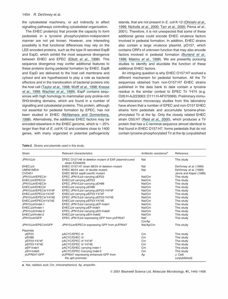
Table 2. Strains and plasmids used in this study.
Strain Relevant characteristics Antibiotic resistancea Reference
JPN15Dtir EPEC O127:H6 tir deletion mutant of EAF plasmid-curedstrain E2348/69,
Nal This study
EHECDtir EHEC O157:H7 strain 86/24 tir deletion mutant Nal DeVinney et al. (1999)UMD619Ætir EHEC 86/24 eae–tir double mutant Nal DeVinney et al. (1999)CVD451 EHEC 86/24 sepB (escN ) mutant Jarvis and Kaper (1996)JPN15Dtir/EPECtir EPEC JPN15Dtir carrying pEP23 Nal/Cm This studyEHECDtir/EPECtir EHECDtir carrying pEP23 Nal/Cm This studyJPN15Dtir/EHECtir EPEC JPN15Dtir carrying pEH86 Nal/Cm This studyEHECDtir/EHECtir EHECDtir carrying pEH86 Nal/Cm This studyJPN15Dtir/EPECtir Y474F EPEC JPN15Dtir carrying pEP23-Y474F Nal/Cm This studyEHECDtir/EPECtir Y474F EHECDtir carrying pEP23-Y474F Nal/Cm This studyJPN15Dtir/EPECtir Y474E EPEC JPN15Dtir carrying pEP23-Y474E Nal/Cm This studyEHECDtir/EPECtir Y474E EHECDtir carrying pEP23-Y474E Nal/Cm This studyJPN15Dtir/Indel-1 EPEC JPN15Dtir carrying pEP-Indel1 Nal/Cm This studyEHECDtir/Indel-1 EHECDtir carrying pEP-Indel1 Nal/Cm This studyJPN15Dtir/Indel-2 EPEC JPN15Dtir carrying pEH-Indel2 Nal/Cm This studyEHECDtir/Indel-2 EHECDtir carrying pEH-Indel2 Nal/Cm This studyJPN15Dtir/GFP EPEC JPN15Dtir expressing GFP from pUFR047 Nal/ This study
Cm/ApJPN15Dtir/EPECtir/GFP JPN15Dtir/EPECtir expressing GFP from pUFR047 Nal/Ap/Cm This study
PlasmidspEP23 pACYC/EPEC tir Cm This studypEH86 pACYC/EHEC tir Cm This studypEP23-Y474F pACYC/EPEC tir Y474F Cm This studypEP23-Y474E pACYC/EPEC tir Y474E Cm This studypEP-Indel1 pACYC/EHEC carrying Indel-1 Cm This studypEH-Indel2 pACYC/EPEC Carrying Indel-2 Cm This studypUFR047-GFP pUFR047 expressing enhanced GFP from
the aph promoterAp J. Celli
(unpublished)
a. Nal, nalidixic acid; Cm, chloramphenicol; Ap, ampicillin.
1454 R. DeVinney et al.
Q 2001 Blackwell Science Ltd, Molecular Microbiology, 41, 1445–1458

results). Therefore, it is tempting to speculate that, by
forming tyrosine phosphorylation-independent pedestals,
EHEC has evolved to become a more efficient pathogen
that is less dependent on host cell signalling pathways,
such as those involving tyrosine kinases, to form
pedestals.
We have shown that, in EPEC, Tir tyrosine phosphoryl-
ation and the region surrounding the phosphorylated
tyrosine 474 are essential features for pedestal formation.
The role played by EPEC Tir tyrosine phosphorylation is
not the result only of the introduction of a negative charge.
In contrast, in EHEC, pedestal formation occurs indepen-
dently of Tir tyrosine phosphorylation and requires the
delivery of additional EHEC factors into the host cell to
circumvent host tyrosine kinase signalling. These findings
represent a fundamental difference in the mechanisms of
pedestal formation by these two closely related
pathogens.
Experimental procedures
Bacterial strains and HeLa cell cultures
The strains used in this study are listed in Table 2. EHEC andEPEC strains used to infect HeLa cells were grown in Luria–
Bertani broth supplemented with appropriate antibiotics asstanding overnight cultures at 378C. HeLa cells (CCL2;
American Type Culture Collection) were cultured in minimalessential medium (MEM) supplemented with 10% fetal calf
serum (FCS), grown at 378C in 5% CO2.
Construction of plasmids expressing EPEC and EHEC tir
The coding regions of the EPEC and EHEC tir genes were
polymerase chain reaction (PCR) amplified using the primersTir0041 and Tir005–, which introduced Bgl II and Xho I
restriction sites respectively (Table 3). Tir0041 hybridized tothe ribosome binding site and 50 end of both tir genes,
whereas Tir005– hybridized to the 30 end sequences thatspan the stop codons. The amplified products were cloned
into the Bam HI–Sal I sites in pACYC184, leaving the tir genesunder the control of the tetracycline resistance gene (TetR)
promoter, thus generating pEP23 (EPEC) and pEH86(EHEC).
Site-directed mutagenesis of EPEC tir Tyr474
To construct both the Tyr-474Phe and the Tyr-474Glumutants of EPEC Tir, pEP23 was used as a template to
PCR amplify a fragment containing the 30 end of the tir genefrom codon 466A, followed by a vector region containing the
pACYC184 Eag I unique restriction site. Oligos DGK0011 orTIR0131 (Table 3), which span an endogenous Sap I
restriction site and introduce single point mutations in the474 codon to create the Y474F and Y474E mutants,
respectively, together with the reverse oligonucleotideRD131–, which is complementary to pACYC184, were
used. Both pEP23 and the mutagenized fragments weredigested with Sap I 1 Eag I. The pEP23 large Sap I–Eag I
fragment, lacking the unmutagenized insert fragment, wasgel purified and ligated with the corresponding digested
mutagenized fragments to create pEP23-Y474F andpEP23-Y474E. The tir mutant genes in these plasmids were
sequenced to confirm the modifications and electroporatedinto EPEC JPN15Dtir and EHECDtir. Expression and stability
of the mutant proteins was assessed by Western blot using ananti-Tir monoclonal antibody (see below).
Construction of EPEC and EHEC Tir chimeric proteins
EPEC Indel-1 was constructed by inverse PCR amplificationof pEP23 using the primers Tir0101 and Tir011–, which
introduce a silent mutation to create a unique Spe I site(Table 3). This resulted in the deletion of an EPEC tir fragment
Table 3. Oligonucleotides used in this study.
Oligonucleotide Sequence
Tir0041 50-GGA AGA TCT AGG AAA GGA GAR ATT TAT GCC TAT TGG-30
(EPEC and EHEC tir 50 end)Tir005– 50-CGG CCT CGA GGA TAT ATT TAG ACG AAA CG-30
(EPEC and EHEC tir 30end)DGK0011 50-GCT CAT CAG CCA GAA GAG CAT ATT TTT GAT GAG-30
(EPEC Y474F)RD131– 50-GCC CAG CCG CGT CGG CCG CCA TGC CGG CGA-30
(pACYC reverse primer for SDM)Tir0131 50-CTA TCG GCT CAT CAG CCA GAA GAG CAT ATT GAA GAT G-30
(EPEC Y474E)Tir0111 50-GTG CCG ACT AGT AAT TCT AAT ACG TCT GTT CAG AAT ATG GGG AAT ACA GAT TCT GTT GTA TAT AGC GTT
ATT CAG AAT TTT TCA G-30
(Indel-1)Tir010– 50-AGA ATT ACT AGT CGG CAC CTG TAG ACT ATT CCG AGC CCC CCC AAC-30
(Indel-1)RDI21 50-CCC CTG CAG ATC CTG GTT ATA GCA CCA TTC AAC AT-30
(Indel-2)RDI2– 50-GGG CTG CAG CGA CCT CAT CAT AAA TAT GCT CTT CTG GCT GAT GAG CCG ACG AAT CAT GCA GCG ATG T-30
(Indel-2)
Pedestal formation by EHEC 1455
Q 2001 Blackwell Science Ltd, Molecular Microbiology, 41, 1445–1458

that spans codons 465S to 482G and the insertion of anEHEC tir sequence from codons 469Q to 489V. The resulting
fragment was digested with Spe I and religated to generatepEP-Indel-1. Likewise, EHEC Indel-2 was constructed by
inverse PCR amplification of pEH86 using the primers RDI21
and RDI2–, which introduce a silent mutation to create a
unique Pst I site (Table 3). This resulted in the deletion ofEHEC tir codons 469Q to 489V and the insertion of EPEC tir
codons 465S to 482G. The resulting fragment was digestedwith Pst I and religated to generate pEH-Indel-2. pEP-Indel-1
and pEH-Indel-2 were transformed into EPEC JPN15Dtir andEHECDtir.
Cellular fractionation and immunoblotting
Cultured HeLa cells were infected with either the JPN15Dtir
strains for 4 h or the EHECDtir strains for 6 h, with the culturemedia replaced after 4 h, washed and solubilized with lysis
buffer containing 50 mM Tris (pH 7.4)21% Triton X-100supplemented with protease inhibitors (Complete EDTA-free;
Calbiochem) and phosphatase inhibitors (1 mM NaVO4,100 nM Microcystin LR). Triton X-100-soluble fractions
(membranes, cytosol and solubilized bacterial proteins)
were separated from the insoluble fraction containing mostof the bacteria and host cytoskeleton by centrifugation.
Samples for alkaline phosphatase treatment were prepared inlysis buffer without phosphatase inhibitors and treated with 2 U
of calf intestinal alkaline phosphatase (NEB) for 1 h at 378C.Samples were analysed by immunoblotting with anti-EPEC Tir
(mouse monoclonal antibodies 2A5, amino-terminus; 2E8,carboxy-terminus), rat anti-EHEC Tir (DeVinney et al., 1999)
and anti-phosphotyrosine (anti-PY, clone 4G10; UBI) asdescribed previously (Rosenshine et al., 1996).
Immunofluorescence microscopy
One millilitre of 2� 104 HeLa cells was added to each well of a
24-well plate containing a 12 mm glass coverslip and grownovernight. Monolayers were infected with 5ml of broth-grown
EHEC or JPN15 strains and incubated for either 4 h or 4 hfollowed by the addition of gentamicin (50mg ml21) for an
additional 3 h to promote pedestal elongation (Rosenshineet al., 1996). Cells were fixed in 2.5% paraformaldehyde
(PFA), permeabilized with 1% Triton-X100–PBS and blockedin 10% normal goat serum20.1% Triton X-100–PBS before
incubation with primary and secondary antisera as describedpreviously. Antisera were used at the following dilutions: rat
anti-EHEC Tir, 1:200; mouse anti-EPEC Tir mAb clone 2A5(Tir N-terminus) or 2E8 (Tir C-terminus), 1:100; mouse anti-
PY, 1:100; goat anti-mouse Alexa 488 (Molecular Probes)1:400; donkey anti-rat tetramethyl rhodamine isothiocyanate
(TRITC; Jackson Labs) 1:400; Alexa 488 or 568 phalloidin(Molecular Probes) 1:300; DAPI (Sigma) 2mg ml21. Samples
were examined using a Zeiss Axioskop microscope, andimages were acquired with an Empix DVC1300 digital camera
and analysed using NORTHERN ECLIPSE imaging software. Theefficiency of pedestal formation by the complemented and
cross-complemented strains was assessed by determiningthe percentage of cells showing tightly focused actin
pedestals beneath microcolonies of at least five adhering
bacteria. Replicate fields of at least 100 cells were counted for
each strain that formed pedestals. In all cases, pedestalformation was observed under all adherent bacterial micro-
colonies. Data is expressed as the mean^SD of threereplicate experiments, and the differences between groups
were determined by ANOVA.
Co-infection experiments
Enhanced GFP under the control of the aphA3 (kanamycin)promoter was excised from pAT113, ligated into the Sal I–
Pst I sites of the low-copy-number plasmid pUFR047 andtransformed into JPN15Dtir/EHECtir to create JPN15Dtir/-
EHECtir/GFP (Table 2). HeLa cells plated at 2� 104 ml21 on12 mm glass coverslips in 24-well plates were infected with
10ml of EPEC JPN15Dtir/EHECtir/GFP either alone or co-infected with the EHEC Tir/intimin double mutant, UMD619D
tir (3ml), the EPEC BFP, tir/intimin triple mutant UMD207Dtir(3ml) or the EHEC type III secretion mutant CVD451 (3ml) for
4 h. Cells were washed, fixed, permeabilized and blocked asdescribed above, before labelling with Alexa 568 phalloidin
and DAPI.
Acknowledgements
The authors thank Wanying Deng and Jean Celli for providing
the GFP construct used in these studies, and Derek Knoecheland Mitchell Uh for their assistance with the site-directed
mutagenesis. We also thank Samantha Gruenheid for critical
reading of the manuscript. This work was supported by a grantfrom the Canadian Institutes for Health Research (CIHR).
R.D. is supported by AHFMR and the Province of Alberta;J.L.P. is supported by DGAPA and CONACyT and is a
Howard Hughes International Scholar; A.G. is supported by aCIHR doctoral research award; and D.L.G. by a Natural
Sciences and Engineering Research Council Scholarship.B.B.F. is a CIHR Distinguished Scientist. and B.B.F. and
J.L.P. are Howard Hughes International Scholars.
References
Abe, A., Heczko, U., Hegele, R.G., and Finlay, B.B. (1998)Two enteropathogenic Escherichia coli type III secreted
proteins, EspA and EspB, are virulence factors. J Exp Med188: 1–10.
Beaudry, M., Zhu, C., Fairbrother, J.M., and Harel, J. (1996)Genotypic and phenotypic characterization of Escherichia
coli isolates from dogs manifesting attaching and effacinglesions. J Clin Microbiol 34: 144–148.
Burland, V., Shao, Y., Perna, N.T., Plunkett, G., Sofia, H.J.,and Blattner, F.R. (1998) The complete DNA sequence and
analysis of the large virulence plasmid of Escherichia coliO157:H7. Nucleic Acids Res 26: 4196–4204.
Cantarelli, V.V., Takahashi, A., Akeda, Y., Nagayama, K., andHonda, T. (2000) Interaction of enteropathogenic or
enterohemorrhagic Escherichia coli with HeLa cells resultsin translocation of cortactin to the bacterial adherence site.
Infect Immun 68: 382–386.Cantey, J.R., and Blake, R.K. (1977) Diarrhea due to
Escherichia coli in the rabbit: a novel mechanism. J Infect
Dis 135: 454–462.
1456 R. DeVinney et al.
Q 2001 Blackwell Science Ltd, Molecular Microbiology, 41, 1445–1458

Carlier, M.F., Nioche, P., Broutin-L’Hermite, I., Boujemaa, R.,
Le Clainche, C., Egile, C., et al. (2000) GRB2 links signaling
to actin assembly by enhancing interaction of neural
Wiskott–Aldrich syndrome protein (N-WASp) with actin-
related protein (ARP2/3) complex. J Biol Chem 275:
21946–21952.
Creighton, T. (1993) Proteins: Structure and Molecular
Properties. New York: WH Freeman.
Dean-Nystrom, E., Bosworth, B., Moon, H., and O’Brien, A.
(1998) Escherichia coli O157:H7 requires intimin for
enteropathogenicity in calves. Infect Immun 66:
4560–4563.
de Grado, M., Abe, A., Gauthier, A., Steele-Mortimer, O.,
DeVinney, R., and Finlay, B.B. (1999) Identification of the
intimin binding domain of Tir of enteropathogenic E. coli
(EPEC). Cell Microbiol 1: 7–18.
DeVinney, R., Stein, M., Reinscheid, D., Abe, A., Rusch-
kowski, S., and Finlay, B.B. (1999) Enterohemorrhagic
Escherichia coli O157:H7 produces Tir, which is translo-
cated to the host cell membrane but is not tyrosine
phosphorylated. Infect Immun 67: 2389–2398.
Donnenberg, M.S., Giron, J.A., Nataro, J.P., and Kaper, J.B.
(1992) A plasmid-encoded type IV fimbrial gene of
enteropathogenic Escherichia coli associated with localized
adherence. Mol Microbiol 6: 3427–3437.
Donnenberg, M.S., Tacket, C.O., James, S.P., Losonsky, G.,
Nataro, J.P., Wasserman, S.S., et al. (1993) Role of the
eaeA gene in experimental enteropathogenic Escherichia
coli infection. J Clin Invest 92: 1412–1417.
Ebel, F., Podzadel, T., Rohde, M., Kresse, A., Kramer, S.,
Deibel, C., et al. (1998) Initial binding of Shiga toxin-
producing Escherichia coli to host cells and subsequent
induction of actin rearrangements depend on filamentous
EspA-containing surface appendages. Mol Microbiol 30:
147–161.
Elliott, S.J., Wainwright, L.A., McDaniel, T.K., Jarvis, K.G.,
Deng, Y.K., Lai, L.C., et al. (1998) The complete sequence
of the locus of enterocyte effacement (LEE) from
enteropathogenic Escherichia coli E2348/69. Mol Microbiol
28: 1–4.
Freeman, N.L., Zurawski, D.V., Chowrashi, P., Ayoob, J.C.,
Huang, L., Mittal, B., et al. (2000) Interaction of the
enteropathogenic Escherichia coli protein, translocated
intimin receptor (Tir), with focal adhesion proteins. Cell Motil
Cytoskeleton 47: 307–318.
Frischknecht, F., Moreau, V., Rottger, S., Gonfloni, S.,
Reckmann, I., Superti-Furga, G., and Way, M. (1999) Actin-
based motility of vaccinia virus mimics receptor tyrosine
kinase signalling. Nature 401: 926–929.
Gauthier, A., de Grado, M., and Finlay, B.B. (2000)
Mechanical fractionation reveals structural requirements
for enteropathogenic Escherichia coli Tir insertion into host
membranes. Infect Immun 68: 4344–4348.
Goosney, D.L., DeVinney, R., Pfuetzner, R.A., Frey, E.A.,
Strynadka, N.C., and Finlay, B.B. (2000) Enteropathogenic
E. coli translocated intimin receptor, tir, interacts directly
with alpha-actinin. Curr Biol 10: 735–738.
Goosney, D.L., DeVinney, R., and Finlay, B.B. (2001)
Recruitment of cytoskeletal and signalling proteins to
enteropathogenic and enterohemorrhagic, E. coli ped-
estals. Infect Immun 69: 3315–3322.
Griffin, P.M., and Tauxe, R.V. (1991) The epidemiology of
infections caused by Escherichia coli O157:H7, other
enterohemorrhagic E. coli, and the associated hemolytic
uremic syndrome. Epidemiol Rev 13: 60–98.
Griffin, P., Ostroff, S., Tauxe, R., Greene, K., Wells, J., Lewis,
J., and Blake, P. (1988) Illnesses associated with
Escherichia coli O157:H7 infections. Ann Intern Medicine
109: 705–712.
Gruenheid, S., DeVinney, R., Bladt, F., Goosney, D., Gelkop,
S., Gish, G.D., et al. (2001) Enteropathogenic E. coli Tir
binds Nck to initiate actin pedestal formation in host cells.
Nature Cell Biol (in press).
Ismaili, A., Philpott, D.J., Dytoc, M.T., Soni, R., Ratnam,
S., and Sherman, P.M. (1995) Alpha-actinin accumu-
lation in epithelial cells infected with attaching and
effacing gastrointestinal pathogens. J Infect Dis 172:
1393–1396.
Ismaili, A., McWhirter, E., Handelsman, M., Brunton, J., and
Sherman, P. (1998) Divergent signal transduction
responses to infection with attaching and effacing Escheri-
chia coli. Infect Immun 66: 1688–1696.
Jarvis, K., and Kaper, J. (1996) Secretion of extracellular
proteins by enterohemorrhagic Escherichia coli via a
putative type III secretion system. Infect Immun 64:
4826–4829.
Kalman, D., Weiner, O.D., Goosney, D.L., Sedat, J.W., Finlay,
B.B., Abo, A., and Bishop, J.M. (1999) Enteropathogenic E.
coli acts through WASP and Arp2/3 complex to form actin
pedestals. Nature Cell Biol 1: 389–391.
Kenny, B. (1999) Phosphorylation of tyrosine 474 of the
enteropathogenic Escherichia coli (EPEC) Tir receptor
molecule is essential for actin nucleating activity and is
preceded by additional host modifications. Mol Microbiol 31:
1229–1241.
Kenny, B., DeVinney, R., Stein, M., Reinscheid, D.J., Frey,
E.A., and Finlay, B.B. (1997) Enteropathogenic E. coli
(EPEC) transfers its receptor for intimate adherence into
mammalian cells. Cell 91: 511–520.
Knutton, S., Baldwin, T., Williams, P.H., and McNeish, A.S.
(1989) Actin accumulation at sites of bacterial adhesion to
tissue culture cells: basis of a new diagnostic test for
enteropathogenic and enterohemorrhagic Escherichia coli.
Infect Immun 57: 1290–1298.
Kondro, W. (2000) E. coli outbreak deaths spark judicial
inquiry in Canada. Lancet 355: 2058.
Kresse, A.U., Rohde, M., and Guzman, C.A. (1999) The EspD
protein of enterohemorrhagic Escherichia coli is required for
the formation of bacterial surface appendages and is
incorporated in the cytoplasmic membranes of target cells.
Infect Immun 67: 4834–4842.
Luo, Y., Frey, E.A., Pfuetzner, R.A., Creagh, A.L., Knoechel,
D.G., Haynes, C.A., et al. (2000) Crystal structure of
enteropathogenic Escherichia coli intimin–receptor com-
plex. Nature 405: 1073–1077.
McDaniel, T., and Kaper, J. (1997) A cloned pathogenicity
island from enteropathogenic Escherichia coli confers the
attaching and effacing phenotype on E. coli K-12. Mol
Microbiol 23: 399–407.
McKee, M., Melton-Celsa, A., Moxley, R., Francis, D., and
O’Brien, A. (1995) Enterohemorrhagic Escherichia coli
O157:H7 requires intimin to colonize the gnotobiotic pig
Pedestal formation by EHEC 1457
Q 2001 Blackwell Science Ltd, Molecular Microbiology, 41, 1445–1458

intestine and to adhere to HEp-2 cells. Infect Immun 63:
3739–3744.
McNamara, B.P., and Donnenberg, M.S. (1998) A novel
proline-rich protein, EspF, is secreted from enteropatho-
genic Escherichia coli via the type III export pathway. FEMS
Microbiol Lett 166: 71–78.
Makino, K., Ishii, K., Yasunaga, T., Hattori, M., Yokoyama, K.,
Yutsudo, C.H., et al. (1998) Complete nucleotide
sequences of 93-kb and 3.3-kb plasmids of an enterohe-
morrhagic Escherichia coli O157:H7 derived from Sakai
outbreak. DNA Res 5: 1–9.
Marches, O., Nougayrede, J.P., Boullier, S., Mainil, J.,
Charlier, G., Raymond, I., et al. (2000) Role of tir and
intimin in the virulence of rabbit enteropathogenic Escheri-
chia coli serotype O103:H2. Infect Immun 68: 2171–2182.
Moon, H.W., Whipp, S.C., Argenzio, R.A., Levine, M.M., and
Giannella, R.A. (1983) Attaching and effacing activities of
rabbit and human enteropathogenic Escherichia coli in pig
and rabbit intestines. Infect Immun 41: 1340–1351.
Moreau, V., Frischknecht, F., Reckmann, I., Vincentelli, R.,
Rabut, G., Stewart, D., and Way, M. (2000) A complex of
N-WASP and WIP integrates signalling cascades that lead
to actin polymerization. Nature Cell Biol 2: 441–448.
Mullins, R.D. (2000) How WASP-family proteins and the
Arp2/3 complex convert intracellular signals into cyto-
skeletal structures. Curr Opin Cell Biol 12: 91–96.
Nataro, J., and Kaper, J. (1998) Diarrheagenic Escherichia
coli. Clin Microbiol Rev 11: 142–201.
Newman, J.V., Zabel, B.A., Jha, S.S., and Schauer, D.B.
(1999) Citrobacter rodentium espB is necessary for signal
transduction and for infection of laboratory mice. Infect
Immun 67: 6019–6025.
Nicholls, L., Grant, T.H., and Robins-Browne, R.M. (2000)
Identification of a novel genetic locus that is required for in
vitro adhesion of a clinical isolate of enterohaemorrhagic
Escherichia coli to epithelial cells. Mol Microbiol 35:
275–288.
Ohnishi, M., Tanaka, C., Kuhara, S., Ishii, K., Hattori, M.,
Kurokawa, K., et al. (1999) Chromosome of the entero-
hemorrhagic Escherichia coli O157: H7; comparative
analysis with K-12 MG1655 revealed the acquisition of a
large amount of foreign DNAs. DNA Res 6: 361–368.
Perna, N.T., Mayhew, G.F., Posfai, G., Elliott, S., Donnen-
berg, M.S., Kaper, J.B., and Blattner, F.R. (1998) Molecular
evolution of a pathogenicity island from enterohemorrhagic
Escherichia coli O157:H7. Infect Immun 66: 3810–3817.
Perna, N., Plunkett, G.I., Burland, V., Mau, B., Glasner, J.,
Rose, D., et al. (2001) Genome sequence of entero-
haemorrhagic Escherichia coli O157:H7. Nature 409:
529–533.
Phillips, A.D., Navabpour, S., Hicks, S., Dougan, G., Wallis,
T., and Frankel, G. (2000) Enterohaemorrhagic Escherichia
coli O157:H7 target Peyer’s patches in humans and cause
attaching/effacing lesions in both human and bovineintestine. Gut 47: 377–381.
Reid, S.D., Herbelin, C.J., Bumbaugh, A.C., Selander, R.K.,and Whittam, T.S. (2000) Parallel evolution of virulence in
pathogenic Escherichia coli. Nature 406: 64–67.Rivero-Lezcano, O.M., Marcilla, A., Sameshima, J.H., and
Robbins, K.C. (1995) Wiskott–Aldrich syndrome proteinphysically associates with Nck through Src homology 3
domains. Mol Cell Biol 15: 5725–5731.Rosenshine, I., Donnenberg, M.S., Kaper, J.B., and Finlay,
B.B. (1992) Signal transduction between enteropathogenicEscherichia coli (EPEC) and epithelial cells: EPEC induces
tyrosine phosphorylation of host cell proteins to initiatecytoskeletal rearrangement and bacterial uptake. EMBO J
11: 3551–3560.Rosenshine, I., Ruschkowski, S., Stein, M., Reinscheid, D.,
Mills, S., and Finlay, B. (1996) A pathogenic bacteriumtriggers epithelial signals to form a functional bacterial
receptor that mediates actin pseudopod formation. EMBO J
15: 2613–2624.Songyang, Z., Shoelson, S.E., Chaudhuri, M., Gish, G.,
Pawson, T., Haser, W.G., et al. (1993) SH2 domainsrecognize specific phosphopeptide sequences. Cell 72:
767–778.Suzuki, T., Miki, H., Takenawa, T., and Sasakawa, C. (1998)
Neural Wiscott–Aldrich syndrome protein is implicated inthe actin-based motility of Shigella flexneri. EMBO J 17:
2767–2776.Tacket, C.O., Sztein, M.B., Losonsky, G., Abe, A., Finlay,
B.B., McNamara, B.P., et al. (2000) Role of EspB inexperimental human enteropathogenic Escherichia coli
infection. Infect Immun 68: 3689–3695.Tarr, P.I., Bilge, S.S., Vary, J.C., Jr, Jelacic, S., Habeeb, R.L.,
Ward, T.R., et al. (2000) Iha: a novel Escherichia coliO157:H7 adherence-conferring molecule encoded on a
recently acquired chromosomal island of conservedstructure. Infect Immun 68: 1400–1407.
Taylor, K., O’Connell, C., Luther, P., and Donnenberg, M.(1998) The EspB protein of enteropathogenic Escherichia
coli is targeted to the cytoplasm of infected HeLa cells.Infect Immun 66: 5501–5507.
Tzipori, S., Gunzer, F., Donnenberg, M., de Montigny, L.,Kaper, J., and Donohue-Rolfe, A. (1995) The role of the
eaeA gene in diarrhea and neurological complications in agnotobiotic piglet model of enterohemorrhagic Escherichia
coli infection. Infect Immun 63: 3621–3627.Wachter, C., Beinke, C., Mattes, M., and Schmidt, M. (1999)
Insertion of EspD into epithelial target cell membranes byinfecting enteropathogenic Escherichia coli. Mol Microbiol
31: 1695–1707.Wolff, C., Nisan, I., Hanski, E., Frankel, G., and Rosenshine, I.
(1998) Protein translocation into host epithelial cells by
infecting enteropathogenic Escherichia coli. Mol Microbiol28: 143–155.
1458 R. DeVinney et al.
Q 2001 Blackwell Science Ltd, Molecular Microbiology, 41, 1445–1458