Pathophysiology of Abdominal Aortic Aneurysms: Insights from the Elastase-Induced Model in Mice with...
15
Pathophysiology of Abdominal Aortic Aneurysms Insights from the Elastase-Induced Model in Mice with Different Genetic Backgrounds ROBERT W. THOMPSON, JOHN A. CURCI, TERRI L. ENNIS, DONGLI MAO, MONICA B. PAGANO, AND CHRISTINE T.N. PHAM Departments of Surgery (Section of Vascular Surgery), Radiology, Cell Biology and Physiology, Medicine, and Pathology and Immunology, Washington University School of Medicine, St. Louis, Missouri, USA ABSTRACT: Abdominal aortic aneurysms (AAAs) represent a complex degenerative disorder involving chronic aortic wall inflammation and destructive remodeling of structural connective tissue. Studies using hu- man AAA tissues have helped identify a variety of molecular mediators and matrix-degrading proteinases, which contribute to aneurysm dis- ease, thereby providing a sound foundation for understanding AAAs; however, these human tissue specimens represent only the “end stage” of a long and progressive disease process. Further progress in understand- ing the pathophysiology of AAAs is therefore dependent in part on the development and application of effective animal models that recapitu- late key aspects of the disease. Based on original studies in rats, transient perfusion of the abdominal aorta with porcine pancreatic elastase has provided a reproducible and robust model of AAAs. More recent applica- tions of this model to mice have also opened new avenues for investigation. In this review, we summarize investigations using the elastase-induced mouse model of AAAs including results in animals with targeted deletion of specific genes and more general differences in mice on different genetic backgrounds. These studies have helped us identify genes that are essen- tial to the development of AAAs (such as MMP9, IL6, and AT1R) and to reveal other genes that may be dispensable in aneurysm formation. Inves- tigations on mice from different genetic backgrounds are also beginning to offer a novel approach to evaluate the genetic basis for susceptibility to aneurysm development. KEYWORDS: abdominal aortic aneurysm; animal models of disease; ge- netically altered mice; elastase; inflammation; extracellular matrix; pro- teinases; cytokines; genetic susceptibility Address for correspondence: Robert W. Thompson, M.D., Section of Vascular Surgery, Department of Surgery, Washington University School of Medicine, 5101 Queeny Tower, One Barnes-Jewish Hospital Plaza, St. Louis, Missouri, 63110. Voice: 314-362-7410; fax: 314-747-3548. e-mail: [email protected] Ann. N.Y. Acad. Sci. 1085: 59–73 (2006). C 2006 New York Academy of Sciences. doi: 10.1196/annals.1383.029 59
-
Upload
independent -
Category
Documents
-
view
1 -
download
0
Transcript of Pathophysiology of Abdominal Aortic Aneurysms: Insights from the Elastase-Induced Model in Mice with...

Pathophysiology of AbdominalAortic Aneurysms
Insights from the Elastase-Induced Model inMice with Different Genetic Backgrounds
ROBERT W. THOMPSON, JOHN A. CURCI, TERRI L. ENNIS,DONGLI MAO, MONICA B. PAGANO, AND CHRISTINE T.N. PHAM
Departments of Surgery (Section of Vascular Surgery), Radiology, Cell Biologyand Physiology, Medicine, and Pathology and Immunology, WashingtonUniversity School of Medicine, St. Louis, Missouri, USA
ABSTRACT: Abdominal aortic aneurysms (AAAs) represent a complexdegenerative disorder involving chronic aortic wall inflammation anddestructive remodeling of structural connective tissue. Studies using hu-man AAA tissues have helped identify a variety of molecular mediatorsand matrix-degrading proteinases, which contribute to aneurysm dis-ease, thereby providing a sound foundation for understanding AAAs;however, these human tissue specimens represent only the “end stage” ofa long and progressive disease process. Further progress in understand-ing the pathophysiology of AAAs is therefore dependent in part on thedevelopment and application of effective animal models that recapitu-late key aspects of the disease. Based on original studies in rats, transientperfusion of the abdominal aorta with porcine pancreatic elastase hasprovided a reproducible and robust model of AAAs. More recent applica-tions of this model to mice have also opened new avenues for investigation.In this review, we summarize investigations using the elastase-inducedmouse model of AAAs including results in animals with targeted deletionof specific genes and more general differences in mice on different geneticbackgrounds. These studies have helped us identify genes that are essen-tial to the development of AAAs (such as MMP9, IL6, and AT1R) and toreveal other genes that may be dispensable in aneurysm formation. Inves-tigations on mice from different genetic backgrounds are also beginningto offer a novel approach to evaluate the genetic basis for susceptibilityto aneurysm development.
KEYWORDS: abdominal aortic aneurysm; animal models of disease; ge-netically altered mice; elastase; inflammation; extracellular matrix; pro-teinases; cytokines; genetic susceptibility
Address for correspondence: Robert W. Thompson, M.D., Section of Vascular Surgery, Departmentof Surgery, Washington University School of Medicine, 5101 Queeny Tower, One Barnes-JewishHospital Plaza, St. Louis, Missouri, 63110. Voice: 314-362-7410; fax: 314-747-3548.
e-mail: [email protected]
Ann. N.Y. Acad. Sci. 1085: 59–73 (2006). C© 2006 New York Academy of Sciences.doi: 10.1196/annals.1383.029
59

60 ANNALS NEW YORK ACADEMY OF SCIENCES
INTRODUCTION
Abdominal aortic aneurysms (AAAs) are a common and life-threatening de-generative disease.1 AAAs occur in 5–9% of the population over the age of 65years and are most frequently associated with cigarette smoking, pulmonaryemphysema, coronary artery disease, and peripheral (occlusive) atherosclero-sis.2–6 AAAs arise over a period of years and most tend to gradually expandwith time, and the risk of aortic rupture increases steadily with progressiveaneurysm growth. Although AAAs can be readily detected in asymptomaticindividuals by abdominal imaging studies, there are currently no specific ther-apies known to alter the natural history of small asymptomatic AAAs. Betterunderstanding of the fundamental pathologic mechanisms that contribute toaneurysmal degeneration is therefore critical toward the development of newtherapeutic approaches.
Aneurysmal degeneration is a complex process involving destructive re-modeling of connective tissue throughout the affected segment of aortic wall.Four interrelated factors have been implicated in this pathological remodel-ing: (a) chronic inflammation within the outer aortic wall accompanied byneovascularization and elevated production of proinflammatory cytokines;(b) excessive local production and dysregulation of matrix-degrading pro-teinases; (c) progressive destruction of structural matrix proteins, particularlyelastin and collagen, which leads to weakening and dilatation of the aorticwall; and (d) depletion of medial smooth muscle cells (SMC), which mayresult in an impaired capacity for connective tissue repair.7 Structural andhemodynamic conditions unique to the human infrarenal aorta may explainthe propensity for AAAs to form in this region, but the recruitment of in-flammatory cells into the elastic media and adventitia appears to be an earlyand crucial step.8 The factors involved in this process likely include oxidizedlipoproteins,9,10 localized tissue hypoxia,11,12 reactive oxygen and nitrogenspecies,13,14 prostanoids and leukotrienes,15–18 and microbial infection19 aswell as elevated local expression of specific cytokines and chemokines asso-ciated with an immunoinflammatory response.20–25 Matrix degradation mayalso release chemoattractant molecules that serve to amplify and localize in-flammatory cells to the outer aortic wall. One example of this is the releaseof elastin-degradation peptides (EDPs) within aneurysm tissue, which canstimulate monocyte chemotaxis through interaction with a 67-kDa cell surfaceelastin-binding protein.26–28 Indeed, the generation of high local concentrationsof EDPs is thought to be one of the mechanisms underlying the developmentof experimental AAAs induced by transient aortic perfusion with pancreaticelastase.
Although studies of human aortic tissue procured during surgical AAArepair have provided valuable clues to pathogenesis, these specimens of “end-stage” disease give limited information on the dynamic changes involvedin progressive aneurysmal degeneration. The development of small animal
THOMPSON et al.: PATHOPHYSIOLOGY OF AAA 61
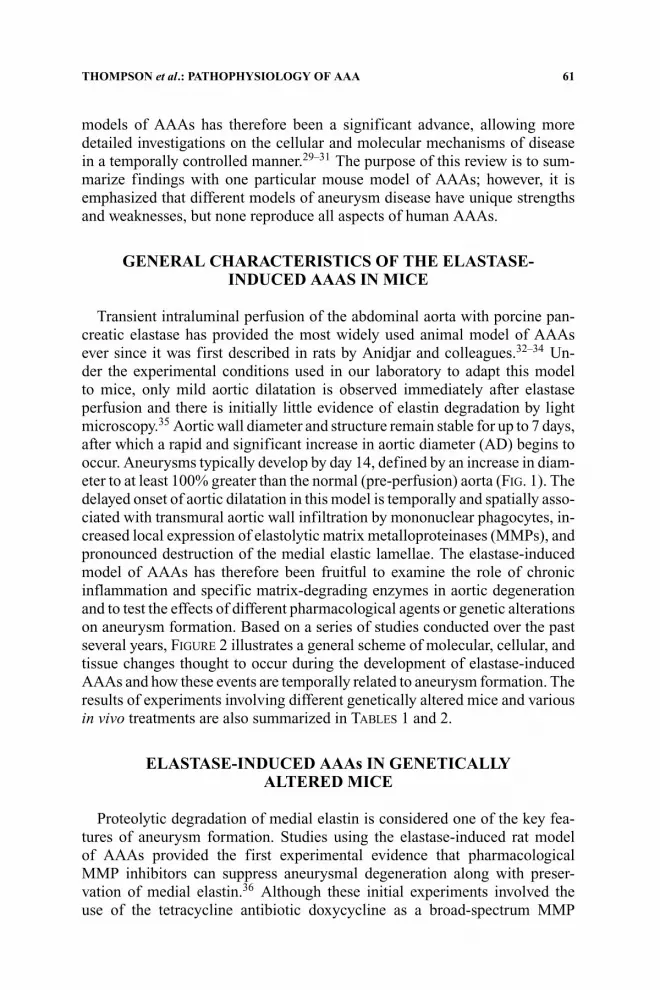
models of AAAs has therefore been a significant advance, allowing moredetailed investigations on the cellular and molecular mechanisms of diseasein a temporally controlled manner.29–31 The purpose of this review is to sum-marize findings with one particular mouse model of AAAs; however, it isemphasized that different models of aneurysm disease have unique strengthsand weaknesses, but none reproduce all aspects of human AAAs.
GENERAL CHARACTERISTICS OF THE ELASTASE-INDUCED AAAS IN MICE
Transient intraluminal perfusion of the abdominal aorta with porcine pan-creatic elastase has provided the most widely used animal model of AAAsever since it was first described in rats by Anidjar and colleagues.32–34 Un-der the experimental conditions used in our laboratory to adapt this modelto mice, only mild aortic dilatation is observed immediately after elastaseperfusion and there is initially little evidence of elastin degradation by lightmicroscopy.35 Aortic wall diameter and structure remain stable for up to 7 days,after which a rapid and significant increase in aortic diameter (AD) begins tooccur. Aneurysms typically develop by day 14, defined by an increase in diam-eter to at least 100% greater than the normal (pre-perfusion) aorta (FIG. 1). Thedelayed onset of aortic dilatation in this model is temporally and spatially asso-ciated with transmural aortic wall infiltration by mononuclear phagocytes, in-creased local expression of elastolytic matrix metalloproteinases (MMPs), andpronounced destruction of the medial elastic lamellae. The elastase-inducedmodel of AAAs has therefore been fruitful to examine the role of chronicinflammation and specific matrix-degrading enzymes in aortic degenerationand to test the effects of different pharmacological agents or genetic alterationson aneurysm formation. Based on a series of studies conducted over the pastseveral years, FIGURE 2 illustrates a general scheme of molecular, cellular, andtissue changes thought to occur during the development of elastase-inducedAAAs and how these events are temporally related to aneurysm formation. Theresults of experiments involving different genetically altered mice and variousin vivo treatments are also summarized in TABLES 1 and 2.
ELASTASE-INDUCED AAAs IN GENETICALLYALTERED MICE
Proteolytic degradation of medial elastin is considered one of the key fea-tures of aneurysm formation. Studies using the elastase-induced rat modelof AAAs provided the first experimental evidence that pharmacologicalMMP inhibitors can suppress aneurysmal degeneration along with preser-vation of medial elastin.36 Although these initial experiments involved theuse of the tetracycline antibiotic doxycycline as a broad-spectrum MMP

62 ANNALS NEW YORK ACADEMY OF SCIENCES
FIGURE 1. Elastase-induced mouse model of AAAs. (A) Technique used for tran-sient intraluminal perfusion of the abdominal aorta with porcine pancreatic elastase. Aftermeasurement of the pre-perfusion AD, a dilute solution of porcine pancreatic elastase isinstilled into the isolated segment of infrarenal abdominal aorta for 5 min at 100 mmHgpressure. The perfusion catheter is removed and flow is restored to the lower extremitiesand the immediate postperfusion AD is measured 5 min later. Final AD measurements areobtained during a repeat laparotomy at various intervals up to 14 days, immediately priorto euthanasia and tissue procurement. (B) Aortic dilatation at various intervals after aorticperfusion with either elastase or heat-inactivated elastase as a control. The initial dilatation(approximately 50–70%) is observed regardless of the perfusion solution with stabilizationof AD up to day 7. Rapid secondary dilatation occurs between day 7 and 14 in mice perfusedwith elastase, but not in controls. AAAs are defined as an overall extent of aortic dilatationgreater than 100% (dashed horizontal line). Data from Pyo et al.35

THOMPSON et al.: PATHOPHYSIOLOGY OF AAA 63
FIGURE 2. Overview of temporal events involved in elastase-induced AAAs. Elastaseperfusion injury initially induces local expression and release of numerous molecular me-diators of acute inflammation through its effects on resident aortic wall cells (EC, SMC,fibroblasts) and initial recruitment of inflammatory cells (PMN). Transition to a chronicinflammatory response involves recruitment of monocyte/macrophages and T cells alongwith increased local activity of several different types of matrix-degrading proteinases. Theeffects of these enzymes on structural aortic wall proteins (elastin and collagen) result inrapid secondary dilatation and aneurysm formation by day 14. Later events involving adap-tive immune responses and connective tissue repair may act to stabilize the aortic wall, butfailure of these processes may result in progressive dilatation.
inhibitor, similar results were obtained using nonantimicrobial tetracyclinederivatives and hydroxamate-based MMP antagonists.37–39
The aneurysm-suppressing effects of doxycycline were confirmed follow-ing development and initial characterization of the elastase-induced mousemodel of AAAs.35 The response to elastase perfusion was subsequentlyexamined in mice with targeted disruption of the MMP-9 or MMP-12genes to discern if either of these elastolytic proteinases is specificallyrequired for the development of experimental AAAs. These experimentsrevealed that the extent of aneurysmal dilatation and the incidence ofAAAs were both substantially reduced in MMP-9 (−/−) mice, but thataneurysm development was unaffected in mice with isolated deficiency ofMMP-12. The reduction in aneurysmal dilatation in MMP-9 (−/−) mice wasassociated with preservation of medial elastin and collagenous fibrosis inthe adventitia, but there was no detectable suppression of elastase-inducedinflammation. Additional studies in mice deficient in tissue inhibitor ofmetalloproteinases-1 (TIMP-1), the principal endogenous inhibitor of MMPs,revealed significant enhancement of elastase-induced AAAs.40,41 These ob-servations thereby demonstrate that expression of a single metalloproteinase(MMP-9) is required for the development of elastase-induced AAAs, helping

64 ANNALS NEW YORK ACADEMY OF SCIENCES
TABLE 1. Genetically altered mice tested in the elastase-induced mouse model of AAAs
Gene targeted AAA phenotype Author (reference)
MMP-9 Resistant Pyo35
MMP-12 Susceptible Pyo35
MMP-9/MMP-12 Resistant Pyo35
TIMP-1 Enhanced Lee, Eskandari40,41
MMP-9/TIMP-1 Enhanced Lee40
MMP-2 Susceptible Unpublished
MMP-8 Susceptible Eliason57
Neutrophil elastase Susceptible Eliason57
Urokinase-PA Susceptible Unpublished
Cat-S Resistant Bartoli48
Cat-C (DPP-I) Resistant Unpublished
NOS-2 (iNOS) Susceptible Lee50
Apo-E Susceptible Steinmetz51
IL-1R Resistant UnpublishedTNFR Resistant UnpublishedIL-6 Resistant Unpublished
IL-10 Enhanced Geraghty58
B cells Susceptible UnpublishedCD8 (T cells) Susceptible UnpublishedCD4 (T cells) Enhanced UnpublishedAT1 (Ang II receptor) Resistant UnpublishedAT2 (Ang II receptor) Susceptible UnpublishedALOX5 (5-Lipoxygenase) Susceptible UnpublishedFLAP Susceptible Unpublished
support the possibility that treatment with MMP inhibitors might have utilityin patients with small AAAs.42–46
The elastase-induced mouse model of AAAs has provided the opportunityto examine the role of several additional proteinases in aneurysmal degenera-tion. For example, studies in mice lacking expression of MMP-2, MMP-8, orurokinase-plasminogen activator (u-PA) revealed no suppression of elastase-induced AAAs. In contrast, recent experiments have revealed novel functionsfor cysteine proteases of the cathepsin family in AAAs such as cathepsin-S(Cat-S), a protease secreted by macrophages and SMC that has potent elastin-degrading activity. Gene expression profiling studies revealed that aortic wallexpression of Cat-S, Cat-K, and Cat-C is markedly increased early after elas-tase perfusion (day 3), whereas expression of the endogenous cathepsin an-tagonist, cystatin-C, is actually diminished.47 Treatment of mice with E-64,a broad-spectrum inhibitor of cysteine proteases, resulted in suppression ofAAAs with preservation of aortic elastin.48 In mice with targeted gene dele-tion of Cat-S, there was a significant reduction in the incidence of AAAsand a 55% reduction in the extent of aortic dilatation compared to wild-typecontrols. Cat-S (−/−) mice also exhibited a minimal postelastase aortic wall

THOMPSON et al.: PATHOPHYSIOLOGY OF AAA 65
TABLE 2. Treatments tested in the elastase-induced mouse model of AAAs
Agent (route) Effects on AAAs Author/reference
Doxycycline (oral) Suppression Pyo35
Doxycycline (local) Suppression Bartoli59
Cigarette smoking Enhanced Buckley60
Simvastatin (ip) Suppression Steinmetz51
Enalapril, lisinopril (oral) Suppression Borhani61
Losartan (oral) Suppression Borhani61
PDTC (ip) Suppression Parodi62
Curcumin (oral) Suppression Parodi63
E-64 (Cathepsin inhibitor) Suppression Bartoli48
Ang II (sc) No Effect Unpublished
Anti-PMN antibody (iv) Suppression Eliason57
Anti-IL-6 antibody (ip) Suppression UnpublishedMK886 (FLAP antagonist) No Effect Unpublished
inflammatory response and striking preservation of medial elastin, but gelatinzymography of aortic tissue extracts revealed no associated reduction inMMP-9 activity. These findings demonstrated for the first time that Cat-S playsa functionally important and necessary role in the development of aneurysmaldegeneration and that the contributions of Cat-S to AAAs are independent ofMMP-9. Additional studies have revealed a similarly crucial but distinct rolefor Cat-C, also known as dipeptidyl peptidase-I (DPP-I). DPP-I is a lysoso-mal enzyme required for activation of granule-associated proteases such asneutrophil elastase, Cat-G, and granzymes as well as mast cell chymases. Thefinding that mice deficient in DPP-I exhibit suppression of elastase-inducedAAAs indicates that DDP-I (or one of its “downstream” proteases) plays apivotal role in the transition from acute to chronic inflammation during thedevelopment of aneurysmal lesions.
The schematic diagram shown in FIGURE 2 illustrates that the early eventsin development of elastase-induced AAAs involve numerous molecular medi-ators including cellular signaling molecules, proinflammatory cytokines, andchemoattractants. It has therefore been of interest to examine mice with tar-geted deficiencies in genes involved in these processes to determine whichmediators might be most important in the unique vascular response to in-jury that results in aneurysm formation. For example, previous studies inrats with pharmacological inhibitors of inducible nitric oxide synthase (iNOS)led to the prediction that nitric oxide produced by this enzyme is critical inthe inflammatory response associated with aneurysm formation,49 yet micelacking expression of iNOS were found to exhibit no resistance to aneurysmformation despite evidence of increased nitric oxide production.50 In otherexperiments it was found that mice with genetically determined hypercholes-terolemia (due to deficiency in apolipoprotein-E) are also fully susceptible to

66 ANNALS NEW YORK ACADEMY OF SCIENCES
TABLE 3. AD measurements in different inbred strains of laboratory mice
Strain n AD pre, mm AD post, mm AD final, mm
129/SvEv 41 0.54 ± 0.01 0.85 ± 0.02 0.98 ± 0.02129/SvJ 6 0.59 ± 0.03 0.92 ± 0.03 1.16 ± 0.08CBA/CaJ 8 0.54 ± 0.01 0.88 ± 0.02 1.10 ± 0.08Balb/cJ 4 0.53 ± 0.00 0.88 ± 0.01 1.08 ± 0.19C3H/HeJ 7 0.56 ± 0.03 0.90 ± 0.03 1.22 ± 0.16C57BL/10SnJ 6 0.52 ± 0.01 0.88 ± 0.02 1.18 ± 0.11FVB/N 5 0.53 ± 0.00 0.88 ± 0.02 1.39 ± 0.08†C57BL/6J 66 0.53 ± 0.00 0.91 ± 0.01 1.38 ± 0.03†Analysis of variance NS NS P ≤ 0.
(ANOVA)∗
Adult male mice from a series of eight different inbred strains underwent transient aortic perfusionwith elastase to induce aneurysmal degeneration. AD measurements were obtained before (AD pre-)and immediately after (AD post-) elastase perfusion with final measurements obtained at 14 days (ADfinal). Data shown represent the mean ± SEM. ∗One-way ANOVA with Student-Newman-Keuls posthoc multiple comparisons test (†P < 0.001 vs. 129/SvEv).
elastase-induced AAAs, but that aneurysm development is suppressed by treat-ment with statins in both normal and Apo-E (−/−) mice.51 This finding likelyreflects the pleiotropic effects of statins acting to interfere with key inflam-matory signaling pathways as a mechanism of suppressing aneurysmal degen-eration. Recent clinical studies have also highlighted the potential benefits oftreatment with statins in patients with AAAs.52,53
Given the acute inflammatory response that follows elastase-induced aorticinjury, it is not surprising that there is a significant increase in mouse aor-tic wall production of proinflammatory cytokines early in the developmentof elastase-induced AAAs such as IL-1�, TNF-�, and IL-6.47 The roles ofIL-1� and TNF-� in this response have been examined in rats, where treat-ment with cytokine-specific inhibitors suppresses elastase-induced AAAs.54
Preliminary work in our laboratory has also led to the observation that elastase-induced AAAs are suppressed in mice lacking expression of the cell-surfacereceptors for IL-1� and TNF-� along with limited development of the aor-tic wall inflammatory response. In other studies we have examined a poten-tial functional role for IL-6 in AAAs. This is of particular interest becauseIL-6 is prominently expressed in explant cultures of human AAA tissue andis significantly elevated in the circulating plasma of patients with AAAs, sug-gesting that circulating IL-6 levels reflect inflammatory processes involved inthe early stages of aneurysm formation.16,25,55,56 We have observed that elas-tase perfusion leads to early increases in IL-6 production within mouse aortictissue (days 3 to 7) along with elevated circulating IL-6 concentrations. In micewith targeted deletion of IL-6, however, there is a significant suppression ofelastase-induced AAAs along with a diminished inflammatory response andpreservation of aortic elastin. Similar findings were observed in wild-type mice

THOMPSON et al.: PATHOPHYSIOLOGY OF AAA 67
FIGURE 3. Extent of aortic dilatation in different inbred strains of laboratory mice.Adult male mice from a series of eight different inbred strains underwent transient aorticperfusion with elastase to induce aneurysmal degeneration and AD measurements were ob-tained (see TABLE 3). The overall extent of aortic dilatation was calculated as the percentageincrease between pre-perfusion AD and final AD on day 14 with the data shown represent-ing the mean ± SEM. Each strain was defined as resistant, intermediate, or susceptible tothe development of elastase-induced AAAs.
treated with an anti-IL-6-neutralizing antibody. Taken together, these findingsdemonstrate that elevated production of IL-6 within the elastase-injured aortictissue may play a crucial role in macrophage recruitment and activation, se-cretion of matrix-degrading proteinases, apoptosis of medial SMC, and otheraspects of aneurysmal degeneration. Application of the elastase-induced modelof AAAs to genetically altered mice has therefore been especially valuable inestablishing the temporal and cellular expression of proinflammatory cytokinesas well as the hierarchy of cytokine-driven aortic wall inflammatory responsesin aneurysm development.
INHERITED VARIATION IN SUSCEPTIBILITY TOELASTASE-INDUCED AAAs
During investigations on elastase-induced AAAs in genetically altered mice,it has become increasingly evident that the extent of aneurysmal dilatationvaries considerably between the different inbred laboratory strains used asbackground controls. For example, the mean extent of aortic dilatation inC57BL/6J background controls used for experiments on IL-6-deficient micewas approximately 150% as compared to only 75% for the 129/SvEv back-ground controls used in experiments on TIMP-1-deficient mice. This observa-tion is important from a practical point of view, emphasizing that it is critical touse control animals on the same genetic background when conducting exper-iments with gene-targeted animals. Furthermore, these observations suggestthat the susceptibility to form AAAs following elastase perfusion is associated,at least in part, with genetically determined inherited traits.

68 ANNALS NEW YORK ACADEMY OF SCIENCES
FIGURE 4. Susceptibility to elastase-induced AAAs in 129/SvEv and C57Bl/6J mice.A series of mice underwent transient aortic perfusion with elastase to induce aneurysmaldegeneration and the overall extent of aortic dilatation was calculated as the percentageincrease on day 14 (see FIG. 3). (A and B) Population distributions for the overall extent ofaortic dilatation in 47 male 129/SvEv mice (mean ± SEM, 104.9 ± 3.8%; median, 100.0%)and 54 male C57Bl/6J mice (mean ± SEM, 158.8 ± 4.4%; median, 155.6%), demonstrat-ing a significant difference in susceptibility to AAAs (P < 0.001). (C) Comparison ofdistribution curves for 129/SvEv and C57Bl/6J mice indicating that animals exhibiting lessthan 85% dilatation can be considered highly resistant and those with more than 185%dilatation can be considered highly susceptible. (D) Population distribution for the overallextent of aortic dilatation in 59 male F1 mice derived from an outcross between 129/SvEvand C57Bl/6J mice demonstrating a degree of susceptibility to AAAs intermediate to thatobserved in the two parental strains (mean ± SEM, 153.1 ± 4.2%; median, 150.0%).
THOMPSON et al.: PATHOPHYSIOLOGY OF AAA 69
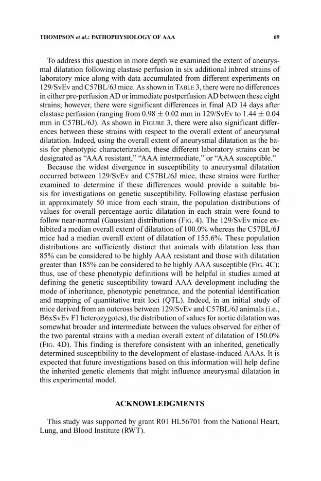
To address this question in more depth we examined the extent of aneurys-mal dilatation following elastase perfusion in six additional inbred strains oflaboratory mice along with data accumulated from different experiments on129/SvEv and C57BL/6J mice. As shown in TABLE 3, there were no differencesin either pre-perfusion AD or immediate postperfusion AD between these eightstrains; however, there were significant differences in final AD 14 days afterelastase perfusion (ranging from 0.98 ± 0.02 mm in 129/SvEv to 1.44 ± 0.04mm in C57BL/6J). As shown in FIGURE 3, there were also significant differ-ences between these strains with respect to the overall extent of aneurysmaldilatation. Indeed, using the overall extent of aneurysmal dilatation as the ba-sis for phenotypic characterization, these different laboratory strains can bedesignated as “AAA resistant,” “AAA intermediate,” or “AAA susceptible.”
Because the widest divergence in susceptibility to aneurysmal dilatationoccurred between 129/SvEv and C57BL/6J mice, these strains were furtherexamined to determine if these differences would provide a suitable ba-sis for investigations on genetic susceptibility. Following elastase perfusionin approximately 50 mice from each strain, the population distributions ofvalues for overall percentage aortic dilatation in each strain were found tofollow near-normal (Gaussian) distributions (FIG. 4). The 129/SvEv mice ex-hibited a median overall extent of dilatation of 100.0% whereas the C57BL/6Jmice had a median overall extent of dilatation of 155.6%. These populationdistributions are sufficiently distinct that animals with dilatation less than85% can be considered to be highly AAA resistant and those with dilatationgreater than 185% can be considered to be highly AAA susceptible (FIG. 4C);thus, use of these phenotypic definitions will be helpful in studies aimed atdefining the genetic susceptibility toward AAA development including themode of inheritance, phenotypic penetrance, and the potential identificationand mapping of quantitative trait loci (QTL). Indeed, in an initial study ofmice derived from an outcross between 129/SvEv and C57BL/6J animals (i.e.,B6xSvEv F1 heterozygotes), the distribution of values for aortic dilatation wassomewhat broader and intermediate between the values observed for either ofthe two parental strains with a median overall extent of dilatation of 150.0%(FIG. 4D). This finding is therefore consistent with an inherited, geneticallydetermined susceptibility to the development of elastase-induced AAAs. It isexpected that future investigations based on this information will help definethe inherited genetic elements that might influence aneurysmal dilatation inthis experimental model.
ACKNOWLEDGMENTS
This study was supported by grant R01 HL56701 from the National Heart,Lung, and Blood Institute (RWT).

70 ANNALS NEW YORK ACADEMY OF SCIENCES
REFERENCES
1. THOMPSON, R.W., P.J. GERAGHTY & J.K. LEE. 2002. Abdominal aortic aneurysms:basic mechanisms and clinical implications. Curr. Prob. Surg. 39: 110–230.
2. ALCORN, H.G., S.K. WOLFSON, JR., K. SUTTON-TYRRELL, et al. 1996. Risk factorsfor abdominal aortic aneurysms in older adults enrolled in The CardiovascularHealth Study. Arterioscler. Thromb. Vasc. Biol. 16: 963–970.
3. LEDERLE, F.A., G.R. JOHNSON, S.E. WILSON, et al. 1997. Prevalence and asso-ciations of abdominal aortic aneurysm detected through screening. AneurysmDetection and Management (ADAM) Veterans Affairs Cooperative Study Group.Ann. Intern. Med. 126: 441–449.
4. BLANCHARD, J.F. 1999. Epidemiology of abdominal aortic aneurysms. Epidemiol.Rev. 21: 207–221.
5. BLANCHARD, J.F., H.K. ARMENIAN & P.P. FRIESEN. 2000. Risk factors for abdominalaortic aneurysm: results of a case-control study. Am. J. Epidemiol. 151: 575–583.
6. LEDERLE, F.A., D.B. NELSON & A.M. JOSEPH. 2003. Smokers’ relative risk for aorticaneurysm compared with other smoking-related diseases: a systematic review.J. Vasc. Surg. 38: 329–334.
7. THOMPSON, R.W. 1996. Basic science of abdominal aortic aneurysms: emergingtherapeutic strategies for an unresolved clinical problem. Curr. Opin. Cardiol.11: 504–518.
8. KOCH, A.E., G.K. HAINES, R.J. RIZZO, et al. 1990. Human abdominal aorticaneurysms. Immunophenotypic analysis suggesting an immune-mediated re-sponse. Am. J. Pathol. 137: 1199–1213.
9. PAPAGRIGORAKIS, E., D. ILIOPOULOS, P.J. ASIMACOPOULOS, et al. 1997. Lipopro-tein(a) in plasma, arterial wall, and thrombus from patients with aortic aneurysm.Clin. Genet. 52: 262–271.
10. SCHILLINGER, M., H. DOMANOVITS, M. IGNATESCU, et al. 2002. Lipoprotein (a) inpatients with aortic aneurysmal disease. J. Vasc. Surg. 36: 25–30.
11. VORP, D.A., P.C. LEE, D.H. WANG, et al. 2001. Association of intraluminal thrombusin abdominal aortic aneurysm with local hypoxia and wall weakening. J. Vasc.Surg. 34: 291–299.
12. SCHILLINGER, M., M. EXNER, W. MLEKUSCH, et al. 2002. Heme oxygenase-1 genepromoter polymorphism is associated with abdominal aortic aneurysm. Thromb.Res. 106: 131–136.
13. MILLER, F.J., JR., W.J. SHARP, X. FANG, et al. 2002. Oxidative stress in humanabdominal aortic aneurysms: a potential mediator of aneurysmal remodeling.Arterioscler. Thromb. Vasc. Biol. 22: 560–565.
14. DALMAN, R.L. 2003. Oxidative stress and abdominal aneurysms: how aortic hemo-dynamic conditions may influence AAA disease. Cardiovasc. Surg. 11: 417–419.
15. HOLMES, D.R., W. WESTER, R.W. THOMPSON, et al. 1997. Prostaglandin E2 synthesisand cyclooxygenase expression in abdominal aortic aneurysms. J. Vasc. Surg.25: 810–815.
16. WALTON, L.J., I.J. FRANKLIN, T. BAYSTON, et al. 1999. Inhibition of prostaglandinE2 synthesis in abdominal aortic aneurysms: implications for smooth musclecell viability, inflammatory processes, and the expansion of abdominal aorticaneurysms. Circulation. 100: 48–54.
17. BAYSTON, T., S. RAMESSUR, J. REISE, et al. 2003. Prostaglandin E2 receptors inabdominal aortic aneurysm and human aortic smooth muscle cells. J. Vasc. Surg.38: 354–359.

THOMPSON et al.: PATHOPHYSIOLOGY OF AAA 71
18. ZHAO, L., M.P. MOOS, R. GRABNER, et al. 2004. The 5-lipoxygenase pathwaypromotes pathogenesis of hyperlipidemia-dependent aortic aneurysm. Nat. Med.10: 966–973.
19. LINDHOLT, J.S., H.A. ASHTON & R.A. SCOTT. 2001. Indicators of infection withChlamydia pneumoniae are associated with expansion of abdominal aorticaneurysms. J. Vasc. Surg. 34: 212–215.
20. PEARCE, W.H., I. SWEIS, J.S. YAO, et al. 1992. Interleukin-1 beta and tumor necrosisfactor-alpha release in normal and diseased human infrarenal aortas. J. Vasc. Surg.16: 784–789.
21. KOCH, A., S. KUNKEL, W. PEARCE, et al. 1993. Enhanced production of the chemo-tactic cytokines interleukin-8 and monocyte chemoattractant protein-1 in humanabdominal aortic aneurysms. Am. J. Pathol. 142: 1423–1431.
22. SZEKANECZ, Z., M.R. SHAH, W.H. PEARCE, et al. 1994. Human atheroscleroticabdominal aortic aneurysms produce interleukin (IL)-6 and interferon-gammabut not IL-2 and IL-4: the possible role for IL-6 and interferon-gamma in vascularinflammation. Agents Actions. 42: 159–162.
23. NEWMAN, K.M., J. JEAN-Claude, H. LI, et al. 1994. Cytokines that activate proteol-ysis are increased in abdominal aortic aneurysms. Circulation 90: II224–II227.
24. MCMILLAN, W.D. & W.H. PEARCE. 1997. Inflammation and cytokine signaling inaneurysms. Ann. Vasc. Surg. 11: 540–545.
25. JONES, K.G., D.J. BRULL, L.C. BROWN, et al. 2001. Interleukin-6 (IL-6) and theprognosis of abdominal aortic aneurysms. Circulation 103: 2260–2265.
26. SENIOR, R.M., G.L. GRIFFIN & R.P. MECHAM. 1980. Chemotactic activity of elastin-derived peptides. J. Clin. Invest. 66: 859–862.
27. HINEK, A. 1994. Nature and the multiple functions of the 67-kD elastin-/lamininbinding protein. Cell. Adhes. Commun. 2: 185–193.
28. HANCE, K.A., M. TATARIA, S.J. ZIPORIN, et al. 2002. Monocyte chemotactic activityin human abdominal aortic aneurysms: role of elastin degradation peptides andthe 67-kD cell surface elastin receptor. J. Vasc. Surg. 35: 254–261.
29. DOBRIN, P.B. 1999. Animal models of aneurysms. Ann. Vasc. Surg. 13: 641–648.30. CARRELL, T.W., A. SMITH & K.G. BURNAND. 1999. Experimental techniques and
models in the study of the development and treatment of abdominal aorticaneurysm. Br. J. Surg. 86: 305–312.
31. DAUGHERTY, A. & L.A. CASSIS. 2004. Mouse models of abdominal aorticaneurysms. Arterioscler. Thromb. Vasc. Biol. 24: 429–434.
32. ANIDJAR, S., J.L. SALZMANN, D. GENTRIC, et al. 1990. Elastase-induced experi-mental aneurysms in rats. Circulation 82: 973–981.
33. ANIDJAR, S., P.B. DOBRIN, M. EICHORST, et al. 1992. Correlation of inflammatoryinfiltrate with the enlargement of experimental aortic aneurysm. J. Vasc. Surg.16: 139–147.
34. ANIDJAR, S., P.B. DOBRIN, G. CHEJFEC, et al. 1994. Experimental study of deter-minants of aneurysmal expansion of the abdominal aorta. Ann. Vasc. Surg. 8:127–136.
35. PYO, R., J.K. LEE, J.M. SHIPLEY, et al. 2000. Targeted gene disruption of ma-trix metalloproteinase-9 (gelatinase B) suppresses development of experimentalabdominal aortic aneurysms. J. Clin. Invest. 105: 1641–1649.
36. PETRINEC, D., S. LIAO, D.R. HOLMES, et al. 1996. Doxycycline inhibition ofaneurysmal degeneration in an elastase-induced rat model of abdominal aorticaneurysm: preservation of aortic elastin associated with suppressed productionof 92 kD gelatinase. J. Vasc. Surg. 23: 336–346.

72 ANNALS NEW YORK ACADEMY OF SCIENCES
37. CURCI, J.A., D. PETRINEC, S.X. LIAO, et al. 1998. Pharmacologic suppression ofexperimental abdominal aortic aneurysms: a comparison of doxycycline and fourchemically modified tetracyclines. J. Vasc. Surg. 28: 1082–1093.
38. MOORE, G., S. LIAO, J.A. CURCI, et al. 1999. Suppression of experimental abdom-inal aortic aneurysms by systemic treatment with a hydroxamate-based matrixmetalloproteinase inhibitor (RS 132908). J. Vasc. Surg. 29: 522–532.
39. BIGATEL, D.A., J.R. ELMORE, D.J. CAREY, et al. 1999. The matrix metalloproteinaseinhibitor BB-94 limits expansion of experimental abdominal aortic aneurysms.J. Vasc. Surg. 29: 130–138.
40. LEE, J.K., C. BUCKLEY, M. BORHANI, et al. 2003. Endogenous expression of TIMP-1regulates development of experimental abdominal aortic aneurysms independentof interactions with gelatinase B (abstract). Circulation (Suppl IV) 108: 166.
41. ESKANDARI, M.K., J.D. VIJUNGCO, A. FLORES, et al. 2005. Enhanced abdominalaortic aneurysm in TIMP-1-deficient mice. J. Surg. Res. 123: 289–293.
42. THOMPSON, R.W., S. LIAO & J.A. CURCI. 1998. Therapeutic potential of tetracyclinederivatives to suppress the growth of abdominal aortic aneurysms. Adv. Dent.Res. 12: 159–165.
43. THOMPSON, R.W. & B.T. BAXTER. 1999. MMP inhibition in abdominal aorticaneurysms: rationale for a prospective randomized clinical trial. Ann. N.Y. Acad.Sci. 878: 159–178.
44. CURCI, J.A., D. MAO, D.G. BOHNER, et al. 2000. Preoperative treatment withdoxycycline reduces aortic wall expression and activation of matrix metallo-proteinases in patients with abdominal aortic aneurysms. J. Vasc. Surg. 31: 325–342.
45. PRALL, A.K., G.M. LONGO, W.G. MAYHAN, et al. 2002. Doxycycline in patientswith abdominal aortic aneurysms and in mice: comparison of serum levels andeffect on aneurysm growth in mice. J. Vasc. Surg. 35: 923–929.
46. BAXTER, B.T., W.H. PEARCE, E.A. WALTKE, et al. 2002. Prolonged administrationof doxycycline in patients with small asymptomatic abdominal aortic aneurysms:report of a prospective (Phase II) multicenter study. J. Vasc. Surg. 36: 1–12.
47. VANVICKLE-CHAVEZ, S. J., W.S. TUNG, T.S. ABSI, et al. 2006. Temporal changes inmouse aortic wall gene expression during the development of elastase-inducedabdominal aortic aneurysms. J. Vasc. Surg. 43(5): 1010–1020.
48. BARTOLI, M.A., G.-P. SHI, T.L. ENNIS, et al. 2004. Role of cathepsin S in de-velopment of experimental abdominal aortic aneurysms (abstract). Arterioscler.Thromb. Vasc. Biol. 24: e64.
49. JOHANNING, J.M., D.P. FRANKLIN, D.C. HAN, et al. 2001. Inhibition of induciblenitric oxide synthase limits nitric oxide production and experimental aneurysmexpansion. J. Vasc. Surg. 33: 579–586.
50. LEE, J.K., M. BORHANI, T.L. ENNIS, et al. 2001. Experimental abdominal aorticaneurysms in mice lacking expression of inducible nitric oxide synthase. Arte-rioscler. Thromb. Vasc. Biol. 21: 1393–1401.
51. STEINMETZ, E.F., C. BUCKLEY, M.L. SHAMES, et al. 2005. Treatment with simvas-tatin suppresses the development of experimental abdominal aortic aneurysmsin normal and hypercholesterolemic mice. Ann. Surg. 241: 92–101.
52. NAGASHIMA, H., Y. AOKA, Y. SAKOMURA, et al. 2002. A 3-hydroxy-3-methylglutaryl coenzyme A reductase inhibitor, cerivastatin, suppressesproduction of matrix metalloproteinase-9 in human abdominal aortic aneurysmwall. J. Vasc. Surg. 36: 158–163.

THOMPSON et al.: PATHOPHYSIOLOGY OF AAA 73
53. SUKHIJA, R., W.S. ARONOW, R. SANDHU, et al. 2006. Mortality and size of abdomi-nal aortic aneurysm at long-term follow-up of patients not treated surgically andtreated with and without statins. Am. J. Cardiol. 97: 279–280.
54. HINGORANI, A., E. ASCHER, M. SCHEINMAN, et al. 1998. The effect of tumor necrosisfactor binding protein and interleukin-1 receptor antagonist on the developmentof abdominal aortic aneurysms in a rat model. J. Vasc. Surg. 28: 522–526.
55. JUVONEN, J., H.M. SURCEL, J. SATTA, et al. 1997. Elevated circulating levels of in-flammatory cytokines in patients with abdominal aortic aneurysm. Arterioscler.Thromb. Vasc. Biol. 17: 2843–2847.
56. ROHDE, L.E., L.H. ARROYO, N. RIFAI, et al. 1999. Plasma concentrations ofinterleukin-6 and abdominal aortic diameter among subjects without aortic di-latation. Arterioscler. Thromb. Vasc. Biol. 19: 1695–1699.
57. ELIASON, J.L., K.K. HANNAWA, G. AILAWADI, et al. 2005. Neutrophil depletioninhibits experimental abdominal aortic aneurysm formation. Circulation 112:232–240.
58. GERAGHTY, P.J., B.C. STARCHER, M. BORHANI, et al. 2001. Interleukin-10 defi-ciency potentiates aortic collagen and elastin degradation in elastase-inducedmurine aortic aneurysm formation (abstract). Arterioscler. Thromb. Vasc. Biol.21: 676.
59. BARTOLI, M.A., F. PARODI, J. CHU, et al. 2006. Localized administration of doxy-cycline suppresses development of experimental abdominal aortic aneurysms.Ann. Vasc. Surg. 20(2): 228–236.
60. BUCKLEY, C., C.W. WYBLE, M. BORHANI, et al. 2004. Accelerated enlargement ofexperimental abdominal aortic aneurysms in a mouse model of chronic cigarettesmoke exposure. J. Am. Coll. Surg. 199: 896–903.
61. BORHANI, M., J.K. LEE, T.L. ENNIS, et al. 2000. Enalapril suppresses experimentalaortic aneurysm formation in mice: evidence for a novel molecular mechanismdistinct from angiotensin converting enzyme inhibition. Surg. Forum. 51: 397–399.
62. PARODI, F.E., D. MAO, T.L. ENNIS, et al. 2005. Suppression of experimental abdom-inal aortic aneurysms in mice by treatment with pyrrolidine dithiocarbamate, anantioxidant inhibitor of nuclear factor-kappaB. J. Vasc. Surg. 41: 479–489.
63. PARODI, F.E., D. MAO, T.L. ENNIS, et al. 2005. Oral administration of diferu-loylmethane (curcumin) suppresses proinflammatory cytokines and destructiveconnective tissue remodeling in experimental abdominal aortic aneurysms. Ann.Vasc. Surg. 41(3): 479–489.