A Syndrome with Congenital Neutropenia and Mutations in G6PC3
16
A novel syndrome with congenital neutropenia caused by mutations in G6PC3 Kaan Boztug, M.D. 1 , Giridharan Appaswamy, M.Sc. *,1 , Angel Ashikov, Ph.D. *,2 , Alejandro A. Schäffer, Ph.D. 4 , Ulrich Salzer, M.D. 5 , Jana Diestelhorst, B.Sc. 1 , Manuela Germeshausen, Ph.D. 1 , Gudrun Brandes, M.D. 3 , Jacqueline Lee-Gossler, M.Sc. 1 , Fatih Noyan, Ph.D. 1 , Anna- Katherina Gatzke, M.Sc. 1 , Milen Minkov, M.D., Ph.D. 6 , Johann Greil, M.D. 7 , Christian Kratz, M.D. 8 , Theoni Petropoulou, M.D. 9 , Isabelle Pellier, M.D. 10 , Christine Bellanné-Chantelot, Pharm.D., Ph.D. 11 , Nima Rezaei, M.D. 12 , Kirsten Mönkemöller, M.D. 13 , Noha Irani-Hakimeh, M.D. 14 , Hans Bakker, Ph.D. 2 , Rita Gerardy-Schahn, Ph.D. 2 , Cornelia Zeidler, M.D. 1 , Bodo Grimbacher, M.D. 15 , Karl Welte, M.D. 1 , and Christoph Klein, M.D., Ph.D. 1 1 Departments of Pediatric Hematology/Oncology, Hannover Medical School, Germany 2 Departments of Cellular Chemistry, Hannover Medical School, Germany 3 Departments of Cell Biology, Hannover Medical School, Germany 4 National Center for Biotechnology Information, NIH, DHHS, Bethesda, MD, USA 5 Department of Rheumatology and Clinical Immunology, University Medical Center Freiburg, Freiburg, Germany 6 St. Anna Children’s Hospital, Vienna, Austria 7 Department of Pediatric Oncology, Hematology and Immunology, Children’s Hospital, University of Heidelberg, Germany 8 Division of, Pediatric Hematology/Oncology, Department of Pediatrics and Adolescent Medicine, University of Freiburg, Germany 9 1 st Department of Pediatrics, Aghia Sophia Children’s Hospital, University of Athens, Greece 10 Department of Pediatric Hematology, Immunology and Oncology, CHU Angers, France 11 Department of Genetics, AP-HP Pitié-Salpétrière, Paris, France Correspondence:, Christoph Klein, MD, PhD, Department of Pediatric Hematology/Oncology, Medical School Hannover, Carl-Neuberg- Straße 1, D-30625 Hannover, Germany, E-mail: [email protected], Phone: +49-511-532-6718, Fax: +49-511-532-9120. * Authors contributed equally Author contributions K.B. designed and performed most of the experiments and identified the first G6PC3 mutation, wrote the initial draft of the manuscript and critically participated in all further revisions of the manuscript. G.A. performed immunoblot analyses and caspase activation assays. A.A. performed phosphatase assays. A.A.S. performed linkage analysis computations, chose microsatellite markers to genotype in the linkage region and wrote parts of the manuscript. U.S. provided laboratory resources for genotyping and tested whether control individuals carry the R253H mutation. J.D. performed sequencing of candidate genes and sequenced patients for ELA2, HAX1, and G6PC3 mutations. M.G. sequenced ELA2, HAX1, G6PC3 and CSFR3 in patient samples. G.B. performed electron microscopy studies. J.L.-G. performed candidate gene sequencing. F.N. helped with cloning of retroviral vectors. A.-K.G. performed E. coli killing assays. M.M., J.G., C.Kr., T.P., I.P., C.B.-C., N.R., K.M. and N.I.-H. obtained clinical samples and provided clinical data. H.B. and R.G.-S. supervised A.A. and were involved in critical discussions. C.Z. cared for patients and collected data in the SCN patient registry. B.G. provided laboratory resources and assisted A.A.S. K.W. provided resources for the SCN registry and significant help to carry out this study. C.Kl. designed and initiated the study, directed the course of investigations, provided laboratory and financial resources, and wrote the manuscript together with K.B. K.B., A.A.S. and C.Kl. analyzed the data in this study. C.Kl. decided to publish this manuscript and vouches for the data. Disclosure K.W. is sponsored by Amgen, receiving royalities on G-CSF patent. NIH Public Access Author Manuscript N Engl J Med. Author manuscript; available in PMC 2009 November 17. Published in final edited form as: N Engl J Med. 2009 January 1; 360(1): 32–43. doi:10.1056/NEJMoa0805051. NIH-PA Author Manuscript NIH-PA Author Manuscript NIH-PA Author Manuscript
-
Upload
independent -
Category
Documents
-
view
1 -
download
0
Transcript of A Syndrome with Congenital Neutropenia and Mutations in G6PC3

A novel syndrome with congenital neutropenia caused bymutations in G6PC3
Kaan Boztug, M.D.1, Giridharan Appaswamy, M.Sc.*,1, Angel Ashikov, Ph.D.*,2, Alejandro A.Schäffer, Ph.D.4, Ulrich Salzer, M.D.5, Jana Diestelhorst, B.Sc.1, Manuela Germeshausen,Ph.D.1, Gudrun Brandes, M.D.3, Jacqueline Lee-Gossler, M.Sc.1, Fatih Noyan, Ph.D.1, Anna-Katherina Gatzke, M.Sc.1, Milen Minkov, M.D., Ph.D.6, Johann Greil, M.D.7, Christian Kratz,M.D.8, Theoni Petropoulou, M.D.9, Isabelle Pellier, M.D.10, Christine Bellanné-Chantelot,Pharm.D., Ph.D.11, Nima Rezaei, M.D.12, Kirsten Mönkemöller, M.D.13, Noha Irani-Hakimeh,M.D.14, Hans Bakker, Ph.D.2, Rita Gerardy-Schahn, Ph.D.2, Cornelia Zeidler, M.D.1, BodoGrimbacher, M.D.15, Karl Welte, M.D.1, and Christoph Klein, M.D., Ph.D.11 Departments of Pediatric Hematology/Oncology, Hannover Medical School, Germany2 Departments of Cellular Chemistry, Hannover Medical School, Germany3 Departments of Cell Biology, Hannover Medical School, Germany4 National Center for Biotechnology Information, NIH, DHHS, Bethesda, MD, USA5 Department of Rheumatology and Clinical Immunology, University Medical Center Freiburg,Freiburg, Germany6 St. Anna Children’s Hospital, Vienna, Austria7 Department of Pediatric Oncology, Hematology and Immunology, Children’s Hospital, Universityof Heidelberg, Germany8 Division of, Pediatric Hematology/Oncology, Department of Pediatrics and Adolescent Medicine,University of Freiburg, Germany9 1stDepartment of Pediatrics, Aghia Sophia Children’s Hospital, University of Athens, Greece10 Department of Pediatric Hematology, Immunology and Oncology, CHU Angers, France11 Department of Genetics, AP-HP Pitié-Salpétrière, Paris, France
Correspondence:, Christoph Klein, MD, PhD, Department of Pediatric Hematology/Oncology, Medical School Hannover, Carl-Neuberg-Straße 1, D-30625 Hannover, Germany, E-mail: [email protected], Phone: +49-511-532-6718, Fax: +49-511-532-9120.*Authors contributed equallyAuthor contributionsK.B. designed and performed most of the experiments and identified the first G6PC3 mutation, wrote the initial draft of the manuscriptand critically participated in all further revisions of the manuscript. G.A. performed immunoblot analyses and caspase activation assays.A.A. performed phosphatase assays. A.A.S. performed linkage analysis computations, chose microsatellite markers to genotype in thelinkage region and wrote parts of the manuscript. U.S. provided laboratory resources for genotyping and tested whether control individualscarry the R253H mutation. J.D. performed sequencing of candidate genes and sequenced patients for ELA2, HAX1, and G6PC3 mutations.M.G. sequenced ELA2, HAX1, G6PC3 and CSFR3 in patient samples. G.B. performed electron microscopy studies. J.L.-G. performedcandidate gene sequencing. F.N. helped with cloning of retroviral vectors. A.-K.G. performed E. coli killing assays. M.M., J.G., C.Kr.,T.P., I.P., C.B.-C., N.R., K.M. and N.I.-H. obtained clinical samples and provided clinical data. H.B. and R.G.-S. supervised A.A. andwere involved in critical discussions. C.Z. cared for patients and collected data in the SCN patient registry. B.G. provided laboratoryresources and assisted A.A.S. K.W. provided resources for the SCN registry and significant help to carry out this study. C.Kl. designedand initiated the study, directed the course of investigations, provided laboratory and financial resources, and wrote the manuscripttogether with K.B.K.B., A.A.S. and C.Kl. analyzed the data in this study. C.Kl. decided to publish this manuscript and vouches for the data.DisclosureK.W. is sponsored by Amgen, receiving royalities on G-CSF patent.
NIH Public AccessAuthor ManuscriptN Engl J Med. Author manuscript; available in PMC 2009 November 17.
Published in final edited form as:N Engl J Med. 2009 January 1; 360(1): 32–43. doi:10.1056/NEJMoa0805051.
NIH
-PA Author Manuscript
NIH
-PA Author Manuscript
NIH
-PA Author Manuscript

12 Immunology, Asthma and, Allergy Research Institute, Tehran University of Medical Sciences,Tehran, Iran13 Department of, General Pediatrics, Children’s Hospital Amsterdamer Straße, Cologne, Germany14 Department of, Laboratory Medicine, Saint Georges University Hospital, Beirut, Lebanon15 Department of, Immunology, Royal Free Hospital and University College London, London, UK
AbstractBackground—Severe congenital neutropenia (SCN) is characterized by early onset of severebacterial infections due to a paucity of mature neutrophils. There is also an increased risk of leukemia.The genetic causes of SCN are unknown in many patients.
Methods—Genome-wide genotyping and linkage analysis were performed on two consanguineouspedigrees with a total of five children affected with SCN. Candidate genes from the linkage intervalwere sequenced. Functional assays and reconstitution experiments were carried out.
Results—All index patients had susceptibility to bacterial infections and myeloid maturation arrestin the bone marrow; some had structural heart defects and venous angiectasia on the trunk andextremities. Linkage analysis of the two index families yielded a combined multipoint LOD scoreof 5.74 on a linkage interval on chromosome 17q21. Sequencing of the candidate gene glucose-6-phosphatase catalytic subunit 3 (G6PC3) revealed a homozygous missense mutation in exon 6 in allaffected children in the two families, abrogating enzymatic activity of Glucose-6-phosphatase.Neutrophils and fibroblasts of patients had increased susceptibility to apoptosis. Myeloid cellsshowed evidence of increased endoplasmic reticulum stress and increased activity of GSK3β. Weidentified seven additional, unrelated SCN patients with syndromic features and distinct biallelicmutations in G6PC3.
Conclusions—Defective function of G6PC3 defines a novel SCN syndrome associated withcardiac and urogenital malformations.
Syndromes associated with congenital neutropenia are a heterogeneous group of disorders 1,2. Severe congenital neutropenia was described more than 50 years ago by Kostmann 3, 4. Inthese syndromes, the paucity of neutrophils in peripheral blood causes life-threatening bacterialinfections early in life. Most patients respond to recombinant human granulocyte-stimulatingfactor (rh-G-CSF), which increases peripheral neutrophil counts and decreases the frequencyand severity of infections5. Nonetheless, patients may remain at risk for both infectiouscomplications and the development of clonal disorders of hematopoiesis such asmyelodysplastic syndrome or acute myeloid leukemia6.
Considerable progress has been made in identifying the molecular defects that cause congenitalneutropenia7, 8. Many patients with severe congenital or cyclic neutropenia have aheterozygous mutation in the neutrophil elastase (ELA2) gene 9–11. We recently identifiedhomozygous mutations in HAX1 in a subgroup of patients with autosomal recessive severecongenital neutropenia12. In addition, mutations in WAS 13, 14 and GFI115 have beenassociated with a phenotype resembling Kostmann’s syndrome. In many patients withcongenital neutropenia, however, the underlying molecular cause remains unknown. Despiterecent insights into the role of apoptosis 12, 16, 17 the mechanisms of neutropenia and the riskof leukemia in severe congenital neutropenia are incompletely understood.
Here we report a syndrome, which to our knowledge has not been previously recognized,associating severe congenital neutropenia with extra-hematopoietic features, which is causedby bi-allelic mutations in the gene encoding the glucose-6-phosphate catalytic subunit 3(G6PC3).
Boztug et al. Page 2
N Engl J Med. Author manuscript; available in PMC 2009 November 17.
NIH
-PA Author Manuscript
NIH
-PA Author Manuscript
NIH
-PA Author Manuscript

MethodsPatients and controls
Blood and bone marrow samples from patients and healthy individuals were taken uponinformed consent. The study was approved by the institutional review board at HannoverMedical School.
Genome-wide linkage analysisWe genotyped microsatellite markers in a whole genome scan for family SCN-I. Equipmentand protocols for genotyping were as described previously12. The genetic linkage analysis wasdone using a combination of quantitative and qualitative syllogisms. The quantitative decisionswere made using LOD scores and optimal recombination fractions computed with the softwareSuperlink18, 19. For LOD score computations, we modeled neutropenia as a fully penetrantautosomal recessive disease with no phenocopies and disease allele frequency 0.001. TheMarshfield map20 was used to select usefully positioned markers for fine mapping. For details,see Supplementary Information.
Amplification and sequence analysis of the G6PC3 geneExons and flanking intron-exon boundaries from candidate genes were PCR-amplified andanalyzed using an ABI Prism 3130 DNA Sequencer and the DNA Sequencing Analysissoftware version 3.4 (Applied Biosystems, Foster City, CA, USA) and Sequencer version 3.4.1(Gene Codes Corporation, Ann Arbor, USA). For primer sequences and details on therestriction length polymorphism analysis to analyze the frequency of the R253H mutation inhealthy controls, refer to Supplementary Methods.
Isolation of early myeloid progenitors from bone marrowPromyelocytes were sorted by FACS as described previously with minor modifcations17.
Real-Time PCR analysisGene expression analysis of G6PC3 and HSPA5/Bip/Grp78 was performed using a Lightcycles2.0 (Roche). See Supplement for details.
Determination of enzymatic activity of wildtype and mutant G6PC3The complete open reading frames of wild type and mutant G6PC3 were PCR amplified andcloned in pYES-cup1 (modified from pYES-NT (Invitrogen) as described in21 and expressedin Saccharomyces cerevisiae. The 100,000 g microsomal fraction was assayed for glucose-6–phosphate (G6P) hydrolysis to glucose by addition of [14C]G6P (MP Biomedicals). Released[14C]glucose was separated from G6P by anion exchange and measured in the eluate by liquidscintillation.
Immunoblot analysesWhole cell lysates from primary granulocytes were separated by SDS-PAGE, blotted andstained with antibodies against phospho-Mcl-1 (Ser159/Thr163), total GSK3βphospho-GSK3β (Ser9) (all from Cell Signaling/New England Biolabs, Frankfurt am Main, Germany),Bip/Grp78 (BD Biosciences, Heidelberg, Germany) and GAPDH (Santa CruzBiotechnologies, Heidelberg, Germany). Please refer to the supplement for further information.
Electron microscopyBone marrow samples from patients and healthy controls were subjected to hypotonic lysis.Fixation and electron microscopy were performed as described previously22.
Boztug et al. Page 3
N Engl J Med. Author manuscript; available in PMC 2009 November 17.
NIH
-PA Author Manuscript
NIH
-PA Author Manuscript
NIH
-PA Author Manuscript

Retroviral gene transfer experimentsThe human G6PC3 cDNA was cloned into a bicistronic retroviral MMP vector23 containingmurine cd24 as a marker gene. RD114-pseudotyped retroviral particles were generated bytripartite transfection of MMP-based vectors together with the envelope plasmid and thepackaging plasmid mPD.old.gag/pol into the HEK 293T cell line. Transduction of CD34+ cellsand myeloid differentiation was performed as described previously12.
Apoptosis assaysApoptosis in peripheral blood neutrophils or in vitro differentiated myeloid cells was inducedusing TNF-α (50 ng/ml), thapsigargin (10μM) or tunicamycin (5 μg/ml; all from Sigma) andassessed by Annexin-V (Invitrogen)/propidium iodide (Sigma) staining. In fibroblasts,apoptosis was induced using 5mM dithiothreitol (Roche). Caspase 3/7 activation was assessedas described previously12. For details, refer to Supplementary information.
ResultsClinical Findings
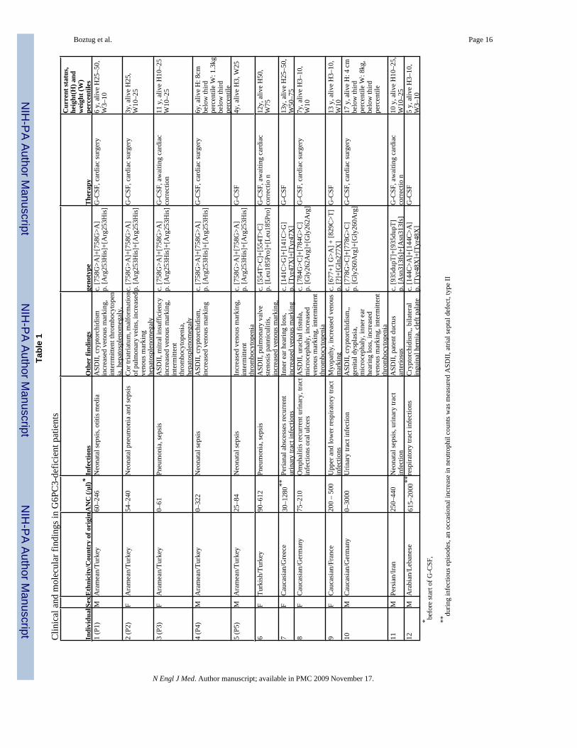
Table 1 lists the main features of the five patients we studied. The siblings P1 and P2, born toconsanguineous parents of Aramean descent, presented with neonatal sepsis. their extendedpedigree is denoted SCN-I (Suppl. Fig. 1). Further workup in their first year of life revealedsevere neutropenia, apparently congenital, with a paucity of mature neutrophils in peripheralblood and bone marrow. Phenotypically, bone marrow smears showed a pathognomonicmaturation arrest at the stage of promyelocytes/myelocytes (Fig. 1a, b and Suppl. Table 1).Erythrocyte counts were normal. Platelet counts in P1 ranged from 73,000–425,000, while P2had normal platelet counts. Both patients had unusually prominent subcutaneous veins and/orvenous angiectasia (Fig 1c); P1 had atrial septal defect (ASD) type II, and P2 had cor triatriatum(Fig. 1d) and hepatosplenomegaly. Genealogical investigations revealed that the SCN-Ipedigree could be extended to include two additional sibships each having one child alsoaffected by severe congenital neutropenia and ASD-II (Patients #3 and 4, Suppl. Fig. 1). Wealso identified a child with severe congenital neutropenia in a second consanguineous pedigree(SCN-II) from the same ethnic background (Patient #5). All patients received recombinanthuman G-CSF (rh-G-CSF) and responded with an increase in peripheral neutrophil counts.
Genetic StudiesMutations in both ELA210 and HAX112 were excluded in all five index patients. Genetic linkageanalysis gave statistical evidence that the gene mutated in SCN-I is located on chromosome17q21 between D17S1299 (36.2Mb, 62.0cM) and D17S1290 (53.7Mb, 82.0cM) (Fig. 2a andSupplementary Results). We carried out a series of fine mapping steps in SCN-I and SCN-IIand were able to genotype an additional 13 microsatellite markers between D17S1299 andD17S1290 in SCN-I, and 11 of these in SCN-II. Supplementary Table 2 shows single-markerLOD scores. Assuming that the same gene is mutated in all five affected children, the maximallinkage interval spanned from D17S1789 (39.1Mb, 63.1cM) to D17S791 (42.2Mb, 64.2cM).Using D17S932, D17S950, and D17S806, the peak multipoint LOD score in SCN-I alone was4.98, and the peak two-pedigree multipoint LOD score was 5.74.
Several candidate genes were identified in the SCN-I linkage interval (Supplementary Table3). Of these, G6PC3, encoding the glucose-6-phosphatase catalytic subunit-3, and located inthe narrowest possible linkage interval, was a plausible candidate, because abnormal glucosemetabolism has been implicated in neutropenia. in glycogen storage disease type Ibpatients24. DNA sequencing revealed a homozygous missense mutation in exon 6 of theG6PC3 gene (c. G758A, p. R253H) (Fig. 2b). This mutation was found in all four affected
Boztug et al. Page 4
N Engl J Med. Author manuscript; available in PMC 2009 November 17.
NIH
-PA Author Manuscript
NIH
-PA Author Manuscript
NIH
-PA Author Manuscript

children in SCN-I and in the affected child in SCN-II. All parents were heterozygous at thisposition, confirming autosomal recessive inheritance of a germline missense mutation. Withrestriction site analysis, the G6PC3R253H allele was not found in 192 healthy central Europeanindividuals. An in silico sequence analysis using SIFT25 predicted that the probability of thismutation being benign was 0.01; analysis with Polyphen26 predicted that the R253H mutationis probably damaging to protein function, as expected since R253 is conserved in multiplespecies including mammals, amphibians, bony fish, and insects.
Functional StudiesEnzymatic Activity of G6PC3R253H—G6PC3wildtype and G6PC3R253H were expressed inSaccharomyces cerevisiae. Microsomes were isolated from yeast transfected withG6PC3wildtype or G6PC3R253H, and assayed for phosphatase activity. G6PC3wildtype
hydrolyzed glucose-6-phosphate and the universal substrate p-Nitrophenylphosphate (pNPP),as demonstrated by radioactive (Fig. 2c) and spectrometric (Suppl. Fig. 3) assays, respectively.In contrast, the level of enzymatic activity of mutant G6PC3R253H did not exceed thephosphatase level in yeast transfected with an empty vector.
Apoptosis: Similar to patients with mutations in ELA210 or HAX112, peripheral bloodneutrophils had an increased rate of spontaneous apoptosis in all five patients tested. Apoptosiswas also markedly accelerated in patients’ neutrophils after induction with either TNF-α (Fig.3a) or tunicamycin (data not shown), as assessed by Annexin-V staining and a test forcaspase-3/7 activation, respectively (see also Suppl. Fig. 4). Since G6PC3 is a ubiquitouslyexpressed gene and since the phenotype of our patients was not restricted to the hematopoieticsystem, we tested non-hematopoietic cells for susceptibility to apoptosis. Skin fibroblasts fromG6PC3-deficient patients displayed an increased susceptibility to apoptosis following DTT-induced stress to the endoplasmic reticulum (Fig. 3b).
To provide further evidence that this novel form of SCN is caused by mutations in G6PC3, weperformed reconstitution experiments to correct premature apoptosis in myeloid cells.CD34+ hematopoietic stem cells from two patients were isolated and transduced with retroviralconstructs containing either the wildtype G6PC3 cDNA sequence and murine CD24 as areporter gene (MMP-G6PC3-mCD24) or the reporter gene only (MMP-mCD24). Upon invitro differentiation in the presence of recombinant human G-CSF and GM-CSF, cells wereexposed to tunicamycin to induce apoptosis and analyzed by flow cytometry by gating onmCD24-positive cells. In control-transduced cells from a patient, exposure to tunicamycininduced a high degree of apoptosis (29.99% Annexin-V positive +4.63% AnnexinV/propidiumiodide double positive cells), whereas in G6PC3-transduced cells, apoptosis was reduced(17.86% + 1.90%) (Fig 3c; Suppl. Fig. 5). We tested the function of neutrophils in G6PC3-deficient neutrophils. Both phagosomal lysis of E. coli and the oxidative burst were comparableto neutrophils from healthy control individuals (Suppl. Fig. 6).
Endoplasmic reticulum stress: Endoplasmic reticulum (ER) stress and the unfolded proteinresponse have been linked to the pathophysiology of aberrant organogenesis27, includingstructural heart defects28, and congenital neutropenia caused by mutations in neutrophilelastase (ELA2)17, 29. We therefore sought evidence for increased endoplasmic reticulum stressin our patients. Transmission electron microscopy of bone marrow cells from all four G6PC3-deficient patients analyzed showed an enlarged rough ER in myeloid progenitor cells ascompared with such cells from a healthy individual (Fig. 4a, 4b), consistent with increased ERstress (Suppl. Fig. 7 shows electron microscopy in other patients). BiP mRNA, another markerof increased ER stress, was measured by RT-PCR in bone marrow promyelocytes isolated byflow cytometry. The BiP mRNA level was increased in promyelocytes from both patients testedcompared to promyelocytes from healthy individuals (Fig. 4c).
Boztug et al. Page 5
N Engl J Med. Author manuscript; available in PMC 2009 November 17.
NIH
-PA Author Manuscript
NIH
-PA Author Manuscript
NIH
-PA Author Manuscript

GSK3β: Recently, a signalling circuit linking glucose, GSK3β, and Mcl1 has beenestablished30, 31. GSK3βcontrols glycogen metabolism, Wnt-signalling, and apoptosis32.Mcl1, an anti-apoptotic member of the Bcl2 family, is involved in maintenance of neutrophilviability33. GSK3βphosphorylates Mcl1, thus facilitating its degradation via theproteasome30. We performed Western blot studies to estimate the levels of GSK3β, BiP, andMcl1 proteins in neutrophils exposed to the ER stress-inducing agent tunicamycin. Weobserved increased levels of Bip (Fig. 4e), an increase in the enzymatically activedephosphorylated form of GSK3β (Fig. 4d and Suppl. Fig. 8), and increased phosphorylationof Mcl1 in in neutrophils from all patients examined (n= 2) (Fig. 4e). To investigate whetherintracellular glucose deprivation causes de-phosphorylation of GSK3β, we inhibited glucosemetabolism in neutrophils from two healthy individuals using 2-deoxyglucose (2DG).Treatment with 2DG induced dephosphorylation of GSK3β(Fig. 4f) and increased apoptosisof neutrophils (Fig. 4g), whereas CD3-positive T lymphocytes were resistant to the effects of2DG (Fig. 4g).
G6PC3 mutations in Other Patients: We assessed the frequency and variety of G6PC3mutations in a cohort of patients with genetically unclassified severe congenital neutropenia.104 such patients were examined for mutations in G6PC3, and seven of these had distinctivebiallelic mutations in G6PC3 (Table 1). The mutations include nonsense mutations (Y47X,Y48X) that would destroy the functional parts of the protein, even if the truncated mRNA istranslated rather than being subjected to nonsense mediated decay. The three additionalmissense mutations were predicted to be deleterious by SIFT22 analysis, with probabilities ofbeing benign of 0.03 for L185P, 0.00 for G262R and 0.00 for G260R, respectively. None ofthese additional patients with G6PC3 mutations had mutations in ELA2 or HAX1, suggestingthat these three genetic defects are distinct variants of severe congenital neutropenia. Of note,none of the G6PC3-deficient patients suffered from hypoglycemia or lactic acidosis(Supplementary Table 4), as seen in patients with glycogen storage disorders.
A comprehensive clinical review of all 12 G6PC3-deficient patients revealed variability ofclinical features. Of the 12 patients, eight had various cardiac malformations, and ten had aphenotype of unusually prominent subcutaneous veins and/or venous angiectasia (Fig. 1 andSupplementary Fig. 2). Five patients had urogenital malformations, including cryptorchidismand urachal fistula. Two patients had inner ear hearing loss. While two patients showed growthdelay, no consistent dysmorphic features were noted (Table 1).
DiscussionWe describe a novel congenital neutropenia syndrome caused by biallelic mutations inG6PC3. Similar to patients with mutations in HAX112 or ELA216, G6PC3-deficient patientshad an evidence of myeloid maturation arrest and increased apoptosis in peripheral neutrophilsOf twelve patients, 8 had structural heart defects (e.g., atrial septal defect type II, cor triatriatum,pulmonary stenosis) and 5 had urogenital defects (e.g., cryptorchidism, urachal fistula). In mostpatients, angiectasia of superficial veins was prominent. The broad spectrum of developmentalaberrations may depend on factors other than the mutant G6PC3. Perhaps increasedsusceptibility to apoptosis also affects cardiac or urogenital development in G6PC3-deficientpatients. There is clinical variation in other neutropenia syndromes such as Cohen syndromeand cartilage hair hypoplasia, even though the two syndromes are genetically homogeneous8.All patients in our cohort responded to treatment with rh-G-CSF, and to date no patient hasdeveloped a clonal hematopoietic disorder.
Three human genes mediating glucose-6-phosphatase activity have been discovered to date:G6PC1, G6PC2, and G6PC3. G6PC1, the classical glucose-6-phosphatase expressed in liver/kidney/small intestine catalyzes the hydrolysis of glucose-6-phosphate, an essential step in the
Boztug et al. Page 6
N Engl J Med. Author manuscript; available in PMC 2009 November 17.
NIH
-PA Author Manuscript
NIH
-PA Author Manuscript
NIH
-PA Author Manuscript

gluconeogenic and glycogenolytic pathways. Patients without G6PC1 activity suffer fromglycogen storage disease type Ia34. G6PC2 is expressed uniquely in pancreas islet cells35, 36
and may be involved in glucose-dependent insulin secretion by controlling free glucoselevels37. Statistically significant associations between noncoding polymorphisms in or nearG6PC2 and the risk of diabetes have been shown38, 39. In contrast to G6PC1 and G6PC2,G6PC3 is ubiquitously expressed40, 41. Glucose-6-phosphate is transported to the endoplasmicreticulum via a glucose-6-phosphate transporter (G6PT)42. Although the stoichiometry andtopological relationships between the catalytic subunits of glucose-6-phosphatase and G6PTremain unclear, their functional link has been recognized42–44. The complex formed betweenG6PT and G6PC1 (potentially also the complex G6PT/G6PC2) appears to maintainnormoglycemia. In contrast, our data show that G6PC3 is needed to maintain neutrophilviability and suggest an important role for glucose in the homeostasis of human neutrophils.
Cheung et al. recently described the phenotype of g6pc3-deficient mice that were generatedby gene-targeting45. These mice had neutropenia and neutrophil dysfunction; another grouphad previously shown that murine g6pc3 deficiency results in lowered plasma cholesterol andelevated glucagon levels46. We could not identify any consistent aberration in neutrophilfunction nor any metabolic aberrations in G6PC3-deficient patients. The underlyingmechanism of increased apoptosis of neutrophils in the absence of G6PC3 involves increasedendoplasmic reticulum stress, usually seen in case of deficient protein folding in theendoplasmic reticulum. In an attempt to counteract potentially toxic effects that may ensue,cells initiate a rescue program which leads, if ineffective, ultimately to apoptosis47.Furthermore, we provide evidence that GSK3β, a key enzyme regulating cellulardifferentiation and apoptosis32, is implicated in this pathway. In the absence of intracellularglucose, GSK3β is activated and thus can phosphorylate the anti-apoptotic molecule Mcl1,thereby mediating its degradation30, 31. Neutrophils from G6PC3-deficient patients contain arelative abundance of non-phosphorylated GSK3β and increased phosphorylation of Mcl1,suggesting that a decrease in antiapoptotic Mcl1 accounts for increased apoptosis in G6PC3-deficient neutrophils. These alterations may at least in part be responsible for the phenotypeof G6PC3 deficiency.
While we cannot rule out that additional mechanisms may contribute to the increased level ofapoptosis in G6PC3-deficient neutrophils, our data suggest that G6PC3 acts via a pathwayinvolving GSK3β to maintain viability of neutrophils. Evidence of increased endoplasmicreticulum stress has previously been reported in patients with mutations in ELA217, 29, andpremature apoptosis of neutrophils is known to cause the phenotype of congenitalneutropenia12, 16, 17. Thus, G6PC3 deficiency provides another example of how increasedapoptosis of neutrophil granulocytes causes congenital neutropenia.
Five arguments support our claim that G6PC3-deficiency causes neutropenia: 1) distinctbiallelic G6PC3 mutations were found in two pedigrees and seven singleton patients withcongenital neutropenia; 2) sequence analysis predicted that all four missense mutations arelikely to affect the function of G6PC3; 3) heterologous expression of G6PC3wildtype andG6PC3R253H in yeast demonstrated that the R253H mutation abrogates enzymatic activity; 4)g6pc3−/− knockout mice show a similar phenotype characterized by neutropenia and increasedmyeloid cell apoptosis and 5) the susceptibility to apoptosis in G6PC3-deficient myeloid cellscould be reduced upon retroviral transfer of the wildtype G6PC3 gene.
Our findings increase the scope of genetic types of congenital neutropenia. and demonstrate anovel role of glucose and/or glucose metabolism in the homeostasis of neutrophil granulocytes..
Boztug et al. Page 7
N Engl J Med. Author manuscript; available in PMC 2009 November 17.
NIH
-PA Author Manuscript
NIH
-PA Author Manuscript
NIH
-PA Author Manuscript

Supplementary MaterialRefer to Web version on PubMed Central for supplementary material.
AcknowledgmentsWe thank the patients and their families for supporting our study. We also thank all colleagues referring and registeringpatients at the Severe Chronic Neutropenia International Registry (SCNIR). The RD114 plasmid was a kind gift fromFrançois-Loïc Cosset. We thank Edelgard Odenwald for analysis of bone marrow smears and Thomas Jack for hishelp with echocardiography. The authors gratefully acknowledge the excellent technical assistance from JessicaPfannstiel, Maren Sievers, Marie Böhm, Marly Dalton, Martina Wackerhahn, Tanja Reinke and Gwendoline Leroyand the excellent collaboration with Dr. Jean Donadieu at the Pediatric Hematology Oncology Service, HôpitalTrousseau, Paris, France.
This research was supported by grants from the Deutsche Forschungsgemeinschaft (DFG KliFo 110–2 (C.K.) and theJunior Research Group “Glycomics” (R.G.-S.). C.K & R.G.-S. are REBIRTH investigators, a DFG cluster ofExcellence. Additional support was provided by BMBF Bone Marrow Failure Syndromes (K.W., C.K.), the IntramuralResearch Program of the U.S. National Institutes of Health, National Library of Medicine (NLM) (A.A.S.), and EUgrant MEXT-CT-2006–042316 (B.G.). K.B. is a recipient of an Else Kröner Memorial fellowship.
References1. Welte K, Zeidler C, Dale DC. Severe congenital neutropenia. Semin Hematol 2006;43:189–95.
[PubMed: 16822461]2. Boztug K, Welte K, Zeidler C, Klein C. Congenital Neutropenia Syndromes. Immunol Allergy Clin
North Am 2008;28:259–75. [PubMed: 18424332]3. Kostmann R. Hereditär reticulos––en ny systemsjukdom. Svenska Läkartidningen 1950;47:2861.4. Kostmann R. Infantile genetic agranulocytosis; agranulocytosis infantilis hereditaria. Acta Paediatr
1956;45 (Suppl 105):1–78. [PubMed: 13313124]5. Bonilla MA, Gillio AP, Ruggeiro M, et al. Effects of recombinant human granulocyte colony-
stimulating factor on neutropenia in patients with congenital agranulocytosis. N Engl J Med1989;320:1574–80. [PubMed: 2471075]
6. Rosenberg PS, Alter BP, Bolyard AA, et al. The incidence of leukemia and mortality from sepsis inpatients with severe congenital neutropenia receiving long-term G-CSF therapy. Blood 2006;107(12):4628–35. [PubMed: 16497969]
7. Boxer LA, Newburger PE. A molecular classification of congenital neutropenia syndromes. PediatrBlood Cancer 2007;49:609–14. [PubMed: 17584878]
8. Schäffer AA, Klein C. Genetic heterogeneity in severe congenital neutropenia: how many aberrantpathways can kill a neutrophil? Curr Opin Allergy Clin Immunol 2007;7:481–94. [PubMed: 17989524]
9. Horwitz M, Benson KF, Person RE, Aprikyan AG, Dale DC. Mutations in ELA2, encoding neutrophilelastase, define a 21-day biological clock in cyclic haematopoiesis. Nat Genet 1999;23:433–6.[PubMed: 10581030]
10. Dale DC, Person RE, Bolyard AA, et al. Mutations in the gene encoding neutrophil elastase incongenital and cyclic neutropenia. Blood 2000;96:2317–22. [PubMed: 11001877]
11. Horwitz MS, Duan Z, Korkmaz B, Lee HH, Mealiffe ME, Salipante SJ. Neutrophil elastase in cyclicand severe congenital neutropenia. Blood 2007;109:1817–24. [PubMed: 17053055]
12. Klein C, Grudzien M, Appaswamy G, et al. HAX1 deficiency causes autosomal recessive severecongenital neutropenia (Kostmann disease). Nat Genet 2007;39:86–92. [PubMed: 17187068]
13. Devriendt K, Kim AS, Mathijs G, et al. Constitutively activating mutation in WASP causes X-linkedsevere congenital neutropenia. Nat Genet 2001;27:313–7. [PubMed: 11242115]
14. Ancliff PJ, Blundell MP, Cory GO, et al. Two novel activating mutations in the Wiskott-Aldrichsyndrome protein result in congenital neutropenia. Blood 2006;108:2182–9. [PubMed: 16804117]
15. Person RE, Li FQ, Duan Z, et al. Mutations in proto-oncogene GFI1 cause human neutropenia andtarget ELA2. Nat Genet 2003;34:308–12. [PubMed: 12778173]
16. Carlsson G, Aprikyan AA, Tehranchi R, et al. Kostmann syndrome: severe congenital neutropeniaassociated with defective expression of Bcl–2, constitutive mitochondrial release of cytochrome c,
Boztug et al. Page 8
N Engl J Med. Author manuscript; available in PMC 2009 November 17.
NIH
-PA Author Manuscript
NIH
-PA Author Manuscript
NIH
-PA Author Manuscript

and excessive apoptosis of myeloid progenitor cells. Blood 2004;103(9):3355–61. [PubMed:14764541]
17. Grenda DS, Murakami M, Ghatak J, et al. Mutations of the ELA2 gene found in patients with severecongenital neutropenia induce the unfolded protein response and cellular apoptosis. Blood2007;110:4179–87. [PubMed: 17761833]
18. Fishelson M, Geiger D. Exact genetic linkage computations for general pedigrees. Bioinformatics2002;18 (Suppl 1):S189–98. [PubMed: 12169547]
19. Fishelson M, Geiger D. Optimizing exact genetic linkage computations. J Comput Biol 2004;11:263–75. [PubMed: 15285892]
20. Broman KW, Murray JC, Sheffield VC, White RL, Weber JL. Comprehensive human genetic maps:individual and sex-specific variation in recombination. Am J Hum Genet 1998;63:861–9. [PubMed:9718341]
21. Ashikov A, Routier F, Fuhlrott J, et al. The human solute carrier gene SLC35B4 encodes a bifunctionalnucleotide sugar transporter with specificity for UDP-xylose and UDP-N-acetylglucosamine. J BiolChem 2005;280(29):27230–5. [PubMed: 15911612]
22. Bohn G, Allroth A, Brandes G, et al. A novel human primary immunodeficiency syndrome causedby deficiency of the endosomal adaptor protein p14. Nat Med 2007;13:38–45. [PubMed: 17195838]
23. Klein C, Bueler H, Mulligan RC. Comparative analysis of genetically modified dendritic cells andtumor cells as therapeutic cancer vaccines. J Exp Med 2000;191:1699–708. [PubMed: 10811863]
24. Beaudet AL, Anderson DC, Michels VV, Arion WJ, Lange AJ. Neutropenia and impaired neutrophilmigration in type IB glycogen storage disease. J Pediatr 1980;97:906–10. [PubMed: 6255119]
25. Ng P, Henikoff S. Accounting for human polymorphisms predicted to affect function. Genome Res2002;12:436–46. [PubMed: 11875032]
26. Ramensky V, Bork P, Sunyaev S. Human non-synonymous SNPs: server and survey. Nucleic AcidsRes 2002;30:3894–900. [PubMed: 12202775]
27. Lin JH, Walter P, Yen TS. Endoplasmic reticulum stress in disease pathogenesis. Annu Rev Pathol2008;3:399–425. [PubMed: 18039139]
28. Glembotski CC. The role of the unfolded protein response in the heart. J Mol Cell Cardiol2008;44:453–9. [PubMed: 18054039]
29. Köllner I, Sodeik B, Schreek S, et al. Mutations in neutrophil elastase causing congenital neutropenialead to cytoplasmic protein accumulation and induction of the unfolded protein response. Blood2006;108:493–500. [PubMed: 16551967]
30. Maurer U, Charvet C, Wagman AS, Dejardin E, Green DR. Glycogen synthase kinase-3 regulatesmitochondrial outer membrane permeabilization and apoptosis by destabilization of MCL-1. MolCell 2006;21:749–60. [PubMed: 16543145]
31. Zhao Y, Altman BJ, Coloff JL, et al. Glycogen synthase kinase 3α and 3βmediate a glucose-sensitiveantiapoptotic signaling pathway to stabilize Mcl-1. Mol Cell Biol 2007;27:4328–39. [PubMed:17371841]
32. Cohen P, Frame S. The renaissance of GSK3. Nat Rev Mol Cell Biol 2001;2:769–76. [PubMed:11584304]
33. Dzhagalov I, St John A, He YW. The antiapoptotic protein Mcl-1 is essential for the survival ofneutrophils but not macrophages. Blood 2007;109:1620–6. [PubMed: 17062731]
34. Lei KJ, Shelly LL, Pan CJ, Sidbury JB, Chou JY. Mutations in the glucose-6-phosphatase gene thatcause glycogen storage disease type 1a. Science 1993;262:580–3. [PubMed: 8211187]
35. Martin CC, Bischof LJ, Bergman B, et al. Cloning and characterization of the human and rat islet-specific glucose-6-phosphatase catalytic subunit-related protein (IGRP) genes. J Biol Chem2001;276:25197–207. [PubMed: 11297555]
36. Arden SD, Zahn T, Steegers S, et al. Molecular cloning of a pancreatic islet-specific glucose-6-phosphatase catalytic subunit-related protein. Diabetes 1999;48:531–42. [PubMed: 10078553]
37. Petrolonis AJ, Yang Q, Tummino PJ, et al. Enzymatic characterization of the pancreatic islet-specificglucose-6-phosphatase-related protein (IGRP). J Biol Chem 2004;279:13976–83. [PubMed:14722102]
Boztug et al. Page 9
N Engl J Med. Author manuscript; available in PMC 2009 November 17.
NIH
-PA Author Manuscript
NIH
-PA Author Manuscript
NIH
-PA Author Manuscript

38. Bouatia-Naji N, Rocheleau G, Van Lommel L, et al. A polymorphism within the G6PC2 gene isassociated with fasting plasma glucose levels. Science 2008;320:1085–8. [PubMed: 18451265]
39. Chen WM, Erdos MR, Jackson AU, et al. Variations in the G6PC2/ABCB11 genomic region areassociated with fasting glucose levels. J Clin Invest 2008;118:2620–28. [PubMed: 18521185]
40. Martin CC, Oeser JK, Svitek CA, Hunter SI, Hutton JC, O’Brien RM. Identification andcharacterization of a human cDNA and gene encoding a ubiquitously expressed glucose-6-phosphatase catalytic subunit-related protein. J Mol Endocrinol 2002;29:205–22. [PubMed:12370122]
41. Guionie O, Clottes E, Stafford K, Burchell A. Identification and characterisation of a new humanglucose-6-phosphatase isoform. FEBS Lett 2003;551:159–64. [PubMed: 12965222]
42. van Schaftingen E, Gerin I. The glucose-6-phosphatase system. Biochem J 2002;362:513–32.[PubMed: 11879177]
43. Lei KJ, Chen H, Pan CJ, et al. Glucose-6-phosphatase dependent substrate transport in the glycogenstorage disease type-1a mouse. Nat Genet 1996;13:203–9. [PubMed: 8640227]
44. Melis D, Fulceri R, Parenti G, et al. Genotype/phenotype correlation in glycogen storage disease type1b: a multicentre study and review of the literature. Eur J Pediatr 2005;164:501–8. [PubMed:15906092]
45. Cheung YY, Kim SY, Yiu WH, et al. Impaired neutrophil activity and increased susceptibility tobacterial infection in mice lacking glucose-6-phosphatase-beta. J Clin Invest 2007;117:784–93.[PubMed: 17318259]
46. Wang Y, Oeser JK, Yang C, et al. Deletion of the gene encoding the ubiquitously expressed glucose-6-phosphatase catalytic subunit-related protein (UGRP)/glucose-6-phosphatase catalytic subunit-betaresults in lowered plasma cholesterol and elevated glucagon. J Biol Chem 2006;281:39982–9.[PubMed: 17023421]
47. Ron D, Walter P. Signal integration in the endoplasmic reticulum unfolded protein response. Nat RevMol Cell Biol 2007;8:519–29. [PubMed: 17565364]
Boztug et al. Page 10
N Engl J Med. Author manuscript; available in PMC 2009 November 17.
NIH
-PA Author Manuscript
NIH
-PA Author Manuscript
NIH
-PA Author Manuscript

Figure 1. Clinical and hematological phenotypePanel A shows severe paucity of mature neutrophils in bone marrow smears from patient P1.In contrast, bone marrow smears from a healthy individual (Panel B) show normal myeloiddifferentiation. Panel C illustrates the phenotype of unusual visibility/angiectasia ofsubcutaneous veins, which was found in all patients from the two pedigrees SCN-I and SCN-II. Panel D shows an atrial septal defect, type II, in patient P3.
Boztug et al. Page 11
N Engl J Med. Author manuscript; available in PMC 2009 November 17.
NIH
-PA Author Manuscript
NIH
-PA Author Manuscript
NIH
-PA Author Manuscript

Figure 2. Haplotype on Chromosome 17q, mutation analysis of G6PC3 and assessment of enzymaticactivity of G6PC3R253H
Panel A indicates the pedigrees from 2 unrelated SCN families (SCN-I and SCN-II). Affectedpatients are marked with filled symbols, whereas unaffected family members are representedby open symbols. P1–P5 refer to the patients described in more detail in Table 1. The linkageinterval is shown in gray for each family. The preferred linkage interval was defined under theassumption that the genetic defect in SCN-I and SCN-II was the same, and ranged from markersD17S930 to D17S950, which contained a total of 36 genes including G6PC3. Panel B showsthe missense mutation (c. G758A, p. R253H) found in an index patient from SCN-I as comparedto the sequence in a healthy control.Panel C represents the enzymatic activity of wild type and G6PC3R253H with [14C]G6P assubstrate compared to an empty vector control measured in microsomes isolated from stablytransfected yeast cells. The presence of 0.1% saponin, a mild detergent preserving themembrane integrity, allows substrate penetration of the microsomes. In the presence of 0.5%triton, membranes are disrupted completely. The difference between the values represents themembrane dependent activity. The results are presented as nmol of glucose produced perminute per gram of yeast cells. Error bars indicate the standard deviation of three independentmicrosomal preparations. * Significantly different with p = 0.012.
Boztug et al. Page 12
N Engl J Med. Author manuscript; available in PMC 2009 November 17.
NIH
-PA Author Manuscript
NIH
-PA Author Manuscript
NIH
-PA Author Manuscript

Figure 3. Increased susceptibility to apoptosis in myeloid cells and fibroblast cell linesPanel A shows a FACS plot indicating that peripheral blood neutrophils from patient P5 hadincreased spontaneous apoptosis as compared to a healthy sibling or a rh-G-CSF treated healthycontrol (HD) (x axis, Annexin-V; y axis, propidium iodide; logarithmic scales in both axes).Similarly, TNF-alpha induced apoptosis was more pronounced in patients.Panel B shows an analysis of enhanced apoptosis in patient fibroblasts after exposure todithiothreitol (DTT) as compared to normal donors.Panel C shows a FACS plot of in vitro differentiated myeloid progenitor cells transduced witha control vector (upper row) or a retroviral vector encoding wildtype G6PC3 (lower row). Cellswere treated with tunicamycin and apoptosis was assessed using Annexin-V (x axis) andpropidium iodide staining (y axis). G6PC3-transduced patient cells from patient P2 show adecreased susceptibility to apoptosis 16 hours after induction with tunicamycin, as comparedto control vector-transduced cells. Data are representative of two independent experiments.
Boztug et al. Page 13
N Engl J Med. Author manuscript; available in PMC 2009 November 17.
NIH
-PA Author Manuscript
NIH
-PA Author Manuscript
NIH
-PA Author Manuscript

Figure 4. Pathophysiologic consequences of G6PC3 deficiencyPanel A shows the aberrantly enlarged rough ER in myeloid progenitor cells as compared toa healthy individual (Panel B). Panel C indicates increased ER stress in G6PC3-deficientpatients, as documented by increased expression levels of Bip mRNA in purifiedpromyelocytes. Panel D represents a Western Blot revealing a decrease in the enzymaticallyactive, dephosphorylated form of GSK3β in patient P5 after induction with tunicamycin. PanelE shows increased phosphorylation of Mcl1 and increased levels of Bip in neutrophils from aG6PC3-deficient patient (P5) at different timepoints after induction with tunicamycin. PanelF shows 2-deoxyglucose (2DG)-induced dephosphorylation of GSK3β in 2 healthy donors.
Boztug et al. Page 14
N Engl J Med. Author manuscript; available in PMC 2009 November 17.
NIH
-PA Author Manuscript
NIH
-PA Author Manuscript
NIH
-PA Author Manuscript

Panel G indicates that healthy neutrophils, but not CD3-positive T cells, are susceptible toapoptosis upon pharmacological glucose-depletion.
Boztug et al. Page 15
N Engl J Med. Author manuscript; available in PMC 2009 November 17.
NIH
-PA Author Manuscript
NIH
-PA Author Manuscript
NIH
-PA Author Manuscript
NIH
-PA Author Manuscript
NIH
-PA Author Manuscript
NIH
-PA Author Manuscript
Boztug et al. Page 16Ta
ble
1
Clin
ical
and
mol
ecul
ar fi
ndin
gs in
G6P
C3-
defic
ient
pat
ient
s
Indi
vidu
alSe
xEth
nici
ty/C
ount
ry o
f ori
gin
AN
C (/μl
)*In
fect
ions
Oth
er fi
ndin
gsge
noty
peT
hera
py
Cur
rent
stat
us,
heig
ht(H
) and
wei
ght (
W)
perc
entil
es1
(P1)
MA
ram
ean/
Turk
ey60
–246
Neo
nata
l sep
sis,
otiti
s med
iaA
SDII
, cry
ptor
chid
ism
incr
ease
d ve
nous
mar
king
,in
terr
mitt
ent t
hrom
bocy
tope
nia
, hep
atos
plen
omeg
aly,
c. [7
58G
>A]+
[758
G>A
]p.
[Arg
253H
is]+
[Arg
253H
is]
G-C
SF, c
ardi
ac su
rger
y6
y, a
live
H25
–50,
W3–
10
2 (P
2)F
Ara
mea
n/Tu
rkey
54–2
40N
eona
tal p
neum
onia
and
seps
isC
or tr
iatri
atum
, mal
form
atio
nof
pul
mon
ary
vein
s, in
crea
sed
veno
us m
arki
nghe
pato
sple
nom
egal
y
c. [7
58G
>A]+
[758
G>A
]p.
[Arg
253H
is]+
[Arg
253H
is]
G-C
SF, c
ardi
ac su
rger
y3y
, aliv
e H
25,
W10
–25
3 (P
3)F
Ara
mea
n/Tu
rkey
0–61
Pneu
mon
ia, s
epsi
sA
SDII
, mitr
al in
suff
icie
ncy
incr
ease
d ve
nous
mar
king
,in
term
itten
tth
rom
bocy
tope
nia,
hepa
tosp
leno
meg
aly
c. [7
58G
>A]+
[758
G>A
]p.
[Arg
253H
is]+
[Arg
253H
is]
G-C
SF, a
wai
ting
card
iac
corr
ectio
n11
y, a
live
H10
–25
W10
–25
4 (P
4)M
Ara
mea
n/Tu
rkey
0–32
2N
eona
tal s
epsi
sA
SDII
, cry
ptor
chid
ism
,in
crea
sed
veno
us m
arki
ngc.
[758
G>A
]+[7
58G
>A]
p. [A
rg25
3His
]+[A
rg25
3His
]G
-CSF
, car
diac
surg
ery
6y, a
live
H: 8
cmbe
low
third
perc
entil
e W
: 1.3
kgbe
low
third
perc
entil
e5
(P5)
MA
ram
ean/
Turk
ey25
–84
Neo
nata
l sep
sis
Incr
ease
d ve
nous
mar
king
,in
term
itten
tth
rom
bocy
tope
nia
c. [7
58G
>A]+
[758
G>A
]p.
[Arg
253H
is]+
[Arg
253H
is]
G-C
SF4y
, aliv
e H
3, W
25
6F
Turk
ish/
Turk
ey90
–612
Pneu
mon
ia, s
epsi
sA
SDII
, pul
mon
ary
valv
est
enos
is p
anni
culit
is,
incr
ease
d ve
nous
mar
king
,
c. [5
54T>
C]+
[554
T>C
]p.
[Leu
185P
ro]+
[Leu
185P
ro]
G-C
SF, a
wai
ting
card
iac
corr
ectio
n12
y, a
live
H50
,W
75
7F
Cau
casi
an/G
reec
e30
–128
0**Pe
riana
l abs
cess
es re
curr
ent
urin
ary
tract
infe
ctio
nsIn
ner e
ar h
earin
g lo
ss,
incr
ease
d ve
nous
mar
king
c. [1
41C
>G]+
[141
C>G
]p.
[Tyr
47X
]+[T
yr47
X]
G-C
SF13
y, a
live
H25
–50,
W50
–75
8F
Cau
casi
an/G
erm
any
75–2
10O
mph
aliti
s rec
urre
nt u
rinar
y, tr
act
infe
ctio
ns o
ral u
lcer
sA
SDII
, ura
chal
fist
ula,
mic
roce
phal
y, in
crea
sed
veno
us m
arki
ng, i
nter
mitt
ent
thro
mbo
cyto
peni
a
c. [7
84G
>C]+
[784
G>C
]p.
[Gly
262A
rg]+
[Gly
262A
rg]G
-CSF
, car
diac
surg
ery
7y, a
live
H3–
10,
W10
9F
Cau
casi
an/F
ranc
e20
0 –
500
Upp
er a
nd lo
wer
resp
irato
ry tr
act
infe
ctio
nsM
yopa
thy,
incr
ease
d ve
nous
mar
king
c. [6
77+1
G>A
] + [8
29C
>T]
p. [?
]+[G
ln27
7X]
G-C
SF13
y, a
live
H3–
10,
W10
10M
Cau
casi
an/G
erm
any
0–30
00U
rinar
y tra
ct in
fect
ion
ASD
II, c
rypt
orch
idis
m,,
geni
tal d
yspl
asia
,m
icro
ceph
aly,
inne
r ear
hear
ing
loss
, inc
reas
edve
nous
mar
king
, int
erm
itten
tth
rom
bocy
tope
nia
c. [7
78G
>C]+
[778
G>C
]p.
[Gly
260A
rg]+
[Gly
260A
rg]G
-CSF
, car
diac
surg
ery
17 y
, aliv
e H
: 4 c
mbe
low
third
perc
entil
e W
: 8kg
,be
low
third
perc
entil
e
11M
Pers
ian/
Iran
250–
440
Neo
nata
l sep
sis,
urin
ary
tract
infe
ctio
nA
SDII
, pat
ent d
uctu
sar
terio
sus
c. [9
35du
pT]+
[935
dupT
]p.
[Asn
313f
s]+[
Asn
313f
s]G
-CSF
, aw
aitin
g ca
rdia
cco
rrec
tio n
10 y
, aliv
e H
10–2
5,W
10–2
512
MA
rabi
an/L
eban
ese
615–
2000
**re
spira
tory
trac
t inf
ectio
nsC
rypt
orch
idis
m,,
bila
tera
lin
guin
al h
erni
a, c
left
pala
tec.
[144
C>A
]+[1
44C
>A]
p. [T
yr48
X]+
[Tyr
48X
]G
-CSF
5 y,
aliv
e H
3–10
,W
3–10
* befo
re st
art o
f G-C
SF,
**du
ring
infe
ctio
us e
piso
des,
an o
ccas
iona
l inc
reas
e in
neu
troph
il co
unts
was
mea
sure
d A
SDII
, atri
al se
ptal
def
ect,
type
II
N Engl J Med. Author manuscript; available in PMC 2009 November 17.