Clonal evolution in relapsed acute myeloid leukaemia revealed by whole-genome sequencing
Upload
tri-londonCategory
view
1download
0
doi:10.1182/blood-2002-04-1288Prepublished online August 29, 2002;
Daniel G Tenen, Wolfgang Hiddemann and Gerhard BehreRajani K Vangala, Marion S Heiss-Neumann, Janki S Rangatia, Sheo M Singh, Claudia Schoch, AML1-ETO in t(8;21) myeloid leukemiaThe myeloid master regulator transcription factor PU.1 is inactivated by
(4212 articles)Neoplasia � (1086 articles)Gene Expression �
Articles on similar topics can be found in the following Blood collections
http://bloodjournal.hematologylibrary.org/site/misc/rights.xhtml#repub_requestsInformation about reproducing this article in parts or in its entirety may be found online at:
http://bloodjournal.hematologylibrary.org/site/misc/rights.xhtml#reprintsInformation about ordering reprints may be found online at:
http://bloodjournal.hematologylibrary.org/site/subscriptions/index.xhtmlInformation about subscriptions and ASH membership may be found online at:
articles must include the digital object identifier (DOIs) and date of initial publication. priority; they are indexed by PubMed from initial publication. Citations to Advance online prior to final publication). Advance online articles are citable and establish publicationyet appeared in the paper journal (edited, typeset versions may be posted when available Advance online articles have been peer reviewed and accepted for publication but have not
Copyright 2011 by The American Society of Hematology; all rights reserved.Washington DC 20036.by the American Society of Hematology, 2021 L St, NW, Suite 900, Blood (print ISSN 0006-4971, online ISSN 1528-0020), is published weekly
For personal use only. by guest on January 27, 2014. bloodjournal.hematologylibrary.orgFrom For personal use only. by guest on January 27, 2014. bloodjournal.hematologylibrary.orgFrom

Vangala et al
1
The myeloid master regulator transcription factor PU.1 is
inactivated by AML1-ETO in t(8;21) myeloid leukemia
Rajani K.Vangala*, Marion S.Heiss-Neumann*, Janki S.Rangatia*, Sheo
M.Singh*, Schoch C*, Daniel G.Tenen$, Wolfgang Hiddemann* and
Gerhard Behre*.
* Department of Internal Medicine III, University Hospital Grosshadern,
Ludwig-Maximilians-University Munich and GSF-National Research Center for
Environment and Health, Munich, 81377, Germany
$ Harvard Institutes of Medicine, Harvard Medical School, Boston, MA 02115,
USA
Corresponding author:
Dr. Gerhard Behre
Dept. of Internal Medicine III, University Hospital Grosshadern, Ludwig-
Maximilians-University Munich
Marchioninistr. 15, D-81377 Munich, Germany
Tel.: +49-89- 7095- 118, Fax: +49-89- 7095-5550, E.Mail: [email protected]
Running title: PU.1 is inactivated by AML1-ETO.
Scientific section heading: Neoplasia
Word Count: 4218 words of text excluding figure legends and references; 196
words in the abstract.
This work was supported by a DFG (German research foundation) grant to G.
B. (Nr. 2042/2-1).
Copyright (c) 2002 American Society of Hematology
Blood First Edition Paper, prepublished online August 29, 2002; DOI 10.1182/blood-2002-04-1288 For personal use only. by guest on January 27, 2014. bloodjournal.hematologylibrary.orgFrom
Vangala et al
2
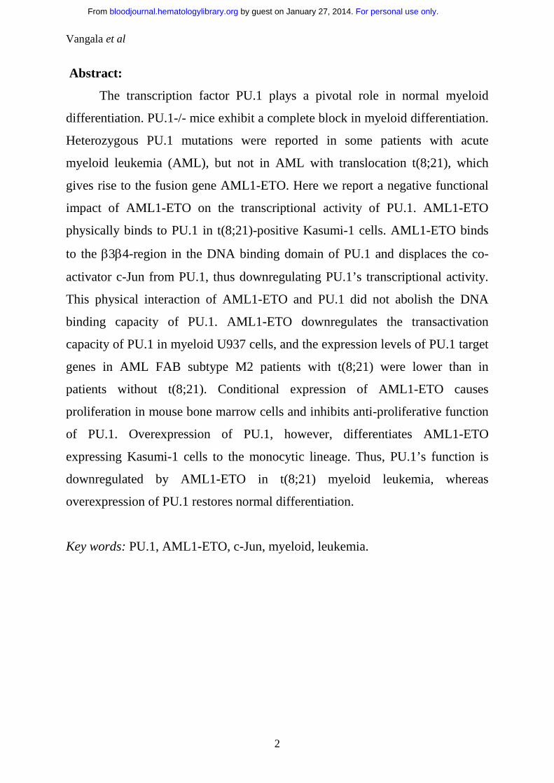
Abstract:
The transcription factor PU.1 plays a pivotal role in normal myeloid
differentiation. PU.1-/- mice exhibit a complete block in myeloid differentiation.
Heterozygous PU.1 mutations were reported in some patients with acute
myeloid leukemia (AML), but not in AML with translocation t(8;21), which
gives rise to the fusion gene AML1-ETO. Here we report a negative functional
impact of AML1-ETO on the transcriptional activity of PU.1. AML1-ETO
physically binds to PU.1 in t(8;21)-positive Kasumi-1 cells. AML1-ETO binds
to the β3β4-region in the DNA binding domain of PU.1 and displaces the co-
activator c-Jun from PU.1, thus downregulating PU.1’s transcriptional activity.
This physical interaction of AML1-ETO and PU.1 did not abolish the DNA
binding capacity of PU.1. AML1-ETO downregulates the transactivation
capacity of PU.1 in myeloid U937 cells, and the expression levels of PU.1 target
genes in AML FAB subtype M2 patients with t(8;21) were lower than in
patients without t(8;21). Conditional expression of AML1-ETO causes
proliferation in mouse bone marrow cells and inhibits anti-proliferative function
of PU.1. Overexpression of PU.1, however, differentiates AML1-ETO
expressing Kasumi-1 cells to the monocytic lineage. Thus, PU.1’s function is
downregulated by AML1-ETO in t(8;21) myeloid leukemia, whereas
overexpression of PU.1 restores normal differentiation.
Key words: PU.1, AML1-ETO, c-Jun, myeloid, leukemia.
For personal use only. by guest on January 27, 2014. bloodjournal.hematologylibrary.orgFrom

Vangala et al
3
Introduction:
The Ets family of transcription factors plays a key role in the growth,
survival, differentiation and activation of hematopoietic cells. This family of
proteins is characterized by presence of an 85 amino acid, 1 winged helix-turn-
helix DNA-binding domain. PU.1 is one of the most important Ets transcription
factors. 2 Its expression is limited to hematopoietic cells, including primitive
CD34+ cells, macrophages, B-lymphocytes, neutrophils, mast cells, and early
erythroblasts. 2 3 in vitro studies suggest that PU.1 regulates the activity of a
number of myeloid and lymphoid-specific promoters and enhancers. 4 5 6 7 8 9 10
PU.1 is a key transcription factor for normal myeloid development as
demonstrated by a complete block of myeloid development in PU.1-/- mice. 11 12
Fetal or newborn PU.1-/- mice have no detectable monocytes/macrophages or
neutrophils. 11 12 We have recently shown that PU.1 is mutated in AML patients. 13 These studies all point to the crucial role of PU.1 in both normal myeloid
differentiation and leukemogenesis.
AML1 is a member of the Runt-like transcription factors (Runx-1, -2 and
–3) named after the Runt protein that regulates segmentation during Drosophila
embryogenesis. 14 15 16 AML1 appears to act as an “organizing” factor for many
promoters and enhancers by interacting with various co-activators and DNA
binding transcription factors. 17 18 19 20 21 22 The AML1 gene is one of the most
frequently translocated or mutated genes in human cancer. 23 24 25 The
t(8;21)(q22;q22) translocation fuses residues 1-177 of AML1 (including the
DNA binding domain) to nearly all of ETO (also known as CBF2T1). 26 ETO is
the human homolog of Drosophila NERVY protein. 27 28 29 The t(8;21) belongs
to the most common chromosomal abnormalities in AML, accounting for 10%
of all AML cases and 40% of the AML French-American-British (FAB) M2
phenotype. 30 31 32 33 AML1 activates transcription from enhancer core motifs
(TGT/cGGY), which are present in a number of genes relevant to myeloid
development, including the M-CSF receptor, GM-CSF, myeloperoxidase and
For personal use only. by guest on January 27, 2014. bloodjournal.hematologylibrary.orgFrom
Vangala et al
4
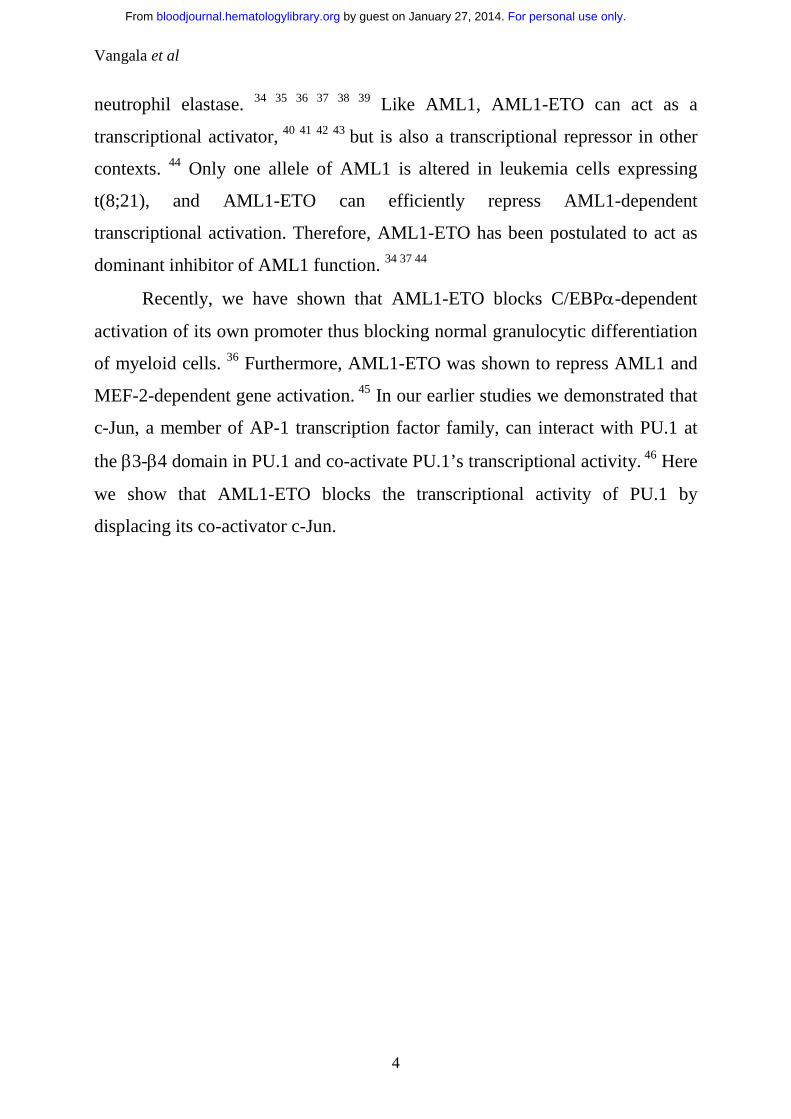
neutrophil elastase. 34 35 36 37 38 39 Like AML1, AML1-ETO can act as a
transcriptional activator, 40 41 42 43 but is also a transcriptional repressor in other
contexts. 44 Only one allele of AML1 is altered in leukemia cells expressing
t(8;21), and AML1-ETO can efficiently repress AML1-dependent
transcriptional activation. Therefore, AML1-ETO has been postulated to act as
dominant inhibitor of AML1 function. 34 37 44
Recently, we have shown that AML1-ETO blocks C/EBPα-dependent
activation of its own promoter thus blocking normal granulocytic differentiation
of myeloid cells. 36 Furthermore, AML1-ETO was shown to repress AML1 and
MEF-2-dependent gene activation. 45 In our earlier studies we demonstrated that
c-Jun, a member of AP-1 transcription factor family, can interact with PU.1 at
the β3-β4 domain in PU.1 and co-activate PU.1’s transcriptional activity. 46 Here
we show that AML1-ETO blocks the transcriptional activity of PU.1 by
displacing its co-activator c-Jun.
For personal use only. by guest on January 27, 2014. bloodjournal.hematologylibrary.orgFrom

Vangala et al
5
Materials and Methods:
Cell lines and cell culture: Human kidney 293T, mouse embryonal
carcinoma F9 and ecotrophic Phoenix cells were maintained in Dulbecco’s
Modified Eagle’s Medium (GIBCO) supplemented with 10% Fetal Bovine
Serum (GIBCO), 1% Glutamine (GIBCO) and 1% Penstrep (GIBCO). Human
monoblastic U937 cells and t(8;21) positive Kasumi-1 cells were cultured in
RPMI 1640 medium (GIBCO) supplemented with 10% Fetal Bovine Serum
(GIBCO).
Bone marrow cells were isolated from the femurs of Balb/C mouse. The
femurs were removed and stripped of the soft tissue, and crushed to release cells
within marrow cavity. The red blood cells were lysed with a 0.15-mol/L solution
of ammonium chloride. The pelleted cells were subjected to low-density
mononuclear cell separation by incubating with density gradient (Histopaque
1083; Sigma) for 10 minutes and centrifuged at 600 rpm for 30 minutes, washed
twice in PBS, followed by culturing in IMDM medium (Stem Cell
Technologies) supplemented with 10% FBS (Stem cell technologies), 50 ng/mL
stem cell factor (R and D systems), 50 ng/mL IL-6 (R and D systems), and 50
ng/mL Flt-3L (R and D systems).
Co-immunoprecipitation assay: 2x10⁷ Kasumi-1 cells were lysed and
200µg of protein was used to perform immunoprecipitation as mentioned by
Mao et al. 45 The following antibodies were used: rabbit IgG (Santa Cruz
Biotechnologies, Cat# sc2027), goat IgG (Santa Cruz Biotechnologies, Cat#
2028), anti-AML1 antibody (Calbiochem, Cat# PC284), anti-PU.1 (Santa Cruz
Biotechnologies, Cat# sc-352), and Protein-A agarose beads (Santa Cruz
Biotechnologies, Cat# sc-2001).
Western blot: 293T cells (after plating in 100mm plates) were transfected
using the LipofectAMINE Plus kit (GIBCO) as per the manufacturer’s protocol.
24h post transfection, cells were harvested and lysed in RIPA lysis buffer, and
immunoblot for PU.1 was performed with 100µg protein as described earlier. 46
For personal use only. by guest on January 27, 2014. bloodjournal.hematologylibrary.orgFrom
Vangala et al
6
47 48 To generate protein lysates, 1x10⁶ F9, Kasumi-1 or 293T cells were lysed
and nuclear extracts were prepared and immunoblot was performed with 100µg
protein for c-Jun (Santa Cruz Biotechnologies, Cat# sc45). Mouse bone marrow
cells transduced with PU.1, AML1-ETO or respective empty vectors were
similarly lysed (RIPA lysis) and 100µg of protein was used for immunoblot
analysis for PU.1 and AML1-ETO (anti-ETO antibody, Santa Cruz
Biotechnologies, Cat# sc9737). Mouse monoclonal anti-β-tubulin purchased
from Roche (Cat# 1111876) was used for immunoblot assay as internal control.
Protein-A peroxidase-conjugated for anti-rabbit (Amersham Pharmacia, cat# NA
9120), or anti-goat peroxidase-conjugated Immunoglobulins (DAKO, code no.
p0449) were used as secondary antibodies.
Reporter constructs and expression plasmids: The human monocyte-
specific M-CSF receptor promoter with or without AML1 binding site,
p(PU.1)4TK, and p(mutPU.1)4TK (PU.1 binding sites and mutated PU.1
binding sites subcloned into pTK61luciferase) were described earlier. 46 As an
internal control plasmid for transient transfection assay, we used the pRL-null
construct driving a Renilla luciferase gene (Promega). 49 Other vectors used
were pECE-PU.1-murine, pcDNA.1-PU.1, pGEX-2TK-PU.1 or β3β4, pS3H-c-
Jun and pSP6-c-Jun, as described previously. 46 50 AML1B-pCMV5 and CBFβ-
pCMV5 were described earlier. 42 AML1-ETO-pcDNA3 was constructed by
enzymatic digestion of AML1-ETO-pCMV5 42 with XbaI and sub-cloning the
resulting 2258bp fragment into the XbaI site of pcDNA3 plasmid (Invitrogen).
Transfection assays: Transient transfections in 293T or F9 cells were
carried out with LipofectAMINE transfection kit (GIBCO) in 24 well plates as
described earlier. 46 47 49 U937 cells were transiently transfected by
electroporation in RPMI medium at 980µF and 280V. Firefly luciferase
activities from the constructs M-CSF receptor promoter luciferase, pXP2,
p(PU.1)4TK, p(mutPU.1)4TK and Renilla luciferase activity from internal
control plasmid pRL-null were determined 24h post transfection using Dual
For personal use only. by guest on January 27, 2014. bloodjournal.hematologylibrary.orgFrom

Vangala et al
7
Luciferase Reporter Assay System (Promega). Results are given as means +
standard error of mean from at least 3 independent experiments.
Protein interaction assays: c-Jun and AML1-ETO were in vitro
transcribed and translated in presence of [35S]-methionine (Amersham
Pharmacia) using the T7/SP6 coupled reticulocyte system (Promega) in
accordance with the manufacturer’s instruction. Glutathione-S-transferase
precipitation assays were performed as described earlier. 46 48
Electrophoretic mobility shift assay: γ32P-ATP (Amersham Pharmacia)
labelled double-stranded oligonucleotides of PU.1 DNA binding site 51 and
AML1 binding site 52 for EMSA were prepared. The assay was performed with
in vitro translated proteins as mentioned earlier. 47 11 For supershift experiments
3µl of either anti-PU.1 or anti-ETO-antibodies were added to the reaction
mixture.
Retroviral transduction assay: 5x10⁶ ecotrophic Phoenix cells were
plated in 10cm plates and transfected with 5µg of PINCO-GFP, PINCO-AML1-
ETO-GFP, pGsam-PU.1-ires-NGFR or pGsam-ires-NGFR vectors using
LipofectAMINE transfection kit (GIBCO). 24hrs post-transfection the
transfection medium was replaced with IMDM medium (supplemented with
10% FBS, 50 ng/mL stem cell factor, 50 ng/mL IL-6, and 50 ng/mL Flt-3L) for
collection of the virus particles. After the viral particle production freshly
isolated mouse bone marrow cells were incubated with viral medium on
Fibronectin coated plates and centrifuged for 30minutes at 1000 x g (this step
was repeated every 12hours). 53 At 60hr after first transduction NGFR and/or
EGFP positive cells were isolated by FACS analysis (Becton Dickinson). To
detect the expression of tNGFR on the cell surface, cells were stained with
mouse anti-human NGFR ( Chemicon, cat# MAB5246)) followed by
phycoerythrin (PE)-conjugated rabbit anti-mouse immunoglobulins (mouse IgG
RPE from Dako, cat# R0439). 1x10⁴ transduced cells sorted for NGFR
positivity were plated in 1.2ml of mouse colony forming medium (Stem Cell
For personal use only. by guest on January 27, 2014. bloodjournal.hematologylibrary.orgFrom

Vangala et al
8
Technologies). After 3, 6, and 12 days of plating live cells were counted by
Trypan blue staining.
Patient material and FACS analysis: Bone marrow cells from AML-M2
patients with or without t(8;21) were obtained after informed consent of the
patients. Mononuclear cells were isolated from the bone marrow by density
gradient centrifugation with Histopaque (Sigma). FACS analysis was performed
with CD11b (Pharmingen, cat# 555388), CD14 (Pharmingen, cat# 555397) and
CD64 (Pharmingen, cat# 555527).
Transfection of Kasumi-1 cells and FACS analysis: Kasumi-1 cells
were electroporated as mentioned above with pGsam-PU.1-ires-NGFR or
pGsam-ires-NGFR vectors and sorted 24h post transfection for NGFR positivity
by FACS (with anti-NGFR antibody from Chemicon, cat# MAB5246 and
mouse IgG RPE from Dako, cat# R0439). Five days after sorting for NGFR
expression, morphological changes were observed by Wright-Giemsa staining of
cells. 1x10⁶ NGFR positive Kasumi-1 cells were incubated with 10µl of
recombinant PE-conjugated mouse monoclonal CD11b (Pharmingen, cat#
555388) or FITC-conjugated mouse monoclonal CD14 (Pharmingen, cat#
555397) in 100µl PBS for 60 min on ice, washed in PBS followed by analysis
on a FACScan flow cytometer (Becton Dickinson) using Cellquest software.
The cells were also analysed for the isotype controls, PE conjugated mouse
IgG1,κ (Pharmingen, cat# 554680) for CD11b-PE and FITC conjugated mouse
IgG1,κ (Pharmingen, cat# 555748) for CD14-FITC. 24hrs post transfection,
5x10⁴ NGFR positive cells were plated in a 6-well plate and passaged with fresh
medium every 24hrs. Cell count for live cells was performed by trypan blue
staining every 24hrs.
For personal use only. by guest on January 27, 2014. bloodjournal.hematologylibrary.orgFrom

Vangala et al
9
Results:
AML1-ETO interacts with PU.1 in vivo and inhibits its
transcriptional activity: To determine whether PU.1 interacts with AML1-
ETO, co-immunoprecipitation assays were performed in Kasumi-1 cells, a
human cell line containing t(8;21). PU.1 co-precipitated with both AML1 and
ETO antibodies but not with IgG control, suggesting that PU.1 interacts with
AML1-ETO in vivo (Fig. 1a). A similar experiment was performed using a PU.1
specific antibody: AML1-ETO co-precipitated with PU.1, but not with rabbit
IgG control (Fig. 1a).
To investigate the functional impact of this in vivo interaction, we
performed transient transfection assays in 293T cells. A M-CSF receptor
promoter luciferase reporter construct which was transactivated 12-fold by PU.1
and 28-fold by PU.1/c-Jun, is completely downregulated by AML1-ETO (Fig.
1b). AML1-ETO had no effects on Serum Response Element (pSRE)/Ras
activity nor on the empty vector (pXP2) as negative controls. The expression
levels of co-transfected PU.1 did not change in presence of AML1-ETO
indicating that the transactivating capacity, but not the expression of co-
transfected PU.1 was downregulated (Fig. 1b).
AML1 does not affect PU.1 or PU.1/c-Jun’s transactivation capacity:
The M-CSF receptor promoter has adjacent AML1 and PU.1 binding sites. 45
AML1-ETO retains the 177 N-terminus amino acids of AML1, suggesting that
AML1 might also have an influence on transactivation of PU.1 or PU.1/c-Jun.
Therefore we addressed if AML1 had any functional impact on PU.1’s or
PU.1/c-Jun’s transactivation capacity using a promoter containing only PU.1
binding sites (p(PU.1)4TK). Transient transfection assays in 293T cells were
performed with p(PU.1)4TK and expression plasmids of PU.1, c-Jun, AML1
and CBFβ. Results (Fig. 1c) show that AML1 did not affect the PU.1 or PU.1/c-
Jun transactivation capacity. In the same experiment AML1 could transactivate
the M-CSF receptor promoter 4-fold in presence of CBFβ (data not shown).
For personal use only. by guest on January 27, 2014. bloodjournal.hematologylibrary.orgFrom

Vangala et al
10
AML1, PU.1, c-Jun and CBFβ had no effects on control vectors
(p(mutPU.1)4TK and pXP2) in the experiments above (data not shown).
AML1-ETO inhibits the co-activation of PU.1 by c-Jun: We have
earlier shown that c-Jun can co-activate PU.1’s transactivation in a JNK-
independent manner. 46 PU.1 induced strong transactivation of p(PU.1)4TK in
293T cells (Fig. 1c). This is possibly due to high expression of its co-activator c-
Jun in these cells. Immunoblot assay for c-Jun indicated that 293T cells have
high amounts of c-Jun (Fig. 2a, lane 1) comparable to Kasumi-1 cells (Fig. 2a,
lane 3). However, F9 cells had no detectable c-Jun protein (Fig. 2a, lane 2).
Therefore further experiments were carried out in F9 cells, which served as a
model cell line for understanding how AML1-ETO might interfere with c-Jun’s
capacity in co-activating PU.1. PU.1/c-Jun could transactivate p(PU.1)4TK (Fig.
2b) and also the M-CSF receptor promoter (Fig. 2c) in F9 cells as described
earlier. 46 In presence of AML1-ETO, PU.1/c-Jun’s capacity of transactivating
the target promoters (Fig. 2b, 2c) was downregulated. c-Jun upregulated the
p(PU.1)4TK promoter in 293T (Fig. 1b) and F9 (Fig. 2b and 2c) cells which
might be due to presence of non-canonical sites in the promoter construct or
unknown factors in these two cell lines collaborating with c-Jun. A similar effect
was also reported earlier. 48 However, this does not influence the final
conclusion.
AML1-ETO displaces c-Jun by binding to the ββββ3ββββ4 region in PU.1:
The results in F9 cells (Fig. 2b, 2c) suggest that AML1-ETO interferes with co-
activation of PU.1 by c-Jun. To investigate this, in vitro protein-protein
interaction assays were performed. c-Jun and AML1-ETO bind to the full length
PU.1 fused to Glutathione-S-transferase (GST) (Fig. 3a). c-Jun was shown to
interact at β3β4 region of the DNA binding domain of PU.1. 48 Therefore, we
performed protein-protein interaction assays using GST-β3β4 and found that
AML1-ETO also binds to GST-β3β4 (Fig. 3b). In competitive protein-protein
interaction assays upon increasing the AML1-ETO protein, c-Jun protein bound
For personal use only. by guest on January 27, 2014. bloodjournal.hematologylibrary.orgFrom

Vangala et al
11
to GST-β3β4 was reduced (Fig. 3b). These results indicate that AML1-ETO
competes c-Jun away from binding to the β3β4 domain of PU.1. Thus c-Jun’s
co-activation function of PU.1 is downregulated and this in turn downregulates
PU.1’s transcriptional activity.
AML1-ETO does not change the DNA binding of PU.1: The protein-
protein interactions described above demonstrate that AML1-ETO directly
interacts with PU.1. The physical interaction of AML1-ETO/PU.1 might
downregulate the DNA binding capacity of PU.1. To address this possibility, we
performed an electrophoretic mobility shift assay (EMSA) using in vitro
translated PU.1 and AML1-ETO and oligonucleotide probes having respective
DNA binding sequences. 51 52 in vitro translated PU.1 binds specifically to the
PU.1 binding oligonucleotide (Fig. 4). Even in presence of AML1-ETO, no
change of DNA binding of PU.1 was observed (Fig. 4), indicating that AML1-
ETO blocks the transactivation capacity, but not PU.1’s DNA binding. In the
same experiment in vitro translated AML1-ETO was found to bind to the AML1
probe (data not shown).
AML1-ETO downregulates PU.1 transcriptional activity in myeloid
cells: All the above transfections were performed in non-myeloid 293T or F9
cells. We asked whether the same effects were also observed in myeloid cells.
Therefore we performed transient transfection assays in myelomonocytic U937
cells. U937 cells were transfected with wild type M-CSF receptor promoter, M-
CSF receptor promoter without AML1 binding site, minimal promoter having
PU.1 binding sites (p(PU.1)4TK), minimal promoter with mutated PU.1 binding
sites (p(mutPU.1)4TK) as control and empty vector with or without AML1-ETO
expression plasmid. We observed that all the promoters were downregulated by
AML1-ETO without any effect on the empty vectors (Fig. 5a). These data
confirm that AML1-ETO downregulates the transcriptional activity of PU.1 in
myeloid cells also. U937 cells express high levels of PU.1 and C/EBPα,
therefore AML1-ETO might not only downregulate PU.1 but also C/EBPα.
For personal use only. by guest on January 27, 2014. bloodjournal.hematologylibrary.orgFrom

Vangala et al
12
Therefore only 50% downregulation of the promoters transfected into U937
cells could be seen. Furthermore, there might be proteins in myeloid cells (in
contrast to 293T or F9 cells), which might interfere with the capacity of AML1-
ETO to block PU.1 function.
Low expression of PU.1 target genes in patients with t(8;21): To
further understand if the downregulation of PU.1/c-Jun’s transactivation
capacity by AML1-ETO leads to downregulation of PU.1’s target genes, we
performed FACS analysis of PU.1 target cell surface markers. 7 51 In AML-M2
patients with t(8;21), CD14, CD11b and CD64 were 4.6, 5.4, and 5.8 fold less
expressed in comparison to patients with normal M2 karyotype (Fig. 5b).
Regulation of CD11b promoter by PU.1 has been shown 51 and further analysis
(by TRANSFAC analysis to identify potential transcription factor binding sites
in a promoter) of the promoter revealed potential AML1 binding sites were
present (data not shown). Downregulation of CD11b might also be due to
downregulation of AML1 in addition to PU.1’s transactivation capacity by
AML1-ETO. Similar analysis of CD14 and CD64 promoters showed (data not
shown) that these gene promoters have PU.1 binding sites but no C/EBPα,
AML1 or MEF binding sites. Therefore, CD14 and CD64 downregulation could
be due to specific downregulation of PU.1’s transactivation capacity by AML1-
ETO in these patients.
AML1-ETO causes proliferation of mouse bone marrow cells by
inhibiting PU.1: To investigate the functional consequences of AML1-ETO
downregulating PU.1’s transactivation capacity, we transduced mouse bone
marrow cells with PU.1 (pGsam-NGFR-PU.1), and AML1-ETO (PINCO-
AML1-ETO-GFP). The cells transduced with AML1-ETO rapidly increased in
number over 12 days, as did the cells overexpressed with AML1-ETO and PU.1
(Fig. 6a). The cells transduced with PU.1 showed no increase in cell number
(Fig. 6a). Furthermore, transduction of AML1-ETO blocks PU.1 induced
monocytic differentiation in mouse bone marrow cells (data not shown). The
For personal use only. by guest on January 27, 2014. bloodjournal.hematologylibrary.orgFrom
Vangala et al
13
expression of transduced genes are shown in Fig. 6b and 6c. Densitometric
quantification of the PU.1 protein expression in the same experiment revealed
downregulation of endogenous PU.1 expression upon overexpression of AML1-
ETO (Fig. 6b). This could be due to AML1-ETO preventing the autoregulation
of PU.1. 54 The expression of AML1-ETO was also quantified (data not shown).
Overexpression of PU.1 initiates differentiation in t(8;21) positive
Kasumi-1 cells: Our data so far shows that AML1-ETO interacts with PU.1 at
the β3β4 region in the DNA binding domain of PU.1 and displaces c-Jun from
binding and co-activating PU.1 (Fig. 2 and 3). Moreover, overexpression of
AML1-ETO downregulated the PU.1 expression in mouse bone marrow cells
(Fig. 6b). It is important to note that Kasumi-1 cells shows high levels of c-Jun
protein expression (Fig. 2a). Hence, we asked whether overexpression of PU.1
could overcome the functional block of PU.1 by AML1-ETO. Transient
overexpression of PU.1 (pGsam-NGFR-PU.1) in t(8;21) bearing Kasumi-1 cells
was performed. FACS sorting (for NGFR) of the transfected cells showed the
PU.1 expression, which was further shown by immunoblot analysis of sorted
cells for PU.1 expression (Fig. 7b). 4-fold overexpression was observed after
transfection (Fig. 7b).
Five days post-transfection of PU.1, morphological changes (Fig. 7a)
were observed by Wright-Giemsa staining of cells. PU.1-transfected cells
differentiated to the monocyte like cells, whereas the empty vector (pGsam-
NGFR) transfected cells showed no morphological change. The PU.1-
transfected Kasumi-1 cells also showed increase in cell surface markers CD11b
(Fig. 7c) (marker for myeloid differentiation) and CD14 (Fig. 7d) (marker for
the monocytic lineage). 24hrs post transfection, the NGFR sorted cells were
further plated and counted for live cells every 24hrs. In PU.1 transfected cells a
decrease in cell number was observed (Fig. 7e).
For personal use only. by guest on January 27, 2014. bloodjournal.hematologylibrary.orgFrom

Vangala et al
14
Discussion:
The importance of PU.1 in myeloid differentiation is well established.
Recently we have reported that PU.1 is mutated in AML patients 13 similar to
C/EBPα 55 suggesting that PU.1 also plays a major role in leukemogenesis.
However, PU.1 was not found to be mutated in AML patients with t(8;21) which
suggest that distinct pathways of inactivation of PU.1 might be occurring in
t(8;21) leukemia. We show here that PU.1 plays a major role in leukemogenesis
in t(8;21) leukemia as it interacts with fusion protein AML1-ETO (Fig. 1a). We
have previously reported a similar phenomenon for C/EBPα, 36 an important
transcription factor in granulocytic differentiation. The physical interaction of
PU.1 and AML1-ETO results in inactivation of PU.1’s transactivation activity
by displacing PU.1’s co-activator c-Jun (Fig. 1b, 2b, 2c and 5a). AML1B was
shown to interact with PU.1 and synergize on M-CSF receptor promoter. 56 10 In
contrast, AML1B does not influence PU.1’s transactivation capacity on a
promoter driven by PU.1 binding sites only (Fig. 1c). These data taken together
suggest that AML1B and PU.1 synergy is possible in the promoters having the
respective binding sites in near proximity for physical interaction, like the M-
CSF receptor promoter.
We observed that AML1-ETO downregulates the transcriptional activity
of PU.1 in myeloid cells (Fig. 5a) and physically interacts at the β3β4 region in
the DNA binding domain of PU.1 (Fig. 3b). Our earlier data show that c-Jun, an
AP-1 transcription factor complex member, binds to the β3β4 region and co-
activates PU.1 in a JNK-independent manner. 46 The competitive protein-protein
interaction experiments with in vitro translated proteins indicate that AML1-
ETO disrupted PU.1/c-Jun interaction in a competitive manner (Fig. 3b), thus
blocking c-Jun from co-activating PU.1. We have described a similar
mechanism for GATA-1 50 and C/EBPα. 48 The role of c-Jun in myeloid
differentiation was shown to be rather important, as it could enhance the extent
of differentiation in U937 cells. 57 In other studies AP-1 and C/EBPβ were
For personal use only. by guest on January 27, 2014. bloodjournal.hematologylibrary.orgFrom

Vangala et al
15
shown to cooperate in regulation of common target genes, including the human
TSG-6, 58 collagenase-1 gene, 59 and TNFα. 60 These data suggest that c-Jun’s
capacity to co-activate PU.1 is also a very important mechanism, which is
downregulated by AML1-ETO.
We observed that physical interaction between AML1-ETO and PU.1 did
not abolish the DNA binding capacity of PU.1 (Fig. 4a), although AML1-ETO
interacted with the PU.1 DNA binding domain. Interestingly, in PU.1’s crystal
structure, 1 the β3β4 domain does not interact with DNA, but is exposed to the
solvent. This structural ability allows PU.1 to retain its DNA binding though
being functionally repressed. We show here that the normal interaction between
co-activators and transcription factors are altered in presence of AML1-ETO,
which could be one of the important mechanisms for disrupted myelopoiesis in
t(8;21) positive leukemia (Fig. 7f).
AML1B and AML1-ETO have been shown to transactivate the M-CSF
receptor, 42 suggesting that interaction between AML1B and AML1-ETO could
be important for leukemogenesis. To investigate the importance of AML1-
ETO/PU.1 interaction in leukemogenesis, transactivation, proliferation and
differentiation assays were performed in cells expressing wild type AML1B
protein. In presence of AML1-ETO the M-CSF receptor promoter was
downregulated and similarly the AML1 site mutated M-CSF receptor promoter
and minimal promoter containing only PU.1 binding sites in U937 cells (Fig.
5a). This could be explained by a dual function of AML1-ETO in regulation of
the M-CSF receptor expression. During normal myeloid differentiation, M-CSF
receptor expression is required for G1-S phase transition, which could be
downregulated by AML1-ETO through the functional interaction with PU.1, and
then AML1-ETO cooperates with AML1B to upregulate the M-CSF receptor
expression for transformation and proliferation of abnormal progenitor cells.
In patients with t(8;21), expression of the cell surface markers CD11b,
CD14 and CD64 was less in comparison to patients without t(8;21) (Fig. 5b).
For personal use only. by guest on January 27, 2014. bloodjournal.hematologylibrary.orgFrom

Vangala et al
16
CD14 and CD64 promoters have putative PU.1 binding sites but not AML1,
C/EBPα or MEF binding sites suggesting that downregulation of PU.1’s
function by AML1-ETO could possibly be an important step in progression
towards leukemia. CD11b, another marker for differentiation was also less
expressed in patients with t(8;21) in comparison to patients without t(8;21) (Fig.
5b). CD11b is regulated by PU.1 and its promoter contains putative binding sites
of AML1. In this case AML1-ETO’s interaction and downregulation of
important myeloid transcription factors like PU.1 and AML1 could explain the
lower CD11b expression. The phenotype of PU.1-/- mice suggest that PU.1 is
critical for myeloid differentiation and development. 4 11 12 Interaction of PU.1
with AML1-ETO and subsequent suppression of PU.1 target genes (Fig. 5b)
might contribute to the phenotypic changes seen in t(8;21). Furthermore, PU.1 is
a self-regulatory protein 54 and AML1-ETO overexpression in mouse bone
marrow cells downregulated the endogenous PU.1 expression (Fig. 6b).
Because AML1-/- mice lack PU.1 expression, 61 AML1-ETO could
downregulate PU.1 expression through repressing AML1 function. However, in
a diseased condition or in presence of AML1-ETO, like in Kasumi-1 cells, the
expression of AML1 and PU.1 genes was still observed. Furthermore, AML1
does not have a downregulatory effect on PU.1 (Fig. 1c). Therefore, the
presence of AML1-ETO does not completely repress the expression levels of
these genes, but may block their functions by protein-protein interactions. To
analyse the functional impact of AML1-ETO on PU.1 in presence of wild type
AML1B protein we performed experiments in cells expressing endogenous
AML1B protein. Overexpression of PU.1 in mouse bone marrow cells leads to a
block in proliferation, but in presence of AML1-ETO this function of PU.1 was
abrogated (Fig. 6a). Recently, it was shown that AML1-ETO expression in
human progenitor cells leads to expansion of human hematopoietic stem cells. 62
Therefore, the block of differentiation and increase in abnormal proliferation of
For personal use only. by guest on January 27, 2014. bloodjournal.hematologylibrary.orgFrom
Vangala et al
17
hematopoietic stem cells could be due to downregulation of PU.1’s activity in
t(8;21) leukemia.
Overexpression of PU.1 in t(8;21) positive Kasumi-1 cells differentiates
them towards the monocytic lineage (Fig.7). Morphologically cells did not
appear to be terminally differentiated even though the cell surface markers
CD11b and CD14 were increased in expression. It has been earlier shown that
short- term activation of PU.1 in multipotent hematopoietic cells leads to
immature eosinophils. 63 However, stable overexpression of PU.1 could lead to
myeloid lineage in hematopoietic progenitor cells. 63 Therefore, higher and
stable expression of PU.1 in Kasumi-1 cells might be needed to terminally
differentiate towards the monocytic lineage. The cell number of PU.1
transfected Kasumi-1 cells decreased over a course of time (Fig. 7e) showing
that PU.1 functions as anti-proliferative factor upon overexpression in Kasumi-1
cells. However, this mechanism needs to be further elucidated. Our data suggest
that the ectopic expression of PU.1 in Kasumi-1 cells overcomes the functional
block by AML1-ETO. PU.1 and C/EBPα are important factors for myeloid
differentiation and AML1-ETO downregulating these two factors could be an
important step towards leukemia. This also suggest the possibility of using these
two factors independently or in combination for therapy of t(8;21) myeloid
leukemias.
For personal use only. by guest on January 27, 2014. bloodjournal.hematologylibrary.orgFrom

Vangala et al
18
Acknowledgments: We thank Dr.Atsushi Iwama, University of Tsukuba, Japan
for providing pGsam-PU.1-ires-NGFR and PGsam-ires-NGFR retroviral
vectors.
For personal use only. by guest on January 27, 2014. bloodjournal.hematologylibrary.orgFrom

Vangala et al
19
Reference List
1. Kodandapani R, Pio F, Ni CZ et al. A new pattern for helix-turn-helix
recognition revealed by PU.1 ETS-doamin-DNA complex. Nature.
1996;380:456-460.
2. Tenen DG, Hromas R, Licht, JD, Zhang DE. Transcription factors, normal
myeloid development and leukemia. Blood. 1997;90:489-499.
3. Chen H, Zhang P, Voso MT et al. Neutrophils and monocytes express
high levels of PU.1 (Spi-1) but not Spi-B. Blood. 1995;85:2918-2928.
4. Anderson KL, Perkin H, Surth CD et al. Transcription factor PU.1 is
necessary for development of thymic and myeloid progenitor-derived
dendritic cells. J Immunol. 2000;164:1855-1861.
5. Behre G, Zhang P, Zhang DE, Tenen DG. Analysis of the modulation of
transcriptional activity in leukemogenesis. Methods. 1999;17:231-237.
6. Hohaus S, Petrovick MS, Voso MT et al. PU.1 (Spi-1) and C/EBP alpha
regulate expression of the granulocyte-macrophage colony-stimulating
factor receptor alpha gene. Mol Cell Biol. 1995;15:5830-5845.
7. Olweus J, Thompson PA, Lund-Johansen F. Granulocytic and monocytic
differentiation of CD34hi cells is associated with distinct changes in the
expression of the PU.1 regulated molecules, CD64 and macrophage
colony-stimulating factor receptor. Blood. 1996;88:3741-3754.
For personal use only. by guest on January 27, 2014. bloodjournal.hematologylibrary.orgFrom

Vangala et al
20
8. Smith LT, Hohaus S, Gonzalez DA, Dziennis SE, Tenen DG. PU.1 (Spi-
1) and C/EBP alpha regulate the granulocyte colony-stimulating factor
receptor promoter in myeloid cells. Blood. 1996;88:1234-1247.
9. Zhang DE, Hetherington CJ, Chen H, Tenen DG. The macrophage
transcription factor PU.1 directs tissue-specific expression of the
macrophage colony-stimulating factor receptor. Mol Cell Biol.
1994;14:373-381.
10. Zhang DE, Hohaus S, Voso MT et al. Function of PU.1 (Spi-1), C/EBP,
and AML1 in early myelopoiesis regulation of multiple myeloid CSF
receptor promoters. Curr Top Microbiol Immunol. 1996;211:137-147.
11. McKercher SR, Torbett BE, Anderson KL et al. Targetted disruption of
the PU.1 gene results in multiple hematopoietic abnormalities. EMBO J.
1996;15:5647-5658.
12. Scott EW, Simon MC, Anastasi J, Singh H. Requirement of transcription
factor PU.1 in the development of multiple hematopoietic lineages.
Science. 1994;265:1573-1577.
13. Mueller BU, Pabst T, Osato M et al. Heterozygous PU.1 mutations are
associated with acute myeloid leukemia. Blood. In press:2002.
For personal use only. by guest on January 27, 2014. bloodjournal.hematologylibrary.orgFrom

Vangala et al
21
14. Crute BE, Lewis AF, Wu Z, Bushweller JH, Speck NA. Biochemical and
biophysical properties of core binding factors 2 (AML1) DNA binding
domain. J Biol Chem. 1996;271:26251-26260.
15. Daga A, Tighe JE, Calabi F. Leukemia/drosophila homology. Nature.
1992;356:484-489.
16. Golling G, Li L, Pepling M, Stebbins M, Gergen JP. Drosophila
homologous of the proto-oncogene product PEBP2/CBF beta regulate the
DNA binding properties of the Runt. Mol Cell Biol. 1996;16:932-942.
17. Hernandez-Munian C, Krangel MS. c-Myb and core binding factor
PEBP2 display functional synergy but bind independently to adjacent
sites in the T-cell receptor enhancer. Mol Cell Biol. 1995;15:3090-3099.
18. Look AT. Oncogenic transcription factors in human acute myeloid
leukemias. Science. 1997;278:1059-1067.
19. Ness SA, Kowenz-Leutz E, Casini T, Graf T, Leutz A. Myb and MF-M:
combinatorial activators of myeloid genes in heterologous cell types.
Genes Dev. 1993;7:749-759.
20. Ogawa E, Inuzuka M, Maruyama M et al. Molecular cloning and
characterization of PEBP2 beta, the heterodimeric partner of novel
Drosophila runt-related DNA binding protein PEBP2. Virology.
1993;194:314-331.
For personal use only. by guest on January 27, 2014. bloodjournal.hematologylibrary.orgFrom

Vangala et al
22
21. Sun W, O'Connell M, Speck NA. Characterization of a protein that binds
multiple sequences in mammalian type C retrovirus enhancers. J Virol.
1993;67:1986-1995.
22. Wang S, Wang Q, Crute BE et al. Cloning and characterization of
subunits of the T-cell receptor and murine leukemia virus enhancer core-
binding factor. Mol Cell Biol. 1993;13:3329-3332.
23. Nucifora G, Birn DJ, Erickson P et al. Detection of DNA rearrangements
in the AML1 and ETO loci and an AML1-ETO fusion mRNA in patients
with t(8;21) acute myeloid leukemia. Blood. 81;1993:883-888.
24. Osato M, Asou N, Abdalla E et al. Biallelic and heterozygous point
mutations in the Runt domain of the AML1/PEBP2αB gene associated
with myeloblastic leukemias. Blood. 1999;93:1817-1824.
25. Song WJ, Sullivan MG, Legare RD et al. Haploinsufficiency of CBFA2
causes familial thrombocytopenia with propensity to develop acute
myelogeneous leukemia. Nat Genet. 1999;23:166- 175.
26. Era T, Asou N, Kunisada T et al. Identification of two transcripts of
AML1-ETO-fused gene in t(8;21) leukemic cells and expression of wild-
type ETO gene in hematopoietic cells. Genes Chromosomes Cancer.
1995;13:25-33.
For personal use only. by guest on January 27, 2014. bloodjournal.hematologylibrary.orgFrom

Vangala et al
23
27. Erickson P Gao J, Chang KS et al. Identification of break point in t(8;21)
acute myelogeneous leukemia and isolation of a fusion transcript, AML1-
ETO, with similarity to drosophila segmentation gene, runt. Blood.
1992;80:1825-1831.
28. Miyoshi H, Kozu T, Shimizu K et al. The t(8;21) translocation in acute
myeloid leukemia results in production of an AML1-MTG8 fusion
transcript. EMBO J. 1993;12:2715-2721.
29. Okuda T, van Deursen J, Hiebert SW, Grosveld G, Downing JR. AML1,
the target of multiple chromosomal translocations in human leukemia, is
essential for normal fetal liver hematopoiesis. Cell. 1996;84:321-330.
30. Bitter MA, Le Beau MM, Rowley JD et al. Associations between
morphology, karyotype, and clinical features in myeloid leukemias. Hum
Pathol. 1987;18:211-225.
31. Downing JR. The AML1-ETO chimeric transcription factor in acute
myeloid leukemia: biology and clinical significance. Br J Haematol.
1999;106:296-308.
32. Lowenberg B, Downing JR, Burnett A. Acute myeloid leukemia. N Engl J
Med. 1999;30:1051-1061.
33. Miyoshi H, Shimizu K, Kazu T, Maseki N, Kaneko Y, Ohki M. t(8;21)
breakpoints on chromosome 21 in acute myeloid leukemia are clustered
within a limited region of a single gene, AML1. Proc. Natl. Acad. Sci.
USA. 1991;88:10431-10434.
For personal use only. by guest on January 27, 2014. bloodjournal.hematologylibrary.orgFrom

Vangala et al
24
34. Frank R, Zhang J, Uchida H, Meyers S, Hiebert SW, Nimer SD. The
AML1-ETO protein blocks transactivation of GM-CSF promoter by
AML1B. Oncogene. 1995;11:2667-2674.
35. Nuchprayoon L, Meyers S, Scott LM, Suzow J, Hiebert S, Friedman AD.
PEBP2/CBF, the murine homolog of human myeloid AML1 and
PEBP2β/CBFβ protooncoproteins, regulates the murine myeloperoxidase
and neutrophil elastase genes in immature myeloid cells. Mol Cell Biol.
1994;14:5558-5568.
36. Pabst T, Mueller BU, Schoch C et al. AML1-ETO downregulates the
granulocytic differentiation factor C/EBPalpha in t(8;21) myeloid
leukemia. Nat.Med. 2001;7:444-451.
37. Yergeau DA, Hetherington CJ, Wang Q et al. Embryonic lethality and
impairment of hematopoiesis in mice heterozygous for AML1-ETO
fusion gene. Nat Genet. 1997;15:303-306.
38. Zhang DE, Fujioka K, Hetherington CJ et al. Identification of a region
which directs the monocytic activity of the colony-stimulating factor 1
(macrophage colony-stimulating factor) receptor promoter and binds
PEBP2/CBF (AML1). Mol Cell Biol. 1994;14:8085-8095.
39. Zhang DE, Hetherington CJ, Meyers S et al. CCAAT enhancer binding
protein (C/EBP) and AML1 (CBFalpha2) synergistically activates the
macrophage colony stimulating factor receptor promoter. Mol Cell Biol.
1996;16:1231-1240.
40. Frank RC, Sun X, Berguido FJ, Jakubowaik A, Nimer SD. The t(8;21)
fusion protein, AML1-ETO transforms NIH3T3 cells and activates AP-1.
Oncogene. 1999;18:1701-1710.
For personal use only. by guest on January 27, 2014. bloodjournal.hematologylibrary.orgFrom

Vangala et al
25
41. Klampfer L, Zhang J, Zelenetz AO, Uchida H, Nimer SD. The AML1-
ETO fusion protein activates transcription of BCL-2. Proc Natl Acad Sci
USA. 1996;93:14059-14064.
42. Rhoades KL, Hetherington CJ, Rowley JD et al. Synergistic up-regulation
of the myeloid-specific promoter for macrophage colony-stimulating
factor receptor by AML1 and the t(8;21)fusion protein may contribute to
leukemogenesis. Proc Natl Acad Sci USA. 1996;93:11895-11900.
43. Rhoades KL, Heterington CJ, Harakawa N et al. Analysis of the role of
AML1-ETO in leukemogenesis using inducible transgenic mouse model.
Blood. 2000;96:2108-2115.
44. Meyers S, Lenny N, Hiebert SW. The t(8;21) fusion protein interferes
with AML1B-dependent transcriptional activity. Mol Cell Biol.
1995;15:1974-1982.
45. Mao, S., Franks RC, Zhang J, Miyazaki Y, Nimer SD. Functional and
physical interactions between AML1 proteins and ETS protein: MEF;
implications for pathogenesis of t(8;21) positive leukemias. Mol Cell
Biol. 1999;19:3635-3644.
46. Behre G, Whitmarsh AJ, Coghlan MP et al. c-Jun is a JNK-independent
coactivator of PU.1 transcription factor. J Biol Chem. 1999;274:4939-
4946.
47. Behre G, Singh SM, Liu H et al. Ras signalling enhances the activity of
C/EBPalpha to induce granulocytic differentiation by phosphorylation of
serine 248. J Biol Chem. 2002;277:26293-26299.
48. Reddy V, Iwama A, Iotzava G et al. The granulocytic inducer
C/EBPalpha inactivates the myeloid master regulator PU.1: possible role
in lineage commitment decisions. Blood. 2002;100:483-490.
For personal use only. by guest on January 27, 2014. bloodjournal.hematologylibrary.orgFrom

Vangala et al
26
49. Behre G, Smith LT, Tenen DG. Use of a promoterless Renilla luciferase
vectors as an internal control plasmid for transient co-transfection assays
of Ras-mediated transcription activation. Biotechniques. 1999;26:24-26.
50. Zhang.P., Behre G, Pan J et al. Negative cross-talk between hematopoietic
regulators: GATA proteins repress PU.1 function. Proc Natl Acad Sci
USA. 1999;96:8705-8710.
51. Pahl HL, Scheibe RJ, Zhang DE et al. The proto-oncogene PU.1 regulates
expression of the myeloid-specific CD11b promoter. J Biol Chem.
1993;268:5014-5020.
52. Meyers S, Downing JR, Hiebert SW. Identification of AML-1 and t(8;21)
translocation protein (AML1-ETO) as sequence-specific DNA-binding
proteins: the runt homology domain is required for DNA binding and
protein-protein interactions. Mol Cell Biol. 1993;13:6336-6345.
53. Grignani F, Kinsella T, Mencarelli A, Gergen JP. High efficiency gene
transfer and selection of human hematopoietic progenitor cells with a
hybrid EBV/retroviral vector expressing the green fluoresence protein.
Cancer Res. 1998;58:14-19.
54. Chen H, Ray D, Zhang P et al. PU.1 (Spi-1) autoregulates its expression
in myeloid cells. Oncogene. 1995;11:1549-1560.
55. Pabst T, Mueller BU, Zhang P et al. Dominant negative mutations of
C/EBPA, enoding CCAAT/enhancer binding protein-alpha (C/EBPalpha),
in acute myeloid leukemia. Nat.Genet. 2001;27:263-270.
56. Petrovick MS, Hiebert SW, Friedman AD et al. Multiple functional
domains of AML1: PU.1 and C/EBPalpha synergize with different
regions of AML1. Mol Cell Biol. 1998;18:3915-3925.
For personal use only. by guest on January 27, 2014. bloodjournal.hematologylibrary.orgFrom
Vangala et al
27
57. Szabo E, Francis J, Birrer MJ. Alterations in differentiation and apoptosis
induced by bufalin in c-Jun overexpressing U937 cells. Int J Oncol.
1998;12:403-409.
58. Klampfer L, Lee TH, Hsu W, Vilcek J, Chen-Kiang S. NF-IL6 and AP-1
cooperatively modulate the activation of TSG-6 gene by tumor necrosis
factor alpha and interleukin-1. Mol.Cell.Biol. 1994;14:6561-6569.
59. Doyle GAR, Pierce RA, Parks WC. Transcriptional induction of
collagenase-1 in differentiated monocyte-like U937 cells is regulated by
AP-1 and upstream C/EBPbeta site. J Biol Chem. 1997;272:11840-11849.
60. Zagariya A, Mungre S, Lovis R et al. Tumor Necrosis Factor Alpha gene
regulation: Enhancement of C/EBPbeta-induced activation by c-Jun. Mol
Cell Biol. 1998;18:2815-2824.
61. Okada H, Watanabe T, Niki M et al. AML1(-/-) embryos do not express
certain hematopoiesis-related gene transcripts including those of the PU.1
gene. Oncogene. 1998;17:2287-2293.
62. Mulloy JC, Cammenga J, MacKenzie KL et al. The AML1-ETO fusion
protein promotes the expansion of human stem cells. Blood. 2002;99:15-
23.
63. Nerlov C, Graf T. PU.1 induces myeloid lineage commitment in
multipotent hematopoietic progenitors. Genes Dev. 1998;12:2403-2412.
For personal use only. by guest on January 27, 2014. bloodjournal.hematologylibrary.orgFrom

Vangala et al
28
Figure legends:
Figure 1: AML1-ETO binds to PU.1 and downregulates PU.1’s
transactivation capacity
a. AML1-ETO binds to PU.1 in vivo: 200µg of Kasumi-1 cell nuclear
extracts were immunoprecipitated with rabbit IgG (lane 1), anti-AML1
antibody (lane 2), goat IgG (lane 3), or anti-ETO antibody (lane4). The
immunoprecipitates were subjected to SDS-PAGE along with in vitro
translated PU.1 (lane 6) and nuclear extracts (NE) (lane 7) and further
subjected to immunoblotting with PU.1 antibody. In the lower panel
Kasumi-1 nuclear extracts were immunoprecipitated with anti-PU.1 (lane
2) or IgG (lane 3) and subjected to SDS-PAGE along with nuclear
extracts of Kasumi-1 cells (lane 4) and blotted with anti-ETO antibody.
b. AML1-ETO inhibits PU.1’s transactivation capacity: 293T cells were
transiently transfected with human monocyte-specific M-CSF receptor
promoter or promoterless vector pXP2 or pSRE (Serum Response
Element) and with expression plasmids of PU.1 (100ng), c-Jun (50ng),
AML1-ETO (20ng) and activated Ras (50ng). Promoter activities (fold)
were determined 24h post transfection and normalized to the activities of
the internal control plasmid pRL0. In the lower panel, it is shown that
AML1-ETO does not change the expression of co-transfected PU.1. 293T
cells were transfected as above and whole cell lysates were subjected to
SDS-PAGE followed by immunoblot assay with PU.1 specific antibody.
c. AML1 does not affect PU.1’s transactivation capacity: 293T cells
transfected with p(PU.1)4TK-luc and expression plasmids of PU.1
(100ng), c-Jun (50ng), AML1 (50ng) or CBFβ (50ng). PU.1, c-Jun,
AML1 and CBFβ had no effects on negative control p(mut.PU.1)4TK
(data not shown).
For personal use only. by guest on January 27, 2014. bloodjournal.hematologylibrary.orgFrom

Vangala et al
29
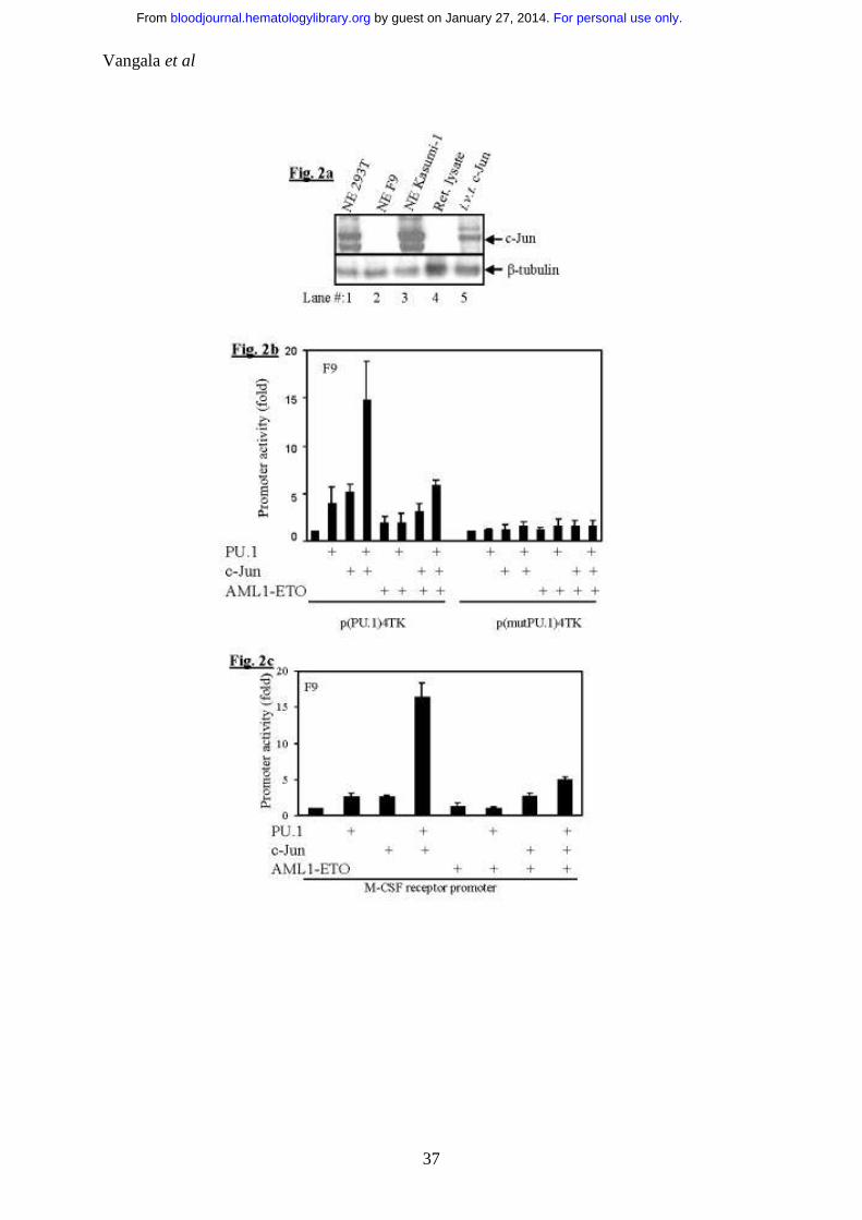
Figure 2: AML1-ETO inhibits co-activation of PU.1 by c-Jun
a. F9 cells do not express c-Jun: 100µg of nuclear extracts of 293T, F9 and
Kasumi-1 cells along with in vitro translated c-Jun were subjected to
SDS-PAGE and immunoblotted for c-Jun.
b. AML1-ETO inhibits PU.1/c-Jun transactivation capacity: F9 cells
were transfected with p(PU.1)4TK, a minimal TK promoter driven by
PU.1 DNA binding sites only or control vector p(mut.PU.1)4TK along
with expression plasmids of PU.1 (100ng), c-Jun (50ng) and AML1-ETO
(20ng).
c. AML1-ETO downregulates the PU.1 regulated M-CSF receptor
promoter activity by inhibiting PU.1/c-Jun function: F9 cells were
transfected with M-CSF receptor promoter and PU.1 (100ng), c-Jun
(50ng) and AML1-ETO (20ng). PU.1, c-Jun and AML1-ETO had no
effects on control vector pXP2 (data not shown).
For personal use only. by guest on January 27, 2014. bloodjournal.hematologylibrary.orgFrom

Vangala et al
30
Figure 3: AML1-ETO displaces the co-activator c-Jun from PU.1 by
binding to the ββββ3ββββ4 region of PU.1
a. AML1-ETO physically binds to PU.1 in vitro: GST pulldown assay was
performed using [35S]Met-labelled in vitro translated c-Jun (lane 1) or
AML1-ETO (lane 4) incubated with equal amounts of bacterially
expressed GST-PU.1 (lanes 2 and 5) or GST+beads (lane 3 and 6). GST-
PU.1 or GST were recovered using glutathione-agarose beads and
separated by SDS-PAGE prior to autoradiography.
b. AML1-ETO displaces c-Jun from binding to the ββββ3ββββ4 domain of
PU.1: Saturating amounts of in vitro translated c-Jun (20µl) were
incubated with GST-β3β4 and increasing amounts (from lane 7-lane 13)
of in vitro translated AML1-ETO (7.5-12.5µl) were incubated.
Densitometric quantification was also performed (given as % input of the
proteins).
For personal use only. by guest on January 27, 2014. bloodjournal.hematologylibrary.orgFrom

Vangala et al
31
Figure 4: AML1-ETO does not change the DNA binding of PU.1
The PU.1 binding sequence from the CD11b promoter was chosen and
labelled with γ32p-dATP (lane 1), incubated with in vitro translated PU.1
(lane 2) or in vitro translated PU.1 and anti-PU.1 antibody (lane 3). As a
competitor, unlabelled probe was used in 100molar excess with (lane 5) and
without anti-PU.1 antibody (lane 4). To investigate if this binding and
supershift is specific for PU.1, similar experiments were performed with
rabbit reticulocyte lysate (lanes 6, 7, 8, 9). In presence of AML1-ETO, PU.1
still binds to its DNA (lane 10) and supershifts with anti-PU.1 antibody (lane
11).
For personal use only. by guest on January 27, 2014. bloodjournal.hematologylibrary.orgFrom

Vangala et al
32
Figure 5: AML1-ETO downregulates PU.1’s transactivation capacity in
myeloid cells and the expression of PU.1’s target genes in AML patients
with t(8;21)
a. AML1-ETO downregulates PU.1’s transactivation in myeloid cells:
U937 cells were electroporated with wild type M-CSF receptor promoter,
M-CSF receptor promoter without (w/o) AML1 binding site,
p(PU.1)4TK, p(mutPU.1)4TK or pXP2 with and without AML1-ETO.
b. Low expression of PU.1 target genes in patients with t(8;21): AML
patients (n = number of patients) with t(8;21) have less positive cells for
cell surface markers regulated by PU.1 as compared to patients without
t(8;21). CD14 and CD64 promoters have PU.1 binding sites, but no
putative C/ΕΒPα, AML1 or MEF binding sites.
For personal use only. by guest on January 27, 2014. bloodjournal.hematologylibrary.orgFrom
Vangala et al
33
Figure 6: The anti-proliferative effect of PU.1 is downregulated by AML1-
ETO in mouse bone marrow cells
a. AML1-ETO causes proliferation in mouse bone marrow cells: Live
transduced mouse bone marrow cells with PU.1, AML1-ETO or PU.1 and
AML1-ETO were counted on day 3, 6, and 12 after Trypan blue staining.
Since both the empty vectors gave the same cell count, only one (PINCO)
vector has been represented as mock.
b. Expression of PU.1 in mouse bone marrow cells: The cells of above
transduction were lysed and immunoblot assays were performed for PU.1
and β-tubulin. NGFR (N) (lysate of empty vector of PU.1), N-PU.1
(NGFR-PU.1 transduced cells), PINCO (P) (lysate of empty vector of
AML1-ETO transduced cells), P-AML1-ETO (lysate of PINCO-AML1-
ETO transduced cells) and P-AML1-ETO+N-PU.1 (lysate of PINCO-
AML1-ETO and NGFR-PU.1 transduced cells). The ratio of PU.1/β-
tubulin was calculated after densitometric quantification of the bands.
For personal use only. by guest on January 27, 2014. bloodjournal.hematologylibrary.orgFrom

Vangala et al
34
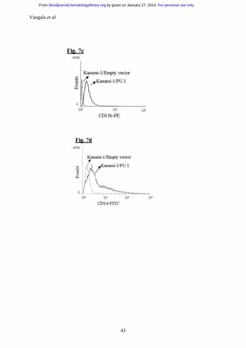
Figure 7: Transient overexpression of PU.1 induces differentiation towards
the monocytic lineage in AML1-ETO positive Kasumi-1 cells:
a. PU.1 induces differentiation in t(8;21) positive Kasumi-1 cells:
Kasumi-1 cells were transiently transfected with PU.1 (pGsam-PU.1-ires-
NGFR) or the empty vector (pGsam-ires-NGFR) and morphological
changes were observed on day 5. Arrows indicate the differentiating cells.
b. PU.1 overexpression in Kasumi-1 cells: Western blot showing PU.1
expression and β-tubulin in transfected Kasumi-1 cells after day 5.
c. PU.1 induces CD11b expression in Kasumi-1 cells: FACS analysis for
the cell surface expression of CD11b, in Kasumi-1 cells transfected with
empty vector or PU.1.
d. PU.1 induces CD14 expression in Kasumi-1 cells: In the same
experiment FACS analysis for the cell surface expression of CD14, in
Kasumi-1 cells transfected with empty vector or PU.1.
e. Kasumi-1 cell number decreases in PU.1 transfected cells: The above
transfected cells were counted by trypan blue staining on day 1, 2, 3, 4,
and 5 after transfection.
f. Model of AML1-ETO blocking PU.1 function: Model of AML1-ETO
interacting with PU.1 and displacing its co-activator c-Jun. This
downregulation of PU.1’s transcriptional activity by AML1-ETO results
in downregulation of PU.1 target genes important for myeloid
differentiation.
For personal use only. by guest on January 27, 2014. bloodjournal.hematologylibrary.orgFrom

Vangala et al
35
For personal use only. by guest on January 27, 2014. bloodjournal.hematologylibrary.orgFrom

Vangala et al
36
For personal use only. by guest on January 27, 2014. bloodjournal.hematologylibrary.orgFrom
Vangala et al
37
For personal use only. by guest on January 27, 2014. bloodjournal.hematologylibrary.orgFrom

Vangala et al
38
For personal use only. by guest on January 27, 2014. bloodjournal.hematologylibrary.orgFrom

Vangala et al
39
For personal use only. by guest on January 27, 2014. bloodjournal.hematologylibrary.orgFrom

Vangala et al
40
For personal use only. by guest on January 27, 2014. bloodjournal.hematologylibrary.orgFrom

Vangala et al
41
For personal use only. by guest on January 27, 2014. bloodjournal.hematologylibrary.orgFrom

Vangala et al
42
For personal use only. by guest on January 27, 2014. bloodjournal.hematologylibrary.orgFrom
Vangala et al
43
For personal use only. by guest on January 27, 2014. bloodjournal.hematologylibrary.orgFrom

Vangala et al
44
For personal use only. by guest on January 27, 2014. bloodjournal.hematologylibrary.orgFrom
Copyright © 2022 FDOKUMEN