Biodegradable Self-Assembling PEG-Copolymer as Vehicle for Poorly Water-Soluble Drugs
Upload
independentCategory
view
0download
0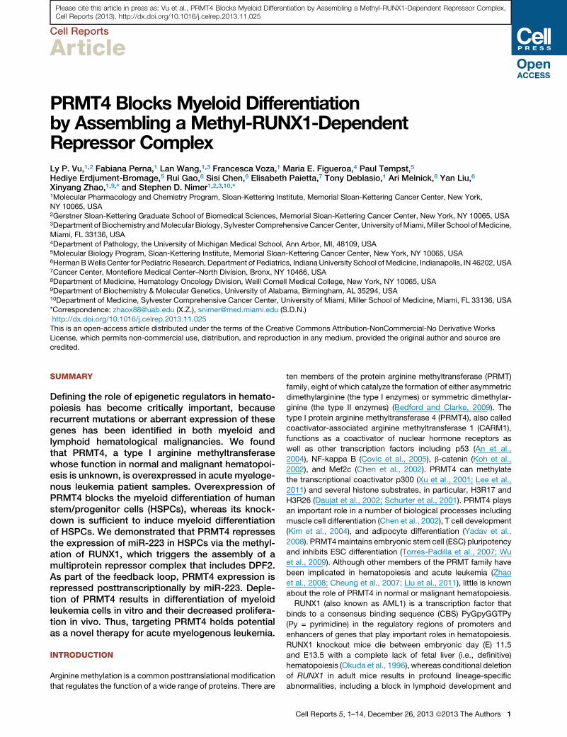
Please cite this article in press as: Vu et al., PRMT4 Blocks Myeloid Differentiation by Assembling a Methyl-RUNX1-Dependent Repressor Complex,Cell Reports (2013), http://dx.doi.org/10.1016/j.celrep.2013.11.025
Cell Reports
Article
PRMT4 Blocks Myeloid Differentiationby Assembling a Methyl-RUNX1-DependentRepressor ComplexLy P. Vu,1,2 Fabiana Perna,1 Lan Wang,1,3 Francesca Voza,1 Maria E. Figueroa,4 Paul Tempst,5
Hediye Erdjument-Bromage,5 Rui Gao,6 Sisi Chen,6 Elisabeth Paietta,7 Tony Deblasio,1 Ari Melnick,8 Yan Liu,6
Xinyang Zhao,1,9,* and Stephen D. Nimer1,2,3,10,*1Molecular Pharmacology and Chemistry Program, Sloan-Kettering Institute, Memorial Sloan-Kettering Cancer Center, New York,
NY 10065, USA2Gerstner Sloan-Kettering Graduate School of Biomedical Sciences, Memorial Sloan-Kettering Cancer Center, New York, NY 10065, USA3Department of Biochemistry andMolecular Biology, Sylvester Comprehensive Cancer Center, University ofMiami,Miller School ofMedicine,
Miami, FL 33136, USA4Department of Pathology, the University of Michigan Medical School, Ann Arbor, MI, 48109, USA5Molecular Biology Program, Sloan-Kettering Institute, Memorial Sloan-Kettering Cancer Center, New York, NY 10065, USA6HermanBWells Center for Pediatric Research, Department of Pediatrics, IndianaUniversity School of Medicine, Indianapolis, IN 46202, USA7Cancer Center, Montefiore Medical Center–North Division, Bronx, NY 10466, USA8Department of Medicine, Hematology Oncology Division, Weill Cornell Medical College, New York, NY 10065, USA9Department of Biochemistry & Molecular Genetics, University of Alabama, Birmingham, AL 35294, USA10Department of Medicine, Sylvester Comprehensive Cancer Center, University of Miami, Miller School of Medicine, Miami, FL 33136, USA
*Correspondence: [email protected] (X.Z.), [email protected] (S.D.N.)
http://dx.doi.org/10.1016/j.celrep.2013.11.025This is an open-access article distributed under the terms of the Creative Commons Attribution-NonCommercial-No Derivative Works
License, which permits non-commercial use, distribution, and reproduction in any medium, provided the original author and source are
credited.
SUMMARY
Defining the role of epigenetic regulators in hemato-poiesis has become critically important, becauserecurrent mutations or aberrant expression of thesegenes has been identified in both myeloid andlymphoid hematological malignancies. We foundthat PRMT4, a type I arginine methyltransferasewhose function in normal and malignant hematopoi-esis is unknown, is overexpressed in acute myeloge-nous leukemia patient samples. Overexpression ofPRMT4 blocks the myeloid differentiation of humanstem/progenitor cells (HSPCs), whereas its knock-down is sufficient to induce myeloid differentiationof HSPCs. We demonstrated that PRMT4 repressesthe expression of miR-223 in HSPCs via the methyl-ation of RUNX1, which triggers the assembly of amultiprotein repressor complex that includes DPF2.As part of the feedback loop, PRMT4 expression isrepressed posttranscriptionally by miR-223. Deple-tion of PRMT4 results in differentiation of myeloidleukemia cells in vitro and their decreased prolifera-tion in vivo. Thus, targeting PRMT4 holds potentialas a novel therapy for acute myelogenous leukemia.
INTRODUCTION
Arginine methylation is a common posttranslational modification
that regulates the function of a wide range of proteins. There are
ten members of the protein arginine methyltransferase (PRMT)
family, eight of which catalyze the formation of either asymmetric
dimethylarginine (the type I enzymes) or symmetric dimethylar-
ginine (the type II enzymes) (Bedford and Clarke, 2009). The
type I protein arginine methyltransferase 4 (PRMT4), also called
coactivator-associated arginine methyltransferase 1 (CARM1),
functions as a coactivator of nuclear hormone receptors as
well as other transcription factors including p53 (An et al.,
2004), NF-kappa B (Covic et al., 2005), b-catenin (Koh et al.,
2002), and Mef2c (Chen et al., 2002). PRMT4 can methylate
the transcriptional coactivator p300 (Xu et al., 2001; Lee et al.,
2011) and several histone substrates, in particular, H3R17 and
H3R26 (Daujat et al., 2002; Schurter et al., 2001). PRMT4 plays
an important role in a number of biological processes including
muscle cell differentiation (Chen et al., 2002), T cell development
(Kim et al., 2004), and adipocyte differentiation (Yadav et al.,
2008). PRMT4maintains embryonic stem cell (ESC) pluripotency
and inhibits ESC differentiation (Torres-Padilla et al., 2007; Wu
et al., 2009). Although other members of the PRMT family have
been implicated in hematopoiesis and acute leukemia (Zhao
et al., 2008; Cheung et al., 2007; Liu et al., 2011), little is known
about the role of PRMT4 in normal or malignant hematopoiesis.
RUNX1 (also known as AML1) is a transcription factor that
binds to a consensus binding sequence (CBS) PyGpyGGTPy
(Py = pyrimidine) in the regulatory regions of promoters and
enhancers of genes that play important roles in hematopoiesis.
RUNX1 knockout mice die between embryonic day (E) 11.5
and E13.5 with a complete lack of fetal liver (i.e., definitive)
hematopoiesis (Okuda et al., 1996), whereas conditional deletion
of RUNX1 in adult mice results in profound lineage-specific
abnormalities, including a block in lymphoid development and
Cell Reports 5, 1–14, December 26, 2013 ª2013 The Authors 1
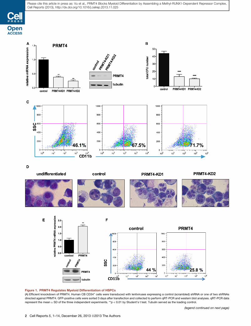
Figure 1. PRMT4 Regulates Myeloid Differentiation of HSPCs
(A) Efficient knockdown of PRMT4. Human CB CD34+ cells were transduced with lentiviruses expressing a control (scrambled) shRNA or one of two shRNAs
directed against PRMT4. GFP-positive cells were sorted 3 days after transfection and collected to perform qRT-PCR and western blot analyses. qRT-PCR data
represent the mean ± SD of the three independent experiments. **p < 0.01 by Student’s t test. Tubulin served as the loading control.
(legend continued on next page)
2 Cell Reports 5, 1–14, December 26, 2013 ª2013 The Authors
Please cite this article in press as: Vu et al., PRMT4 Blocks Myeloid Differentiation by Assembling a Methyl-RUNX1-Dependent Repressor Complex,Cell Reports (2013), http://dx.doi.org/10.1016/j.celrep.2013.11.025
Please cite this article in press as: Vu et al., PRMT4 Blocks Myeloid Differentiation by Assembling a Methyl-RUNX1-Dependent Repressor Complex,Cell Reports (2013), http://dx.doi.org/10.1016/j.celrep.2013.11.025
reduced megakaryocytic production, with little effect on adult
hematopoietic stem cells (HSCs). RUNX1 is one of the most
frequently altered genes in acute leukemia, either by chromo-
somal translocations such as the t(8;21) (Blyth et al., 2005) or
by point mutations or deletions that occur in 4%–10%of patients
with sporadic or therapy-related myelodysplastic syndrome and
acute myelogenous leukemia (AML) (Osato, 2004). Furthermore,
RUNX1 point mutations are found in affected individuals with the
inherited FPD/AML syndrome (familial platelet disorder with
propensity to AML) (Song et al., 1999). Posttranslational modifi-
cations, including ubiquitination, phosphorylation, acetylation,
and methylation fine-tune RUNX1 function (Wang et al., 2009);
for instance, arginine methylation of an RTAMR motif in RUNX1
by PRMT1 abrogates SIN3A binding, thereby potentiating
RUNX1-dependent transcriptional activation of its target genes
(Zhao et al., 2008). Similarly, microRNAs such as miR-17-5p,
miR-20a, and miR-106a can regulate RUNX1 protein expression
and thereby control aspects of hematopoietic cell differentiation
(Fontana et al., 2007).
The myeloid-specific microRNA-223 (miR-223) has been
shown to affect granulocytic differentiation. Loss of miR-223
impairs granulocytic maturation (Johnnidis et al., 2008), whereas
miR-223 overexpression promotes myeloid differentiation (Fazi
et al., 2005). miR-223 expression has been shown to be tran-
scriptionally regulated by NF-IA (Fazi et al., 2005), by C/EBPs
and PU.1 (Fukao et al., 2007) and by E2F1 (Pulikkan et al.,
2010). Fazi et al. reported that the AML1-ETO fusion protein
represses miR-223 expression by binding to a RUNX CBS
located upstream of the pre-miR-223 (Fazi et al., 2007). They,
and others, have found that miR-223 expression is downregu-
lated in AML patient samples (Fazi et al., 2007; Pulikkan et al.,
2010; Eyholzer et al., 2010).
In this study, we have identified PRMT4 as a regulator of the
myeloid differentiation process in stem/progenitor human he-
matopoietic cells (HSPCs). PRMT4 forms a regulatory loop
with miR-223, to reciprocally suppress the expression of each
other during myeloid differentiation. We also show that RUNX1
is a substrate of PRMT4 and that methylation of RUNX1 by
PRMT4 (on arginine 223 [R223]) leads to the recruitment of a
DPF2-containing repressor complex that binds critical miR-223
transcriptional regulatory elements (that are distinct from the
element identified by Fazi et al., 2007) and represses miR-223
expression. A decrease in PRMT4 expression is critical for
normal myeloid differentiation, but this normal event can be
subverted by malignant hematopoietic cell transformation.
(B) Downregulation of PRMT4 decreases CFU formation. 104 of the control or PRM
forming units (CFUs) was scored 2 weeks after the plating. The data represent th
t test.
(C) Downregulation of PRMT4 promotes themyeloid differentiation of HSPCs. GFP
for 7 days. Myeloid differentiation was determined by fluorescence-activated ce
(D) Downregulation of PRMT4 promotes the myeloid differentiation of HSPCs. C
containing medium. Cells growing in basic culture were used as the control for m
(E) Overexpression of PRMT4 was demonstrated at the mRNA and protein levels
control (GFP alone) or GFP and HA-PRMT4. GFP+ cells were sorted after 3 days
qRT-PCR data represent the mean ± SD of the three independent experiments.
(F) Overexpression of PRMT4 blocks themyeloid differentiation of HSPCs. GFP+ C
7 days. Myeloid differentiation was determined by FACS analysis of CD11b expr
See also Figure S1.
Thus, targeting PRMT4 represents a potential therapeutic
approach to promote the differentiation of AML blast cells.
RESULTS
PRMT4 Regulates Myeloid DifferentiationTo examine its function in hematopoiesis, we knocked down
PRMT4 in human cord blood (CB)-derived, CD34+ hematopoiet-
ic stem/progenitor cells (HSPCs), using lentiviral vectors that
express GFP and short hairpin RNAs (shRNAs) directed against
PRMT4. We assayed the extent of PRMT4 knockdown (KD) in
the GFP-positive transduced cells and found a 70%–80%
decrease in PRMT4 expression for the KD1 and KD2 short
hairpin RNAs, respectively (Figure 1A). The PRMT4-KD cells
generated far fewer CFUs when plated in methylcellulose (Fig-
ure 1B) and showed enhanced myeloid differentiation after
7 days in myeloid-promoting liquid culture (which contains
stem cell factor [SCF], FLT-3 ligand, interleukin [IL]-3, IL-6, gran-
ulocyte-macrophage colony stimulating factor [GM-CSF], and
granulocyte colony stimulating factor [G-CSF]) with 60%–70%
of the KD cells being CD11b positive and CD13 positive, versus
40% (and 14%) of the control cells being CD11b (or CD13)
positive (Figures 1C and S1B). Consistent with the immunophe-
notypic evidence, morphologic evidence also showed more
mature myeloid cells following PRMT4 KD (Figure 1D), with the
KD cells showing a condensed nucleus and clear nuclear lobula-
tion. In addition, PRMT4 KD mildly impaired erythroid differenti-
ation under erythroid-promoting culture conditions (SCF and
erythropoietin) (Figure S1E). Consistent with its effect on CFU
generation, a decrease in the numbers and size of the cobble-
stone areas was seen at week 5, reflecting impaired HSPC
self-renewal when PRMT4 was knocked down (Figure S1D).
We next examined whether PRMT4 overexpression blocks
myeloid differentiation. Indeed, we found a marked reduction
in CD11b-positive cells generated from PRMT4-overexpressing
CD34+ cells (compared to the control cells) after 7 days in
myeloid differentiation promoting cultures (Figure 1E). Thus,
PRMT4 appears to be an important negative regulator of normal
myeloid differentiation.
PRMT4 Is Regulated Posttranscriptionally by miR-223during Myeloid DifferentiationGiven the prominent effect of PRMT4 on myeloid differentiation,
we assessed changes in PRMT4 expression during normal
in vitro HSPC differentiation. We cultured human CBCD34+ cells
T4 knockdown cells were plated inmethylcellulose. The total number of colony
e mean ± SD of the three independent experiments. ***p < 0.001 by Student’s
+ CD34+ cells were cultured inmyeloid-promoting cytokine containingmedium
ll sorting (FACS) analysis of CD11b expression.
ellular morphology was evaluated after 7 days in myeloid-promoting cytokine
yeloid differentiation.
. Human CB CD34+ cells were transduced with retroviruses expressing either
of transfection and collected to perform qRT-PCR and western blot analyses.
**p < 0.01 by Student’s t test.
D34+ cells were cultured inmyeloid-promoting cytokine containingmedium for
ession.
Cell Reports 5, 1–14, December 26, 2013 ª2013 The Authors 3
(legend on next page)
4 Cell Reports 5, 1–14, December 26, 2013 ª2013 The Authors
Please cite this article in press as: Vu et al., PRMT4 Blocks Myeloid Differentiation by Assembling a Methyl-RUNX1-Dependent Repressor Complex,Cell Reports (2013), http://dx.doi.org/10.1016/j.celrep.2013.11.025
Please cite this article in press as: Vu et al., PRMT4 Blocks Myeloid Differentiation by Assembling a Methyl-RUNX1-Dependent Repressor Complex,Cell Reports (2013), http://dx.doi.org/10.1016/j.celrep.2013.11.025
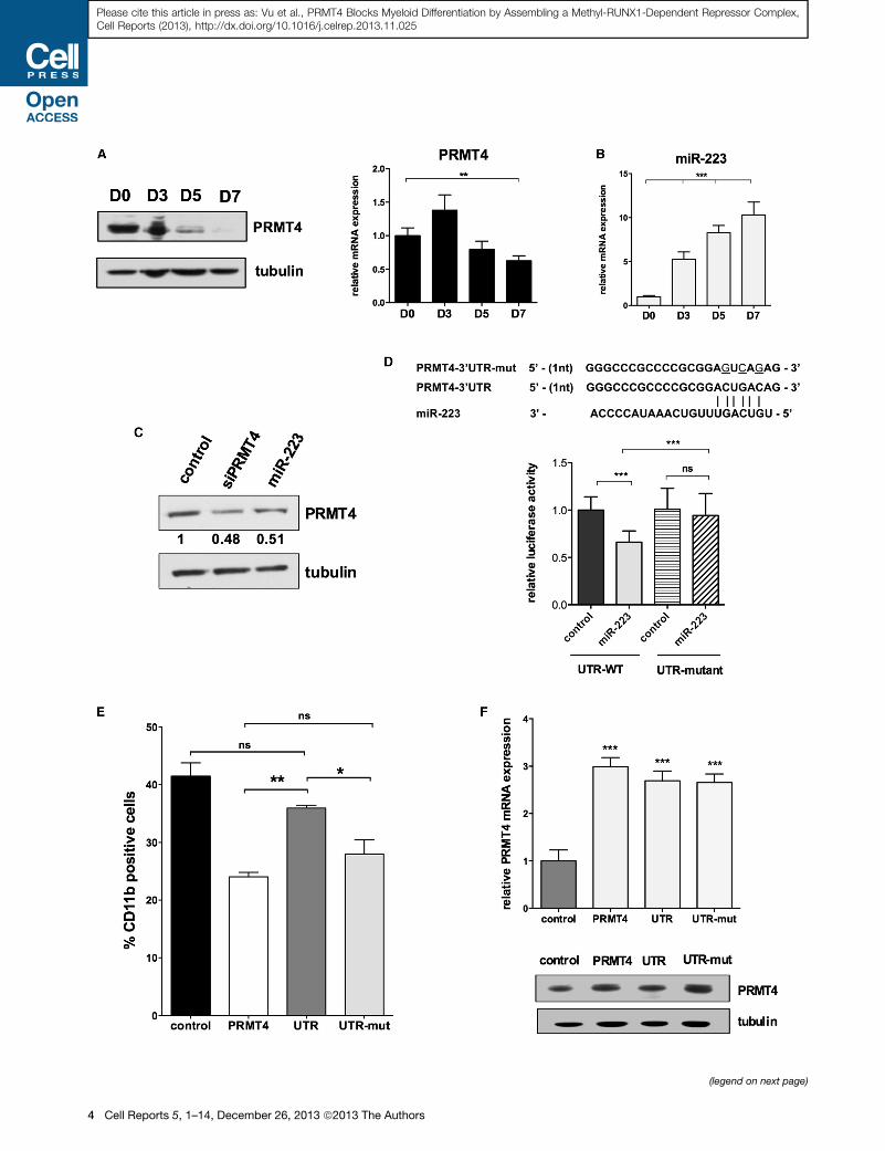
in myeloid-promoting liquid culture and observed a significant
decrease in PRMT4 protein levels over a 7 day period (Figure 2A).
PRMT4 mRNA levels varied only slightly during this process,
suggesting that PRMT4 is being regulated posttranscriptionally.
MicroRNA target prediction programs (TargetScan release 5.2
[Figure S2A] and PITA) suggested that PRMT4 is a potential
target of several microRNAs, including miR-223, a myeloid-
specific microRNA. Interestingly, a seed sequence for miR-223
is found in the 30 UTR region of PRMT4, which is located adjacent
to the stop codon of the PRMT4 open reading frame (ORF)
(16–22 nt from the stop codon); this location could confer a
strong, translational regulatory effect (Grimson et al., 2007; Eula-
lio et al., 2008). Indeed, we found that miR-223 expression
steadily increases during myeloid differentiation (Figure 2B),
concomitant with decreasing PRMT4 protein expression. To
determine whether miR-223 regulates PRMT4 expression, we
transiently transfected CD34+ cells with a short hairpin encoding
the mature miR-223 for 24 hr and monitored PRMT4 expression,
using siPRMT4 as a short-hairpin-positive control (and a scram-
bled short hairpin as a negative control). PRMT4 protein levels
decreased by 50% in the miR-223-overexpressing cells, similar
to that seen in the cells expressing small interfering RNA (siRNA)
directed against PRMT4 (Figure 2C).
To validate that PRMT4 is directly targeted by miR-223, we
cloned its full-length 30 UTR into a luciferase reporter plasmid
(UTR-WT), using the same construct with a mutated miR-223
targeting sequence (UTR-mut), which cannot bind miR-223, as
negative control. We expressed these constructs in 293T cells,
with either the miR-223 short hairpin or the control short hairpin
and found that miR-223 decreased the luciferase activity of the
UTR-WT, with no effect on the UTR-mut reporter plasmid (Fig-
ures 2D and S2C). Thus, miR-223 directly targets PRMT4 by
binding to its recognition sequence in the 30 UTR; this suggests
that PRMT4 and miR-223 form a regulatory loop to regulate
myeloid differentiation.
To determine how important the regulation of PRMT4 expres-
sion by miR-223 is for the effects of PRMT4 on myeloid differen-
tiation, we overexpressed the PRMT4-ORF with either the WT or
the mutant PRMT4 30 UTR, in human HSPCs. Although theWT 30
Figure 2. PRMT4 Is a Potential Target Gene of miR-223 during Myeloid
(A) PRMT4 protein expression is progressively downregulated during myeloid d
myeloid differentiation (right). Isolated CD34+ cells were cultured in myeloid-prom
(D) 0, 3, 5, and 7. Western blot and qRT-PCR analyses were performed. qRT-PCR
Student’s t test. Tubulin served as the loading control.
(B) miR-223 expression steadily increases during myeloid differentiation. qRT-PCR
by Student’s t test.
(C) Overexpression of miR-223 or siRNA directed against PRMT4 lowers PRMT4 p
otideswere transiently expressed inCD34+ cells and 24hr postelectroporation; the c
(D) Putative miR-223 binding site in the PRMT4 30 UTR is shown at the top (based
with a reporter plasmid containing either the wild-type 30 UTR-PRMT4 or the m
without miR-223. Renilla luciferase values are normalized based on the value of fir
0.001 by Student’s t test.
(E) Control of PRMT4 expression by miR-223 is essential to regulate PRMT4
transduced with retroviruses expressing control-GFP alone; GFP-PRMT4-ORF o
were cultured in myeloid-promoting cytokine containing medium for 7 days. Myel
percentage of CD11b-positive cells was quantified as mean ± SD based on thre
(F) qRT-PCR and western blot analyses of PRMT4 expression in control CD34+ ce
mut. Tubulin served as the loading control.
See also Figure S1.
UTR abrogated the PRMT4 imposed block inmyeloid differentia-
tion, when PRMT4was expressed without the 30 UTR, or with the
mutant 30 UTR, the block in myeloid differentiation was seen
(Figure 2E). These effects correlate with PRMT4 protein levels
(Figure 2F) and demonstrate that the regulation of PRMT4 by
miR-223 is important to its functionduringmyeloiddifferentiation.
PRMT4 Represses miR-223 ExpressionGiven the known transcriptional regulatory role of PRMT4, to
determine how PRMT4 controls myeloid differentiation, we first
examined the expression level of several differentiation-specific
‘‘master’’ transcription factors, including PU.1, C/EBPa, KLF4,
andGATA1 in PRMT4-KD cells. Althoughwe found no significant
changes (Figure S3A), we observed a consistent increase inmiR-
223 expression in the PRMT4-KD cells (Figure 3A). Because
upregulation of miR-223 has been reported to promote the
myeloid differentiation of leukemia cells (Fazi et al., 2007), we
overexpressed miR-223 in normal CB CD34+ cells, and saw a
significant increase in CD11b-positive cells (51.2% versus
38.1% for the control cells) (Figure 3B) as well as decreased
PRMT4 expression (Figure S2B). We observed decreased miR-
223 expression, when PRMT4 is overexpressed (Figure 3C).
When we knocked down miR-223 expression, we found a
modest but consistent reduction in CD11b-positive cells
(38.6% ± 3.1% versus 44.8% ± 1.8%, p < 0.05) (Figures 3D
and S3B), which suggests that other microRNAs may compen-
sate formiR-223 duringmyeloid differentiation. This is consistent
with the phenotype of miR-223 knockout mice, where miR-223
is important, but not essential, for granulocytic maturation and
function (Johnnidis et al., 2008).
To determine whether PRMT4 regulates the transcription of
miR-223, we quantified the level of miR-223 primary transcript
(pri-miR-223) and found that PRMT4 expression does reci-
procally regulate pri-miR-223 levels in CD34+ cells (Figure S3C).
In addition, gene expression analysis of PRMT4 KD cells
(GSE46056) revealed a gene signature consistent with an upre-
gulation of myeloid differentiation (Figure S3D). Thus, PRMT4
regulates myeloid differentiation, at least in part, by modulating
miR-223 expression.
Differentiation of HSPCs
ifferentiation (left), whereas PRMT4 mRNA level decreases modestly during
oting cytokine containingmedium and collected at sequential time points: days
data represent the mean ± SD of three independent experiments. **p < 0.01 by
data represent the mean ± SD of three independent experiments. ***p < 0.001
rotein levels in HSPCs. miR-223, siRNA against PRMT4, and control oligonucle-
ells were collected and assayed for PRMT4 expression bywestern blot analyses.
on TargetScan.org release 5.2). Luciferase activity in 293T cells cotransfected
utated 30 UTR (3-UTR-mut, which lacks the seed miR-223 sequence) with or
efly luciferase. Mean ± SD from three independent experiments is shown. ***p <
function during normal myeloid differentiation. Human CB CD34+ cells were
r GFP-PRMT4 30 UTR or GFP-PRMT4 30 UTR-mut. Sorted GFP+ CD34+ cells
oid differentiation was determined by FACS analysis of CD11b expression. The
e independent experiments. *p < 0.05; **p < 0.01 by Student’s t test.
lls or CD34+ cells expressing PRMT4-ORF, PRMT4 30 UTR, or PRMT4 30 UTR-
Cell Reports 5, 1–14, December 26, 2013 ª2013 The Authors 5
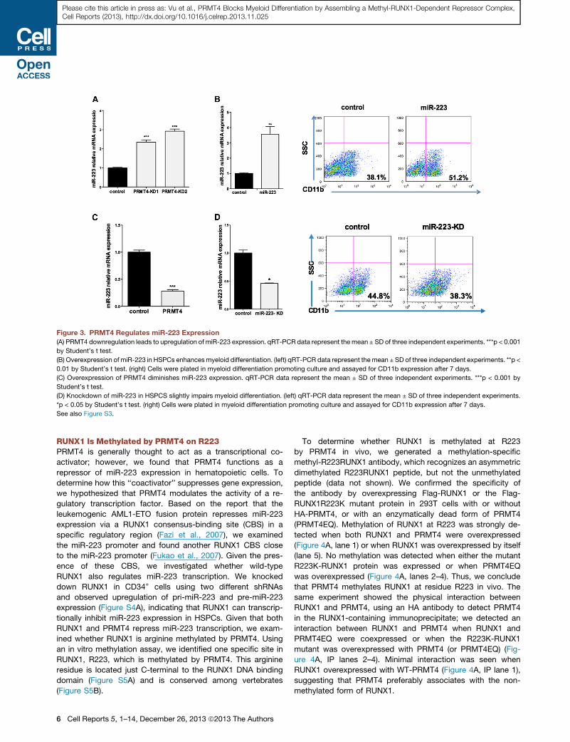
Figure 3. PRMT4 Regulates miR-223 Expression
(A) PRMT4 downregulation leads to upregulation of miR-223 expression. qRT-PCR data represent the mean ± SD of three independent experiments. ***p < 0.001
by Student’s t test.
(B) Overexpression of miR-223 in HSPCs enhancesmyeloid differentiation. (left) qRT-PCR data represent the mean ± SD of three independent experiments. **p <
0.01 by Student’s t test. (right) Cells were plated in myeloid differentiation promoting culture and assayed for CD11b expression after 7 days.
(C) Overexpression of PRMT4 diminishes miR-223 expression. qRT-PCR data represent the mean ± SD of three independent experiments. ***p < 0.001 by
Student’s t test.
(D) Knockdown of miR-223 in HSPCS slightly impairs myeloid differentiation. (left) qRT-PCR data represent the mean ± SD of three independent experiments.
*p < 0.05 by Student’s t test. (right) Cells were plated in myeloid differentiation promoting culture and assayed for CD11b expression after 7 days.
See also Figure S3.
Please cite this article in press as: Vu et al., PRMT4 Blocks Myeloid Differentiation by Assembling a Methyl-RUNX1-Dependent Repressor Complex,Cell Reports (2013), http://dx.doi.org/10.1016/j.celrep.2013.11.025
RUNX1 Is Methylated by PRMT4 on R223PRMT4 is generally thought to act as a transcriptional co-
activator; however, we found that PRMT4 functions as a
repressor of miR-223 expression in hematopoietic cells. To
determine how this ‘‘coactivator’’ suppresses gene expression,
we hypothesized that PRMT4 modulates the activity of a re-
gulatory transcription factor. Based on the report that the
leukemogenic AML1-ETO fusion protein represses miR-223
expression via a RUNX1 consensus-binding site (CBS) in a
specific regulatory region (Fazi et al., 2007), we examined
the miR-223 promoter and found another RUNX1 CBS close
to the miR-223 promoter (Fukao et al., 2007). Given the pres-
ence of these CBS, we investigated whether wild-type
RUNX1 also regulates miR-223 transcription. We knocked
down RUNX1 in CD34+ cells using two different shRNAs
and observed upregulation of pri-miR-223 and pre-miR-223
expression (Figure S4A), indicating that RUNX1 can transcrip-
tionally inhibit miR-223 expression in HSPCs. Given that both
RUNX1 and PRMT4 repress miR-223 transcription, we exam-
ined whether RUNX1 is arginine methylated by PRMT4. Using
an in vitro methylation assay, we identified one specific site in
RUNX1, R223, which is methylated by PRMT4. This arginine
residue is located just C-terminal to the RUNX1 DNA binding
domain (Figure S5A) and is conserved among vertebrates
(Figure S5B).
6 Cell Reports 5, 1–14, December 26, 2013 ª2013 The Authors
To determine whether RUNX1 is methylated at R223
by PRMT4 in vivo, we generated a methylation-specific
methyl-R223RUNX1 antibody, which recognizes an asymmetric
dimethylated R223RUNX1 peptide, but not the unmethylated
peptide (data not shown). We confirmed the specificity of
the antibody by overexpressing Flag-RUNX1 or the Flag-
RUNX1R223K mutant protein in 293T cells with or without
HA-PRMT4, or with an enzymatically dead form of PRMT4
(PRMT4EQ). Methylation of RUNX1 at R223 was strongly de-
tected when both RUNX1 and PRMT4 were overexpressed
(Figure 4A, lane 1) or when RUNX1 was overexpressed by itself
(lane 5). No methylation was detected when either the mutant
R223K-RUNX1 protein was expressed or when PRMT4EQ
was overexpressed (Figure 4A, lanes 2–4). Thus, we conclude
that PRMT4 methylates RUNX1 at residue R223 in vivo. The
same experiment showed the physical interaction between
RUNX1 and PRMT4, using an HA antibody to detect PRMT4
in the RUNX1-containing immunoprecipitate; we detected an
interaction between RUNX1 and PRMT4 when RUNX1 and
PRMT4EQ were coexpressed or when the R223K-RUNX1
mutant was overexpressed with PRMT4 (or PRMT4EQ) (Fig-
ure 4A, IP lanes 2–4). Minimal interaction was seen when
RUNX1 overexpressed with WT-PRMT4 (Figure 4A, IP lane 1),
suggesting that PRMT4 preferably associates with the non-
methylated form of RUNX1.
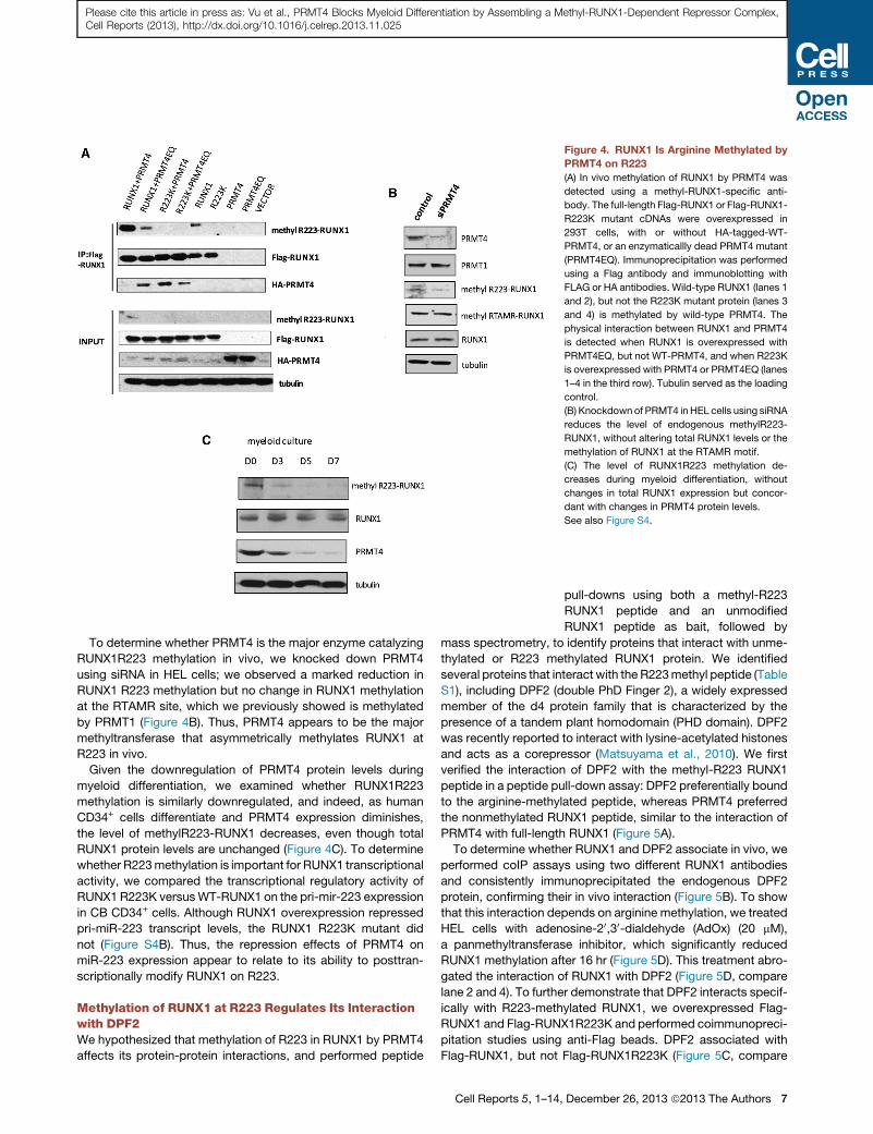
Figure 4. RUNX1 Is Arginine Methylated by
PRMT4 on R223
(A) In vivo methylation of RUNX1 by PRMT4 was
detected using a methyl-RUNX1-specific anti-
body. The full-length Flag-RUNX1 or Flag-RUNX1-
R223K mutant cDNAs were overexpressed in
293T cells, with or without HA-tagged-WT-
PRMT4, or an enzymaticallly dead PRMT4 mutant
(PRMT4EQ). Immunoprecipitation was performed
using a Flag antibody and immunoblotting with
FLAG or HA antibodies. Wild-type RUNX1 (lanes 1
and 2), but not the R223K mutant protein (lanes 3
and 4) is methylated by wild-type PRMT4. The
physical interaction between RUNX1 and PRMT4
is detected when RUNX1 is overexpressed with
PRMT4EQ, but not WT-PRMT4, and when R223K
is overexpressed with PRMT4 or PRMT4EQ (lanes
1–4 in the third row). Tubulin served as the loading
control.
(B) Knockdown of PRMT4 in HEL cells using siRNA
reduces the level of endogenous methylR223-
RUNX1, without altering total RUNX1 levels or the
methylation of RUNX1 at the RTAMR motif.
(C) The level of RUNX1R223 methylation de-
creases during myeloid differentiation, without
changes in total RUNX1 expression but concor-
dant with changes in PRMT4 protein levels.
See also Figure S4.
Please cite this article in press as: Vu et al., PRMT4 Blocks Myeloid Differentiation by Assembling a Methyl-RUNX1-Dependent Repressor Complex,Cell Reports (2013), http://dx.doi.org/10.1016/j.celrep.2013.11.025
To determine whether PRMT4 is the major enzyme catalyzing
RUNX1R223 methylation in vivo, we knocked down PRMT4
using siRNA in HEL cells; we observed a marked reduction in
RUNX1 R223 methylation but no change in RUNX1 methylation
at the RTAMR site, which we previously showed is methylated
by PRMT1 (Figure 4B). Thus, PRMT4 appears to be the major
methyltransferase that asymmetrically methylates RUNX1 at
R223 in vivo.
Given the downregulation of PRMT4 protein levels during
myeloid differentiation, we examined whether RUNX1R223
methylation is similarly downregulated, and indeed, as human
CD34+ cells differentiate and PRMT4 expression diminishes,
the level of methylR223-RUNX1 decreases, even though total
RUNX1 protein levels are unchanged (Figure 4C). To determine
whether R223methylation is important for RUNX1 transcriptional
activity, we compared the transcriptional regulatory activity of
RUNX1 R223K versusWT-RUNX1 on the pri-mir-223 expression
in CB CD34+ cells. Although RUNX1 overexpression repressed
pri-miR-223 transcript levels, the RUNX1 R223K mutant did
not (Figure S4B). Thus, the repression effects of PRMT4 on
miR-223 expression appear to relate to its ability to posttran-
scriptionally modify RUNX1 on R223.
Methylation of RUNX1 at R223 Regulates Its Interactionwith DPF2We hypothesized that methylation of R223 in RUNX1 by PRMT4
affects its protein-protein interactions, and performed peptide
Cell Reports 5, 1–14
pull-downs using both a methyl-R223
RUNX1 peptide and an unmodified
RUNX1 peptide as bait, followed by
mass spectrometry, to identify proteins that interact with unme-
thylated or R223 methylated RUNX1 protein. We identified
several proteins that interact with the R223methyl peptide (Table
S1), including DPF2 (double PhD Finger 2), a widely expressed
member of the d4 protein family that is characterized by the
presence of a tandem plant homodomain (PHD domain). DPF2
was recently reported to interact with lysine-acetylated histones
and acts as a corepressor (Matsuyama et al., 2010). We first
verified the interaction of DPF2 with the methyl-R223 RUNX1
peptide in a peptide pull-down assay: DPF2 preferentially bound
to the arginine-methylated peptide, whereas PRMT4 preferred
the nonmethylated RUNX1 peptide, similar to the interaction of
PRMT4 with full-length RUNX1 (Figure 5A).
To determine whether RUNX1 and DPF2 associate in vivo, we
performed coIP assays using two different RUNX1 antibodies
and consistently immunoprecipitated the endogenous DPF2
protein, confirming their in vivo interaction (Figure 5B). To show
that this interaction depends on arginine methylation, we treated
HEL cells with adenosine-20,30-dialdehyde (AdOx) (20 mM),
a panmethyltransferase inhibitor, which significantly reduced
RUNX1 methylation after 16 hr (Figure 5D). This treatment abro-
gated the interaction of RUNX1 with DPF2 (Figure 5D, compare
lane 2 and 4). To further demonstrate that DPF2 interacts specif-
ically with R223-methylated RUNX1, we overexpressed Flag-
RUNX1 and Flag-RUNX1R223K and performed coimmunopreci-
pitation studies using anti-Flag beads. DPF2 associated with
Flag-RUNX1, but not Flag-RUNX1R223K (Figure 5C, compare
, December 26, 2013 ª2013 The Authors 7
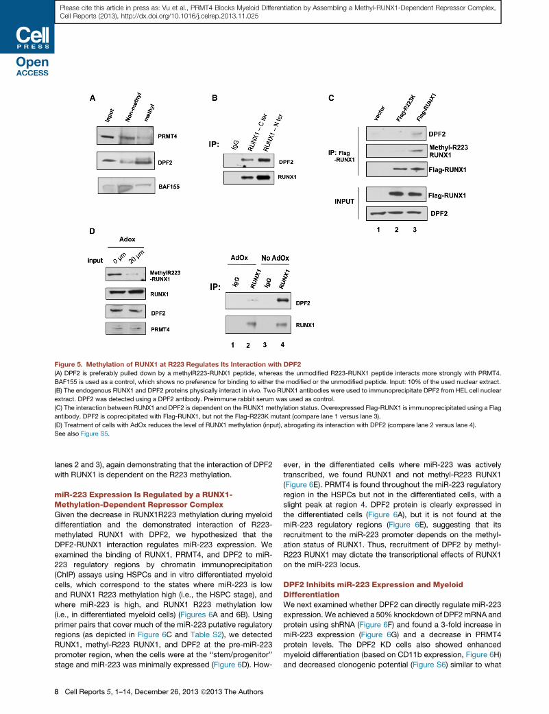
Figure 5. Methylation of RUNX1 at R223 Regulates Its Interaction with DPF2
(A) DPF2 is preferably pulled down by a methylR223-RUNX1 peptide, whereas the unmodified R223-RUNX1 peptide interacts more strongly with PRMT4.
BAF155 is used as a control, which shows no preference for binding to either the modified or the unmodified peptide. Input: 10% of the used nuclear extract.
(B) The endogenous RUNX1 and DPF2 proteins physically interact in vivo. Two RUNX1 antibodies were used to immunoprecipitate DPF2 from HEL cell nuclear
extract. DPF2 was detected using a DPF2 antibody. Preimmune rabbit serum was used as control.
(C) The interaction between RUNX1 and DPF2 is dependent on the RUNX1 methylation status. Overexpressed Flag-RUNX1 is immunoprecipitated using a Flag
antibody. DPF2 is coprecipitated with Flag-RUNX1, but not the Flag-R223K mutant (compare lane 1 versus lane 3).
(D) Treatment of cells with AdOx reduces the level of RUNX1 methylation (input), abrogating its interaction with DPF2 (compare lane 2 versus lane 4).
See also Figure S5.
Please cite this article in press as: Vu et al., PRMT4 Blocks Myeloid Differentiation by Assembling a Methyl-RUNX1-Dependent Repressor Complex,Cell Reports (2013), http://dx.doi.org/10.1016/j.celrep.2013.11.025
lanes 2 and 3), again demonstrating that the interaction of DPF2
with RUNX1 is dependent on the R223 methylation.
miR-223 Expression Is Regulated by a RUNX1-Methylation-Dependent Repressor ComplexGiven the decrease in RUNX1R223 methylation during myeloid
differentiation and the demonstrated interaction of R223-
methylated RUNX1 with DPF2, we hypothesized that the
DPF2-RUNX1 interaction regulates miR-223 expression. We
examined the binding of RUNX1, PRMT4, and DPF2 to miR-
223 regulatory regions by chromatin immunoprecipitation
(ChIP) assays using HSPCs and in vitro differentiated myeloid
cells, which correspond to the states where miR-223 is low
and RUNX1 R223 methylation high (i.e., the HSPC stage), and
where miR-223 is high, and RUNX1 R223 methylation low
(i.e., in differentiated myeloid cells) (Figures 6A and 6B). Using
primer pairs that cover much of the miR-223 putative regulatory
regions (as depicted in Figure 6C and Table S2), we detected
RUNX1, methyl-R223 RUNX1, and DPF2 at the pre-miR-223
promoter region, when the cells were at the ‘‘stem/progenitor’’
stage and miR-223 was minimally expressed (Figure 6D). How-
8 Cell Reports 5, 1–14, December 26, 2013 ª2013 The Authors
ever, in the differentiated cells where miR-223 was actively
transcribed, we found RUNX1 and not methyl-R223 RUNX1
(Figure 6E). PRMT4 is found throughout the miR-223 regulatory
region in the HSPCs but not in the differentiated cells, with a
slight peak at region 4. DPF2 protein is clearly expressed in
the differentiated cells (Figure 6A), but it is not found at the
miR-223 regulatory regions (Figure 6E), suggesting that its
recruitment to the miR-223 promoter depends on the methyl-
ation status of RUNX1. Thus, recruitment of DPF2 by methyl-
R223 RUNX1 may dictate the transcriptional effects of RUNX1
on the miR-223 locus.
DPF2 Inhibits miR-223 Expression and MyeloidDifferentiationWe next examined whether DPF2 can directly regulate miR-223
expression. We achieved a 50% knockdown of DPF2mRNA and
protein using shRNA (Figure 6F) and found a 3-fold increase in
miR-223 expression (Figure 6G) and a decrease in PRMT4
protein levels. The DPF2 KD cells also showed enhanced
myeloid differentiation (based on CD11b expression, Figure 6H)
and decreased clonogenic potential (Figure S6) similar to what
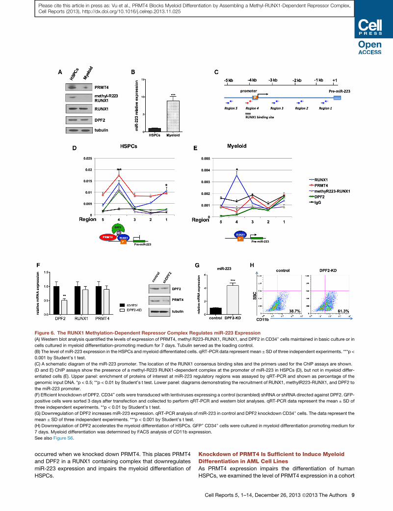
Figure 6. The RUNX1 Methylation-Dependent Repressor Complex Regulates miR-223 Expression
(A) Western blot analysis quantified the levels of expression of PRMT4, methyl R223-RUNX1, RUNX1, and DPF2 in CD34+ cells maintained in basic culture or in
cells cultured in myeloid differentiation-promoting medium for 7 days. Tubulin served as the loading control.
(B) The level of miR-223 expression in the HSPCs and myeloid differentiated cells. qRT-PCR data represent mean ± SD of three independent experiments. ***p <
0.001 by Student’s t test.
(C) A schematic diagram of the miR-223 promoter. The location of the RUNX1 consensus binding sites and the primers used for the ChIP assays are shown.
(D and E) ChIP assays show the presence of a methyl-R223 RUNX1-dependent complex at the promoter of miR-223 in HSPCs (D), but not in myeloid differ-
entiated cells (E). Upper panel: enrichment of proteins of interest at miR-223 regulatory regions was assayed by qRT-PCR and shown as percentage of the
genomic input DNA. *p < 0.5; **p < 0.01 by Student’s t test. Lower panel: diagrams demonstrating the recruitment of RUNX1, methylR223-RUNX1, and DPF2 to
the miR-223 promoter.
(F) Efficient knockdown of DPF2. CD34+ cells were transduced with lentiviruses expressing a control (scrambled) shRNA or shRNA directed against DPF2. GFP-
positive cells were sorted 3 days after transfection and collected to perform qRT-PCR and western blot analyses. qRT-PCR data represent the mean ± SD of
three independent experiments. **p < 0.01 by Student’s t test.
(G) Downregulation of DPF2 increases miR-223 expression. qRT-PCR analysis of miR-223 in control and DPF2 knockdown CD34+ cells. The data represent the
mean ± SD of three independent experiments. ***p < 0.001 by Student’s t test.
(H) Downregulation of DPF2 accelerates the myeloid differentiation of HSPCs. GFP+ CD34+ cells were cultured in myeloid differentiation promoting medium for
7 days. Myeloid differentiation was determined by FACS analysis of CD11b expression.
See also Figure S6.
Please cite this article in press as: Vu et al., PRMT4 Blocks Myeloid Differentiation by Assembling a Methyl-RUNX1-Dependent Repressor Complex,Cell Reports (2013), http://dx.doi.org/10.1016/j.celrep.2013.11.025
occurred when we knocked down PRMT4. This places PRMT4
and DPF2 in a RUNX1 containing complex that downregulates
miR-223 expression and impairs the myeloid differentiation of
HSPCs.
Knockdown of PRMT4 Is Sufficient to Induce MyeloidDifferentiation in AML Cell LinesAs PRMT4 expression impairs the differentiation of human
HSPCs, we examined the level of PRMT4 expression in a cohort
Cell Reports 5, 1–14, December 26, 2013 ª2013 The Authors 9
(legend on next page)
10 Cell Reports 5, 1–14, December 26, 2013 ª2013 The Authors
Please cite this article in press as: Vu et al., PRMT4 Blocks Myeloid Differentiation by Assembling a Methyl-RUNX1-Dependent Repressor Complex,Cell Reports (2013), http://dx.doi.org/10.1016/j.celrep.2013.11.025
Please cite this article in press as: Vu et al., PRMT4 Blocks Myeloid Differentiation by Assembling a Methyl-RUNX1-Dependent Repressor Complex,Cell Reports (2013), http://dx.doi.org/10.1016/j.celrep.2013.11.025
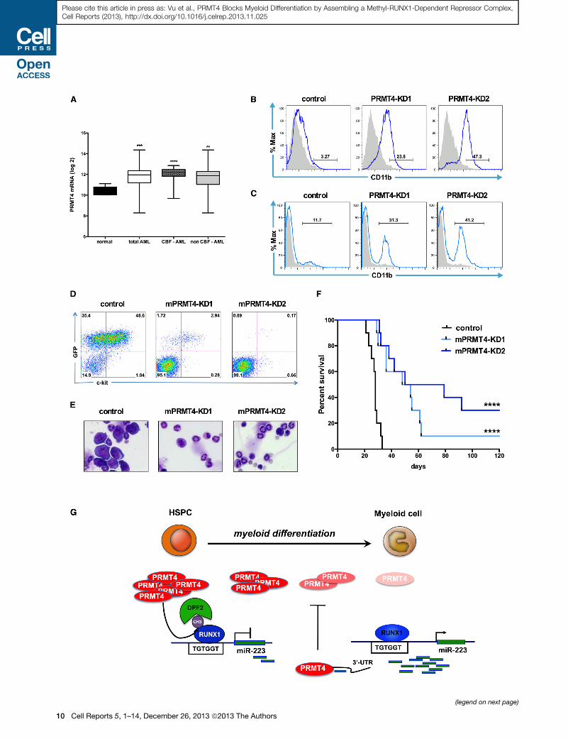
of 318 AML patient samples (GSE24505) (Figueroa et al., 2010).
PRMT4 levels were significantly upregulated in the AML
samples, compared to the control group (n = 5) (Figure 7A). A
high level of PRMT4 expression was seen in core-binding factor
(CBF) AMLs (which express either AML1-ETO or CBFb-
SMMHC), which exhibit low-level miR-223 expression, but,
overall, about 70% of the AML patients had at least a 2-fold in-
crease in PRMT4 expression (Figure S7A).
Several studies have shown that overexpression of miR-223
can promote the granulocytic differentiation of NB4 acute
promyelocytic leukemia (APL) cells (Fazi et al., 2005). We
knocked down PRMT4 in NB4 cells and triggered myeloid
differentiation with a significant increase in the number of
CD11b-positive cells (3.3% control versus 23.8% and 47.3%)
(Figure 7B) as well as changes in cellular morphology and an
upregulation of miR-223 expression (Figures S7B and S7C). A
similar induction of differentiation was also observed in the
ATRA-resistant NB4-R4 cells following PRMT4-KD (Rosenauer
et al., 1996) (Figures S7D and S7E). Because therapeutic target-
ing of most leukemia fusion proteins, including AML1-ETO
remains elusive, we tested whether targeting PRMT4 can pro-
mote the differentiation of AML1-ETO-expressing cells (Fig-
ure 7A). We knocked down PRMT4 in t(8;21)-positive Kasumi-1
cells and found increased differentiation (Figures 7C and S7E).
We also saw significant apoptosis of all three cell lines when
PRMT4 is knocked down (Figure S7F), suggesting that PRMT4
not only impairs the differentiation of these leukemia cells, but
it is also critical for their survival.
Knockdown of PRMT4 Reduces the Leukemia BurdenIn VivoTo investigate the in vivo role of PRMT4 in leukemogenesis, we
used shRNA-expressing lentiviruses to knock down PRMT4 in
the AML1-ETO9a (AE9a)-driven mouse AML model (Yan et al.,
2006; Wang et al., 2011). Leukemia cells growing in culture
Figure 7. Downregulation of PRMT4 Is Sufficient to Induce Myeloid Di
(A) PRMT4 expression is upregulated in AML patient samples. The graph shows
cells isolated from five healthy donors (normal) or 318 AML patients. CBF, core-b
0.001, **p < 0.01 by Student’s t test.
(B) Knockdown of PRMT4 triggers the myeloid differentiation of NB4 cells. NB4 ce
shRNA directed against PRMT4. Sorted GFP-positive cells were cultured for 3 da
Figure S7D.
(C) Knockdown of PRMT4 triggers the myeloid differentiation of Kasumi-1 cells. K
shRNA or shRNA directed against PRMT4. Sorted GFP-positive cells were culture
shown in Figure S7D.
(D) FACS analysis showed far fewer GFP+ ckit+ cells in peripheral blood (PB) of th
cells at week 3. AE9a expressing mouse leukemia cells were transduced with
mPRMT4-KD1 and mPRMT4-KD2). Transduced cells were sorted for expression
sublethal irradiation.
(E) Bone marrow (BM) morphology shows marked reduction in the number of le
AE9a-control cells.
(F) Knockdown of PRMT4 prolongs the survival of AE9a transplanted mice. Theme
group (54 days and 64.5 days versus 28 days p < 0.0001).
(G) A schematic model showing how PRMT4 regulates myeloid differentiation of
myeloid differentiation. PRMT4 inhibits myeloid differentiation by assembling
expression. When HSPCs undergo myeloid differentiation, PRMT4 expression
allowing it to be transcribed. At that stage, the higher expression of miR-223
reinforcing the myeloid differentiation process.
See also Figure S7.
were transduced with two different shRNAs, and R70%–80%
KD was achieved (Figure S7G). The transduced AE9a-
mPRMT4KD and control cells were injected into sublethally
irradiated C57Bl/6 mice (day 0). We observed decreased
numbers of immature GFP+ c-kit+ blast cells in the peripheral
blood of the AE9a-mPRMT4KD mice compared to the control
mice at week 3 (Figure 7D). Morphological analysis of the bone
marrow (Figure 7E) and peripheral blood (Figure S7I) also
showed a marked reduction in blast cells at week 4 with lower
white blood cell counts, and less anemia and thrombocytopenia
(Figure S7H), compared to the AE9a-control mice. This trans-
lated to a significant increase in median survival, from 28 days
for the control group, to 51 days and 64.5 days for the AE9a-
mPRMT4-KD1 and AE9a-mPRMT4-KD2 groups, respectively
(p < 0.0001; Figure 7F). This demonstrates an important role
for PRMT4 in leukemogenesis and identifies it as an important
therapeutic target.
DISCUSSION
We have found that PRMT4 is highly expressed in HSPCs, where
it functions as an inhibitor of myeloid differentiation (Figure 7G).
In these cells, PRMT4 methylates RUNX1 at R223, promoting
the assembly of a DPF2-containing transcriptional corepressive
complex and repressing transcription at the miR-223 locus. As
HSPCs undergo myeloid differentiation, PRMT4 expression
decreases, reducing the amount of R223-methyl RUNX1, which,
in turn, decreases the presence of DPF2 at the miR-223
promoter region, thereby allowing miR-223 to be transcribed.
The ability of miR-223 to target the 30 UTR of PRMT4 allows
the further upregulation of miR-223 expression, which further
decreases PRMT4 and sustains the myeloid differentiation
process. Although PRMT4 promotes differentiation in several
biological systems including T cell, adipocyte, andmuscle devel-
opment, it blocks differentiation in the hematopoietic system,
fferentiation in Leukemia Cells
the log2 expression of PRMT4 from transcript profiling of CD34+ bone marrow
inding factor. CBF-AML n = 57. Non-CBF-AML n = 261. ****p < 0.0001, ***p <
lls were transduced with lentiviruses expressing control (scrambled) shRNA or
ys prior to FACS analysis of CD11b expression. Quantitative data are shown in
asumi-1 cells were transduced with lentiviruses expressing control (scrambled)
d for 3 days prior to FACS analysis of CD11b expression. Quantitative data are
e mice transplanted with AE9a-mPRMT4-KD cells in compare to AE9a-control
shRNA scramble control (AE9a-control), or shRNAs against PRMT4 (AE9a-
of RFP and the sorted cells were injected into recipient mice that had received
ukemic cells in mice transplanted with AE9a-mPRMT4-KD cells, compared to
dian survival was extended in the knockdown groups, compared to the control
human HSPCs. PRMT4 and miR-223 form a regulatory loop that is critical for
a methyl R223-RUNX1-DPF2 repressor complex that suppresses miR-223
is downregulated, releasing the miR-223 locus from transcription repression,
targets the PRMT4 30 UTR to further decrease PRMT4 expression, thereby
Cell Reports 5, 1–14, December 26, 2013 ª2013 The Authors 11

Please cite this article in press as: Vu et al., PRMT4 Blocks Myeloid Differentiation by Assembling a Methyl-RUNX1-Dependent Repressor Complex,Cell Reports (2013), http://dx.doi.org/10.1016/j.celrep.2013.11.025
allowing HSPCs to maintain stemness. This role is consistent
with its role in embryonic stem cells, where PRMT4 functions
to maintain pluripotency (Torres-Padilla et al., 2007; Wu et al.,
2009).
The changes in PRMT4 expression and miR-223 during
myeloid differentiation allowed us to define a link between an
arginine methyltransferase and the expression of a microRNA
(miR-223), with PRMT4 and miR-223 forming a regulatory loop
to influence myeloid differentiation. MicroRNAs that target
enzymes involved in epigenetic regulation are being identified,
and PRMT4 can join the list of targets, which thus far includes
Dnmt3A (miR-29) (Fabbri et al., 2007; Garzon et al., 2009) and
EZH2 (miR-101) (Varambally et al., 2008). Although we have
identified the specific sequence in the PRMT4 30 UTR that
contributes to its regulation by miR-223, the 30 UTR region of
PRMT4 has binding sequences for other microRNAs that could
also target its expression. Whether these microRNAs also
contribute to the regulation of PRMT4 during hematopoiesis
will require further study.
The initial signals that trigger the downregulation of PRMT4
expression, which helps drive the process of myeloid differenti-
ation, remain to be determined. Once the decrease in PRMT4
activity occurs, increased expression of miR-223 (and likely
other targets of the methyl-RUNX1 repressor complex) occurs,
which can further downregulate PRMT4 protein levels and activ-
ity, and promote differentiation. This regulatory feedback loop
therefore pushes the differentiation process forward. It is known
that PRMT4 enzymatic activity can be regulated by posttransla-
tional modifications (Cheung et al., 2008; Higashimoto et al.,
2007); thus, upstream signaling pathways (such as PI3K/AKT;
Supplemental Results) could control PRMT4 enzymatic activity
and expression.
A fundamental aspect of transcriptional regulation has been
to define how a given protein can function either as an activator
or a repressor. We have recently shown that AML1 (RUNX1)-
ETO, a well-known leukemia-associated TF fusion protein
generally thought to function as a transcriptional repressor, has
activator functions as well that are critical to its leukemogenic
properties (Wang et al., 2011). Our study of PRMT4 provides
further evidence for a flexible model of how proteins regulate
gene expression. PRMT4 has been thought of as a ‘‘secondary’’
coactivator molecule that helps activate transcription of its target
genes via methylation of histone H3. We have identified a
transcriptional repressor function for PRMT4 and provide a
molecular basis for this function, which involves the methylation
of a nonhistone substrate, namely RUNX1. The interaction of
PRMT4 with RUNX1 appears to be transient, i.e., a kind of ‘‘hit
and run.’’ However, the recruitment of PRMT4 to the chromatin
of its target genes could be more stable, either due to its binding
histones or other chromatin associated factors.
It is clear that RUNX1 can assemble a variety of multiprotein
complexes that affects its transcriptional regulatory functions.
These complexes are regulated by various posttranscriptional
modifications. The association of RUNX1 with mSIN3A is disrup-
ted by the PRMT1-dependent methylation of RUNX1 on R206
and R210 (Zhao et al., 2008). Similarly, the methylation of C/
EBPb by PRMT4 interfered with its association with both the
SWI/SNF and Mediator complexes (Kowenz-Leutz et al., 2010).
12 Cell Reports 5, 1–14, December 26, 2013 ª2013 The Authors
In contrast to that model, we show that the methylation
of RUNX1 by PRMT4 actually promotes protein-protein
interactions.
We found the preferential binding of DPF2 to R223-methylated
RUNX1 and that, by recruiting DPF2, RUNX1 can repress miR-
223 expression. This function of DPF2 is consistent with its ability
to act as a cosuppressor of nuclear receptor-mediated tran-
scription regulation, by binding HDAC1 (Matsuyama et al.,
2010). Although DPF2 has been implicated in transcriptional
regulation, little is known about its biological functions. Here,
DPF2 appears to be another important regulator of myeloid
differentiation that can cooperate with PRMT4 to maintain the
‘‘stemness’’ of HSPCs. As both PRMT4 (Torres-Padilla et al.,
2007; Wu et al., 2009) and DPF2 are expressed in ES cells
(Ho et al., 2009), they may also cooperatively regulate gene
expression in ES cells.
Targeting of the differentiation (and apoptotic) processes has
become a promising therapeutic approach in the treatment of
hematologic malignancies like AML, which are characterized
by a block in differentiation. We were able to differentiate
myeloid leukemic cells by knocking down PRMT4 and observed
this effect even in ATRA-resistant cell lines. By utilizing the
AML1-ETO driven leukemia model, we showed that knocking
down of PRMT4 not only induced myeloid differentiation but
also triggered apoptosis, leading to improved survival in an
in vivo mouse AML model. These findings strongly suggest
that targeting PRMT4 function could hold potential as a novel
therapy of acute myelogenous leukemia.
EXPERIMENTAL PROCEDURES
Purification, Infection, and Culture of HSPC-CD34+ Cells
CD34+ HSPCs were purified by positive selection using the Midi MACS
(magnetic-activated cell sorting) LS+ separation columns and isolation Kit (Mil-
tenyi) starting with mononuclear cells that were isolated from cord blood (CB)
by Ficoll-Hypaque Plus density centrifugation. CD34+ cells were cultured in
Iscove’s modified Dulbecco’s medium (IMDM, Cellgro) 20%BIT 9500medium
(STEMCELL Technologies) supplemented with SCF (100 ng/ml), FLT-3 ligand
(10 ng/ml), IL-6 (20 ng/ml), and TPO (100 ng/ml) as the basic culture. CD34+
cells were infected with high-titer lentiviral concentrated suspensions, with
8 mg/ml polybrene (Sigma-Aldrich). To differentiate HSPCs, cells were cultured
under the myeloid-promoting conditions: SCF (100 ng/ml), FLT-3 ligands
(10 ng/ml), IL-3 (20 ng/ml), IL-6 (20 ng/ml), GM-CSF (20 ng/ml), and G-CSF
(20 ng/ml) and the erythroid-promoting conditions Epo (6 IU/ml) and SCF
(100 ng/ml). Cytokines were purchased from PeproTech.
Peptide Pull-Down Assay
Methylated (Acetyl-TPNPR [Asymmetric-dimethyl] ASLNHS-C-amide) and
nonmethylated (Acetyl-TPNPRASLNHS-C-amide) peptides were synthesized,
quantified, and conjugated to SulfoLink agarose (Pierce). For each pull-down
reaction, 10mg of HEL cell nuclear extract was used with 10 mg peptide bound
beads in H lysis buffer (20 mM HEPES [pH 7.9], 150 mM NaCl, 1 mM MgCl2,
1%NP40, 10mMNaF, 0.2mMNaVO4, 10mM b-glycerol phosphate, 5%glyc-
erol) with freshly added 1 mM DTT and proteinase inhibitor cocktail (Roche).
After rotating overnight at 4�C, the beads were washed five times with the
binding solution. The bound protein was then eluted with 1 3 SDS sample
buffer and analyzed on 4%–12% NUPAGE gels.
ChIP Assays
Approximately 4 3 106 cells were used per ChIP reaction after crosslinking
with 1% formaldehyde for 10 min at room temperature. ChIP assays were per-
formed according the previously reported methodology (Zhao et al., 2008).
Please cite this article in press as: Vu et al., PRMT4 Blocks Myeloid Differentiation by Assembling a Methyl-RUNX1-Dependent Repressor Complex,Cell Reports (2013), http://dx.doi.org/10.1016/j.celrep.2013.11.025
After purification, the associated DNA was subjected to quantitative RT-PCR
(qRT-PCR) to detect specific DNA sequences. Quantitative results are repre-
sented as percentages relative to 5% DNA input. Table S2 provides primer
sequences.
In Vivo Transplantation of AE9a Leukemia Cells
AE9a-expressing mouse leukemia cells were generated based on the work of
Wang et al. (2011). These cells were transduced with lentiviruses expressing
RFP and shRNAs against PRMT4 or a scrambled control shRNA. Transduced
cells were sorted for RFP positivity, and 105-sorted cells were injected into
female C57Bl/6 recipient mice that has been sublethally irradiated with 475
cGy via tail vein. All animal studies were performed on IACUC approved animal
protocols.
Statistic
Statistical analyses were carried out using Prism 5.0 for Macintosh. All data are
shown as mean ± SD. The mean values of each group were compared by
Student’s t test.
ACCESSION NUMBERS
RNA sequencing data has been deposited in the NCBI Gene Expression
Omnibus under accession number GSE46056.
SUPPLEMENTAL INFORMATION
Supplemental Information includes Supplemental Experimental Procedures,
Supplemental Results, seven figures, and two tables and can be found with
this article online at http://dx.doi.org/10.1016/j.celrep.2013.11.025.
ACKNOWLEDGMENTS
We thank Michael Kharas, Minkui Luo, Ross Levine, and the members of the
Nimer laboratory for providing thoughtful suggestions and comments. We
thank the MSKCC Flow Cytometry core facility for their assistance and the
Eastern Cooperative Oncology Group (ECOG) bank for making their database
of AML patient samples available. The mass spectrometry work was sup-
ported by NCI Cancer Center support grant P30 CA08748 to Microchemistry
and Proteomics Core Laboratory, MSKCC. We would like to thank Elizabeth
Chang for help with mass spectrometry sample preparation. This study was
funded by a grant from the National Institutes of Health (R01CA166835) and
by the American Society of Hematology Scholar Award (to F.P.).
Received: June 19, 2013
Revised: October 2, 2013
Accepted: November 13, 2013
Published: December 12, 2013
REFERENCES
An, W., Kim, J., and Roeder, R.G. (2004). Ordered cooperative functions of
PRMT1, p300, and CARM1 in transcriptional activation by p53. Cell 117,
735–748.
Bedford, M.T., and Clarke, S.G. (2009). Protein arginine methylation in
mammals: who, what, and why. Mol. Cell 33, 1–13.
Blyth, K., Cameron, E.R., andNeil, J.C. (2005). The RUNX genes: gain or loss of
function in cancer. Nat. Rev. Cancer 5, 376–387.
Chen, S.L., Loffler, K.A., Chen, D., Stallcup, M.R., and Muscat, G.E.O. (2002).
The coactivator-associated arginine methyltransferase is necessary for
muscle differentiation: CARM1 coactivates myocyte enhancer factor-2.
J. Biol. Chem. 277, 4324–4333.
Cheung, N., Chan, L.C., Thompson, A., Cleary, M.L., and So, C.W.E. (2007).
Protein arginine-methyltransferase-dependent oncogenesis. Nat. Cell Biol.
9, 1208–1215.
Cheung, W.D., Sakabe, K., Housley, M.P., Dias, W.B., and Hart, G.W. (2008).
O-linked beta-N-acetylglucosaminyltransferase substrate specificity is regu-
lated by myosin phosphatase targeting and other interacting proteins.
J. Biol. Chem. 283, 33935–33941.
Covic, M., Hassa, P.O., Saccani, S., Buerki, C., Meier, N.I., Lombardi, C.,
Imhof, R., Bedford, M.T., Natoli, G., and Hottiger, M.O. (2005). Arginine meth-
yltransferase CARM1 is a promoter-specific regulator of NF-kappaB-depen-
dent gene expression. EMBO J. 24, 85–96.
Daujat, S., Bauer, U.-M., Shah, V., Turner, B., Berger, S., and Kouzarides, T.
(2002). Crosstalk between CARM1 methylation and CBP acetylation on
histone H3. Curr. Biol. 12, 2090–2097.
Eulalio, A., Huntzinger, E., and Izaurralde, E. (2008). Getting to the root of
miRNA-mediated gene silencing. Cell 132, 9–14.
Eyholzer, M., Schmid, S., Schardt, J.A., Haefliger, S., Mueller, B.U., and Pabst,
T. (2010). Complexity of miR-223 regulation by CEBPA in human AML. Leuk.
Res. 34, 672–676.
Fabbri, M., Garzon, R., Cimmino, A., Liu, Z., Zanesi, N., Callegari, E., Liu, S.,
Alder, H., Costinean, S., Fernandez-Cymering, C., et al. (2007). MicroRNA-
29 family reverts aberrant methylation in lung cancer by targeting DNA meth-
yltransferases 3A and 3B. Proc. Natl. Acad. Sci. USA 104, 15805–15810.
Fazi, F., Rosa, A., Fatica, A., Gelmetti, V., De Marchis, M.L., Nervi, C., and
Bozzoni, I. (2005). A minicircuitry comprised of microRNA-223 and transcrip-
tion factors NFI-A and C/EBPalpha regulates human granulopoiesis. Cell
123, 819–831.
Fazi, F., Racanicchi, S., Zardo, G., Starnes, L.M., Mancini, M., Travaglini, L.,
Diverio, D., Ammatuna, E., Cimino, G., Lo-Coco, F., et al. (2007). Epigenetic
silencing of the myelopoiesis regulator microRNA-223 by the AML1/ETO
oncoprotein. Cancer Cell 12, 457–466.
Figueroa, M.E., Abdel-Wahab, O., Lu, C., Ward, P.S., Patel, J., Shih, A., Li, Y.,
Bhagwat, N., Vasanthakumar, A., Fernandez, H.F., et al. (2010). Leukemic
IDH1 and IDH2 mutations result in a hypermethylation phenotype, disrupt
TET2 function, and impair hematopoietic differentiation. Cancer Cell 18,
553–567.
Fontana, L., Pelosi, E., Greco, P., Racanicchi, S., Testa, U., Liuzzi, F., Croce,
C.M., Brunetti, E., Grignani, F., and Peschle, C. (2007). MicroRNAs 17-5p-
20a-106a control monocytopoiesis through AML1 targeting and M-CSF
receptor upregulation. Nat. Cell Biol. 9, 775–787.
Fukao, T., Fukuda, Y., Kiga, K., Sharif, J., Hino, K., Enomoto, Y., Kawamura, A.,
Nakamura, K., Takeuchi, T., and Tanabe, M. (2007). An evolutionarily
conserved mechanism for microRNA-223 expression revealed by microRNA
gene profiling. Cell 129, 617–631.
Garzon, R., Liu, S., Fabbri, M., Liu, Z., Heaphy, C.E.A., Callegari, E., Schwind,
S., Pang, J., Yu, J., Muthusamy, N., et al. (2009). MicroRNA-29b induces global
DNA hypomethylation and tumor suppressor gene reexpression in acute
myeloid leukemia by targeting directly DNMT3A and 3B and indirectly
DNMT1. Blood 113, 6411–6418.
Grimson, A., Farh, K.K.-H., Johnston, W.K., Garrett-Engele, P., Lim, L.P., and
Bartel, D.P. (2007). MicroRNA targeting specificity in mammals: determinants
beyond seed pairing. Mol. Cell 27, 91–105.
Higashimoto, K., Kuhn, P., Desai, D., Cheng, X., and Xu, W. (2007). Phosphor-
ylation-mediated inactivation of coactivator-associated arginine methyltrans-
ferase 1. Proc. Natl. Acad. Sci. USA 104, 12318–12323.
Ho, L., Ronan, J.L.,Wu, J., Staahl, B.T., Chen, L., Kuo, A., Lessard, J., Nesvizh-
skii, A.I., Ranish, J., and Crabtree, G.R. (2009). An embryonic stem cell
chromatin remodeling complex, esBAF, is essential for embryonic stem cell
self-renewal and pluripotency. Proc. Natl. Acad. Sci. USA 106, 5181–5186.
Johnnidis, J.B., Harris, M.H., Wheeler, R.T., Stehling-Sun, S., Lam, M.H.,
Kirak, O., Brummelkamp, T.R., Fleming, M.D., and Camargo, F.D. (2008).
Regulation of progenitor cell proliferation and granulocyte function by micro-
RNA-223. Nature 451, 1125–1129.
Kim, J., Lee, J., Yadav, N., Wu, Q., Carter, C., Richard, S., Richie, E., and Bed-
ford, M.T. (2004). Loss of CARM1 results in hypomethylation of thymocyte
Cell Reports 5, 1–14, December 26, 2013 ª2013 The Authors 13
Please cite this article in press as: Vu et al., PRMT4 Blocks Myeloid Differentiation by Assembling a Methyl-RUNX1-Dependent Repressor Complex,Cell Reports (2013), http://dx.doi.org/10.1016/j.celrep.2013.11.025
cyclic AMP-regulated phosphoprotein and deregulated early T cell develop-
ment. J. Biol. Chem. 279, 25339–25344.
Koh, S.S., Li, H., Lee, Y.-H., Widelitz, R.B., Chuong, C.-M., and Stallcup, M.R.
(2002). Synergistic coactivator function by coactivator-associated arginine
methyltransferase (CARM) 1 and b-catenin with two different classes of
DNA-binding transcriptional activators. J. Biol. Chem. 277, 26031–26035.
Kowenz-Leutz, E., Pless, O., Dittmar, G., Knoblich, M., and Leutz, A. (2010).
Crosstalk between C/EBPbeta phosphorylation, arginine methylation, and
SWI/SNF/Mediator implies an indexing transcription factor code. EMBO J.
29, 1105–1115.
Lee, Y.-H., Bedford, M.T., and Stallcup, M.R. (2011). Regulated recruitment of
tumor suppressor BRCA1 to the p21 gene by coactivator methylation. Genes
Dev. 25, 176–188.
Liu, F., Zhao, X., Perna, F., Wang, L., Koppikar, P., Abdel-Wahab, O., Harr,
M.W., Levine, R.L., Xu, H., Tefferi, A., et al. (2011). JAK2V617F-mediated phos-
phorylation of PRMT5 downregulates its methyltransferase activity and
promotes myeloproliferation. Cancer Cell 19, 283–294.
Matsuyama, R., Takada, I., Yokoyama, A., Fujiyma-Nakamura, S., Tsuji, N.,
Kitagawa, H., Fujiki, R., Kim, M., Kouzu-Fujita, M., Yano, T., and Kato, S.
(2010). Double PHD fingers protein DPF2 recognizes acetylated histones
and suppresses the function of estrogen-related receptor a through histone
deacetylase 1. J. Biol. Chem. 285, 18166–18176.
Okuda, T., van Deursen, J., Hiebert, S.W., Grosveld, G., and Downing, J.R.
(1996). AML1, the target of multiple chromosomal translocations in human
leukemia, is essential for normal fetal liver hematopoiesis. Cell 84, 321–330.
Osato, M. (2004). Point mutations in the RUNX1/AML1 gene: another actor in
RUNX leukemia. Oncogene 23, 4284–4296.
Pulikkan, J.A., Dengler, V., Peramangalam, P.S., Peer Zada, A.A., Muller-
Tidow, C., Bohlander, S.K., Tenen, D.G., and Behre, G. (2010). Cell-cycle regu-
lator E2F1 and microRNA-223 comprise an autoregulatory negative feedback
loop in acute myeloid leukemia. Blood 115, 1768–1778.
Rosenauer, A., Raelson, J.V., Nervi, C., Eydoux, P., DeBlasio, A., and Miller,
W.H., Jr. (1996). Alterations in expression, binding to ligand andDNA, and tran-
scriptional activity of rearranged and wild-type retinoid receptors in retinoid-
resistant acute promyelocytic leukemia cell lines. Blood 88, 2671–2682.
Schurter, B.T., Koh, S.S., Chen, D., Bunick, G.J., Harp, J.M., Hanson, B.L.,
Henschen-Edman, A., Mackay, D.R., Stallcup, M.R., and Aswad, D.W.
14 Cell Reports 5, 1–14, December 26, 2013 ª2013 The Authors
(2001). Methylation of histone H3 by coactivator-associated arginine methyl-
transferase 1. Biochemistry 40, 5747–5756.
Song, W.-J., Sullivan, M.G., Legare, R.D., Hutchings, S., Tan, X., Kufrin, D.,
Ratajczak, J., Resende, I.C., Haworth, C., Hock, R., et al. (1999). Haploinsuffi-
ciency of CBFA2 causes familial thrombocytopenia with propensity to develop
acute myelogenous leukaemia. Nat. Genet. 23, 166–175.
Torres-Padilla, M.-E., Parfitt, D.-E., Kouzarides, T., and Zernicka-Goetz, M.
(2007). Histone arginine methylation regulates pluripotency in the early mouse
embryo. Nature 445, 214–218.
Varambally, S., Cao, Q., Mani, R.-S., Shankar, S., Wang, X., Ateeq, B.,
Laxman, B., Cao, X., Jing, X., Ramnarayanan, K., et al. (2008). Genomic loss
of microRNA-101 leads to overexpression of histone methyltransferase
EZH2 in cancer. Science 322, 1695–1699.
Wang, L., Huang, G., Zhao, X., Hatlen, M.A., Vu, L., Liu, F., and Nimer, S.D.
(2009). Post-translational modifications of Runx1 regulate its activity in the
cell. Blood Cells Mol. Dis. 43, 30–34.
Wang, L., Gural, A., Sun, X.-J., Zhao, X., Perna, F., Huang, G., Hatlen, M.A., Vu,
L., Liu, F., Xu, H., et al. (2011). The leukemogenicity of AML1-ETO is dependent
on site-specific lysine acetylation. Science 333, 765–769.
Wu, Q., Bruce, A.W., Jedrusik, A., Ellis, P.D., Andrews, R.M., Langford, C.F.,
Glover, D.M., and Zernicka-Goetz, M. (2009). CARM1 is required in embryonic
stem cells to maintain pluripotency and resist differentiation. Stem Cells 27,
2637–2645.
Xu, W., Chen, H., Du, K., Asahara, H., Tini, M., Emerson, B.M., Montminy, M.,
and Evans, R.M. (2001). A transcriptional switch mediated by cofactor methyl-
ation. Science 294, 2507–2511.
Yadav, N., Cheng, D., Richard, S., Morel, M., Iyer, V.R., Aldaz, C.M., and
Bedford, M.T. (2008). CARM1 promotes adipocyte differentiation by coactivat-
ing PPARgamma. EMBO Rep. 9, 193–198.
Yan, M., Kanbe, E., Peterson, L.F., Boyapati, A., Miao, Y., Wang, Y., Chen,
I.M., Chen, Z., Rowley, J.D., Willman, C.L., and Zhang, D.E. (2006). A previ-
ously unidentified alternatively spliced isoform of t(8;21) transcript promotes
leukemogenesis. Nat. Med. 12, 945–949.
Zhao, X., Jankovic, V., Gural, A., Huang, G., Pardanani, A., Menendez, S.,
Zhang, J., Dunne, R., Xiao, A., Erdjument-Bromage, H., et al. (2008). Methyl-
ation of RUNX1 by PRMT1 abrogates SIN3A binding and potentiates its tran-
scriptional activity. Genes Dev. 22, 640–653.
Copyright © 2022 FDOKUMEN