HACE1: A novel repressor of RAR transcriptional activity
21
HACE1: A Novel Repressor of RAR Transcriptional Activity Jianhua Zhao 1 , Zhenping Zhang 2 , Zivjena Vucetic 2 , Kenneth J. Soprano 1 , and Dianne Robert Soprano 2,* 1 Department of Microbiology and Immunology, Temple University School of Medicine, Philadelphia, PA 19140 2 Department of Biochemistry, Temple University School of Medicine, Philadelphia, PA 19140 Abstract The diverse biological actions of retinoic acid (RA) are mediated by RA receptors (RARs) and retinoid X receptors (RXRs). While the coregulatory proteins that interact with the ligand- dependent AF-2 in the E region are well studied, the ligand-independent N-terminal AF-1 domain- interacting partners and their influence(s) on the function of RARs are poorly understood. HECT domain and Ankyrin repeat Containing E3 ubiquitin-protein ligase (HACE1) was isolated as a RARβ 3 AB region interacting protein. HACE1 interacts with RARβ 3 both in in vitro GST pull- down and in cell-based coprecipitation assays. The interaction sites map to the N terminus of RARβ 3 and the C terminus of HACE1. HACE1 functionally represses the transcriptional activity of RARα 1 , RARβ isoforms 1, 2 and 3, but not RARγ 1 in luciferase reporter assays. In addition, HACE1 represses the endogenous RAR-regulated genes CRABP II, RIG1 and RARβ 2 , but not RAI3 in CAOV3 cells. Mutation of the putative catalytic cysteine (C876 of LF HACE1), which is indispensable for its E3 ubiquitin ligase activity, does not alter the repressive effect of HACE1 on the transcriptional activity of RARβ 3 . On the other hand, HACE1 inhibits the RA dependent degradation of RARβ 3 . It is possible that the repression of RAR-regulated transcription by HACE1 is due to its ability to inhibit the RA-dependent degradation of RARs. Keywords retinoic acid; RAR; HACE1; transcription; AF-1 Introduction The biological function of retinoic acid (RA) is mediated by specific nuclear receptors termed retinoic acid receptors (RARs) and retinoid X receptors (RXRs). Like all members of the steroid/thyroid hormone nuclear receptor superfamily, RARs and RXRs share a common architecture consisting of five or six structurally and functionally distinct regions, termed A to F. Briefly, the N-terminal AB region is the least conserved and contains a ligand- independent transcriptional activation function (AF-1). The C region is the most conserved and contains the DNA binding domain (DBD) that is responsible for binding to the retinoic acid response element (RARE) located in the promoter regions of RAR-regulated genes. Region D represents the hinge that connects regions C and E. Region E is the second most conserved and contains the ligand binding pocket, a dimerization surface, a ligand- dependent transactivation function (AF-2) and binding surfaces for coregulatory proteins. Corresponding author: Dianne R. Soprano, Ph.D., Department of Biochemistry, Temple University School of Medicine, 3420 N. Broad Street, Philadelphia, PA 19140, Telephone: 215-707-3266, Fax: 215-707-7536, [email protected]. NIH Public Access Author Manuscript J Cell Biochem. Author manuscript; available in PMC 2009 September 2. Published in final edited form as: J Cell Biochem. 2009 June 1; 107(3): 482–493. doi:10.1002/jcb.22146. NIH-PA Author Manuscript NIH-PA Author Manuscript NIH-PA Author Manuscript
Transcript of HACE1: A novel repressor of RAR transcriptional activity
HACE1: A Novel Repressor of RAR Transcriptional Activity
Jianhua Zhao1, Zhenping Zhang2, Zivjena Vucetic2, Kenneth J. Soprano1, and DianneRobert Soprano2,*
1Department of Microbiology and Immunology, Temple University School of Medicine,Philadelphia, PA 191402Department of Biochemistry, Temple University School of Medicine, Philadelphia, PA 19140
AbstractThe diverse biological actions of retinoic acid (RA) are mediated by RA receptors (RARs) andretinoid X receptors (RXRs). While the coregulatory proteins that interact with the ligand-dependent AF-2 in the E region are well studied, the ligand-independent N-terminal AF-1 domain-interacting partners and their influence(s) on the function of RARs are poorly understood. HECTdomain and Ankyrin repeat Containing E3 ubiquitin-protein ligase (HACE1) was isolated as aRARβ3 AB region interacting protein. HACE1 interacts with RARβ3 both in in vitro GST pull-down and in cell-based coprecipitation assays. The interaction sites map to the N terminus ofRARβ3 and the C terminus of HACE1. HACE1 functionally represses the transcriptional activityof RARα1, RARβ isoforms 1, 2 and 3, but not RARγ1 in luciferase reporter assays. In addition,HACE1 represses the endogenous RAR-regulated genes CRABP II, RIG1 and RARβ2, but notRAI3 in CAOV3 cells. Mutation of the putative catalytic cysteine (C876 of LF HACE1), which isindispensable for its E3 ubiquitin ligase activity, does not alter the repressive effect of HACE1 onthe transcriptional activity of RARβ3. On the other hand, HACE1 inhibits the RA dependentdegradation of RARβ3. It is possible that the repression of RAR-regulated transcription byHACE1 is due to its ability to inhibit the RA-dependent degradation of RARs.
Keywordsretinoic acid; RAR; HACE1; transcription; AF-1
IntroductionThe biological function of retinoic acid (RA) is mediated by specific nuclear receptorstermed retinoic acid receptors (RARs) and retinoid X receptors (RXRs). Like all members ofthe steroid/thyroid hormone nuclear receptor superfamily, RARs and RXRs share a commonarchitecture consisting of five or six structurally and functionally distinct regions, termed Ato F. Briefly, the N-terminal AB region is the least conserved and contains a ligand-independent transcriptional activation function (AF-1). The C region is the most conservedand contains the DNA binding domain (DBD) that is responsible for binding to the retinoicacid response element (RARE) located in the promoter regions of RAR-regulated genes.Region D represents the hinge that connects regions C and E. Region E is the second mostconserved and contains the ligand binding pocket, a dimerization surface, a ligand-dependent transactivation function (AF-2) and binding surfaces for coregulatory proteins.
Corresponding author: Dianne R. Soprano, Ph.D., Department of Biochemistry, Temple University School of Medicine, 3420 N.Broad Street, Philadelphia, PA 19140, Telephone: 215-707-3266, Fax: 215-707-7536, [email protected].
NIH Public AccessAuthor ManuscriptJ Cell Biochem. Author manuscript; available in PMC 2009 September 2.
Published in final edited form as:J Cell Biochem. 2009 June 1; 107(3): 482–493. doi:10.1002/jcb.22146.
NIH
-PA Author Manuscript
NIH
-PA Author Manuscript
NIH
-PA Author Manuscript

The C-terminal region F is absent in RXR and its function in RAR is unknown. [For review,see Chambon, 1996]
The transcriptional activity of RAR is dependent on both the AB region (AF-1) and the Eregion (AF-2), however the role of only the E region is well understood. In the absence RA,AF-2 recruits corepressors such as NCoR/SMRT that help to maintain the chromatin in acompacted state due to their histone deacetylase activity. Upon RA binding to the ligandbinding pocket, a major conformational change occurs in the ligand binding domain causingthe release of corepressors and recruitment of coactivators such as SRC, CBP and SWI/SNF.Coactivators function to decompact chromatin by histone modification or shifting ofnucleosomes. In addition, some coactivators can interact with certain general transcriptionalfactors [For review, see Bastien and Rochette-Egly, 2004]. Once repressive chromatin hasbeen decompacted, it has been proposed that coactivators dissociate and generaltranscription factors including PolII associate with the promoter regions of target genes andinitiate their transcription [Dilworth and Chambon, 2001].
In contrast to AF-2, the amino terminal AB regions of RARs are very variable in both sizeand amino acid sequence along with lacking predictable secondary structures. Themechanism underlying the regulation of transcription by AF-1 is poorly understood. It hasbeen hypothesized that the unfolded AB regions of nuclear receptors can adopt the correctfunctional secondary structures in the presence of specific interacting proteins by theinduced fit model [For review, see Warnmark et al., 2003; Kumar and Thompson, 2003].Limited studies with the AB regions of RARs indicate that their function can be modulatedby modifications such as phosphorylation and by binding interacting proteins.Phosphorylation of serine residues buried in the proline rich region is important for thetranscriptional activity of RARγ2 and RARα1 [Rochette-Egly et al., 1997; Bastien et al.,2000; f et al., 2002a; Gianni et al., 2002b]. In addition, two AB region interacting proteins,Acinus S' and Vinexin β, repress the transcriptional activity of RARs [Vucetic et al., 2008;Bour et al., 2005].
HECT domain and Ankyrin repeat Containing E3 ubiquitin-protein ligase (HACE1) wasinitially identified as a novel E3 ubiquitin ligase whose expression is greatly reduced insporadic Wilms' tumors [Angelesio et al., 2004]. Cys 876 is reported to function as thecatalytic cysteine residue and UbcH7 as the partner E2 enzyme in in vitro ubiquitinationassays [Angelesio et al., 2004]. More recently, HACE1 was reported to be a tumorsuppressor [Zhang et al., 2007]. Genetic inactivation of HACE1 in mice resulted in thedevelopment of spontaneous, late-onset tumors. Knockdown of HACE1 expression by shorthairpin RNAs in HEK293 cells resulted in increased colony formation in soft agar and amarked increase in tumorigenicity in vivo.
Based on the hypothesis that AF-1 interacting proteins can modulate the function of RARs,we isolated HACE1 as an AB region-interacting protein in a yeast two-hybrid screen. In thecurrent study, we have demonstrated an interaction between HACE1 and RARβ3 by both invitro and cell based assays. HACE1 represses RAR-dependent transcription of a RARE-driven reporter gene and several endogenous RAR-regulated genes. Finally, HACE1 inhibitsthe RA-regulated degradation of RARβ3. It is possible that the repression of RAR-regulatedtranscription by HACE1 is due to its ability to inhibit the RA-dependent degradation ofRARs.
Zhao et al. Page 2
J Cell Biochem. Author manuscript; available in PMC 2009 September 2.
NIH
-PA Author Manuscript
NIH
-PA Author Manuscript
NIH
-PA Author Manuscript
Materials and MethodsReagents
RA powder was a generous gift from Hoffmann-LaRoche. Ciglitazone was purchased fromCayman Chemicals, G418 from Sigma, trichostatin A (TSA) from Cayman Chemicals, andcycloheximide from Alexis Biochemicals.
Plasmid constructsHuman HACE1 constructs used are Long Form (LF) HACE1 in pCMX-XL4 (purchasedfrom Origene), LF HACE1 in Invitrogen destination vectors including pcDNA3.1/nV5-DEST (5′ V5), pDEST27 (5′ GST mammalian expression) and pDEST15 (5′ GST bacterialexpression), Short Form (SF) HACE1 (obtained as a generous gift from Kazusa ResearchInstitute, Japan) in pcDNAhisC and pGEX-KG. RARβ3 constructs include RARβ3 inpOPRSVICAT, pET29a, pDEST27 and pcDNA3.1/nV5-DEST. All RAR constructs used inGST pull down assays were in pET29a. All RARβ constructs used in transactivation assaywere in pOPRSVICAT, RARα1 and RARγ1 were in pSG5. Estrogen receptor (ER)α, thyroidreceptor (TR)α and peroxisome proliferative activated receptor (PPAR)γ in pCMX were agenerous gift from Dr. Ronald Evans, Salk Institute. Mutation of Cys 876 to Ala in LF-HACE1 and Cys 529 to Ala in SF-HACE1 was performed using the Quik-Change Kit fromStratagene.
Cell cultureCos1, NIH3T3 and CAOV3 cells were cultured in DMEM medium supplemented with 10%fetal bovine serum, 2 mM glutamine, 100 μg/ml penicillin, and 100 units/ml streptomycin.The cells were maintained in an incubator at high humidity, 5% CO2, and 37°C. Stabletransfection of CAOV3 cells was performed using the calcium phosphate method aspreviously described [Ravikumar et al., 2007] using LF HACE1 in pcDNA 3.1/nV5-DEST.Colonies were selected by G418. Individual colonies were isolated and screened by westernblot for V5-HACE1 expression using V5 antibody. For electroporation of CAOV3 cells,6×106 cells were resuspended in electroporation buffer EmbryoMax (Chemicon) containing10 μg V5-LF HACE1 expression plasmid DNA and transferred into an electroporationcuvette (4 mm). The cells were electroporated using GenePulse/Xcell (Biorad) with thefollowing parameters: 1000μF, 230v. Cells were placed in fresh medium and incubated at37°C with 5% CO2 and humidity overnight. The culture medium was replaced the next dayand the cells were treated with ethanol or 1 μM RA, and RNA were extracted after 16 hr.The efficiency of electroporation was determined by immunohistochemistry using V5antibody.
GST pull-down assaysGlutathione S-transferase (GST) pull-down assays were performed essentially as previouslydescribed [Vucetic et al., 2008]. GST–LF HACE1 (amino acids 1 to 909), GST–SF HACE1(amino acids 1 to 562), GST–AR (amino acids 1 to 358 of LF), GST–SFNT (amino acids 1to 356 of SF) and GST–SFCT (amino acids 356 to 562) were expressed in BL21-DE3 E.coli cells and purified using glutathione-agarose beads (Sigma). Full-length mouse RARα1,RARβ1, RARβ2, RARβ3, RARβ4, RARγ1, RXRα, RARβ deletion mutants including RARβC-F (amino acids 115-482 of RARβ3) RARβ D-F (amino acids 181-482 of RARβ3); RARβ1AB (amino acids 1 to 101); RARβ2 AB (amino acids 1 to 94); RARβ3 AB (amino acids 1 to118); RARβ4 A-D (amino acids 1 to 141); RARβ3 A-C (amino acids 1-168); RARβ3 A-D(amino acids 1 to 224), ERα, TRα, and PPARγ were in vitro transcribed and translated usingTnT kit (Promega) and [35S]-methionine (1,175 Ci/mmol; Perkin-Elmer Life and AnalyticalSciences) following the manufacturer's protocol.
Zhao et al. Page 3
J Cell Biochem. Author manuscript; available in PMC 2009 September 2.
NIH
-PA Author Manuscript
NIH
-PA Author Manuscript
NIH
-PA Author Manuscript

Transactivation assaysTransactivation assays were performed as previously described [Vucetic et al., 2008]. Cos1cells were transfected using GenJet (Genscript) with 0.1 μg RARE-Luc reporter DNA(Panomics), 0. 01 μg pRL DNA (Promega); 0.3 μg RAR expression construct DNA; and 3μg pCMX-XL4-LF HACE1 DNA or pcDNA3.1/His-SF HACE1 DNA or empty vectorDNA. For the SP1 and PPARγ transactivation assays, the transfections were performed with0.1μg SP1-Luc reporter DNA (Panomics) or PPARγ-Luc reporter DNA (Panomics), 0.01 μgpRL DNA, 0.3 μg RARβ3 expression construct DNA (for SP1 assay) or pCMX-PPARγDNA (for PPARγ assay), and 3 μg pCMX-XL4-LF HACE1 DNA or empty vector DNA.Twenty-four hr after transfection, cells were treated with 1 μM RA, 1 μM ciglitazone, 100nM TSA, or ethanol carrier for 24 hr and then harvested. For each experiment, the fireflyluciferase activity was normalized to renilla luciferase activity. The change in normalizedfirefly luciferase activity was calculated relative to cells that were transfected with emptyvector DNA and treated with ethanol, which was set as 1 arbitrarily. Values are the mean +/-standard deviation of three independent experiments assayed in triplicate. P values weregenerated using pairwise student T test.
RNA isolation and real-time PCRFor quantitative reverse transcription-PCR (RT-qPCR), CAOV3 cells were treated withethanol or 1 μM RA for 16 hr. Total RNA was isolated using RNA-Bee RNA isolationreagent (Tel-Test, Inc.) according to the manufacturer's instructions. One microgram of totalRNA was used in the reverse transcription reaction with oligo(dT) primers supplied in theAdvantage RT-for-PCR kit (Clontech). Subsequently, 10 μl of the RT reaction mixture wasused for quantitative real-time PCR using SYBR green PCR chemistry (AppliedBiosystems) according to manufacturer's instructions. Specific PCR primers weresynthesized (IDT) and optimized for amplification of each gene. Changes in gene expressionwere calculated using relative quantitation of each target against the endogenousglyceraldehyde-3-phosphate dehydrogenase (GAPDH) standard. The cycling parameterswere 95°C for 10 min and then 40 cycles of 95°C for 45 sec, 55°C for 45 sec, and 72°C for90 sec. Values are the mean +/- standard deviation of triplicate samples. Primers are: RARβ,GTCACCGAGATA AGA ACTGTGTTA and ACTCAGCTGTCATTTCATAGCTCTC;RAI3, TGCTCACAA AGCAACGAAAC and TGGTTCTGCAGCTGAAAATG; RIG1,GAGATTTTCCGCCTTGGCTAT and CCGTTTCACCTCTGCACTGTT; CRABPII,CCCGAATTCATGCCCAACTTCTCTGG andAGTGGATCCTCACTCTCGGACGTAGA; HPRT, TTCTTTGCTGACCTGCTGG andTCCCCTGTTGACTGGTCAT; GAPDH, AGAAGACTGTGGATGGCCCC andAGGTCCACCACCCTGTGGC.
CoprecipitationCos1 cells were cotransfected with V5-HACE1 or GST-HACE1 expression plasmid DNA,GST-RARβ3 or V5-RARβ3 plasmid DNA, RXRα-pSG5 DNA and RARE-luc DNA byGenJet transfection method (Genscript). Twenty-four hr after transfection, cells were treatedwith 1 μM RA for an additional 24 hr. Whole cell protein extracts were prepared and 50%glutathione beads were added to the whole cell lysate and incubated overnight at 4°C. Thebeads were washed five times with TNE buffer (0.05M Tris-HCl pH8.0, 0.15M NaCl, 1%NP40, 2mM EDTA pH 8.0). Proteins were released from beads and resolved on 9% SDS-PAGE. Western blots were performed using anti-V5 or anti-GST as primary antibodies,donkey anti-mouse IRdye 800CW or donkey anti-rabbit IRDye 680CW as secondaryantibodies, and detected using a LI-COR Odyssey instrument.
Zhao et al. Page 4
J Cell Biochem. Author manuscript; available in PMC 2009 September 2.
NIH
-PA Author Manuscript
NIH
-PA Author Manuscript
NIH
-PA Author Manuscript
Protein stability assayCos1 cells were cotransfected with V5-LF HACE1 DNA or empty vector DNA, V5-RARβ3DNA, RXRα-pSG5 DNA and RARE-luc DNA by GenJet transfection method. Forty-eighthr after transfection, cells were treated with 10 μg/ml cycloheximide and 1μM RA orethanol. Whole cell extracts were made at 0, 2, 4, 8 and 16 hr after treatment. The celllysates obtained at various time points were resolved on a 9% SDS-PAGE gel. Western blotswere performed using anti-V5 or anti-GAPDH primary antibodies, donkey anti-mouseIRdye 800CW or donkey anti-rabbit IRDye 680CW secondary antibodies, and detectedusing a LI-COR Odyssey instrument. The levels of V5-RARβ3 and V5-LF HACE1 werequantitated using Odyssey software. The half-life of the protein (50% of protein at time 0)was calculated using linear equations generated from the quantitated protein density overtime. Values are the mean +/- standard deviation of three independent experiments. P valueswere generated using pairwise student T test.
ResultsIdentification of HACE1 as a RARβ3 AB Domain Interacting Protein
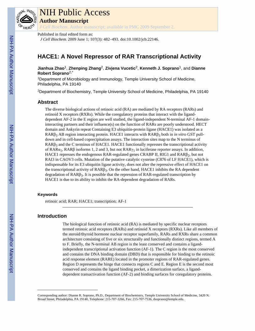
In order to identify proteins that interact with and potentially regulate the activity of the N-terminus of RARs, a yeast two-hybrid screen of an 11-day mouse embryo cDNA library wasperformed using the A region and first seven amino acids of the B region of RARβ3. Amongthe positive clones isolated in the yeast two-hybrid screen, we identified a clone thatdisplays 100% sequence identity to nucleotides 2624-3050 of Mus musculus HACE1(NM_172473) cDNA. This clone encodes the 98-carboxyl terminal amino acids of HACE1protein (amino acids 811-909) including the putative catalytic cysteine residue (C876) in theHECT domain [Anglesio et al., 2004] (Figure 1A).
Both HACE1 protein and cDNA sequences are highly conserved between Mus musculus(NM_172473) and Homo sapiens (NM_020771). Their amino acid and nucleotide sequencesdisplay 97.3% (884 /909 amino acids) and 91.2% (2490 /2729 nucleotides) sequenceidentity, respectively. The carboxyl terminal region of human and mouse HACE1 proteincorresponding to the region deduced from the yeast two-hybrid clone shows 100% aminoacid sequence identity. GenBank contains sequences for two isoforms of Homo sapiensHACE1. Human Long Form (LF) HACE1 cDNA (NM_020771) is 4614 base pairs andencodes a 909 amino acid protein and human Short Form (SF) HACE1 cDNA is 5321 basepairs and encodes a 562 amino acid protein. Both transcripts are generated fromchromosome 6 at 6q21 presumably by differential promoter usage. The two isoforms ofHACE1 share both the HECT domain in the carboxyl terminal end (amino acids 555-909 ofLF and amino acids 208-562 of SF) and the functionally unknown region (amino acids12-207 of SF and amino acids 360-554 of LF). However, these two isoforms differ at theiramino terminal ends. Amino acids 1-359 of LF HACE1 contain a region with six ankyrinrepeats (amino acids 66-217) while amino acids 1-11 of SF HACE1 have no knownfunctional domain (Figure 1A).
Interaction between HACE1 and RARsTo confirm the physical interaction between HACE1 and RARβ3, we performed an in vivocoprecipitation assay in Cos1 cells. Figure 1B demonstrates that V5-RARβ3 associates invivo with GST-LF HACE1 but not with GST alone. Similarly, V5-LF HACE1 associates invivo with GST-RARβ3 but not with GST alone.
To further study the physical interaction between HACE1 and other nuclear receptors, weperformed in vitro GST pull-down assays. As seen in Figure 1C, RARβ isoforms 1, 2 and 3specifically bound both full length GST-SF HACE1 and GST-LF HACE1 but not GST
Zhao et al. Page 5
J Cell Biochem. Author manuscript; available in PMC 2009 September 2.
NIH
-PA Author Manuscript
NIH
-PA Author Manuscript
NIH
-PA Author Manuscript
alone, while RARβ4 did not specifically interact with either GST-SF HACE1 or GST-LFHACE1. As expected for a negative control, luciferase did not interact with full length GST-SF HACE1, GST-LF HACE1 or GST alone. In addition, GST-SF HACE1 interacts withRARα1, RARγ1, ERα and TRα but not with RXRα1 and PPARγ (Figure 1D). Finally, theinteraction between RARβ3 and SF HACE1 is not affected by the RAR ligand, RA (data notshown).
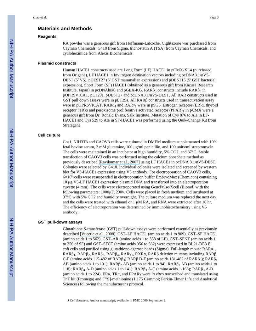
Mapping of the Interaction Site between HACE1 and RARβ3We further mapped the interaction site on both HACE1 and RARβ3 by performing GSTpull-down assays using GST-tagged full length and portions of HACE1 (Figure 1A) and[35S]-methionine labeled full length and portions of RARβ (Figure 2A). As seen in Figures1C & 1E, AR (amino acids 1-358 of LF HACE1) and SFNT (amino acids 1-356 of SFHACE1) do not appear to interact with any of the isoforms of RARβ, while SFCT (aminoacids 356-562 of SF HACE1) interacts with RARβ isoforms 1, 2 and 3 with similarintensity. Taken together these data suggest that only the C terminus of HACE1 (aminoacids 356-562 of SF HACE1, amino acids 703-909 of LF HACE1) interacts with RARβ.This is consistent with the fact that the RARβ3 interacting clone identified in the yeast two-hybrid screen encodes the carboxyl terminal region of LF HACE1 (amino acids 811-909).
Figure 2 demonstrates that the A-D regions of RARβ3 (panel B) as well as A-C regions ofRARβ3 (panel C) interact with SF HACE1 while neither C-F regions nor D-F regions ofRARβ interact with SF HACE1 (panel E). However, the AB regions of RARβ1, RARβ2 andRARβ3 showed weak or no interaction with SF HACE1 (panel D). Taken together, the Nterminus (A-C regions) of RARβ3 contains the interacting site however the C region in theC-F regions of RARβ does not interact with SF HACE. This is consistent with the fact thatthe A/partial B regions were originally used as the bait in the yeast two-hybrid screen.Taking into consideration the fact that the AB regions are a small peptide whoseconformation may not be optimal for interaction with HACE1 when fused to the C terminalend of GST, the interaction site most likely resides in the AB regions and the additionalamino acids in the C region (GST-RARβ3 A-C) or GAL4 DBD (yeast two-hybrid bait) mayallow the AB region to fold properly.
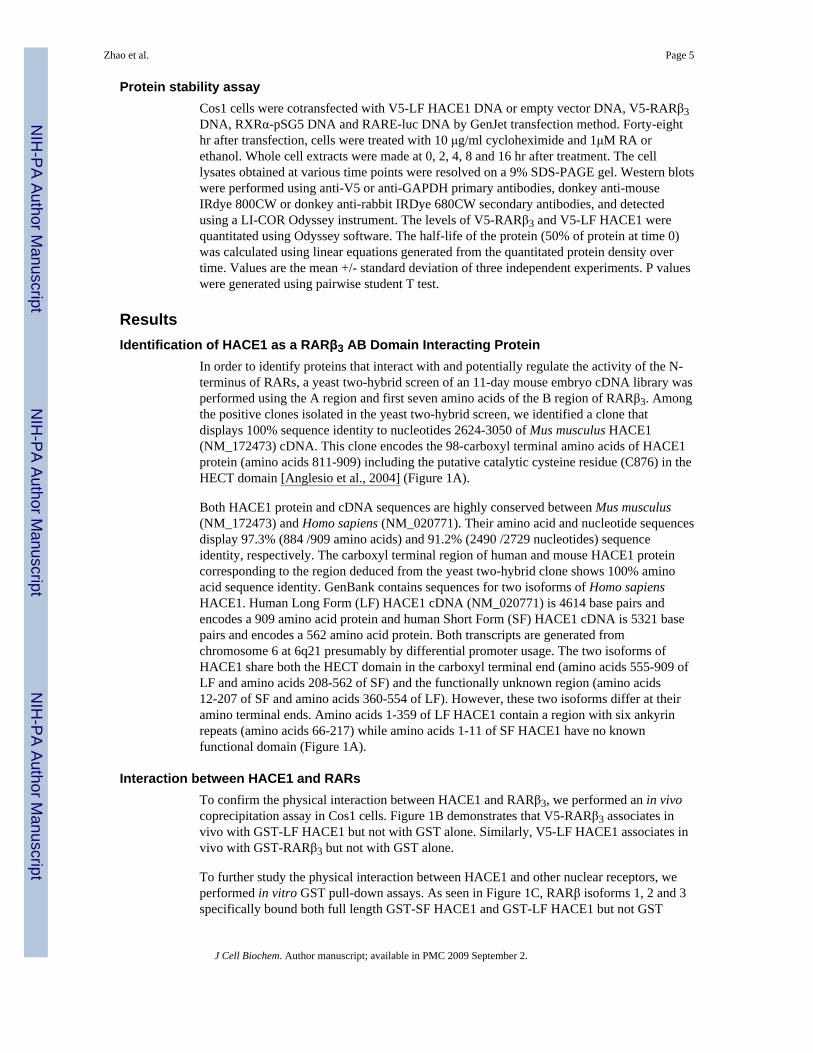
Effect of HACE1 on RAR/RARE Dependent TranscriptionTo test the functional role of the HACE1-RAR interaction, we first assessed the effect ofHACE1 on the transcriptional activity of RAR using a luciferase reporter which is controlledby a minimal TA promoter and DR5 type RARE in Cos1 cells. LF HACE1 caused arepression in the transcriptional activity of both endogenous RARs and exogenous RARβ3when the cells were treated with RA (Figure 3A & 3B). This repression of RARβ3transcriptional activity was dependent on the amount of HACE1 expression plasmid DNAtransfected into the cells, with the highest amount of LF HACE1 DNA resulting in a ∼80%repression of RARβ3 transcriptional activity over mock control (Figure 3A).
In addition, we chose three luciferase reporter controls (SP-1 luc, PPRE-luc and empty luc)to examine the specificity of the effect of LF HACE1 on RAR-dependent transcription. Asseen in Figure 3B, LF HACE1 has no effect on SP1-dependent, PPARγ-dependent andempty luciferase reporter (data not shown) transcriptional activity in Cos1 cells. In additionto Cos1 cells, transactivation assays were also performed in NIH3T3 cells (Figure 3B). Wealso observed that LF HACE1 repressed the transcriptional activity of both endogenousRAR and exogenous RARβ3 with ∼50-60% repression over mock control. On the otherhand, LF HACE1 had little effect on the transcriptional activity of PPARγ and SP1. Takentogether these results demonstrate that LF HACE1 acts as a specific repressor of RAR/RARE dependent transcription.
Zhao et al. Page 6
J Cell Biochem. Author manuscript; available in PMC 2009 September 2.
NIH
-PA Author Manuscript
NIH
-PA Author Manuscript
NIH
-PA Author Manuscript
Finally, we also examined the effect of LF HACE1 and SF HACE1 on the transcriptionalactivity of other RAR subtypes including RARα1, RARβ isoforms 1-3 and RARγ1 using thetranscriptional transactivation assay (Figure 3C). Both LF HACE1 and SF HACE1 repressedthe transcriptional activity of RARα1 and RARβ isoforms 1-3 significantly; however theyhad no effect on RARγ1 transcriptional activity.
Analysis of RAR-Regulated Gene Expression in HACE1-Expressing CAOV3 CellsWe stably overexpressed V5-LF HACE1 in CAOV3 cells (see Supplemental Figure 1) andexamined the effect of LF HACE1 expression on the mRNA levels of endogenous RAR-regulated genes including cellular retinoic acid binding protein (CRABP) II, retinoidinducible gene (RIG) 1, RARβ2 and retinoic acid induced (RAI) 3. All four genes contain afunctional DR5 RARE in their promoter region [Astrom et al., 1994; Tao et al., 2004; Jianget al., 2005; Zelent et al., 1991]. As seen in Figure 4, left panel, overexpression of LFHACE1 repressed the RA-dependent induction of CRABP II, RIG1 and RARβ2 mRNAlevels in all five independently isolated HACE1 overexpressing CAOV3 clones (H1, H4,H9, H11 and H14) but not that of RAI3. No change was observed in the mRNA level of thecontrol housekeeping gene hypoxanthine phosphoribosyltransferase (HPRT).
In addition, the effect of LF HACE1 on RAR regulated gene expression was measured inCAOV3 cells transiently expressing V5-LF HACE1. Electroporation efficiency wasmeasured by immunohistochemistry using V5 antibody. Approximately 90% of the cellswere positive for V5-LF HACE1 expression. Similar to that observed in CAOV3 cells stablyexpressing HACE1, transient expression of HACE1 repressed the RA-dependent inductionof CRABP II, RIG1 and RARβ2 in HACE1 overexpressing CAOV3 cells after 16 hr RAtreatment, but not that of RAI3 (Figure 4, right panel). No change was observed in themRNA level of the control housekeeping gene HPRT.
How HACE1 Functions as a RAR Transcriptional RepressorSince HACE1 was reported to be an E3 ubiquitin ligase, we asked whether its function as anE3 ubiquitin ligase plays a role in mediating repression of RAR transcriptional activity. Wemutated the catalytic cysteine residue to alanine in SF HACE1 (Cys529) and LF HACE1(Cys876) (Anglesio et al., 2004), and examined the ability of Cys529Ala SF HACE 1 tobind RARβ3 and Cys876Ala LF HACE1 to repress RARβ3–dependent transcription. Asseen in Figure 5A and 5B, both wild type HACE1 and the putative catalytic cysteinemutants of HACE1 interacted similarly with RARβ3 and displayed a similar level ofrepression of RARβ transcriptional activity. This suggests that the effect of HACE1 onRARβ transcriptional activity is not mediated by the catalytic cysteine residue associatedwith its E3 ubiquitin ligase activity.
We next asked whether HACE1 represses the transcriptional activity of RARs bydeacetylation of histone proteins. Since TSA is a commonly used histone deacetylaseinhibitor, we asked whether TSA treatment could relieve HACE1 repressive effect on thetranscriptional activity of RARβ3. As seen in Figure 5C, treatment with TSA increasesRARβ3 transcriptional activity in the presence and absence of LF HACE1, however LFHACE1 represses the activity of RARβ3 activity with same level of repression (80%) both inthe presence and in the absence of TSA. This suggests that the repressive effect of HACE1on the transcriptional activity of RARβ3 was not due to histone deacetylation.
RARs are degraded by the proteasome system in response to retinoids and RARγ2degradation is indispensable for the induction of RARγ2 transcriptional activity [For review,see Bour et al., 2007]. We therefore asked whether LF HACE1 affects RARβ3transcriptional activity by modulating RARβ3 protein stability. As expected, in the absence
Zhao et al. Page 7
J Cell Biochem. Author manuscript; available in PMC 2009 September 2.
NIH
-PA Author Manuscript
NIH
-PA Author Manuscript
NIH
-PA Author Manuscript
of LF HACE1, RARβ3 in the ethanol treated cells has a half life of 7.0+/-1.1 hr while in theRA treated cells the half life is reduced to 4.5+/-0.6 hr (Figure 6). Interestingly, in thepresence of LF HACE1, RARβ3 displays a similar half life in both the ethanol and RAtreated cells, 7.0+/-1.1 hr and 6.9+/-1.0 hr, respectively (Figure 6). Note that the half-life ofRARβ3 in the presence of LF HACE1 with or without RA treatment is the same as that ofthe ethanol treated cells in the absence of LF HACE1 Finally there is no difference in thehalf-life of LF HACE1 in the presence or absence of RA. These studies suggest that HACE1is repressing the transcription of RARβ3 by inhibiting the RA-dependent degradation ofRARβ3.
DiscussionIn this report, we have demonstrated an interaction between LF HACE1 and RARβ3 by bothin vitro GST pull-down and in vivo cell based copurification assays. The sites of interactionbetween HACE1 and RARβ3 map to the common C-terminus of the two HACE1 isoforms(amino acids 356-562 of SF HACE1, amino acids 703-909 of LF HACE1) and the N-terminus (ABC regions) of RARβ3. In addition, HACE1 interacts with RARβ1, RARβ2,RARβ3, RARα1, RARγ1, ERα and TRα, but not with RARβ4, RXRα and PPARγ in in vitroGST pull-down assays. Functionally both HACE1 isoforms repress the transcriptionalactivity of RARα1, RARβ isoforms 1, 2 and 3, but not RARγ1 in transactivation assays. LFHACE1 also represses the expression of the endogenous RAR-regulated genes RIG1,CRABP II and RARβ2, but not RAI3 in CAOV3 cells. This repression of RAR-dependentgene expression does not appear to be mediated by the E3 ubiquitin ligase activity ofHACE1 nor by deacetylation of histone proteins. On the other hand, HACE1 inhibited theRA-dependent degradation of RARβ3.
It was surprising that the AB regions of RARβ isoforms 1, 2, 3 have very weak binding toHACE1 in in vitro GST pull-down assays. On the other hand, the ABC regions of RARβ3strongly bound HACE1. Failure of RARβ4 to bind HACE1 suggests that amino acids in theA region are necessary for HACE1 binding since the A region of this receptor is truncated,consisting of only 4 amino acids [Nagpal et al., 1992]. Furthermore, since the C-F regions ofRARβ did not bind to HACE1, it is likely that the C region without the A and B regions isnot sufficient to bind HACE1. The addition of the DBD (C region) of RARβ3 in the GSTpull-down assays or the GAL4 DBD in the yeast two-hybrid screen may enable the ABregions to properly fold to allow binding of HACE1. This is supported by studies utilizingnuclear magnetic resonance, circular dichroism spectroscopy and limited proteolysis ofseveral steroid nuclear receptor N terminal domains /AF-1s (GR, ERα, ERβ, PR) whichindicate that this region exists in an unfolded state ]Dahlman-Wright et al., 1995; Warnmarket al., 2000; Birnbaumer et al., 1983; Bain et al., 2000]. Interestingly, when the AB regionsand DBD of either PR or AR were expressed, they tended to be more structured than theAF-1 domain alone [Lavery and McEwan, 2005; Bain et al., 2000; Kumar et al., 1999].Taken together, it is likely that the AB regions of RARβ contains the binding site/surface forHACE1 however additional amino acid residues in the C region enable proper structuralfolding of the AB region in order to bind HACE1. Such a model would be consistent withour finding that HACE1 binds to TR and ER as well as all three RAR subtypes. Since theAB regions of TR and ER shares little primary amino acid sequence identity with the ABregions of the RARs, it is possible that the conformation of the combined ABC regions ofTR and ER share common feature(s) with that of RARs that permit binding to HACE1. Theamino terminal regions of several nuclear receptors, including ER and AR, undergo atransition to a proper folded state upon interaction with coregulatory proteins [Warnmark etal., 2001; Reid et al., 2002; Warnmark et al., 2003]. Such features would not be present inthe conformation of the ABC regions of RXR and PPARγ.
Zhao et al. Page 8
J Cell Biochem. Author manuscript; available in PMC 2009 September 2.
NIH
-PA Author Manuscript
NIH
-PA Author Manuscript
NIH
-PA Author Manuscript

HACE1 functionally represses the transcriptional activity of RARα1, RARβ isoforms 1, 2and 3, but not RARγ1 in transactivation assays using a luciferase reporter gene under thecontrol of a DR5 RARE. It is unclear why HACE1 fails to repress the RARγ1 regulatedtranscription of the luciferase reporter gene despite strong in vitro binding of HACE1 andRARγ1. It is possible that RARγ1 interacts in Cos1 cells with specific cofactor(s) that blocksits binding to HACE1. In addition, HACE1 represses the expression of the endogenousRAR-regulated genes RIG1, CRABP II and RARβ2, but not RAI3. Each of these RAR-regulated genes contains a DR5 RARE in their promoter region [Jiang et al., 2005; Tao etal., 2004; Zelent et al., 1991; Astrom et al., 1994]. RARα and/or RARβ have been shown tobe involved in the transcription of CRABP II, RIG1 and RARβ2. CRABPII has been shownto be regulated by RARα [Astrom et al., 1994], RIG1 DR5 interacts with RARα/RXR in invitro EMSA assays [Jiang et al., 2005], and RARβ2 expression can be regulated by all of thethree RAR isotypes (Taneja et al., 1996). However, it is unknown which subtype of RAR isinvolved in regulation of RAI3 transcription [Tao et al., 2004]. These data demonstrate thatHACE1 displays a type of specificity in the modulation of RAR-regulated genes that mightbe related to a RAR subtype functional selectivity and/or promoter context.
Although HACE1 was reported to be an E3 ubiquitin ligase [Anglesio et al., 2004], mutationof the catalytic Cys 876 (LF HACE1)/Cys 529 (SF HACE1) reported to be responsible forits ubiquitin ligase activity to an Ala does not affect HACE1-dependent repression ofRARβ3 transcriptional activity or HACE1/RARβ3 binding, respectively. The E3 ubiquitinligase activity of HACE1 is dependent on the conserved HECT domain that is the signaturedomain for this class of HECT containing E3 ligases. Structurally HECT domains form twolobes. The N-lobe interacts with E2 ubiquitin transferase and the C-lobe is responsible fortransferring ubiquitin to its substrate [Huang et al., 1999; Wang et al., 1999; Verdecia et al.,2003]. Other well studied HECT domain containing E3 ubiquitin ligases interact with theirsubstrates through their N terminal domains rather than their HECT domains (for example,RLD domain of HERC5, WW domain of NEDD family and LXXLL motif of E6-AP)[Huang et al., 1999; Wang et al., 1999]. Interaction of HACE1 with RARβ3 via its Cterminal HECT domain is not consistent with HACE1 ubiquitinating RARβ3 and couldpossibly block its E3 ubiquitin ligase activity. Taken together, our data do not support thenotion that HACE1 affects the transcriptional activity of RARs through its E3 ubiquitinligase activity.
It has been shown that proteins with E3 ubiquitin ligase activity have other functions inaddition to ubiquitination of target proteins. HERC1 has been shown to bind and act asguanine nucleotide exchange factor for ARF1. However, ARF1 does not appear to bedegraded by HERC1 [Rosa et al., 1996]. In spite of the importance of Cbl E3 ubiquitinligase activity in the degradation of a number of proteins such as EGFR and Vav, Cbl alsofunctions as an adaptor molecule by forming complexes with numerous proteins. Inaddition, Cbl is also involved in activation of MAP kinases [For review, see Swaminathanand Tsygankov, 2006].
Both RAR and RXR are degraded by the 26S proteasome system in response to retinoids[Boudjelal et al., 2000; Kopf et al., 2000; Osburn et al., 2001; Tanaka et al., 2001; Gianni etal., 2002; Gianni et al., 2003; Bour and Rochette-Egly, 2007]. Similarly, our datademonstrate that RA potentiates the degradation of RARβ3 (half life of 4.5+/-0.6 hrs for RAtreated sample versus 7.0+/-1.1 hrs for ethanol treated sample). Interestingly, HACE1inhibits this RA-dependent increase in RARβ3 degradation.
It is possible that the repression of RARβ3 transcriptional activity by HACE1 is due to itsability to inhibit the RA-dependent degradation of RARβ3. Prior reports have demonstratedthat the proteolytic function of the proteasome system on RARγ2 upon RA treatment is
Zhao et al. Page 9
J Cell Biochem. Author manuscript; available in PMC 2009 September 2.
NIH
-PA Author Manuscript
NIH
-PA Author Manuscript
NIH
-PA Author Manuscript
intimately linked with the transcriptional activity of RARγ2 [Gianni et al., 2003; Gianni etal., 2002a & b]. When RARγ2 degradation is blocked, the RA-dependent transcriptionalactivity of RARγ2 is dramatically impaired. In addition, E6-AP, a HECT domain containingE3 ubiquitin ligase for ER, has dual roles including degradation of ER and transactivation ofER activity [For review, see Ramamoorthy and Nawaz, 2008]. When the ubiquitin ligaseactivity of E6AP is abolished, the transcriptional activity of ER is repressed. In addition,inhibition of proteosome degradation significantly diminished the ligand-inducedtranscriptional activity of many of the nuclear receptors including AR, ER, PR, RARα, TR,PPAR and RXR [For review, see Alarid, 2006]. Moreover, it has been postulated that theproteosome system may orchestrate the dynamics of ER mediated transcription bymodulating the degradation of the ER and cofactor complexes on chromatin [Shang et al.,2000; Metevier et al., 2003; Reid et al., 2003].
We still do not understand how HACE1 inhibits the RA dependent degradation of RARβ3.One possible mechanism is that the interaction of HACE1 and RARβ3 may interfere withthe function of its real E3 ubiquitin ligase preventing ubiquitination of RARβ3. One exampleof this phenomenon is the interaction of calmodulin with ER enhances the stability of ERthrough interfering with the interaction of E6AP and ER [Li et al., 2006]. Alternatively, theinteraction of HACE1 with RAR may interfere with a signal for RAR degradation, such asphosphorylation. Phosphorylation of serine residues in the proline rich region in B domainwas suggested to be required for both RARγ2 degradation and RARγ2 transactivation[Gianni et al., 2003; Gianni et al., 2002a&b]. If the interaction of HACE1 with RAR blocksthe binding and/or action of kinases on critical amino acid residues then the degradation ofthe receptor may be prevented and transcription will be repressed. Interestingly, vinexin βinteracts with unphosphorylated RARγ and represses its transcriptional activity [Bour et al.,2005].
In conclusion, our data demonstrate that the N-terminal AB region of RARs can bindHACE1. In addition, HACE1 represses the transcriptional activity of RARs and inhibits theRA-dependent degradation of RARs. Finally, these data suggest that HACE1 has additionalfunction(s) beyond its role as an E3 ubiquitin ligase.
Supplementary MaterialRefer to Web version on PubMed Central for supplementary material.
AcknowledgmentsThis work was supported by National Institutes of Health grants DK67558 (to D.R.S.), DK44517 (to D.R.S.) andCA64945 (to K.J.S.).
Grant Support: NIH grants DK67558, DK44517 and CA64945
ReferencesAlarid ET. Lives and times of nuclear receptors. Mol Endocrinol. 2006; 20:1972–1981. [PubMed:
16423879]Anglesio MS, Evdokimova V, Melnyk N, Zhang L, Fernandez CV, Grundy PE, Leach S, Marra MA,
Brooks-Wilson AR, Penninger J, Sorensen PH. Differential expression of a novel ankyrincontaining E3 ubiquitin-protein ligase, Hace1, in sporadic Wilms' tumor versus normal kidney.Hum Mol Genet. 2004; 13:2061–2074. [PubMed: 15254018]
Astrom A, Pettersson U, Chambon P, Voorhees JJ. Retinoic acid induction of human cellular retinoicacid-binding protein-II gene transcription is mediated by retinoic acid receptor-retinoid X receptor
Zhao et al. Page 10
J Cell Biochem. Author manuscript; available in PMC 2009 September 2.
NIH
-PA Author Manuscript
NIH
-PA Author Manuscript
NIH
-PA Author Manuscript

heterodimers bound to one far upstream retinoic acid-responsive element with 5-base pair spacing. JBiol Chem. 1994; 269:22334–22339. [PubMed: 8071361]
Bain DL, Franden MA, McManaman JL, Takimoto GS, Horwitz KB. The N-terminal region of thehuman progesterone A-receptor. Structural analysis and the influence of the DNA binding domain. JBiol Chem. 2000; 275:7313–7320. [PubMed: 10702302]
Bastien J, Adam-Stitah S, Riedl T, Egly JM, Chambon P, Rochette-Egly C. TFIIH interacts with theretinoic acid receptor gamma and phosphorylates its AF-1-activating domain through cdk7. J BiolChem. 2000; 275:21896–21904. [PubMed: 10748061]
Bastien J, Rochette-Egly C. Nuclear retinoid receptors and the transcription of retinoid-target genes.Gene. 2004; 328:1–16. [PubMed: 15019979]
Birnbaumer M, Schrader WT, O'Malley BW. Assessment of structural similarities in chick oviductprogesterone receptor subunits by partial proteolysis of photoaffinity-labeled proteins. J Biol Chem.1983; 258:7331–7337. [PubMed: 6345521]
Boudjelal M, Wang Z, Voorhees JJ, Fisher GJ. Ubiquitin/proteasome pathway regulates levels ofretinoic acid receptor gamma and retinoid X receptor alpha in human keratinocytes. Cancer Res.2000; 60:2247–2252. [PubMed: 10786691]
Bour G, Lalevee S, Rochette-Egly C. Protein kinases and the proteasome join in the combinatorialcontrol of transcription by nuclear retinoic acid receptors. Trends Cell Biol. 2007; 17:302–309.[PubMed: 17467991]
Bour G, Plassat JL, Bauer A, Lalevee S, Rochette-Egly C. Vinexin beta interacts with the non-phosphorylated AF-1 domain of retinoid receptor gamma (RARgamma) and repressesRARgamma-mediated transcription. J Biol Chem. 2005; 280:17027–17037. [PubMed: 15734736]
Chambon P. A decade of molecular biology of retinoic acid receptors. FASEB J. 1996; 10:940–954.[PubMed: 8801176]
Dahlman-Wright K, Baumann H, McEwan IJ, Almlof T, Wright AP, Gustafsson JA, Hard T.Structural characterization of a minimal functional transactivation domain from the humanglucocorticoid receptor. Proc Natl Acad Sci USA. 1995; 92:1699–1703. [PubMed: 7878043]
Dilworth FJ, Chambon P. Nuclear receptors coordinate the activities of chromatin remodelingcomplexes and coactivators to facilitate initiation of transcription. Oncogene. 2001; 20:3047–3054. [PubMed: 11420720]
Gianni M, Bauer A, Garattini E, Chambon P, Rochette-Egly C. Phosphorylation by p38MAPK andrecruitment of SUG-1 are required for RA-induced RAR gamma degradation and transactivation.EMBO J. 2002a; 21:3760–3769. [PubMed: 12110588]
Gianni M, Kopf E, Bastien J, Oulad-Abdelghani M, Garattini E, Chambon P, Rochette-Egly C. Down-regulation of the phosphatidylinositol 3-kinase/Akt pathway is involved in retinoic acid-inducedphosphorylation, degradation, and transcriptional activity of retinoic acid receptor gamma 2. J BiolChem. 2002b; 277:24859–24862. [PubMed: 12032135]
Gianni M, Tarrade A, Nigro EA, Garattini E, Rochette-Egly C. The AF-1 and AF-2 domains of RARgamma 2 and RXR alpha cooperate for triggering the transactivation and the degradation of RARgamma 2/RXR alpha heterodimers. J Biol Chem. 2003; 278:34458–34466. [PubMed: 12824162]
Huang L, Kinnucan E, Wang G, Beaudenon S, Howley PM, Huibregtse JM, Pavletich NP. Structure ofan E6AP-UbcH7 complex: insights into ubiquitination by the E2-E3 enzyme cascade. Science.1999; 286:1321–1326. [PubMed: 10558980]
Jiang SY, Wu MS, Chen LM, Hung MW, Lin HE, Chang GG, Chang TC. Identification andcharacterization of the retinoic acid response elements in the human RIG1 gene promoter.Biochem Biophys Res Commun. 2005; 331:630–639. [PubMed: 15850806]
Kopf E, Plassat JL, Vivat V, de The H, Chambon P, Rochette-Egly C. Dimerization with retinoid Xreceptors and phosphorylation modulate the retinoic acid-induced degradation of retinoic acidreceptors alpha and gamma through the ubiquitin-proteasome pathway. J Biol Chem. 2000;275:33280–33288. [PubMed: 10869350]
Kumar R, Thompson EB. Transactivation functions of the N-terminal domains of nuclear hormonereceptors: protein folding and coactivator interactions. Mol Endocrinol. 2003; 17:1–10. [PubMed:12511601]
Zhao et al. Page 11
J Cell Biochem. Author manuscript; available in PMC 2009 September 2.
NIH
-PA Author Manuscript
NIH
-PA Author Manuscript
NIH
-PA Author Manuscript
Lavery DN, McEwan IJ. Structure and function of steroid receptor AF1 transactivation domains:induction of active conformations. Biochem J. 2005; 391:449–464. [PubMed: 16238547]
Li L, Li Z, Howley PM, Sacks DB. E6AP and calmodulin reciprocally regulate estrogen receptorstability. J Biol Chem. 2006; 281:1978–1985. [PubMed: 16314411]
Metivier R, Penot G, Hubner MR, Reid G, Brand H, Kos M, Gannon F. Estrogen receptor-alphadirects ordered, cyclical, and combinatorial recruitment of cofactors on a natural target promoter.Cell. 2003; 115:751–763. [PubMed: 14675539]
Nagpal S, Zelent A, Chambon P. RAR-beta 4, a retinoic acid receptor isoform is generated from RAR-beta 2 by alternative splicing and usage of a CUG initiator codon. Proc Natl Acad Sci USA. 1992;89:2718–2722. [PubMed: 1313565]
Osburn DL, Shao G, Seidel HM, Schulman IG. Ligand-dependent degradation of retinoid X receptorsdoes not require transcriptional activity or coactivator interactions. Mol Cell Biol. 2001; 21:4909–4918. [PubMed: 11438648]
Ramamoorthy S, Nawaz Z. E6-associated protein (E6-AP) is a dual function coactivator of steroidhormone receptors. Nucl Recept Signal. 2008; 6:1–9.
Ravikumar S, Perez-Liz G, Del Vale L, Soprano DR, Soprano KJ. Insulin receptor substrate-1 is animportant mediator of ovarian cancer cell growth suppression by all-trans retinoic acid. CancerRes. 2007; 67:9266–9275. [PubMed: 17909034]
Reid G, Hubner MR, Metivier R, Brand H, Denger S, Manu D, Beaudouin J, Ellenberg J, Gannon F.Cyclic, proteasome-mediated turnover of unliganded and liganded ERalpha on responsivepromoters is an integral feature of estrogen signaling. Mol Cell. 2003; 11:695–707. [PubMed:12667452]
Reid J, Kelly SM, Watt K, Price NC, McEwan IJ. Conformational analysis of the androgen receptoramino-terminal domain involved in transactivation. Influence of structure-stabilizing solutes andprotein-protein interactions. J Biol Chem. 2002; 277:20079–20086. [PubMed: 11896058]
Rochette-Egly C, Adam S, Rossignol M, Egly JM, Chambon P. Stimulation of RAR alpha activationfunction AF-1 through binding to the general transcription factor TFIIH and phosphorylation byCDK7. Cell. 1997; 90:97–107. [PubMed: 9230306]
Rosa JL, Casaroli-Marano RP, Buckler AJ, Vilaro S, Barbacid M. p619, a giant protein related to thechromosome condensation regulator RCC1, stimulates guanine nucleotide exchange on ARF1 andRab proteins. EMBO J. 1996; 15:4262–4273. [PubMed: 8861955]
Shang Y, Hu X, DiRenzo J, Lazar MA, Brown M. Cofactor dynamics and sufficiency in estrogenreceptor-regulated transcription. Cell. 2000; 103:843–852. [PubMed: 11136970]
Swaminathan G, Tsygankov AY. The Cbl family proteins: ring leaders in regulation of cell signaling. JCell Physiol. 2006; 209:21–43. [PubMed: 16741904]
Taneja R, Roy B, Plassat JL, Zusi CF, Ostrowski J, Reczek PR, Chambon P. Cell-type and promoter-context dependent retinoic acid receptor (RAR) redundancies for RARβ2 and Hoxa-1 activation inF9 and P19 cells can be artefactually generated by gene knockout. Proc Natl Acad Sci USA. 1996;93:6197–6202. [PubMed: 8650243]
Tanaka T, Rodriguez de la Concepcion ML, De Luca LM. Involvement of all-trans-retinoic acid in thebreakdown of retinoic acid receptors alpha and gamma through proteasomes in MCF-7 humanbreast cancer cells. Biochem Pharmacol. 2001; 61:1347–1355. [PubMed: 11331070]
Tao Q, Cheng Y, Clifford J, Lotan R. Characterization of the murine orphan G-protein-coupledreceptor gene Rai3 and its regulation by retinoic acid. Genomics. 2004; 83:270–280. [PubMed:14706456]
Verdecia MA, Joazeiro CA, Wells NJ, Ferrer JL, Bowman ME, Hunter T, Noel JP. Conformationalflexibility underlies ubiquitin ligation mediated by the WWP1 HECT domain E3 ligase. Mol Cell.2003; 11:249–259. [PubMed: 12535537]
Vucetic Z, Zhang ZP, Zhao J, Wang F, Soprano KJ, Soprano DR. Acinus-S' represses retinoic acidreceptor (RAR)-regulated gene expression through interaction with the B domains of RARs. MolCell Biol. 2008; 28:2549–2558. [PubMed: 18250153]
Wang G, Yang J, Huibregtse JM. Functional domains of the Rsp5 ubiquitin-protein ligase. Mol CellBiol. 1999; 19:342–352. [PubMed: 9858558]
Zhao et al. Page 12
J Cell Biochem. Author manuscript; available in PMC 2009 September 2.
NIH
-PA Author Manuscript
NIH
-PA Author Manuscript
NIH
-PA Author Manuscript

Warnmark A, Gustafsson JA, Wright AP. Architectural principles for the structure and function of theglucocorticoid receptor tau 1 core activation domain. J Biol Chem. 2000; 275:15014–15018.[PubMed: 10747977]
Warnmark A, Treuter E, Wright AP, Gustafsson JA. Activation functions 1 and 2 of nuclear receptors:molecular strategies for transcriptional activation. Mol Endocrinol. 2003; 17:1901–1909.[PubMed: 12893880]
Warnmark A, Wikstrom A, Wright AP, Gustafsson JA, Hard T. The N-terminal regions of estrogenreceptor alpha and beta are unstructured in vitro and show different TBP binding properties. J BiolChem. 2001; 276:45939–45944. [PubMed: 11595744]
Zelent A, Mendelsohn C, Kastner P, Krust A, Garnier JM, Ruffenach F, Leroy P, Chambon P.Differentially expressed isoforms of the mouse retinoic acid receptor beta generated by usage oftwo promoters and alternative splicing. EMBO J. 1991; 10:71–81. [PubMed: 1846599]
Zhang L, Anglesio MS, O'Sullivan M, Zhang F, Yang G, Sarao R, Mai PN, Cronin S, Hara H, MelnykN, Li L, Wada T, Liu PP, Farrar J, Arceci RJ, Sorensen PH, Penninger JM. The E3 ligase HACE1is a critical chromosome 6q21 tumor suppressor involved in multiple cancers. Nat Med. 2007;13:1060–1069. [PubMed: 17694067]
Zhao et al. Page 13
J Cell Biochem. Author manuscript; available in PMC 2009 September 2.
NIH
-PA Author Manuscript
NIH
-PA Author Manuscript
NIH
-PA Author Manuscript

Figure 1. Interaction of SF HACE1, LF HACE1 and HACE1 regions with RARs and othernuclear receptorsA. Schematic representation of full length LF HACE1 (909 amino acids), SF HACE1 (562amino acids) and regions of HACE1 including AR (amino acids 1-358 of LF HACE1),SFNT (amino acids 1-356 of SF HACE1) and SFCT (amino acids 356-562 of SF HACE1).B. Interaction of LF HACE1 and RARβ3 in vivo. Cos1 cells were cotransfected with V5-LFHACE1 DNA and GST-RARβ3 DNA or empty GST vector DNA, or V5- RARβ3 DNAalong with GST-LF HACE1 DNA or empty GST DNA as indicated. Protein complexeswere purified using glutathione beads and resolved by SDS-PAGE. Western blots wereperformed using anti-V5 or anti-GST primary antibodies, donkey anti-mouseIRDdye800CW or donkey anti-rabbit IRDye680CW secondary antibodies, and detectedusing LI-COR Odyssey instrument. C. Interaction of LF HACE1, AR and SF HACE1 withRARβ isoforms. D. Interaction of SF HACE1 with RARα1, RARγ1 and several steroid/thyroid hormone receptors including ER, TR and PPARγ. E. Interaction of SF HACE1,SFNT, SFCT with RARβ isoforms. GST pull-down assays were performed with purifiedGST fused LF HACE1, SF HACE1 and HACE1 regions or GST alone and the indicated invitro transcribed and translated [35S]-methionine labeled nuclear receptors and luciferase.Input (5% for panels C and E and 10% for panel D) and purified GST fusion proteins(panels C and E) are also shown.
Zhao et al. Page 14
J Cell Biochem. Author manuscript; available in PMC 2009 September 2.
NIH
-PA Author Manuscript
NIH
-PA Author Manuscript
NIH
-PA Author Manuscript
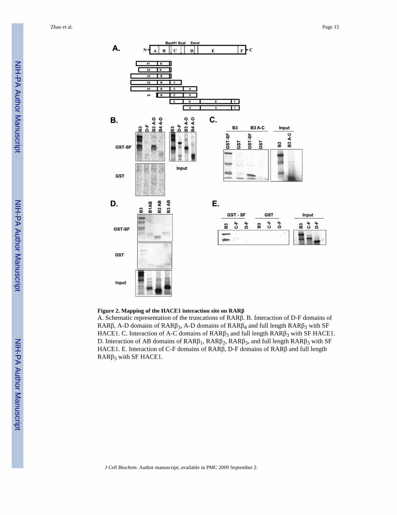
Figure 2. Mapping of the HACE1 interaction site on RARβA. Schematic representation of the truncations of RARβ. B. Interaction of D-F domains ofRARβ, A-D domains of RARβ3, A-D domains of RARβ4 and full length RARβ3 with SFHACE1. C. Interaction of A-C domains of RARβ3 and full length RARβ3 with SF HACE1.D. Interaction of AB domains of RARβ1, RARβ2, RARβ3, and full length RARβ3 with SFHACE1. E. Interaction of C-F domains of RARβ, D-F domains of RARβ and full lengthRARβ3 with SF HACE1.
Zhao et al. Page 15
J Cell Biochem. Author manuscript; available in PMC 2009 September 2.
NIH
-PA Author Manuscript
NIH
-PA Author Manuscript
NIH
-PA Author Manuscript
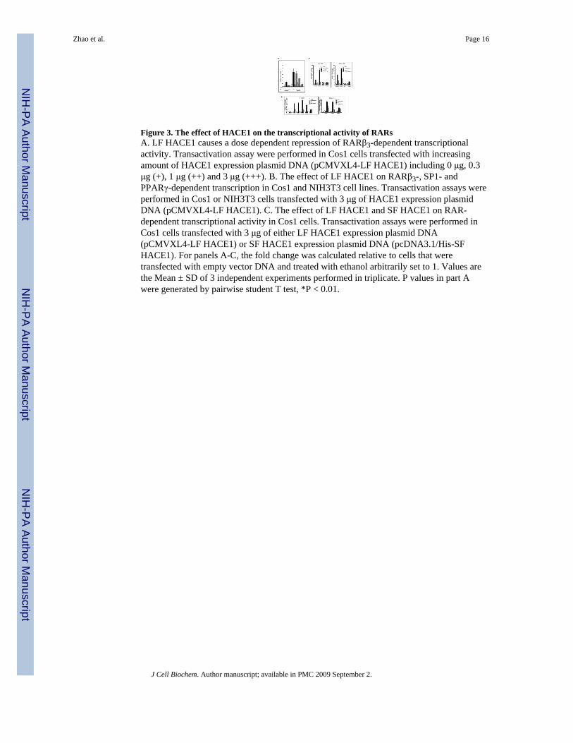
Figure 3. The effect of HACE1 on the transcriptional activity of RARsA. LF HACE1 causes a dose dependent repression of RARβ3-dependent transcriptionalactivity. Transactivation assay were performed in Cos1 cells transfected with increasingamount of HACE1 expression plasmid DNA (pCMVXL4-LF HACE1) including 0 μg, 0.3μg (+), 1 μg (++) and 3 μg (+++). B. The effect of LF HACE1 on RARβ3-, SP1- andPPARγ-dependent transcription in Cos1 and NIH3T3 cell lines. Transactivation assays wereperformed in Cos1 or NIH3T3 cells transfected with 3 μg of HACE1 expression plasmidDNA (pCMVXL4-LF HACE1). C. The effect of LF HACE1 and SF HACE1 on RAR-dependent transcriptional activity in Cos1 cells. Transactivation assays were performed inCos1 cells transfected with 3 μg of either LF HACE1 expression plasmid DNA(pCMVXL4-LF HACE1) or SF HACE1 expression plasmid DNA (pcDNA3.1/His-SFHACE1). For panels A-C, the fold change was calculated relative to cells that weretransfected with empty vector DNA and treated with ethanol arbitrarily set to 1. Values arethe Mean ± SD of 3 independent experiments performed in triplicate. P values in part Awere generated by pairwise student T test, *P < 0.01.
Zhao et al. Page 16
J Cell Biochem. Author manuscript; available in PMC 2009 September 2.
NIH
-PA Author Manuscript
NIH
-PA Author Manuscript
NIH
-PA Author Manuscript

Figure 4. Effect of overexpressing HACE1 on the mRNA levels of RAR responsive genes instable HACE1 expressing CAOV3 clones (Left Panels) and transiently HACE1 expressingCAOV3 cells (Right Panel)Wild type CAOV3 cells (WT), empty control clone (CC1) and LF HACE1 overexpressingclones (H1, H4, H9, H11 and H14), (Left Panels), or CAOV3 cells electroporated witheither LF HACE1 expression plasmid DNA or control empty plasmid DNA (Right Panels)were treated with 10-6 M RA or ethanol for 16 hr. RNA was extracted and reversetranscribed. The expression levels of CRABP II, RIG1, RARβ2, RAI3 and HPRT weredetermined by RT-qPCR. The expression level of each gene tested was normalized to theendogenous GAPDH levels and the fold changes upon RA treatment calculated relative tothe gene expression level in the respective ethanol treated sample. Left panel, each clonewas assayed one time; right panel, values are Mean +/- SD.
Zhao et al. Page 17
J Cell Biochem. Author manuscript; available in PMC 2009 September 2.
NIH
-PA Author Manuscript
NIH
-PA Author Manuscript
NIH
-PA Author Manuscript

Figure 5. The role of E3 ubiquitin ligase activity (Panels A and B) and histone deacetylaseactivity (Panel C) in the repression of RARβ3 transcriptional activity by HACE1A. GST-pulldown assays were performed with wild type GST-SF HACE 1 or GST-SFHACE1 C529A and in vitro transcribed and translated [35S]-methionine labeled RARβ3. B.Cos1 cells were co-transfected with DNA constructs including HACE1 expression plasmid(pCMVXL4-LF HACE1), HACE1 CA mutant expression plasmid (pCMVXL4-LF HACE1C876A) or empty plasmid along with pTL-RARE-luc reporter plasmid, pRL reporterplasmid, and RARβ3 expression plasmid (pOPRSVICAT- RARβ3) or empty expressionplasmid. C. Cos1 cells were cotransfected with expression plasmid of LF HACE1(pCMVXL4-LF HACE1) or empty expression plasmid, pTL-RARE-luc reporter plasmid,
Zhao et al. Page 18
J Cell Biochem. Author manuscript; available in PMC 2009 September 2.
NIH
-PA Author Manuscript
NIH
-PA Author Manuscript
NIH
-PA Author Manuscript

pRL reporter plasmid and expression plasmid of RARβ3. Twenty-four hr after transfection,cells were treated with combinations of ethanol/10-6 M RA and 100ng/ml TSA for anadditional 24 hr. For panel B, the fold changes were calculated relative to cells that weretransfected with empty expression plasmid DNA and treated with ethanol arbitrarily set to 1.For panel C, the fold changes were calculated relative to cells that were transfected withempty expression plasmid DNA, treated with ethanol and no TSA arbitrarily set to 1. Forpanels B and C, values are the Mean + SD of 3 independent experiments performed intriplicate.
Zhao et al. Page 19
J Cell Biochem. Author manuscript; available in PMC 2009 September 2.
NIH
-PA Author Manuscript
NIH
-PA Author Manuscript
NIH
-PA Author Manuscript

Figure 6. Analysis of RARβ3 half life in the absence and presence of HACE1Cos1 cells were transfected with V5-LF HACE1 DNA or empty vector DNA, V5-RARβ3DNA, RXRα DNA and RARE-luc DNA. Cells were treated with 10 μg/ml cycloheximide,and 10-6 M RA or ethanol for the indicated times. (A). Whole cell extracts were preparedfrom cells with indicated times of treatment and western blots were performed using V5 orGAPDH primary antibodies, donkey anti-mouse IRdye 800CW or donkey anti-rabbit IRDye680CW secondary antibodies, and detected using a LI-COR Odyssey instrument. GAPDHwas used as a loading control. A representative figure from one of three independentexperiments is shown. (B). Densitometric values of each band from western blots werequantitated using LI-COR software. V5-RARβ3 and V5-HACE1 values were normalizedwith corresponding GAPDH values. The normalized values of V5-RARβ3 and V5-HACE1were plotted. A representative plot from one of three independent experiments is shown,time 0 was set to 1 arbitrarily. (C). Half life (protein level = 50% of time 0) of RARβ3 was
Zhao et al. Page 20
J Cell Biochem. Author manuscript; available in PMC 2009 September 2.
NIH
-PA Author Manuscript
NIH
-PA Author Manuscript
NIH
-PA Author Manuscript
calculated based on the linear equations generated from quantitated protein density overtime from three independent experiments. Values are the Mean ± SD of 3 independentexperiments. P value was generated by pairwise student T test, *P < 0.01.
Zhao et al. Page 21
J Cell Biochem. Author manuscript; available in PMC 2009 September 2.
NIH
-PA Author Manuscript
NIH
-PA Author Manuscript
NIH
-PA Author Manuscript