LFB1/HNF1 acts as a repressor of its own transcription
7
4284-4290 Nucleic Acids Research, 1994, Vol. 22, No. 20 LFB1/HNF1 acts as a repressor of its own transcription Giulia Piaggio*, Licia Tomei1, Carlo Toniattil, Raffaele De Francesco1, Jutta Gerstner2,+ and Riccardo Cortese1 Istituto 'Regina Elena' Centro Ricerca Sperimentale, Laboratorio Oncogenesi Molecolare, via Delle Messi d'Oro 156, 00158 Rome, 1IRBM- Istituto di Ricerche di Biologia Molecolare P.Angeletti, via Pontina Km 40.600, 00040 Pomezia, Rome, Italy and 2EMBL, European Molecular Biology Laboratory, MeyerhofstraBe 1, 6200 Heidelberg, Germany Received May 27, 1994; Revised and Accepted September 5, 1994 ABSTRACT LFB1/HNF1 is a hepatocyte-enriched trans-activator involved in the regulation of many liver-specific genes. We report the cloning and characterization of a rat genomic DNA fragment containing about 3.5 kb of the LFB1/HNF1 gene 5'-flanking region. This DNA segment is capable of directing the liver-specific expression of a reporter gene in transfection assays. More interestingly, the basal activity of the LFB1/HNF1 promoter in cultured hepatoma cell lines is down- regulated by exogenously added LFB1/HNF1 protein itself. The ability to repress transcription starting from its own promoter requires the integrity of the N-terminal LFB1/HNF1 DNA-binding domain. Contrary to the expectations, in vitro binding experiments failed to demonstrate any specific and functional interaction of purified LFB1/HNF1 with the -3.5 kb promoter sequence. In addition to the DNA-binding domain, a 60 aa region contained in the C-terminus of the protein and distinct from the previously characterized activation domains, is also required for the repressing function. INTRODUCTION The liver provides a useful model system in which to study the mechanisms that govern the differential expression of tissue- specific genes (1). Several cDNAs coding for factors that play an important role in controlling the hepatocyte-specific expression have recently been cloned. They include the Liver Factor BI/Hepatocyte Nuclear Factor 1 (LFB1/HNFl; 2,3) and the related factor LFB3/vHNFI (4,5), the Drosophila fork head homologue HNF3 (6), HNF4, a member of the steroid hormone receptor superfamily (7) and the leucine-zipper containing factors C/EBP (8) and DBP (9). The interaction of these transcription factors with a given array of cis-acting elements on the promoter region of liver-specific genes modulates their expression rate. This interaction may in fact activate or repress promoter activity according to the stage of development, hormonal induction or pathological condition (10). The comprehension of the molecular EMBL accession no. X67649 mechanisms underlying hepatocyte-specific transcriptional regulation may provide insight into the events leading to liver development and differentiation. LFB1/HNF1 was initially identified as a hepatocyte factor interacting with the promoter regions of many liver-specific genes (10,11). Although its expression was shown not to be limited to hepatic cells, both in vivo and in vitro studies have confirmed this protein to be one of the major determinants of the liver phenotype (5,12-14). LFBI/HNF1 binds as a dimer to specific palindromic sites (2,11,15). The functional architecture of the LFB1/HNF1 protein has been extensively studied in our laboratory. A protein fragment that contains the N-terminal 281 residues of LFBI/HNF1 binds to DNA with affinity and specificity comparable to those of the native protein (2). Further dissection of this minimal DNA- binding domain revealed a unique tripartite DNA binding structure that includes an unusually long homeodomain, a region related to the POU-specific A-box, and a short N-terminal dimerization domain (15,16). The C-terminus of the molecule (aa 282-628) contains at least three regions that have been shown to be indispensable for the transcription activation function: the serine-rich ADI (Activation Domain I, aa 547-628), the proline- rich ADIH (aa 282-318) and the glutamine-rich ADHI (aa 440-506) (15,17,18). The cloning and functional characterization of the rat and mouse LFBI/HNFI promoter region was reported (19, 20). It has been shown that the activity of this promoter in cultured hepatoma cell lines can be enhanced by co-transfection with cDNAs coding for the hepatocyte-enriched trans-activators HNF4 and HNF3 (19, 20). Autoregulation is a common mechanism through which the correct balance between transcription factors is established and mantained during the developmental stages of the cells or in response to external stimuli (21-23). Conflicting results have been reported about the role of LFBI/HNF1 in regulating the transcriptional activity of the promoter of its gene (24-26). In this paper we show evidences of a negative autoregulatory loop, through which LFBI/HNFI negatively controls transcription * To whom correspondence should be addressed 'Present address: Leica Vertrieb GmbH, Lilienthalstral3e 39-45, D-6140 Bensheim, Germany 1994 Oxford University Press
-
Upload
independent -
Category
Documents
-
view
3 -
download
0
Transcript of LFB1/HNF1 acts as a repressor of its own transcription
4284-4290 Nucleic Acids Research, 1994, Vol. 22, No. 20
LFB1/HNF1 acts as a repressor of its own transcriptionGiulia Piaggio*, Licia Tomei1, Carlo Toniattil, Raffaele De Francesco1, Jutta Gerstner2,+and Riccardo Cortese1Istituto 'Regina Elena' Centro Ricerca Sperimentale, Laboratorio Oncogenesi Molecolare, via DelleMessi d'Oro 156, 00158 Rome, 1IRBM- Istituto di Ricerche di Biologia Molecolare P.Angeletti,via Pontina Km 40.600, 00040 Pomezia, Rome, Italy and 2EMBL, European Molecular BiologyLaboratory, MeyerhofstraBe 1, 6200 Heidelberg, Germany
Received May 27, 1994; Revised and Accepted September 5, 1994
ABSTRACTLFB1/HNF1 is a hepatocyte-enriched trans-activatorinvolved in the regulation of many liver-specific genes.We report the cloning and characterization of a ratgenomic DNA fragment containing about 3.5 kb of theLFB1/HNF1 gene 5'-flanking region. This DNA segmentis capable of directing the liver-specific expression ofa reporter gene in transfection assays. Moreinterestingly, the basal activity of the LFB1/HNF1promoter in cultured hepatoma cell lines is down-regulated by exogenously added LFB1/HNF1 proteinitself. The ability to repress transcription starting fromits own promoter requires the integrity of the N-terminalLFB1/HNF1 DNA-binding domain. Contrary to theexpectations, in vitro binding experiments failed todemonstrate any specific and functional interaction ofpurified LFB1/HNF1 with the -3.5 kb promotersequence. In addition to the DNA-binding domain, a 60aa region contained in the C-terminus of the protein anddistinct from the previously characterized activationdomains, is also required for the repressing function.
INTRODUCTIONThe liver provides a useful model system in which to study themechanisms that govern the differential expression of tissue-specific genes (1). Several cDNAs coding for factors that playan important role in controlling the hepatocyte-specific expressionhave recently been cloned. They include the Liver FactorBI/Hepatocyte Nuclear Factor 1 (LFB1/HNFl; 2,3) and therelated factor LFB3/vHNFI (4,5), the Drosophila fork headhomologue HNF3 (6), HNF4, a member of the steroid hormonereceptor superfamily (7) and the leucine-zipper containing factorsC/EBP (8) and DBP (9). The interaction of these transcriptionfactors with a given array of cis-acting elements on the promoterregion of liver-specific genes modulates their expression rate.This interaction may in fact activate or repress promoter activityaccording to the stage of development, hormonal induction orpathological condition (10). The comprehension of the molecular
EMBL accession no. X67649
mechanisms underlying hepatocyte-specific transcriptionalregulation may provide insight into the events leading to liverdevelopment and differentiation.LFB1/HNF1 was initially identified as a hepatocyte factor
interacting with the promoter regions of many liver-specific genes(10,11). Although its expression was shown not to be limitedto hepatic cells, both in vivo and in vitro studies have confirmedthis protein to be one of the major determinants of the liverphenotype (5,12-14).LFBI/HNF1 binds as a dimer to specific palindromic sites
(2,11,15). The functional architecture of the LFB1/HNF1 proteinhas been extensively studied in our laboratory. A protein fragmentthat contains the N-terminal 281 residues of LFBI/HNF1 bindsto DNA with affinity and specificity comparable to those of thenative protein (2). Further dissection of this minimal DNA-binding domain revealed a unique tripartite DNA bindingstructure that includes an unusually long homeodomain, a regionrelated to the POU-specific A-box, and a short N-terminaldimerization domain (15,16). The C-terminus of the molecule(aa 282-628) contains at least three regions that have been shownto be indispensable for the transcription activation function: theserine-rich ADI (Activation Domain I, aa 547-628), the proline-rich ADIH (aa 282-318) and the glutamine-rich ADHI (aa440-506) (15,17,18).The cloning and functional characterization of the rat and
mouse LFBI/HNFI promoter region was reported (19, 20). Ithas been shown that the activity of this promoter in culturedhepatoma cell lines can be enhanced by co-transfection withcDNAs coding for the hepatocyte-enriched trans-activators HNF4and HNF3 (19, 20).
Autoregulation is a common mechanism through which thecorrect balance between transcription factors is established andmantained during the developmental stages of the cells or inresponse to external stimuli (21-23). Conflicting results havebeen reported about the role of LFBI/HNF1 in regulating thetranscriptional activity of the promoter of its gene (24-26). Inthis paper we show evidences of a negative autoregulatory loop,through which LFBI/HNFI negatively controls transcription
* To whom correspondence should be addressed
'Present address: Leica Vertrieb GmbH, Lilienthalstral3e 39-45, D-6140 Bensheim, Germany
1994 Oxford University Press
Nucleic Acids Research, 1994, Vol. 22, No. 20 4285
driven from a 3500 bp fragment of its own promoter. We showthat an intact DNA-binding domain and a region contained inthe C-terminal moiety of the protein are both required for theautorepressing activity of LFB1/HNF1, even though directbinding to the promoter sequence could not be demonstrated.
MATERIALS AND METHODSIsolation of the LFB1/HNF1 promoter region2.5 x 106 recombinant phage plaques from an amplifiedEMBL-3 rat genomic library were screened by hybridization witha labelled DNA fragment encompassing positions 1/1840 of theLFBl/HNF1 cDNA (X18; 2). Plating of the phages and duplicatelifts were done by standard procedures (27). Lifts were presoakedin 6 xSSC (0.9 M NaCl, 0.09 M Na Citrate), 1 xDenhardt's,100 /tg/ml salmon sperm DNA and hybridized in the same buffercontaining the labelled probe for 16 h at 650C. Filters werewashed three times in 6 x SSC, 0.1 % SDS and once in 0.2 x SSC,0.1% SDS for 30'at 65°C. The DNA was purified from onepositive clone (X4-5) (28) after tertiary screening andcharacterized by restriction digestion revealing a 30 kb longinsert. Southern blot analysis identified a 3.7 kb SacI-SacIfragment, hybridizing with the X18 probe. This fragment wassubcloned in the SacI site of the pUC 19 vector (construct S2/12)and sequenced.
DNA sequencingTo obtain the complete sequence of the LFB1/HNF1 5'-flankingregion, several overlapping fragments from the 3.7 kb insert ofthe S2/12 construct (SacI-EcoRI, positions 1/1271; PstI-PstI,positions 220/2205 and 2205/3760; Hincd-EcoRI, positions457/1271; AluI-AluI, positions 691/750 and 1316/1885;SmaI-Sacd, positions 3138/3772; Sau3AI-Sau3AI, positions2561/3005) were subcloned into pUC19. Each insert wassequenced on both strands by the dideoxy method (29), usingforward and reverse M13 primers flanking the cloning sites, orinternal synthetic primers.
Plasmid constructionsThe plasmid pE8U-3.5CAT, used in the transfection experiments,was obtained by sub-cloning the 3598bp SacI/SpeI fragment(positions -3510/+88, relatively to the transcription starts site)from the S2/12 plasmid, in the SmaI site of the pE8UCATpolylinker (30), upstream of the chloramphenicol acetyl-transferase (CAT) gene, after filling-in with Klenow polymerase.The strong transcription terminator from the mos gene, containedin the pE8UCAT vector, minimizes artifactual readthroughtranscription from cryptic vector promoters.The RSV-LFB 1 construct, expressing the full-lenght
LFBl/HNF1 protein under the control of the Rous Sarcoma VirusLTR promoter (RSV) in the pGM-4 vector has been alreadydescribed (31). To construct the plasmid H2-HNF4, theBamHI-BamHI fragment from pLEN4S (kindly provided byJ.E.Darnell), containing the full-lenght HNF4 protein, was filled-in with Klenow and cloned into the NheI-XhoI sites, bothblunted, of the pH2 expression vector (5). For the constructionof the hybrid protein TAT/LFB1, a DNA fragment coding forthe aminoacid region from A21 to F38 of the HIV TAT proteinhas been obtained by filling-in of two partially complementarysingle stranded oligos. This fragment has been cloned into theNcoI -SphI sites, both blunt-ended with Klenow enzyme, of the
LFB1/HNF1 mutant A1 in the BlueScript vector (15;bsTAT/LFB 1). The Al vector was previously mutagenized todestroy a second NcoI site present in the LFB1/HNF1 codingsequence. Th NheI-Bgll fragment from bsTAT/LFB1,containing the TAT/LFB1 chimaera in the context of full-lengthLFB1/HNF1 protein, was subsequently cloned into theXbaI-BamHI sites of pGM-4 (RVS-TAT/LFBI).The construction of RSV-GAL4/LFBl and the LFBl/HNF1
C-terminal deletions, including the RSV-LFBIAC281 whichexpresses the LFB1/HNF1 DNA binding domain, has beenalready described in detail (18).
Cell cultures, DNA transfections and CAT assaysHepG2 and HeLa cell lines were cultured in Dulbecco's modifiedEagle's medium + 10% fetal calf serum (FCS) at 5% CO2. H4II and H5 were cultured in a 1:1 mixture of F12/NCTC-135media (GIBCO/FLOW) + 10% FCS at 7% CO2.DNA transfections were performed with the calcium phosphate
precipitation technique as previously reported (32). Each cell linewas transfected with an aliquot of a precipitate prepared induplicate. In each experiment 200 ng of CMV-luciferase plasmidwere transfected as an internal control for transfection efficiency.Cells were harvested 36 h after transfection; CAT activity wasassayed in whole-cell extracts according to Gorman et al. (33)and normalized to the luciferase activity present in the sameextract, measured as described (34).
Western immunoblotNuclear extracts from HepG2 cells, not transfected or transfected,were prepared as described (18) and separated on a 10% PA-GE/SDS gel. The gel was then electroblotted on nitrocellulosemembrane and subsequently immunostained using a 1: 1 mixtureof a polyclonal antibody generated against the last 30 aminoacidsat COOH-terminal and a polyclonal antibody generated againstthe DNA binding domain of the LFB1/HNF1 protein.
RESULTSLFB1/HNF1 acts as a negative regulator of its own promoterIn order to isolate the LFB1/HNF1 promoter sequence, a DNAsegment spanning the 5' region of the rat LFB1/HNF1 cDNA(XI18; 2) was used as a probe to screen a rat genomic EMBL-3library. A positive clone (X4-5) containing a 3.7 kb Sacd-Sacdinsert was isolated and sequenced. The isolated DNA sequence(not shown) contained 185 nucleotides of the LFB1/HNF1 cDNAcoding sequence up to the previously characterized SacI site (2)and about 3.5 kb of the 5'-untranslated region.To test the competence of this DNA segment to direct tissue-
specific transcription the 5' flanking region of the LFBl/HNF1gene from nucleotide -3515 to + 88 (relatively to thetranscription start site) was linked to the bacterial chloramphenicolacetyltransferase (CAT) reporter gene in the pE8UCATeukaryotic expression vector (30). The activity of the resultingconstruct, called pE8U-3.5CAT, was tested in transfectionexperiments using cell lines of hepatic (HepG2) and non hepaticorigin (HeLa). The results indicate that the -3.5kb LFB1/HNF1promoter segment is capable of directing efficient transcriptionin human hepatoma cells (HepG2, Fig. lA), in which theendogenous LFB1/HNF1 gene is expressed, but not in cells,which do not contain any detectable LFB l/HNF1 activity (HeLacells; data not shown). This finding suggests that the -3.5 kb
4286 Nucleic Acids Research, 1994, Vol. 22, No. 20
.x
15O*
- I (XI -
;00
-3500 +88 0-
I L CA-romoteTTT
L _ B E .44\..
11 10 10 10 10 pg
IO 10 10 10
- - 1 2 5
fl I
.... i.- IZS 1'N
}tR ,.}V F
Figure 1. LFBl/HNF1 represses its own promoter activity. A) HepG2 cells were
transfected with 10 ttg of the pE8U-3.5CAT plasmid carrying the -3.5 kbLFBl/HNF1 promoter fragment and increasing amounts (specified on the figure)of a construct expressing the full length LFBl/HNFI protein under the controlof the Rous Sarcoma Virus LTR promoter (RSV-LFB1). Data in the right panelare expressed as percentage of the LFBl/HNFl promoter efficiency in the absenceof cotransfected LFBl/HNFI expression plasmid. A representative CAT assayis shown in left panel. B) Transfection in HepG2 cells with the 10 jig of theLFBlIHNF1-responsive construct CRP-CAT and the specified amounts of RSV-LFB1. The efficiency of trans-activation of the CRP promoter by LFBl/HNFlis reported as fold increase over its basal activity. C) CAT assay from HepG2cells transfected in with 10 itg of the RSV-CAT construct and the specified amountof RSV-LFB1. CAT activities were measured as described in Materials andMethods 36 hours after transfection. The values in panel A and panel B are themean of those obtained from 3 independent experiments.
5'-flanking region of the LFBl/HNFl gene contains cis-actingelements sufficient for its cell-type specific expression at leastex vivo.
In order to test the possibility that an autoregulatory loop mightbe involved in the control of the LFB1/HNFl gene expression,pE8U-3.5CAT was used as reporter plasmid and transfectedtogether with increasing amounts of an LFB1/HNF1 expressionconstruct (RSV-LFB1, 31) in both HeLa and HepG2 cells. NoCAT activity was detectable when HeLa were used as recipientcells, even at the highest concentration of the cotransfectedLFBI/HNFI expression plasmid (data not shown). This resultindicates that LFBl/HNF1 is not capable of acting as a positiveregulator of its own transcription. On the contrary, increasingamounts of RSV-LFB1 incrementally decreased the -3.5 kbpromoter activity in HepG2 cells (Fig. lA). At the highestconcentration used, more than 80% reduction of the promoteractivity was observed. In a series of cotransfection experimentsperformed in parallel we confirmed that RSV-LFB1 directs thesynthesis of a stable protein, capable of transactivating an
0.
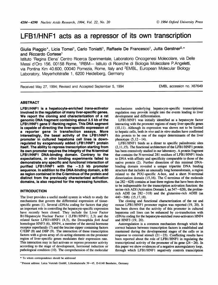
Figure 2. Trans-inhibition of the LFB1/HNF1 promoter activity in HeLa cells.The pE8U-3.5CAT reporter plasmid (10 1g) was transfected in HeLa cellstoghether with 10 itg of an expression vector encoding for the HNF4 protein(H2-HNF4) and the indicated amounts of RSV-LFB1. Cells were collected 36h after transfecion and CAT activities were measured. Top panel: the averageresults from 2 independent experiments are shown. Values (in arbitrary units)are expressed as the amount of CAT conversion corrected by the transfectionefficiency as determined by the luciferase activity in the same cell extracts. Bottompanel: a representative CAT assay is shown.
LFB1/HNF1-responsive promoter (CRP-CAT; 5) both in HepG2(Fig. iB) and HeLa cells (5,18). It has to be pointed out that norepression could be observed under the same experimentalconditions on different promoters, such as RSV (Fig. iC) andCMV (not shown), which do not contain LFB1/HNFI DNA-binding sites.
It has been shown that the liver-specific transcription factorHNF4 is a major determinant of the cell-specific expression ofthe LFBl/HNF1 gene (19,20). Consistent with these reports, wehave found that HNF4 is also able to transactivate the -3.5kbLFB1/HNF1 promoter fragment: a strong CAT gene expressionfrom this promoter fragment has been obtained in HeLa cellsin cotransfection experiments with increasing amounts of theHNF4 protein expressed under the control of the H2 promoter(H2-HNF4; Fig.2). The possibility to obtain the expression ofthe LFBI/HNFI promoter in non-hepatic cells upon thecotransfection of the HNF4 protein, has been used to verifywhether the autorepression activity of LFB1/HNF1 was restrictedto cells of hepatic origin. We therefore performed experimentsin HeLa cells in which the expression of the -3.5kb LFB1promoter was allowed by the cotransfection of H2-HNF4 andthen challenged with increasing amounts of RSV-LFB1. Theresults, reported in Fig. 2, show that LFBI/HNF1 is able to trans-inhibit its own promoter expression even in cells where the geneis not normally expressed, suggesting that the observedautorepression is not a tissue-specific phenomenon.
2(00 -
(
X
Nucleic Acids Research, 1994, Vol. 22, No. 20 4287
A
75-
-3500 +88
L-.XFB I ll-proilotel, o
E<31--- LFBt]}'.5.0 (0628
B
1z 1.1'F IvH',vil(X}
I10 1 0) 1 1 LIF1
- 0.05 0.5 1
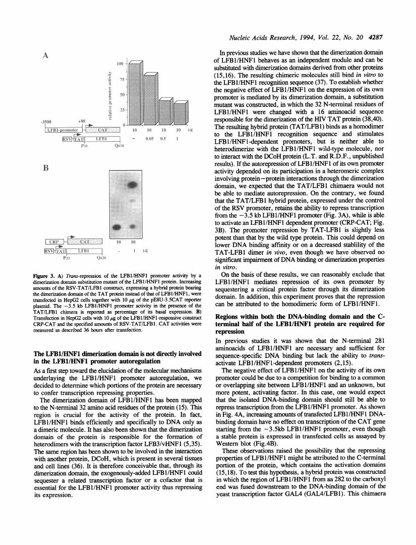
Figure 3. A) Trans-repression of the LFB1I/HNF1 promoter activity by a
dimerization domain substitution mutant of the LFB1/HNF1 protein. Increasingamounts of the RSV-TAT/LFB1 construct, expressing a hybrid protein bearingthe dimerization domain of the TAT protein instead of that of LFBl/HNF1, were
transfected in HepG2 cells together with 10 Ag of the pE8U-3.5CAT reporterplasmid. The -3.5 kb LFB1/HNF1 promoter activity in the presence of theTAT/LFB1 chimera is reported as percentage of its basal expression. B)Transfection in HepG2 cells with 10 ytg of the LFB1/HNF1-responsive constructCRP-CAT and the specified amounts of RSV-TAT/LFB1. CAT activities were
measured as described 36 hours after transfection.
The LFB1/IHNF1 dimerization domain is not directly involvedin the LFBl/HNF1 promoter autoregulationAs a first step toward the elucidation of the molecular mechanismsunderlaying the LFBl/HNFI promoter autoregulation, we
decided to determine which portions of the protein are necessary
to confer transcription repressing properties.The dimerization domain of LFBI/HNFl has been mapped
to the N-terminal 32 amino acid residues of the protein (15). Thisregion is crucial for the activity of the protein. In fact,LFBl/HNFl binds efficiently and specifically to DNA only as
a dimeric molecule. It has also been shown that the dimerizationdomain of the protein is responsible for the formation ofheterodimers with the transcription factor LFB3/vHNF1 (5,35).The same region has been shown to be involved in the interactionwith another protein, DCoH, which is present in several tissuesand cell lines (36). It is therefore conceivable that, through itsdimerization domain, the exogenously-added LFBI/HNFI couldsequester a related transcription factor or a cofactor that isessential for the LFB1/HNF1 promoter activity thus repressingits expression.
In previous studies we have shown that the dimerization domainof LFB1/HNF1 behaves as an independent module and can besubstituted with dimerization domains derived from other proteins(15,16). The resulting chimeric molecules still bind in vitro tothe LFB1/HNF1 recognition sequence (37). To establish whetherthe negative effect of LFBl/HNF1 on the expression of its ownpromoter is mediated by its dimerization domain, a substitutionmutant was constructed, in which the 32 N-terminal residues ofLFB1/HNF1 were changed with a 16 aminoacid sequenceresponsible for the dimerization of the I{V TAT protein (38,40).The resulting hybrid protein (TAT/LFB1) binds as a homodimerto the LFB1/HNF1 recognition sequence and stimulatesLFBl/HNFl-dependent promoters, but is neither able toheterodimerize with the LFBl/HNF1 wild-type molecule, norto interact with the DCoH protein (L.T. and R.D.F., unpublishedresults). If the autorepression of LFBl/HNFl of its own promoteractivity depended on its participation in a heteromeric complexinvolving protein -protein interactions through the dimerizationdomain, we expected that the TAT/LFB1 chimaera would notbe able to mediate autorepression. On the contrary, we foundthat the TAT/LFB1 hybrid protein, expressed under the controlof the RSV promoter, retains the ability to repress transcriptionfrom the -3.5 kb LFB1/HNF1 promoter (Fig. 3A), while is ableto activate an LFB1/HNF1 dependent promoter (CRP-CAT; Fig.3B). The promoter repression by TAT-LFB 1 is slightly lesspotent than that by the wild type protein. This could depend onlower DNA binding affinity or on a decreased stablility of theTAT-LFB1 dimer in vivo, even though we have observed nosignificant impairment ofDNA binding or dimerization propertiesin vitro.On the basis of these results, we can reasonably exclude that
LFBl/HNF1 mediates repression of its own promoter bysequestering a critical protein factor through its dimerizationdomain. In addition, this experiment proves that the repressioncan be attributed to the homodimeric form of LFBl/HNFI.
Regions within both the DNA-binding domain and the C-terminal half of the LFB1/HNF1 protein are required forrepressionIn previous studies it was shown that the N-terminal 281aminoacids of LFBl/HNF1 are necessary and sufficient forsequence-specific DNA binding but lack the ability to trans-activate LFB1/HNF1-dependent promoters (2,15).The negative effect of LFB1/HNF1 on the activity of its own
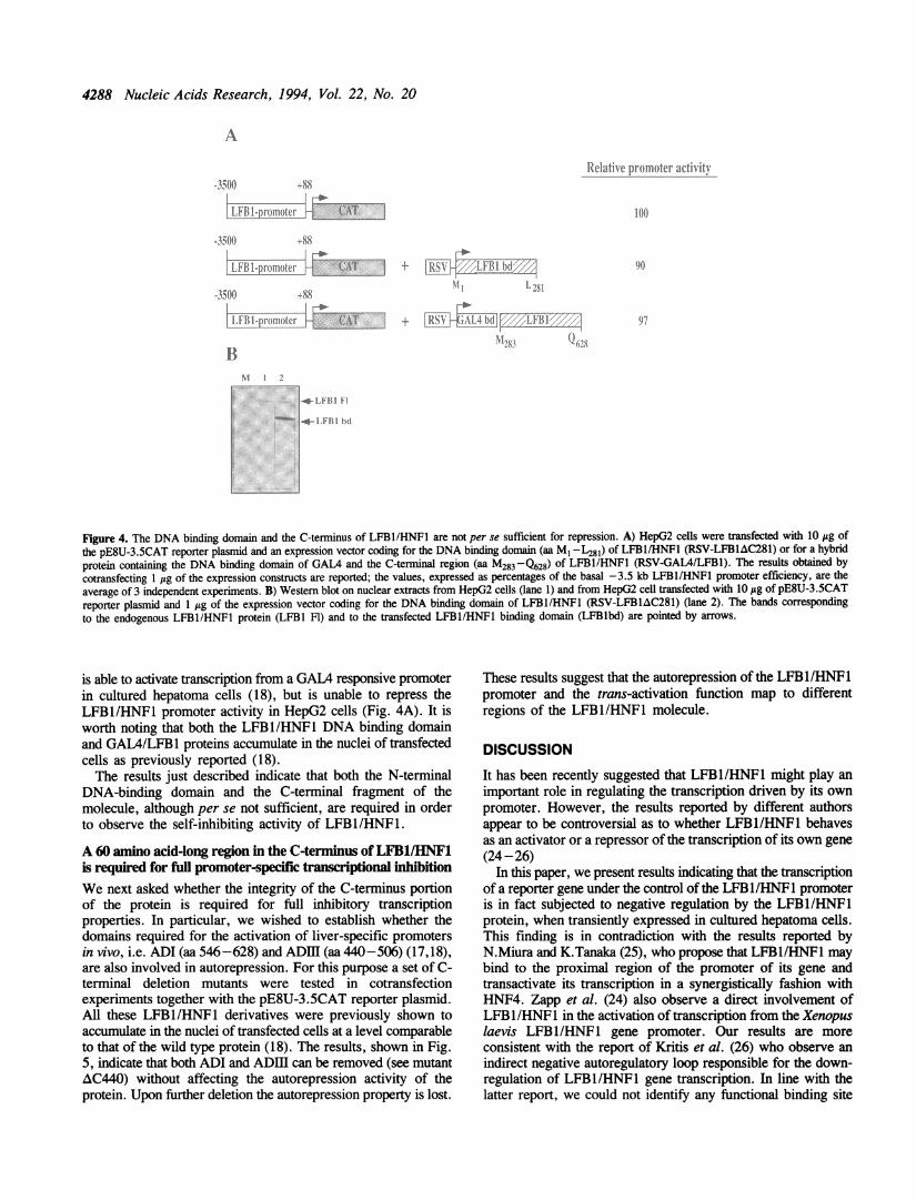
promoter could be due to a competition for binding to a commonor overlapping site between LFBl/HNF1 and an unknown, butmore potent, activating factor. In this case, one would expectthat the isolated DNA-binding domain should still be able torepress transcription from the LFB1/HNF1 promoter. As shownin Fig. 4A, increasing amounts of transfected LFBl/HNF1 DNA-binding domain have no effect on transcription of the CAT genestarting from the -3.5kb LFB1/HNFl promoter, even thougha stable protein is expressed in transfected cells as assayed byWestern blot (Fig.4B).These observations raised the possibility that the repressing
properties of LFB1/HNF1 might be attributed to the C-terminalportion of the protein, which contains the activation domains(15,18). To test this hypothesis, a hybrid protein was constructedin which the region of LFBl/HNF1 from aa 282 to the carboxylend was fused downstream to the DNA-binding domain of theyeast transcription factor GAL4 (GAL4/LFB1). This chimaera
I Ll ("
4288 Nucleic Acids Research, 1994, Vol. 22, No. 20
A
*3.500 'ISs
b~~~~~~iRip}ilue X
_v.ronmou [
-3.500) 55
11II1.p1 jiollocr ( ,.k
-350)) +8%
LI' I a -
BI 2
Relaltive_promollter .tctivitx
+ 11RSV idOl-'-A'hO
I ,.'fi5
Figure 4. The DNA binding domain and the C-terminus of LFBI/HNFI are not per se sufficient for repression. A) HepG2 cells were transfected with 10 Ag ofthe pE8U-3.5CAT reporter plasmid and an expression vector coding for the DNA binding domain (aa M1-L281) of LFBI/HNFl (RSV-LFBlAC281) or for a hybridprotein containing the DNA binding domain of GALA and the C-terminal region (aa M283 -Q628) of LFBI/HNFl (RSV-GAL4/LFB1). The results obtained bycotransfecting 1 jig of the expression constructs are reported; the values, expressed as percentages of the basal -3.5 kb LFBl/HNFl promoter efficiency, are theaverage of 3 independent experiments. B) Western blot on nuclear extracts from HepG2 cells (lane 1) and from HepG2 cell transfected with 10 sg of pE8U-3.5CATreporter plasmid and 1 /g of the expression vector coding for the DNA binding domain of LFBI/HNFI (RSV-LFBIAC281) (lane 2). The bands correspondingto the endogenous LFBI/HNFI protein (LFB1 Fl) and to the transfected LFBI/HNFI binding domain (LFBlbd) are pointed by arrows.
is able to activate transcription from a GAL4 responsive promoterin cultured hepatoma cells (18), but is unable to repress theLFB1/HNFL promoter activity in HepG2 cells (Fig. 4A). It isworth noting that both the LFB1/HNF1 DNA binding domainand GAL4/LFB1 proteins accumulate in the nuclei of transfectedcells as previously reported (18).The results just described indicate that both the N-terminal
DNA-binding domain and the C-terminal fragment of themolecule, although per se not sufficient, are required in orderto observe the self-inhibiting activity of LFB1/HNF1.
A 60 amino acid-long region in the C-terminus ofLFB1/HNF1is required for full promoter-specific transcriptional inhibitionWe next asked whether the integrity of the C-terminus portionof the protein is required for full inhibitory transcriptionproperties. In particular, we wished to establish whether thedomains required for the activation of liver-specific promotersin vivo, i.e. ADI (aa 546-628) and ADM (aa 440-506) (17,18),are also involved in autorepression. For this purpose a set of C-terminal deletion mutants were tested in cotransfectionexperiments together with the pE8U-3.5CAT reporter plasmid.All these LFB1/HNF1 derivatives were previously shown toaccumulate in the nuclei of transfected cells at a level comparableto that of the wild type protein (18). The results, shown in Fig.5, indicate that both ADI and ADHI can be removed (see mutantAC440) without affecting the autorepression activity of theprotein. Upon further deletion the autorepression property is lost.
These results suggest that the autorepression of the LFB1/HNF1promoter and the trans-activation function map to differentregions of the LFBl/HNF1 molecule.
DISCUSSIONIt has been recently suggested that LFBI/HNF1 might play animportant role in regulating the transcription driven by its ownpromoter. However, the results reported by different authorsappear to be controversial as to whether LFB1/HNF1 behavesas an activator or a repressor of the transcription of its own gene(24-26)
In this paper, we present results indicating that the transcriptionof a reporter gene under the control ofthe LFB1/HNF1 promoteris in fact subjected to negative regulation by the LFBl/HNF1protein, when transiently expressed in cultured hepatoma cells.This finding is in contradiction with the results reported byN.Miura and K.Tanaka (25), who propose that LFB1/HNF1 maybind to the proximal region of the promoter of its gene andtransactivate its transcription in a synergistically fashion withHNF4. Zapp et al. (24) also observe a direct involvement ofLFB1/HNF1 in the activation of transcription from the Xenopuslaevis LFB1/HNF1 gene promoter. Our results are moreconsistent with the report of Kritis et al. (26) who observe anindirect negative autoregulatory loop responsible for the down-regulation of LFB1/HNF1 gene transcription. In line with thelatter report, we could not identify any functional binding site

Nucleic Acids Research, 1994, Vol. 22, No. 20 4289
CAT ACTIVITY0 2 4
.-A.
1 1 3
1 281
LFBI/HNFI
628
Figure 5. A region different from the activation domains is required forautorepression. C-terminal deletions of the LFBl/HNF1 protein were transfectedin HepG2 cells with 10 itg of the pE8U-3.5CAT reporter plasmid. Cells were
harvested 36 h after transfection and the -3.5 kb promoter efficiency was
determined from the CAT activity in whole-cell extracts. The results obtainedby cotransfecting 1 zg of the expression construct are reported. The values are
expressed as the amount of CAT conversion corrected by the luciferase activityin the same cell extract (arbitrary units). The data represent an average of at least2 independent experiments. A schematic representation of the LFBI/HNF1 proteinis shown: open boxes: dimerization domain, pseudo POU-A and homeodomain;hatched boxes: Activation Domain I (aa 547-628) and III (aa 440-506).
for LFB1/HNF1 on its 3.5 kb promoter region: we scanned theentire -3.5 kb promoter in search of protein/DNA interactionsby DNaseI footprinting and gel-shift experiments with a set ofoverlapping DNA fragments using purified LFB1/HNFL protein.We could observe only a weak binding site centered at position-2677 from the transcriptional start site. However, this site hasno involvement in the LFB1/HNF1 self-inhibition, since itsdeletion in the pE8U-2.6CAT construct affects neither theefficiency of transcription nor the autorepression (data notshown). In addition computer assisted analysis of the -3.5kbLFB1/HNF1 promoter sequence has not revealed the presence
of canonical LFB1/HNF1 DNA binding sites.We have attempted to rule out relatively trivial explanations
for this finding, such as competition for a limiting amount ofa common target that is required for activation. Competition fora general transcription factor, a phenomenon known as
'squelching', seems unlikely on the basis of the followingobservations: in the same experimental system and at the same
relative concentrations, LFB1/HNF1 is a potent trans-activatorof liver-specific promoters and does not inhibit promoters thatdo not contain LFB1/HNF1 cis-acting elements (i.e. the RSVpromoter). In addition, a construct carrying the activationdomains of LFB1/HNF1 fused to a heterologous DNA-bindingdomain would be sufficient to mediate squelching, whereas theGAL4/LFB1 fusion protein did not have any effect on the activityof the LFBI/HNF-1 promoter. We can also rule out thepossibility that the over-expressed LFB 1/HNFl is sequesteringa related transcriptional activator or an essential cofactor, suchas DCoH, via its dimerization domain, since a mutant proteinin which this region has been substituted by the TAT dimerizationdomain is still capable of repression. Moreover, it is unlikelythat LFB1/HNFl acts as a repressor of its own gene expressionsimply by displacing a more potent activator of transcription fromits own promoter, because a truncated LFBI/HNFI molecule
carrying only the DNA-binding domain, while able to repressthe transcription from LFBl/HNF1-dependent promoters (31),was inactive on the LFB1/HNF1 promoter itself.
In addition to the DNA-binding domain we show that asequence present in the carboxy-terminal region is also necessaryfor the autorepression effect. This region, tentatively locatedbetween aa 380 and 440, coincides with neither of the previouslycharacterized liver-specific activation domains. One attractivepossibility is that binding of LFBl/HNF1 to its own promoteroccurs only if stabilized by protein-protein interactions, mediatedby this latter region, with a second, unknown transcriptionactivator: this protein-protein interaction would ultimately resultin 'quenching' (40) the transcriptional potential of the secondactivator.
Autoregulation appears to be a common feature of genes codingfor eukaryotic transcription factors: both positive and negativeautoregulatory loops have been found to play an important rolein modulating transcription factor expression (reviewed in 41 and42). Many examples are known of positive and negativeautoregulation of transcription factors: the muscle-specific factorMyoDi activates its own endogenous gene when transfected intofibroblast cells inducing their conversion to myoblasts (43); theerythroid-specific factor GATA-1 (23) and the pituitary-specificPOU-protein Pitl/GHF1 (22,44) have both been shown topositively regulate expression from their own promoter.Moreover, c-Fos generally represses its own transcription (21).
Several reports have contributed to outline a network ofregulatory interactions among liver-specific transcription factors(19,20). Transcription of the LFB1/HNFL gene depends on thepresence of an active HNF4, thus indicating that, in the hierarchyof developmental regulation, HNF4 comes before theLFB1/HNF1 factor. In this paper we present evidence suggestingthat another level of possible regulation of the transcription ofthe LFB1/HNF1 gene might be mediated by LFB1/HNF1 itself.It is worth noting that also LFB3/vHNF1 is capable of exertinga negative effect on LFB1/HNF1 transcription in HepG2 cells(21 and G.P., unpublished observations). In previous work wehave shown that LFB3/vHNF1 mRNA, and the protein itself,is detected before LFB1/HNF1 during the early stages ofdevelopment and during organ-specific differentiation (5,14). Itcould be speculated that the autoregulation of LFB 1/HNF1 andthe negative effect of LFB3/vHNF1 on LFB1/HNF1 transcriptionmight play a role in guaranteeing the precise timing of thesequential activation of these two factors and their correct relativeabundance, during critical stages of development anddifferentiation.
Further work will be required to elucidate the detailedmechanism by which this activity is brought about and to assessits biological relevance. At this stage, this phenomenon is justanother tessera to be added to the complex mosaic ofinterdependent regulatory mechanisms potentially involved inliver-specific gene expression.
ACKNOWLEDGEMENTSWe are grateful to N.La Monica, J.Jiricni and C.Gaetano forcritically reading the manuscript. We also want to acknowledgeYves Cully for helping us in the preparation of the figures. Thiswork was partially supported by a grant no. BI02CT930103
-3500 +88
LFIl-promoler rCAT
=-d =}THI ZJEJ-
pE8U-3.SCAT+
AC516
AC506
AC440
AC380
AC318

4290 Nucleic Acids Research, 1994, Vol. 22, No. 20
REFERENCES
1. Lai, E. and Damell, J.R. (1991) TIBS 16, 427-430.2. Frain, M., Swart, G., Monaci, P., Nicosia, A., Stampffi, S., Frank,R., and
Cortese, R. (1989), Cell 59, 145-157.3. Chouard, T., Blumenfeld., M., Bach, I, Vanderkerckhove, J., Cerenghini,
S., and Yaniv, M.( 1990), Nucl. Acids Res. 18, 5853-5863.4. Bach, I., Mattei, M.G., Cerenghini, S., and Yaniv, M. (1991) Nucl. Acids
Res.19, 3553-3559.5. De Simone, V., De Magistris, L., Lazzaro, D., Gerstner, J., Monaci, P.,
Nicosia, A., and Cortese, R. (1991), EMBO J. 10, 1435-1443.6. Lai, E., Prezioso, V.R., Tao, W., and Darnell, J.E. (1991), Genes & Dev.
5, 416-427.7. Sladek, F.M., Zhong, W., Lai, E. and Darnell, J.E. (1990) Genes & Dev.
4, 2353-2365.8. Landschulz, W.H., Johnson, P.F., Adashi, E.Y., Graves, B.J., and
McKnight, S.L. (1988) Genes & Dev. 2, 786-800.9. Mueller, C.R., Maire, P., and Schibler, U. (1990) Cell 61, 279-291.
10. De Simone, V., and Cortese, R. (1991), Cur. Op. in Cell Biol. 3,960-965.11. Mendel, D.B., and Crabtree, G.R. (1991) J. Biol. Chem. 266, 677-680.12. Blumenfeld, M., Maury, M., Chouard, T., Yaniv, M, and Condamine, H.
(1991), Development 113, 589-599.13. Xanthopoulos, K.G., Prezioso, V.R., Chen, W.S., Sladek, F.M., Cortese,
R., and Darnell, J.E. (1991) Proc. Natl. Acad. Sci.USA 88, 3707-3711.14. Lazzaro, D., De Simone, V., De Magistris, L., Lehtonen, E., and Cortese,
R. (1992) Development 114, 469-479.15. Nicosia, A., Monaci, P., Tomei, L., De Francesco, R., Nuzzo, M.,
Stunnenberg, H., and Cortese, R. (1991) Cell 61, 1225-1236.16. De Francesco, R., Pastore, A., Vecchio, G., and Cortese, R. (1991),
Biochemistry 30, 143-147.17. Raney, A.K., Easton, A.J., Milich, D.R., and McLachlan, A. (1991) J.
Virol. 65, 5774-5781.18. Toniatti, C., Monaci, P., Nicosia, A., Cortese, R., and Ciliberto, G. (1992)
DNA and Cell Bio. 12, 199-208.19. Tian, J.-M. and Schibler, U. (1991) Genes & Dev. 5, 2225-2234.20. Kuo, C.J., Conley, P.B., Chen, L., Sladek, F.M., Damell Jr., J.E., and
Crabtree G.R. (1992), Nature 355, 457-460.21. K6nig, H., Ponta, H., Rahmsdorf, U., Buscher, M., Schonthal, A.,
Rahmsdorf, H.J., and Herrlich, P. (1989), EMBO J. 8, 2559-2566.22. McCormick, A., Brady, H., Theill, L.E. and Karin, M. (1990) Nature 345,
829-832.23. Tsai, S.-F., Strauss, E., and Orldn, S.H. (1991) Genes & Dev. 5, 2352-2365.24. Zapp, D., Bartkowski, S., Holewa, B., Zoidl, C., Klein-Hitpass, L., and
Ryffel, G.U. (1993), Mol. Cell. Biol. 13, 6416-6426.25. Miura, N. and Tabnaka, K. (1993) Nucd. Acids Res. 21, 3731-3736.26. Kritis, A.A., Ktisaki, F., Barda, D., Zannis, V.I. and Talianidis, I. (1993)
Nucl. Acids Res. 21, 5882-5889.27. Sambrook, J., Fritsh., E.F., and Maniatis, T. (1989) Molecular cloning:
a laboratory Manual. Cold Spring Harbor University Press. Cold SpringHarbor.
28. Manfioletti, G., and Schneider, C. (1988), Nud. Acids Res. 16, 2873-2884.29. Sanger, F., Nicklen, S., and Coulson, A.R. (1977) Proc. Natl. Acad. Sci.
USA 74, 5463-5467.30. Piaggio, G., and De Simone, V. (1990) Focus 12, 83-84.31. Toniatti, C., Denartis, A., Monaci, P., Nicosia, A., and Ciliberto, G. (1990)
EMBO J. 9, 4457-4475.32. De Simone, V., Ciliberto, G., Hardon, E., Paonessa, G., Palla, F., Lundberg,
L., and Cortese, R. (1987), EMBO J. 6, 2759-2766.33. Gorman, C.M., Moffat, L.F., and Howard, B.H. (1982), Mol.Cell.Biol.
2, 1044-1051.34. De Wet, J.R., Wood, K.V., De Luca, M., Helsinki, D.R., and Subramani,
S. (1987), Mol.Cell.Biol. 7, 725-737.35. Rey-Campos, J., Chouard, T., Yaniv, M., and Cerenghini, S. (1991) EMBO
J. 10, 1445-1457.36. Mendel, D.B., Khavari, P.A., Conley, P.B., Graves, M.K., Hansen, L.P.,
Admond, A., and Crabtree, G.R. (1991) Science 254, 1762-1767.37. Tomei, L., Cortese, R., and De Francesco, R. (1992) EMBO J. 11,
4119-4129.38. Frankel, A.D., Bredt, D.S., and Pabo, C.O. (1988), Proc. Natl. Acad.
Sci.USA 85, 6297-6300.39. Frankel, A.D., Bredt, D.S., and Pabo, C.O. (1988), Science 240, 7073.40. Levine, M., and Manley, L.J. (1989) Cell 59, 405-408.41. Selfing, E. (1989) Trends in Genetics 5, 131-133.42. Falvey, E., and Schibler, U. (1990), FASEB J. 5, 309-314.
43. Thayer, M.J., Tapscott, S.J., Davis, R.L., Wright, W.E., Lassar, A.B.,Weintraub, H. (1989) Cell 58, 241-248.
44. Chen, R., Ingraham, H.A., Treacy, M.N., Albert, V.R., Wilson, L., andRosenfeld, M.G. (1990). Nature 346, 583-586.