Identification of the gene transcription repressor domain of Gli3
12
Identification of the gene transcription repressor domain of Gli3 Robert Tsanev, Piret Tiigimägi, Piret Michelson, Madis Metsis, Torben Østerlund ⁎ , and Priit Kogerman Department of Gene Technology, Tallinn University of Technology, Akadeemia tee 15, 12618 Tallinn, Estonia. Abstract Gli transcription factors are downstream targets of the Hedgehog signaling pathway. Two of the three Gli proteins harbor gene transcription repressor function in the N-terminal half. We have analyzed the sequences and identified a potential repressor domain in Gli2 and Gli3 and have tested this experimentally. Overexpression studies confirm that the N-terminal parts harbor gene repression activity and we mapped the minimal repressor to residues 106 till 236 in Gli3. Unlike other mechanisms that inhibit Gli induced gene transcription, the repressor domain identified here does not utilize Histone deacetylases (HDACs) to achieve repression, as confirmed by HDAC inhibition studies and pull-down assays. This distinguishes the identified domain from other regulatory parts with negative influence on transcription. Abbreviations β-gal, β-galactosidase; Ci, Cubitus interruptus; DMEM, Dulbecco’s modified Eagle’s medium; FCS, fetal calf serum; HDAC, histone deacetylases; Hh, Hedgehog; PHS, Pallister–Hall syndrome; TSA, trichostatin-A Keywords Gli3; Transcription; Domain; Hedgehog; Signaling; Development 1 Introduction The morphogens of the Hedgehog (Hh) family are crucial for numerous developmental processes during embryogenesis as well as postnatally. In fact the Hh pathway is one of the four major signaling systems that are controlling the major developmental processes. Gli proteins are Zn-finger transcription factors and are targets as well as mediators of the Hh signaling pathway [1]. Mammals have three Gli genes encoding Gli1, Gli2 and Gli3 that are orthologs of the Drosophila transcription factor Cubitus interruptus (Ci). Ci is mainly a transcriptional activator in its full-length form that dominates in the presence of Hh. In the absence of Hh, a portion of Ci is proteolytically cleaved to produce an N-terminal gene repressor form. In a similar fashion Gli2 and Gli3 can undergo proteolysis to produce a gene © 2009 Elsevier B.V.. This document may be redistributed and reused, subject to certain conditions. ⁎Corresponding author. E-mail: [email protected]. This document was posted here by permission of the publisher. At the time of deposit, it included all changes made during peer review, copyediting, and publishing. The U.S. National Library of Medicine is responsible for all links within the document and for incorporating any publisher-supplied amendments or retractions issued subsequently. The published journal article, guaranteed to be such by Elsevier, is available for free, on ScienceDirect. Sponsored document from Febs Letters Published as: FEBS Lett. 2009 January 05; 583(1): 224–228. Sponsored Document Sponsored Document Sponsored Document
-
Upload
independent -
Category
Documents
-
view
1 -
download
0
Transcript of Identification of the gene transcription repressor domain of Gli3
Identification of the gene transcription repressor domain of Gli3
Robert Tsanev, Piret Tiigimägi, Piret Michelson, Madis Metsis, Torben Østerlund⁎, and PriitKogermanDepartment of Gene Technology, Tallinn University of Technology, Akadeemia tee 15, 12618 Tallinn, Estonia.
AbstractGli transcription factors are downstream targets of the Hedgehog signaling pathway. Two of the threeGli proteins harbor gene transcription repressor function in the N-terminal half. We have analyzedthe sequences and identified a potential repressor domain in Gli2 and Gli3 and have tested thisexperimentally. Overexpression studies confirm that the N-terminal parts harbor gene repressionactivity and we mapped the minimal repressor to residues 106 till 236 in Gli3. Unlike othermechanisms that inhibit Gli induced gene transcription, the repressor domain identified here doesnot utilize Histone deacetylases (HDACs) to achieve repression, as confirmed by HDAC inhibitionstudies and pull-down assays. This distinguishes the identified domain from other regulatory partswith negative influence on transcription.
Abbreviationsβ-gal, β-galactosidase; Ci, Cubitus interruptus; DMEM, Dulbecco’s modified Eagle’s medium; FCS,fetal calf serum; HDAC, histone deacetylases; Hh, Hedgehog; PHS, Pallister–Hall syndrome; TSA,trichostatin-A
KeywordsGli3; Transcription; Domain; Hedgehog; Signaling; Development
1 IntroductionThe morphogens of the Hedgehog (Hh) family are crucial for numerous developmentalprocesses during embryogenesis as well as postnatally. In fact the Hh pathway is one of thefour major signaling systems that are controlling the major developmental processes. Gliproteins are Zn-finger transcription factors and are targets as well as mediators of the Hhsignaling pathway [1]. Mammals have three Gli genes encoding Gli1, Gli2 and Gli3 that areorthologs of the Drosophila transcription factor Cubitus interruptus (Ci). Ci is mainly atranscriptional activator in its full-length form that dominates in the presence of Hh. In theabsence of Hh, a portion of Ci is proteolytically cleaved to produce an N-terminal generepressor form. In a similar fashion Gli2 and Gli3 can undergo proteolysis to produce a gene
© 2009 Elsevier B.V..This document may be redistributed and reused, subject to certain conditions.
⁎Corresponding author. E-mail: [email protected] document was posted here by permission of the publisher. At the time of deposit, it included all changes made during peer review,copyediting, and publishing. The U.S. National Library of Medicine is responsible for all links within the document and for incorporatingany publisher-supplied amendments or retractions issued subsequently. The published journal article, guaranteed to be such by Elsevier,is available for free, on ScienceDirect.
Sponsored document fromFebs Letters
Published as: FEBS Lett. 2009 January 05; 583(1): 224–228.
Sponsored Docum
ent Sponsored D
ocument
Sponsored Docum
ent
repressor form. The full-length forms of Gli2 and Gli3 act as gene activators. A repressor formof Gli1 cannot be generated and Gli1 is considered to be a strong gene activator. The dominatingrole of Gli2 appears to be gene activation whereas Gli3 often has a gene repression role,mediated by the N-terminal part. In humans, several GLI3 morphopathies have been described,which can be broadly divided into two classes: Greig’s syndrome caused by total loss of GLI3function and Pallister–Hall syndrome (PHS)/other postaxial polydactylies that are presumedto be caused by abnormally high repressor generation. The first identified mutations causingPHS were found in the GLI3 gene [2]. Since then several Gli3 mutations have been identifiedin the same region (exons 12–14). Both original mutations are single nucleotide deletions thatlead to frame shift and premature translational stop [2]. The produced peptide has 691 residues(compared to the 1596 residue full-length protein) but contain alternative residues in the lastapproximately 20 residues, encoded after the mutations [2]. It was shown that thecorresponding peptide Gli3-PHS (residues 1–674) indeed has strong gene repressor activity,which may explain the phenotypes of these patients [3]. Due to its vast impact on celldifferentiation and proliferation aberrant Hh signaling is involved in many cancers and severalgene members of the pathway are either proto-oncogenes or tumor suppressors [4]. A thoroughanalysis of the Gli proteins is therefore important in order to understand the associateddevelopmental biology and pathology as well as related carcinogenesis.
To further analyze the repressor function in the PHS part of Gli3 and to identify the specificrepressor sequence, we made a series of GLI3 constructs and evaluated their activity in cellulargene regulation assays. This led to the identification of a specific repressor domain in GLI3also conserved in GLI2 but not in GLI1. The repressor function of this domain is not dependenton histone deacetylases (HDAC) and therefore works through a different mechanism.
2 Materials and methods2.1 DNA constructs
Gli1, Gli1(1–407), Gli3, Gli3-PHS, Gli3ΔRD and Gli3-PHSΔRD all of human origin werecloned into pcDNA3.1His expression vector (some of these were described before [3,5]). The12GliRE-luc and β-galactosidase (β-gal) constructs were described before [5]. The Gli3repressor domain (residues 106–246) and shorter versions were subcloned into the pFA vectorin frame with the DNA Binding Domain (DBD) of yeast Gal4 (Stratagene, La Jolla, CA, USA).As Gal4 reporter construct was used the pMN-Luc plasmid containing a thymidine kinasepromoter with five tandem repeats of the yeast GAL4 binding sites that control expression ofthe firefly luciferase gene. For recruitment of HDAC in gene silencing we employed the C-terminal HDAC dependent repressor domain of the rat REST protein [6] cloned in frame withGAL4 DBD in pFA.
2.2 Cell cultureHEK293 cells were grown and transfected as previously described [7]. The cells weremaintained in Dulbecco’s modified Eagle’s medium (DMEM) supplemented with 10% fetalcalf serum (FCS) (PAA Laboratories, Pasching, Austria), streptomycin and penicillin(100 units/ml; Invitrogen, Carlsbad, CA, USA). Cells were grown at 37 °C and 5.0% CO2 incell culture incubator. One day before transfection cells were plated into the required growthplates.
Shh-Light2 cells were grown in the same medium as HEK293 supplemented with G 418(400 μg/ml; Sigma–Aldrich, St. Louis, MO, USA) and Zeocine (100 mg/ml; Invitrogen). At24 h post transfection the medium was changed to low-serum medium (0.5% of FCS; PAALaboratories).
Tsanev et al. Page 2
Published as: FEBS Lett. 2009 January 05; 583(1): 224–228.
Sponsored Docum
ent Sponsored D
ocument
Sponsored Docum
ent
2.3 Luciferase assaysTransfections for luciferase assays were performed in 24-well plates. Assessment of Gli1, Gli3and Gli3-PHS in HEK293 cells was done as previously described [5,7]. Assessment in Shh-L2 cells was performed as described [8], using the incorporated luciferase gene as measurementof gene activation and the co-transfected β-gal as control. Transfections were done with thesame amount of total DNA by using empty vector to compensate.
For measurement of the Gli3-RD deletion constructs we transfected HEK293 cells also usingthe β-gal construct as control. The amount of reporter plasmid (pMN-Luc) used was 300 ngper well and the effector plasmids (pFA Gal4 fusions with RD segments) were 30 ng per well.For normalization we used 100 ng of pCMV-β-gal. As a transfecting agent we usedpolyethyleneimine (PEI; Sigma–Aldrich) 1 μg per well. DNA and PEI were mixed in 50 μl ofDMEM. An additional 150 μl of DMEM was added to the DNA/PEI mixture and then appliedto the cells. After 2 h the medium was exchanged for DMEM with 10% FCS. On the followingday the medium was changed again and where required, trichostatin-A (TSA) was added at0.2, 0.5, and 1 μM. Cells were harvested after an additional 24 h. Firefly luciferase and β-galassays were performed in Ascent FL fluoroskan with the Luciferase Assay Kit (BioTherma,Darlarö, Sweden) and Galacto-Light Plus System (Applied Biosystems, Foster City, CA,USA). In assays measuring HDAC induced gene silencing we used the REST expressing pFAvector as positive control of HDAC recruitment.
2.4 Immunoprecipitation and HDAC enzymatic assayTo test the association of Gli3-RD with HDAC we immunoprecipitated Gli3-RD and measuredHDAC enzymatic activity of the precipitate. As a positive control we immunoprecipitatedSin3A that is known to be in a complex with HDAC1 and HDAC2.
We transfected HEK293 cells with Gli3-RD in 15 cm culture dishes. The DNA/PEI complexwas prepared as follows: 30 μg of Gli3-RD DNA was mixed with 60 μg of PEI per plate in500 μl of DMEM. After 10 min of incubation 9 ml of DMEM was added to the DNA/PEImixture and then applied to the culture dish. After 2 h the medium was exchanged for DMEMwith 10% FCS. On the following day, the medium was changed again and cells were lysed inPBS with 1% Triton X-100 (Sigma–Aldrich) after an additional 24 h.
For immunoprecipitation of Gli3-RD 5 μl of anti-Gal4 polyclonal antibodies (Santa CruzBiotechnology, Santa Cruz, CA, USA) were incubated with 30 μl Protein G agarose(Amersham Biosciences, Bucks, UK). After 1 h 1 ml of cell lysate was added andimmunoprecipitation was performed at 4 °C overnight. The immunoprecipitation of Gli3-RDwas analyzed by Western blot using Gal4 monoclonal antibody (Santa Cruz Biotechnology).Sin3A was immunoprecipitated and detected as described above using an anti mSin3Aantibody (Santa Cruz Biotechnology).
HDAC activity was measured using a fluorescent substrate Fluor de Lys (Biomol, PlymouthMeeting, PA, USA) that contains an acetylated lysine side chain. This substrate was incubatedwith immunoprecipitated Gli3-RD or mSin3A. If the immunoprecipitate contain HDAC’s thesubstrate is deacetylated and a fluorophore is produced. Assays were performed according tomanufacturer’s instructions, and measured in Tecan GENios pro microplatespectrofluorometer (Tecan Group, Männedorf, Switzerland) with the Magellan V5.03 system(Tecan Group).
3 Results and discussionThe Gli3-PHS part (residues 1–673) was shown to contain gene repressor activity [3]. Usinga two-hybrid screening technology it was shown that almost the same part (residues 1–613) in
Tsanev et al. Page 3
Published as: FEBS Lett. 2009 January 05; 583(1): 224–228.
Sponsored Docum
ent Sponsored D
ocument
Sponsored Docum
ent
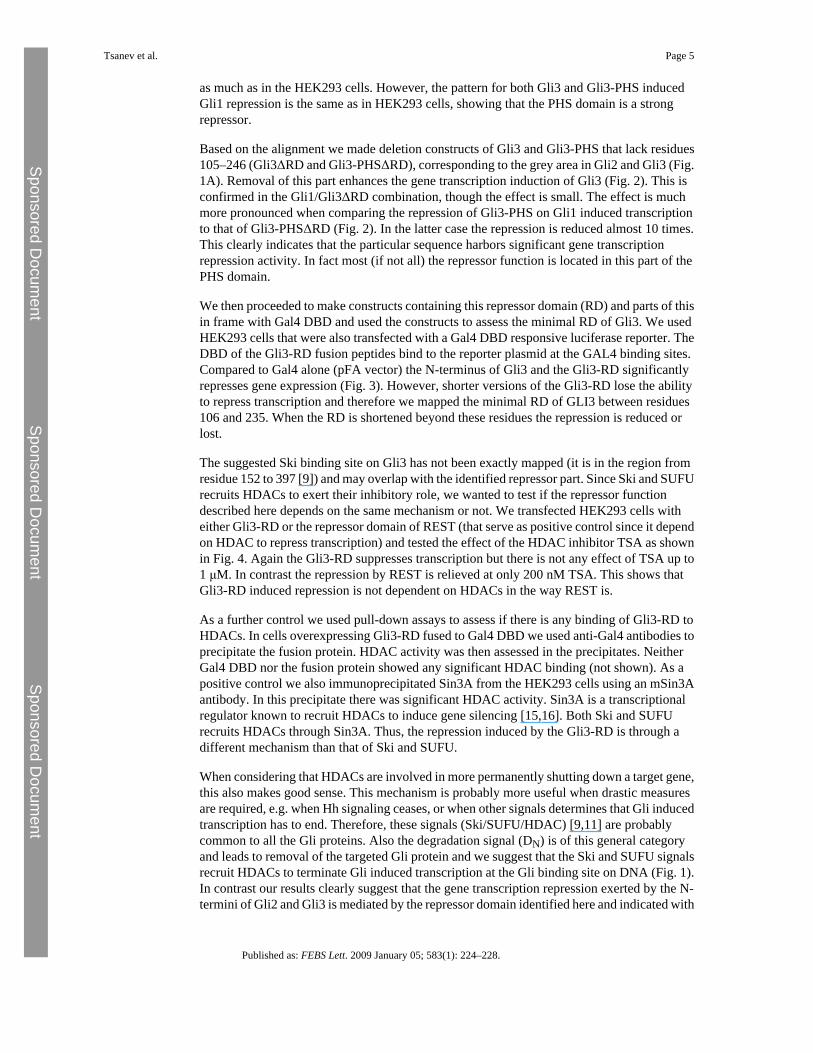
Gli3 binds to Ski [9]. The Ski binding site on Gli3 was determined to the region from residue152 to 397 using pull-down assays [9]. Since Ski is known to be part of a gene repressorcomplex including HDAC, it was suggested that Gli3 exerts its repressor activity throughbinding of Ski and recruitment of HDAC [9]. However, most of the Ski binding region isconserved between all three Gli proteins, suggesting that Ski binding and HDAC recruitmentis part of a general transcription termination signal common to all Gli proteins. Likewise, theSUFU binding site (BS) on Gli proteins (SYGH) is also found in all three Gli proteins [10].Also SUFU is known to recruit HDAC through recruitment of SAP18 and Sin3A [11] andtherefore, SUFU binding may also be regarded as a general mechanism to turn off Gli mediatedtranscription. Recently, two sites in Gli1 were identified as responsible for protein degradation[12]. One peptide (degron) was in the C-terminal part (DC) whereas the other was found in theN-terminal part (DN). In fact the DN peptide is located very close to the SUFU binding site andis conserved also in Gli2 and Gli3. A previous study identified the peptide 94–280 of Gli2 asa repressor part and removal of a corresponding part in Gli3 (residues 1–344) had strongpositive effect on transcription [13]. In contrast, when this part of Gli1 (residues 1–134) wasremoved there was no effect on transcription as compared to wild type protein [13]. The lastapproximately 100 residues of this region is conserved between the three Gli proteins, andthose are the parts that overlap with the identified Ski binding part, contain DN and the SYGHpeptide. Fig. 1A shows a schematic alignment of the N-terminal parts of mammalian Gliproteins, until the end of the Zn-fingers (corresponding to the PHS-domain), with indicationsof the respective domains describe above. From this work and a previous paper [3] it issuggested that the gene repressor function is localized to the N-terminal (grey) part of Gli2 andGli3. The physiological significance was shown by regulation of PTCH1 transcription [3]. Wesuggest that the other mechanisms with negative gene transcription activity (Ski BS, SUFUBS and DN peptide) that are common to all Gli proteins, are general means to terminate Gliinduced transcription. At least two of these pathways (Ski and SUFU through interaction withSAP18 and Sin3A) probably recruit HDACs to terminate transcription and increase thedegradation of the Gli protein.
As stated above it has previously been shown that the Gli3-PHS domain is a repressor of bothbasal and Gli3 induced transcription [3]. However, since Gli3 only induces a modesttranscriptional activation we wanted to test the PHS domain together with the much strongertranscriptional activator Gli1. Therefore, in HEK293 cells Gli1, Gli3 and Gli3-PHS weretransfected alone or in combinations together with a Gli-luciferase reporter construct [5]. Asshown before [5,7] Gli1 is an effective activator of transcription whereas Gli3 only activatesweakly (Fig. 1B). The transcriptional activation of Gli1 is strongly inhibited by co-expressingGli3-PHS. Expression of full-length Gli3 also leads to repression of Gli1 induced transcription,although significant activity is seen. Gli3 on its own gives much lower activity but it appearsthat repression of Gli1 is a more pronounced effect (3–4 times Gli3 induction vs. 6 timesrepression). In other words, not only does the Gli3-PHS have repressor activity on its own[3] it also strongly repress Gli1 induced transcription. Expression of the Gli1 peptidecorresponding to Gli3-PHS (residues 1–407) only weakly suppresses Gli1 inducedtranscription, which is likely to be due to competitive expression and suggest that the N-terminal of Gli1 does not exert any significant repressor function.
In order to analyze the constructs in a more in vivo-like setting, we turned to the Shh-Light2(Shh-L2) cells that have a Gli-inducible luciferase reporter construct incorporated into thegenome. [14]. Transfection of these cells is less efficient and the transcriptional induction byGli1 is much lower than in the HEK293 cells (Fig. 2). The induction posed by Gli3 is alsolower but the difference is not as pronounced as in HEK293 cells. This may indicate that inthe Shh-L2 cells the transcriptional regulation of the reporter is different from the vector-basedone used in HEK293 cells. Alternatively, the differences could reflect differences in the ratiosof the expressed Gli peptides. In the Shh-L2 cells Gli3 does not affect Gli1 induced transcription
Tsanev et al. Page 4
Published as: FEBS Lett. 2009 January 05; 583(1): 224–228.
Sponsored Docum
ent Sponsored D
ocument
Sponsored Docum
ent
as much as in the HEK293 cells. However, the pattern for both Gli3 and Gli3-PHS inducedGli1 repression is the same as in HEK293 cells, showing that the PHS domain is a strongrepressor.
Based on the alignment we made deletion constructs of Gli3 and Gli3-PHS that lack residues105–246 (Gli3ΔRD and Gli3-PHSΔRD), corresponding to the grey area in Gli2 and Gli3 (Fig.1A). Removal of this part enhances the gene transcription induction of Gli3 (Fig. 2). This isconfirmed in the Gli1/Gli3ΔRD combination, though the effect is small. The effect is muchmore pronounced when comparing the repression of Gli3-PHS on Gli1 induced transcriptionto that of Gli3-PHSΔRD (Fig. 2). In the latter case the repression is reduced almost 10 times.This clearly indicates that the particular sequence harbors significant gene transcriptionrepression activity. In fact most (if not all) the repressor function is located in this part of thePHS domain.
We then proceeded to make constructs containing this repressor domain (RD) and parts of thisin frame with Gal4 DBD and used the constructs to assess the minimal RD of Gli3. We usedHEK293 cells that were also transfected with a Gal4 DBD responsive luciferase reporter. TheDBD of the Gli3-RD fusion peptides bind to the reporter plasmid at the GAL4 binding sites.Compared to Gal4 alone (pFA vector) the N-terminus of Gli3 and the Gli3-RD significantlyrepresses gene expression (Fig. 3). However, shorter versions of the Gli3-RD lose the abilityto repress transcription and therefore we mapped the minimal RD of GLI3 between residues106 and 235. When the RD is shortened beyond these residues the repression is reduced orlost.
The suggested Ski binding site on Gli3 has not been exactly mapped (it is in the region fromresidue 152 to 397 [9]) and may overlap with the identified repressor part. Since Ski and SUFUrecruits HDACs to exert their inhibitory role, we wanted to test if the repressor functiondescribed here depends on the same mechanism or not. We transfected HEK293 cells witheither Gli3-RD or the repressor domain of REST (that serve as positive control since it dependon HDAC to repress transcription) and tested the effect of the HDAC inhibitor TSA as shownin Fig. 4. Again the Gli3-RD suppresses transcription but there is not any effect of TSA up to1 μM. In contrast the repression by REST is relieved at only 200 nM TSA. This shows thatGli3-RD induced repression is not dependent on HDACs in the way REST is.
As a further control we used pull-down assays to assess if there is any binding of Gli3-RD toHDACs. In cells overexpressing Gli3-RD fused to Gal4 DBD we used anti-Gal4 antibodies toprecipitate the fusion protein. HDAC activity was then assessed in the precipitates. NeitherGal4 DBD nor the fusion protein showed any significant HDAC binding (not shown). As apositive control we also immunoprecipitated Sin3A from the HEK293 cells using an mSin3Aantibody. In this precipitate there was significant HDAC activity. Sin3A is a transcriptionalregulator known to recruit HDACs to induce gene silencing [15,16]. Both Ski and SUFUrecruits HDACs through Sin3A. Thus, the repression induced by the Gli3-RD is through adifferent mechanism than that of Ski and SUFU.
When considering that HDACs are involved in more permanently shutting down a target gene,this also makes good sense. This mechanism is probably more useful when drastic measuresare required, e.g. when Hh signaling ceases, or when other signals determines that Gli inducedtranscription has to end. Therefore, these signals (Ski/SUFU/HDAC) [9,11] are probablycommon to all the Gli proteins. Also the degradation signal (DN) is of this general categoryand leads to removal of the targeted Gli protein and we suggest that the Ski and SUFU signalsrecruit HDACs to terminate Gli induced transcription at the Gli binding site on DNA (Fig. 1).In contrast our results clearly suggest that the gene transcription repression exerted by the N-termini of Gli2 and Gli3 is mediated by the repressor domain identified here and indicated with
Tsanev et al. Page 5
Published as: FEBS Lett. 2009 January 05; 583(1): 224–228.
Sponsored Docum
ent Sponsored D
ocument
Sponsored Docum
ent

grey in Fig. 1A. We also suggest that the Ski binding site is strictly localized in the regioncommon to all three Gli proteins, but a more exact mapping of the Ski binding site requiresfurther experimentation. It remains to be investigated by which mechanism Gli3-RD regulatestranscription. Perhaps the domain recruits other proteins than HDACs or interacts (physicallyor functionally) with the transcriptional machinery. It has been shown that Gli3 interacts withand regulate gene transcription via mediator [17] and perhaps the repressor domain it able toinfluence this interaction. However, the mediator binding site is localized at the C-terminalpart of Gli3 [17] and the Gli3-PHS like repressor that is generated in vivo is not likely to bindmediator, but may exert its repressor function by an independent mechanism. It is clear thatthe repression is not dependent on HDACs and investigations of the mechanism of repressionhave been initiated.
AcknowledgementsWe thank Lagle Kasak and Kersti Olspert for technical assistance and Dr. Kalju Vanatalu, for fruitful discussionsduring the study. This study was supported by the Wellcome Trust and Estonian Science Foundation (Grants 5552,5933 to P.K. and 7279 to T.Ø.).
References[1]. Hooper J.E. Scott M.P. Communicating with Hedgehogs. Nat. Rev. Mol. Cell Biol. 2005;6:306–
3317. [PubMed: 15803137][2]. Kang S. Graham J.M. Olney A.H. Biesecker L.G. GLI3 frameshift mutations cause autosomal
dominant Pallister–Hall syndrome. Nat. Genet. 1997;15:266–268. [PubMed: 9054938][3]. Shin S.H. Kogerman P. Lindström E. Toftgárd R. Biesecker L.G. GLI3 mutations in human disorders
mimic Drosophila Cubitus interruptus protein functions and localization. Proc. Natl. Acad. Sci.USA 1999;96:2880–2884. [PubMed: 10077605]
[4]. Rubin L.L. de Sauvage F.J. Targeting the Hedgehog pathway in cancer. Nat. Rev. Drug Discov.2006;5:1026–1033. [PubMed: 17139287]
[5]. Kogerman P. Grimm T. Kogerman L. Krause D. Undén A.B. Sandstedt B. Toftgård R. ZaphiropoulosP.G. Mammalian suppressor-of-fused modulates nuclear-cytoplasmic shuttling of Gli-1. Nat. CellBiol. 1999;1:312–319. [PubMed: 10559945]
[6]. Huang Y. Myers S.J. Dingledine R. Transcriptional repression by REST: recruitment of Sin3A andhistone deacetylase to neuronal genes. Nat. Neurosci. 1999;2:867–872. [PubMed: 10491605]
[7]. Østerlund T. Everman D.B. Betz R.C. Mosca M. Nöthen M.M. Schwartz C.E. Zaphiropoulos P.G.Toftgård R. The FU gene and its possible protein isoforms. BMC Genomics 2004;5:49. [PubMed:15268766]
[8]. Maloveryan A. Finta C. Østerlund T. Kogerman P. A possible role of mouse Fused (STK36) inHedgehog signaling and Gli transcription factor regulation. J. Cell Commun. Signal. 2007;1:165–173. [PubMed: 18600476]
[9]. Dai P. Shinagawa T. Nomura T. Harada J. Kaul S.C. Wadhwa R. Khan M.M. Akimaru H. Sasaki H.Colmenares C. Ishii S. Ski is involved in transcriptional regulation by the repressor and full-lengthforms of Gli3. Genes Develop. 2002;16:2843–2848. [PubMed: 12435627]
[10]. Dunaeva M. Michelson P. Kogerman P. Toftgard R. Characterization of the physical interaction ofGli proteins with SUFU proteins. J. Biol. Chem. 2003;278:5116–5122. [PubMed: 12426310]
[11]. Cheng S.Y. Bishop J.M. Suppressor of Fused represses Gli-mediated transcription by recruiting theSAP18-mSin3 corepressor complex. Proc. Natl. Acad. Sci. USA 2002;99:5442–5447. [PubMed:11960000]
[12]. Huntzicker E.G. Estay I.S. Zhen H. Lokteva L.A. Jackson P.K. Oro A.E. Dual degradation signalscontrol Gli protein stability and tumor formation. Genes Develop. 2006;20:276–281. [PubMed:16421275]
[13]. Sasaki H. Nishizaki Y. Hui C. Nakafuku M. Kondoh H. Regulation of Gli2 and Gli3 activities byan amino-terminal repression domain: implication of Gli2 and Gli3 as primary mediators of Shhsignaling. Development 1999;126:3915–3924. [PubMed: 10433919]
Tsanev et al. Page 6
Published as: FEBS Lett. 2009 January 05; 583(1): 224–228.
Sponsored Docum
ent Sponsored D
ocument
Sponsored Docum
ent

[14]. Taipale J. Chen J.K. Cooper M.K. Wang B. Mann R.K. Milenkovic L. Scott M.P. Beachy P.A.Effects of oncogenic mutations in smoothened and patched can be reversed by cyclopamine. Nature2000;406:1005–1009. [PubMed: 10984056]
[15]. Laherty C.D. Yang W.-M. Sun J.-M. Davie J.R. Seto E. Eisenman R.N. Histone deacetylasesassociated with the mSin3 corepressor mediate mad transcriptional repression. Cell 1997;89:349–356. [PubMed: 9150134]
[16]. Nagy L. Kao H.Y. Chakravarti D. Lin R.J. Hassig C.A. Ayer D.E. Schreiber S.L. Evans R.M.Nuclear receptor repression mediated by a complex containing SMRT, mSin3A, and histonedeacetylase. Cell 1997;89:373–380. [PubMed: 9150137]
[17]. Zhou H. Kim S. Ishii S. Boyer T.G. Mediator modulates Gli3-dependent Sonic Hedgehog signaling.Mol. Cell Biol. 2006;26:8667–8682. [PubMed: 17000779]
Tsanev et al. Page 7
Published as: FEBS Lett. 2009 January 05; 583(1): 224–228.
Sponsored Docum
ent Sponsored D
ocument
Sponsored Docum
ent
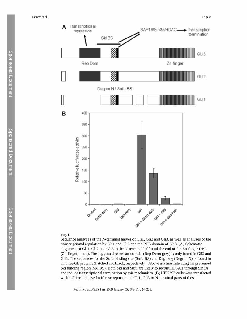
Fig. 1.Sequence analyzes of the N-terminal halves of Gli1, Gli2 and Gli3, as well as analyzes of thetranscriptional regulation by Gli1 and Gli3 and the PHS domain of Gli3. (A) Schematicalignment of Gli1, Gli2 and Gli3 in the N-terminal half until the end of the Zn-finger DBD(Zn-finger; lined). The suggested repressor domain (Rep Dom; grey) is only found in Gli2 andGli3. The sequences for the Sufu binding site (Sufu BS) and DegronN (Degron N) is found inall three Gli proteins (hatched and black, respectively). Above is a line indicating the presumedSki binding region (Ski BS). Both Ski and Sufu are likely to recruit HDACs through Sin3Aand induce transcriptional termination by this mechanism. (B) HEK293 cells were transfectedwith a Gli responsive luciferase reporter and Gli1, Gli3 or N-terminal parts of these
Tsanev et al. Page 8
Published as: FEBS Lett. 2009 January 05; 583(1): 224–228.
Sponsored Docum
ent Sponsored D
ocument
Sponsored Docum
ent

corresponding to the PHS domain, or combinations to assess the effects of these ontranscription. Error bars indicate the standard deviations of triplicate analyzes.
Tsanev et al. Page 9
Published as: FEBS Lett. 2009 January 05; 583(1): 224–228.
Sponsored Docum
ent Sponsored D
ocument
Sponsored Docum
ent

Fig. 2.Analyzes of the repressor function of the Gli3-PHS domain and the repressor domain (RD) inShh-L2 cells. Shh-L2 cells (with an incorporated Gli responsive luciferase reporter gene) weretransfected with Gli1, Gli3, Gli3-PHS, Gli3ΔRD, Gli3-PHSΔRD or combinations of these toassess the effect on transcription alone or on the Gli1 induced transcription. The analyzes wereperformed at least three times and error bars indicate the standard deviations.
Tsanev et al. Page 10
Published as: FEBS Lett. 2009 January 05; 583(1): 224–228.
Sponsored Docum
ent Sponsored D
ocument
Sponsored Docum
ent
Fig. 3.Determination of the minimal repressor domain of Gli3. The repressor domain (residues 105–246) or parts of this were expressed together with the DBD of Gal4 and assessed for repressionof Gal4 induced transcription in HEK293 cells (mock). Also a larger part of the Gli3 N-terminalpart was measured since this is known to have significant repressor function (residues 1–480).The analyzes were performed three to five times and error bars show the standard deviations.
Tsanev et al. Page 11
Published as: FEBS Lett. 2009 January 05; 583(1): 224–228.
Sponsored Docum
ent Sponsored D
ocument
Sponsored Docum
ent

Fig. 4.HDAC recruitment study of the Gli3 repressor domain. HEK293 cells were transfected withGli3-RD (squares) or the repressor domain of REST (positive control, triangles) and treatedwith increasing amounts of the HDAC inhibitor TSA. As negative control we used cellstransfected with empty vector (diamonds). The analyzes were performed three to six times andthe error bars indicate standard deviations.
Tsanev et al. Page 12
Published as: FEBS Lett. 2009 January 05; 583(1): 224–228.
Sponsored Docum
ent Sponsored D
ocument
Sponsored Docum
ent