Quantitative properties of sovereign default models: Solution methods matter
Upload
khangminh22Category
view
0download
0
CONFERENCE
ON EMERGENCY
MEDICINE
ASIAN
CONGRESS
EMERGENCY
MEDICINE
TURKISH
Regnum Carya Belek, Antalya / Turkey
November 22-25, 2017
www.acem2017.org
&&13th
Regnum Carya Belek, Antalya / Turkey
November 22-25, 2017
www.acem2017.org
ACEM2017november 2017
antalya, Turkey
A S I A NSOCIETY FOREMERGENCYM E D I C I N E
PROGRAM AND ABSTRACT BOOK

9th ASIAN CONFERENCE ON EMERGENCY MEDICINE9th ASIAN CONFERENCE ON EMERGENCY MEDICINE
13th TURKISH EMERGENCY MEDICINE CONGRESS13th TURKISH EMERGENCY MEDICINE CONGRESS
November 22-25, 2017, Regnum Carya Belek, Antalya / TurkeyNovember 22-25, 2017, Regnum Carya Belek, Antalya / Turkey ACEM2017
&&
Table of Contents
WELCOME MESSAGE
COMMITTEES
SPONSORS ACKNOWLEDGMENT
CONTACT
SCIENTIFIC PROGRAM
LECTURES
POSTER ABSRTACTS
ORAL ABSTRACTS

9th ASIAN CONFERENCE ON EMERGENCY MEDICINE9th ASIAN CONFERENCE ON EMERGENCY MEDICINE
13th TURKISH EMERGENCY MEDICINE CONGRESS13th TURKISH EMERGENCY MEDICINE CONGRESS
November 22-25, 2017, Regnum Carya Belek, Antalya / TurkeyNovember 22-25, 2017, Regnum Carya Belek, Antalya / Turkey ACEM2017
&&
Welcome Message
Dear Colleagues,
We would like to cordially thank you for joining us at the 9th Asian Conference on Emergency Medicine & 13th Turkish Emergency Medicine Congress. The Asian Conference for Emergency Medicine is the most important international meeting organized by Asian Society for Emergency Medicine. We are very excited about getting together with you in our congress held from November 22 to 25, 2017 at the Regnum Carya Belek, Antalya, Turkey.
Emergency Medicine has been recognized as a major medical specialty in Turkey for over 20 years. From the very beginning of this process, the Emergency Medicine Association of Turkey has always been the biggest supporter of all emergency medical professionals. Publications of our association and our continuous medical education activities are the main reference source for Emergency Medicine Training institutions, Emergency Medicine specialists and practitioners, and for Emergency Medicine residents.
In ACEM 2017, important academicians, editors and world-renowned names of the Emergency Medicine community came together as speakers and participants. The conference is organized in cooperation with the Asian Society for Emergency Medicine, International Federation for Emergency Medicine and other regional and international societies.
We aimed to create an environment that fosters a dialogue among participants and the experts, leading to idea exchange and discussions, collaborations and most importantly friendship. As you already know, there are several sessions in di�erent formats such as plenary meeting, hands-on courses, panel discussions, round table meetings, to name a few.
We are very pleased with the interest of the community with more than 1.000 participants from 36 di�erent countries and submission of 902 interesting research manuscripts and case studies in di�erent topics related to emergency medicine. Your participation and your contribution has been our strength, thank you one more time for being a part of this memorable conference.
We wish you will have a socially enjoyable, scienti�cally rich, comfortable and warm environment. Hope you will feel completely at home, in Antalya, and in Turkey.
Sincerely,
Prof. Dr. Yıldıray ÇETEPresident - Emergency Medicine Association of Turkey &President Elect - Asian Society for Emergency Medicine

9th ASIAN CONFERENCE ON EMERGENCY MEDICINE9th ASIAN CONFERENCE ON EMERGENCY MEDICINE
13th TURKISH EMERGENCY MEDICINE CONGRESS13th TURKISH EMERGENCY MEDICINE CONGRESS
November 22-25, 2017, Regnum Carya Belek, Antalya / TurkeyNovember 22-25, 2017, Regnum Carya Belek, Antalya / Turkey ACEM2017
&&
EMERGENCY MEDICINE ASSOCIATION OF TURKEY(EMAT)
EXECUTIVE COMMITTEE
ASIAN SOCIETY FOR EMERGENCY MEDICINE (ASEM)EXECUTIVE COMMITTEE
PresidentYıldıray ÇETE
Vice PresidentArzu DENİZBAŞI
General SecretaryBülent ERBİL
TreasurerCem OKTAY
Board Members (*)Ersin AKSAYMurat ÇETİN
Özgür ÇEVRİMMüge GÜNALP ENEYLİ
Özlem KÖKSALHakan TOPAÇOĞLU
Özcan YAVAŞİ
* Aligned in order of the surnames.
PresidentWai-mau CHOI
Taiwan Society of Emergency Medicine
President ElectYıldıray ÇETE
Emergency Medicine Association of Turkey
Honorary SecretarySwee-han LIM
Society for Emergency Medicine in Singapore
Assistant SecretaryArif Alper ÇEVİK
Emergency Medicine Association of Turkey
Honorary TreasurerAxel SIU
Hong Kong Society for Emergency
Medicine and Surgery
Assistant SecretarySrinath Kumar Tumsi SANJEEVA
Society for Emergency Medicine in India
Immediate Past PresidentProf. Takeshi SHIMAZU
Japanese Association for Acute Medicine
Committees
*Please search for related section, by typing name, institution or word.

9th ASIAN CONFERENCE ON EMERGENCY MEDICINE9th ASIAN CONFERENCE ON EMERGENCY MEDICINE
13th TURKISH EMERGENCY MEDICINE CONGRESS13th TURKISH EMERGENCY MEDICINE CONGRESS
November 22-25, 2017, Regnum Carya Belek, Antalya / TurkeyNovember 22-25, 2017, Regnum Carya Belek, Antalya / Turkey ACEM2017
&&
Committees
CONGRESS SCIENTIFIC COMMITTEE
Abu Hassan Asaari ABDULLAH (Malaysia)Ersin AKSAY (Turkey)
Rıdvan ATİLLA (Turkey)Rasha BUHUMAID (U.A.E.)
Yıldıray ÇETE (Turkey)Arif Alper ÇEVİK (Turkey)Wai Mau CHOI (Taiwan)
Arzu DENİZBAŞI (Turkey)Serkan Emre EROĞLU (Turkey) Cheng Chung FANG (Taiwan)
Saleh FARES (U.A.E.)Sabariah Faizah JAMALUDDIN (Malaysia)
Somchai KANCHANASUT (Thailand)Pairoj KHRUEKARNCHANA (Thailand)
Akio KIMURA (Japan)Tamorish KOLE (India)
Özlem KÖKSAL (Turkey)T.S. Srinath KUMAR (India)
Kang Hyun LEE (Korea)Swee Han LIM (Singapore)
Faith Joan MESA-GAERLAN (Philippines)Reynante MIRANO (Philippines)
Cem OKTAY (Turkey)Takeshi SHIMAZU (Japan)
Axel SIU (Hong Kong)Do Ngoc SON (Vietnam)Özgür SÖĞÜT (Turkey)
Mohan TIRU (Singapore)Ludwig TSOI (Hong Kong)
Hyuk Jun YANG (Korea)
* Aligned in order of the surnames.
*Please search for related section, by typing name, institution or word.

9th ASIAN CONFERENCE ON EMERGENCY MEDICINE9th ASIAN CONFERENCE ON EMERGENCY MEDICINE
13th TURKISH EMERGENCY MEDICINE CONGRESS13th TURKISH EMERGENCY MEDICINE CONGRESS
November 22-25, 2017, Regnum Carya Belek, Antalya / TurkeyNovember 22-25, 2017, Regnum Carya Belek, Antalya / Turkey ACEM2017
&&
Committees
* Aligned in order of the surnames.
CONGRESS ORGANIZING COMMITTEE
Abu Hassan Asaari ABDULLAH (Malaysia)Gökhan AKSEL (Turkey)
Yusuf Ali ALTUNCI (Turkey)Gürkan ALTUNTAŞ (Turkey)Yunus Emre ARIK (Turkey)
Mehmet Ali ASLANER (Turkey)Barış Murat AYVACI (Turkey)
Rasha BUHUMAID (U.A.E.)Wai Mau CHOI (Taiwan)
Mustafa Emin ÇANAKÇI (Turkey)Murat ÇETİN (Turkey)
Arif Alper ÇEVİK (Turkey)Özgür ÇEVRİM (Turkey)Bulut DEMİREL (Turkey)Halil DOĞAN (Turkey)
Cheng Chung FANG (Taiwan)Melis EFEOĞLU SACAK (Turkey)
Bülent ERBİL (Turkey)Mehmet ERGİN (Turkey)
Serkan Emre EROĞLU (Turkey) Müge GÜNALP ENEYLİ (Turkey)
Saleh FARES (U.A.E.)Enes GÜLER (Turkey)
Süleyman İBZE (Turkey)Sabariah Faizah JAMALUDDIN (Malaysia)
Gültekin KADI (Turkey)
Somchai KANCHANASUT (Thailand)Sinan KARACABEY (Turkey)
Pairoj KHRUEKARNCHANA (Thailand)Akio KIMURA (Japan)
Mehmet KOÇAK (Turkey)Tamorish KOLE (India)
T.S.Srinath KUMAR (India)Kang Hyun LEE (Korea)
Swee Han LIM (Singapore)Faith Joan MESA-GAERLAN (Philippines)
Reynante MIRANO (Philippines)Ayhan ÖZHASENEKLER (Turkey)
Erkman SANRI (Turkey)Takeshi SHIMAZU (Japan)
Axel SIU (Hong Kong)Do Ngoc SON (Vietnam)Mohan TIRU (Singapore)
Hakan TOPAÇOĞLU (Turkey)Ludwig TSOI (Hong Kong)
Ebru ÜNAL AKOĞLU (Turkey)Hyuk Jun YANG (Korea)Özcan YAVAŞİ (Turkey)Murat YAZICI (Turkey)Yasin YILDIZ (Turkey)
Çağdaş YILDIRIM (Turkey)
*Please search for related section, by typing name, institution or word.

9th ASIAN CONFERENCE ON EMERGENCY MEDICINE9th ASIAN CONFERENCE ON EMERGENCY MEDICINE
13th TURKISH EMERGENCY MEDICINE CONGRESS13th TURKISH EMERGENCY MEDICINE CONGRESS
November 22-25, 2017, Regnum Carya Belek, Antalya / TurkeyNovember 22-25, 2017, Regnum Carya Belek, Antalya / Turkey ACEM2017
&&
Sponsors Acknowledgment
Sponsors are listed alphabetically and categorized by the contribution
*Please search for related section, by typing name, institution or word.

9th ASIAN CONFERENCE ON EMERGENCY MEDICINE9th ASIAN CONFERENCE ON EMERGENCY MEDICINE
13th TURKISH EMERGENCY MEDICINE CONGRESS13th TURKISH EMERGENCY MEDICINE CONGRESS
November 22-25, 2017, Regnum Carya Belek, Antalya / TurkeyNovember 22-25, 2017, Regnum Carya Belek, Antalya / Turkey ACEM2017
&&
Contact
Organization Secretariat
Congress Scientific Secretariat
Erkan GÖKSU, MD. Prof.Akdeniz University Faculty of Medicine,
Department of Emergency Medicine, Antalya - Turkey
Serkan Emre EROĞLU, MD. Assoc. Prof.University of Health Sciences Ümraniye Training
and Research Hospital Clinic of Emergency Medicine, Istanbul - Turkey
Axel SIU, MD.North District Hospital Accident and Emergency Department, Hong Kong
Koşuyolu Mah. Mahmut Yesari Cad. No:6434718 Kadıköy / İstanbul - Turkey
Phone: +90 (216) 414 11 11Fax: +90 (216) 414 65 44
*Please search for related section, by typing name, institution or word.

9th ASIAN CONFERENCE ON EMERGENCY MEDICINE9th ASIAN CONFERENCE ON EMERGENCY MEDICINE
13th TURKISH EMERGENCY MEDICINE CONGRESS13th TURKISH EMERGENCY MEDICINE CONGRESS
November 22-25, 2017, Regnum Carya Belek, Antalya / TurkeyNovember 22-25, 2017, Regnum Carya Belek, Antalya / Turkey ACEM2017
&&
Scientific Program
*Please search for related section, by typing name, institution or word.

9th ASIAN CONFERENCE ON EMERGENCY MEDICINE9th ASIAN CONFERENCE ON EMERGENCY MEDICINE
13th TURKISH EMERGENCY MEDICINE CONGRESS13th TURKISH EMERGENCY MEDICINE CONGRESS
November 22-25, 2017, Regnum Carya Belek, Antalya / TurkeyNovember 22-25, 2017, Regnum Carya Belek, Antalya / Turkey
&&
22 November 2017, Wednesday
COURSES
10:00 - 15:00 ULTRASOUND WORKSHOP HALL E COURSE COORDINATORS AND TRAINERS: Mohamad Moussa (U.S.A.), Abdel Noureldin (U.S.A.), Aslıhan Yürüktümen Ünal (Turkey), Özlem Dikme (Turkey), Betül Gülalp (Turkey)
10:00 - 11:00 Session 1 11:00 - 11:30 Co�ee Break
11:30 - 12:30 Session 2
12:30 - 13:30 Lunch
13:30 - 15:00 Session 3
10:00 - 15:00 TRIAGE WORKSHOP HALL F COURSE COORDINATOR AND TRAINER: Cem Oktay (Turkey)
10:00 - 11:00 Session 1 11:00 - 11:30 Co�ee Break
11:30 - 12:30 Session 2
12:30 - 13:30 Lunch
13:30 - 15:00 Session 3
10:00 - 15:00 NIV-IMV WORKSHOP HALL G COURSE COORDINATORS AND TRAINERS: Başak Bayram (Turkey), Neşe Çolak Oray (Turkey)
10:00 - 11:00 Session 1 11:00 - 11:30 Co�ee Break
11:30 - 12:30 Session 2
12:30 - 13:30 Lunch
13:30 - 15:00 Session 3
ACEM2017
*Please search for related section, by typing name, institution or word.

9th ASIAN CONFERENCE ON EMERGENCY MEDICINE9th ASIAN CONFERENCE ON EMERGENCY MEDICINE
13th TURKISH EMERGENCY MEDICINE CONGRESS13th TURKISH EMERGENCY MEDICINE CONGRESS
November 22-25, 2017, Regnum Carya Belek, Antalya / TurkeyNovember 22-25, 2017, Regnum Carya Belek, Antalya / Turkey
&&
22 November 2017, Wednesday
COURSES
10:00 - 15:00 ECMO WORKSHOP HALL H COURSE COORDINATORS AND TRAINERS: Mohamad Moussa (U.S.A.), Abdel Noureldin (U.S.A.), Aslıhan Yürüktümen Ünal (Turkey), Özlem Dikme (Turkey), Betül Gülalp (Turkey)
10:00 - 11:00 Introduction to ECMO 11:00 - 11:30 Co�ee Break
11:30 - 12:30 ECMO Device and Catheters
12:30 - 13:30 Lunch
13:30 - 15:00 ECMO Workshop - Practical Session
10:00 - 15:30 İLERİ HAVAYOLU KURSU HALL I * This course will be held in Turkish / Bu kurs Türkçe olarak gerçekleştirilecektir.
KURS KOORDİNATÖRLERİ VE EĞİTMENLERİ:
Barış Murat Ayvacı (Turkey), Volkan Arslan (Turkey), Mustafa Yazıcıoğlu (Turkey)
10:00 - 11:00 Havayolu Anatomisi / Supraglottik ve Cerrahi Havayolu Yönetimi 11:00 - 11:30 Kahve Molası
11:30 - 12:30 Videolaringoskopi/Fiberoptik Havayolu Yönetimi
12:30 - 13:30 Öğle Yemeği
13:30 - 15:00 RSI/DSI/Zor Havayolu Tanımı ve Yönetimi İstasyon 1 / Videolaringoskopi - Fiberoptik Havayolu Yönetimi - Endotrakeal Entübasyon - Buji ile Entübasyon Mustafa Yazıcıoğlu (Turkey) İstasyon 2 / Supraglottik Havayolları Barış Murat Ayvacı (Turkey) İstasyon 3 / Cerrahi Havayolu Yönetimi Volkan Arslan (Turkey)
ACEM2017
*Please search for related section, by typing name, institution or word.

9th ASIAN CONFERENCE ON EMERGENCY MEDICINE9th ASIAN CONFERENCE ON EMERGENCY MEDICINE
13th TURKISH EMERGENCY MEDICINE CONGRESS13th TURKISH EMERGENCY MEDICINE CONGRESS
November 22-25, 2017, Regnum Carya Belek, Antalya / TurkeyNovember 22-25, 2017, Regnum Carya Belek, Antalya / Turkey
&&
22 November 2017, Wednesday
HALL B
15:30 - 17:00 OPENING PLENARY SESSION: Present and Future Perspective of Emergency Medicine MODERATOR: Lim Swee Han (Singapore)
15:30 - 16:00 Emergency Medicine in Asian Countries Wai Mau Choi (Taiwan)
16:00 - 16:30 Emergency Medicine in Europe Luis Garcia-Castrillo Riesgo (Spain) 16:30 - 17:00 Canary in the Coal Mine Michael Bullard (Canada) 17:00 - 18:00 OPENING CEREMONY
18:00 - 19:00 OPENING COCKTAIL (Exhibition Area)
ACEM2017
*Please search for related section, by typing name, institution or word.

9th ASIAN CONFERENCE ON EMERGENCY MEDICINE9th ASIAN CONFERENCE ON EMERGENCY MEDICINE
13th TURKISH EMERGENCY MEDICINE CONGRESS13th TURKISH EMERGENCY MEDICINE CONGRESS
November 22-25, 2017, Regnum Carya Belek, Antalya / TurkeyNovember 22-25, 2017, Regnum Carya Belek, Antalya / Turkey
&&
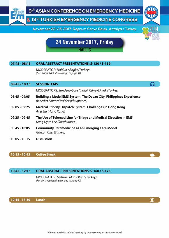
07:45 - 08:45 ORAL ABSTRACT PRESENTATIONS: S-001 / S-010
MODERATOR: Mustafa Burak Sayhan (Turkey) (For abstract details please go to page 44)
08:45 - 10:15 SESSION: Resuscitation
MODERATORS: Khusrav Bajan (India), Cem Oktay (Turkey)
08:45 - 09:05 BLS (CPR) Training 2017 Updates: What are the Latest Sciences and Guidelines Lim Swee Han (Singapore)
09:05 - 09:25 New Approach to Education and Training of Home Bystander CPR Sang Do Shin (South Korea)
09:25 - 09:45 Are we Ready for Drones? Sowjanya Patibandla (India)
09:45 - 10:05 ACLS is all about A-C-L-S: From Team of Expert to Expert Teams Matthew Huei Ming Ma (Taiwan)
10:05 - 10:15 Discussion
10:45 - 12:15 PLENARY SESSION: Trauma MODERATORS: Sabariah Faizah Jamaluddin (Malaysia), Levent Avşaroğulları (Turkey)
10:45 - 11:15 Trauma Care: Pitfalls to Avoid Jim Holliman (U.S.A.)
11:15 - 11:45 Controversies in ATLS Cem Oktay (Turkey)
11:45 - 12:15 Educational and Clinical Impact of ATLS Fikri Abu Zidan (United Arab Emirates)
10:15 - 10:45 Co�ee Break
12:15 - 13:15 Lunch
23 November 2017, Thursday
HALL A
ACEM2017
*Please search for related section, by typing name, institution or word.

14:45 - 15:15 Co�ee Break
9th ASIAN CONFERENCE ON EMERGENCY MEDICINE9th ASIAN CONFERENCE ON EMERGENCY MEDICINE
13th TURKISH EMERGENCY MEDICINE CONGRESS13th TURKISH EMERGENCY MEDICINE CONGRESS
November 22-25, 2017, Regnum Carya Belek, Antalya / TurkeyNovember 22-25, 2017, Regnum Carya Belek, Antalya / Turkey
&&
13:15 - 14:45 SESSION: Disaster Medicine
MODERATORS: Axel Siu (Hong Kong), Niyazi Özüçelik (Turkey)
13:15 - 13:35 New Triage Tools for Mass Disasters Goh Siang Hiong (Singapore)
13:35 - 13:55 Largest Refugee Population in the World; Syrians in Turkey Erkan Günay (Turkey)
13:55 - 14:15 Disaster Risk Reduction Research Capacity Building Program in the Philippines Teodoro J. Herbosa (Philippines)
14:15 - 14:35 Challenges in Conducting Disaster Simulations Ali Haedar (Indonesia)
14:35 - 14:45 Discussion
16:00 - 17:30 SESSION: Cardiac Emergencies
MODERATORS: Mahmood Mohammed Ghanaim (United Arab Emirates) Selahattin Kıyan (Turkey)
16:00 - 16:20 Update on Management of STEMI – What You Need to Know Before Your Next Shift Lim Swee Han (Singapore)
16:20 - 16:40 Management of Acute Heart Failure in ED Salvatore Di Somma (Italy)
16:40 - 17:00 ACS 2017: Update Mehmet Ergin (Turkey)
17:00 - 17:20 Chest Pain Evaluation and Stress Imaging in ED Patients Annitha Annathurai (Singapore)
17:20 - 17:30 Discussion
15:15 - 16:00 INDUSTRY SPONSORED SYMPOSIUM: GSK
Optimal Bronchodilatation: Emergency Approach to Asthma Patients in Accompanying Guidelines Serkan Emre Eroğlu (Turkey)
17:30 - 18:30 SELECTED ORAL ABSTRACT PRESENTATIONS: S-076 / S-083
MODERATOR: Levent Avşaroğulları (Turkey) (For abstract details please go to page 51)
18:30 - 19:30 Türkiye Acil Tıp Derneği Genel Kurul
23 November 2017, Thursday
HALL A
ACEM2017
*Please search for related section, by typing name, institution or word.

9th ASIAN CONFERENCE ON EMERGENCY MEDICINE9th ASIAN CONFERENCE ON EMERGENCY MEDICINE
13th TURKISH EMERGENCY MEDICINE CONGRESS13th TURKISH EMERGENCY MEDICINE CONGRESS
November 22-25, 2017, Regnum Carya Belek, Antalya / TurkeyNovember 22-25, 2017, Regnum Carya Belek, Antalya / Turkey
&&
08:45 - 10:15 SESSION: General Medicine
MODERATORS: Gregor Prosen (Slovenia), Hakan Topaçoğlu (Turkey)
08:45 - 09:05 Medical Clearance of the Psychiatry ED Patients Mahmoud Aljufaili (Oman)
09:05 - 09:25 Acid Base Disorders at ED Ankur Verma (India)
09:25 - 09:45 Antimicrobial Stewardship in the ED Can Özen (Turkey)
09:45 - 10:05 Tumor Lysis Syndrome Imad Majzoub (Lebanon)
10:05 - 10:15 Discussion
13:15 - 14:45 SESSION: Pediatrics
MODERATORS: Ludwig Tsoi (Hong Kong), Özlem Köksal (Turkey)
13:15 - 13:30 Procedural Sedation and Analgesia in Pediatric Patients Süha Türkmen (Turkey)
13:30 - 13:45 Airway Management in Pediatric Patients Imad Majzoub (Lebanon)
13:45 - 14:15 Pediatric ECMO Salih Özçobanoğlu (Turkey)
14:15 - 14:30 Pediatric Emergency Medicine - Need of the Hour S.Srinath Kumar (India)
14:30 - 14:45 Discussion
07:45 - 08:45 ORAL ABSTRACT PRESENTATIONS: S-011 / S-020 MODERATOR: Süha Türkmen (Turkey) (For abstract details please go to page 45)
10:45 - 12:15 SELECTED ORAL ABSTRACT PRESENTATIONS: S-041 / S-050 MODERATOR: Arzu Denizbaşı (Turkey) (For abstract details please go to page 48)
10:15 - 10:45 Co�ee Break
12:15 - 13:15 Lunch
23 November 2017, Thursday
HALL B
ACEM2017
*Please search for related section, by typing name, institution or word.

15:15 - 16:00 INDUSTRY SPONSORED SYMPOSIUM: Daiichi-Sankyo MODERATOR: Yıldıray Çete (Turkey)
Approach to STEMI in Light of 2017 European Guidelines Adnan Abacı (Turkey)
9th ASIAN CONFERENCE ON EMERGENCY MEDICINE9th ASIAN CONFERENCE ON EMERGENCY MEDICINE
13th TURKISH EMERGENCY MEDICINE CONGRESS13th TURKISH EMERGENCY MEDICINE CONGRESS
November 22-25, 2017, Regnum Carya Belek, Antalya / TurkeyNovember 22-25, 2017, Regnum Carya Belek, Antalya / Turkey
&&
14:45 - 15:15 Co�ee Break
16:00 - 17:30 SESSION: General Medicine
MODERATORS: Jae Ho Lee (South Korea), Nalan Metin Aksu (Turkey)
16:00 - 16:20 Point of Care Testing (POCT) in Emergency Department - A Revolutionary Tool Sandeep Gore (India)
16:20 - 16:40 The Patient You Saw Yesterday : Short-Term Death After Emergency Department Discharge Murat Ersel (Turkey)
16:40 - 17:00 Critical Decision Making in Emergency Medicine Fikri Abu Zidan (United Arab Emirates)
17:00 - 17:20 Urgent Pain Management Anindya Dasgupta (India)
17:20 - 17:30 Discussion
23 November 2017, Thursday
HALL B
17:30 - 18:30 SELECTED ORAL ABSTRACT PRESENTATIONS: S-084 / S-090 MODERATOR: Nurettin Özgür Doğan (Turkey) (For abstract details please go to page 52)
ACEM2017
*Please search for related section, by typing name, institution or word.

9th ASIAN CONFERENCE ON EMERGENCY MEDICINE9th ASIAN CONFERENCE ON EMERGENCY MEDICINE
13th TURKISH EMERGENCY MEDICINE CONGRESS13th TURKISH EMERGENCY MEDICINE CONGRESS
November 22-25, 2017, Regnum Carya Belek, Antalya / TurkeyNovember 22-25, 2017, Regnum Carya Belek, Antalya / Turkey
&&
08:45 - 10:15 SESSION: ED Management
MODERATORS: Hady Zgheib (Lebanon), Ahmet Demircan (Turkey)
08:45 - 09:05 How to Start an Emergency Department Shift; A Useful Checklist Rıdvan Atilla (Turkey)
09:05 - 09:25 Emergency Medicine - Quality Standards With Space Technology: Where and How? S.Srinath Kumar (India)
09:25 - 09:45 How can Mobile Health Applications Help Emergency Patients and Workers? Jae Ho Lee (South Korea)
09:45 - 10:15 Discussion
07:45 - 08:45 ORAL ABSTRACT PRESENTATIONS: S-021 / S-030
MODERATOR: Zeynep Kekeç (Turkey) (For abstract details please go to page 46)
10:45 - 12:15 SELECTED ORAL ABSTRACT PRESENTATIONS: S-051 / S-060
MODERATOR: Orhan Çınar (Turkey) (For abstract details please go to page 49)
10:15 - 10:45 Co�ee Break
12:15 - 13:15 Lunch
23 November 2017, Thursday
HALL C
ACEM2017
*Please search for related section, by typing name, institution or word.

9th ASIAN CONFERENCE ON EMERGENCY MEDICINE9th ASIAN CONFERENCE ON EMERGENCY MEDICINE
13th TURKISH EMERGENCY MEDICINE CONGRESS13th TURKISH EMERGENCY MEDICINE CONGRESS
November 22-25, 2017, Regnum Carya Belek, Antalya / TurkeyNovember 22-25, 2017, Regnum Carya Belek, Antalya / Turkey
&&
13:15 - 14:45 SESSION: EM Training
MODERATORS: Gotsadze Giorgi (Georgia), Cuma Yıldırım (Turkey)
13:15 - 13:35 Development of Emergency Medicine in Malaysia: "From Embryo to Dancing in the Storm" Dato Sri Abu Hassan A Abdullah (Malaysia)
13:35 - 13:55 Emergency Medicine Clerkship: Universal Guidelines and Local Applications Arif Alper Çevik (United Arab Emirates)
13:55 - 14:15 EM Residency Training in India - Our Experience Imron Subhan (India)
14:15 - 14:35 EM Reorganizastion in Slovenia Gregor Prosen (Slovenia)
14:35 - 14:45 Discussion
16:00 - 17:30 SESSION: Toxicology
MODERATORS: Narendra Nath Jena (India) , Özlem Güneysel (Turkey)
16:00 - 16:20 Toxic Acidosis Mohammed Almalki (Saudi Arabia)
16:20 - 16:40 Red Flags in the Management of Poisoning with Synthetic Cannabinoids Arzu Denizbaşı (Turkey)
16:40 - 17:00 Diagnosis and Treatment of Illness due to Chemical Agents and Bioterrorism Can Özen (Turkey)
17:00 - 17:20 KLIA2 Murder with Venomous Agent X Dato Sri Abu Hassan A Abdullah (Malaysia)
17:20 - 17:30 Discussion
14:45 - 15:15 Co�ee Break
17:30 - 18:30 ORAL ABSTRACT PRESENTATIONS: S-091 / S-099
MODERATOR: Müge Günalp Eneyli (Turkey) (For abstract details please go to page 53)
23 November 2017, Thursday
HALL C
ACEM2017
*Please search for related section, by typing name, institution or word.

08:45 - 10:15 OTURUM: Pediatrik Aciller
MODERATÖR: İsmet Parlak (Turkey)
08:45 - 09:05 Pediatrik Sepsis Güncellemesi İsa Kılıçaslan (Turkey)
09:05 - 09:25 Kaçırılmaması Gereken Döküntüler Recep Dursun (Turkey)
09:25 - 09:45 Pediatrik Ana�laksi Yönetimi Mustafa Burak Sayhan (Turkey)
09:45 - 10:05 Bu Çocuk Topallıyor Murat Yeşilaras (Turkey)
10:05 - 10:15 Tartışma
9th ASIAN CONFERENCE ON EMERGENCY MEDICINE9th ASIAN CONFERENCE ON EMERGENCY MEDICINE
13th TURKISH EMERGENCY MEDICINE CONGRESS13th TURKISH EMERGENCY MEDICINE CONGRESS
November 22-25, 2017, Regnum Carya Belek, Antalya / TurkeyNovember 22-25, 2017, Regnum Carya Belek, Antalya / Turkey
&&
07:45 - 08:45 SELECTED ORAL ABSTRACT PRESENTATIONS: S-031 / S-040
MODERATOR: Murat Pekdemir (Turkey) (For abstract details please go to page 47)
10:15 - 10:45 Co�ee Break
10:45 - 12:15 ORAL ABSTRACT PRESENTATIONS: S-061 / S-075
MODERATOR: Özcan Yavaşi (Turkey) (For abstract details please go to page 50)
12:15 - 13:15 Lunch
23 November 2017, Thursday
HALL D
ACEM2017
*Please search for related section, by typing name, institution or word.

13:15 - 14:45 OTURUM: Nörolojik Aciller
MODERATÖR: Bülent Erdur (Turkey)
13:15 - 13:35 TIA Eve Gider mi? Ahmet Ak (Turkey)
13:35 - 13:55 Nöbet Geçirdi ve Bize Geldi Şimdi İyi: Ne Yapalım? Neşe Nur User (Turkey)
13:55 - 14:15 Başağrısı Güncellemesi Gökben Cetin (Turkey)
14:15 - 14:35 Akut İnme Tanısında AS’de MRG Gerekli mi? Okhan Akdur (Turkey)
14:35 - 14:45 Tartışma
16:00 - 17:30 OTURUM: Resüsitasyon
MODERATÖR: Turgut Deniz (Turkey)
16:00 - 16:20 Adrenalin Nereye Koşuyor? Özge Duman Atilla (Turkey)
16:20 - 16:40 Kompresyon Cihazları Nurdan Acar (Turkey)
16:40 - 17:00 Kaliteyi Nasıl Takip Edelim: Kardiyak Arrest Serkan Doğan (Turkey)
17:00 - 17:20 Post-kardiyak Arrest Kateterizasyon: Kime, Ne Zaman? Kaan Çelik (Turkey)
17:20 - 17:30 Tartışma
9th ASIAN CONFERENCE ON EMERGENCY MEDICINE9th ASIAN CONFERENCE ON EMERGENCY MEDICINE
13th TURKISH EMERGENCY MEDICINE CONGRESS13th TURKISH EMERGENCY MEDICINE CONGRESS
November 22-25, 2017, Regnum Carya Belek, Antalya / TurkeyNovember 22-25, 2017, Regnum Carya Belek, Antalya / Turkey
&&
14:45 - 15:15 Co�ee Break
17:30 - 18:30 ORAL ABSTRACT PRESENTATIONS: S-100 / S-109
MODERATOR: Bülent Erbil (Turkey) (For abstract details please go to page 54)
23 November 2017, Thursday
HALL D
ACEM2017
*Please search for related section, by typing name, institution or word.

08:45 - 10:15 SESSION: Literature Updates
MODERATORS: Ankur Verma (India), Orhan Çınar (Turkey)
08:45 - 09:05 Shock State and Fluid Responsiveness in Trauma Haldun Akoğlu (Turkey)
09:05 - 09:25 Disaster Medicine : 10 Best Research You Must Know Tamorish Kole (India)
09:25 - 09:45 The Best Publications of the Last 5 Years on Prehospital Medicine Marc Sabbe (Belgium)
09:45 - 10:05 Current Literature Updates in Neurology Gotsadze Giorgi (Georgia)
10:05 - 10:15 Discussion
9th ASIAN CONFERENCE ON EMERGENCY MEDICINE9th ASIAN CONFERENCE ON EMERGENCY MEDICINE
13th TURKISH EMERGENCY MEDICINE CONGRESS13th TURKISH EMERGENCY MEDICINE CONGRESS
November 22-25, 2017, Regnum Carya Belek, Antalya / TurkeyNovember 22-25, 2017, Regnum Carya Belek, Antalya / Turkey
&&
07:45 - 08:45 ORAL ABSTRACT PRESENTATIONS: S-110 / S-119
MODERATOR: Cem Ertan (Turkey) (For abstract details please go to page 55)
10:15 - 10:45 Co�ee Break
10:45 - 12:00 Plenary Session
MODERATOR: Jim Holliman (U.S.A.), Arif Alper Çevik (United Arab Emirates)
10:45 - 11:15 Team Management in the ER Yasumitsu Mizobata (Japan)
11:15 - 11:45 Emergency Physician: Should Always Be Fully Charged Mahmood Mohammed Ghanaim (United Arab Emirates)
11:45 - 12:00 Discussion
12:15 - 13:30 Lunch
24 November 2017, Friday
HALL A
ACEM2017
*Please search for related section, by typing name, institution or word.

13:30 - 15:00 SESSION: Critical Care and Sepsis
MODERATORS: Anindya Dasgupta (India), Özge Ecmel Onur (Turkey)
13:30 - 13:50 Fluid Therapy in Sepsis – How NORMAL is Normal Saline Mohan Tiru (Singapore)
13:50 - 14:10 Current Practices in Sepsis Management in the ED Faith Joan M. Gaerlan (Philippines)
14:10 - 14:30 Scoring in ED Yusuf Ali Altuncı (Turkey)
14:30 - 14:50 Role of Procalcitonin and Lactate Clearance in Sepsis Patients Assad Suliman Shujaa (Saudi Arabia)
14:50 - 15:00 Discussion
16:00 - 17:30 SESSION: Geriatrics EM
MODERATORS: Kang Hyun Lee (South Korea), Zeynep Kekeç (Turkey)
16:00 - 16:20 The Geriatric Patient and the ED Marc Sabbe (Belgium)
16:20 - 16:40 Trauma Care in the Elderly Sanajy Jaiswal (India)
16:40 - 17:00 Polypharmacy in the Elderly Hady Zgheib (Lebanon)
17:00 - 17:20 Appropriately Triaging and Prioritizing Elderly ED Patients Michael Bullard (Canada)
17:20 - 17:30 Discussion
9th ASIAN CONFERENCE ON EMERGENCY MEDICINE9th ASIAN CONFERENCE ON EMERGENCY MEDICINE
13th TURKISH EMERGENCY MEDICINE CONGRESS13th TURKISH EMERGENCY MEDICINE CONGRESS
November 22-25, 2017, Regnum Carya Belek, Antalya / TurkeyNovember 22-25, 2017, Regnum Carya Belek, Antalya / Turkey
&&
15:00 - 15:15 Co�ee Break
15:15 - 16:00 INDUSTRY SPONSORED SYMPOSIUM: Roche
MODERATOR: Yıldıray Çete (Turkey)
The Role of Procalcitonin for Clinical Decision Making in Emergency Department Önder Ergönül (Turkey)
24 November 2017, Friday
HALL A
ACEM2017
*Please search for related section, by typing name, institution or word.

9th ASIAN CONFERENCE ON EMERGENCY MEDICINE9th ASIAN CONFERENCE ON EMERGENCY MEDICINE
13th TURKISH EMERGENCY MEDICINE CONGRESS13th TURKISH EMERGENCY MEDICINE CONGRESS
November 22-25, 2017, Regnum Carya Belek, Antalya / TurkeyNovember 22-25, 2017, Regnum Carya Belek, Antalya / Turkey
&&
07:45 - 08:45 ORAL ABSTRACT PRESENTATIONS: S-120 / S-129
MODERATOR: İsa Kılıçaslan (Turkey) (For abstract details please go to page 56)
08:45 - 10:15 SESSION: Imaging
MODERATORS: Ali Haedar (Indonesia), Özgür Karcıoğlu (Turkey)
08:45 - 09:05 Point of Care Ultrasound in Critically ill Patients (What is the Limit?) Fikri Abu Zidan (United Arab Emirates)
09:05 - 09:25 Clinical Decision Rules to Reduce Unnecessary Imaging in Trauma Mohan Tiru (Singapore)
09:25 - 09:45 Incorporating Basic Point of Care UIltrasound Into Medical School Curriculum Saurav Mahanta (India)
09:45 - 10:05 Blunt Aortic Injury Erhan Akpınar (Turkey)
10:05 - 10:15 Discussion
10:15 - 10:45 Co�ee Break
10:45 - 12:15 SESSION: Education
MODERATORS: Tamorish Kole (India), Arif Alper Çevik (United Arab Emirates)
10:45 - 11:05 Emergency Medicine Training: Challenges in Outcome-based Education Faith Joan M. Gaerlan (Philippines)
11:05 - 11:25 Simulate to Stimulate & Competency for E�ciency" the Training Culture for Emergency Physician Dato Sri Abu Hassan A Abdullah (Malaysia)
11:25 - 11:45 Great Expectations Elif Dilek Çakal (Turkey)
11:45 - 12:00 Confessions of the Professor: Lessons for Research and Lessons for Life Gregori Luke Larkin (U.S.A.)
12:00 - 12:15 Discussion
24 November 2017, Friday
HALL B
ACEM2017
*Please search for related section, by typing name, institution or word.

12:15 - 13:30 Lunch
15:00 - 15:15 Co�ee Break
9th ASIAN CONFERENCE ON EMERGENCY MEDICINE9th ASIAN CONFERENCE ON EMERGENCY MEDICINE
13th TURKISH EMERGENCY MEDICINE CONGRESS13th TURKISH EMERGENCY MEDICINE CONGRESS
November 22-25, 2017, Regnum Carya Belek, Antalya / TurkeyNovember 22-25, 2017, Regnum Carya Belek, Antalya / Turkey
&&
13:30 - 15:00 SESSION: Neurology
MODERATORS: Imad Majzoub (Lebanon), Şahin Aslan (Turkey)
13:30 - 13:50 Improving Door to Needle Time in Acute Ischemic Stroke with Mobile App Based Strategy for Team Coordination and Monitoring Fabith Moideen (India)
13:50 - 14:10 What’s New in Stroke? Dina Shah (India)
14:10 - 14:30 Post-Cardiac Arrest Neuroprognostication Murat Arsava (Turkey)
14:30 - 14:50 Managing Status Epilepticus in ED Sanjukta Dutta (India)
14:50 - 15:00 Discussion
24 November 2017, Friday
HALL B
ACEM2017
*Please search for related section, by typing name, institution or word.
12:15 - 13:30 Lunch
9th ASIAN CONFERENCE ON EMERGENCY MEDICINE9th ASIAN CONFERENCE ON EMERGENCY MEDICINE
13th TURKISH EMERGENCY MEDICINE CONGRESS13th TURKISH EMERGENCY MEDICINE CONGRESS
November 22-25, 2017, Regnum Carya Belek, Antalya / TurkeyNovember 22-25, 2017, Regnum Carya Belek, Antalya / Turkey
&&
07:45 - 08:45 ORAL ABSTRACT PRESENTATIONS: S-130 / S-139
MODERATOR: Haldun Akoğlu (Turkey) (For abstract details please go to page 57)
08:45 - 10:15 SESSION: EMS
MODERATORS: Sandeep Gore (India), Cüneyt Ayrık (Turkey)
08:45 - 09:05 Building a Model EMS System: The Davao City, Philippines Experience Benedict Edward Valdez (Philippines)
09:05 - 09:25 Medical Priority Dispatch System: Challenges in Hong Kong Axel Siu (Hong Kong)
09:25 - 09:45 The Use of Telemedicine for Triage and Medical Direction in EMS Kang Hyun Lee (South Korea)
09:45 - 10:05 Community Paramedicine as an Emerging Care Model Gürkan Özel (Turkey)
10:05 - 10:15 Discussion
10:15 - 10:45 Co�ee Break
10:45 - 12:15 ORAL ABSTRACT PRESENTATIONS: S-160 / S-175
MODERATOR: Mehmet Mahir Kunt (Turkey) (For abstract details please go to page 60)
24 November 2017, Friday
HALL C
ACEM2017
*Please search for related section, by typing name, institution or word.

15:15 - 16:00 INDUSTRY SPONSORED SYMPOSIUM: Astra Zeneca
2017 Updated European Society of Cardiology Guidelines Approach to the Patient with Acute Coronary Syndrome in ER Bilgehan Karadağ (Turkey)
13:30 - 15:00 SESSION: Trauma
MODERATORS: Yasumitsu Mizobata (Japan), Erdem Çevik (Turkey)
13:30 - 13:50 Hemostatic Agents Ahmed Humaid (United Arab Emirates)
13:50 - 14:10 War-related Traumas, on Scene Management, Care and Health Personnel Safety Volkan Ülker (Turkey)
14:10 - 14:30 Fluid Resuscitation in Trauma Levent Avşaroğulları (Turkey)
14:30 - 14:50 Trauma Registry : Does it Help in Trauma Care? Sabariah Faizah Jamaluddin (Malaysia)
14:50 - 15:00 Discussion
16:00 - 17:30 SESSION: Airway
MODERATORS: Imron Subhan (India), Bülent Erbil (Turkey)
16:00 - 16:20 Di�cult Airway in Trauma Patients Abdel Noureldin (U.S.A.)
16:20 - 16:40 RSI Consepts and Controversies Orhan Çınar (Turkey)
16:40 - 17:00 Post Intubation Care in ED-Pearls and Pitfals Narendra Nath Jena (India)
17:00 - 17:20 Non-Invasive Ventilation-Beyond the Horizon Khusrav Bajan (India)
17:20 - 17:30 Discussion
15:00 - 15:15 Co�ee Break
9th ASIAN CONFERENCE ON EMERGENCY MEDICINE9th ASIAN CONFERENCE ON EMERGENCY MEDICINE
13th TURKISH EMERGENCY MEDICINE CONGRESS13th TURKISH EMERGENCY MEDICINE CONGRESS
November 22-25, 2017, Regnum Carya Belek, Antalya / TurkeyNovember 22-25, 2017, Regnum Carya Belek, Antalya / Turkey
&&
24 November 2017, Friday
HALL C
ACEM2017
*Please search for related section, by typing name, institution or word.
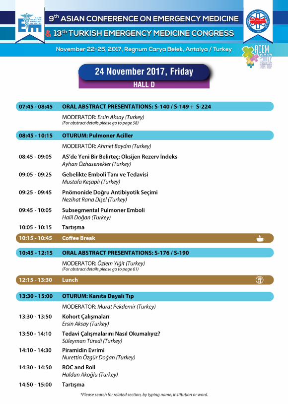
07:45 - 08:45 ORAL ABSTRACT PRESENTATIONS: S-140 / S-149 + S-224
MODERATOR: Ersin Aksay (Turkey) (For abstract details please go to page 58)
9th ASIAN CONFERENCE ON EMERGENCY MEDICINE9th ASIAN CONFERENCE ON EMERGENCY MEDICINE
13th TURKISH EMERGENCY MEDICINE CONGRESS13th TURKISH EMERGENCY MEDICINE CONGRESS
November 22-25, 2017, Regnum Carya Belek, Antalya / TurkeyNovember 22-25, 2017, Regnum Carya Belek, Antalya / Turkey
&&
08:45 - 10:15 OTURUM: Pulmoner Aciller
MODERATÖR: Ahmet Baydın (Turkey)
08:45 - 09:05 AS’de Yeni Bir Belirteç: Oksijen Rezerv İndeks Ayhan Özhasenekler (Turkey)
09:05 - 09:25 Gebelikte Emboli Tanı ve Tedavisi Mustafa Keşaplı (Turkey)
09:25 - 09:45 Pnömonide Doğru Antibiyotik Seçimi Nezihat Rana Dişel (Turkey)
09:45 - 10:05 Subsegmental Pulmoner Emboli Halil Doğan (Turkey)
10:05 - 10:15 Tartışma
13:30 - 15:00 OTURUM: Kanıta Dayalı Tıp
MODERATÖR: Murat Pekdemir (Turkey)
13:30 - 13:50 Kohort Çalışmaları Ersin Aksay (Turkey)
13:50 - 14:10 Tedavi Çalışmalarını Nasıl Okumalıyız? Süleyman Türedi (Turkey)
14:10 - 14:30 Piramidin Evrimi Nurettin Özgür Doğan (Turkey)
14:30 - 14:50 ROC and Roll Haldun Akoğlu (Turkey)
14:50 - 15:00 Tartışma
10:15 - 10:45 Co�ee Break
10:45 - 12:15 ORAL ABSTRACT PRESENTATIONS: S-176 / S-190
MODERATOR: Özlem Yiğit (Turkey) (For abstract details please go to page 61)
12:15 - 13:30 Lunch
24 November 2017, Friday
HALL D
ACEM2017
*Please search for related section, by typing name, institution or word.

15:00 - 15:15 Co�ee Break
9th ASIAN CONFERENCE ON EMERGENCY MEDICINE9th ASIAN CONFERENCE ON EMERGENCY MEDICINE
13th TURKISH EMERGENCY MEDICINE CONGRESS13th TURKISH EMERGENCY MEDICINE CONGRESS
November 22-25, 2017, Regnum Carya Belek, Antalya / TurkeyNovember 22-25, 2017, Regnum Carya Belek, Antalya / Turkey
&&
16:00 - 17:30 OTURUM: Uzman Atışması
MODERATÖR: Serkan Emre Eroğlu (Turkey)
16:00 - 16:15 SAK Ön Tanısında Normal Non-Kontrast BT: LP mi? CTA mi? LP Özgür Çevrim (Turkey) CTA Mehmet Ali Aslaner (Turkey)
16:15 - 16:30 Occult Pnömotoraks: Tüp Torakostomi mi? Gözlem mi? Tüp Torakostomi Erkman Sanrı (Turkey) Gözlem Murat Çetin (Turkey)
16:30 - 16:45 Submasif Embolide Yarı Doz Trombolitik Yarı Doz Trombolitik Yasin Yıldız (Turkey) Karşıt Görüş Onur Karakayalı (Turkey)
16:45 - 17:00 Burun Tamponu Yerleştirilen Hastada Sistemik Antibiyotik Gerekli mi? Sistemik Antibiyotik Gerekli Özgür Tatlı (Turkey) Sistemik Antibiyotik Gerekli Değil Özgür Dikme (Turkey)
17:00 - 17:15 BT Negatif Majör Künt Travma Ne Yapmalı? Eve Gitmeli Sevilay Ünver (Turkey) Takip Edilmeli Gürkan Altuntaş (Turkey)
17:15 - 17:30 Tartışma
17:30 - 18:30 ORAL ABSTRACT PRESENTATIONS: S-206 / S-215
MODERATOR: Rıdvan Atilla (Turkey) (For abstract details please go to page 63)
24 November 2017, Friday
HALL D
ACEM2017
*Please search for related section, by typing name, institution or word.

9th ASIAN CONFERENCE ON EMERGENCY MEDICINE9th ASIAN CONFERENCE ON EMERGENCY MEDICINE
13th TURKISH EMERGENCY MEDICINE CONGRESS13th TURKISH EMERGENCY MEDICINE CONGRESS
November 22-25, 2017, Regnum Carya Belek, Antalya / TurkeyNovember 22-25, 2017, Regnum Carya Belek, Antalya / Turkey
&&
10:45 - 12:15 ORAL ABSTRACT PRESENTATIONS: S-191 / S-205
MODERATOR: Mehmet Ergin (Turkey) (For abstract details please go to page 62)
07:45 - 08:45 ORAL ABSTRACT PRESENTATIONS: S-150 / S-159
MODERATOR: Neşe Çolak Oray (Turkey) (For abstract details please go to page 59)
17:30 - 18:30 ORAL ABSTRACT PRESENTATIONS: S-216 / S-225
MODERATOR: Mutlu Kartal (Turkey) (For abstract details please go to page 64)
24 November 2017, Friday
HALL E
ACEM2017
12:15 - 13:30 Lunch
10:15 - 10:45 Co�ee Break
*Please search for related section, by typing name, institution or word.

08:00 - 08:45 ORAL ABSTRACT PRESENTATIONS: S-226 / S-233
MODERATOR: Özgür Karcıoğlu (Turkey) (For abstract details please go to page 65)
9th ASIAN CONFERENCE ON EMERGENCY MEDICINE9th ASIAN CONFERENCE ON EMERGENCY MEDICINE
13th TURKISH EMERGENCY MEDICINE CONGRESS13th TURKISH EMERGENCY MEDICINE CONGRESS
November 22-25, 2017, Regnum Carya Belek, Antalya / TurkeyNovember 22-25, 2017, Regnum Carya Belek, Antalya / Turkey
&&
09:00 - 10:30 SESSION: Cardiac Emergencies – II
MODERATORS: Annitha Annathurai (Singapore), İbrahim Türkçüer (Turkey)
09:00 - 09:20 Pre-Hospital ECG - Expedite PPCI AMI Ludwig Tsoi (Hong Kong)
09:20 - 09:40 Cardiac Tests in the ED and Their In�uence on Outcome Cem Ertan (Turkey)
09:40 - 10:00 Syncope: An Update Tamorish Kole (India)
10:00 - 10:20 Antiplatelets and Antilipids Management in ACS Assad Suliman Shujaa (Saudi Arabia)
10:20 - 10:30 Discussion
11:00 - 12:30 PLENARY SESSION: Triage in the ED
MODERATORS: Teodoro J. Herbosa (Philippines), Müge Günalp Eneyli (Turkey)
11:00 - 11:30 A Real Story of Triage of Mass Casualties from Iraq Shakir Katea (Iraq)
11:30 - 12:00 Triage in Disaster Saurabh Kole (India)
12:00 - 12:30 Pysician in Triage Cem Oktay (Turkey)
10:30 - 11:00 Co�ee Break
25 November 2017, Saturday
HALL A
ACEM2017
*Please search for related section, by typing name, institution or word.

08:00 - 08:45 ORAL ABSTRACT PRESENTATIONS: S-234 / S-240
MODERATOR: Nezihat Rana Dişel (Turkey) (For abstract details please go to page 66)
9th ASIAN CONFERENCE ON EMERGENCY MEDICINE9th ASIAN CONFERENCE ON EMERGENCY MEDICINE
13th TURKISH EMERGENCY MEDICINE CONGRESS13th TURKISH EMERGENCY MEDICINE CONGRESS
November 22-25, 2017, Regnum Carya Belek, Antalya / TurkeyNovember 22-25, 2017, Regnum Carya Belek, Antalya / Turkey
&&
09:00 - 10:30 SESSION: ED Management
MODERATORS: Gregory Luke Larkin (U.S.A.), Murat Orak (Turkey)
09:00 - 09:20 KPI’s Implementation in ER Dina Shah (India) 09:20 - 09:40 Stepping into EM Leadership Role? - Know How to Succeed S. Saravana Kumar (India) 09:40 - 10:00 Emergency Physician Compensation Serkan Şener (Turkey)
10:00 - 10:20 Health and Wellness Among Emergency Medicine Residents Gül Pamukçu (Türkiye)
10:20 - 10:30 Discussion
11:00 - 12:30 OTURUM: Travma
MODERATÖR: Cemil Kavalcı (Turkey)
11:00 - 11:20 Künt Göğüs Travması: Kime Tomogra�? İbrahim Toker (Turkey)
11:20 - 11:40 Travmada Nöroresüsitasyon Müge Sönmez (Turkey)
11:40 - 12:00 Bu Hastanın Niye Boyunluğu ve Sırt Tahtası Yok! Mehmet Tahir Gökdemir (Turkey)
12:00 - 12:20 Blast Yaralanmaları Ömer Faruk Demir (Turkey)
12:20 - 12:30 Tartışma
10:30 - 11:00 Co�ee Break
25 November 2017, Saturday
HALL B
12:30 - 13:00 Closing Reception
ACEM2017
*Please search for related section, by typing name, institution or word.

09:00 - 10:30 SESSION: General EM
MODERATORS: Salvatore Di Somma (Italy), Müge Günalp Eneyli (Turkey)
09:00 - 09:20 Predicting Manpower Increase of Emergency Department for In�uenza Epidemics Using Ensemble Learning Algorithms Ping-Wun Huang (Taiwan)
09:20 - 09:40 Reversal of Oral Anticoagulants Mehmet Ali Karaca (Turkey)
09:40 - 10:00 Pearls, Pitfalls and Updates on Thyroid Emergencies Alzamani Mohammad Idrose (Malaysia)
10:00 - 10:20 Capnography in the ED Sai Surendar (India)
10:20 - 10:30 Discussion
10:30 - 11:00 Co�ee Break
9th ASIAN CONFERENCE ON EMERGENCY MEDICINE9th ASIAN CONFERENCE ON EMERGENCY MEDICINE
13th TURKISH EMERGENCY MEDICINE CONGRESS13th TURKISH EMERGENCY MEDICINE CONGRESS
November 22-25, 2017, Regnum Carya Belek, Antalya / TurkeyNovember 22-25, 2017, Regnum Carya Belek, Antalya / Turkey
&&
08:00 - 08:45 ORAL ABSTRACT PRESENTATIONS: S-241 / S-248
MODERATOR: Ahmet Demircan (Turkey) (For abstract details please go to page 67)
11:00 - 12:30 ORAL ABSTRACT PRESENTATIONS: S-257 / S-272 + S-291, S-292
MODERATOR: Erdem Çevik (Turkey) (For abstract details please go to page 69)
25 November 2017, Saturday
HALL C
ACEM2017
*Please search for related section, by typing name, institution or word.

9th ASIAN CONFERENCE ON EMERGENCY MEDICINE9th ASIAN CONFERENCE ON EMERGENCY MEDICINE
13th TURKISH EMERGENCY MEDICINE CONGRESS13th TURKISH EMERGENCY MEDICINE CONGRESS
November 22-25, 2017, Regnum Carya Belek, Antalya / TurkeyNovember 22-25, 2017, Regnum Carya Belek, Antalya / Turkey
&&
09:00 - 10:30 OTURUM: Pecha-Kucha
MODERATÖR: Ersin Aksay (Turkey)
Akut Apandisit Keselim mi? AB’mi Verelim? Mehmet Akçimen (Turkey)
Bell’s Palsi: Antiviral Başlayalım mı? Birdal Yıldırım (Turkey)
Banyo Tuzları Hakkında Herşey Emine Akıncı Emektar (Turkey)
Göremiyorum: Sorun Nerede? Ahmet Kenan Türkdoğan (Turkey)
Ajite Hastayla Baş Etme Stratejileri Sinem Avcı (Turkey)
Kontrast Nefropatisi: Kafam Karıştı Özcan Yavaşi (Turkey)
HFNO (High Frequency Nasal Ventilation) Gökhan Aksel (Turkey)
Tek Dozda Hayat Kurtaran İlaçlar Bulut Demirel (Turkey)
Senkopta Yüsek Riskli EKG Mehmet Ali Karaca (Turkey)
10:30 - 11:00 Co�ee Break
11:00 - 12:30 ORAL ABSTRACT PRESENTATIONS: S-273 / S-290 + S-293, S-294
MODERATOR: Özge Ecmel Onur (Turkey) (For abstract details please go to page 70)
08:00 - 08:45 ORAL ABSTRACT PRESENTATIONS: S-249 / S-256
MODERATOR: Özlem Yiğit (Turkey) (For abstract details please go to page 68)
25 November 2017, Saturday
HALL D
ACEM2017
*Please search for related section, by typing name, institution or word.

*Please search for related section, by typing name, institution or word.
LECTURES

*Please search for related section, by typing name, institution or word.
SAK ÖN TANISINDA NORMAL NON-KONTRAST BT: LP Mİ? CTA Mİ?
Mehmet Ali Aslaner
Baş ağrısı acil servislere karşımıza sıklıkla gelen başvuru şikayetleri arasında yer almaktadır (1). Bu hastalarda öncelikle ekarte etmemiz gereken acil tanılarımızdan biri ise Subaraknoid Kanama’dır (SAK).1 SAK’ların %75’i anevrizma rüptürüne bağlı olarak gelişmektedir. %20’sinde kanamaya yol açan neden saptanamazken %5’lik bir bölümüne arteriyovenöz malformasyonlar neden olmaktadır. 2
Klasik bilgi, BT’nin SAK’ı tanımadaki duyarlılığının semptomların başlangıcından sonraki ilk 12-24 saatte yüksek olduğunu belirtilmektedir. Bu süre içerisinde BT’nin sensitivitesi %98 olarak belirtilmiştir, 24. saatin ardından ise %90’lara düştüğü bilinmektedir. BT normal olarak tespit edilen hastalarda ise SAK şüphesi devam ediyorsa, LP yaparak ksantrokromi ve eritrosit sayımı yapmak gerekir. Son zamanlarda bu konuyla alakalı yapılan çalışmalarda LP’ye gerek kalmadan sadece BT ile hastaları taburcu edebilir miyiz sorusu üzerine çalışmalar yapılmaktadır. 2011 yılında Perry ve ark.nın yaptığı çok merkezli çalışmada, başağrısının başladığı ilk 6 saatte çekilen BT’de hiçbir hastada 3 ay içerisinde mortalite veya morbidite gözlenmemiş.3 Yine LP’nin invazifliğinden kaçınabilirmiyiz diye düşünen Cormack ve ark.’nın derlemesine göre, son yapılan çalışmalarda acil servise baş ağrısı şikayetiyle başvuran nörolojik muayenesi intakt olan ve
anevrizma için yüksek riski olmayan hastalarda BT sonrası BT anjiografi protokolünün anevrizma ve arteriyovenöz malformasyonlara bağlı SAK’ı ekarte etmekte %99 başarılı olduğu gösterilmiştir. 4 Sonuç olarak şuuru açık herhangi bir nörolojik defisiti olmayan ve ağrı başlangıcının ilk 6 saatinde BT’de özellik olmayan BT anjiografidede anevrizma ya da arteriyo venöz malformasyon tespit edilmeyen hastalarda SAK artık yüksek olasılıkla dışlanmaktadır ve LP’nin komplikasyonları göz önünde bulundurulduğunda yapılmayabilir. Kaynaklar:
1. Baş Ağrısında Lomber Ponksiyon (LP) Tarih mi Oluyor? (Accessed 05.09.2017, at https://www.acilci.net/bas-agrisinda-lomber-ponksiyon-lp-tarih-mi-oluyor/.) 2. Al-Shahi R, White PM, Davenport RJ, Lindsay KW. Subarachnoid haemorrhage. Bmj 2006;333:235-40. 3. Perry JJ, Stiell IG, Sivilotti MLA, et al. Sensitivity of computed tomography performed within six hours of onset of headache for diagnosis of subarachnoid haemorrhage: prospective cohort study. BMJ 2011;343. 4. McCormack RF, Hutson A. Can computed tomography angiography of the brain replace
lumbar puncture in the evaluation of acute-onset headache after a negative noncontrast cranial computed tomography scan? Academic emergency medicine : official journal of the Society for Academic Emergency Medicine 2010;17:444-51.

*Please search for related section, by typing name, institution or word.
APPROPRIATELY TRIAGING AND PRIORITIZING ELDERLY ED PATIENTS
Michael Bullard
The baby boomers have now reached the age of 65 and are the fastest growing group in most western populations expected to represent 20% of the population by 2030. An even greater rise in ED
visits by this complex group require greater resource utilization and have higher admission rates.1
Several challenges need to be understood to appropriately prioritize these patients and prevent under-triage. There are many factors associated with aging and chronic disease, but I will focus on 3 key areas:
i) Aging affects homeostatic mechanisms making vital sign interpretation more difficult. a. Respiratory: Aging lungs are less responsive to hypoxia and hypercapnia, have less
elastic recoil and more dead space. A respiratory rate of greater than 27 breaths / minute is a more sensitive predictor than pulse of blood pressure in identifying critically ill patients.2
b. Hemodynamic: Myocardial thickening, arterial wall stiffness, and hypertension increase workload on the heart. The resting heart rate increases with age, while wider pulse pressures and a decreased response to catecholamines can cause orthostatic hypotension and also a tepid response to critical illness. A systolic blood pressure of less than 110 mmHg often represents hypotension in the older population especially among trauma victims.3
c. Temperature: The combination of a less robust immune system, decreased metabolic rate and decreased muscle mass often limits the ability of older adults to mount a fever response. Subtle temperature changes, including hypothermia, may indicate a serious infection.4
ii) A number of factors complicate pain assessment including changes in pain perception,5 an increased risk of persistent pain, and difficulty in assessing patients with cognitive impairment.6
iii) Domains of care requiring special consideration include: a. Atypical presentations of common diseases: Acute coronary syndromes often present
without chest pain. Sepsis may present with non-specific symptoms and apparently normal vital signs. Pneumonia patients may present without respiratory difficulties, pain or fever. Patients with an acute surgical abdomen often deny significant pain.7
b. Cognitive impairment: Studies of ED patients over age 65 and 70 have reported delirium rates of 9.6% and 10% respectively.8 Another study noted 16% of patients with mental
status impairment and 6% with both delirium and dementia.9 Delirium often went unrecognized by the treating physician and some were discharged home. Early recognition of acute cognitive changes by the triage nurse, coupled with communication to the care team is very valuable.
c. Falls and trauma: Elderly ED trauma presentations are rising along with morbidity and mortality. Unlike younger patients the major mechanism of injury is falls often due to general weakness, impaired gait or vision, an acute medical event, medication, or balance issues.10 The severity of the injury and the impact of comorbidities, are often underappreciated and resulting in undertriage.11
d. Polypharmacy: In the US 44% of men and 57% of women over 65 are on 5 or more
medications often leading to adverse drug events (ADEs).12 ADEs account for up to 10% of elderly ED presentations. Drugs causing ADEs include cardiovascular, diuretics, antibiotics, hypoglycemics, sedatives, opioid analgesics, anticholinergics and anti-inflammatory medications.

*Please search for related section, by typing name, institution or word.
References:
1. Aminzadeh F, Dalziel W. Older adults in the emergency department: a systematic review of patterns of use, adverse outcomes, and effectiveness of interventions. Ann Emerg Med 2002;39(3):238-47.
2. Ridley S. The recognition and early management of critical illness. Ann R Coll Surg Engl 2005;87(5):315–22.
3. Brown JB, Gestring ML, Forsythe RM, et al. Systolic blood pressure criteria in the National Trauma Triage Protocol for geriatric trauma: 110 is the new 90. J Trauma Acute Care Surg 2015;78(2):352-59.
4. Keating HJ 3rd, Klimek JJ, Levine DS, et al. Effect of aging on the clinical significance of fever in ambulatory adult patients. J Am Geriatr Soc 1984;32(4):282–87.
5. Moore AR, Clinch D. Underlying mechanisms of impaired visceral pain perception in older people. JAGS 2004;52:132-36.
6. Zwakhalen SM, Hamers JP, Abu-Saad HH, Berger MP. Pain in elderly people with severe dementia: a systematic review of behavioural pain assessment tools. BMC Geriatrics 2006;6:3.
7. Samaras N, Chevalley T, Samaras D, et al. Older patients in the emergency department: a review. Ann Emerg Med 2010;56(3):261-69.
8. Elie M, Rousseau F, Cole M, et al.. Prevalence and detection of delirium in elderly emergency department patients. CMAJ. 2000;163: 977-81.
9. Hustey FM, Meldon SW. The prevalence and documentation of impaired mental status in elderly emergency department patients. Ann Emerg Med. 2002;39:248-53
10. Bonne S, Schuerer DJE. Trauma in the older adult: epidemiology and evolving geriatric trauma principles. Clin Geritr Med 2013;29:137-50.
11. Chang DC, Bass RR, Cornwell EE, et al. Undertriage of elderly trauma patients to state-designated trauma centers. Arch Surg 2008;8:776–81.
12. Haynes BD, Klein-Schwartz W, Barreuto F. Polypharmacy and the geriatric patient. Clin Geriatr Med 2007;23:371-90.

*Please search for related section, by typing name, institution or word.
CANARY IN THE COAL MINE: EMERGENCY MEDICINE CHALLENGES IN NORTH AMERICA
Michael Bullard Emergency Medicine in North America developed in parallel in the US and Canada in the 1970s, to provide the necessary resources and skill sets to support the emerging Emergency Medical System (EMS) and evolving cardiac arrest and trauma care and medical technological advances in diagnostic imaging and therapeutics like thrombolysis.1 While Canada have a single payer system and the US a multi-payer one, the principles and provision of Emergency Medicine are very similar.
Emergency Medicine (EM) has suffered from its own successes. Emergency Departments (EDs) are the Gatekeepers of the health care system sitting at the interface between the community and the inpatient world. Unlike all other disciplines and components of the health care system, the ED never closes and never refuses patient access to care. With every new acute and critical care innovation, the ED not only receives these patients but often need to be able to rapidly rule in or rule out the diagnosis and ensure timely access to care. Some examples include thrombolysis and angioplasty for STEMIs and CVAs, the golden hour for trauma victims, procedural sedation for fracture reductions and procedures, RSI and ultrasound guided central lines or nerve blocks.2 In Canada from our inception of EM, we have mandated the presence of front line emergency physician attending staff 24/7/365. Thus,
the ED acts as the “canary in the coal mine” both highlighting and falling victim to the many care delivery shortfalls in the system.3 Globally EDs have struggled to deal with severe overcrowding for many years to the detriment of patient outcomes.4 Canary in the coal mine examples:
1. Abrogation of responsibility - 1970s Anesthesia said “let us know when the patient is stable” and Surgeon said “I’ll see them in the morning” for unstable trauma victims
2. FP availability - many ED patients have no (unable to find) primary care physician 3. Investigation delays - primary care physicians often refer patients to ED for faster access to
diagnostic imaging studies 4. Poor community planning - elderly patients who are failing at home or in long term care are
transferred to the ED 5. Neglected patient populations - those with acute on chronic substance abuse problems or
mental health problems are generally directed to the ED 6. Bankers hours - cancer patients with acute or unexplained complications come to the ED for
care 7. Siloed care providers - home care and community palliative care teams refer patients to the
ED, especially on Friday afternoon While many of these patients are sick and the ED is an appropriate entry to acute care, each of them also are clear indicators of “failure” on the part of the system. The latest and most challenging group are the elderly who are on an ever-steeper decline towards mortality. Unless there is an acute reversible medical condition, the ED is not the best environment to try to clarify goals of care, determine ongoing care needs (independent vs dependent), and provide the necessary time and ancillary service assessment team to make critical decisions.5,6

*Please search for related section, by typing name, institution or word.
References:
1. Elyas, R. 31. The birth of a new specialty: The history of emergency medicine in Canada. Clinical & Investigative Medicine 2007;30(4):S44. ISSN 1488-2353. Available at: <http://cimonline.ca/index.php/cim/article/view/2791>.
2. Objectives of training in the specialty of emergency medicine. Royal College of Physicians and Surgeons of Canada.
http://www.royalcollege.ca/cs/groups/public/documents/document/y2vk/mdaw/~edisp/tztest3rcpsced000895.pdf
3. Marcus BS, Venkat A. Ethical pain in the emergency department: the canary in the coal mine. Pain Manag. 2015;5(4):251-60. doi: 10.2217/pmt.15.18. Epub 2015 Jun 10.
4. Berstein SL, Aronsky D, Duseja R, et al. The effect of emergency department crowding on clinically oriented outcomes. Acad Emerg Med 2009;16(1):1-10
5. Aminzadeh F, Dalziel WB. Older adults in the emergency department: a systematic review of patterns of use, adverse outcomes, and effectiveness of interventions. Ann Emerg Med 2002;39:238-47.
6. Samaras N, Chevalley T, Samaras D, et al. Older patients in the emergency department: a
review. Ann Emerg Med 2010;56:261-69.

*Please search for related section, by typing name, institution or word.
ElJf DJlek Çakal Please remember the day you started your residency training: What was that you were dreaming? What did you expect from the years ahead of yourself? And finally through numerous shifts and countless patients and until the chaos that once felt alien became familiar and even convenient, which little pieces did your residency training add to you – one way or another? Most of all, what do we offer to our residents, now that we are educators? In this presentation, in light of quotes from real residents and hot topics from medical education
literature, the speaker will attempt to decipher the expectations of the residents’ expectations of the residency training, compare them with our actual practice and offer practical suggestions to anyone in audience involved in medical education. Sources: Understanding medical education: Evidence, theory, and practice. Swanwick, T. (2013). General medical council – National training Surveys

*Please search for related section, by typing name, institution or word.
EMERGENCY MEDICINE CLERKSHIP: UNIVERSAL GUIDELINES AND LOCAL APPLICATIONS
Arif Alper Cevik Emergency Medicine (EM) is a unique specialty. As educators, we should support this specialty and its residents continuously on every level. Quality of the selected residents helps to specialty's reputation, confidence, quality of the health care given and its power to change the system. Therefore, providing good EM clerkship experience to medical students may influence their decision to choose EM as a future career opportunity. The more the applicants, the more chance to select good candidates for
residency training. Although there are multiple variations, residency training standards one way or another is similar. However, we see more variations in EM clerkships even in the same country. There are very limited guidelines for EM clerkship curriculum (1, 2). Still, there is a need to improve local standards with these limited guidelines. Applying the basics of medical education, using technology effectively, obtaining continues data with multi-level assessments, and finally using all data for the feedback improves the students' knowledge and creates a valuable learning experience for them. This presentation covers our local experience under the guidance of universal guidelines. We believe our experience will give an idea to other directors of EM about how to improve their teaching and student experience during their clerkship.
1. Manthey DE, Ander DS, Gordon DC, Morrissey T, Sherman SC, Smith MD, Rimple D, Thibodeau LG. Emergency medicine clerkship curriculum: an update and revision. Academic Emergency Medicine. 2010;17(6):638-43. 2. Hobgood C, Anantharaman V, Bandiera G, Cameron P, Halperin P, Holliman J, Jouriles N, Kilroy D, Mulligan T, Singer A. International Federation for Emergency Medicine model curriculum for medical student education in emergency medicine. Emergency Medicine Australasia. 2009;21(5):367-72.

*Please search for related section, by typing name, institution or word.
RSI CONCEPTS AND CONTROVERSIES
Orhan Çınar
Rapid sequence intubation (RSI) is the cornerstone of emergency airway management. Although it is a well-defined procedure, new drugs and technologies are changing the RSI practices. This talk will be about the current concepts and controversies including the rocuronium vs succinylcholine discussion, the effect of videolaryngocopes, prehospital RSI and apneic oxygenation.

*Please search for related section, by typing name, institution or word.
TEK DOZDA HAYAT KURTARAN İLAÇLAR
Bulut Demirel Acil tıp doğası itibari ile hızlı karar vermeyi ve hızlı tedavi etmeyi amaçlayan bir tıp bilimidir. Hastaların tanısının hızlı bir şekilde belirlenmesi elzemdir. Fakat bir başka elzem ve vazgeçilmez olan durum ise hastalığın tedavisinin zaman kaybedilmeden başlanmasıdır. Nöroloji, kardiyoloji, ortopedi, enfeksiyon hastalıkları, toksikoloji ve diğer acil hastalıkların sayılamayacak kadar çok tedavi yöntemi vardır. Fakat öyle ilaçlar vardır ki gönlümüzde ve aklımızda mucizeler yarattığı için yer etmiştir. Acil tıp denilince birçoğumuzun aklına kardiyo-pulmoner resüsitasyon gelmektedir. Adrenalin vazgeçilmez ve mucizevi bir ilaç olarak karşımızda durmaktadır. Naloksan uygulaması sonrası hastanın durumunun düzeldiğini gözlemlemeyen var mı? Yada atropin verilmiş bir organofosfat zehirlenmesinin klinik iyileşmesine şahit olmayanınız var mı? İşte bu ‘Tek dozda hayat kurtaran ilaçlar!’ ı kısa kısa hatırlatmak isteriz.
LİFE SAVİNG DRUGS İN A SİNGLE DÖŞE
Emergency medicine is a medical science aiming to make quick decisions and to treat as fast as possible because of its nature. It is essential to determine the diagnosis of the patients quickly. But another essential and indispensable condition is that the treatment of the disease begins without any loss of time. Neurology, cardiology, orthopedics, infectious diseases, toxicology and other emergency diseases have many treatments that cannot be counted. But there are medicines which are in our minds and in our minds because it creates miracles. In emergency medicine cardio-pulmonary resuscitation is nearly a daily routine. Adrenaline is indispensable and stands as a miraculous drug. Is anyone not observing that the patient's condition improved after naloxone administration? Do you witnessed of clinical improvement of an organophosphate poisoning given atropine? We would like to briefly remind you of these 'life saving drugs in a single dose!'

*Please search for related section, by typing name, institution or word.
THE PATIENT YOU SAW YESTERDAY : SHORT-TERM DEATH AFTER EMERGENCY DEPARTMENT DISCHARGE
Murat Ersel
Emergency departments are high risk areas for medical errors leading to death. In early 90’ies it has been reported every 13 patient per 100.000 died after ED discharge (1). In 2007 Sklar et al. reported that 30 per 100.000 patients died within 7 days of ED discharge, 20 per 100,000 unexpectedly and 9 per 100,000 with a potentially contributory medical error, atypical presentations, exacerbation of chronic disease, abnormal vital signs, and substance abuse were found as common themes(2). In an another study Baker et al. found a mortality rate of 0,2 %, for patients discharged directly from the emergency department and subsequently died within 30 days. Almost for the half of the patients who died, dead had been occured with in first 8 days after discharge (3). Sklar et al found that most patients die of conditions that were expected to cause death, such as cancer, or they die from a completely unrelated cause. Unfortunatly, about half of deaths, appeared to be unexpected and related to the visit, and also about 60% of these cases were associated with a possible error (2). In a study with a large database originated from Medicar resources mean age of patients who died within 7 days after ED discharged had been reported as 69. In the same study, diagnoses such as altered mental status, dyspnea and malaise/fatigue were found more associated among early deaths compared with other reasons of emergency department visits. Atherosclerotic heart disease, myocardial infarction, chronic obstructive pulmonary diseases were identifed as leading causes of short time death after ED discharge (4). The reasons for early term and unexpected deaths after ED discharge must be investigated in large databases and in different communities. So, patients under risk might be identified clearly, probabily this may help emergency physicians for safely discharge of the patients from the ED’s. References 1. Kefer MP, Hargarten SW, Jentzen J. Death after discharge from the emergency department. Ann Emerg Med. 1994;24:1102-1107 2. Sklar DP, Crandall CS, Loeliger E, Edmunds K, Paul I, Helitzer DL. Unanticipated death after discharge home from the emergency department. Ann Emerg Med. 2007;49:735-745. 3. Baker M, Clancy M. Can mortality rates for patients who die within the emergency department, within 30 days of discharge from the emergency department, or within 30 days of admission from the emergency department be easily measured? Emergency Medicine Journal 2006;23:601-603. 4. Obermeyer Z, Cohn B, Wilson M, Jena AB, Cutler DM. Early death after discharge from emergency departments: analysis of national US insurance claims data. BMJ. 2017; 356: j239. doi: 10.1136/bmj.j239.

*Please search for related section, by typing name, institution or word.
EMERGENCY PHYSICIAN: SHOULD ALWAYS BE FULLY CHARGED
Mahmood Ghanaim
People all over the world rely heavily on the 24-hour access to care provided by hospital emergency departments (EDs), and this need is growing.
• ED visits have increased by nearly 20 percent over the past decade.
• Almost half of hospital care begins in the ED.
• The majority of ED patients require immediate care.
Emergency medicine requires thought processes that incorporate risk stratification, assessment of urgency and need for immediate treatment or resuscitation, and often leads to treatment before diagnosis. Management of the undifferentiated but acutely ill or injured patient often mandates intervention based on presenting problem rather than specific pathology. Are the intellectual processes and cognitive models used in the emergency medicine paradigm different to the classically taught medical model?
Several studies have shown that there is information loss during interruptions, and that multitasking creates higher memory load, both of which contribute to medical error. Nowhere is this more critical than in the emergency department (ED), where the emphasis of clinical decision is on the timely evaluation and stabilization of patients. Introduce emergency physicians to the use of interruption management strategies as a method of handling the frequent interruptions they are exposed to. Use of these strategies when high-risk primary tasks are performed may reduce the disruptiveness of some interruptions and improve patient safety.
Physicians are interested in how to best meet the needs of the population, in continually improving the care provided to their clients. To do so requires that they also care for themselves including managing the effects of sleep deprivation and fatigue. It is a complex issue that requires multifaceted solutions. Strategies must address physician fatigue at an individual, organizational/institutional and system level.
1. Canadian Medical Association: MANAGEMENT OF PHYSICIAN FATIGUE -2014 2. Always There, Ready To Care; The 24/7 Role of America’s Hospitals. American Hospital
association, 2015. 3. Skaugset LM, Farrell S, Carney M. Can You Multitask? Evidence and Limitations of Task
Switching and Multitasking in Emergency Medicine. Annals of emergency medicine. 2015.
4. Emergency Physician Use of Cognitive Strategies to Manage Interruptions; Ann Emerg Med. 2017 Jun 7. pii: S0196-0644(17)30512-7.
5. Emergency Physician Shift Work; American college of Emergency physicians, June 2017. 6. Akerstedt T, Wright KP Jr. Sleep loss and fatigue in shift work and shift work disorder. Sleep
Med Clin. 2009;4:257-271.

*Please search for related section, by typing name, institution or word.
TRIAGE TOOLS FOR DISASTER MANAGEMENT (A RELOOK)
Goh Siong Hiang The speaker will provide an overview of mass disaster triage algorithms that have been used, their international adaptability and inter-observer reliability. Examples will include Triage Sieve, Triage Sort, Jumpstart, SALT, etc Mention will also be made of triage algorithms for HAZMAT, radiation, lightning strikes and smoke inhalation scenarios. Some examples of triage tags and electronic triage communications systems used internationally will be shown too.

*Please search for related section, by typing name, institution or word.
ABSTRACT FOR THE NINTH ASIAN CONFERENCE ON EMERGENCY MEDICINE
C. James Holliman
Objectives of this presentation are to present how institutional and individual commitment to care for injured patients is essential, understand the importance of an ongoing performance improvement program in the care of the trauma patient, show how the failure to follow the fundamental principles of trauma resuscitation leads to pitfalls, understand the importance of early recognition of resource limitation and transfer to definitive care at an accredited trauma center, and demonstrate how the
tertiary survey helps prevent missing injuries. The five major potential pitfalls in trauma resuscitation include lack of institutional and individual commitment to the care of critically injured patients, having an underdeveloped performance improvement plan, failure to follow the fundamental principles of trauma resuscitation during the primary survey, failure to recognize local resource limitations and make an early decision to transfer to definitive care, and failure to perform a tertiary survey (meaning a complete, comprehensive head to toe physical re-exam for injuries) to prevent missing injuries. Trauma resuscitation pitfalls to avoid include: delay in recognizing a compromised airway, failing to recognize hypoxia early, failing to recognize the presence of decompensated shock and initiate timely appropriate treatment, failing to transfuse blood and blood products early, spending too much time doing resuscitation-related procedures in the emergency department that could be better performed in
the operating room, lack of early surgical consultation for patients demonstrating signs or symptoms of shock, failing to treat hypoxia and hypotension aggressively in the patients with traumatic brain injury, and not preventing hypothermia in the trauma patient. Trauma performance improvement plans should identify problem events, classify these events by determination, grade, and preventability, and develop action plans which assign accountability, track and trend in a measureable way, and reanalyze the action plan to determine its success or progress.
*Please search for related section, by typing name, institution or word.
GERIATRIC TRAUMA
Sanjay Jaiswal Geriatric trauma is not just about the injury …. It is different! Whenever the word ‘Trauma’ comes to our mind we automatically focus on the mechanism of injury; whether it is blunt or penetrating? Although trauma is classified in most of the curriculum on this basis, there is one more criterion that should be considered while approaching these situations: What is the age of the patient? It is of significance because if they belong to geriatric population, important factors should be considered which may affect assessment and management of the patient. By 2050 it is projected that elderly will constitute around 20% of the population across the globe. Due to this rapid growth of geriatric population there is a great impact on the economy because of the unique medical requirements. Presently, trauma is the 7th leading cause of death amongst elderly. Even though elderly patients are less likely to be injured in comparison to adults, they are more likely to have fatal outcomes from the injuries. Knowledge of the changes that occur with ageing, appreciation of the injury patterns seen in elderly can lead to improved outcomes. This approach can be developed among Emergency physicians by knowing how the consequences of traumatic injury are different in geriatric population than in adults and by recognising elements of Comprehensive geriatric assessment which can lead to improvement in geriatric care across the world.

*Please search for related section, by typing name, institution or word.
TRAUMA REGISTRY: DOES IT HELP TRAUNMA CARE
Sabariah Faizah Jamaluddin Trauma registries are databases that document acute care delivered to patients with injuries. They are designed to provide information that can be used to improve the efficiency and quality of trauma care. Trauma registry helps set a system for tracking and reviewing outcomes and trends. The information from trauma registry, such as types of injuries, ages , location of injury, will enable where the need to focus the injury prevention programs and education.
Assessing and monitoring the quality of care in trauma patient through quality indicators would allow identifying opportunities for improvement whose implementation would improve outcomes in hospital mortality, functional outcomes and quality of life of survivors The information contained in the trauma registries is essential to know the current health care reality, identify opportunities for improvement and contribute to the clinical and epidemiological research. Trauma registries also offer distinct advantages when assessing the effectiveness of trauma systems. Detailed injury data and statistical comparisons provide advantages over population-based or preventable death studies.

*Please search for related section, by typing name, institution or word.
POST INTUBATİON- PEARLS & PİTFALLS
Narendra Nath Jena
Intubation is an important intervention in the ED. In addition to maintaining an appropriate ventilation strategy after intubation, it is crucial that we use appropriate post-intubation sedation and analgesia regimens for the continued care of these critical patients. This article will review some common pearls and pitfalls that we must be taken care of Post intubation.
Hypotension shouldn’t prevent us from providing adequate pain control and analgesia. Fentanyl
has less hemodynamic instability than other opioids and does not rely on renal clearance, making it a good analgesic for this situation. Ketamine is also an appropriate choice here for its pro-hemodynamic effects which will not worsen the hypotension and can actually increase blood pressure .
Pain and stress increase sympathetic tone, causing a rise in heart rate, blood pressure, intracranial pressure, and oxygen demands.Providers should be especially diligent in patients receiving a long-acting paralytic as they are unable to communicate any sort of discomfort. One tip in avoiding these errors is to order the post-intubation medications while ordering RSI medications.
Ketamine is associated with bronchodilation which will help open reactive airways while providing sedation and analgesia. It preserves airway reflexes which is helpful if we want to try to avoid intubation in the first place. Opioids can also be used in this situation for pain, but be aware of the
potential side effect of chest wall rigidity. For sedation after seizure intubation there are two good choices: benzodiazepines and propofol.
Traditionally we reach for benzodiazepines; these are first line medications to treat both seizures and delirium tremors (DT) in addition to being a sedative for the mechanically ventilated patient. Consider starting the patient on a midazolam drip. An alternative tool is propofol for additional seizure control/continued sedation in refractory status or unstable alcohol withdrawal. Another benefit is that as quickly as propofol works, it is also rapidly cleared. This makes propofol an excellent sedative choice in patients that will need neurologic reassessment.
For a critically-ill hypertensive patient with ICH, blood pressure control is important and this must include adequate pain control with fentanyl to prevent increasing sympathetic response and
intracranial pressure..... References: 1. Sessler CN, Gosnell MS, Grap MJ et al. The Richmond agitation-sedation scale: validity and
reliability in adult intensive care unit patients. Am J Resp Crit Care Med. 2002; 186:1338-1344. 2. Lawner B, Farzad A. Sedation of the mechanically ventilated patient in the Emergency
Department. EM Critical Care / EB Medicine. 2014;(4)2. www.ebmedicine.net 3. Barr J, Fraser GL, Puntillo K, et al. Clinical practice guidelines for the management of pain,
agitation, and delirium in adult patients in the intensive care unit. Crit Care Med. 2013; 41(1):263-306.
4. Fraser GL, Devlin JW, Worby CP, et al. Benzodiazepine versus nonbenzodiazepine-based sedation for mechanically ventilated, critically ill adults: A systematic review and meta-analysis of randomized trials. Crit Care Med. 2013;41(9 Suppl):S30-S38.
5. Asad EP, Martin JR, Erstad BL. Ketamine for analgosedation in the intensive care unit: a systemic review. J Intensive Care Med. December 2015; epub ahead of print.

*Please search for related section, by typing name, institution or word.
GEBELERDE PULMONER EMBOLİ-TANI VE TEDAVİSİ
Mustafa Keşaplı
Pulmoner emboli (PE),gebelik ve postpartum dönemde anne ölümlerinin %20-30’undan sorumludur. Gebelik esnasında tromboembolik olay insidansının 1000 doğumda 2 olduğu belirtilmektedir. Gebe olmayanlara göre VTE riski gebelerde 4-50 kat artar. PE özellikle gelişmiş ülkelerde gebeliğe bağlı anne ölümlerinin 6.sırada nedenidir.
Gebelik ile beraber gelişen fizyolojik değişimler,(örneğin ortalama %70 gebe de dispne gelişir) PE’nin klinik olarak saptanmasını zorlaştırır. Gebe hasta da,semptom olmadan şok tablosu gelişebilir yada ani kardiak arrest olabilir. Akut başlangıçlı dispne, plöretik göğüs ağrısı ve hemoptizi varlığında PE öncelikle düşünülmelidir. Ölümleri önlemenin yolu, klinisyenin tanı için süphe duyması erken antikoagülasyon tedavisi başlanmasıdır. Gebelerde, özellikle geliştirilmiş, pretest olasılık araçları yada validite edilmiş klinik bulgu klavuzları mevcut değildir.Modifiye edilmiş Well’s skorlama dışlamak için kullanılabilir. D-Dimer, gebelikte arttığı için özellikle ilk 20 haftada artışı anlamlı değildir. Ancak bu dönemde yüksek olmayışı dışlama kriteri olarak kullanılabilir. PE şüphesi olan gebelerde ve gebe olmayanlarda arteryel kan gazı tanısal değildir. Hem PE’de hem de gebelikte respiratuar alkaloz ortak özelliktir. Gebelikte üçüncü trimesterde sırtüstü konumdayken PaO2 yatar pozisyonda daha düşük olabileceği için, arteryel kan dik konumdayken alınmalıdır
Tanısal uygulanacak görüntüleme yöntemlerinin radyasyon içeriğinden dolayı,hekimler ve hastalar tereddütlü davranmaktadırlar Göğüs radyografisi,başlangıç olarak, diğer sebepleri de ekarte etmek için önerilmektedir. Hastada alt ekstremite de kompresyon ultrasonografi tanıya yardımcıdır. Ekokardiyografi, tanıyı destekleme, masif pulmoner tromboemboli varlığını değerlendirme ve tedavi planının belirlenmesinde yardımcı olabilmektedir. Hastada normal PA AC grafisinin ardından, düşük doz radyasyon vermek için ventilasyon/perfüzyon sintigrafisi önerilmektedir. Patolojik AC grafisinden sonra Pulmoner CT angiografi çekilmesi önerilmektedir. Tanı da gold standart pulmoner CT angiografidir.(Alınan radyasyon miktarından ziyade meme kanseri riskini arttırmaktadır.) MRI,sensitivite – spesifite ve kullanılabilirliği konusundaki çalışmalar yeterli düzeyde değildir. Kesin tanı,yüksek olasılık,V/Q scan yada pıhtının CT ile gösterilmesi ile konur. Tanı yöntemleri ile ilgili çalışmalar genellikle retrospektif çalışmalara dayanmaktadır. İyi dizayn edilmiş prospektif çalışmalara ihtiyaç vardır.
Tedavi de heparin antikoagülasyonu uygulanır.Heparin plasentayı geçmediği için gebelik dönemi boyunca rahatlıkla kullanılabilir. Düşük moleküler heparinler, kiloya göre düzenlenerek kullanılır. Warfarin, teratojenik etkisinden dolayı kullanılmaz. Yeni kuşak oral antikoagülan ajanlar, gebelikte kontrendikedir. Antikoagülasyon için kesin bir kontrendikasyon bulunduğunda,vena Kava’ya filtre yerleştirilmesi düşünülebilir. Trombolitik tedavi hemodinamisi unstable olan kritik hastalarda kullanılabilir.

*Please search for related section, by typing name, institution or word.
Referanslar
1. 2014 ESC Guidelines on the diagnosis and management of acute pulmonary embolism, European Heart Journal doi:10.1093/eurheartj/ehu283
2. Up to Date.Pulmonary embolism in pregnancy: Epidemiology, pathogenesis, and diagnosis.This topic last updated: Feb 15, 2017.
3. Pulmanary embolism diagnostic of pregnant patients:what is the recommended clinical pathway considering the clinical walue and associated risk of available imaging tests?Georgias Z et all,Physica Media,Doi;10.1016/j.ejmp.2017.07.017
4. Türk Thorax derneği,Pulmoner Tromboembolizm Tanı ve Tedavi Uzlaşı Raporu,2015 5. Gebelerde pulmoner tromboembolinin tanı ve tedavisi,Tuberk Thorax,2015;63(2):132-
139,Doi:10.5578/tt.6342

*Please search for related section, by typing name, institution or word.
Saurav Mahanta Dr. Saurav Mahanta has been working in the field of emergency and critical care medicine since 2013. After having worked in various hospitals across southern India, he joined Narayana Health City as an associate consultant in the Department of Emergency Medicine in 2016. He is a certified trainer for National Ultrasound Life Support (NULS) and National Trauma Life Support (NTLS) under the Society for Emergency Medicine, India (SEMI). He is also a certified American Heart Association (AHA) instructor for Advanced Cardiac Life Support (ACLS) program. He has presented papers and posters in national and international emergency medicine conferences across a variety of topics and is
actively involved in promotion of emergency medicine in India. In his free time, you will either find him travelling to the various national parks in India, or eating out with his friends in town.

*Please search for related section, by typing name, institution or word.
TEAM MANAGEMENT FOR THE ACUTE MEDICINE
Yasumitsu Mizobata, Naoki Shinyama, Takafumi Terada, Tomohiro Noda, Takeichi Hagiwara INTRODUCTION Teamwork has been suggested as a promoting approach to improve the quality of care process in the emergency departments. However, for teamwork to yield expected results, implementation must produce behavior changes. It has been recommended to train health-care professionals as the team to improve their non-technical
skills. Simulation-based training has been proposed as a safe and effective training method. It has the potential to facilitate communication, cooperation and leadership skills, which are essential for the teamwork in the acute medical care. Multi-professional team training is expected to allow for more natural team interactions and reinforce understanding across the disciplines. Although simulation-based training of the multi-disciplinary team is recommended, the question is whether the initiative has an effect on the daily practical situation. We produced the simulation-based training of non-technical skills for the acute medicine (NoTAM) course for the physicians and nurses who take care of patients as the team in the emergency department. The aim of this study is to evaluate the effects of the NoTAM course for the practical clinical activities in the emergency department.
METHOD NoTAM course is the three-hour program, composed by thirty-minutes didactic lecture, thirty-minutes demonstration video and discussion which emphasize how to perform the non-technical skills in the emergency department, and two case simulation-based scenario training using high performance human simulator. Two attending physicians, one or two intern residents, and two nurses participated the NoTAM course as one team. Between October and December in 2016, six NoTAM course were carried for the 12 attending physicians, 11 nurses, and 8 intern residents. The surveys concerning the team performance were investigated for the all participants. Patient treatments were recorded in the emergency department before (n=27, January to March 2016) and after (n=27, January to March 2017) the NoTAM course training implementation. The performance
of the patient care and non-technical skills were scored. For the statistical analysis, Wilcoxon ranked analysis was performed, and p value less than 0.05 was considered as significant. RESULTS The answer of the survey demonstrated that all the disciplines concern about the poor communication, leadership, and team performance in the daily practice. After the NoTAM course, they clearly understood the team dynamics and predicted to be able to perform better non-technical skills. However, the performance scores in the practical patient care did not show any changes in patient assessment (2.4 [2.1 - 2.9] vs. 2.5 [2.1 - 3.1], p=0.537), in non-technical skills (2.2 [1.9 - 2.5] vs. 2.2 [2.0 - 2.7], p=0.537), nor in total performance (2.3 [2.0 - 2.6] vs. 2.3 [2.0 - 2.7], p=0.454; middle [25% - 75% IQR]; pre vs. post). Only the score of the briefing significantly improved (2.1 [1.7 - 2.7] vs. 2.4 [2.1
- 2.9], p=0.038) after the NoTAM implementation. CONCLUSIONS The simulator based teamwork training brought better understanding of the team management for the multidisciplinary emergency medical staffs. The changes of the performance in the daily practical activities may require repeated training and practical trial in the daily patient care. Key words: teamwork, simulation, non-technical skill, leadership, briefing

*Please search for related section, by typing name, institution or word.
CAN A MOBILE APP HELP TO IMPROVE DOOR TO NEEDLE TIME IN THE TREATMENT OF ACUTE ISCHEMIC STROKE?
Fabith Moideen
Introduction: The time between presentation to the hospital and administration of recombinant tissue plasminogen activator (rtPA) for intravenous thrombolysis of acute ischemic stroke is called the door to needle time (DTN). International guidelines recommend that the door to needle time should not exceed 60mins. However, less than one third of acute ischemic stroke patients who receive tPA are
treated within guideline-recommended door-to-needle times. The benefit of intravenous tPA in acute ischemic stroke is strongly time-dependent and time to revascularization is critical in improving outcomes in stroke patients. In 2017, we introduced a unique mobile application in our centre to facilitate entry of patient parameters, along with a timer and team synchronisation as a potential solution to improve door to needle time. Objective: To study the effectiveness of a mobile app based strategy to improve door to needle time in the treatment of acute ischemic stroke. Methodology: Consecutive patients admitted to Emergency Department at Baby Memorial Hospital
between April-September 2017 with acute ischemic stroke were entered in mobile app with patient parameters, timer, NIH stroke scale (NIHSS), thrombolysis checklist and tPA dose calculator. The app also enabled team synchronisation by notifying all on call members and team leaders of the patient movement in real time, along with sharing of radiological images. Door to needle time (DNT) captured from the app was entered in a spreadsheet and compared to previous values from our center. Mean values were compared using the unpaired t test (two-tailed). Findings: A total of 25 patients were thrombolysed while using the mobile app. The mean DNT was 46.96 min with 84% being thrombolysed within 60mins.These patients were
compared with 25 patients who were thrombolysed between December 2015 - December 2016, where the mean DNT was 58 min, with only 60% being thrombolysed within the first 60mins. A mean DNT decrease of 12 minutes was seen with 1.4 times increase in DNT < 60 min. This difference was statistically significant (p=0.0481, unpaired t test). The causes of delay were identified and included delays in-patient shifting and imaging. Result and Conclusion: We have been able to demonstrate significant improvement in door to needle time by using a mobile app at our center as a tool to improve team performance, and identify causes of delay.

*Please search for related section, by typing name, institution or word.
HEALTH AND WELLNESS AMONG EMERGENCY MEDICINE RESIDENTS
Gül Pamukçu Günaydın
Burnout effects majority of physicians in all countries and specialties at some point in their carreer. EM Residents burnout rates are one of the highest reaching to %70 because of night sifts, long work hours, huge patient load, exams, and not being ableto make clinical decisions independently and having little control over their work schedule. Residency is important for wellness because it is a time physicians develop patterns and lifelong habits.
SAEM defines wellness as skills, attitudes and beliefs that allow one to enjoy practicing EM for a long time in balance with life. Interventions to improve resident wellness can be divided in to individual level, residency programs level, national or world wide level.
Individually we need to increase healthy and decrease unhealty habits. Having a primary physician, sleep, nutrition, water intake and exercise, rest, keep a positive mind, have a social network and to get help when in need is particularly important. Maintaining a work/ life balance is not sometimes easy so don’t feel guilty if you can’t do all the advised. In program level establishing a wellness program includes having faculty and a resident responsible for the program, assessing for burnout regularly, involving residents in decision making and responding to resident needs. Including topics of wellness to training program and social activities to create an ED Family is also
recommended. Yet most of the problems are system based and nowadays physician wellness is considered important only for patient safety and quality of care.
A cultural change towards resident wellness is necessary. Policy changes don’t happen overnight so we need to work together to build a more humane healthcare workplace.

*Please search for related section, by typing name, institution or word.
EM REORGANISATION IN SLOVENIA
Gregor Prosen Affiliations: 1) Center for Emergency medicine Maribor, C. Proletarskih brigad 21, 2000 Maribor, Slovenia 2) University of Maribor Medical faculty, Dpt. for Emergency Medicine, Taborska ul. 8, 2000 Maribor,
Slovenia Abstract Background Emergency medicine in Slovenia has traditionally been divided into a primary care level and a secondary level. The primary, or prehospital level has traditionally been operated by general practitioners (GP’s) in local community health centres. The secondary level has involved inpatient wards divided according to each speciality (i.e.surgery, internal medicine, infectious diseases, neurology etc.) Until 2016, prehospital EMS care was provided by physician-led EMS teams, that outside of the largest urban centres were running an EMS system in parallel with general practice. The same prehospital emergency network was also providing 24/7 “after hour” clinics throughout Slovenia (approx. 20-30km apart). Slovenia is still one of few European countries without a centralised national dispatch centre - emergency calls are currently still answered by local community health centres. By the end of 2015, ten “general” Emergency Departments (ED) modelled on the British “A&E”, were opened in ten of twelve Slovenian hospitals. They were 80% financed by European Union regional cohesion funds. This establishment of Emergency Departments, was major milestone for development of EM in Slovenia and future reorganisation of Emergency Medicine and the healthcare system in general. Future developments Faced with the need for the legal placement of ED’s, updated legislation regarding the Emergency Medical system was passed in October, 2015. The major pillars of this legislation for structure and future developments are:
- EDs follow a unified, functional design divided into four “units”: for accidents, diseases, “fast track”, and resuscitation. Entry is always through a unified triage, using Manchester’s triage system (with minor Slovenian modifications).
- Prehospital EMS vehicles now consist of fleets both with and without a physician on board. They are to be dispatched according to the Norwegian dispatch index (with Slovenian modifications). A unified dispatch centre is currently being built and staff is being recruited, with a scheduled opening in 2018. A unified, IT-supported dispatch system will also finally provide much needed objective data on prehospital interventions and enable a better planning of the EMS network. With this renewed EMS network, it will be possible to finally free GP’s of their parallel work. This change will need political support, which is currently lacking. In addition, a renewed EMS network will enable the gradual evolution to a majority non-physician-staffed EMS system.
- In smaller towns without a general hospital (i.e. without hospital-based ED), the establishment of “satellite ED’s” [stand-alone ED] was proposed, with closure of even smaller “after-hour GP clinics” in neighbouring towns. Such “satellite ED’s” would be better equipped (PoC-Lab, PoC-US) and staffed, thus regionalising care. These developments are just in the preliminary stages of progress.
- Most importantly, the number of EM residents is slowly, but steadily rising. It is projected that in next 10-15 years, newly-graduated EM specialists will slowly take over the staffing in ED’s and satellite ED’s in an evolutionary manner.
Summary With the establishment of general ED’s and a thriving EM speciality, the newly accelerated era of EM development and reorganisation has started in Slovenia. Resident education and ED financing will be major determinants of the speed and trajectory at which EM will continue to develop in Slovenia.

*Please search for related section, by typing name, institution or word.
APPROPRIATELY TRIAGING AND PRIORITIZING ELDERLY ED PATIENTS
Marc Sabbe Several studies report that 12-21% of all ED admissions are persons aged 75 years or above. In addition, population aging will increase these percentages substantially in the upcoming decades. However, the implications of this phenomenon are larger than just increasing patient volumes in a setting already burdened with crowding. It has been demonstrated that managing older ED patients takes more time and resources, as they
often suffer from vague complaints, multi-morbidities and social problems due to functional and/or cognitive decline. Still, despite specific investments, outcome of elderly is often less prosperous than those of younger patients. Up to one out of three older adults experience an adverse event after an episode of ED care, including hospitalisation, nursing home admission, functional decline and death. Important factors contributing to the incidence of adverse events are inappropriate disposition planning and occurrence of specific geriatric syndromes (e.g. delirium, functional impairment, decreased cognitive skills…). Since these syndromes frequently receive less attention in the ED due to prioritisation, time and expertise constraints, opportunities for improving care lie in reorienting the disease focused to a more geriatric friendly ED model. Implementing comprehensive geriatric assessment (CGA) in ED care can improve the timely
recognition of geriatric problems. It is defined as “a multidimensional interdisciplinary diagnostic process focused on determining a frail elderly person’s medical, psychosocial and functional capabilities in order to develop a coordinated and integrated plan for treatment and long-term follow-up”. This approach has particularly demonstrated to be beneficial on patient outcomes after hospitalisation on an acute geriatric ward. However, its effect on the ED remains inconclusive due to heterogeneity in study methods, interventions and settings. In this presentation, new evolutions on triaging and prioritizing frail elderly in the ED will be discussed.
Braes T, Moons Ph, Lipkens P, Sterckx W, Sabbe M, Flamaing J, Boonen S, Milisen K: Screening for risk of unplanned readmission in older patients admitted to the hospital: predictive value of three instruments. Aging Clin Exp Res 22: 345–351, 2010 Deschodt M, Devriendt E, Sabbe M, Knockaert D Deboutte P, Boonen S, Flamaing J and Milisen K. Characteristics of older adults admitted to the Emergency department and their risk factors for ED readmission. BMC Geriatrics 15(1): 54-64, 2015. Devriendt E, De Brauwer I, Vandersaenen L, Heeren P, Conroy S, Boland B, Sabbe M, Flamaing J,
Milisen K. Geriatric support on the emergency department: a national survey in Belgium. BMC Geriatr 17(1): 68, 2017. Devriendt E, Deschodt M, Delaere M, Flamaing J, Sabbe M and Milisen K. Does the get up and go test improve diagnostic characteristics of the triage risk screening tool or Rowland questionnaire in older patients admitted to the ED? European Journal of Emergency Medicine. Accepted.
Devriendt E, Heeren P, Deschodt M, Sabbe M, Fieuws S, Flamaing J, Conroy S, Parker S and Milisen K. Effectiveness of comprehensive geriatric assessment based interventions in the Emergency Department: a systematic review and meta-analysis. In preparation.

*Please search for related section, by typing name, institution or word.
THE BEST PUBLICATIONS OF THE LAST 5 YEARS ON PREHOSPITAL MEDICINE
Marc Sabbe Prehospital care is without any doubt, one of many parts of Emergency Medicine. The basis of our daily clinical work is generated from evidence out of scientific research. Moreover, one of the important challenges facing modern medicine is the "knowledge-transfer gap", the gulf that separates the results of clinical research from the reality of day-to-day medical decision-making. However, until recently, only limited amounts of evidence existed in prehospital medicine. Many
reasons such as limited funding, practical aspects to organize research projects and ethical dilemmas (informed consent) exist to explain the limited amount of evidence. As the number of published research papers in the field of prehospital care is rising, although mainly about resuscitation, it is becoming of clinical interest to select those that bring innovative techniques or therapy, or confirm that existing interventions are beneficial. In this presentation, some cutting edge papers will be presented that are of interest to minimize the knowledge-performance gap. Kudenchuk PJ et al. Amiodarone, lidocaine or placebo in out-of-hospital cardiac arrest. NEJM 374 (18), 2016.
Gates S et al. Prehospital randomized assessment of a mechanical compression device in out-of-hospital cardiac arrest (PARAMEDIC): a pragmatic, cluster randomized trial and economic evaluation. Health technology assessment. 21(11), 2017. Bernard SA et al. Induction of therapeutic hypothermia during out-of-hospital cardiac arrest using a rapid infusion of cold saline. The RINSE Trial. Circulation 134: 797-805, 2016.

*Please search for related section, by typing name, institution or word.
NEW APPROACH TO EDUCATION AND TRAINING OF HOME BYSTANDER CPR
Sang Do Shin Background The out-of-hospital cardiac arrest (OHCA) is the serious public problem in most countries. The bystander cardiopulmonary resuscitation (CPR) and public automatic external defibrillation (PAD) is the two core programs to enhance the survival after OHCA. Recent scientific discoveries are the better effectiveness and efficacy of hand-only CPR and dispatcher-assisted CPR (DA-CPR). Public
bystanders and first responders show the higher performance and good quality in CPR than home bystander. Home bystanders have more difficulty to perform the higher quality CPR due to fear and emotional upset, physical limitations, and standing alone than two or three at the witness time. Main contents DA-CPR increase the bystander CPR rates and result in improvement in outcomes after OHCA. Successful implementation of DA-CPR shows the two or three times of bystander CPR rates then before implementation. However, the home bystanders has more benefit from DA-CPR because the home bystanders are women, elderly rather than young man who are common in public places. The Home Education and Resuscitation Outcomes Study (HEROS) in Seoul is an example program for home bystanders. HEROS has the target for special training, home bystanders including women
and elderly population who are likely to forget CPR method, who reluctant to follow up CPR instruction, who are difficult to do higher quality CPR. The program is the new BLS program including DA-CPR process in the training, so-called DA-BLS, in addition to general BLS training. A simulation training and clinical researches show the HEROS training increase the CPR quality than general BLS. Conclusion The HEROS project and DA-BLS is the new approach to improve the CPR quality of DA-CPR for home bystanders.

*Please search for related section, by typing name, institution or word.
TRAVMADA NÖRORESÜSTASYON
Müge Sönmez
Nörotravma, travmatik beyin hasarı (TBH), genç erişkin yaş grubunda mortalitenin birincil nedenidir ve kontüzyon, epidural-subdural-subaraknoid kanama, difüz aksonal hasar, kafatası kırıkları ve travmatik spinal kord hasarı’ndan oluşmaktadır [1, 2]. Acil serviste nörotravma hastasına yaklaşım primer bakı ile başlar ve havayolu, solunum ve dolaşım ile ilgili problemler spinal stabilizasyon ile eş zamanlı kontrol altına alındıktan sonra hastanın bilinç düzeyini tanımlayan Glaskow Koma Skala Skoru, pupil çap ve reaksiyonu, motor fonksiyon ve duyu muayenesinden oluşan nörolojik odaklı muayene ile devam etmelidir [2]. Bu hastaların nöroresüstasyonununun temel amacı birincil hasarın ilerlemesinin ve ikincil beyin hasarının önlenmesidir [3]. Fizyolojik hedeflerin optimizasyonu (%94<SaO2<%100, PaCO2 35-45 mm Hg, 50-69 yaş arası SKB > 100 mm Hg ve 15-49 yaş arası ile >70 yaş > 110 mm Hg, pH 7.35-7.45, intrakranial basınç (İKP) < 20 mm Hg, kan şekeri 80-180 mg/dL, serebral perfüzyon basıncı (SPB) > 60 mm Hg, serum Na 135-145, INR < 1.4, trombosit > 75 x 103/mm3, Hgb > 8 mg/dL) ile yüksek mortalite ve morbidite ile ilişkili sekonder hasarlar (hipotansiyon, hipoksi-hiperoksi, ateş, koagülopati, hipo-hiperglisemi) önlenebilir [4]. Nörolojik kliniğin bozulması İKP artışının göstergesidir dolayısıyla İKP’ın sıkı kontrolünü (baş elevasyonu, ateş-ağrı ve ajitasyonun kontrolü, fizyolojik parametrelerin optimizasyonu, hiperozmolar tedavi, eksternal ventriküler drenaj, cerrahi dekompresyon) gerektirmektedir [5]. Barbitürat koması ve terapötik hipotermi dirençli İKP yüksekliğinde denenebilir [5]. Risk faktörlerinin (GKS<10, intrakranial lezyon varlığı, penetran kafa travması,deplase kemik kırığı, ilk 24 saat içinde nöbet varlığı) dışında nöbet profilaksisi önerilmemektedir ve benzodiyazepinler ilk tercih edilecek ajanlardır [6]. Nörojenik şok T6 seviyesinin üzerindeki spinal yaralanmalarda ortaya çıkabilen sempatik tonus kaybı ile karakterizedir ancak öncelikle hipovolemik şok dışlanmalıdır, spinal şok ise alt spinal seviyelerdeki spinal kord travması ile ilişkili olup motor, duyu defisiti ve reflekslerin kaybı ile karakterizedir [7]. Tüm bu müdahalelerin zamanında ve uygun şekilde yapılması sonucunda başarılı bir nöroresüstasyon ile mortalite ve morbidite önlenebilir. Referanslar 1. Stein DM, Roddy V, Mark J, Smith WS, Weingart SD. Emergency Neurological Life Support: Traumatic Spine Injury. Neurocrit Care 2012;17:102-11. 2. ATLS Subcommittee; American College of Surgeons’ Committee on Trauma; International ATLS working group. Advanced trauma life support (ATLS®): the ninth edition. J Trauma Acute Care Surg. 2013 May;74(5):1363-6. 3. Wan-Tsu WC, Badjatia N. Neurotrauma. Emerg Med Clin N Am 2014;32:889-905. 4. Carney N, Totten AM, O’Reilly C, Ullman JS, Hawryluk GWJ, bell MJ, et al. Guidelines for the Management of Severe Traumatic Brain Injury, Fourth Edition. Neurosurgery 2016;0(0):1-10. 5. Emergency Neurological Life Support: Elevated ICP or Herniation. 2014. Available at http://enlsprotocols.org/files/ICP.pdf. Accessed 14 September 2017. 6. Torbic H, Forni A, Anger KE, et al. Use of antiepileptics for seizure prophylaxis after traumatic brain injury. Am J Heal Pharm 2013;70(9):759–66. 7. Jia X, Kowalski RG, Sciubba DM, Geocadin RG. Critical care of traumatic spinal cord injury. J Intensive Care Med 2013; 28:12.

*Please search for related section, by typing name, institution or word.
Ludwig Tsoi AMI is the number one killer in urban cities and time is muscle. The current practice for patients presenting with chest pain is “load and go”, where they will be transported to a local A&E, and ECG will only be done after arrival. The major drawback being, time will be wasted in the ambulance and A&E waiting for ECG to be done. The current pilot program is a joint project between ambulance, machine vendor, A&E and cardiologists. In a 1-year pilot project in 2016, 732 patients with prehospital ECG were performed for analysis. It was shown that the vast majority was category III patients (79%), 53 of them would require cardiologist consultation in A&E. The sensitivity and specificity of the
machine were calculated and discussed. Of those admitted for STEMI analysis, the door-to-ballon time was calculated. The 30-day mortality rate was lower in the pilot group (8% vs 12-20% of historic data).

*Please search for related section, by typing name, institution or word.
ACID BASE DISORDERS
Ankur Verma
Frequently in the Emergency department, physicians are faced with reading ABG’s and deciphering what is going on with the patient. Acid Base disorders are very commonly encountered in our setting and sometimes just slight derangements in some of the values make the patient a candidate for intensive care admission. Reading into the acid-base disorders may be anxiety ridden for the clinician either due to inexperience, lack of understanding of the physiology, or how to co relate the findings with the underlying pathologies of the patient. Traditionally the Henderson-Hasselbach equation was relied upon to understand the physiologies and determine the pH and proton concentrations. Lately, the Stewartian methodology which relies on the relationship between ions and gives a clearer picture to the clinician regarding the exact status of the patient is being advocated and used. Acid base disorders usually accompany the critically ill patient. Sometimes they are self limiting after the control of the provoking insults (increased lactates in seizures) or may require aggressive therapy of the underlying pathologies (sepsis, trauma, hemorrhagic shock). To give Sodium bicarbonate to an acidotic (metabolic) patient is a key controversy that still takes place between ED physicians and ICU’s including our own. If we just understand the physiology and the effects of bicarbonate coupled with the literature out there, one would be staying away from giving bicarbonate to the patient. Acid base disorders are an unavoidable yet important part of an Emergency physicians’ daily life. Understanding it may take time but when mastered can make life easier.

*Please search for related section, by typing name, institution or word.
KONTRAST NEFROPATİSİ: KAFAM KARIŞTI
Özcan Yavaşi Hasta bakımının bir parçası olarak, bir çok tanısal ve girişimsel işlemde iyotlu kontrast maddeler kullanılmaktadır. Bunun potansiyel bir etkisi kontrast nefropatisi (KN) olup hastane kökenli akut böbrek hasarının en sık formlarından biridir (1). KN, intravenöz kontrast verildikten 48-72 saat sonra başka bir neden olmaksızın, serum kreatinin değerinde bazale göre %25 artış veya mutlak serum kreatinin değerinde 0.5 mg/dL (44 µmol/L) artışla karakterize böbrek fonksiyonunda bozulma olarak tanımlanmaktadır (2). KN’nin patofizyolojisinde kontrastın sitotoksik etkisi veya renal vazokontruksiyonun renal tubüllerde neden olduğu hipoksik hasar sorumlu tutulmakla beraber, intravasküler kontrast uygulaması sonrası görülen akut böbrek hasarının birlikte eşzamanlı bulunan risk faktörlerine bağlı olarak ortaya çıktığı ve tesadüfen kontrastla ilişkili olduğu da belirtilmektedir (3). Bu risk faktörleri hasta ilişkili (yaş, kronik böbrek hastalığı, diyabet, hipertansiyon, metabolik sendrom, anemi ve renal transplantasyon gibi) veya kontrast madde ilişkili (kontrast miktarı, ozmolarite, iyonisite, viskozite ve molekül yapısı gibi) olabilir. Kontrast öncesi veya sonrası KN’sini önceden tahmin etmek için veya bir kişinin KN geliştirme riskini belirlemek için, birçok klinik risk skoru hesaplaması önerilmektedir. Bunlar arasında Mehran ve Bartholomew skorları en sık kullanılanlardır. KN tedavisinin temel basamağını önleyici yöntemler oluşturur. Hidrasyon, sodyum bikarbonat, N-asetil sistin, statinler, askorbik asit, vazodilatörler, zorlu diürez ve renal replazman tedavisi gibi birçok yöntem bu amaçla denenmekte olup sistematik derleme ve metaanalizlerde bunların birbirine üstünlüklerinin tartışmalı olduğu görülmektedir. Referanslar 1. Nash K, Hafeez A, Hou S: Hospital-acquired renal insufficiency. Am J Kidney Dis 2002; 39:930-6. 2.Kitajima K, Maeda T, Watanabe S, Sugimura K. Recent issues in contrast-induced nephropathy. Int J Urol 2011; 18:686-90. 3.McDonald JS, McDonald RJ, Carter RE, Katzberg RW, Kallmes DF, Williamson EE. Risk of intravenous contrast material-mediated acute kidney injury: a propensity score-matched study stratified by baseline-estimated glomerular filtration rate. Radiology 2014; 271:65-73. 4. Basu A. Contrast-Induced Nephropathy.at: emedicine.medscape.com/article/246751-overview. Accessed: August 15, 2017

*Please search for related section, by typing name, institution or word.
BELL’S PALSY DE ANTİVİRAL TEDAVİ YARARLI MI
Birdal Yıldırım Fasiyal sinir paralizilerin birçok farklı nedeni olabilir. Bu nedenler arasında; genetik faktörler, viral infeksiyona bağlı gelişen vasküler iskemi ve inflamasyon, otoimmun hastalıklar, temporal kemik fraktürleri, baş-boyun tümörleri, santral sinir sistemi lezyonları yer almaktadır. Fakat bu bilinen nedenlere karşın fasiyal paralizilerin büyük kısmı “idiopatik” ya da “Bell paralizisi” olarak karşımıza çıkmaktadır. Bell paralizisinde hastaların yaklaşık %80-85'inde ilk üç ay içerisinde spontan ve tam yada tama yakın iyileşme görülür. Fakat prognozu olumsuz etkileyen faktörlerde göz önüne alındığında bu hastalara en geç üç gün içinde tedavi başlanması gerektiği kabul edilmektedir. Tedavide kortikosteroidlerin erken dönemde kullanılmaya başladığında etkili olduğu bilinmektedir. Kortikosteroid tedavisi ile birlikte ya da tek başına antiviral ajanların da tedavide rutin olarak kullanılması ile ilgili, kesin ve yeterli klinik kanıtlar ise henüz tartışmalıdır. Biz de bu sunum ile Bell’s paralizinin tedavisinde antiviral tedavinin yararını mevcut literatürler ışığında tartışmayı amaçladık. Referanslar 1. Baugh RF, Basura GJ, Ishii LE, et al. Clinical practice guideline: Bell’s palsy. Otolaryngol Head Neck Surg. 2013;149(3 suppl):S1-27. 2. de Almeida JR, Guyatt GH, Sud S, et al.Management of Bell palsy: clinical practice guideline. CMAJ. 2014;186:917-922. 3. Lockhart P, Daly F, Pitkethly M, et al. Antiviral treatment for Bell’s palsy (idiopathic facial paralysis). Cochrane Database Syst Rev. 2009;(4):CD001869. 4. Abdelghany AM, Kamel SB. The effect of prednisolone and/or acyclovir in relation to severity of Bell’s palsy at presentation. Egyptian J Ear Nose Throat Allied Sci. 2013;14:155-159. 5. Gagyor I, Madhok VB, Daly F, et al. Withdrawn: antiviral treatment for Bell’s palsy (idiopathic facial paralysis). Cochrane Database Syst Rev. 2015;(9):CD001869. 6. Kim HJ, Kim SH, Jung J, et al. Comparison of acyclovir and famciclovir for the treatment of Bell’s palsy. Eur Arch Otorhinolaryngol; 2016.

*Please search for related section, by typing name, institution or word.
SUBMASİF PULMONER EMBOLİ’DE YARI DOZ TROMBOLİTİK
Yasin YILDIZ Submasif pulmoner emboli (sPE) vakalarının yönetiminde, trombolitik kullanımı ve hangi dozda kullanılacağı halen tartışmalıdır. Bir meta-analizde, trombolitik ile yalnızca antikoagülasyon karşılaştırılmış; mortalite daha az, kanama daha fazla saptanmıştır (1). PEITHO çalışmasında, tenekteplaz + antikoagülasyon ile sadece antikoagülasyon karşılaştırılmıştır (2). Mortalitede fark saptanmazken, ≥75 yaş hastalarda major
kanama ve intrakraniyal kanamanın arttığı bulunmuştur. MOPETT çalışmasında, yarı doz tPA + modifiye doz antikoagülasyon ile plasebo + antikoagülasyon karşılaştırılmıştır (3). Kanama açısından fark saptanmamışken, yarı dozda daha az pulmoner hipertansiyon ve major/intrakraniyal kanama saptanmıştır. Bir başka meta-analizde, tam ile yarı doz litik karşılaştırıldığında; yarı doz grubunda mortalite daha az bulunmuştur (4). SEATTLE-2 çalışmasında, ultrason (USG) yardımlı katater ile düşük doz trombolitik çalışılmıştır ve sağ ventrikül dilatasyonu ve pulmoner hipertansiyon oranları daha az bulunmuştur (5). Bu bilgiler ışığında, sPE vakalarında litik kullanımı mortaliteyi azaltmabilmektedir ancak litiğin kanama riskini, özellikle 75 yaş ve üzerindeki hastalarda arttırdığı, buna karşın yarı doz litik kullanımının
hedeflenen sonlanım noktalarına ulaştırdığı ve bunu yaparken de major kanama riskini arttırmadığı görülmektedir. Dahası, yarı doz kullanımının, tam doza göre daha az mortal olduğu saptanmıştır. Yarı doz da olsa, söz konusu olan bir trombolitik ajan olduğu ve en korkulan yan etkisi kanama olduğu için, bu protokol, seçilmiş hasta gruplarında uygulanmalıdır. Bu seçilmiş hastalarda, yaşam kalitesini arttırması, çalışmalarda daha az kanama oranları saptanması nedeniyle, yarı doz trombolitik kullanımı uygun bir seçenek olarak öne çıkmaktadır. Kaynaklar: (1). Chatterjee, S, et al. (2014). Thrombolysis for pulmonary embolism and risk of all-cause mortality, major bleeding, and intracranial hemorrhage: a meta-analysis. Jama, 311(23), 2414-2421.
(2). Meyer, G, et al. (2014). Fibrinolysis for patients with intermediate-risk pulmonary embolism. New England Journal of Medicine, 370(15), 1402-1411. (3). Sharifi, M., Bay, C., Skrocki, L., Rahimi, F., & Mehdipour, M. (2013). Moderate pulmonary embolism treated with thrombolysis (from the “MOPETT” Trial). The American journal of cardiology, 111(2), 273-277. (4). Zhang, Z., et al. (2014). Lower dosage of recombinant tissue-type plasminogen activator (rt-PA) in the treatment of acute pulmonary embolism: a systematic review and meta-analysis. Thrombosis research, 133(3), 357-363. (5). Piazza, G., et al. (2015). A prospective, single-arm, multicenter trial of ultrasound-facilitated, catheter-directed, low-dose fibrinolysis for acute massive and submassive pulmonary embolism: the
SEATTLE II study. JACC: Cardiovascular Interventions, 8(10), 1382-1392.

*Please search for related section, by typing name, institution or word.
EDUCATIONAL AND CLINICAL IMPACT OF ATLS
Fikri M. Abu-Zidan, Subash Gautam, Frank Branicki
Background: ATLS is one of the most common courses taught worldwide. We aim to assess the impact of teaching ATLS courses in the United Arab Emirates on educational, clinical, research, and international collaboration areas. Material and methods: We have started teaching ATLS in 2004. More than 2000 doctors have been trained in three centers. More than 85% were residents and specialists [1]. They stand in the first line of managing multiple trauma patients. Results: Teaching ATLS in UAE was helpful in increasing the knowledge of our doctors and to develop and update their management plans. Accordingly, it was made obligatory for all doctors who manage trauma patients in most of our major hospitals to attend this course. Our ATLS training had a major impact on other countries in our region as it helped to start ATLS training in Egypt, Syria, Lebanon, Oman, India, Pakistan, Iran, and Iraq. Furthermore, it helped us to perform high level educational research on the value of ATLS using evidenced based approach [2]. Conclusion: ATLS courses are very useful for doctors who treat multiple trauma patients although they may not alone improve trauma death rates and disability. Our experience supports its use and spread worldwide. References 1) Abu-Zidan FM, Mohammad A, Jamal A, Chetty D, Gautam SC, van Dyke M, Branicki FJ. Factors affecting success rate of Advanced Trauma Life Support (ATLS) courses. World J Surg. 2014;38:1405-10. 2) Mohammad A, Branicki F, Abu-Zidan FM. Educational and clinical impact of Advanced Trauma Life Support (ATLS) courses: a systematic review. World J Surg. 2014;38:322-9.

*Please search for related section, by typing name, institution or word.
CRITICAL DECISION MAKING IN EMERGENCY MEDICINE
Fikri M Abu-Zidan
Background: Critical decision making (CDM) is a complex process in which decisions are made depending on the patient, health care providers and the environment in which care was given. The aim of this presentation is to understand the process of CDM so as to improve our clinical outcome in critically ill patients.
Methods: A subjective theoretical analysis of the critical care provider’s experience when he/she takes critical decisions when managing critically ill patients is reported. Results: Critical decision making is a complex process in which emotions, knowledge, experience, managerial and technical skills, and the interests of a specific provider are practiced in different clinical scenarios with different environments and their legal system. This skill will vary between different providers. CDM are done quickly and on limited amount of information. A delicate balance between time and collected information can improve the outcome of critical decisions. The effects of these decisions can not be reversed. This has profound effects on the life of patients. Understanding theoretical medicine and decision theory may improve our decision making.
Conclusions: Critical decision making is a complex dynamic process in which the provider’s experience, the patients’ population and environment are blended together. The theory of how we make critical decisions should be taught to emergency physicians so as to improve their outcome.

*Please search for related section, by typing name, institution or word.
POINT OF CARE ULTRASOUND IN CRITICALLY ILL PATIENTS: WHAT IS THE LIMIT?
Fikri M Abu-Zidan The use of Point-of-Care ultrasound (POCUS) has dramatically expanded during the last three decades. More acute care physicians are currently eager to learn and practice POCUS. POCUS can be used as a timely decision making tool in critically-ill patients. It is a non-invasive, quick, and accurate beside diagnostic tool that can be performed even on unstable patients. It can be done repeatedly without the risk of radiation. Furthermore, it is less expensive than CT scanning and MRI. This presentation will highlight the value and scope of using POCUS in critically-ill patients by acute care physicians in critical clinical scenarios. POCUS done by acute care physicians is different from that done by radiologists. It is a physiological study rather than an anatomical study, it is available when really needed, its scope has dramatically expanded beyond routine radiological examinations (including measuring the IVC and chest sonography), and it became as an actual extension of the clinical examination. Acute care physicians are capable to instantaneously correlate the sonographic findings with the clinical findings and make timely critical decisions that may save the life of a patient. The young generation of acute care physicians should be prepared and trained to use POCUS in their routine practice.

*Please search for related section, by typing name, institution or word.
POSTER ABSTRACT

*Please search for related section, by typing name, institution or word.
[P-004]
Opportunity to Instill The Knowledge and Psychomotor Skills in Performing CPR Among Secondary School Children in Malaysia Mohd Hisham Bin Mohd Isa1, Azlan Helmy Bin Abdul Samat1, Nurul Shaliza Binti Shamsudin2
1Department of Emergency Medicine, Faculty of Medicine, University Kebangsaan Malaysia Medical Centre, Kuala Lumpur, Malaysia 2Emergency & Trauma Department, Hospital Tawau, Sabah Introduction: Bystander CPR has been demonstrated to improve the chance of survival among out-of hospital cardiac arrest (OHCA). However OHCA patients do not receive a bystander CPR due to inadequate knowledge and skills in performing CPR.This provides an opportunity for education to reduce the barrier for a low bystander CPR rate and by inclusion into the curriculum will help to disseminate and propagate the knowledge and skills to public nationwide. Objective: The aim is to assess the baseline knowledge and psychomotor skills of CPR among secondary school children in Malaysia Material/Methodology: Questionnaires were distributed to 44 secondary school students in Kuala Lumpur, Malaysia. Assessment of psychomotor skills was done on a standard adult manikin using hands-only CPR checklist. The completed data were statistically analyzed to assess CPR knowledge and psychomotor skills to performing bystander CPR. Results: Theoretical knowledge regarding the correct steps of performing CPR and number of chest compression per minute were high (100%) and all of them knew the national emergency telephone number correctly. However, majority of these students were unable to identify correct hand placement (43.2%), effective chest compression (50%), check for response (70.5%) and scene safety (63.6%). In the assessment of psychomotor skills, most of them were unable to perform most of the CPR steps correctly. The students were unable to perform scene safety (93.2%), check for breathing (63.6%), Correct hand placement (75%), adequate depth of compression (97.7%) and adequate rate of compression (95.5%). Conclusion: Majority of this young generation in Kuala Lumpur, Malaysia did not have acceptable level of knowledge and skills on cardiopulmonary resuscitation. Therefore this study has created an opportunity to instill knowledge and psychomotor skills of CPR to secondary school students in Malaysia to strengthen the first part of the chain of survival in OHCA. Keywords: Basic life support (BLS); Cardiopulmonary resuscitation (CPR), Secondary school, Malaysia

*Please search for related section, by typing name, institution or word.
Theoretical Knowledge of CPR
Percentage of students that answered individual questions pertaining to CPR

*Please search for related section, by typing name, institution or word.
[P-005]
Evaluation of Cardiopulmonary Resuscitation in the Emergency Service Made a Retrospective Analysis of Patients Yılmaz Zengin1, Songül Araç2, Murat Orak1, Mehmet Üstündağ1, Cahfer Güloğlu1
1Emergency Medicine Department, Medical School, Dicle University, Diyarbakir, Turkey 2Department of Internal Medicine, Diyarbakır Training and Research Hospital, Diyarbakir, Turkey Purpose: The entire procedures to be applied in Cardiac Arrest (CA) treatment are named as Cardiopulmanory Resuscitation (CPR). We tried to present the survival markers in both in-hospital and out-of-hospital CAs. Material-Method: 285 patients who met our criteria from 511 patients who applied between the dated 01.01.2013 and 31.05.2015, were retrospectively analyzed from clinical and sociodemographic point of view. The patients are separated as in-hospital and out-of-hospital CAs. The result was registered as survival and exitus. Findings: In this study, the number of patients surviving is 71(24.9%), while the number of exitus patients is 214(75.1%). 14%(n=10) of surviving patients were discharged. 41.1%(n=117) of our patients were female, while 58.9%(n=168) of them were male. The average age of the patients was 54.7±23.6. The most frequent time of application is between 12:01-18:00, with a ratio of 32.7%(n=93) in all patients. The most common comorbidity is coronary artery disease 25.3%(n=72). 47.3% (n=26) of the out-of-hospital CAs were applied with defibrillation; while 58.3%(n=134) of the in-hospital CAs were applied with it. Defibrillation, adrenalin, amiadaron and lidocaine has no affect on survival and mortality ratio (p=0.49, p=0.354). No meaningful differences were recognized in terms of the biochemical and bloodgas parameters. Result: The presence of hypertension is meaningful in favor of survival, while the gender: male and application made between 06:01-12:00 is meaningful in favor of mortality. It was ascertained that the practises applied in the scene, in terms of out-of-hospital CAs, are not meaningful concerning the survival and mortality. However, the cardiac rhythm of the same patients following the transfer, being pulseless electrical activity and being asystole, as well as the CPR being performed for and over 21 minutes, are correlated with the increased mortality. As for the in-hospital CAs, the CPR being performed for and over 21 minutes is correlated with the increased mortality. Keywords: Cardiopulmonary resuscitation, CPR, emergency department

*Please search for related section, by typing name, institution or word.
[P-006]
A Retrospective Study of Orotracheal Intubation in an Academic Emergency Department in the Philippines Jzan Rose Sta Brigida Cruz Emergency Medicine St Lukes Medical Center Quezon City Background: Urgent airway management remains to be the cornerstone of the practice of Emergency Medicine (EM). A delay in providing adequate oxygenation may compromise the likelihood of a good patient outcome. Objectives: This study was undertaken to determine the (1) success rate of orotracheal intubation (OTI) as airway management in an academic emergency department in the Philippines, (2) current practice and techniques used in emergent OTI and (3) rate of successful OTI first attempts. Methods: A retrospective chart review of all patients requiring advanced airway management performed by emergency medicine consultants and residents in St. Luke’s Medical Center – Quezon City (SLMC-QC) from January 2016 to October 2016. Each procedure was manually logged with the following information: patient age, gender, technique for airway management, indication for intubation, medication/s used, number of attempt/s made, physician’s level of training. Results: 154 patients required OTI at the ED during the study period, aged twenty-five (25) years old to 101 years old. Medical emergencies (93%) outnumber traumas (7%) as indications for definitive airway. Ninety-three (65%) patients were intubated by EM residents, fifty-one (35%) patients by EM consultants. A success rate of 87% (60/69) and a 13% (9/69) failure rate among consultants was observed. Success rates among residents are first year: 76% (53/70), second year 78% (28/36), third year 100% (16/16), fourth year 74% (14/19), overall success rate is 82%. Nineteen percent (28/144) of attempts by emergency physicians utilized Rapid Sequence Intubation (RSI) with a 92% success. Other techniques used are: OTI without medication, 17% (25/144), OTI with sedation 61% (88/144), cricothryrodotomy 0.7% (1/144) and videolaryngoscopy 0.14% (2/144). Conclusion: This demonstrates a high success rate in performing OTI by emergency medicine physicians in SLMC-QC. There is no significant difference in success rates per year of training, technique used, thereby validating high competency among residents. Keywords: Orotracheal intubation, emergency medicine

*Please search for related section, by typing name, institution or word.
Intubation attempts by emergency medicine residents: Successful versus failed intubation
A total of 130 intubation attempts were made by emergency medicine residents for the same 10 month period.
Shown in this figure is the proportion of the attempts done per level of training, including the percentages of
success and failed intubations. Success rates per year level were as follows: first year: 76% (53/70), second year
78% (28/36), third year 100% (16/16) and fourth year 74% (14/19). An overall success rate of 82% was
observed.

*Please search for related section, by typing name, institution or word.
[P-007]
Acute Respiratory Distress Syndrome Due to Vitriol Inhalation Ahmet Sefa Yeter, Muge Gunalp Eneyli, Ahmet Burak Oguz, Onur Polat, Sinan Genc, Serdar Gurler, Muhammed Can Sen Ankara University School of Medicine, Department of Emergency Medicine, Ankara, Turkey Introduction: Nitric acid(vitriol) is a caustic substance that used for various industrial applications such as metal refining, cleaning, electroplating. It dissolves into nitric oxide and nitrogen dioxide that causes local tissue injury in skin, lower respiratory tract etc. Nitric oxide can cause simple medical issues but it can be lethal for some patients when inhaled. Especially in metal workers, dermal or inhalational injury were reported. Here we present an acute respiratory distress syndrome (ARDS) case caused by inhalation of nitric acid in a copper processer. Case: A 78-year-old man admitted to emergency service with acute onset dyspnea. The patient had only hypertension without left ventricular heart failure. Patient was copper processer. The patient’s recent medical history was significant: he inhaled nitric acid during copper tinning. Patient’s vital signs were; heart rate: 116/min, respiratory rate: 22/min, blood pressure: 130/76 mmHg, oxygen saturation of 78%. ECG was sinus tachycardia. In physical examination there was diffuse ralles in bilateral hemithorax. Chest X-ray revealed bilateral patchy infiltration(Figure-1a). All laboratory values were within normal limits; except for a serum creatinine level of 1,5 mg/dl and leukocyte level 15,5*109/L. On admission pO2/FiO2 ratio was 136 mmHg. Thorax CT revealed bilateral patchy consolidations(Figure-1b). Patient was diagnosed as ARDS and started non invasive mechanical ventilation in CPAP PSV mode (PSV:18 mmHg, PEEP:10 mmHg) was initiated. After 2 hours of NIMV treatment, patient transferred to intensive care unit. On 11 day follow up, patient discharged. After one month, chest X-ray(Figure-1c) and thorax CT(Figure-1d) showed normalization. Discussion: Nitric oxide can lead various medical issues in a spectrum from simple dermal irritation to lethal respiratory pathologies. Patients who exposed to nitric oxide should be examined and monitored for any inhalational side effects such as ARDS. In such patients, emergency physicians should consider for non invasive and invasive mechanical ventilation immediately. Keywords: ARDS, nitric acid, vitriol

*Please search for related section, by typing name, institution or word.
Figure 1
Chest X-ray and CT images of patient; a: X-ray on admission, b: CT image on admission, c: X-ray on follow-up,
d: CT image on follow-up

*Please search for related section, by typing name, institution or word.
[P-009]
"One In and One Out": Heterotropic Pregnancy Alzamani Mohammad Idrose, Fareena Zahari Hospital Kuala Lumpur, Kuala Lumpur, Malaysia Introduction: We share a case of in which a pregnant lady collapsed, resuscitated and revived with surgical intervention. Case Description: A 31 years old Chinese lady with Gravida 1 Para 0 at 8 weeks of pregnancy based on last menses was referred to our centre from private hospital with the chief complaint of abdominal pain and giddiness. Patient collapsed at the private hospital and CPR commenced for 20 minutes after which she regained return of spontaneous circulation. The full blood count(fbc) there showed a hemoglobin level of 7.4 g/dL and the urine pregnancy test was positive. Patient was sent to our centre intubated after that with the diagnosis of ruptured ectopic pregnancy. Upon arrival, patient was intubated and sedated. The blood pressure was 80/54, and the heart rate was 120. Patient was pale and had poor pulse volume with cold peripheries. Ultrasound at the emergency department showed intrauterine growth sac. Free fluid was noted over the right Morrison pouch and at the pouch of douglas. Repeated fbc showed hemoglobin at 6.3 g/dL. The arterial blood gas showed severe metabolic acidosis with pH 6.9 and Hco3 4.3. Patient was transfused with safe O blood. The obstetrics and gynaecology team was referred promptly and patient was pushed straight to the operation theatre. Intraoperatively patient had heterotrophic pregnancy (right ruptured tubal pregnancy + missed miscarriage intrauterine pregnancy). Tubal repair and dilation and curettage were performed. Post surgery, patient was placed in the intensive care unit (ICU). Patient recovered and discharged to normal ward after 4 days in the ICU. After another 3 days in normal ward, patient was discharged well. Conclusion: Heterotropic pregnancy should be considered in patients with anaemic hemoglobin drop despite ultrasound finding of intrauterine growth sac. This is a rare condition and fast surgical intervention can save patient’s life. Keywords: Heterotropic pregnancy ultrasound anaemia

*Please search for related section, by typing name, institution or word.
[P-010]
The Impact of Smart Phone Application on CPR Quality in a Manikin Study Erkan Goksu1, Hüseyin Sevil1, Aydan Şenfer1, Nesip Reyhan1, Duygu Kefal1, Esma Gültürk2 1Akdeniz University School of Medicine Department of Emergency Medicine, Antalya, Turkey 2Akdeniz University School of Paramedics, Antalya, Turkey Introduction: Out-of-hospital cardiac arrest is an important public health problem with high mortality and morbidity rates. Advanced Cardiac Life Support 2015 guidelines emphasize the importance of high quality CPR. Chest compression rate of at least 100-120 compressions/min, chest compression depth of minimum 5 cm but less than 6 cm, the allowing for complete recoil and the minimizing of interruptions are important endpoints in the performance of high-quality CPR. Although targets for good quality CPR are emphasized in American and European Resuscitation Guidelines, sub-optimal CPR is commonly performed even by health-care professionals. Real-time feedback devices may be helpful as an assist device to monitor the CPR quality and may guide the practitioners along the CPR process. Recently some of the medical companies released soft wares for smart phones that a performer may follow chest compression rate, depth and recoil. The aim of this present study is to compare the CPR quality metrics while performing chest compression only CPR (CCPR) with and without an audiovisual feedback smart phone application in a manikin model. Results: A total of 70 students from first year of paramedic school agreed to participate to this prospective randomized study. Every participant was randomized to CCPR either with smart phone application open, closed or no phone between hands. Number of chest compression per minute and appropriate chest compression rate was statistically significant. Conclusion: The smart phone application as an audio visual feedback device did not have a clinical significance in a manikin model with novice practitioners. Keywords: Resuscitation, cpr quality, smart phone
Comparison of Compression Quality Parameters
Smartphone application is on
Smartphone application is closed
No phone between hands
P value
Number of Chest compression/min (SD)
109±6.7 114±11.8 115±9.7 <0.001
Compression with adequate rate (%)
84±22 65±37 64±35 0.006
Chest compression depth (mm) 45.4±8.9 45.7±8.5 45.5±8.5 0.96
Recoil (%) 73±34 81±24 77±30 0.61
Deep enough compressions 33.8±39 35±36 34±38 0.93
Flow fraction 99±1.9 99±1.2 99±0.8 0.40
Compression score 64±30 60±33 64±30 0.79

*Please search for related section, by typing name, institution or word.
[P-013]
A Rare Cause of Abdominal Pain: Omental Infarction Ibrahim Toker1, Ipek Gülin Ağar1, Tuğba Tümer2, Güllü Akbaydoğan Dündar3, Hüseyin Narcı1, Cüneyt Ayrık1 1Department of Emergency Medicine, Mersin University, Faculty of Medicine, Mersin, Turkey 2Department of Radiology, Mersin University, Faculty of Medicine, Mersin, Turkey 3Emergency service, Mardin State Hospital, Mardin, Turkey Introduction: Omental infarction is a rare disease that can cause acute abdominal pain and the diagnosis of omental infarction can be difficult for emergency physicians. The misdiagnosis may lead to unnecessary surgical intervention. Case: A 30-year-old woman was brought to our emergency department (ED) due to right upper abdominal pain that had started 10 days earlier and became aggravated one day before presentation. On the anamnesis of patient, she had a normal spontaneous vaginal delivery a month and a half ago. There was no prior history of significant diseases. The patient’s vital signs on admission were a temperature of 36,8°C, blood pressure of 117/72 mmHg, heart rate of 78 beats per minute, and respiratory rate of 18 breaths per minute, and her oxygen saturation was 99% while breathing room air. On abdominal examination, she had right upper quadrant tenderness and voluntary guarding without rebound tenderness on palpation. The remaining physical examination results were normal. Laboratory findings on admission were white blood cell count 7,8x10³, hemoglobin 10,5 g/dL, C-reactive protein level 83 mg/dL. The other results of the hematologic tests were normal. The abdominal computed tomography (CT) was showed an area of an interspersed fatty lesion with hyperattenuating streaky infiltration. This finding suggested the diagnosis of omental infarction (Figure 1). This finding supported the diagnosis of omental infarct. Emergent surgical consultation was requested and the patient was undertaken to outpatient surgical follow-up. Conclusion: Because of omental infarct can be in any part of the abdomen, especially the right upper quadrant. It should be considered in the differential diagnosis of acute abdominal pain. Keywords: Omentum, infarction, abdominal pain

*Please search for related section, by typing name, institution or word.
Figure 1
Contrast-enhanced abdominal computed tomography image of a patient showed an area of an interspersed fatty
lesion with hyperattenuating streaky infiltration (white arrow).

*Please search for related section, by typing name, institution or word.
[P-014]
Propofol Induced Seizure-Like Phenomena Zeynep Karakaya, Fatih Esad Topal, Emre Özkaya, Umut Payza, Pınar Yesim Akyol Department of Emergency Medicine, Izmir Kâtip Celebi University Atatürk Training and Research Hospital, Izmir, Turkey Introduction: Propofol is used for general anesthesia induction from sedation-requiring radiology procedures to long-lasting cases. It has been shown that propofol is an anticonvulsant effect and is useful for status epilepticus. However, it has also been reported that some patients have seizure-like activity. We present a case in which a seizure-like pattern develops in the tonic clonic contraction one minute after the propofol is administered for endoscopy in a young patient who did not have epileptic disease history and had previously had a similar seizure activity. Case: A 29-year-old 76-kg male patient scheduled for endoscopy due to gastroesophageal reflux did not have an additional systemic disease and history of trauma prior to the anesthesia examination. The patient did not use any medication before the operation. For general anesthesia induction, 3 mg / kg iv propofol was administered by slow injection. While ventilation with the face mask was provided, the emergency room was introduced from the endoscopy unit upon commencement of generalized tonic-clonic contractions within one minute of the propofol injection. Physical examination revealed a heart rate of 84 beats / min, blood pressure: 120/70 mmHg in the postictal state. Laboratory examinations, ECG, and PA chest X-ray were normal. Discussion: Propofol is a commonly used anesthetic agent in sedation all over the world for both day-surgery and long-term operation and for induction of anesthesia. It is very often preferred because of the fast sedation and the rapid termination of the effect, especially in day surgery. The seizure activity in various forms such as propofolin opistotonus, tremor, myoclonus, and generalized tonic clonic seizures have been reported in the literatüre. It should be known that propofol, which is used especially in the treatment of long-standing epileptic seizures such as status epilepticus, may also create seizure-like activity at the same time. Keywords: Propofol, seizure, emergency medicine

*Please search for related section, by typing name, institution or word.
[P-015]
Yeni Jenerasyon Sentetik Bir Kannabinoid, "Skunk"; Mods Olgusu Mehmet Muzaffer İslam, Büşra İhtiyar, Serkan Emre Eroğlu Sağlık Bilimleri Üniversitesi Ümraniye Eğitim ve Araştırma Hastanesi, Acil Tıp Kliniği, İstanbul Giriş: Multiorgan yetmezliği sendromu (MODS) pek çok sebebe bağlı olarak akut gelişen, organ sistemlerinde biyokimyasal ve klinik olarak gözlenebilen hipometabolik ve immünsüprese bir durumdur. Sentetik kannabinoid kullanımı da MODS sebeplerinden birisidir. Bu bildiri ile yeni bir sentetik kannabinoid türevi olan “skunk” kulanımı sonrasında gelişen MODS'a ait olgu sunulmak istenmektedir. Olgu: 32 yaşında erkek hasta 112 ekipleri tarafından genel durum bozukluğu ile acil servise getirildi. Bilinci açık, konfü, dezoryante ve saldırgan olan hastanın fizik muayenesinde arteryel tansiyonu 50/20mmHg, nabzı 120/dk, O2 satürasyonu %50, genel durumu orta-iyi, Glasgow Koma Skalası 14, pupiller izokorik ve dilate, ışık refleksleri bilateral pozitif olarak saptandı. Solunum değerlendirmesinde özellikle bazal zonlarda inspiratuar ve ekspiratuar kaba raller mevcuttu olup EKG’sinde V1 ve aVR derivasyonlarında 2mm ST elevasyonu ve ekstremite ve prekordiyal derivasyonlarda yaygın ST depresyonları saptandı.Sıvı resüsitasyonu ve oksijen desteği başlanan hastaya AKS açısından 300mg asetilsalisilat ve 60mg prasugrel peroral olarak verilirken, noradrenalin infüzyonu ve lipid tedavisi de başlandı.Anamnez derinleştirildiğinde piyasaya yeni sürülen “skunk” isimli bir sentetik kannabinoid türevi kullandıktan sonra genel durumunun bozulduğu saptandı. Kannabinoid kullanımına bağlı intoksikasyon ve sonrasında MODS gelişimi düşünülen hasta kardiyoloji,dahiliye ve anestezi hekimlerince de görüldü.Kardiyoloji bölümü, hastanın mevcut EKG değişikliklerini ve troponin yüksekliğini; var olan hipotansiyona bağlarken, akut koroner sendromu düşünmedi.Yoğun bakım şartlarında takibi uygun görülen hasta takibinde, bütün riskler kendisine anlatılmasına karşın hastaneden izinsiz şekilde yürüyerek ayrıldı.Yaklaşık 5 saat sonra 112 ekipleri tarafından kardiyak arrest şeklinde evinden alınan hasta,acil servisdeki 40 dakikaiık CPR’a yanıt vermeyerek eksitus kabul edildi. Sonuç: Multiorgan yetmezliğine gidiş,kannabinoid kullanımı sonrasında anstabil şekilde acil servise başvuran hastalarda akılda tutulması gereken hayati bir durumdur. Bu sebeple bu hastalardan mutlaka hemogram, geniş biyokimya, koagülasyon markerları, kan gazı ve kadiyak markerlar, vital değerler ve EKG ile birlikte alınmalı ve takibi yapılmalıdır. Tansiyon stabilizasyonu için sıvı resüsitasyonu ve gerekirse inotrop desteği, lipit tedavisi ve yoğun bakım takibi ise bu hastalar için temel acil yaklaşımını oluşturmaktadır. Anahtar Kelimeler: Skunk, MODS, sentetik kannabinoid

*Please search for related section, by typing name, institution or word.
Saatlere göre kan tetkiki sonuçları
Başvuru Esnasındaki Kan Tetkikleri
2. Saat Kan Gazı
3. Saat Kan Tetkileri
4. Saat Kan Gazı
Arrest Olarak Geldiğinde Alınan Kan Tetkikleri
WBC (K/µL) 15.2
25
Pıhtılı Numune
HGB (gr/dl) 13.7
12.4
Pıhtılı Numune
Na (mEq/L) 141
134
129
K (mEq/L) 5.8
7
8
Kre (mg/dl) 1.94
2.1
3.5
BUN (mg/dl) 47
52
72
AST (U/L) 1195
4851
16105
ALT (U/L) 1068
4023
6881
Troponin (ng/dl) (cut-off value:0.0262 ng/dl)
0.59
1.17
6.4
INR 1.98
Pıhtılı Numune
3.2
pH 7.12 (venöz) 7.12 (venöz)
7.17 (arteryel)
7.17 (arteryel)
6.84 (arteryel) (entübe hasta)
pO2 (mm/Hg) 11 22.4 64 78 93
pCO2 (mm/Hg) 90 78 67 70 109
HCO3 (mmol/L) 19 18 19.8 20.3 9.6
ABE (mmol/L) -3.9 -6.2 -5.4 -4.9 -18
Laktat (mmol/L) 6.7 6.1 3.1 3 16

*Please search for related section, by typing name, institution or word.
[P-017]
A Case for Diagnostic Tool in ED: Ocular Ultrasonography Tuğba Atmaca Temrel1, Serkan Şahin2, Osman Çelik2, Ayhan Özhasenekler1, Şervan Gökhan1 1Ankara Yildirim Beyazit University, Department of Emergency Medicine, Ankara 2Ankara Ataturk Research and Training Hospital, Department of Emergency Medicine, Ankara, Turkey Ultrasonography (US) is a useful imaging technique for the emergency physicians. Bedside US can be applied mainly as E-FAST on trauma patient in the resuscitation room, which provides rapid and accurate diagnosis. Other fields of US use is on ocular and soft tissue/musculoskeletal system. In our case, we are presenting lens dislocation and glob perforation detected by ocular US for the patient with right orbital trauma after motor vehicle accident. 47-year-old female patient was brought to ED after motor vehicle accident. The physical examination revealed right periorbital ecchymosis and edema, hemorrhage in conjunctiva. No light reflex was obtained in the pupil examination of the right eye. Left eye light reflex was normal. Distortion of glob integrity and lens dislocation were detected in right eye by ocular US (Figure 1). Left eye didn’t have any abnormality (Figure 2). The computed tomography of brain showed a fracture in the posterior aspect of the right orbital wall and deterioration of the integrity of the right globe, fracture in the right occipital region, and bilateral maxillary wall fracture (Figure 3) Patient had consultation with neurosurgery, ophthalmology and otorhinolaryngology. She was brought to operation room. The use of US for diagnostic purposes in most EDs has become an important tool for evaluation. The fact is that US can be applied quickly, available easily, don’t have any radiation and be repeated at bedside, which put US in the resuscitation room in evaluation of critical patient. Ocular US is also used in trauma patients, which make diagnosis for many pathological conditions such as intraorbicular or periorbital foreign body, glob rupture, hyphema, lens dislocation, retinal detachment, vitreous hemorrhage, papilledema. Increase in intracranial pressure can also be detected by US. The widespread use of US in emergency physicians is expected to have positive effects on patient mortality and morbidity Keywords: Ultrasound, ocular ultrasonography, trauma

*Please search for related section, by typing name, institution or word.
[P-018]
Scapular Fracture Secondary to Electrocution Eren Onur Karavin, Zuhal Kalyon, Çiğdem Özpolat, Sinan Karacabey, Haldun Akoğlu, Özge Onur, Arzu Denizbaşı Marmara University Faculty of Medicine, Department of Emergency Medicine, Istanbul, Turkey Introduction: Scapular fractures are uncommon injuries and associated with high-energy trauma. Direct trauma has been reported as the most common cause of scapular fractures. We present a case of scapular fracture caused by electric shock. Case Report: A 44-year-old male, was admitted to ED after having a work-related electrical injury. He declared of touch naked wires with his left hand. After contact with the naked wires, he was immediately thrown away from the source, and fell on the ground. He was in contact with the wire no longer than two seconds, and there was not direct trauma to the scapula. On examination, no entry and exit wounds were found, and no neurovascular deficit was present. He was in sinus rhythm with a normal ECG. He had pain on his left shoulder. Radiographic exam revealed the fracture of the left scapula. After a 24-hour monitorization, he was discharged with a broad arm sling. Conclusion: Electrical injuries are relatively common. Injuries due to electricity occur by three mechanisms: Direct effect of electrical current on body tissues, conversion of electrical energy to thermal energy, resulting in deep and superficial burns, and blunt mechanical injury from lightning strike, muscle contraction, or as a complication of a fall after electrocution. Appropriate therapy includes management of trauma and a detailed physical assessment. Scapular fractures have been reported after seizures, cardiopulmonary resuscitation and electroconvulsive therapy. The usual cause of skeletal injury following electrocution is a fall resulting from the shock. Scapular fracture as a result of electric shock without associated direct trauma are rare. Some authors have described an indirect mode of injury, the cause being muscle pull. We should suspect about scapular fracture of the patient who has a shoulder pain after electric shock. Keywords: Scapular fracture, trauma, electric shock

*Please search for related section, by typing name, institution or word.
[P-019]
Fracture of Vertebrae in Ankylosing Spondylitis: 3 Cases Emre Üstsoy, Özge Can, Funda Karbek Akarca Department of Emergency Medicine, Ege University, Izmir, Turkey Introduction: Ankylosing spondylitis is a systemic inflammatory disease that affects the vertebrae and sacroiliac junction, causing fusion of the vertebrae [1,2]. Developing osteoporosis and loss of flexibility increase the risk of spinal fractures and neurological injuries even in low-energy traumas [3]. Patients with ankylosing spondylitis often have transvertebral fractures [4]. Case1: The 54-year-old male patient with ankylosing spondylitis was admitted to the emergency department with a 3-step fall of the back. On physical examination, spinal sensitivity was present in the lumbar spine, and there was no motor sensory deficit in the lower extremity. On X-Ray and computerized tomography showed that L1-2 vertebrae ‘chalk stick’ fracture. Case2: A 67-year-old female patient with ankylosing spondylitis has a fall to ground 2 weeks before. She presented to emergency with a neck pain and right-hand sensory deficits. Partial deplaced fracture and posterior dislocation were observed in the C7 vertebra corpus. Case3: A 70-year-old female patient with ankylosing spondylitis was caught urgently due to her vomiting during transportation with an ambulance from outside the province. He was diagnosed as having fallen 1 month ago and was found to have no pathology. Spinal sensitivity along the entire cervical spine, hypoesthesia of the upper extremity was determined. On CT was showed the C7 plateau fracture. Discussion: Vertebral fractures and major complications are developed even in minor traumas in patients with ankylosing spondylitis [5]. X-Ray imaging may not sufficient due to abnormalities in bone structure in patients with ankylosing spondylitis [4]. These atypical unstable fractures occur because of the loss of flexibility and fragility of the osteoporotic spine [6]. Early diagnosis for possible intervention is important because of the high mortality rate. Early detection of fractures is important in preventing complications. Early spinal stabilization should be achieved in trauma patients with ankylosing spondylitis and early diagnosis is important. Keywords: "Ankylosing spondylitis, fracture of vertebrae, chalk stick fracture

*Please search for related section, by typing name, institution or word.
[P-021]
Is Every Intraabdominal Free Air a Surgical Emergency? Akile Zengin1, Ertunç Altuntaş2, Mehmet Korkmaz3, Sezgin Zeren1, Murat Seyit4 1Department of General Surgery, Faculty of Medicine, Dumlupinar University, Kütahya, Turkey 2Department of General Surgery, Dumlupınar University Evliya Çelebi Training and Research Hospital, Kütahya, Turkey 3Department of Radiology, Faculty of Medicine, Dumlupinar University, Kütahya, Turkey 4Department of Emergency, Dumlupınar University Evliya Çelebi Training and Research Hospital, Kütahya, Turkey Pneumatosis cystoides intestinalis (PCI) is a rare condition characterized by the presence of air in the submucosal and/or subserosal layer of the intestinal wall. Clinical signs and imaging studies could mimic gastrointestinal perforation. PSI might disappear spontaneously, persist for many years, or recur after treatment. In the present case, a 44-year-old male patient presented to the emergency department (ED) with complaints of abdominal pain, nausea and vomiting. Intravenous contrast-enhanced abdominal tomography (CT) revealed free air in the perihepatic, perisplenic, and periintestinal areas. (figure 1) Since the patient was not considered as acute abdomen, medical treatment was decided. On the third day of follow-up, biochemical parameters returned to normal and the physical examination showed no abnormality. The patient was discharged on the fifth day of his hospitalization. Two days after his discharge, the patient was referred to the ED with fever, unconsciousness and vomiting. CT scans revealed abscess formation located adjacent to the spleen and intra-abdominal free air. He was operated urgently with pre-diagnosis of seconder peritonitis and perforation. During the operation, the abscess in spleen was drained and no sign of intestinal perforation was found. On postoperative day 1, the patient developed hypoxia and tachycardia and passed away. About 50% of the patients with PCI benefit from conservative treatment, and surgery should be considered in cases of obstruction or acute abdomen. In the present case, emergency operation was performed after detecting intra-abdominal abscess in CT scans at ED, with the signs of sepsis. Abscess drainage in the presence of interventional radiology has not been preferred due to the risk of possible perforation. We believe that this case will be useful in terms of emphasizing the im-portance of patient examination and anamnesis and alerting young surgeons. Keywords: Free air, perforation, pneumatosis cystoides intestinalis

*Please search for related section, by typing name, institution or word.
Figure 1
free air is indicated with white arrow

*Please search for related section, by typing name, institution or word.
[P-022]
Superior Mesenteric Artery Syndrome: A Rare Cause of Gastrointestinal Obstruction in Emergency Service Ali Kemal YILDIZ, Sercan YALÇINLI, Funda Karbek AKARCA, Murat ERSEL Ege University Faculty of Medicine, Department of Emergency Medicine, Izmir, Turkey Introduction: Superior mesenteric artery syndrome (SMAS) is rare cause of upper gastrointestinal obstruction in emergency service. Patients usually present with chronic abdominal pain, vomiting and loss of weight. Rarely, acute gastric dilatation and related metabolic complications can be presenting chief complaint. Here we would like to discuss a case that presented to emergency department (ED) with deterioration of symptoms and ultimately diagnosed as SMAS. Case: Thirty years old male patient with no prior surgical history, internal disease, drug use or allergies presented to ED with intractable abdominal pain, nasuea and vomiting. Patient had this recurrent symptom for two years. He was unable to pass gas or feces for 24 hours. Patient denied dysuria, fever, diarrhea and reported relief with vomiting. The patient appeared mildy dehydrated and asthenic. His examination revealed epigastric tenderness. No other local tenderness like McBurney’s point or Murphy’s sign observed. Normal salin and symptomatic treatment administired but no symptomatic relief was obtained. Laboratory studies were normal except 15000 mm3 leucocyte count. Abdominal plain radiography revealed an air-fluid level. Bloated stomach, dilated duodenal segments and duodenal obstruction was shown on computerized tomography with intravenous contrast due to a narrow-angled SMA. A nasogastric tube was placed to stomach. Fluid and electrolyte replacement were continued. Nasogastric tube collected 1500 ml bilous fluid. After drainage patient reported relief of his symptoms. A consultation was made and patient was referred to General Surgery floor for follow-up. Conclusion: SMA syndrome is a rare clinical entity in ED and should be considered in patients with chronic and recurrent abdominal pain, vomiting and weight loss. A target-specific evaluation may accelerate the treatment process when the risk factors and comorbid diseases were presented. Keywords: Superior mesenteric artery, vomiting, intestinal obstruction
CT SCAN showing duodenal obstruction
CT Scan showing duodenal obstruction (black arrow) due to SMAS

*Please search for related section, by typing name, institution or word.
[P-023]
Iliopsoas Abcess Secondary to Ventriculoperitoneal Shunt Atanur Ince, Çiğdem Özpolat, Sinan Karacabey, Erkman Sanrı, Serhad Ömercikoğlu, Kerem Ali Kabaroğlu, Musa Adanç, Melis Efeoğlu Marmara University, Pendik Research and Training Hospital, Istanbul, Turkey Introduction: Abdominal complications of ventriculoperitoneal (VP) shunts include shunt infection, followed by CSF pseudocyst, abscess, and infected fluid collection, migration of the peritoneal end of the shunt tube, Iliopsoas abscess is a collection of pus in the iliopsoas compartment. It is an uncommon and a potentially fatal condition. Iliopsoas abcess with VP shunt is so rare. Here we describe an unusual case of iliopsoas abscess in a patient with ventriculoperitoneal shunt. Case: A 46-year-old woman presented with a 3-day history of right groin pain, general malaise and anorexia. She had a past medical history of placement a VP shunt after a benign tumor excision and total abdominal hysterectomy with bilateral salpingo oopherectomy operation. She had unremarkable physical examination except for moderate tenderness in the right lower quadrant and groin region. Laboratory findings were as follows: Hemoglobin 11.1 g/dL; white blood cell count 19.000/mm3 with a predominant left shift; CRP 95 mg/dL. A Computed Tomography (CT) scan of the abdomen was obtained and revealed an intra-abdominal loculated fluid collection. She was consulted to urology and then went to interventional radiology for drainage of the abscess and intravenous antibiotic regimen was administered for two weeks. Discussion: Abdominal abscess is a rare shunt-related complication with few reported cases. Psoas abscess is often difficult to identify and nonspesific presentation of the disease delays the diagnose and treatment. It must be kept in mind as a differential diagnose especially in immuncompromised patients. Keywords: VP shunt, intraabdominal abcess

*Please search for related section, by typing name, institution or word.
[P-024]
A Rare Cause of Abdominal Pain: Epiploic Appendagitis Ayça Açıkalın, Ömer Taşkın, Nezihat Rana Dişel Çukurova Üniversitesi Tıp fakültesi, Acil Tıp AD, Adana, Türkiye Epiploic appendagitis is a rare condition caused by the acute inflammation of an appendix epiploica. In surgical case series, 57% of cases occur in the rectosigmoid, 26% in the ileocecum, 9% in the ascending colon, 6% in the transverse colon, and 2% in the descending colon. Obesity and strenuous exercise may cause for the development of epiploic appendagitis. Here we report a 22-year-old male who was admitted to the emergency department (ED) with an abdominal pain in left lower quadrant which was ongoing for 2 days. There was a mild tachycardia, tenderness and guarding on the physical examination; high white blood count in the laboratory test. Subsequently, the patient was diagnosed with epiploic appendagitis, which was confirmed by contrasted computed abdominal tomography. The patient was then hospitalized in general surgery ward for pain management and follow up. Eventually after three days follow-up, the patient was discharged without any further complications related to epiploic appendagitis. Epiploic appendagitis is a rare medical condition presenting with abdominal pain which can cause unnecessary abdominal surgery. Treatment modalities include medical interventions other than surgical exploration. It is needed to keep in mind this diagnose in the ED for differential diagnosis of abdominal pain to protect patients from unnecessary surgical interventions. Keywords: Epiploic appendagitis, abdominal CT, abdominal pain
Batın BT görüntüsü

*Please search for related section, by typing name, institution or word.
[P-027]
Karın Ağrısı ile Orantısız Karın Muayene Bulguları ve Gecikmiş Tanı Deniz Tengerek, Hatice Şeyma Akca, Serkan Emre Eroğlu, Burcu Yılmaz Sağlık Bilimleri Üniversitesi, Ümraniye Eğitim ve Araştırma Hastanesi, Acil Tıp Kliniği, İstanbul Giriş: Karın ağrısı, acil servis başvuru sebeplerinin ciddi bir çoğunluğu oluşturmaktadır ve ayrıcı tanı yelpazesi oldukça geniştir. Etkili anamnez, fizik muayene, gerektiğinde laboratuar bulguları ve görüntülemeler ile tanıya ulaşabilmek çoğu zaman mümkündür. Bu bildiride, minimal karın muayene bulgulu, geç farkedilen duedonum perforasyonu olgusuna yer verilmek istenmiştir. Olgu: 62 yaşında erkek hasta, şiddetli karın ağrısı şikayeti ile başvurdu. Vital parametreleri normal sınırlarda olan hastada, batında minimal generalize hassasiyet ve rektal dijital muayenede koyu yeşil renkli gayta bulaşı dışında anlamlı pozitif bir bulguya rastlanmadı. Hidrasyon ve kontrol karın muayeneleri ile izlenilen hastanın tetkiklerinde beyaz küre: 13300 K/uL, hemoglobin 8.94 g/dL, laktat 4.4 harici diğer değerler normal sınırlarda idi. Karın ağrısı rahatlamayan hastaya mezenter iskemi ön tanısıyla yapılan batın bilgisayarlı tomografi (BT) anjiyografisinde serbest hava görüldü. Görüntüler radyoloji uzmanına sözel olarak danışıldı.BT yorumlaması daha çok retroperitonda olmak üzere, retroperiton ve intraperitoneal alanda serbest sıvı, duedonum rüptürü, mide ve duedonum içeriğinin boşalması şeklinde yapıldı. Hastaya uygun antibiyoterapi sağlandı. Genel cerrahi konsültasyonu yapıldı. Hastanın batın muayenesi tekrarlarında hassasiyetin bir miktar arttığı gözlendi fakat defans, rebaund veya tahta karına rastlanmadı. Hasta operasyon amaçlı genel cerrahi servisine yatırıldı. Sonuç: Dev duodenal ülser perforasyonları tüm perforasyonların %1-2’sini oluşturur. Ancak bu perforasyonların morbiditesi ve mortalitesi yüksektir. Metastatik kolon kanserinde Bevazicumab tedavisine bağlı ortaya çıkan intestinal perforasyon vakası gösterilmiştir. Dev mesane taşına bağlı spontan mesane perforasyonunun anlatıldığı 73 yaşındaki erkek hastada genel durumda bozulma ve karın ağrısı şikayetleri mevcuttu (3). Gastrointestinal kanal perforasyonu peptik ülser hastalığı, travma, iyatrojenik, yabancı cisim, apandisit, enflamasyon, tümör gibi nedenlerle ortaya çıkar ve erken tanı ve gecikmeden cerrahi müdahale gerektirir. Acil serviste hastanın stabilizasyonu sonrası cerrahi onarım için yönlendirme esastır. Bizim vakamızda karın ağrısı şikayeti olmayıp hastanın vitalleri stabildi. Karın ağrısı şikâyeti ile yapılan acil servis başvurularında tipik muayene bulguları olmadan da patolojilerin gelişebileceği unutulmamalıdır. Vakamızda olduğu gibi peritonit gelişmesine rağmen tahta karın bulgusunun gelişmemesi, batın muayenesinin nispeten rahat olması, hasta bazlı değerlendirmenin önemini göstermektedir. Anahtar Kelimeler: Perforasyon, duedonum, nonspesifik fizik muayene

*Please search for related section, by typing name, institution or word.
iv kontrastlı bilgisayarlı tomografi

*Please search for related section, by typing name, institution or word.
[P-028]
Left Side Pain: Splenic Venous Thrombus Without Upper Gastrointestinal Bleeding Duygu Ege, Mücahit Avcil, Aslı Yelgin, Mehmet Kıy, Talha Tatlı Emergency Department, Medicine Faculty, Adnan Menderes University, Aydin, Turkey Splenic venous thrombosis(SVT) is often related to a pancreatic pathology. SVT may also occur due to intrinsic endothelial damage to the splenic vein from thrombosis or neoplasm, and erosion of a pseudocyst into the splenic vein, or it may result from hyper coagulopathy that leads to venous stasis and thrombosis. Although the clinically described triad of SVT includes splenomegaly, normal liver enzymes and upper GI bleeding, SVT is diagnosed most often on imaging. In this case we would like to present our 30-year-old female patient who had isolated severe left sight pain and had applied two different hospitals before us and discharged with renal colic prescriptions. 30-year-old female patient who had complaint of isolated severe left sight pain and had discharged with renal colic prescriptions from two different hospitals before us. Her general condition was good and her vitals were stable. Her pain was continuous characteristic. Her first admission serum parameters were haemoglobin 8,2gr/dL, leukocyte 7580/mkrL, thrombocyte 431000/mkrL, LDH 193U/L, amylase 140U/L, lactate1,5mmol/L, other parameters were normal. Total urine analysis was normal and bedsite ultrasonography couldn’t find any sign of urinary calculus. After 4 hours follow, we couldn’t cut her pain and her control serum parameters amylase increased to 888U/L, LDH to 818U/L. After this, in computed tomography(CT) splenic long axis was 125mm and in splenic venous thrombosis was seen and in doppler ultrasound collateral venous was seen. We hospitalized her to general surgery ward and detected hyper couagulopathy tests were positive. After 3 days following she discharged without surgery. We thought that collateral veins prevented to develop portal hypertension, varicose congestion, gastrointestinal bleeding and spleen infarct. In this case we want to emphasize that it was the characteristic feature of pain that prompted us to use CT with a clinical suspicion. Keywords: Abdominal pain, splenic venous thrombosis

*Please search for related section, by typing name, institution or word.
[P-029]
Karın Ağrısı Etiyolojilerinde Nadir Bir Sebep: Renal Arter Embolisi Emel Erkuş Sirkeci, Eliz Mazhar, Dilek Ejder, Hikmet Neşe Ergün, Özgür Sirkeci Near East University Hospital, Cyprus Giriş: Renal arter embolisi oldukça nadir görülen ve genellikle tanıda gözden kaçırılabilen bir klinik antitedir. (1) Patogenezinde atrial fibrilasyon, koroner arter hastalığı, kardiyomiyopatiler, mitral ve aort kapak hastalıkları, önceden geçirilmiş tromboembolik olaylar gibi bir çok risk faktörü rol oynar. (2) Bu zeminde oluşan kardiyak trombus, renal arteri oklüze eder ve renal infarkta yol açar. Bunlar sonucunda hastanın şikayetleri karın ağrısı, sırt ve yan ağrısı gibi sık görülen non-spesifik semptomlardır. Bu nedenle de hastalığın tanısı zorlaşır, gecikir ve mortalite artar. (3) Hastalığın tanısında iyive kapsamlı bir anamnez almak, risk faktörlerini belirlemek ve ayırıcı tanıda renal arter embolisini akla getirmek büyük önem taşır. Bizim olgumuzda da hastanın şikayetlerinin ve genel durumunun düzelmemesi ile ileri tetkiklerin istenmesi sonucunda tanı konulmuştur. Olgu: 64 yaşında erkek hasta, başvurusundan bir saat önce ani olarak başlayan ve giderek artan karın ve sağ yan ağrısı ile acil servise başvurdu. Hasta acil servise başvurduğunda vital bulguları; nabız:84, tansiyon: 110/70, ateş: 36.5, GKS:15, bilinci açık, oryante ve koopere hasta. Hastanın muayenesinde batın sağ alt kadranda hassasiyeti mevcut, rebound ve defans negatifti, sağ kostovertebral açı hassasiyeti var; S1+, S2+, üfürüm duyulmadı. Hastanın özgeçmişinde koroner stent öyküsü var. EKG’sinde ve laboratuar tetkiklerinde bir anormallik saptanmadı. Hastaya ayakta batın grafisi çekildi, hava sıvı seviyelenmesi görülmedi. Hastaya semptomatik tedavi verilmesine rağmen şikayetlerinin devam etmesi üzerine kontrastlı bilgisayarlı tomografi çekildi. Sonucunda sağ böbrekte multifokal infarktı düşündüren düzensiz sınırlı hipodens alanlar saptandı. Ayrıca lezyonlarda portal ve arteriyel fazlarda kontrast tutulumu gözlenmedi. (Resim.1) Hastada renal arter embolisi düşünülürek kardiyoloji bölümüne interne edildi. Sonuç: Uygulanan tedavilere rağmen karın ağrısı şikayetleri rahatlamayan hastalarda tanıya ulaşabilmek için bir sonraki ileri tetkik olarak tomografi ile görüntüleme çok değerlidir. (4) Yan ağrısı ile başvuran hastalarda; AF, KAH, yakın zamanda geçirilmiş tromboembolik olay gibi risk faktörleri de mevcutsa renal arter embolisi gibi nadir görülen tanılar akla getirilmelidir. Anahtar Kelimeler: Renel arter, emboli, karın ağrısı

*Please search for related section, by typing name, institution or word.
Resim1

*Please search for related section, by typing name, institution or word.
[P-030]
Which Came First the Chicken or the Egg? Ileus and Aortic Dissection Emir Ünal, Oğuzhan Demir, Su Izel Sesigür, Erkman Sanrı, Sinan Karacabey, Musa Adanç, Arzu Denizbaşı Marmara University Faculty of Medicine, Department of Emergency Medicine, Istanbul, Turkey Introduction: Intestinal obstruction occurs when the normal flow of intraluminal contents is interrupted. Mechanisms of intestinal obstruction can be categorized into mechanical obstruction of lumen and inadequate propulsive motility. We present a case of aortic dissection manifested as ileus, or just the opposite? Case: A 73-year-old man presented to the ED with cramping abdominal pain, distension, and vomiting for 4-hours, and constipation lasting for seven days. He stated that he had to strain too much to defecate. His medical history included controlled HT, DM and prostate cancer. He had no surgical history. His vitals were stable. In physical examination his lungs were clear to auscultation and his heart sounds were normal, with no added sounds. He has abdominal tenderness in all quadrants. Both femoral pulses were palpable and equal. The neurological examination revealed no abnormalities. Ileus was suspected and contrast-enhanced abdominal CT was performed. On CT, ileus and an aortic dissection were diagnosed. Placement of a nasogastric tube and enema treatment relieved the abdominal distention. Patient was admitted to cardiovascular surgical ICU. Conclusion: According to the literature, ileus is one of the rare causes of aortic dissection. However, secondary mesenteric ischemia due to dissection may also lead to ileus. When the order of symptomatology was investigated, constipation was found as the primary problem. Even though it is not possible to know which one is the primary pathology, it is important to know the mechanisms responsible to lead one another. Therefore, EPs should have higher level of suspicion in patients with dissection for ileus, and in ileus patients for dissection. In this case we think the cause of aortic dissection was the maneuvers that raise blood pressure (sneezing, straining, coughing). Keywords: Aortic dissection, ileus, computed tomography

*Please search for related section, by typing name, institution or word.
[P-031]
Acute Pancreatitis Secondary to Pulse Steroid Treatment in a Patient with Multiple Sclerosis: A Case Report Erkan Atak1, Huseyin Acar2, Kadriye Acar4, Ibrahim Ulas Ozturan3 1Kocaeli State Hospital, Emergency Medicine Department, Kocaeli, Turkey 2Izmir Ataturk Research and Training Hospital, Emergency Medicine Department, Izmir, Turkey 3Kocaeli University Medical Faculty, Emergency Medicine Department, Kocaeli, Turkey 4Tunceli State Hospital, Emergency Medicine Department, Tunceli, Turkey Background: Although corticosteroid induced acute pancreatitis is a rarely seen condition it is important to recognize the etiology for an appropriate treatment. In this report, we present a case of corticosteroid induced pancreatitis and aimed to draw attention on this subject. Case: Thirty-six years old male was presented to emergency department complaining of abdominal pain, nausea and vomiting. The pain was dull, constant and radiating to his back. The patient was recently diagnosed with multiple sclerosis and had been taking 1000 mg/day methylprednisolon for five days. The patient was diagnosed acute pancreatitis secondary to corticosteroid treatment and was hospitalized. Corticosteroid treatment was stopped in collaboration with neurologists. After the six days of treatment, the symptoms was resolved entirely and abnormal laboratory findings was normalized. Conclusion: Corticosteroid associated acute pancreatitis has been reported as a rare condition. In patients using oral corticosteroids have been shown to be a significant increase in the risk of development of acute pancreatitis compared with non-users. High probability of drug advers effect and exclusion of other causes of acute pancreatitis led us to diagnosis of corticosteroid induced pancreatitis. Keywords: Pancreatitis, corticosteroid, emergency department

*Please search for related section, by typing name, institution or word.
[P-032]
Different Presentation of Chest Pain: Myocardial or Renal Infarct? Evvah Karakılıç, Nazlı Özcan, Filiz Baloğlu Kaya, Engin Özakın, Çağlar Kuas, Emre Çatal Department of Emergency Medicine, School of Medicine, Eskişehir Osmangazi University, Eskişehir, Turkey Introduction: Despite the development of diagnostic methods, renal infarction still presents difficulties for emergency departments in cases where atypical presentations are not considered to be the cause of preliminary diagnosis. We aimed to present a rare presentation of renal infarction in a patient with coronary artery disease and a typical chest pain in this case. Case: A 72-year-old male with hypertension, diabetes and coronary artery disease and coronary artery by-pass greft operation history was admitted to the emergency department with chest pain with no additional complaints. His pain was on the left side of the chest, especially the compressive character. The blood pressure was 140/80 mmHg, pulse rate 80/min, respiration 22/min, fever 36,2 ˚C, saturation 96%. On physical examination; the patient’s conscious was open, lung sounds were natural and second degree murmur on the mitral valve area of the chest was detected. Neurological examination and other system examination revealed no abnormality. On follow-up, the patient's pain did not regress, no pathological finding was found in the echocardiography, and no pathological findings, except WBC: 15200, were found in cardiac markers and other laboratory findings. Thoracoabdominal CT revealed an infarction in the left kidney lower lobe and a 5 mm sized calculus in the left ureteovesical junction. The patient was consultated with urology and cardiovascular surgery clinics after the enoxaparin 6000 Anti-xa IU sc application. Conclusion: Patients with renal infarction were more likely to admit with abdominal or side pain, fever, nausea and vomiting; Atypical presentation such as chest pain should be kept in mind. Keywords: Abdominal pain, chest pain, renal infarction

*Please search for related section, by typing name, institution or word.
[P-033]
A Simple Mistake in Acute Abdomen: Nonsteroid Antiinflammatory Drugs Evvah Karakılıç, Pakize Gözde Gök, Ezgi Akçacı Baran, Kübra Parpucu, Nurdan Acar, Engin Özakın Department of Emergency Medicine, School of Medicine, Eskişehir Osmangazi University, Eskişehir, Turkey Introduction: Acute abdominal pain may be a sign of intraabdominal pathologies. In uncomplicated clinical situations, acute abdomen diagnosis can be bypassed. Here, we aimed to present the case of an acute abdomen, which is skipped by the guidance of the patient's history and used NSAID, and went with course of mortality. Case: A 34-year-old female patient with Behcet's disease and the use of colchicine was brought to the emergency due to general condition disorder and stomach ache. The patient complained of low back pain starting 1 day before after the heavy lifting and applied to the external centers 3 times. Patient was prescribed NSAID and thiocolchicoid for low back pain and was discharged. When she arrived at the emergency, the overall condition was moderate-poor and there was minimal prevalence of tenderness in the abdominal examination with defenses but no rebounds. Sensitivity was detected on bilateral flank area and cutis marmoratus-like spottings on the skin. A suspicious pelvic fluid appearance was detected at bedside ultrasound in accordance with the RUSH protocol. Proper hydration support was started and blood samples were submitted. In the CT of the patient with negative pregnancy test; perihepatic, perisplenic and pelvic fluid were detected. Ileal and jejunal dilatation and occasional air fluid level were observed. This was significantly evaluated in favor of ileus. Transition zone was not observed, but free air was detected in the abdomen. With these finding the situation was evaluated as perforation. The patient was treated with general surgery and antibiotics were started. Positive inotrop and blood support were initiated due to the hypotensive nature of the patient. Cardiac arrest developed when the patient was scheduled for surgery. Conclusion: Careful physical examination and follow-up are needed in patients with abdominal pain in emergency. Especially the medication with NSAID may mislead the physician. Keywords: Acute abdomen, nonsteroid antiinflammatory drugs, perforation

*Please search for related section, by typing name, institution or word.
[P-034]
Mental Retarde Bir Hastada Chilaiditi Sendromu: Olgu Sunumu Ezgi Akçacı Baran1, Engin Özakın1, Pakize Gözde Gök1, Kübra Parbucu1, Betül Tiryaki Baştuğ2, Nurdan Acar1 1Eskişehir Osmangazi Üniversitesi, Acil Tıp Anabilim Dalı, Eskişehir 2Eskişehir Osmangazi Üniversitesi, Radyoloji Anabilim Dalı, Eskişehir Giriş: Chilaiditi işareti kolonun (transvers kolon veya hepatik fleksura) veya ince bağırsakların karaciğer ve diyafram arasına yer değiştirmesiyle ortaya çıkan, tesadüfen görülen durumdur (1). Genelde asemptomatik olup hafif karın ağrısından akut intermitan barsak obstrüksiyonuna kadar bir çok belirti oluşturabilir (2). Chilaiditi sendromu, Chilaiditi işaretine klinik semptomların eşlik ettiği tıbbi durumu ifade eder (3-4). Olgu sunumumuzda bulantı, kusma ve karın ağrısı şikayeti ile başvuran mental retarde bir hastada Chilaiditi sendromuna dikkat çekmek istedik. Olgu: 29 yaşında mental retarde erkek hasta, 1 aydır süren, son 3 gündür şiddetlenen, bulantı ve kusmanın eşlik ettiği karın ağrısı nedeniyle acil servise başvurdu. Özgeçmişinde epilepsi ve kronik konstipasyonu mevcuttu. Vital bulguları stabil olan hastanın fizik muayenesinde, batın sağ üst kadranda hassasiyet ve timpanik ses dışında patolojik bulgu saptanmadı. Çekilen akciğer ve ayakta direk batın grafisinde (şekil 1) yükselmiş sağ diyaframla birlikte, karaciğer üzerinde ve diyafram altında hava; ve kontrastlı batın bilgisayarlı tomografisinde kolonun hepatodiyafragmatik interpozisyonu (şekil 2) tespit edildi. Konservatif tedavi sonrası şikayetleri gerileyen, takibinde komplikasyon gelişmeyen, oral gıda alımını tolere edebilen hasta, gaz-gaita deşarjı olduğu görülmesi üzerine eldeki radyolojik görüntülemeler eşliğinde Chilaiditi sendromu tanısı düşünülerek gastroenteroloji polikliniğinde kontrol önerisiyle taburcu edildi. Tartışma ve Sonuç: Chilaiditi sendromu oluşumunda 3 faktörün etkili olduğu düşünülmektedir; birincisi karaciğerde pitosis, küçük karaciğer, süspansör ligamentin relaksasyonu, ikincisi diyafram kaslarında zayıflama ve dejenerasyon, frenik sinir paralizisi, tüberküloz ve amfizeme bağlı intratorasik basınçtaki değişiklikler, üçüncüsüde megakolon, konjenital nedenlerle kolonun anormal mobilizasyonudur (5). Transvers ve sigmoid kolon volvulusu, abdominal travma, obezite, konjenital hipotiroidi, salmonellozis, skleroderma, gastrointestinal malignite, asit, hipertansiyon, iskemik kalp hastalıkları, postnekrotik siroz ve kronik akciğer hastalıkları ile beraber olabilir (6-8). Mental yetersizliği olan hastaların %8.8’inde Chilaiditi sendromu görülmüştür. Bunun nedeninin aşırı hava yutma ve buna bağlı distansiyon olduğu düşünülmüştür (9). Çoğu olgu asemptomatik olup, başka amaçlarla çekilen akciğer grafileri ile tanıdan şüphelenilmektedir. Klinik olarak semptomatik olgularda ise gastrointestinal yakınmalar ön plandadır. Anahtar Kelimeler: Chilaiditi sendromu, karın ağrısı, mental retardasyon

*Please search for related section, by typing name, institution or word.
x-ray ve BT

*Please search for related section, by typing name, institution or word.
[P-035]
A Silent Cause of Sepsis: Chronic Mesenteric Ischemia Ezgi Ceylan, Hande Candemir, Sinan Karacabey, Çiğdem Özpolat, Haldun Akoğlu, Özge Onur, Arzu Denizbaşı Marmara University Faculty of Medicine, Department of Emergency Medicine, Istanbul, Turkey Introduction: Intestinal blood flow reduction cause mesenteric ischemia and there is two types of ischemia, acute or chronic depending on the time course of symptoms. Chronic mesenteric ischemia is a rare condition that may easily transform into a life-threatening acute form with subsequent bowel infarction, sepsis and death. Case: A 75-year-old man admitted to ER with abdominal pain and constipation which started a week ago. He had a history of COPD and pulmonary embolism. He was on SC LMWH for pulmonary embolism. His vital signs were normal. Laboratory findings were normal except Lactate level 7.6 mmol/L. CT angiography revealed a fusiform aneurysm of 40 mm diameter on abdominal aorta, and mural thrombus in celiac artery, SMA and right iliac artery. Anticoagulation and antibiotics were administered and surgical consultation was performed. During follow-up, the patient was progressed into a shock state with BP of 80/40 mmHg, HR of 134 bpm. Laboratory findings were as follows: WBC 21000/mL, Lactate 12 mmol/L, AST 5675 U/L, ALT 3659 U/L. The patient was admitted to ICU with the diagnosis of sepsis and MODS. Discussion: The classic symptoms of mesenteric ischemia are postprandial abdominal pain and weight loss. Other nonspecific symptoms include nausea, vomiting, diarrhea, constipation and flatulence. The diagnosis is usually late in its course due to the slow progression of the disease and the abundance of mesenteric collaterals. Classically, patients have leukocytosis, acidosis, and elevated lactate; however, this occurs in a minority of patients. In daily clinical practice, conventional angiography is considered as the gold standard. But for chronic intestinal ischemia ultrasonography, computerized tomography and magnetic resonance angiography have more important place. Treatment options are anticoagulant therapy, open surgical repair and endovascular revascularization techniques. Patients should undergo definitive treatment because of the risk of continued weight loss, acute infarction, perforation, sepsis, and death. Keywords: Mesenteric ischemia

*Please search for related section, by typing name, institution or word.
[P-036]
A Case with an Uncommon Cause ff Abdominal Pain: Mesenteric Vein Thrombosis Fadime Güllü Ercan Haydar1, Eyüp Karaoğlu2, Burhanettin Burak Yurt2, Gül Pamukçu Günaydın1, Gülhan Kurtoğlu Çelik2 1Ankara Atatürk Training and Research Hospital, Emergency Service, Ankara, Turkey 2Ankara Yıldırım Beyazıt University, Faculty of Medicine, EM Department, Ankara, Turkey Objective: Abdominal pain is one of the most frequent reason for emergency room (ER) visit. No etiology can be detected in 42% of these referrals. We present a case with abdominal pain for a week. Case: A 61-year-old female patient had been admitted to the ER with complaints of increasing abdominal pain and distention for one week, red-colored stool that began in the morning. The patient had diabetes mellitus and hypertension. In her medical history, she was operated with umbilical hernia 4 times, most recently 5 years ago. On physical examination, the general condition was fair, conscious and co-operative. There was a incision on the midline, with common tenderness but no defence or rebound in the examination of the abdomen. Fresh red colored blood was detected on the rectal examination. In laboratory tests; Hemoglobin 15.1 g / dl (Normal Range: 13.5-18), white blood cells 18,3 K / uL (NR: 4-11), blood sugar 418 mg / dl (NR: 74-106) and lactate in arterial blood gas was 4.6 mmol / L (NR: 0.5-1.5). Direct abdominal graph at standing position showed gas distension on the midline. Ultrasonography of the abdomen revealed enlargement of the small intestine and thickening of the intestinal wall and edema. Intravenous contrast enhanced abdominal computed tomography showed the thrombus at the portal vein which start from hilary region and progressed to superior mesenteric vein and branches. CT also showed that thickening of the wall at distal jejunum and ileal segments in concordance with ischemia. Emergent operation was performed by general surgery. Conclusion: Mesenteric vein thrombosis is a rare cause of abdominal pain but it can be fatal. Especially in elderly patients with severe abdominal pain which does not correlate with the clinical findings and bloody stool; mesenteric vein thrombosis should be kept in mind in differential diagnosis. Keywords: Abdominal pain, bloody stool, mesenteric vein thrombosis

*Please search for related section, by typing name, institution or word.
[P-037]
Cough-related Diaphragm Rupture: A Case Report Fatih Tanrıverdi1, Ayhan Özhasenekler1, Şeyda Tuba Savrun1, Alp Şener2, Tuğba Atmaca Temrel2 1Ankara Yıldırım Beyazıt University, Faculty of Medicine, Emergency Medicine Department, Ankara, Turkey 2Ankara Atatürk Training and Research Hospital, Emergency Service, Ankara, Turkey Introduction: Diaphragm is a dome-shaped, strong and flat muscle that provides 70% of breathing. Diaphragm injuries occur very rarely and often due to trauma. In this case, a patient with diaphragmatic rupture due to prolonged cough presented. Case: A 45-year-old female patient was admitted to the emergency department with complaints of sudden onset abdominal pain and swelling. The patient was admitted to the chest diseases polyclinic with complaints of cough, purulent sputum and fever five days ago and pharmacotherapy was started. The patient's cough complaints were never regressed for five days. The patient’s blood pressure was 110/60 mmHg, pulse 85/min, respiration rate 21/min, saturation 90, fever 36.5C. The patient was conscious, cooperative, normal orientation to time, place, space and Glasgow coma scale was 15. On the breathing examination there was no breath sound on the left lung. Rupture in the left diaphragm and intra-abdominal organs into the intrathoracic area and left atelectasis was detected in Chest computed tomography. No abnormality in blood test. 2lt / min oxygen and analgesia treatment was started to the patient. The patient was referred to thoracic surgery department by 112. Conclusion: Diaphragm injuries can also occur in situations other than trauma, which cause sudden changes in intraabdominal pressure. It is among the diseases that do not cause clinical symptoms but can be diagnosed with suspicion. It should be kept in mind that patients with prolonged cough or increased dyspnea with sudden onset dyspeptic complaints may have diaphragm rupture. Keywords: Cough, diaphragm rupture, abdominal pain

*Please search for related section, by typing name, institution or word.
[P-038]
İlioçekal İnvaginasyon Hacı Mehmet Çalışkan1, Serdar Şahin2, Burak Çelik3, Serdar Süha Dönmez3, Zamir Kemal Ertürk3 1Ahi Evran Üniversitesi Tıp Fakültesi Eğitim ve Araştırma Hastanesi, Acil Tıp A.D, Kırşehir, Türkiye 2Ahi Evran Üniversitesi Tıp Fakültesi Eğitim ve Araştırma Hastanesi, Genel Cerrahi, Kırşehir, Türkiye 3Ahi Evran Üniversitesi Tıp Fakültesi Eğitim ve Araştırma Hastanesi, Acil Servisi, Kırşehir, Türkiye Giriş: İnvaginasyon bağırsak segmentlerinin içi içe girmesidir. İnvaginasyon daha sıklıkla çocukluk çağında ortaya çıkan bir hastalık olup erişkin yaşta oldukça nadiren görülmektedir ve erişkinlerde tüm bağırsak obstrüksiyonlarının %1’inden sorumludur. Genellikle terminal ileumda ve tümörlere sekonder ileoçekal invaginasyon şeklinde gözlenmektedir. Biz bu bildirimizde acil servisimize karın ağrısı, hafif kanlı dışkılama ve gaz çıkaramama nedenleri ile başvuran ileoçekal invaginasyon olgusunu sunduk. Olgu: 61 yaşında bayan hasta acil servisimize şiddetli karın ağrısı, hafif kanlı dışkılama ve gaz çıkaramama nedenleri ile başvurdu hastanın hikayesinden ağrısının 2 haftadır ara ara şiddetli kolik tarzı ağrıları olduğu ve bazen 24 saat sürdüğü öğrenildi. Hatta bu karın ağrısı ile 2 defa acile başvurduğunu ve renal kolik ya da gaz sancısı olabileceğinin söylendiğini ifade etti. Fizik muayenesinde şiddetli karın ağrısı, yaygın hassasiyet, defans ve rebaund mevcuttu. Alınan kan tahlillerinde Wbc: 6.99 10^3, Hgb: 12.1gr/dL, Htc: 37.5% olup ayakta direkt batın grafisinde yer yer hava sıvı seviyesi görülmesi ve akut batın bulguları nedeni ile hastaya genel cerrahi konsültasyonu istendi. Genel cerrahi uzmanı hastanın mevcut bulguları ile intestinal obstrüksiyon ya da invaginasyon olabileceğini belirterek tomografi ile değerlendirmek istedi. Hastaya çekilen tomografisindeki ileoçekal invaginasyon ve tümör görüntüsü nedeni ile hastanın onkolojik cerrahi merkezi olan bir merkezde opere olması için sevkini uygun gördü ve hasta başka bir merkeze sevk edildi. Sevk edildiği merkezden alınan bilgilere göre hastanın kabul edip takibe alındıktan yaklaşık 12 saat sonra invaginasyonunun kendiliğinden açıldığı obstrüksiyonun düzeldiği öğrenildi ve hastaya terminal ileumdaki tümör açısından operasyon planlandığı öğrenildi. Sonuç: İnvaginasyon ileri yaşlarda çok nadiren gözlenebilir ve hastalar kolik ağrılar ile acil servise başvurabilirler beraberinde kanlı dışkılama olabilir. Acil servise benzer şikayetlerle başvuran hastalarda ayrıcı tanılar arasında invaginasyon da akla gelmelidir. Anahtar Kelimeler: İnvaginasyon, karın ağrısı

*Please search for related section, by typing name, institution or word.
invaginasyon BT görüntüsü

*Please search for related section, by typing name, institution or word.
[P-040]
A Rarely Seen Pathology in Left Side Pain: Splenic Infarct Hulusi Kardaş, Mustafa Silcan, Alper Torun, Oral Tekin, Vermi Değerli Izmir Bozyaka Education and Research Hospital, Izmir, Turkey Introduction: Abdominal pains comprise the big part of the emergency department applications. Renal colic is the diagnosis that comes to mind most frequently and firstly in patients who admitted to emergency services with left side pain. However, for patients who do not respond to symptomatic treatment it should not be hesitated to conduct further investigations. Case: A 35-year-old woman was admitted to our emergency department with complaints of left sided pain, nausea and vomiting 2 times. The patient’s vitals were stable and the examination showed costostebral angle sensitivity on the left. The history of the patient showed that there was an operation for the ileum tumor six months ago, so the patient follow up was performed by the ileostomi bag. There was no pathologic finding in the ileostomy site of the patient with normal abdominal examination, it was learned that the gaita discharge frequency and consistency were normal. WBC: 12000, CRP: 413, ure-creatine: 83-1.2 and urinary examination were also found normal. In the patient who didn’t have renal stone and hydronephrosis in USG, there was a parapelvic cyst in the left lower of the kidney, and an anechoic area was found in which the caliectasy can not be distinguished. Splenic infarct and renal infarct were preliminary diagnoses in our minds, so the abdominal CT with contrast was scanned and it showed that splenic upper pontine infarct and partial thrombosis in SMA. Patient was scheduled for operation and admitted to general surgery service. Result: Alternative diagnoses other than renal colic should always be remembered in patients with side pain. USG and abdominal CT should be used to diagnose less common pathologies such as splenic infarct. Keywords: CT, side pain, splenic infarct

*Please search for related section, by typing name, institution or word.
Figure 1
[P-041] Gençlerde Karın Ağrısının Nadir Nedeni: Divertikülit Huriye Yürük Misirlioğlu, Yasemin Özdamar, Ilter Ağaçkıran Hacettepe Üniversitesi Tıp Fakültesi, Acil Tıp Anabilim Dalı, Ankara 36 yaşında sağlıklı erkek hasta, acil servise başvurdu. Gelişinden yaklaşık 4 saat önce başlayan karın ağrısı ve 2 kez sulu dışkılama şikayeti mevcuttu. Gelişinde 39.1 derece ateşi ve 117/dk nabzı dışında vital bulguları stabildi. Hastanın fizik muayenesinde batını yaygın defansifti ve rebound mevcuttu. Hastanın tam kan sayımında 16100 lökositi mevcuttu, biyokimya ve tam idrar tetkiki analizleri normal sınırlardaydı. Çekilen ayakta direk batın grafisinde diyafram altı serbest hava mevcut olması üzerine hastaya oral ve intravenöz opak madde verilerek abdominal bilgisayarlı tomografi çekildi. Tomografi sonucu şu şekilde raporlandı: sigmoid kolonda perfore divertikülit, intrabadominal serbest hava, sigmoid kolonda çok sayıda divertikül. Hasta genel cerrahi bölümüne konsülte edildi ve başarılı bir cerrahi girişimin ardından hasta kolostomi açılarak taburcu edildi. Sonuç: Divertikülozis ve komplikasyonları genellikle yaşıl popülasyonda görülmekle birlikte karın ağrısı ile başvuran genç hastalarda da düşünülmeli ve ileri inceleme ile tanı netleştirilmelidir. Anahtar Kelimeler: Divertikülit, karın ağrısı, acil servis

*Please search for related section, by typing name, institution or word.
[P-042]
Differential Diagnosis in Pregnant Patient Admitted to The Emergency Service with Abdominal Pain Hüseyin Emre Arslan1, Akile Zengin1, Sezgin Zeren1, Murat Seyit2, Kadriye Beril Yüksel3, Emine Kadıoğlu2 1Department of General Surgery, Dumlupinar University Medical Faculty, Kutahya, Turkey 2Clinic of Emergency, Ministry Health Dumlupinar University Evliya Celebi Training and Research Hospital, Kutahya, Turkey 3Department of Obstetrics and Gynecology, Dumlupinar University Medical Faculty, Kutahya, Turkey Acute abdominal pain is one of the most frequent cause of admission to the emergency service among patients aged 15 years or older; and it accounts 4-8% of all emergency service admissions. Although most commonly considered pre-diagnosis in patients admitted to emergency service with right lower quadrant pain is acute appendicitis; acute gastrointestinal, genitourinary and gynaecological pathologies may also cause similar symptoms. In a young female patient; acute gynaecological pathologies, ovarian torsion, haemorrhagic ovarian cysts, pelvic inflammatory disease (PID), ectopic pregnancy must also be considered in differential diagnosis of right lower quadrant pain. In this case report, a 26-year old pregnant patient with hypovolemic shock signs and a pre-diagnosis of acute appendicitis for reminding ovarian cyst rupture among differential diagnoses is presented. A 5 weeks pregnant patient admitted to emergency service with abdominal pain and genital discharge persisted for 1 week was pre-diagnosed with acute appendicitis who was evaluated with ultrasonography (US). Intra-abdominal and pelvic free fluid as well as short septa-like structures in lower right quadrant and a left adnexal cyst approximately 4.5 cm in diameter were detected in US. Patient was consulted to a gynaecologist/obstetrician and no sign of acute gynaecological pathology was detected; however, patient underwent urgent surgery due to hypovolemic shock after haemoglobin level decreased to 6.8 mg/dl during monitoring. Ovarian cyst excision was performed by the gynaecologist/obstetrician after diffusing free intraperitoneal haemorrhagic fluid was aspirated. A normal appendix vermiform is and a mass-like lesion in the left ovarian were observed during exploration. To conclude, although modern medicine is predominantly based on laboratory and radiological findings; physical examination always preserves its value. A carefully obtained anamnesis and performing a proper physical examination are crucial in terms of differential diagnosis (e.g. acute appendicitis, ovarian cyst rupture, etc.). In addition, possibility of hypovolemic state in ovarian cyst rupture must be kept in mind. Keywords: Acute appendicitis, ovarian cyst rupture, hypovolemic shock

*Please search for related section, by typing name, institution or word.
[P-043]
Correlation of Clinical Manifestation of Renal Colic with Ureteral Stone Size and Location Juneyoung Lee1, Min Joung Kim2 1Department of Emergency Medicine, Incheon St. Mary's Hospital, The Catholic University of Korea, Seoul, Republic of Korea. 2Department of Emergency Medicine, Yonsei University College of Medicine, Seoul, Republic of Korea Objective: This study was designed to correlate the size and position of ureteral stones to their clinical manifestation Material-Method: This study was a retrospective review of medical records of 201 patients who visited the emergency department complaining of renal colic and diagnosed with ureteral stone(s) by use of computed tomography from July to December 2011. The size and position of a stone and the corresponding ipsilateral hydro nephrosis, as well as the number and type of administered analgesics, were evaluated Results: Ureteral stones were located at the ureteropelvic junction in 2.0% of cases, at the proximal ureter between the ureteropelvic junction and the iliac vessels in 41.8% of cases, at the ureter crossing external iliac vessels in 1.0% of cases, at the distal ureter between the iliac vessels and the ureter vesical junction in 12.9% of cases, and at the ureter vesical junction in 42.3% of cases. Where the ureter crossed external iliac vessels, the mean size of stones was significantly larger in the upper ureter, as 5.3±2.3 mm, than in the lower ureter, as 4.2±1.8 mm. Pain duration and stone size were significant factors associated with hydro nephrosis by logistic regression analysis. Ketorolac was selected as the first-line analgesics in 169 patients (94.9%). The factor affecting the response to analgesic treatment was stone size. Patients needing analgesic administration three times or more, had smaller stones. Conclusion: Most of the ureteral stones observed were in the proximal ureter and at the ureter vesical junction. Hydro nephrosis occurred more often in patients who had longer periods of pain and larger stones. Patients with smaller stones needed more frequent administration of pain killers. Keywords: Ureteral calculi, hydro nephrosis, analgesics
Location of ureteral stones
UPJ: Ureteropelvic junction UEIV: Ureter crossing external iliac vessel UVJ: Ureter vesical junction

*Please search for related section, by typing name, institution or word.
Logistic regression analysis of the factors affecting hydro nephrosis
Variables Significance Odds ratio 95% confidence interval 95% confidence interval
lower upper
Univariate analysis
Age 0.999 1.000 0.979 1.021
Sex 0.147 0.630 0.337 1.177
Pain duration 0.012* 1.066 1.014 1.119
Pain scale 0.786 0.980 0.846 1.134
CVAT 0.225 0.685 0.371 1.263
Intractable pain 0.684 1.142 0.601 2.170
Stone location 0.063 0.582 0.329 1.030
Stone size 0.012* 1.213 1.044 1.408
Side 0.134 1.547 0.875 2.736
Visible in KUB 0.963 0.986 0.548 1.775
Multivariate analysis
Pain duration 0.018* 1.060 1.010 1.113
Stone size 0.030* 1.185 1.016 1.381
CVAT; Costovertebral angle tenderness, KUB; Kidney ureter bladder

*Please search for related section, by typing name, institution or word.
[P-045]
An Unusual Case of Thoracic Empyema after Laparoscopic Cholecystectomy That is Complicated by Dropped Gallstone Meltem Songür Kodik, Büşra Sapmaz, Murat Ersel Ege University, Faculty of Medicine, Department of Emergency Medicine, Izmir, Turkey Introduction: Empyema is an infrequent complication following laparoscopic cholecystectomy. This complication has been reportedly related to the dropped gallstones eroding through the diaphragm or migrating through previous defects in it. Here, we describe a case of delayed empyema after laparoscopic cholecystectomy in an elderly woman, who readily had defects in her right hemi diaphragm. Case: A-66-year-old female patient applied to the Emergency Service with the complaints of palpitation and fever. In the physical exam, there was a distinct sensitivity in the right upper quadrant. In pulmonary auscultation, the sounds of respiration on the right side were decreased. On her chest x-ray, a massive fluid was seen in the right chest (Fig. 1). Pulmonary thromboembolic, pneumonia diagnosis, and Thoracic Anjio CT was planned to elucidate the ethology of the pleural effusion of the patient. The patient was admitted to the General Surgery Department for examination and treatment. Discussion and Conclusion: Empyema may present as a complication after laparoscopic cholecystectomy and it is caused commonly by a super- infection of a reactional right-sided pleural effusion, atelectasis or pneumonia. In this case, we were facing right diaphragm rupture 10 days after an uncomplicated laparoscopic cholecystectomy without dropped gallstones or signs of preoperative bleeding. Chest radiograph is a good screening examination, but only 50% of patients show an abnormality. CT scan is the best imaging modality to diagnose diaphragmatic hernias, but specificity is only 50% for the right side. Diaphragmatic rupture should be kept in mind in patients presenting with such complaints after laparoscopic cholecystectomy. Keywords: Laparoscopic cholecystectomy, empyema, diaphragmatic rupture
Fig. 1-2-3

*Please search for related section, by typing name, institution or word.
[P-046]
ARareCauseofAbdominalPain:RenalArteryTrombosis
(Nadir bir karın ağrısı sebebi: Renal Arter Trombozu)
MuratSeyit1,HarunYıldırım1,IsmailÖzmen2,MehmetKorkmaz31DumlupınarUniversityEvliyaÇelebiTrainingandResearchHospital,DepartmentofEmergencyMedicine,Kütahya,Turkey2DumlupınarUniversityEvliyaÇelebiTrainingandResearchHospital,DepartmentofİnternalMedicine,Kütahya,Turkey3DumlupınarUniversityEvliyaÇelebiTrainingandResearchHospital,DepartmentofRadiology,Kütahya,Turkey
RenalArteryThrombosis(RAT)isarareandseriousclinicalconditionthatmaycauselossofrenalparenchyma.PrevioushistoryoftraumaoratrialfibrillationhasusuallybeenobservedincaseswithRAT.Oneoftheleadingandfrequentreasonsformisdiagnosisisnon-specificexaminationfindingsandlaboratorytestresults.Latediagnosisusuallyresultsinlossofrenalparenchyma.ThroughthiscasepresentationitisaimedtodrawattentiontodiagnosisofRATdiagnosedona44-year-oldfemalepatient
presentingtoemergencywithnonspecificcomplaintssuchasrightsidepainandnausea.
Objective:TodrawattentiontoRenalArteryThrombosis(RAT)
Case: A 44-year-old female presented to emergency with right side pain and nausea at the evening hours. On examination, as the patient has urethral trace pain, complete blood count, complete urin analysis, and biochemistry analysis were demanded. The patient was given hydration and a dose of 50 mg pethidine until the results obtained. There was no history of specific characteristics of the patient. There was no prior problem of urinary, cardiovascular or respiratory systemin the patient or in her family and she did not any history of medications. Complete blood countresults were normal. Laboratory investigation showed leukocyte count 9.2/ul, hemoglobin 11.3 g/dl, and PLT 399g/dl, which were at normal range. No biochemical abnormality was recognized. Upon her decreasing pain, the patient was discharged with abdominal pain advices. Again in the morning, when the patient presented to emergency with increased pain, she underwent abdominal computed tomography. CT image reveals hypodense infarct area in right kidney parenchyma (Black arrow). The patient has 2 renal arteries and thrombus within lumenin one of the arteries was identified (Red circle). The other renal artery is normal (Blue circle). Following CT results, the urology, cardiovascular surgery, and internal medicine services were consulted. The patient was admitted to the general medicine service for follow-up.
Conclusion:Itwasconcludedthatdiagnosisofrenalarterythrombosisshouldbeconsideredespecially
inpatientspresentedwithrenalcolicandthepatientswithnormalurinalysisresults.

*Please search for related section, by typing name, institution or word.
[P-047]
Chilaiditi Sendromu Osman Mahir Okur, Salih Kocaoğlu, Abuzer Coşkun, Sedat Özbay Sivas Numune Hastanesi, Sivas, Giriş: Chilaiditi sendromu transvers kolon, hepatik fleksura veya ince barsakların karaciğer ve diyafragma arasına arasına yer değiştirmesi sonucu görülen bir durumdur. Nadir bir anomalidir. Genel populasyonda tesadüfen % 0,003- %0,025 arasında görülür (1). Çoğunlukla asemptomatik olmakla birlikte hafif karın ağrısından akut intermittant barsak obstruksiyonuna kadar çeşitli semptomlar oluşturabilirler (2). Biz vakamızda sağ diyafragma altında serbest hava görülmesi üzerine ilçe hastanesinden akut perforasyon ön tanısıyla sevk edilen bir hastayı inceledik Olgu: 75 yaşında erkek hasta 112 ile ilçe hastanesinden acil servisimize akut batın ön tanısıyla sevk edildi. Hastanın ilçe hastanesinde çekilen akciğer grafisinde sağ diyafragma altında serbest hava saptanmıştır. Hastanın fizik muayenesinde yaygın hassasiyet ve distansiyon mevcuttu. Bulantı, kusma, kabızlık tariflemiyordu. Ayrıntılı anamnezinde benzer ağrının uzun zamandır olduğu ve ara ara rahatsız ettiğini belirtmekteydi. Hempgram incelemesinde hemoglobin 12.6 g/dl, lökosit 5.600/mm3, trombosit 190.000/mm3; biyokimya incelemesi CRP 0.2 mg/dl üre 69 mg/dl, kreatinin 1.6 mg/dl, amilaz 195 U/L dışında diğer değerler normaldi. Akciğer grafisinde sağ diyafragma altında serbest hava ile uyumlu görüntü saptandı. Bunun üzerine kontrastsız batın bt çekilen hastanın tomografisinde sağ hemidiyafragmada eventrasyon ile uyumlu görünüm mevcuttu ve sağ fleksura karaciğer anteriorunda izlenmişti. Tomografisi Chilaiditi Sendromu ile uyumlu olan hasta genel cerrahi ile konsülte edildi. Acil cerrahi girişim gerektirmeyen hasta konservatif tedavi ile rahatlaması üzerine acil servisten taburcu edildi. Tartışma ve Sonuç: Chilaiditis sendromunda olguların büyük çoğunluğunun yakınması olmaz ce radyolojik incelemeler sırasında tesadüfen saptanır. Etyolojide diyafragma kaslarında zayıflama, dejenerasyon, tüberküloz veya amfizema bağlı intertorasik basınç değişiklikleri, karaciğerde pitozis, süspansör ligamentin relaksasyonu, küçük karaciğer ve megakolon gib faktörler gösterilmiştir. Semptomlar karın ağrısı, distansiyon, bulantı, kusma ve konstipasyondur. Karın ağrısı şiddetli olup volvulus, inkarserasyon, perforasyon ile sonuçlanarak akut batına yol açabilir (3). Ayrıca diyafragma altında gaz görünümü olan tipik radyolojik görüntüsü nedeniyle pnömoperitoneum ile karışabilir. Tedavisi konservatifdir. Semptomlar yatak istirahati, nazogastrik dekompresyon, sıvı replasmanı ve lavman ile genellikle düzelir. Cerrahi müdahale nadiren gerekli olur. Anahtar Kelimeler: Chilaiditi sendromu, akut batın, serbest hava

*Please search for related section, by typing name, institution or word.
[P-048]
Flank Pain ≠ Uretheral Stone: A Rare Case of Ruptured Renal Angiomyolipoma Öykü Bakalim, Ilhan Uz, Enver Özçete, Murat Ersel Ege University Faculty of Medicine, Department of Emergency Medicine, Izmir, Turkey Introduction: Angiomyolipoma is a benign renal neoplasm which mostly found incidentally on imaging studies. Most small angiomyolipoma lesions are asymptomatic, while the relatively rarer symptomatic angiomyolipomas may manifest themselves as a palpable abdominal mass and cause hematuria or flank pain. Case: A 31-year-old male was admitted to the ED with an acute pain present for four hours in the left flank. Physical examination showed costovertebral angle tenderness upon palpation. Computed tomography (CT) was performed for the patient with persistent pain. CT scan revealed mass lesions indicating angiomyolipomas, which contained areas that showed exophytic growing from the inferior pole of the left kidney towards the anterior pole and had a fatty tissue density of about 10x9 cm. Hemorrhagic components were observed in the perirenal region around the angiomyolipoma (Figure 1). Upon admitting the patient to the Urology Department, arterial embolization of the bleeding branch was successfully performed by the Interventional Radiology Department. Discussion: Angiomyolipoma may present itself with hypochondrial pain, hematuria or palpable masses. There is a correlation between bleeding and its size. Studies showed that patients with major bleeding had significantly larger tumors than did those without bleeding. Major bleeding was more prevalent in sporadic angiomyolipomas with a diameter of >6 cm, while patients with tumor sizes less than 4 cm were asymptomatic. In agreement with the previous cases, the patient admitted to our Emergency Room had a ruptured angiomyolipoma of 10x9 cm; however, unlike the cases in previous studies, the patient did not show any clinical sign of hemorrhagic shock or hematuria. Selective transcatheter arterial embolization before laparoscopic partial nephrectomy can decrease the difficulty of the surgery, complications and the risk of re-bleeding. Conclusion: Emergency physicians should also consider ruptured angiomyolipoma in patients with persistent flank pain, albeit its rarity. Keywords: Ruptured renal angiomyolipoma, flank pain

*Please search for related section, by typing name, institution or word.
Figure 1.
Ruptured angiomyolipoma
[P-049]
Acute Pancreatitis Mimicking Acute Inferior St-Segment Elevated Myocardial Infarction Özcan Yavaşi1, Kenan Yavuz2, Özlem Bilir1, Mümin Murat Yazıcı1, Ensar Topaloğlu1, Enes Güler1, Muhammet Ali Ayvaz3 1Recep Tayyip Erdogan University, Faculty of Medicine, Department of Emergency Medicine, Rize, Turkey 2Rize State Hospital, Department of Emergency Medicine, Rize, Turkey 3Recep Tayyip Erdogan University, Faculty of Medicine, Department of Gastroenterology, Rize, Turkey Introduction: Epigastric pain has a wide spectrum etiology. It may arise both from abdominal and cardiopulmonary pathologies. Acute pancreatitis is an inflammatory disease of pancreatic parenchyme. Although it is commonly a local disease, it may cause systemic complications due to release of cytokines. It has been shown to be associated with ECG changes that can mimic acute myocardial ischemia. These ECG changes, in association with the epigastric pain and the occasional hemodynamic instability found in acute pancreatitis, have frequently prompted invasive coronary evaluation and even thrombolytic therapy. We report a case that was transferred to our emergency department (ED) as acute inferior ST-segment elevated myocardial infarction but the final diagnoses were acute cholecystitis and acute pancreatitis. Case: A previously healthy 64-year-old man presented to ED with epigastric pain of 5-6 hours. His vital signs were: Blood pressure, 130/80 mmHg; pulse rate, 72 bpm; respiratory rate, 16 breaths/min; oxygen saturation, 99%; and body temperature, 37 °C. He had epigastric tenderness and Murhy sign was positive. Electrocardiogram showed minimal ST segment elevation on inferior derivations without any reciprocal changes. Laboratory results were: WBC, 19 103 /uL; direct/total bilirubin, 0.70/1.82 mg/dL; amylase, 2286 U/L; CKMB,1.67 ng/dL; Troponin, 0.003 ng/dL, C-reactive protein, 1.47 mg/dL; and slightly elevated liver enzymes. Contrast-enhanced computed tomography

*Please search for related section, by typing name, institution or word.
was compatible with acute cholecystitis and diffuse edematous pancreatitis. He was started supportive therapy. Control cardiac enzymes were normal with normalized ECG changes. He was hospitalized to gastroenterology clinic and discharged 5 days later without any complication. Conclusion: Acute pancreatitis may cause subtle ST segment elevation, arhythmia, conduction anomalies, time changes on T-wave and QT interval. Vagal reflexes such as cardiobiliary reflex, metabolic and electrolyte anomalies, toxic effects of pancreatic enzymes on myocardium, coronary atery vasospasm, hemodynamic instability and systemic inflammatory response are suggested as the mechanisms of ECG changes during acute pancreatitis. Keywords: Acute colecystitis, acute pancreatitis, ST segment elevation

*Please search for related section, by typing name, institution or word.
[P-050]
Diagnostic Challenge of Subhepatic Appendicitis: Case Report Sıddıka Nihal Toprak, Beril Köse, Savaş Eriş Mersin Toros Devlet Hastanesi; Acil Servis, Mersin, Turkey Introduction: Diagnostic uncertainty due to nonclassical evolution of acute appendicitis may occur when the appendix is anatomically mal-located. The various positions are retrocecal (65.3%), pelvic (31%), subcecal (2.3%), preileal (1%) and postileal (0.4%). The rarer types include subhepatic, lateral pouch, mesocoeliac, left-sided (associated with situs viscerum inversus), intraherniary and lumbar appendicitis (appendix is posterior, lying against the peritoneum behind or below the caecum). Case: A 31-year-old female had a history tip1 Diabetes mellitus presented in emergency service with a pain in right flunk and upper abdomen with fever 38,5 °C. A day before the emergency service application she admitted to infectious diseases policlinic due to fever and abdominal pain then got amoxicilline and paracetamol recipe. Pain was severe in nature and wasn’t associated with nausea and vomiting. Clinical examination showing pulse rate 95/minute with normal blood pressure, discomfort right costovertebral and tender right hypochondrium. Blood tests were requested and these showed a white cell count of 16.9×10⁹ /L, biochemical and urine laborotory tests were normal. Chest X ray was normal. Abdominal computed tomography (CT) demonstrated a subhepatic appendicitis (Picture 1). General surgery consultation was done and she was taken up for appendectomy. On surgery, ceacum and appendix was found at subhepatic region with inflamed and perforated appendix. Appendectomy was performed. Patient recovered well after surgery. Discussion: In 1955, King reported the first case of subhepatic appendicitis due to nondescent of the caecum. Since then, only a few isolated cases have been described in the literature. There have been reports of intestinal mal-rotation rather than nondescent of the caecum as a cause of this anatomical variant. Subhepatic appendicitis does not present in the classical way and as such can be mistaken for other conditions including biliary pathology. Conclusion: Early utilisation of abdominal CT scanning may help in establishing earlier diagnosis. Keywords: Emergency, subhepatic appendicitis

*Please search for related section, by typing name, institution or word.
subhepatic appendicitis

*Please search for related section, by typing name, institution or word.
[P-051]
Rectus Sheath Hematoma Due to Dual Antiplatelet Therapy Including Ticagrelor Sinan Genç, Müge Günalp Eneyli, Ahmet Burak Oğuz, Onur Polat Department of Emergency Medicine, School of Medicine, Ankara University, Ankara, Turkey Introduction: Rectus sheath hematoma (RSH) is a rare clinical entity that results from accumulation of blood within the rectus sheath. Several risk factors including trauma, asthma, chronic obstructive pulmonary disease (COPD), anticoagulation or antiplatelet therapy have been associated with the formation of rectus sheath hematomas. In this case report we describe a case of RSH associated with dual anti-platelet therapy (DAPT) including ticagrelor. Case presentation: A 86-year old male patient was admitted to emergency department with abdominal pain with a palpable mass at left upper quadrant. His medical history was significant for coronary artery disease, hypertension, hypercholesterolemia and COPD. His medications were atorvastatin 40 mg/day, asetyl salicilic acid 100 mg/day, ramipril 5 mg/day and metoprolol 50 mg/q12. One month ago, he was diagnosed with NSTEMI and ticagrelor 90 mg/q12 was added his medical treatment. His physical examination was revealed a palpable and tender abdominal mass approximately 5 cm diameter at left upper quadrant of abdomen. Laboratory results were; hemoglobin:11,9 g/dL, hematocrit: 37,4%, platelets: 184.000/µL, aPTT: 30,6 sec, PT: 12,5 sec and INR: 1,1. Contrast enhanced abdominal CT was significant for a hematoma in 5x3 cm dimensions in the left rectus sheath. Patient was remained hemodynamically stable without any significant change in vital signs, serially measured hemoglobin or hematocrit levels. He was treated conservatively with bedrest and analgesia. Conclusion: DAPT is the cornerstone of treatment for post acute coronary syndrome and PCI to prevent acute stent thrombosis. Ticagrelor is an oral ADP receptor antagonist exhibiting its antiplatelet effects by binding reversibly to platelet ADP P2Y12 receptors thus preventing platelet activation and aggregation. Minor bleeding, transient bradycardia, dyspnea, pulmonary hemorrhage, subdural hematoma and omental bleeding have been reported with ticagrelor treatment. DAPT including ticagrelor provide a stronger anti-thrombotic effect although rare bleeding conditions such as RSH may be encountered with DAPT. Keywords: Hematoma, ticagrelor

*Please search for related section, by typing name, institution or word.
[P-052]
Rectum Perforation Due to Migration of Biliary Stent: Case Report Tanju Biçer, Taygun Baykal, Muhammed Fatih Cırıl, Abdullah Taşcı, Güven Yılmaz, Ayşe Çağla Özmert Toplu, Mehmet Koçak, Tuba Cimili Öztürk, Demet Devrimsel Doğan, Cemalettin Sakal, Ahmet Aykut, Rohat Ak Acil Tıp Kliniği, Fatih Sultan Mehmet Eğitim ve Araştırma Hastanesi, Istanbul, Turkey Endoscopically placed biliary stents (EPBS) are the most common procedure in both benign and malign biliary obstruction cases as non-surgical treatment. Risk of complication due to biliary stents is less than 1%. Migration of EPBS is a well-recognized complication of endoscopic retrograde cholangiopancreatography. However, less than 1% of migrated stents cause intestinal perforation. We present a case of a migrated biliary stent that resulted in rectum perforation and biliary peritonitis. Computed tomography is a good choice for diagnosis the migration of biliary stents and perforation of gastrointestinal tracts. It should be kept in mind for as a differential diagnosis of acute abdomen in patients with biliary stent. Keywords: Biliary stents, intestinal perforation

*Please search for related section, by typing name, institution or word.
[P-053]
Nadir Görülen bir Reaktif Trombositoz Sebebi: Divertikülit Volkan Mumcu1, Orçun Çil1, Ömer Salt2 1TEV Sultanbeyli Devlet Hastanesi, İstanbul 2Trakya Üniversitesi Acil Tıp Ana Bilim Dalı, Edirne Giriş: Divertikül; kolon duvarının kese biçiminde olan çıkıntısıdır. Divertikülit; bir yada birkaç divertikülün peridivertiküler yada perikolik alana perfore olması sonrasında gelişen inflamasyon sonucunda oluşan bir durumdur. Klinik olarak; hafif semptomlarla giden iyi sınırlı tablodan, serbest perforasyon ve generalize peritonit duruma kadar ağırlaşabilen bulgularla seyredebilir. Reaktif (Sekonder) Trombositoz; enfeksiyon, doku hasarı, akut inflamasyon, kronik inflamatuar hastalıklar, demir eksikliği, anemi, splenomegali, ilaçlar sebebiyle görülebilen bir trombosit yüksekliğidir (>= 400 X 103/uL). Bizim vakamızda; reaktif trombositoz nedenleri arasında genellikle belirtilmeyen ve karın ağrısı, lökositoz ve trombositoz ile acil servisimize başvuran bir divertikülit vakasını sunmayı amaçladık. Olgu: Bilinen kronik bir hastalığı olmayan 48 yaşında kadın hasta acil servisimize 3 gündür olan karın ağrısı şikayeti ile başvurdu. Hastanın gelişinde TA: 120/80mmHg, Nabız: 80/dk, Ateş: 38,o C idi. Hastanın yapılan muayenesinde sol orta ve alt kadranlarda hassasiyet, defans mevcut idi rebound saptanmadı. Hastadan kan tetkikleri, postero-anterior akciğer grafisi, ayakta direk karın grafisi, tüm batın ultrasonografisi istendi. Yapılan laboratuar tetkiklerinde Lökosit: 25.480 uL, Hemoglobin: 10,8 gr/dl, Trombosit: 2272 X 103/uL, CRP: 5,1 mg/dl olarak saptandı. Hastanın yapılan ultrasonografisinde patolojik bir bulguya rastlanılmadı. Akciğer ve ayakta direk karın grafilerinde patoloji saptanmadı. Trombosit değerinin yüksek olmasından dolayı periferik yayma istendi. Periferik yaymanın incelenmesi sonucunda yüksek olasılıkla reaktif trombositoz olarak değerlendirildi. Hastanın ağrısının devam etmesi üzerine intravenöz kontrastlı tüm batın tomografisi çekildi. Tomografi sonucunda; ‘Desendan kolon etrafında yağlı dokularda heterojenite ve reaktif olduğu düşünülen duvar kalınlık artışı, periçekal bölgede ve desendan kolon etrafında yüksek dansitede (HU:50) serbest sıvı izlendi. Bu bulgular eşliğinde divertikülit ve divertikülite bağlı komplikasyonlar’ olarak yorumlandı. Tartışma: Reaktif trombositozun enfektif süreçlerde ortaya çıktığı bilinmektedir. Vakamızda da divetikülit enfektif süreç olduğundan dolayı reaktif trombositoz ortaya çıkmıştır. Bu yüzden karın ağrısı ile gelen hastalarda trombositoz saptanması ciddiye alınmalı ve altından ciddi bir enfektif patoloji çıkabileceği akıldan çıkarılmamalıdır. Anahtar Kelimeler: Reaktif trombositoz, divertikülit

*Please search for related section, by typing name, institution or word.
[P-054]
A Rare Cause of GI Bleeding: Aortoenteric Fistula Yasemin Öztürk, Ali Çelik, Murat Doğanay, Çiğdem Özpolat, Haldun Akoğlu, Özge Onur, Arzu Denizbaşı Marmara University Faculty of Medicine, Department of Emergency Medicine, Istanbul, Turkey Introduction: Aorta-enteric fistula (AEF) is a very mortal and rare condition. AEFs are characterized by a duct between the aortic lumen and bowels that can form spontaneously or as a complication of an invasive surgical procedure. The most common cause of secondary fistula is aortic reconstructive surgery. We report the case of a secondary aorta-enteric fistula. Case: A 61-year-old male presented to the ER with acute abdominal pain and bloody stool. He had surgery for an abdominal aortic aneurysm previous year, CAD and HT in his medical history. Patient was awake, oriented, and cooperative at admission. His medication list includes ASA. His vital signs were normal except a regular pulse rate of 130/min. On physical examination there was a mild epigastric discomfort without rebound tenderness. Bloody stool was found on digital rectal examination. His skin was cold, sweaty, and pale. Abnormal laboratory findings were hemoglobin level of 6.8 g/dL, #WBC count 19.300/m^3, #PLT 347000/mm^3, and INR 1.15. After hemodynamic stabilization, IV PPI treatment and ES transfusion contrast-enhanced CT was performed, which revealed an aorta-duodenal fistula. Fistula was closed by the interventional radiology and cardio-vascular surgery clinics. After five days follow-up, the patient was discharged without further problems. Conclusion: Early diagnosis and aggressive surgical treatment of AEFs are crucial for patient survival. Gastrointestinal bleeding caused by AEF is a very rare and highly fatal condition. As such, a high index of suspicion for AEF is required for prompt diagnosis and treatment of this life-threatening condition. Keywords: GI bleeding, aortoenteric fistulae

*Please search for related section, by typing name, institution or word.
[P-055]
Spontaneous Rectus Sheath Hematoma Due to Warfarin Usage Yasin Özpınar, Ahmet Oktay, Yasemin Özdamar, İlter Ağaçkıran, Dilek Sarıoğlu, Mehmet Ali Karaca The Department of Emergency Medicine, Hacettepe University, Ankara, Turkey Rectus sheath hematoma (RSH) is a rare cause of acute abdominal pain. RSH is usually self-limiting but potentially life-theatening clinical condition. The purpose of this case to present our experience about spontaneous RSH due to warfarin usage. Case: A 71-year-old man receiving long-term warfarin therapy for atrial fibrillation presented with a 1-day histyory of acute right-side abdominal pain. On examination, his vital signs were stabile, he had a tender mass at he right lower area. Complete blood count showed a normal white blood cell and plateled count, hemoglobin level of 11,5 g/dL, INR of 3,24 and APTT of 30,9 s. US of the abdomen showed an echogenic mass of 50x40 mm that was thought RSH in the right lower area. He was consulted to the department of general surgery and anticoagulation was stopped. The patient without hemoglobin drop on follow-up was discharged with the recommendation of general surgery and cardiology policlinic control. Closure of the appendix for AF is planned. Conclusion: Spontaneous RSH should be considered in patients who are using anticoagulation medications and present with acute abdominal pain. Keywords: Rectus sheath hematoma, warfarin, abdominal pain

*Please search for related section, by typing name, institution or word.
[P-056]
A Rare Cause of Acute Flank Pain: Spontaneaus Renal Artery Thrombosis Yusuf Şahin, Pınar Yeşim Akyol, Zeynep Karakaya, Fatih Esad Topal, Umut Payza, Mahmut Şahin Katip Çelebi University, Atatürk Research and Training Hospital, Izmir, Turkey Introduction: Many patients admitted to emergency medicine with severe abdominal pain. Renal artery thrombosis (RAT) is a rare, but serious condition with a reported incidence of % 0,0007 (1) and often misdiagnosed. RAT can have multiple risk factors similar to thrombosis risk factors. Howewer, spontaneaus renal artery thrombosis without any cause is extremly rare. We will present a case with acute flank pain admitted to the emergency department (ED). Case: A 71-year old male was admitted to the ED with persistent left lower abdominal pain spreading to lumbar region. He is presented with no previous medical disorders and trauma history. On examination, the patient was afebrile, normotensive and in normal sinus rhytm. His abdominal exam revealed tenderness to palpation in the left lower quadrant with no rebaund. The remainder of the exam was normal. Laboratory data showed no leucocytosis, mildly elevated CRP to 11,60 mg/dL (normal range:0,01-0,82), AST: 47 U/L (normal range: 5-34 U/L), ALT: 62 U/L (normal range: 0-55 U/L). Lactate dehydrogenase was mildy high (440 U/L). The coagulation profile included antithrombin 3, C protein antigen, S protein antigen were all in normal limits. Computed tomography angiogram of abdomen was performed, which showed infarction of left kidney and thrombus in left renal artery(Figure). A thrombotic occlusion of the renal artery was detected by angiography and stenting were succesfully after 1 week. Then, he was discharged with anticoagulation. Conclusion: Thrombolytics can also be considered in cases diagnosed within about 6 hours of onset of symptoms. Surgical interventions such as nephrectomy can be avoided with early diagnosis and initiations of thrombolysis and anticoagulation. We aim to keep in mind rare conditions in differential diagnosis of acute abdomen. Keywords: Abdomen, acute flank pain, renal artery thrombosis

*Please search for related section, by typing name, institution or word.
Figure
Coronal view of the renal infarction and trombus in left renal artery

*Please search for related section, by typing name, institution or word.
[P-057]
Time to Initial Physician Evaluation in Different Parts of an Emergency Department with Demographic Partitioning Dominic Jenkins1, Ashad Hannan1, Ankush Pathare1, Raheel Qureshi1, Landric Benjamin Dsouza1, Stephen Hodges Thomas2 1Emergency Department, Hamad General Hospital, Hamad Medical Corporation, Doha, Qatar 2Weill Cornell Medical College in Qatar, Education City, Doha, Qatar Objectives: In the State of Qatar, cultural and other factors combine to dictate a degree of ED partitioning by demographics. Aim of the study was to assess tMD performance in various demography-defined areas of the study ED and demonstrate tMD assessment methodology that may be useful to other partitioned EDs. Methods: Study was a retrospective database analysis that was collected automatically by the ED's electronic medical record (EMR). Data was then imported into the statistical software package Stata (version 14MP, StataCorp, College Station, Texas USA). Descriptive analysis of continuous variables used median and interquartile range (IQR). Multivariate logistic regression model was used to assess the demographic and operational variables effects on the dependent variable "met tMD target" and then analyzed associations between the patient location and the likelihood of being seen within the applicable tMD goal was observed. Results: During the study defined, six months' period, the ED census was 208,377 of which 176,996 patients were categorized as CTAS 3-5 and their data was included in the study. The median age of study population was 31.6 years (25.0-41.0) and 124,707(70.5%) were male. Of the total, 13,915 (7.9%) were below 15 years and 36,259 (20.5%) were GCC nationals. The median tMD was 86 (36-182) minutes. Age, sex and nationality were found to be significant variables affecting tMD on uni-variate analysis. These variables retained significance on adjusted multivariate logistic regression too. In addition, comparisons of all groups against the baseline were significant at p <.0001 using contrast analysis. Conclusions: In conclusion, the tMD varied in different areas of the ED, for reasons that appeared to be unrelated to patient acuity or ED operational stressors. The methods used to depict and analyze tMD were found useful in the study center. Further studies can help assist partitioned ED's to execute self-assessments using similar methods. Keywords: Emergency department, demographic, ED

*Please search for related section, by typing name, institution or word.
[P-059]
Agreement Between Physician Raters with Regard to Association Between Emergency Department Care Quality and Unexpected Return Visits Raheel Qureshi, Isma Qureshi, Mohamed Abbasy, Waseem Malik, Benny Ponappan, Sohaib Chaudhry, Amjad Gauhar, Sameer Pathan, Dominic Jenkins, Stephen H Thomas Emergency Medicine Department, Hamad General Hospital, Hamad Medical Corporation, Doha, Qatar Objective: The aim of the study was to assess the unexpected return visits (URV) rate attributed to emergency physicians (EP) diagnostic or therapeutic errors, and to determine the EM physicians’ inter-group agreement while categorizing the URVs. Methods: Retrospective review was conducted based on electronic medical records (EMR) database of an Emergency department with annual attendance of 460,000 patient visits. The EMR automatically track any revisit within 48 hours of discharge from the ED. Two teams of three EM physicians reviewed EMR for each identified URV independently. The cases were categorized as Category-1: URV clearly unrelated to EP quality of care; Category-2: reasonable chance that the URV was attributable to EP quality of care; or Category-3: uncategorizable into either of the above. The results were presented as proportions with 95% CI for categorical outcomes and kappa (κ) testing was utilized as an indicator of inter-group agreement. Results: Total number of URV was 1753 with an URV rate of 5.1% (95% CI, 4.8-5.3) of monthly census (34,649). The n of category-2 URVs were 615 (1.7%, 95% CI 1.6- 1.9%) during the study month. Out of total URVs, 575 (32.8%, 95% CI 30.6-35.1%) were registered as Left without being seen (LWBS) on the initial visit. None of the URV patients died within a month from index visit (0.0%, one-sided 97.5% CI 0.0-0.2%), and none of the LWBS were admitted to the ICU (0%, one-sided 97.5% CI 0.0-0.6%). The κ calculation for the two groups agreement was.94 (95% CI.93-.96). Conclusion: In conclusion, the current study found that URVs were frequently due to factors that were exceedingly unlikely due to physician care quality on the initial ED visit. The study also found that there was nearly perfect agreement between independent groups of physicians tasked to categorized URVs. Keywords: Emergency physicians, unexpected return visits, emergency medicine

*Please search for related section, by typing name, institution or word.
[P-060]
Inaccuracy in Electronic Medical Record-Reported Wait Times to Initial Emergency Physician Evaluation Tinsy George, Mona Elgharbawy, Ahmed Fathi, Sameer Pathan, Dominic Jenkins, Stephen Thomas Emergency Department, Hamad General Hospital, Hamad Medical Corporation, Doha, Qatar Objectives: This time frame between registration and initial evaluation by an emergency physician, time-to-physician (tMD), is of importance from clinical, patient-satisfaction, and operational efficiency perspectives. There have been many studies assessing tMD reduction, but There are sparse data addressing the accuracy of tMD reported by electronic medical records (EMRs). The current study was set out to test the accuracy of EMR-reported tMD. Methods: The study was conducted at an academic urban ED in Qatar. The EMR-assigned tMD (tMDEMR) was defined by default as the time of tracking-board assignment of a physician to a patient. Research assistants also used stopwatches to assess actual tMD (tMDTimed). The primary study methodology was descriptive, to assess for presence and magnitude of difference between tMDEMR and tMDTimed. Nonparametric Kruskal-Wallis testing (p <.05 defining significance) was used to assess for association between the difference of the two tMD indicators and patient age group, triage acuity, ED area, shift time, and chief complaint category. Results: For all 100 patients, the tMDEMR exceeded the tMDTimed; the range of differences was 2 to 255 minutes and the median (IQR) was 21 minutes (10-45). The tMDEMR was longer than (i.e. overestimated) tMDTimed by at least 10 minutes in 2/3rds of cases. There was no association between the magnitude of tMDEMR overestimate of tMDTimed and any of the following: ambulance arrival (p =.0219), pediatric status (p =.589), chief complaint category (p =.447), triage acuity (p =.318), or area of the ED (p =.328). Conclusions: At the study site, the EMR consistently overestimated human-timed tMD to a degree that could be judged clinically and operationally significant. Individual ED efforts to streamline the important endpoint of tMD, should include similar accuracy checks as it is not appropriate to presume that tMD data from an EMR are accurate. Keywords: Emergency department, time to physician, performance indicator

*Please search for related section, by typing name, institution or word.
[P-061]
International Conference in Emergency Medicine and Public Health - ICEP-Q 2016 Zain Ali Bhutta1, Saleem Farook1, Shazia Nadeem2, Landric B Dsouza1, Guillaume Alinier3, Sameer Pathan1, Stephen Hodges Thomas4 1Emergency Department, Hamad General Hospital, Hamad Medical Corporation, Doha, Qatar 2Ministry of Public Health, Doha, Qatar 3University of Hertfordshire, Hatfield, United Kingdom 4Weill Cornell Medical College in Qatar, Education City, Doha, Qatar The International Conference in Emergency Medicine and Public Health 2016 (ICEP-Q 2016) was held at the Qatar National Convention Center (QNCC), Doha, Qatar from January 14 to 18, 2016. ICEP-Q 2016 was a combined effort by the largest healthcare provider in Qatar, Hamad Medical Corporation (HMC) and the Ministry of Public Health (MoPH), formerly the Supreme Council of Health (SCH). The event spanned over a period of five days with two days of preconference workshops on 19 topics conducted by leading professionals from the fields of Emergency Medicine, Public Health, Medical Education, Management, Emergency Nursing, and Prehospital Care. The program saw an amalgamation in the form of parallel tracks, free papers, workshops and moderated posters. Difficulties in preventive healthcare in Qatar were highlighted during the conference such as, the WHO emergency care program focusing on the interface between emergency medicine and public health, the emergency preparedness during a natural disaster and response time of health sectors, simulating training for professionals for mass causalities incidents and the emergency medicine and public health partnership in disaster management. The major achievements in Qatar through the collaborative work of emergency medicine and public health were highlighted during ICEP-Q2016. Pioneers in their own respective fields established a relationship which is likely to be beneficial in the future. In line with ‘Qatar’s National Vision 2030’ to deliver world-class healthcare for the population of Qatar, this collaborative venture was perceived as an important milestone. Keywords: Emergency medicine, public health, conference

*Please search for related section, by typing name, institution or word.
[P-062]
Esophageal Foreign Bodies: Case report Ecem Deniz Kırkpantur1, Ayse Dilara Kocaturk Termeli1, Huseyin Ozer1, Ferhat Yildiz1, Muhammet Fatih Ciril1, Mehmet Kocak1, Tuba Cimilli Ozturk1, Ozge Ecmel Onur2 1Fatih Sultan Mehmet Research and Education Hospital, Emergency Medicine, Istanbul, Turkey 2Marmara University Hospital, Emergency Medicine, Istanbul, Turkey Foreign body (FB) ingestion, including food bolus impaction, is frequently encountered in clinical practice. Although FB ingestion usually may occur in children, it may be seen in adults. Generally, the ingested FBs pass spontaneously; however, it was speculated that 10–20% of FB ingestion cases require medical intervention. Foreign body ingestion is usually considered a serious medical condition because of possible lethal complications. Herein we report a case whose neck computed tomography discovered a mass of FB in the upper esophagus which was found to be a chicken bone during endoscopic procedure. The case suggests that physicians need to perform adequate history and if unavailable or in doubt imaging studies need to be done to identify the FB and site of impaction. Keywords: Foreign body, emergency medicine, endoscopy

*Please search for related section, by typing name, institution or word.
Computed Tomography demonstrated proximal esophageal foreign body.

*Please search for related section, by typing name, institution or word.
[P-063]
A Case of Angioedema Associated with Benidipine Hydrochloride Use Fatih Ahmet Kahraman1, Gülhan Kurtoğlu Çelik2, İsmail Erkan Aydın1, Murat Genç1, Mehmet Ergin2 1Ankara Ataturk Training and Research Hospital Emergency Service, Ankara, Turkey 2Ankara Yıldırım Beyazıt University Faculty of Medicine Emergency Medicine Department, Ankara, Turkey Introduction: Urticaria and angioedema occurs in 7 per 100.000 emergency department admissions. They are vascular reactions that occur by allergic, inflammatory, or infectious mechanisms due to several endogenous or exogenous conditions. Herein, we present an angioedema case due to a calcium channel blocker, which occurs more rarely than reactions with other medications. Case Report: A 77-year-old woman presented to emergency department with swelling affecting her lips and face. Her past history was notable for hypertension. Her previous medication was changed to benidipine hydrochloride (a calcium channel blocker) 1 week earlier. On physical examination, she had edema in lips and the lower half of her face (Figure 1); her oropharynx and respiratory sounds were normal and she was free of uvula edema. Based on her personal history and physical examination, angioedema due to calcium channel blocker was considered as the initial diagnosis and the medication was immediately stopped. She was monitored and an I.V cannula was placed, through which she was administered antihistaminic and methylpredinisolone. Her antihypertensive medication was changed. As her symptoms did not abate, however, a dermatology consultation was obtained and she was admitted to the dermatology department for further care and follow-up. Edema was eliminated during her follow-up and she was discharged on antihistaminic medications. Conclusion: A thorough knowledge of urticaria and angioedema enables a timely diagnosis and treatment and prevents ordering unnecessary tests in emergency department. The recent increase in the number of medications used in modern medicine has inevitably increased the incidence of drug-related adverse reactions. Hospital admission is recommended when there is unresponsiveness to symptomatic treatment or when airway management is jeopardized. As allergic reactions due to calcium channel blockers are rarely encountered and thus remain undiagnosed in emergency department, angioedema caused by these agents may cause serious morbidity and mortality. Keywords: Angioedema, calcium channel blocker

*Please search for related section, by typing name, institution or word.
[P-064]
Warfarin Induced Peritonsillar Hematoma Fatih Ahmet Kahraman1, Serkan Şahin1, Gülhan Kurtoğlu Çelik2, Gül Pamukçu Günaydın1, Tuğba Atmaca Temrel2 1Ankara Ataturk Training and Research Hospital, Emergency Medicine Service, Ankara, Turkey 2Ankara Yıldırım Beyazıt University, Faculty of Medicine Emergency Medicine Department, Ankara, Turkey Background: Peritonsillar hematoma is a rare but life-threatening complication of oral anticoagulants. It has been well documented that, when effective anticoagulant therapy is employed in treating thromboembolic disease, hemorrhage is a possible complication that can be spontaneous without a history of trauma. Case: A 81-year-old female on chronic warfarin therapy for recent aortic valve replacement presented to the emergency department with pain on the right side of his throat and difficulty in swallowing. She denied recent trauma, epistaxis, melena or hematuria. She did not have any cough, chest pain, difficulty breathing, stridor or drooling. Her complaints continued for about a month. On admission, the patient had a blood pressure 134/72 mmHg, heart rate of 104/min, temparature of 36. 6 °C, peripheral O2 saturation of 97% and respiratory rate of 24/min. Examination of the patient’s oral cavity revealed a dark-red swelling on right tonsilla. The patient’s laboratory tests showed activate Partial Thromboplastin Time (aPTT) 41.1 secs, Prothrombin Time (PT) 93.4 secs, International Normalized Ratio (INR) 4.4, hemoglobin (Hb) 12.2 gr/dL, creatinine 0.6 mg/dL and no electrolyte imbalance. Computed tomography of the neck showed enlargement of the right peritonsillar hematoma. The patient consulted with otorhinolaryngologist and was admitted to the inpatient clinic for follow-up. The patient was discharged following the regression of peritonsillar hematoma without any additional problems during his hospital admission. Conclusion: Anticoagulants are commonly used for the treatment and inhibition of arterial and venous thrombosis and thrombosis due to heart valve prostheses. Early detection of peritonsillar hematomas is important because it can lead to life problems such as airway obstruction. Keywords: Anticoagulant, hematoma, warfarin

*Please search for related section, by typing name, institution or word.
[P-065]
Hematoma in the Neck due to Blunt Neck Trauma and Safety of Airway Fatih Tanrıverdi1, Alp Şener2, Sueda Zaman1, Gülhan Kurtoğlu Çelık1, Tuğba Atmaca Temrel2 1Ankara Yıldırım Beyazıt University, Faculty of Medicine, Emergency Medicine Department, Ankara, Turkey 2Ankara Atatürk Training and Research Hospital, Emergency Service, Ankara, Turkey Introduction: Approach to blunt and penetrating neck trauma is very complicated. An injury that seems very mild may quickly become life threatening. Missed or delayed diagnoses can result in complications or even death. In this case report, a severe neck hematoma due to the seat belt is presented. Case: A 62-year-old male patient was brought to our emergency department by 112 ambulance due to an traffic accident (in-car). The patient was conscious, cooperative, normal orientation to time, place, space. In the patient with tachycardia and tachypnea, the hematoma area was seen in the dimensions of 10x8 cm, crossing the midline in the front, preventing the SCM muscles’ appearing on the right side of the neck. Tenderness was presented at bilateral cost and crepitations were presented at upper levels of thorax. Breath sounds are bilaterally equal both hemithorax. No significant pathology was observed in FAST. The patient was entubated for safety of airway, because the hematoma of his neck was rapidly expanding. During intubation, initial part of trachea was seen to contract by 50%. A 10x10x30 cm hematoma on the right SCM was detected on the neck CT. A hypodense area in the superolateral part of the right lobe of the liver suggesting laceration detected at abdominal tomography. Department of otolaryngology and cardiovascular surgery was recommended conservative treatment for the hematoma. There was pneumothorax on the right. The patient underwent tube thoracostomy. There was significant air leak and hemorrhagic and defibrinated drainage of approximately 400 cc at the beginning. Emergency operation was not recommended from the department of general surgery, cardiovascular surgery, thoracic surgery and otolaryngology. The patient was admitted to intensive care unit. Discussion and Conclusion: The neck hematomas due to blunt trauma must be closely monitored. Attention should be paid to don’t miss the systemic injuries with aggressive airway management Keywords: Blunt neck trauma, neck hematoma, airway management

*Please search for related section, by typing name, institution or word.
[P-066]
Foreign Body on the Posterior Tracheal Wall Mustafa Emin Çanakçı1, Barış Öztürk1, Filiz Baloğlu Kaya2, Şeyhmus Kaya1, Tuğrul Zeytin1 1Emergency Service, Eskisehir State Hospital, Eskisehir, Turkey 2Department of Emergency Medicine, Eskisehir Osmangazi University Medical School, Eskisehir, Turkey Introduction: When a metal object hits the body, it might cause both surface wounds and fatal pathologies. This paper presents a case with the imaging of an atypical emplacement after the patient was hit on the neck by a metal object. Case: A 32-year-old male patient came to the Emergency Department (ED) after a small foreign body hit his neck. He had no complaints other than dysphagia. In his physical examination, his arterial pressure was 120/80 mmHg, pulse was 80 bpm, respiratory rate was 24 breaths/min, temperature was 36.7 C°, and sPO2 was 98%. The patient’s voice and speech were normal. His trachea was in midline. There was an external skin laceration (0.5 cm) on the cricotyroid membrane. In this area, no tangible foreign body was detected. Subcutaneous emphysema did not reveal crepitation. The breath sounds were normal. No murmur was heard from both carotids. No pathological finding was detected in his other examinations. Bedside USG did not show foreign body around the trachea. The plain neck radiography exposed a radiopaque image matching the foreign body in the lower epiglottis behind the airway. The CT revealed a hyperdense space matching the foreign body in front of C5, on the posterior tracheal wall. The foreign body did not impair the integrity of the posterior tracheal wall, did not cause subcutaneous airing and pneumomediastinum. The ear, nose and throat specialist examined the patient with endoscopy, which did not reveal any foreign body, bleeding or erosion. That the foreign body moved from the lateral side of the trachea to its posterior side was considered. The patient was hospitalized for follow-up. No further problems developed and the patient was discharged. Conclusion: If the presence of a foreign body is clinically under consideration, ED physicians should use the necessary advanced imaging examinations for these patients. Keywords: Foreign body, trachea

*Please search for related section, by typing name, institution or word.
TRACHEA FOREIGN BODY

*Please search for related section, by typing name, institution or word.
[P-068]
A Case of Successful Treatment for Tracheobronchial Obstruction with Small Pieces of Rice Cake by using Broncoscopy: Consideration of Endoscopic Techniques Required by Emergency Physicians Tatsuo Takama1, Koki Umeda1, Daisaburou Yanagi2, Takayuki Shirakusa3, Hideaki Yoshihara1 1Emergency and Critical Care Center, Kagoshima City Hospital, Japan 2Department of Cardiology, Fukuseikai Hospital, Japan 3Department of Thoracic surgery, Fukuseikai Hospital, Japan Background: Here we report a case of successful management of tracheobronchial obstruction due to small pieces of rice cake. Case: A 90-year-old man collapsed suddenly while eating traditional Japanese soup containing rice cakes. When placed in the ambulance, he displayed pulseless electrical activity (PEA); however, spontaneous circulation was recovered after cardiopulmonary resuscitation (CPR) with tracheal intubation and adrenaline administration by emergency medical services. He was brought to our hospital and presented with severe hypoxemia. Chest computed tomography (CT) showed a hyperdense foreign body obstructing the tracheal bifurcation and the right main bronchus. Emergency bronchoscopy revealed that the foreign body comprised small pieces of rice cake, meat, and leaves of vegetables which were ingredients of the soup. We could not remove the foreign body through only suction and normal biopsy forceps owing to adhesion of the rice cake to the tracheal wall. Hence, we used a basket catheter to shred the rice cake into smaller pieces. This facilitated removal of the foreign body, following which the severe hypoxemia improved immediately. Conclusions: Tracheobronchial obstruction due to rice cake can cause rapid suffocation; therefore, complex and prompt manipulation of forceps through the bronchoscope is required. Emergency physicians must endeavor to proficiently manipulate various types of forceps, catheters and bronchoscope for emergency airway management. Keywords: Foreign body, emergency bronchoscopy, training of bronchoscopy

*Please search for related section, by typing name, institution or word.
Bronchial foreign body
CT showed a foreign body obstructing the tracheal bifurcation and the right main bronchus.

*Please search for related section, by typing name, institution or word.
[P-070]
The effect of honeys on infected wound healing Oğuz Eroğlu1, Turgut Deniz1, Sedat Kaygusuz2, Orhan Murat Koçak3, Faruk Metin Çomu4, Üçler Kısa5 1Kirikkale University Faculty of Medicine, Department of Emergency Department, Kirikkale, Turkey 2Kirikkale University Faculty of Medicine, Department of Infection Disease, Kirikkale, Turkey 3Kirikkale University Faculty of Medicine, Department of Ph.D. , Kirikkale, Turkey 4Kirikkale University Faculty of Medicine, Department of Physiology, Kirikkale, Turkey 5Kirikkale University Faculty of Medicine, Department of Medical Biochemistry, Kirikkale, Turkey Aim: This experimental study was conducted to investigate the effects of treatments of mad honey, bloossom honey and nitrofurazone on infected wound healing. Methods: The study was performed with the approval of the local animal experiment ethics committee. A total of 40 male albino-wistar rats were randomly divided into four groups: Group MH(Mad honey), BH(Bloossom honey), N(Nitrofurazone) ve C(Control). All rats were anaesthetized with intraperitonally xylasine and ketamine. A circular skin incision of 1cm in diameter was made in back region of rats. The grafts which included slime-producin S. Epidermidis were inserted to incision area, then sutured to the skin with 4.0 polypropylene stitches. Infection in the wound area was confirmed 48 hours later. Rats were dressed twice in a day with the treatment materials. On days 7th and 14th, rats were randomly sacrificed and tissue samples taken. Tissue samples were assessed for hydroxyproline(HP), tensile strength(TS) and intensity measurement. Results: Compared with the control group, the HP levels were higher in the treatment group (Group MH, BH, N) at 7th and 14th days. “Group X day” interaction was found in the HP levels(p=0,015). In group MH and N, increase of HP levels between the 7th and 14th days significantly higher than those of other groups(p<0.05). It has been shown that group C intensity significantly lower than the other groups, and intensity of group MH was significantly higher than the other groups. There was significant “group X day” interaction in the intensity(p=0,006). TS of the 7th day significantly lower than the 14th day(p=0,022). There wasn’t neither main effect of group nor “group X day” interaction in TS. Conclusion: Honey treatment is successful to heal infected wound. Especially, effectivity of mad honey in wound healing is not lower than nitrofurazone. The effect of mad honey on infected wound healing also needs further investigations. Keywords: Mad honey, wound healing, hydroxyproline

*Please search for related section, by typing name, institution or word.
[P-071]
The Side Effects for Hirudotherapy on Patient Using Low Molecular Weight Heparin Gülten Kara, Meltem Songür Kodik, Funda Karbek Akarca Ege University, Faculty of Medicine, Department of Emergency Medicine, Izmir, Turkey Introduction: Medical leech has been used by alternative medicine since ancient times, but it has also been used in modern medical care in last century. Treatment with leeches known as hirudotherapy in Latin language. Leech saliva has been shown to contain a variety of useful constituents including anticoagulants (e.g. hirudin, calin, factor Xa inhibitor, apryse) clot-busters, anti-inflammatory agents, local anesthesia, and reduce scar tissue permeability. This report introduces and discusses some complications from leech bite, as internal skin hemorrhage. Case: 78-year-old woman presented to our emergency department with bilateral leg pain and swelling. The patient has been used innohep because she had diagnosed deep vein thrombosis before five days ago. Because of these she has been applied leeches bilateral her legs. After emergency management, because of unstopped localized bleeding on her left leg crural muscles the patient was hospitalized in emergency service. There was hematoma from popliteal region to left ankle. The left popliteal region and the same leg posterior area lesions were erythematosus, edematous, and plaque-like shaped, as well as ecchymotic and hemorrhagic areas at the center of the region. There was no active bleeding or local fever. Peripheral pulses were palpable. The ultrasound was not compatible with deep vein thrombosis. Discussion and Conclusion: Leech species that weight 3-6,5 grams, is 10-15 cm in length and consists of 34 segments. H. Medicinalis sucks blood without causing pain, by dissecting the skin with 3 sharp teeth located in its upper jaw, simultaneously releasing mediators in to the bite via its saliva. These mediators are hirudin, calin, and bdellin. Also leeches have an established role in improving tissue viability in grafts and flaps, reattachment or salvage of several of anatomical appendages. H. Medicinalis may cause external bleeding, not only ecchymosis and various skin lesions, such as in our patient. Keywords: Hirudotherapy, leech, anticoagulants

*Please search for related section, by typing name, institution or word.
[P-072]
Deep Neck Infection After Use of Medical Leech: A Case Report Seda Guney Pinar, Ibrahim Ulas Ozturan, Ibrahim Ozcalimli, Serkan Yilmaz Kocaeli University Medical Faculty, Emergency Medicine Department, Kocaeli, Turkey Backround: Medical leech therapy was used to treat variety of diseases since B.C 15th century. Contemporary medicine uses the medical leeches in the treatment of venous congestion, to ensure tissue perfusion after tissue flap and replantation surgeries, and in the field of pain medicine. In order to decrease the complication of the treatment, sterile medical leech is produced by several medical companies. Case: Twenty-two years old male was presented to emergency department (ED) with swelling and pain in the neck. He had a history of chronic otitis media which had been treating with tympanostomy tube and ciprofloxacin at the admission of ED. Medical leech was applied on the neck a week before the swelling was started. Vital signs are within normal limit except temperature of 38.2 Co and heart rate of 105 bpm. Purulent discharge from the right ear and painful, indurated, erythematous, fixed rigid mass was noticed upon physical examination. Laboratory tests revealed white blood cells of 15.400, CRP of 15.9 mg/dL, glucose of 320 mg/dL. Contrast enhanced neck computed tomography (CT) showed an abscess which eroded to occipital bone and compressed to right internal jugular vein. Otomastoiditis was also seen on the CT. The patient was hospitalized and antibiotics was started after the drainage of the abscess. There was no pathogen identified from the blood and abscess cultures. Clinical findings and abnormal laboratory results was normalized in two weeks. Conclusion: Medical leeches may carry infective pathogens in the stomach despite their sterile production. The most isolated pathogen is Aeromonas Hydrophila and it is sensitive to 3th generation cephalosporin and quinolone. Bacterial colonization may be suppressed in the immunocompromised patients or in the patients who is on the antibiotics, however severe infections may be occurred in these patient population. Keywords: Medical leech, alternative medicine, emergency department

*Please search for related section, by typing name, institution or word.
[P-073]
Hemopneumothorax After Trigger Point Injection for Fibromyalgia Sinan Genç1, Müge Günalp Eneyli1, Onur Polat1, Ahmet Burak Oğuz1, Serkan Enön2 1Ankara University School of Medicine, Department of Emergency Medicine, Ankara, Turkey 2Ankara University School of Medicine, Department of Thoracic Surgery, Ankara, Turkey Introduction: Trigger point injections (TPI) and acupuncture are common procedures in chronic back pain management and usually considered safe. TPI into cervical and thoracic regions can be associated with life threatening complications including intrathecal injection, epidural abscess, cardiac tamponade and hemopneumothorax. Needling therapy into thoracic region can be associated with pneumothorax because of pleural manipulation or lung injury from the needles. Because of wide spread use of acupuncture treatment, pneumothorax is a well-known complication after acupuncture and acupoint injections. Besides only a few cases of trigger point injection related pneumothoraces have been previously reported. Case presentation: A 45-year old woman was admitted to emergency department with dyspnea and dizziness. Her medical history was significant for fibromyalgia and she underwent TPI therapy for right shoulder and back pain 24 hours before admission. On admission to emergency department, she was tachypnoeic, with a respiratory rate of 24 breaths/min, a pulse rate of 110/min, blood pressure 90/60 mm Hg, her physical examination was remarkable for decreased chest expansion, dullness to percussion and reduced breath sounds over the right hemithorax. CT of thorax was significant for hemopneumothorax at right hemithorax and collapsed right lung markedly in the right lower lobe. In the operating room she was intubated and Video-assisted thoracoscopic surgery was performed. On the apex of right hemithorax close to right subclavian artery an actively bleeding adhesion was seen and and bleeding was stopped with electrocauterization. The patient was discharged from the hospital on the 11th day of admission with a fully expanded lung and normal lung functions. Conclusion: Healthcare professionals need to be aware of hemopneumothorax to ensure early diagnosis and management while performing TPI on the chest wall. Keywords: Fibromyalgia, hemopneumothorax, trigger point injection

*Please search for related section, by typing name, institution or word.
[P-074]
Bitkisel Ürünlerin Kullanımına Bağlı Anjioödem: Olgu Sunumu Songül Araç1, Mustafa İpek2, Murat Orak2, Mehmet Üstündağ2 1Diyarbakır Gazi Yaşargil Eğitim ve Araştırma Hastanesi, Acil Tıp, Diyarbakır 2Dicle Üniversitesi Tıp Fakültesi, Acil Tıp Anabilim Dalı, Diyarbakır Giriş: Günümüzde birçok hastalığın korunma ve seyrini değiştirmede tam bir başarı sağlanamaması hastaları farklı arayışlara yönlendirmektedir. Bu arayışların başında bitkiler yer almaktadır. Bitkilerin bu amaçla kullanılmasına fitoterapi denilmektedir. Birçok ülkede fitoterapi sentetik ilaçlara “doğal bir alternatif” olarak kabul edilmektedir. Hem kolay ulaşılabilir olması hem de zarar vermeyeceği yönündeki görüşler bitkilere olan güveni arttırmıştır. Bu güven sonucu bilinçsiz yaygın kullanım, toplum sağlığını tehlikeye atacak pek çok soruna yol açabilir. Biz bu olguda bitkisel ürünlerin lokal kullanımına bağlı gelişen anjioödem tablosuyla başvuran hastayı sunmayı amaçladık. Olgu: Kırk beş yaşında kadın hasta, gün içerisinde gittikçe artan göz etrafı şişlik ve yüzde kızarıklık şikâyeti ile acil servise başvurdu. Öyküsünde yaklaşık 6 aydır saç dökülmesi ve kaşlarda dökülmesinin olduğunu medikal tedaviye yanıt alamadığını ve bu nedenle televizyon programından gördüğü elma sirkesi, fındık yağı ve çörek otu yağı karışımını yaklaşık 5 dakika kadar saçlı deri ve kaşlarına uyguladığını ve sonrasında şikayetlerinin başladığını belirtti. Hastanın fizik bakısında; vital bulguları stabil iken saçlı deride kızarıklık, saçlarda dökülme, göz etrafında şişlik ve yüzde kızarıklık mevcut idi (Şekil 1). Diğer sistem muayeneleri doğal idi. Laboratuvar değerlerinde lökositoz ve CRP yüksekliği mevcut olup, diğer laboratuvar parametreleri normal sınırlardaydı. Tedavide sistemik antihistaminik ve steroid başlandı. Tarafımızca iki gün takip edilen hasta şifa ile taburcu edildi. Sonuç: Bitkilerle tedavi, tüm dünyada ve ülkemizde yaygın olarak kullanılmaktadır. Ancak bilinenin aksine pek çok sağlık sorununa yol açabilmesi nedeniyle sağlık profesyonellerinin üzerinde önemle durması gereken bir konudur. Anahtar Kelimeler: Anjioödem, fitoterapi
resim 1

*Please search for related section, by typing name, institution or word.
[P-075]
A Case of Lightning Stroke at The Beach In Japan Hayato Tachibana, Sachiko Miyakawa, Hiroshi Sakurai, Atsushi Miyazato, Junichi Kawanami, Shinichi Morimoto, Kazushi Takayama, Rei Takaesu, Hiroyuki Tsuchiya, Shuji Tomiyama, Daiki Ota, Takehiro Umemura Okinawa Prefectural Nanbu Medical Center & Children's Medical Center 118-1 Arakawa, Haebaru-cho, Shimajiri-gun, Okinawa Prefecture, Japan A 40-year-old male was struck by lightning at the beach to be in cardiopulmonary arrest. Bystander CPR promptly initiated by the lifeguards and defibrillation by AED was performed twice. The waveform was PEA when Emergency Corps and Helicopter Emergency Medical Service crews contacted the patient. He was resuscitated to spontaneous circulation after adrenaline administered by IV and he was taken to our medical emergency center. In ICU, multidisciplinary therapy such as hypothermia treatment was performed for PCAS (Post Cardiac Arrest Syndrome), and he weaned from ventilatory support on the 8th day of administration. He discharged from ICU on the 14th day to find the after effect of disease such as muscle weakness and higher brain dysfunction. Head MRI of the 24th day showed the sign of hypoxic encephalopathy. As rehabilitation continued, functional prognosis was improved up to Glasgow-Pittsburgh cerebral performance category (CPC) 2, Overall performance category (OPC) 2. He moved to another hospital on 51st day for more rehabilitation. Damage caused by lightning stroke in the living body is referred to as lightning injury, which is relatively a rare case in Japan, therefore there are few case reports. The cause of most fatal causes of lightning injury is ventricular fibrillation (VF) and in this case, we confirmed the waveform of VF recorded in AED used by bystanders. We experienced a case in which chain of survival was successfully effective to cardiopulmonary arrest from lightning stroke, and we will report it with some bibliographic consideration. Keywords: Lightning stroke, chain of survival

*Please search for related section, by typing name, institution or word.
[P-076]
Buz Dağının Görülmeyen Kısmı; Blast Yaralanma Özgen Gönenç Çekiç1, Selen Bayrak Tamer2, Vildan Özer2, Burcu Sağlam2, Yunus Karaca2, Aynur Şahin2 1Fatih Devlet Hastanesi, Acil Tıp, Trabzon 2Karadeniz Teknik Üniversitesi Tıp Fakültesi, Acil Tıp Anabilimdalı, Trabzon Patlama yaralanmaları sonucu oluşan doku hasarından sorumlu mekanizmalar; penetran yaralanma, termal yanık hasarı sonucu oluşan yaralanmalar ve blast yaralanmalardır. Bu vakada aracının lastiğini değiştirirken lastiğin yüzüne doğru patlamasıyla oluşan maksillofasiyal blast yaralanma sunulmaktadır. Otuz yaşında erkek hastada aracının lastiğini değişirken aniden yüzüne patlaması sonucu maksillofasiyal blast yaralanma meydana gelmiştir. Hastanın fizik muayenesinde bilinci açık, sol yanakta 10 cm lik yüzeyel kesi, sol gözde periorbital hematom mevcut idi(resim). Göz hareketleri her yöne doğal, duyu muayenesi normaldi. Bilateral zigomatik ark devamlılığı palpasyonda doğal idi. Hastanın diğer sistem muayeneleri normal idi. Çekilen beyin bilgisayarlı tomografisinde beyin parankimi normal görünümdeydi. Maksillofasiyal BT’sinde sol maksiller sinüs duvarında, sol orbita lateral duvarında, sol zigomatik arkta deplase lineer fraktür mevcuttu. Ayrıca mandibula korpus sol kesiminde ve sol ramusta lineer fraktür mevcuttu. Hastada rekonstrüktif cerrahi amaçlı yatış yapıldı. Blast yaralanmalar hasar mekanizmasına göre dört gruba ayrılır. Birincil blast yaralanmalarda doku hasarı, doğrudan yüksek basınç nedeniyle olur. Timpanik membran perforasyonu (en sık), akciğer yaralanması ve içi boş organ yaralanmaları sık görülen yaralanma alanlarıdır. Bununla birlikte, oküler yaralanmalar (glob rüptürü, konjüktival hemoraji vb.) ve myokardial kontüzyon gibi kardiyak yaralanmalar da görülebilmektedir. İkincil blast yaralanma patlama ile ortaya çıkan parçacıkların doğrudan teması neticesinde oluşan yaralanmalardır. Bu yaralanmalar sıklıkla mortal seyreder. Künt ya da penetran travmaya ait yaralanmaları ortaya çıkarır. Üçüncül blast yaralanma tipinde, yaralı sıklıkla bulunduğu yerden farklı yere savrulmuştur. Benzer bir şekilde, başka bir obje de kendisine doğru savrularak kendisini yaralamış olabilir. Dördüncül blast yaralanma kimyasal temas, yanık, duman inhalasyonu gibi çevresel nedenler söz konusudur. Bu gibi patlayıcı mekanizmalarla olan blast yaralanmalarında hasta mutlaka ayrıntılı bir fizik muayeneden geçirilmeli, dıştan açıkça görülen hasarın arkasında mutlaka özellikle iç organ hasarını ikincil ve üçüncül yaralanmaları ve nörolojik patolojileri ayrıntılı değerlendirilmeli gözden kaçırılmamalıdır. Anahtar Kelimeler: Blast travma, patlama
resim Patlama Sonrası Fasial Görünüm

*Please search for related section, by typing name, institution or word.

*Please search for related section, by typing name, institution or word.
[P-079]
Wolf-Parkinson-White (WPW) Syndrome Arife Erdoğan, Caner Sağlam Department of Emergency Medicine, Izmir Bozyaka Research and Training Hospital, Izmir, Turkey Introduction: Wolf-Parkinson-White (WPW) syndrome is one of the common causes of supraventricular tachycardia (SVT). WPW syndrome is a re-entrant tachycardia that the tachycardia ring is developing between the atrioventricular (AV) node in the physiologic cardiac conduction system and the accessory pathway (AP). WPW syndrome is pathologically characterized by delta wave which is associated with short PR interval and wide QRS complex. Although this cardiac pathology is often asymptomatic, it can cause slightly serious morbidity and mortality. In this syndrome, there can be seen various arrhythmias such as paroxysmal supraventricular tachycardia, ventricular fibrillation and atrial fibrillation. Case: A 32-year-old woman patient was admitted to our emergency department because of syncope. According to the medical history, it was determined that she has a palpitation and amnesia. There was no chest pain and dyspnea. Parameters of patient were measured as tension 130/80, SpO2 99%, fever 36.5, pulse rate 230 / min. Narrow complex tachycardia which is regular with rate of 225/min was seen in the ECG. Diltiazem was administered to the patient at a dose of 0.25 mg/kg. After treatment, short PR interval, wide QRS and delta wave were seen in the ECG. The patient was diagnosed with WPW syndrome and hospitalized in coronary intensive care. Conclusion: WPW syndrome is one of the preexcitation syndromes. There is a risk of sudden death in symptomatic cases. Cardioversion should be done if there is hemodynamic disorder. Parenteral propafenone, flecainide, ibutilide, or prokainamide may be used in patients who have hemodynamic stability. When a pre-excitatory AF is present, intravenous amiodarone, β-blockers, non-dihydropyridine calcium channel antagonists, digoxin and adenosine are contraindicated because of they can block AV node and may accelerate the accessory pathway. Thus, WPW which is a preexcitation syndrome that is rare but it may be fatal should be thought during SVT treatment. Keywords: WPW Syndrome, Supraventricular tachycardia, Pre-excitation

*Please search for related section, by typing name, institution or word.
[P-080]
Methotrexate Induced Deep Venous Thrombosis Çiğdem Arslan1, Özgür Çevrim2, Yunus Emre Arık2 1Gaziosmanpaşa Taksim Education and Researh Hospital Emerceny Department Istanbul, Turkey 2Şişli Hamidiye Etfal Training and Research Hospital, Emergency Department, Istanbul, Turkey Introduction: Deep vein thrombosis is common problem at emergency medicine services. This conditon related with immobilisation, hiperkoagulability, malignity etc. In our case is drug induced deep vein thrombosis, although he hasn’t any of these risk factors with deep vein thrombosis Case: 27 years old patient was presented with left leg pain to the emergency. The pain was started from the upper leg 2 days ago and spreaded to the lower leg. He was diagnosed with Psoriasis a year ago and given medical treatment. The 200 mg tablets of cyclosporin was given. Than cyclosporin was stopped because the renal function test results were getting to worse during the long-term follow-up. The new prescription was folic acid tablet and Methotrexate 2,5 mg tablets two times a week. BP: 110/60 mmHg, Temperature: 36,7 C Pulse: 96/min RR: 18/min. The physical examination: There are widespread psoriasis rashes in the arms and legs. The pulses are equal and palpable in bilateral arteria tibialis posterior and arteria dorsalis pedis. Homans is positive in the left leg. There is a little diameter difference in the lower left leg. Laboratuary: There is no abnormal laboratuary results. Imaging; The left lower extremity venous doppler ultrasonography: Hyperechoic thrombus are observed along the trace of left vena femoralis communis and femoralis superficialis with no respond to compression. In the literature we found a case with sinus vein thrombosis after single dose intrathecal injection of methotrexate. The emergency medicine phsicians must keep in mind thrombosis who used methotrexate for any reasons. 1*Cerebral venous and sinus thrombosis with cerebrospinal fluid circulation block after the first methotrexate administration by lumbar puncture. Bienfait HP1, Gijtenbeek JM, van den Bent MJ, de Bruin HG, Voogt PJ, Pillay M Keywords: Methotrexate, emergency medicine, thrombosis

*Please search for related section, by typing name, institution or word.
[P-082]
Graves Dissease And Wellens' Sendrome Dilek Sarıoğlu, Yasemin Özdamar, İlter Ağaçkıran, Mehmet Ali Karaca Hacettepe Üniversitesi, Acil tıp Anabilim Dalı, Ankara, Turkey Cardiovascular semptoms are an integral and often the predominant clinical presentation of patients with hyperthyroidism. Palpitations resulting from both an increase in the rate and force of cardiac contractility are present in most patients. Peripheral edema and congestive heart failure are also known to occur in patient with hyperthyroidism. Less frequently angina-like chest pain in patients with hyperthyroidism may be secondary to coroner spasm. We report a patient who developed recurrent angina and had resting ECG which was compatible with Wellens’ sendrome and underwent percutaneus coronary intervention with a stent. Case: A 45-year-old man presented with a complaint of intermittent, sharp, substernal chest pain of a month duration; the pain occured at with exercise and nights. He had a history of hyperthyroidism and smoke and no risk factör of cardiac dissease. His vital signs were stabile. He had normal physical examination findings. There was no active chest pain when he arrived. There was a Wellens’ T wave changes on the ECG taken at that time (figüre 1). He described active chest pain in follow-up and the ECG taken at that time was compatible with anterolateral MI (figüre 2). Laboratory results showed troponin 0.06 and TSH 0.015. He was boarded in the cardiology department and underwent angiography. Angiographically LAD 70% obstruction was detected and the patient was stented (figüre 3). Conclusion: In patients with Graves' disease or thyrotoxicosis risk, Wellens’ sign can be seen when there is chest pain. ST elevation with exertion may be seen in these patients without complete congestion. Keywords: Graves, wellwns
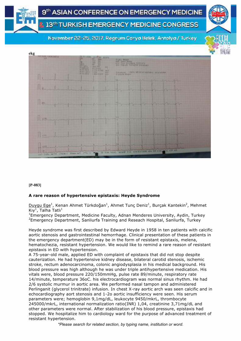
*Please search for related section, by typing name, institution or word.
ekg
[P-083]
A rare reason of hypertensive epistaxis: Heyde Syndrome Duygu Ege1, Kenan Ahmet Türkdoğan1, Ahmet Tunç Deniz1, Burçak Kantekin2, Mehmet Kıy1, Talha Tatlı1 1Emergency Department, Medicine Faculty, Adnan Menderes University, Aydin, Turkey 2Emergency Department, Sanliurfa Training and Reseach Hospital, Sanliurfa, Turkey Heyde syndrome was first described by Edward Heyde in 1958 in ten patients with calcific aortic stenosis and gastrointestinal hemorrhage. Clinical presentation of these patients in the emergency department(ED) may be in the form of resistant epistaxis, melena, hematochezia, resistant hypertension. We would like to remind a rare reason of resistant epistaxis in ED with hypertension. A 75-year-old male, applied ED with complaint of epistaxis that did not stop despite cauterization. He had hypertensive kidney disease, bilateral carotid stenosis, ischemic stroke, rectum adenocarcinoma, colonic angiodysplasia in his medical background. His blood pressure was high although he was under triple antihypertensive medication. His vitals were, blood pressure 220/150mmHg, pulse rate 89/minute, respiratory rate 14/minute, temperature 36oC. his electrocardiogram was normal sinus rhythm. He had 2/6 systolic murmur in aortic area. We performed nasal tampon and administered Perlinganit (glycerol trinitrate) infusion. In chest X-ray aortic arch was seen calcific and in echocardiography aort stenosis and 1-2o aortic insufficiency were seen. His serum parameters were; hemoglobin 9,1mg/dL, leukocyte 9450/mkrL, thrombocyte 245000/mkrL, international normalization ratio(INR) 1,04, creatinine 3,71mg/dL and other parameters were normal. After stabilization of his blood pressure, epistaxis had stopped. We hospitalize him to cardiology ward for the purpose of advanced treatment of resistant hypertension.

*Please search for related section, by typing name, institution or word.
We wanted to present Heyde syndrome in ED with angiodysplasia, aortic stenosis and anemia, which is a rare cause in the patient with complaints of resistant hypertension and hemorrhage in this case. Keywords: Epistaxis, Heyde syndrome

*Please search for related section, by typing name, institution or word.
[P-084]
Anevrizma Ne Kadar Büyüyebilir? Emel Erkuş Sirkeci, Çimen Elias, Hikmet Neşe Ergün, Özgür Sirkeci, Eliz Mazhar, Dilfüza Abdurshurova, Zakaria Suliman Near East University Hospital, Cyprus Aortada anevrizma lokalize bir alanda normal çapın %50’den fazla genişlemiş (1,5 kat) olmasıdır. Abdominal aort anevrizması (AAA) lokalize bir alanda kalıcı olarak infrarenal aort çapının 3 cm’yi aşması olarak ifade edilebilir. 50 yaş üstü erkek hastalarda %5 oranında görülmektedir. 1 Genellikle AAA yılda 0,5 cm genişleme gösterir ve doğal seyri sonucunda rüptür gelişir. Rüptür aşamasına kadar çoğunlukla asemptomatik seyir gözlenir. Rüptür riski çap ile orantılıdır ve 5,5 cm üzerindeki anevrizmalarda artmıştır. Olguların rüptür gelişmeden, elektif şartlarda tedavi edilmeleri gerektirmektedir.1,2,3 AAA larının tedavisinde anevrizma çapının büyük önemi vardır. 4 Anevrizma çapı ile orantılı olarak yükselen yıllık rüptür riski, 4 cm’nin altında %0, 4,5 cm için %1, 5,5 cm için %11 ve 6,5 cm için de %26 olarak belirtilmiştir. Görüldüğü gibi çap artışı ile rüptür riski arasında logaritmik bir ilişki mevcuttur. 53 yaşında erkek hasta, halsizlik ve iştahsızlık ile acil servisimize başvurdu. Hasta sorgulandığuında aralıklı olarak karın ağrısının da aralıklı iki gündür olduğunu ifade ediyor. Hastanın özgeçmişinde özellik yok. Muayenesinde hastanın vitalleri; ateş:37,5, tansiyon:105/70, SpO2:93, nabız:75, şuur açık, oryante ve koopere, GKS:15. Hastanın orofarenks bölgesi muayenesi hafif hiperemik. Akciğer muayenesinde bazallerde raller ve ronküs duyuluyor. Batın muayyenesinde hastanın epigastriumda ağrısının dışında sol üst kadranda pulsasyon alınması üzerine abdominal aort olabileceği düşünülerek ileri tetkik planlandı. Hastanın çekilen elektrokardiyografisinde sinüs taşikardisi mevcuttu.Hastanın iki gündür yeterli oral alımı olmadığı ve idrara çıkamadığını ifade etti.Hastanın kan tahlillerinde akut böbrek yetmezliği lehine üre kreatinin değerlerinde artış saptandı.Hastaya direkt akciğer grafisi ve sonrasında batın ultrasonografi planlandı ve tedavisine hidrasyon ve proton pompa inhibitörü eklenerek devam edildi.Hastanın renal hasarı olmadığı ancak 8,7 cm ebatında abdominal aortada anevrizma saptandı.Hasta ileri takip için kalp damar cerrahisine interne edildi. Hastalar acil servise anamnezlerinden acil bir durumu ifade ederek ya da hastalıkları olmadığını söyleyerek gelseler bile yeterli bir muayene ve tetkikle hastaların hayatları yeni tanılarla kurtarılabilinir. Anahtar Kelimeler: Aort, anevrizma, diseksiyon

*Please search for related section, by typing name, institution or word.
[P-085]
Syncope? Seizure? or Both? A Unique Case of Complete Heart Block Emir Ünal, Zuhal Kalyon, Su Izel Sesigür, Erkman Sanrı, Haldun Akoğlu, Özge Onur, Arzu Denizbaşı Marmara University Faculty of Medicine, Department of Emergency Medicine, Istanbul, Turkey Introduction: The similarity of the clinical presentation of patients with seizures and syncope makes it difficult to answer the question of cardiac or neurological origin. The challenge becomes more difficult considering the epileptic neural discharges triggering cardiac arrhythmias that might create syncope. We present a case of repeating seizures secondary to AV block progressing to asystole during collapse attacks diagnosed by real-time monitorization. Case: An 80-year-old female patient admitted to ED with a brief episode of unconsciousness lasting about 1 minute. Physical examination and ECG was normal. She was on beta blockers and ACE inhibitors for HT. Due to suspicion of an intermittent arrhythmia, the patient was put on cardiac monitorization in the ED. During monitorization, acute loss of consciousness lasting 6-7 seconds has occured with a lateralizing gaze. During the attack, carotid pulse could not be obtained and the monitor showed asystolic rhythm. The rhythm was returned with the return of the consciousness without any intervention and a complete AV block (50/minute) was observed. Similar attacks repeated in a short time period, therefore an emergency external pacemaker was implanted to the patient, and admitted to coronary ICU. However, asystolic attacks unresponsive to pacing ended up with the demise of the patient. Discussion: In patients with loss of consciousness, seizures and syncopes, close cardiac monitorization is indicated to rule-out cardiac arrhythmias and blocks. Seizures should not be thought as a sign to rule out cardiac causes. Keywords: Syncope, bradycardia, complete heart block

*Please search for related section, by typing name, institution or word.
[P-086]
Atrial Fibrillation After Low-Tension Electrical Injury Ertuğ Günsoy, Aslıhan Yürüktümen Ünal Akdeniz University Hospital Emergency Medicine Electrical injury can cause various cardiac dysrhythmias such as asystole, ventricularfibrillation, sinus tachycardia, and heart blocks. However, supraventricular tachyarrhythmias, such as atrial fibrillation, are rare. It is very rare that atrial fibrillation develops after low-voltage electrical shock. İn this case the patient who developed atrial fibrillation after impactwith 220 volts is spontaneously returned to normal sinus rhythm. Keywords: Atrial fibrillation, low tension electrical injury, cardiac dysrhythmias
fig
fig 1: acute atrial fibrillation (AF) with rapid ventricular response

*Please search for related section, by typing name, institution or word.
[P-088]
Brugada Syndrome with Atypical Complaints: Case Report Güven Yılmaz, Muhammed Fatih Cırıl, Ayse Cagla Ozmert Toplu, Izzet Ustaalioğlu, Ali Göksu, Ozden Seven, Gülbahar Güler, Ferhat Yıldız, Ecem Deniz Kırkpantur, Mehmet Koçak, Tugba Cimilli Öztürk, Ozge Onur SBU Fatih Sultan Mehmet Training and Research Hospital, Emergency Medicine Department, Istanbul, Turkey Brugada syndrome is an autosomal dominant genetical disease which is characterised by abnormal ECG and risk of sudden cardiac death. Typical finding of brugada is presence of ST segment elevation in precordial leads with concomitant right bundle branch block on ECG. Misdiagnosis or difficulty in diagnosis are generally encountered in routine clinical practice for Brugada Syndrome. Herein we would like to share a case who presented to our emergency medicine with atypical complaints such as pain in both shoulders for two days. Our aim is to overview of Brugada syndrome in the context of the latest published literature. In conclusion, BS is rare but can lead to fatal arrhythmias. Therefore, according to this report the emergency physicians should be aware of atypical complaints that may point out Brugada syndrome. Keywords: Atypical presentation, brugada syndromes, bundle brinch block

*Please search for related section, by typing name, institution or word.
[P-092]
Silent Death: Aortic dissection Hulusi Kardaş, Serhat Akay, Emrah Yılmaz, Mehmet Eman, Mustafa Silcan Izmir Bozyaka Education and Research Hospital, Izmir, Turkey Introduction: No matter how busy the emergency service is, the first task of an Emergency Medicine specialist is to make a diagnosis of the life-threatening situation. Aortic dissection in a patient with back pain should be considered as a primary differential diagnosis. Patient with back pain in our case was discharged after symptomatic treatment with myalgia diagnosis at external center for 3 days. Case: A 44-year-old male patient was admitted to the emergency service with a pain which was continued for 3 days, increased with movement and palpation and started between both of the scapulae spreading to sides. Patient with arterial blood pressure: 130/70 had no tachycardia-tachypnea, his saturation was normal. There is no drug used regularly by the patient without any illness on his history. On physical examination, respiratory sounds were natural and there was no ral-rhonchus, peripheral pulses were bilateral open. The ECG was normal sinus rhythm with non-ST change. Our preliminary diagnoses were pneumothorax, aortic dissection, myocardial ischemia, pneumonia and myalgia therefore lung film and blood including cardiac markers and D-dimer were taken. Thorax-abdomen tomography with contrast was performed with D-dimer level: 1984, creatine kinase: 2248, creatine kinase-MB: 19.3, troponin I: 0. In the sections obtained in the aortic phase, the anterior-posterior diameter of the aorta was measured as 4 cm, arcus aorta 3 cm, descending aorta 4.5 cm. From the level of the aortic valve to the level of the diaphragm, a double lumen view of the dissecting flap was observed. Morphology De-Bakey was evaluated as type 3. The patient who was hospitalized in the vascular surgery services was taken into surgery. After 4 days he was discharged by healing. Results: Aortic dissection is the pathology that should be remembered first in every patient who comes to the emergency department with back, chest and abdominal pain. Keywords: Aortic dissection, back pain, D-dimer

*Please search for related section, by typing name, institution or word.
Figure 1

*Please search for related section, by typing name, institution or word.
[P-094]
Tipik Göğüs Ağrısında Farklı Bir Tanı; Koroner Sinüs Ritmi: Olgu Sunumu İzzet Ustaalioglu, Guven Yılmaz, Muhammed Fatih Cırıl, Ali Goksu, Ayşe Çağla Özmert Toplu, Ferhat Yıldız, Hüseyin Özer, Mehmet Koçak, Tuba Cimilli Öztürk, Özge Ecmel Onur SBU Fatih Sultan Mehmet Training and Research Hospital, Emergency Medicine Department, Istanbul, Turkey Giriş: Kısa PR sendromları (KPRS), sinoatriyal nodun baskılanması nedeni ile atrioventriküler nodun uyarı çıkarma özelliğinin sinoatriyal nodu aştığı durumlarda veya ektopik bir odak olması durumda ortaya çıkar. Sinoatriyal nod pacemaker özelliğini kaybetmiştir. Vaka: 45 yaşında erkek hasta yarım saat önce başlayan, uykudan uyandıran baskı tarzında göğüs ağrısı ile acil servisimize başvurdu. Daha önce benzer ağrısı olmamış. Hipertansiyondan başka ek hastalığı olmayan sigara içicisi hastanın ailesinde koroner arter hastalığı öyküsü mevcut. Gelişinde hemodinamik olarak stabil olan hastanın EKG’sinde PR aralığı kısa (0,04 ms), D2, D3, aVF’de negatif P dalgaları mevcuttu. Yapılan Ekokardiyografisinde EF: %55 segmenter hareket kusuru ve lateral duvarda hipokinezi saptanması üzerine hasta primer anjio işlemine alındı. Tartışma: PR aralığı <120 ms olması durumunda pre-eksitasyonu (atriyum ve ventriküller arası aksesuar yolak varlığı) veya AV nodal (junctional) ritim akla gelmelidir. Pre-eksitasyon sendromlarından en sık görülenleri Wolff-Parkinson-White (WPW), Lown-Ganon-Levine (LGL) ve koroner sinüs ritmidir. Ayırıcı tanıda EKG’de hız, P ve QRS dalga morfolojisilerine dikkatli bakılmalıdır. Wolff-Parkinson-White sendromunun karakteristik özellikleri kısa PR aralığı, normal P dalgaları, geniş QRS kompleksi ve QRS kompleksine hızlı geçiş olan delta dalgasıdır. LGL sendromu ise normal P dalgaları ve QRS kompleksleri ile birlikte çok kısa PR aralığı ve delta dalgası yokluğuyla karakterizedir. LGL sendromunda beraberinde taşikardi atakları (özellikle AVNRT) görülebilir. Koroner sinüs ritminde uyarılar atriumdaki ektopik bir odaktan çıktığı için P dalgaları sinüs P’sinden farklıdır. Bazen D2, D3 ve aVF’de negatif olabilir. AV nodal ritimde ise kısa PR aralığı ile birlikte P dalgaları yoktur veya anormaldir. Sonuç: KPRS ani kardiyak ölüm gibi risklerinden dolayı özellikle acil servis doktorlarının uygun anamnez ile EKG bulgularını iyi değerlendirip KPRS tanılarına hâkim olmaları gerekmektedir. Anahtar Kelimeler: Kısa PR sendromu, göğüs ağrısı, koroner sinüs ritmi

*Please search for related section, by typing name, institution or word.
[P-095]
What Lies Beneath: An Unusual Case of Aortic Dissection Presenting as an Acute Ischemic Stroke Keith Rollo R. Nario, MD; Peter F. Quilala, MD, FPCEM Department of Emergency Medicine, St. Luke’s Medical Center Keith Rollo Rimando Nario, Peter Flores Quilala Department of Emergency Medicine, St. Luke's Medical Center, Quezon City, Philippines Aortic dissection is a life-threatening emergency commonly occurring due to injury on the inner wall of the artery. This disease has a wide range of symptoms but typically causes severe pain on the chest, abdomen or back and usually accompanied by physical examination findings of hemodynamic instability. However, it can also cause neurologic symptoms usually due to decreased cerebral perfusion. Aortic dissection masquerading as an ischemic stroke is the diagnostic and therapeutic dilemma in this case of a 70-year-old female presenting with neurologic deficits and hemodynamic instability. Timely diagnosis and recognition with the help of point of care ultrasound prevented mismanagement because in this age of thrombolytic therapy, misdiagnosis could be catastrophic. This case report highlights the importance of point of care ultrasound and recognizing atypical presentations of acute aortic dissection. This reminds emergency physicians to be vigilant in knowing alternate reasons for patients presenting with stroke symptoms to avoid misdiagnosis and mismanagement. Keywords: Aortic dissection, point of care ultrasound, ischemic stroke

*Please search for related section, by typing name, institution or word.
[P-096]
Aortic Dissection in Differential Diagnosis in The Case Of Pericardial Tamponade Kübra Parpucu, Gözde Gök, Filiz Baloğlu Kaya, Ezgi Akçacı Baran, Engin Özakın, Evvah Karakılıç Department of Emergency Medicine, School of Medicine, Eskişehir Osmangazi University, Eskişehir, Turkey. Introduction: Aortic dissection (AD) is defined as the separation of the aortic wall layers. Cardiac tamponade (CT) is a clinical syndrome caused by fluid accumulation in the pericardial space, resulting in reduced ventricular filling followed by hemodynamic deterioration. The CT observed in AD is seen with proximal aortic dissection extending to the aortic root. Mortality in AD complicated by CT is 60%. Pericardiocentesis in these patients increases blood flow by creating a pressure gradient between the dissection and the pericardial space. This may lead to hemodynamic deterioration and prolongation of the dissection. In this case, we wanted to remind that the AD should be discarded in the patients who are scheduled for pericardiocentesis. Case: A 67-year-old woman was brought to the emergency. No pathology was found on physical examination except for the deep heart sound and weakness of the left lower extremity peripheral pulses. The bedside ultrasound revealed a pericardial fluid that was 2 cm in the thickest area and causing collapse. Although the abdominal aortic diameters were normal, a suspicious flap appearance was observed. In the aortogram; there was a dissection starting from ascending aorta and continued throughout the aorta. The coronary arteries appeared starting from the true-, while the left renal artery was observed starting from the false lumen. The patient was planned to undergo replacement of the ascending aorta by cardiac surgery. Although postoperative inotropic support, the case was hypotensive, and had no urine output, therefore cardiac arrest developed. Discussion: The possibility of AD should be kept in mind in patients presenting with complaints such as urgent dyspnea, chest, back and neck pain. AD is an absolute contraindication for pericardiocentesis and should be discarded by imaging methods such as bedside ultrasonography. Keywords: Pericardial tamponade, aortic dissection, differential diagnosis

*Please search for related section, by typing name, institution or word.
[P-099]
Two Thrombosis Case Without Any Thrombi: Aortic Dissection Merve Akın, Özge Can, G. Selahattin Kıyan Department of Emergency Medicine, Ege University, Izmir, Turkey Introduction: The classic presentation of an acute aortic dissection is the sudden onset of severe and tearing chest pain. There were a few cases with atypical findings or no pain in the literature. We report the two cases that present the ED with neurologic symptoms and diagnosed as an aortic dissection. Case 1: 46-year-old male patient admitted to the ED with dizziness, diplopia, and nausea. His physical examination was normal. On his history, type 3 aortic dissection was determined. While following in the ED, the patient had become quadriparesis. Cranial and neck and thorax angiography were examined. CT was showed that both vertebral arteries proximal had a dissection and in the thorax CT there was Stanford type A dissection. The patient was hospitalized in ICU. Case 2: 50 y male patient presented to the ED with acute episode of altered mental status, headache. His vital signs were normal. On his physical examination, he was only disorientated, no any neurologic deficit. His ECG was normal. His cranial CT was normal. Hypotension US protocol (RUSH) had performed him. And aorta enlargement determined. CT scan showed Stanford type A dissection. He was hospitalized. Discussion: Aortic dissection is characterized by sudden onset and tearing of the chest, back, or abdominal pain (1). These symptoms are specific to the diagnosis. However, 10% of the aortic dissection can occur with no pain and organ-related symptoms (1,2). A few cases of aortic dissection with neurological symptoms have been reported in the literature. Neurological symptoms were classified according to the affected area; spinal artery obstruction, or reduce spinal cord blood flow, a decrease in cerebral blood flow due to hypotension or progression of the aortic arch to the dissection can cause paraparesis or paraplegia is detected (3). Conclusion: Emergency physicians should remember aortic dissection, patients with neurological deficits. Keywords: Aort dissection, atipical presentation, neurological symptoms

*Please search for related section, by typing name, institution or word.
[P-100]
Genç Senkopta Atlanmaması Gereken Ölümcül Bir Tanı: Brugada Sendromu Mine Kayacı Yıldız1, Yasin Yıldız2, Serkan Emre Eroğlu1 1Sağlık Bilimleri Üniversitesi, Ümraniye Eğitim ve Araştırma Hastanesi, Acil Tıp Kliniği, İstanbul. 2Sağlık Bilimleri Üniversitesi, Sultan Abdülhamid Han Eğitim ve Araştırma Hastanesi, Acil Tıp Kliniği, İstanbul Giriş: Senkop ani,geçici ve kendiliğinden düzelen bilinç ve tonus kaybı olup acil kliniklerinde sık görülen bir klinik problemdir.Kardiyak aritmiler veya yapısal kalp hastalıklarına bağlı senkop başlıca nedenlerdendir.EKG,senkop yakınması olan her hastada yapılması gereken noninvaziv bir test olup,senkop etiyoloji araştırmasında organik kalp hastalığı olup olmaması, aritmi şüphesi ve senkop sıklığı testlerin seçiminde önemlidir(1).Biz burada,acil servisimize epistaksis nedenli başvuran,anamnezi derinleştirildiğinde senkop öyküsü veren ve EKG ile Brugada Sendromu tanısı konan bir hastayı sunmayı amaçladık. Olgu: 18 yaşında erkek hasta,epistaksisle 112 tarafından acilimize getirildi.Gelişinde aktif epistaksisi olmayan hastanın vitalleri stabildi.Anamnez derinleştirildiğinde,son 20 gündür hemen hergün,günde 7-8 kez epistaksisinin olduğu ve ara ara baygınlık geçirdiği zamanların da olduğu ve bu nedenli acile mükerrer başvurularının olduğu öğrenildi.Bu başvurusunda da senkop tarifleyen hastanın dedesinin 20'li yaşlarda ani ölümle kaybedildiği öğrenildi.Çekilen EKG'de V1-2'de Brugada tip-2 paterni görüldü(Resim A). Hemodinamik açıdan stabil olan hastanın Kardiyolog tarafından yapılan EKO'sunda kardiyak patoloji saptanmadı. Laboratuvar değerleri normal aralıklarda ve senkop etyolojisi açısından diğer sistem muayeneleri doğal olan hastanın kontrol EKG'sinde Brugada paterninin düzeldiği görüldü (Resim B). Hasta, kardiyoloğun da önerisi ile, Ajmalin testi yapılmak üzere, Kardiyoloji Dal Hastanesi'ne yönlendirildi. Tartışma: Brugada Sendromu (BrS), ilk olarak 1992'de Brugada kardeşler tarafından tanımlanmıştır ve ventiküler fibrilasyona bağlı ani kardiyak ölüm riskinin arttığı nadir bir genetik hastalıktır(2).Gözlemsel çalışmalarda prevelansı yaklaşık 5/10000 olarak saptanmıştır(3).Tanısı, kendiliğinden veya ilaç provakasyonu sonrası sağ prekordiyal derivasyonların(V1-3) en az birinde saptanan >2mm ST segment elevasyonu ile konur(3).BrS'den şüphelenilen hastalarda tanı koymak için sodyum kanal blokerleri sıklıkla kullanılmaktadır.Diğer sodyum kanal blokerlerine göre,Ajmalin'in tanısal değerinin daha fazla olması sebebiyle,bu amaçla tüm dünyada en sık Ajmalin kullanılmaktadır(4).BrS, genellikle erişkin yaşta ortaya çıkar,ortalama ani ölüm yaşı 41±15'tir.Ani ölümün önlenmesindeki tek tedavi seçeneği ICD implantasyonudur(5). Sonuç: Özellikle gençlerde,senkop etiyolojisi araştırılırken aile öyküsü iyi sorgulanmalı ve mutlaka EKG çekilip,sadece iskemik değişiklikler değil,ani kardiyak ölüme yol açabilecek aritmiler açısından da dikkatli olunmalıdır. Herhangi bir medikal tedavisi bulunmayan BrS'nin EKG paternleri, acil hekimleri tarafından tanınmalıdır. Anahtar Kelimeler: Brugada sendromu, EKG, senkop

*Please search for related section, by typing name, institution or word.
Resim A ve B
A. Geliş EKG'si: V1-2'de Brugada Tip 2 Paterni B. Kontrol EKG: Normal

*Please search for related section, by typing name, institution or word.
[P-102]
What is the Reason for Syncope Oğuzhan Demir, Fatih Mehmet Akbaş, Serhad Ömercikoğlu, Sinan Karacabey, Özge Onur, Haldun Akoğlu, Arzu Denizbaşı Marmara University, Pendik Research and Training Hospital, Istanbul, Turkey Introduction: Syncope is a risk factor for sudden cardiac death (SCD) in many conditions associated with structural heart disease as well as inherited heart disease. The ECG in patients with syncope should be examined carefully for signs of structural heart disease, such as myocardial infarction or cardiomyopathy; signs of conduction system disease, such as bundle branch block or atrioventricular block; and signs of primary electrical disease. Here we present a case of primary electrical disease and mention for underlying causes of syncope. Case: A 46 year of women presented with syncope to emergency department. Her glaskow Coma Scale was 15 at presentation. Her vital signs and physical examination were normal. She had syncope attacks in her past. Episodes of atrial fibrillation (AF) was diagnosed. She had no treatment or evaluation. ECG in ED revealed demonstrated a Wolff Parkinson White (WPW) pattern combined with unexplained syncope. None of the examinations, including biochemical profiles, brain computed tomography was pathologic. Due to high risk of sudden death, for further follow-up, she admitted to coronary unit. Discussion: Syncope, despite being induced by various mechanisms, has been considered an alarming sign of sudden death of WPW syndrome. AF was the first event in only 6% of patients with WPW and episodes of atrial fibrillation (AF) increase risk of sudden death in WPW. The electrocardiogram (ECG) is one of the key clinical variables and specific electrocardiographic presentations must be searched in the patient with syncope. Keywords: Syncope, WPW
*Please search for related section, by typing name, institution or word.
[P-103]
Painless Aortic Dissection Presenting with Loss of Consciousness Oğuzhan Demir, Murat Doğanay, Ali Celik, Erkman Sanrı, Haldun Akoğlu, Özge Onur, Arzu Denizbaşı Marmara University Faculty of Medicine, Department of Emergency Medicine, Istanbul, Turkey Introduction: Acute aortic dissection is one of the most fatal cardiovascular emergencies. Classically seen as sudden, severe chest, back, or abdominal pain. However, there are several cases presenting with atypical sypmtoms. In this case we present a case of aortic dissection with loss of consciousness. Case: 77-year-old female with history of hypertension admitted to ED with loss of consciousness. Relatives declared she lost consciousness half an hour ago, without any prior complaints. They denied history of seizure, trauma or syncope. Vitals were bp:65/30mmHg hr:72bpm fever:36.5 C gcs:12. Her other neurological examinations were normal. There were no pulse deficit or extremity blood pressure difference. In non-contrast cranial CT, no pathologies were present. no abnormality. A CT pulmonary angiogram was ordered to rule-out PTE despite negative echo findings, which revealed the dissection of ascending aorta. Discussion: Differential diagnosis of a syncope or loss of consciousness encompasses several life-threatening conditions. Immediate diagnosis and early treatment can be crucial. Key in the management of acute aortic dissection is to maintain a high level of suspicion for this diagnosis. Keywords: Aortic dissection, computed tomography
*Please search for related section, by typing name, institution or word.
[P-104]
Aortic Dissection in The Emergency Room, in A Young Male with Atypical Presentation Selahattin Gürü1, Adil Emre Gezer2, Fatih Tanrıverdi2, Mehmet Ergin2, Şervan Gökhan2 1Ankara Atatürk Training and Research Hospital, Emergency Department 2Yıldırım Beyazıt University, Emergency Department, Ankara, Turkey Introduction: Aortic dissection (AD); A life-threatening disease, which the intima layer of the aortic wall ruptures. Chest pain is typical, beginning suddenly, spreading to the neck and jaw, and defined "very severe and tearful" by the patients. It is twice as common in males as it is the most common elderly. In this article, a case with atypical aortic dissection will be presented. Case: A 28-year-old male presented to our emergency room on the 5th day of mild chest pain that spread to the lower right armpit and dyspeptic complaints. There were no risk factors in the medical history. Arterial blood pressure measurement results were 143/82 mmHg on the upper right limb and 112/70 mmHg on the left. In the last 4 days, he was examined by various tests including upper gastrointestinal endoscopy. D-dimer level was 1100,63 ng/mL (>500 ng/mL). Thoracoabdominal CT was taken, intimal separation and dissection flaps were seen from the end of the aortic arch to the level of the diaphragm. He was discharged safely on the 10th day following the surgery. Discussion: AD can be rapidly fatal. It is most commonly seen between the ages of 60 and 70. Although we present the case that the dissection developed at the age of 28 years. For patients with unidentified chest and back pain, aortic dissection should be considered in the differential diagnosis. For aortic dissection, values below 500 ng / mL are considered to have a high negative predictive value in aortic dissection. In this case we have been investigated further by the reason of high d-dimer value. Conclusion: AD is a serious pathology for emergency department. It should not be forgotten that there may be atypical applications of 10% as well as a typical clinical presentation for AD. D-dimer should be considered during the decision-making process. Keywords: Aortic dissection, emergency room

*Please search for related section, by typing name, institution or word.
[P-106]
Chest Pain triggered by Emotional Stress: Takotsubo Cardiomyopathy or Broken Heart Syndrome Su Izel Sesigür, Emir Ünal, Sinan Karacabey, Haldun Akoğlu, Özge Onur, Arzu Denizbaşı Marmara University Faculty of Medicine, Department of Emergency Medicine, Istanbul, Turkey Introduction: A takotsubo (Japanese) is a device with a narrow neck and large, balloon-shaped body, made of ceramic, used to capture octopus. The mystery of the disease continues due to the lack of sufficient information about the clinical course and prognosis of this syndrome, which exact cause of is still unknown and is caused by emotional and physical stress. We present a case of takotsubo cardiomyopathy who admitted to our ED with chest pain triggered by emotional stress. Case: A 54-year-old female was admitted to our ED after a successful resuscitation on a plane. On admission, she was conscious with full orientation and cooperation. Physical examination revealed tenderness on chest wall, possibly related to CPR. Sinus rhythm was present in the ECG, without any ST segment changes. She stated that she has no cardiac history, but her husband has died recently and she was returning from his funeral. During the flight, she had a sudden feeling of discomfort on her chest. She was monitorized by a doctor and nurse on flight and a VF was diagnosed. She was defibrillated 4 consecutive times and CPR was resumed until sinus rhythm and ROSC. Bedside echocardiogram in the ED revealed decreased EF, apical dyskinesia and ballooning. She was admitted to the CICU with the diagnosis of takotsubo cardiomyopathy. Discussion: In Turkey and world, ST changes were observed as ECG findings in Takotsubo cardiomyopathies. In our patient, ST change and T wave changes were not observed in the ECG as it is seen in the literature. This is not enough to exclude Takotsubo Cardiomyopathy. If there is no serious stenosis in the vessels of patients undergoing coronary angiography due to ACS, ventriculography or echocardiography should be considered as soon as possible to evaluate Takotsubo's syndrome. Keywords: Broken heart syndrome, takotsubo cardiomyopathy, acute myocardial infarction

*Please search for related section, by typing name, institution or word.
[P-107]
Prehospital ECPR in the Mobile Operating Room for Emergency surgery saved a woman’s life Akihide Konn, Tatsuya Nodagashira, Kensuke Fuijita, Yutaro Kurihara Hachinohe City Hospital, Department of emergency and Critical Care Medicine, Japan Introduction:Mobile Operating Room for Emergency surgery (MOREs) can be potentially effective for saving lives by starting Extracorporeal cardiopulmonary resuscitation (ECPR) from the site near the scene. MOREs brings 3Drs and the ECMO devices. We construct a simplified theater for 5 minutes. MOREs enable early ECMO implantation and emergency thoracotomy and laparotomy. Case report:A 45-years-old woman accidentally felt from the bridge and drifted down the river to the sea in winter. The EMS pulled her up from the sea and found her in cardiac arrest. A rapid response car dispatched in advance and started the ACLS. MOREs with 3 doctors and 2 clinical engineers dispatched because she needed prehospital ECPR. We started ECPR in MOREs 5 minutes after unsuccessful standard ACLS resuscitation. She was transferred to the hospital for 20 minutes and rewarmed, then the return of spontaneous circulation was achieved. Days2, she was weaned from ECMO and mechanical ventilator. Days4, she was discharged from the intensive care unit and a few days later, she left the hospital with CPC 1. Conclusion: Prehospital ECPR in MOREs can shorten the time to restart circulation when it takes a lot of time to transfer the patient to the hospital. Keywords: Extracorporeal cardiopulmonary resuscitation, ECMO

*Please search for related section, by typing name, institution or word.
[P-110]
Survival Rate and Outcomes of Out-Of-Hospital Cardiac Arrest at St. Luke’s Medical Center from February 2015 to June 2016: A Retrospective Cohort Study Maria Cristina Pascual Angeles Emergency Medicine Department, St. Luke's Medical Center, Quezon City, Philippines The study aims to provide data involving out-of-hospital cardiac arrest patients brought to the emergency department as confirmed by the absence of pulse, unresponsiveness and apnea. This will include cardiac arrests of both presumed cardiac and non-cardiac etiology. It will also point out the need for improvement in emergency medical system to achieve better patient survival and outcome in the country. General Objective: To determine the survival rate and outcomes of out-of-hospital cardiac arrest seen at St. Luke’s Medical Center from February 2015 to June 2016. Specific Objectives: To determine the Out-Of-Hospital Cardiac Arrest as to patient information (Age, Gender, Medical history). Second, to determine the pre-hospital event and Emergency department resuscitation information of cardiac arrest patients. Lastly, to determine the disposition, survival and outcome of cardiac arrested patients. Methodology: Retrospective Cohort Study. The study was conducted at St. Luke’s Medical Center, Emergency Department, a private tertiary hospital in Quezon City. Patient demographics, initiation of cardiopulmonary resuscitation and outcome of resuscitation at Emergency Department were obtained using a cardiac arrest registry form derived from the Pan Asian Resuscitation Outcomes Study (PAROS) Clinical Research Network (CRN). Results: The study showed that majority of the Out-Of-Hospital Cardiac Arrest patients brought to Emergency Department were males, mostly with a non-traumatic cause, commonly observed in 45-64 age group, with highest incidences were found to be unwitnessed. The most common presumed etiology is cardiac in origin. Conclusion: Survival Rate remains to be low despite identifying several factors that greatly affect patient’s outcome. Future researchers should focus more on prompt delivery of interventions of known effectiveness, especially to those who have an immediate access to perform advance resuscitative efforts to increase the chance of patient’s survival with improved outcome. Keywords: Out-Of-Hospital-Cardiac-Arrest (OHCA)
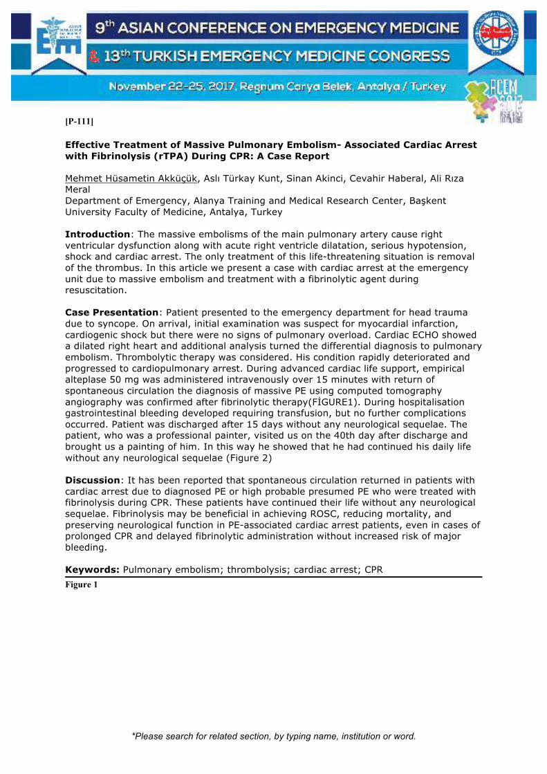
*Please search for related section, by typing name, institution or word.
[P-111]
Effective Treatment of Massive Pulmonary Embolism- Associated Cardiac Arrest with Fibrinolysis (rTPA) During CPR: A Case Report Mehmet Hüsametin Akküçük, Aslı Türkay Kunt, Sinan Akinci, Cevahir Haberal, Ali Rıza Meral Department of Emergency, Alanya Training and Medical Research Center, Başkent University Faculty of Medicine, Antalya, Turkey Introduction: The massive embolisms of the main pulmonary artery cause right ventricular dysfunction along with acute right ventricle dilatation, serious hypotension, shock and cardiac arrest. The only treatment of this life-threatening situation is removal of the thrombus. In this article we present a case with cardiac arrest at the emergency unit due to massive embolism and treatment with a fibrinolytic agent during resuscitation. Case Presentation: Patient presented to the emergency department for head trauma due to syncope. On arrival, initial examination was suspect for myocardial infarction, cardiogenic shock but there were no signs of pulmonary overload. Cardiac ECHO showed a dilated right heart and additional analysis turned the differential diagnosis to pulmonary embolism. Thrombolytic therapy was considered. His condition rapidly deteriorated and progressed to cardiopulmonary arrest. During advanced cardiac life support, empirical alteplase 50 mg was administered intravenously over 15 minutes with return of spontaneous circulation the diagnosis of massive PE using computed tomography angiography was confirmed after fibrinolytic therapy(FİGURE1). During hospitalisation gastrointestinal bleeding developed requiring transfusion, but no further complications occurred. Patient was discharged after 15 days without any neurological sequelae. The patient, who was a professional painter, visited us on the 40th day after discharge and brought us a painting of him. In this way he showed that he had continued his daily life without any neurological sequelae (Figure 2) Discussion: It has been reported that spontaneous circulation returned in patients with cardiac arrest due to diagnosed PE or high probable presumed PE who were treated with fibrinolysis during CPR. These patients have continued their life without any neurological sequelae. Fibrinolysis may be beneficial in achieving ROSC, reducing mortality, and preserving neurological function in PE-associated cardiac arrest patients, even in cases of prolonged CPR and delayed fibrinolytic administration without increased risk of major bleeding. Keywords: Pulmonary embolism; thrombolysis; cardiac arrest; CPR
Figure 1

*Please search for related section, by typing name, institution or word.
24 hour later CT image with thrombus after the fibrinolytic therapy
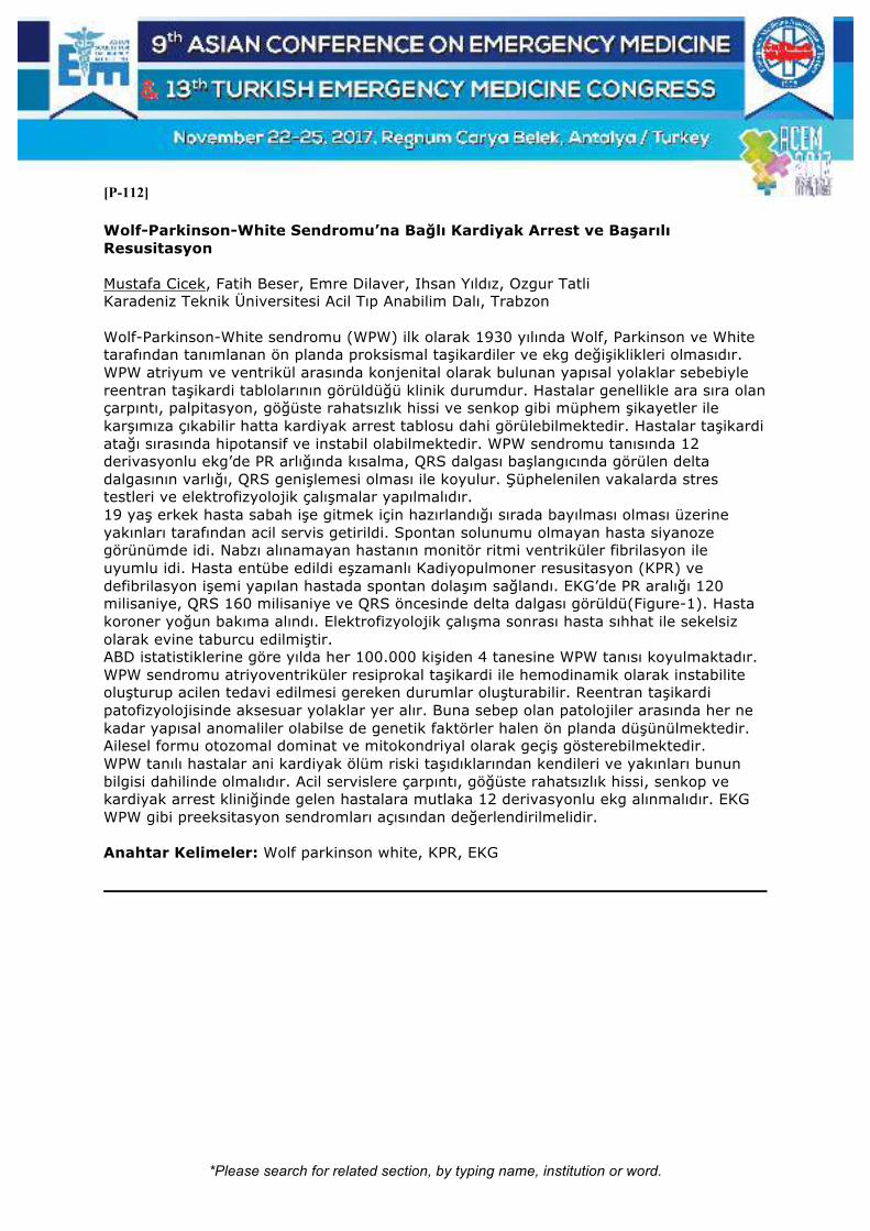
*Please search for related section, by typing name, institution or word.
[P-112]
Wolf-Parkinson-White Sendromu’na Bağlı Kardiyak Arrest ve Başarılı Resusitasyon Mustafa Cicek, Fatih Beser, Emre Dilaver, Ihsan Yıldız, Ozgur Tatli Karadeniz Teknik Üniversitesi Acil Tıp Anabilim Dalı, Trabzon Wolf-Parkinson-White sendromu (WPW) ilk olarak 1930 yılında Wolf, Parkinson ve White tarafından tanımlanan ön planda proksismal taşikardiler ve ekg değişiklikleri olmasıdır. WPW atriyum ve ventrikül arasında konjenital olarak bulunan yapısal yolaklar sebebiyle reentran taşikardi tablolarının görüldüğü klinik durumdur. Hastalar genellikle ara sıra olan çarpıntı, palpitasyon, göğüste rahatsızlık hissi ve senkop gibi müphem şikayetler ile karşımıza çıkabilir hatta kardiyak arrest tablosu dahi görülebilmektedir. Hastalar taşikardi atağı sırasında hipotansif ve instabil olabilmektedir. WPW sendromu tanısında 12 derivasyonlu ekg’de PR arlığında kısalma, QRS dalgası başlangıcında görülen delta dalgasının varlığı, QRS genişlemesi olması ile koyulur. Şüphelenilen vakalarda stres testleri ve elektrofizyolojik çalışmalar yapılmalıdır. 19 yaş erkek hasta sabah işe gitmek için hazırlandığı sırada bayılması olması üzerine yakınları tarafından acil servis getirildi. Spontan solunumu olmayan hasta siyanoze görünümde idi. Nabzı alınamayan hastanın monitör ritmi ventriküler fibrilasyon ile uyumlu idi. Hasta entübe edildi eşzamanlı Kadiyopulmoner resusitasyon (KPR) ve defibrilasyon işemi yapılan hastada spontan dolaşım sağlandı. EKG’de PR aralığı 120 milisaniye, QRS 160 milisaniye ve QRS öncesinde delta dalgası görüldü(Figure-1). Hasta koroner yoğun bakıma alındı. Elektrofizyolojik çalışma sonrası hasta sıhhat ile sekelsiz olarak evine taburcu edilmiştir. ABD istatistiklerine göre yılda her 100.000 kişiden 4 tanesine WPW tanısı koyulmaktadır. WPW sendromu atriyoventriküler resiprokal taşikardi ile hemodinamik olarak instabilite oluşturup acilen tedavi edilmesi gereken durumlar oluşturabilir. Reentran taşikardi patofizyolojisinde aksesuar yolaklar yer alır. Buna sebep olan patolojiler arasında her ne kadar yapısal anomaliler olabilse de genetik faktörler halen ön planda düşünülmektedir. Ailesel formu otozomal dominat ve mitokondriyal olarak geçiş gösterebilmektedir. WPW tanılı hastalar ani kardiyak ölüm riski taşıdıklarından kendileri ve yakınları bunun bilgisi dahilinde olmalıdır. Acil servislere çarpıntı, göğüste rahatsızlık hissi, senkop ve kardiyak arrest kliniğinde gelen hastalara mutlaka 12 derivasyonlu ekg alınmalıdır. EKG WPW gibi preeksitasyon sendromları açısından değerlendirilmelidir. Anahtar Kelimeler: Wolf parkinson white, KPR, EKG

*Please search for related section, by typing name, institution or word.
Figure-1: KPR sonrası alınan EKG, PR kısalığı, QRS öncesi Delta Dalgası

*Please search for related section, by typing name, institution or word.
[P-115]
Don’t Neglect to Check Position of Central Venous Catheter After Procedure Alp Şener1, Osman Çelik2, Mehmet Ergin2, Fatih Tanrıverdi2, Gül Pamukçu Günaydın1 1Emergency Medicine, Ankara Atatürk Training and Research Hospital, Ankara, Turkey 2Emergency Medicine, Ankara Yıldırım Beyazıt University, Faculty of Medicine, Ankara, Turkey Introduction: Central venous catheters can help with diagnosis and treatment of the critically ill patients. However, this procedure has some complications including malpositioning that has been reported to be 5% to 12% and may result in potentially fatal complications. This case report describes a catheter malposition and our repositioning procedure. Case: A 89-year-old man presented with pneumonia. We got a decision to place a central venous catheter to provide and monitor fluid resuscitation and to give positive inotropic support with medications. Because the patient's neck was right-faced and immobile, we had placed catheter in left internal jugular vein. After placing the catheter, a chest x-ray image had shown that the catheter was in left subclavian vein (Figure- left side). We measured the diverting parts length on chest x-ray image then drawed catheter back as measured. After that, the catheter had rotated about 180 degrees and pushed through. After repositioning, second chest x-ray had shown our catheter tip in right atrium (Figure- right side). No other complications were observed. Conclusion: Left subclavian vein malposition is an uncommon and relatively benign complication of central venous catheterization. It is important to diagnose this situation early and manage with. Keywords: Central venous catheter, malpositioning, left subclavian vein
Figure. Malposition on left side and corrected position on right side

*Please search for related section, by typing name, institution or word.
[P-116]
A Young Man with Acute Fulminant Myocarditis Ayhan Özhasenekler1, Eyüp Karaoğlu1, Gülhan Kurtoğlu Çelik1, Gül Pamukçu Günaydın2, Tuğba Atmaca Temrel2 1Yıldırım Beyazıt University, Faculty of Medicine, Emergency Medicine Department, Ankara, Turkey 2Ankara Atatürk Training and Research Hospital, Emergency Service, Ankara, Turkey Objective: Fulminant myocarditis (FM) is an inflammatory process in the myocardium, which causes acute onset heart failure. Its course is mostly subclinical with nonspecific symptoms. It can be mortal if it develops quickly in cases with cardiac insufficiency and/ or cardiogenic shock. We present a young case with acute heart failure. Case: A 30-year-old male admitted with dyspnea, sweating, fever and weakness for last three days. Blood pressure was 90/60 mmHg, pulse 110 beats/min, respiration 22 breaths/min, body temperature 37.5 C. In the physical exam, he was consciousness and alert. In the laboratory tests; white blood cell count 12554 K/uL (4-11), thrombocyte 50.000/mm3 (150-400), INR 1.57 (0.8-1.2), CRP 328 mg/L (0-5), procalcitonin 98 ng/mL (<0.5), urea 107 mg/dL (10-48.5), creatine 3.7 mg dL (<0.7) Troponin was 143 ng/mL (0-14) and mass CKMB 2.8 ng/ml (0-4.9). Patient having normal sinus rhythm on ECG had preliminary diagnosis of myocarditis. The patient who had 20% of ejection fraction (EF) and minimal pericardial effusion on echocardiography was admitted to the coronary ICU. Hypotensive patient who did not respond to the vasopressor treatment was connected to intraaortic balloon pump and intracardiac biopsy was obtained. Patient with worsening findings, acidosis and hypoxia was intubated and extracorporeal membrane oxygenation (ECMO) was applied. The biopsy result was evaluated as 'eosinophilic myocarditis' and the viral markers of the patient were negative. After vital signs recovered and urea creatinine levels returned to normal, he was discharged sat 33rd day of admission. Conclusion: Acute FM should not be missed in cases with symptoms of fever and cardiopulmonary insufficiency. It is clinically characterized by acute onset and severe hemodynamic impairment in a previously healthy person. FM should be rapidly diagnosed and mechanical supportive care should be initiated with cardiac inotropes and ECMO, intraaortic balloon pumps or ventricular assist devices (VAD) as necessary. Keywords: Fulminant Myocarditis, Heart Failure, Eosinophilic Myocarditis

*Please search for related section, by typing name, institution or word.
[P-117]
Hypermagnesemia: An Ignored Electrolyte Disturbance in the Emergency Department Duygu Ferek Emir1, Ibrahim Ulas Ozturan1, Busra Temiz1, Murat Pekdemir1, Serkan Yilmaz1, Nurettin Ozgur Dogan1, Necmi Eren2, Metin Ergul2 1Kocaeli University Medical Faculty, Emergency Medicine Department, Kocaeli, Turkey 2Kocaeli University Medical Faculty, Nephrology Department, Kocaeli, Turkey Background: Hypermagnesemia is a rare and usually overlooked electrolyte disturbance in the emergency department (ED) which can cause poor outcomes such as cardiac arrhythmia, respiratory depression and death. Since hypermagnesemia symptoms are nonspesific and vague, delayed diagnosis may lead to increased mortality. In this report, we aimed to emphasize the critical effects of an overlooked electrolyte. Case: A 89 years old female patient was presented to ED with two days history of severe abdominal pain and constipation. Past medical history was consisted of ischemic cerebrovascular disease and chronic constipation. The patient was using over the counter laxative for ten years. Initial evaluation revealed a lethargic female with a distended and tender abdomen and decreased bowel sounds. Vital signs were within normal limit except blood pressure of 171/82 mmHg and heart rate of 101 bpm. Laboratory tests revealed creatinine of 1,77 mg/dL, sodium of 138,2 mEq/L, potassium of 5,25 mEq/L and calcium of 6,7 mg/dL. Abdominal computed tomography (CT) was performed to differentiate the acute abdomen. CT showed generalized gas distention without bowel obstruction or perforation. Massive gas and stool evacuation was occurred and increased rectal tonus was noticed upon rectal examination. Serum magnesium and inorganic phosphorus levels were elicited to clarify hypocalcemia and constipation with increased rectal tonus. Serum magnesium level was of 4,02 mg/dL and inorganic phosphorus of 13,6 mg/dL. Hypermagnesemia was attributed to chronic use of milk of magnesia (magnesium hydroxide) with decreased renal function. Absence of bowel movement might also have contributed to increased serum magnesium levels. Conclusion: Milk of magnesia is commonly used over the counter laxative. As use of laxative is common in chronic constipated patients, it also has an abuse potential for weight loose. Since the magnesium is not a commonly assessed electrolyte in the ED, missed diagnosis can cause severe consequences. Keywords: Hypermagnesemia, constipation, emergency department

*Please search for related section, by typing name, institution or word.
[P-118]
Baş-Boyunda Siyanoz: Vena Cava Superior Sendromu Mustafa Silcan, Duygu Ziyan, Bora Karabağ, Özkan Erarslan, Barış Güçyetmez İzmir Bozyaka Eğitim ve Araştırma Hastanesi, Acil Tıp Kliniği, İzmir Giriş: Vena Cava Superior, baş, boyun, üst extremiteler ve toraksın üst bölümünün venöz drenajını sağlayan ana venöz damardır. Benign veya malign (akciğer kanserleri, lenfomalar, metastaslar, germ hücreli tümörler) hastalıklara bağlı olarak ekstrinsik kompresyon, lümen içi trombüs, direk invasyon yada ven duvarının infiltrasyonu veya bunların kombinasyonları sebep olur. Ayrıca enfeksiyöz nedenler, subclavian anevrizma, septık tromboz, pacemakera bağlı tromboz, santral venöz kateter neden olabilir. Olgu: Acil servisimize nefes darlığı ve boyunda yüzde morarma şikayetiyle başvuran hasta. Diyabetes mellitus, hemodiyaliz bağımlı kronik böbrek yetmezliği mevcut. Genel durum orta, bilinç açık kopere oryante idi. Tansiyon arteriel: 180\80, nabız:90, 02 saturasyonu: 85, olarak tespit edildi. Hastanın muayenesinde dispenik, takipneik boyun venlerinde dolgunluk, baş boyunda ödem ve siyanoz tespit edildi. Akciğerde ekspiryumda uzama tespit edildi. Ral ronkus saptanmadı. Pretibial ödem yok. batın muayenesi olağan defans rebound yok, pulsatil kitle yok. Sağ subclavian kateteri çalışmadığı için değiştirilmiş ve sağ femoral diyaliz kateteri yerleştirilmiş. Üst ekstremite venöz dopler ultrasonografide: Sağda venöz akım yavaşlamış olup lümende " ekojenik blood" görünümü mevcuttur. Bazilik ve sefalik venler kompresibledir, tromboz bulgusu saptanmamıştır. Toraks anjiografi incelemesinde: Vena kava superiorun trombüs ile oklüde görünümde olduğu dikkati çekmektedir, mediastende kollateral vasküler yapılar izlenmiştir. Vena kava superiorun hemen medial kesiminde santral kateterin varlığı mevcuttur. Vena kava superior sendromuna uymaktadır. Sonuç: Vena kava süperior sendromu, acil olarak müdahale gerektiren durumlar arasındadır ve ven akımın rahatlatılması, hastanın ve hastalığın palyasyonu açısından en önemli basamağı oluşturmaktadır. Vena kava superior obstrüksiyonu tedavisi durumun akut oluşu ve altta yatan etyolojiye göredir. Malignansi nedeni ile olan VKSS tedavisinde medikal tedavi, diüretikler, kortikosteroidler, başın yukarda tutulması ve takiben tümör tipine bağlı olarak kemoterapi yada radyoterapi başlanır. Malignansiye bağlı VKSS’da obstrüksiyon semptomlarını geriletmek ve hastanın yaşam kalitesini düzeltmek için endovasküler girişimlerin kullanılması tedavinin ana unsuru haline gelmiştir. Anahtar Kelimeler: Siyanoz, vena cava sendromu, dispne

*Please search for related section, by typing name, institution or word.
Resim-1

*Please search for related section, by typing name, institution or word.
[P-119]
A Study Revealing the Overestimation of Hemoglobin By Arterial Blood Gas Analyser When Compared to Value Obtained by Conventional Methods Siva Kumar Aytha Fortis Hospital, Anandapur, Kolkata, India Background: Maintaining the patient's hemoglobin levels in the optimal range is the need of the hour in critical care setup. Delay in the treatment of anemia poses the significant risk in the overall mortality, morbidity of the patient in critical state. Early detection of the range of anemia and prompt treatment benefits the overall prognosis of the patient. Objective: The objective of this study was to compare the hemoglobin values obtained by point of care arterial blood gas analyser (Hb_ABG) vs those obtained by venous sampling through conventional methods. (Hb_V) Materials and methods: We have used ABL 800 classic for ABG analysis and sodium lauryl sulfate method for estimation of venous hemoglobin levels for this prospective observational study. We have collected the arterial and venous samples from 48 patients from January, 2017 to July, 2017 and the hemoglobin values are estimated with the respective methods mentioned above. The patients with hemoglobinopathies, patient population in whom the sampling of arterial and venous blood took more than 10 min are excluded from study to limit the confounding factors. Results: Mean value of Hb_ABG is 11.37 with the standard deviation (SD) of 2.65, mean value of Hb_V is 10.52 with the SD 2.52. Pearson's product-moment correlation coefficient between the Hb_ABG and Hb_V is 0.963 (95% confidence interval), which is significantly positive with p-value <2.2e-16. Regression equation between the Hb_ABG and Hb_V is: Hb_V=0.1142 + 0.9154*
Hb_ABG (coefficient of determination being 0.927) Conclusions: Hemoglobin value obtained by point of care ABG is good indicator for estimating the severity of anemia. However, our study has hypothesised that ABG analyser overestimates the hemoglobin levels when compared to gold standard conventional methods to some extent. This helps the clinician to preestimate the severity of anemia and offer the aggressive management whenever indicated. Keywords: Hemoglobin, arterial blood gas analysis, anemia
Hb_ABG (X-axis) Vs Hb_V (Y-axis)

*Please search for related section, by typing name, institution or word.
Statistical analysis done with statistical software R, version 3.4.1 (2017-06-30)

*Please search for related section, by typing name, institution or word.
[P-120]
Risk Analysis on Emergency Preparedness Among Health Emergency Managers in The Province of Negros Occidental Emmanuel Logrono Galor Department of Emergency Medicine, East Avenue Medical Center, Quezon City, Philippines Objective: Main thrust of this descriptive evaluative study is to analyze risks on emergency preparedness among the 31 local health units in the Province of Negros Occidental in central Philippines. Materials: Hazard, vulnerable and capacity assessments were based through standardized questionnaire cognizant with Department of Health and; generated from the tools of Department of Science and Technology. Methods: These variable assessments were integrated arriving with a risk analysis further expressed through a risk index (local health preparations anticipating emergencies and disasters). Higher indices will more be at health risk from injuries, illnesses and deaths in the community compared to those with lower indices. Results: Study showed that health emergency managers are from the technical and nursing division of the local health unit (70.97%) where majority are females (51.61%). Majority (87.10%) do not have any specialties aside from their current professional degree. This study reflected that municipality of Cauayan has the highest index of risk (2.41) followed by Murcia (2.04) where it needs further emergency preparedness. Seven local health units are poorly prepared (1.00-1.49). Majority of the local health units (19 out of 31) in the province are satisfactorily prepared with indices of risk between 0.50 – 0.99. Municipality of Calatrava in the north, La Carlota City in the midland and Hinigaran health unit in the south are very prepared (<0.49) in health emergencies and disasters. Conclusion: Projection of probable hazard outcomes and vulnerability awareness enable health emergency managers establish preparedness by capacity development. Health emergency managers should be recognized in the institutionalized local disaster risk reduction and management council to support the local adoption of health emergency policies. Local health units should be prepared for the least addressed Mental Health and Psychosocial Support Services cluster in possible emergencies and disasters. Keywords: Risk analysis, emergency preparedness, disaster medicine

*Please search for related section, by typing name, institution or word.
Risk Index of Negros Occidental, Philippines.
Figure (left) shows the integrated hazard, vulnerable and capacity assessments expressed through a Risk Index.
Higher indices are reflected with increased risk to injury, illness or death during emergencies and disasters.
*Please search for related section, by typing name, institution or word.
[P-122]
‘Needless Needle’: The Rare Hazard of Shisha Smoking Alzamani Mohammad Idrose, Muhammad Syakeer Fazli Hospital Kuala Lumpur, Kuala Lumpur, Malaysia Introduction: Shisha smokers sustain unhealthy risks associated with the heavy smoke inhaled into the lungs. We present a usual case in which a needle used to poke the sisha alumunium foil was ingested into the alimentary tract. Case Description: A 36 years old Syrian who was previously well presented to our centre with the chief complaint of accidentally ingesting a needle. He came to our centre 3 hours after the ingestion. The needle was used to poke aluminium foil of the shisha and subsequently put in between his teeth. He was having fun with his friend and laughed and in the process ingested the needle. At our centre, his vital signs were all normal. He did not complain any pain or discomfort. X-Rays taken showed a 3 centimeters needle over the left gastric area. Surgical referral was made. Patient was observed for 6 hours. Subsequently, decision for surgery was made as the needle remained static. Lessons Learnt and Conclusion: Accidental needle ingestion is unusual. Needle does not travel within the gut easily and tend to get stuck. Surgical intervention is required to remove the foreign body. Keywords: Shisha, needle, ingestion

*Please search for related section, by typing name, institution or word.
[P-123]
Nadir Görülen bir Klinik Tablo: Malign Karsinoid Sendrom Mustafa Cicek, Sinan Paslı, Burcu Sağlam, Metin Yadigaroğlu, Aynur Şahin, Yunus Karaca Karadeniz Teknik Üniversitesi. Acil Tıp Anabilim Dalı, Trabzon Karsinoid Sendrom serotonin ürünleri salınımı sonrası hepatik metabolizasyon kapasitesi aşılır ve dolaşıma serotonin ürünleri geçer. Bu durum flushing, diyare, nefes darlığı ve dermatit gibi klinik ile seyreden bir tablo oluşturur. Bu vakada 50 yaşında metastatik karsinoid tümör tanılı hastada görülen karsinoid sendrom kliniği aktarılmıştır. Elli yaşında erkek hasta 2.5 yıldır primer akciğer kaynaklı karaciğer metastatik karsinoid tümör tanısı ile takipli olan hasta son günlerde başlayan göbek üzeri seviyede kızarıklık, gözlerde sulanma, günde 5-6 kez olan kan veya mukus içermeyen sulu ishal, 3-4 kez olan yediklerini içerir tarzda kusma, nefes darlığı, dudaklarda şişme nedeniyle acil servise başvurdu. Genel durumu orta-iyi bilinç muayenesi normal olan hastanın vücut sıcaklığı 37,6 CO, nabız 127/dk, arteryel tansiyon 105/70 mmHg, Solunum sayısı 36/dk ve parmak ucu O2 saturasyonu %96 idi. Baş ve boyun bölgesinde flushing tarzı kızarıklık, gözlerde sulanma, dudaklar ve uvulada hafif seviyede ödemli idi. Gövde ve üzerinde basmakla solan papüler karakterde flushing tarzı döküntüler mevcut idi. Akciğer seslerinde yaygın yerleşimli hafif ronküsleri olan hastanın batın muayenesi doğal barsak sesleri hiperaktif idi. Antihistaminik, bronkodilatatör ve steroid tedavisi verildi. Sandostatin tedavisine devam edildi. Barsak hiperaktivitesi nedeniyle loperamid tedavisi uygulanan hasta onkoloji servisine yatırıldı. Malign karsinoid sendrom karsinoid tümör tanılı hastaların %10’undan daha azında meydana gelebilen vazoaktif hormon sekresyonlarının sebep olduğu flushing tarzı kızarıklık, ishal ve astım atağı benzeri klinik ile karşımıza çıkan bir hastalıktır. Medikal tedavide ocreotit ve interferonlar kullanılmaktadır. Diğer tedavi seçenekleri histamin salgısını azaltmak için H1 antihistaminik ajanlar, cilt döküntüleri için H2 reseptör blokörleri ve kardiyak outputu arttırmak, hipertansiyon ve taşikardiyi düzenlemek aynı zamanda ishal tablosunu geriletmek için alfa-2 adrenerjik blokörleridir. Malign karsinoid sendrom acil servislere sık flushing tarzı cilt döküntüsü, taşikardi, kusma ve ishal atakları ile gelen hastalarda mutlak olarak düşünülmeli ve tedavi edilmesi gereken bir durumdur. Hastaların tedavilerinin gecikmesi durumunda hepatik yetmezlik, böbrek yetmezliği ve kalp yetmezliği nedeniyle mortal seyir olabilmektedir. Anahtar Kelimeler: Karsinoid sendrom, onkoloji, flushing

*Please search for related section, by typing name, institution or word.
Flushing

*Please search for related section, by typing name, institution or word.
[P-124]
Efficiency of Classical Versus Video-Assisted Training on “Approaching Patients with Multiple Trauma” in Emergency Medicine Technician Students Birdal Güllüpınar1, Sıdıka Nihal Toprak2, Ataman Köse3, Aydın Sarıhan4, Gökçen Serin5, Beril Köse2, Giray Altınok3 1Department of Emergency Medicine, İzmir Bozyaka Training and Research Hospital, İzmir, Turkey 2Department of Emergency Medicine, Mersin Toros State Hospital, Mersin, Turkey 3Department of Emergency Medicine, Mersin University, Mersin, Turkey 4Department of Emergency Medicine, Istanbul Medipol University, Istanbul, Turkey 5Department Of Anaesthesiology and Reanimation, Mersin Toros State Hospital, Mersin, Turkey Objectives: Our objective fort his study is to determine the accuracy of class 11 undergraduates in the Emergency Medicine Technician (EMT) students’ performance after a classical and video-assisted encounter; compare the effectiveness of video-assisted teaching (VAT) and classical teaching (CT) processes just after and 3 months after. Various teaching methods are used in medical education and their superiorities are investigated to each other Methods: Of the 51 in the EMT program, 46 volunteered to participate in the study of ‘’Airway and Breathing Management in Trauma Patients (ABMTP)’’ and “General Approach to Multiple Trauma Patients (GABMTP)” from the Advanced Trauma Life Support (ATLS) training program in May 2015. After three months in September 2015, the same tests were performed on the same groups. Results: The VAT group showed higher before and just-after scores as compared to the CT group (p=0.007 and p=0.005, respectively), while the 3-month scores were higher in the CT group than in the VAT group, however, the difference was not statistically significant (p=0.265). Conclusions: Our study demonstrated that even by employing low-cost technology, practical skills of the students can be improved by VAT. Therefore, this VAT should be adopted in a wider scale in undergraduate educational programs. Keywords: Education methods; effieciency; airway

*Please search for related section, by typing name, institution or word.
Flowchart for the study groups

*Please search for related section, by typing name, institution or word.
[P-125]
Transition of a Middle Eastern Emergency Department to an Academic Physician Model Improves Patient Left-Without-Being-Seen Rate Dominic Jenkins, Sameer Pathan, Jibin Moinudheen, Raheel Qureshi, Isma Qureshi, Saleem Farook, Abdullatif Al Khal, Stephen Hodges Thomas Hamad Medical Corporation, Qatar Objectives: One of the major end point for ED performance assessment is minimizing the left without being seen (LWBS) proportion. the aim of this study was to assess previously unstudied aspect of LWBS which is to determine whether increasing the proportion of a shift’s on-duty ED physicians who were trainees, had any effect on the shift’s % LWBS. Methods: The study was conducted at a busy (annual census: 452,757) urban ED over one-year. Since late 2015, the ED also follows physician staffing – there are approximately 70 all-grade physicians /24-hour. The methodology employed multivariate linear regression (p <.05 defining significance) to identify and adjust for myriad LWBS influencers related to patient care. Results: As analyzed over 1098 shifts (i.e. 3 daily shifts for a 366-day leap year), the shifts’ median (interquartile range,) LWBS was 8.9% (IQR 5.3% to 13.5%). LWBS was not adversely impacted by increasing trainee presence in the ED; the opposite finding was noted. In univariate analysis, the proportion of on-duty trainee EM physicians was significantly (p <.001) associated with improved (i.e. lower) LWBS. It was also confirmed in a multivariate regression model that adjusted for calendar-related variables and patient census, numbers of on-duty physicians, number of ED boarders (i.e. pending admissions), and acuity as assessed by triage severity score and ambulance arrival status. According to a multivariate model that adjusted for all of the statistically significant and confounding LWBS influencers, each increase of 1% (absolute) in trainees’ proportion of overall on-duty physician coverage was associated with an absolute % LWBS decrease of 2.1% (95% CI 0.43% to 3.8%, p =.014). Conclusions: At the study site, partial replacement of board-certified specialist-grade EM physicians with EM residents and fellow trainees was associated with statistically and operationally significant improvement in LWBS. Keywords: Emergency department operation data; emergency department; left without being seen; emergency medicine physician; emergency medicine trainees; middle east

*Please search for related section, by typing name, institution or word.
[P-126]
Simulation as a Method of Medical Education for Basic Life Support (BLS) and Advanced Cardiac Life Support (ACLS) Training Mehmet Umut Akbulut1, Erkman Sanrı1, Sinan Karacabey1, Serkan Emre Eroğlu2, Haldun Akoğlu1, Özge Onur1, Arzu Denizbaşı1 1Marmara University Faculty of Medicine, Department of Emergency Medicine, Istanbul, Turkey 2Umraniye Training and Research Hospital, Department of Emergency Medicine, Istanbul, Turkey Objective: In this study our objective is to analyze the effectiveness of simulation medical education method in basic life support and advanced cardiac life support training. Material-Methods: This prospective survey analysis was performed at the medical school of Marmara University. All 6th grade medical students were enrolled to participate in the simulation labs for BLS and ACLS training during their emergency medicine internship. A high-fidelity mannequin was used for the simulation sessions. All subjects took a pretest before the simulation session, and they took the same test in 2 months time after the simulation. Subjects with missing test data were excluded. The retention of the education material was analyzed. Results: Study population included 78 students. Thirty-eight of the subjects were male (48.7%). All groups were similar regarding to baseline demographic characteristics. The median (IQR) posttest score was significantly higher then the median (IQR) pretest score (90 (70,100) vs 70 (50,80); p<0.001, respectively). Conclusion: Simulation education for basic life support and advanced cardiac life support is an effective education method for medical students and high degree of information retention is present after 2 months of lag time. Keywords: Simulation education, medical training, mannequin
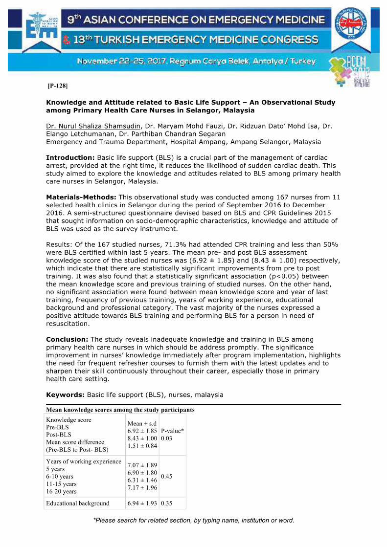
*Please search for related section, by typing name, institution or word.
[P-128]
Knowledge and Attitude related to Basic Life Support – An Observational Study among Primary Health Care Nurses in Selangor, Malaysia Dr. Nurul Shaliza Shamsudin, Dr. Maryam Mohd Fauzi, Dr. Ridzuan Dato’ Mohd Isa, Dr. Elango Letchumanan, Dr. Parthiban Chandran Segaran Emergency and Trauma Department, Hospital Ampang, Ampang Selangor, Malaysia Introduction: Basic life support (BLS) is a crucial part of the management of cardiac arrest, provided at the right time, it reduces the likelihood of sudden cardiac death. This study aimed to explore the knowledge and attitudes related to BLS among primary health care nurses in Selangor, Malaysia. Materials-Methods: This observational study was conducted among 167 nurses from 11 selected health clinics in Selangor during the period of September 2016 to December 2016. A semi-structured questionnaire devised based on BLS and CPR Guidelines 2015 that sought information on socio-demographic characteristics, knowledge and attitude of BLS was used as the survey instrument. Results: Of the 167 studied nurses, 71.3% had attended CPR training and less than 50% were BLS certified within last 5 years. The mean pre- and post BLS assessment knowledge score of the studied nurses was (6.92 ± 1.85) and (8.43 ± 1.00) respectively, which indicate that there are statistically significant improvements from pre to post training. It was also found that a statistically significant association (p<0.05) between the mean knowledge score and previous training of studied nurses. On the other hand, no significant association were found between mean knowledge score and year of last training, frequency of previous training, years of working experience, educational background and professional category. The vast majority of the nurses expressed a positive attitude towards BLS training and performing BLS for a person in need of resuscitation. Conclusion: The study reveals inadequate knowledge and training in BLS among primary health care nurses in which should be address promptly. The significance improvement in nurses’ knowledge immediately after program implementation, highlights the need for frequent refresher courses to furnish them with the latest updates and to sharpen their skill continuously throughout their career, especially those in primary health care setting. Keywords: Basic life support (BLS), nurses, malaysia
Mean knowledge scores among the study participants
Knowledge score Pre-BLS Post-BLS Mean score difference (Pre-BLS to Post- BLS)
Mean ± s.d 6.92 ± 1.85 8.43 ± 1.00 1.51 ± 0.84
P-value* 0.03
Years of working experience 5 years 6-10 years 11-15 years 16-20 years
7.07 ± 1.89 6.90 ± 1.80 6.31 ± 1.46 7.17 ± 1.96
0.45
Educational background 6.94 ± 1.93 0.35

*Please search for related section, by typing name, institution or word.
Diploma Bachelor Degree Master Degree PHD Others i.e. PMR/ SPM/STPM
7.83 ± 0.75 9.00 ± 0.00 0.00 ± 0.00 6.74 ± 1.75
Professional category Community Nurse Staff Nurse Sister Matron
6.59 ± 1.80 7.14 ± 1.87 6.00 ± 2.35 7.64 ± 1.45
0.08
Previous training Yes No
7.10 ± 1.77 6.46 ± 1.96
0.04
Frequency of training Never Once More than once
6.56 ± 1.92 7.04 ± 1.81 7.71 ± 1.41
0.15
Years of last training 1. BLS/AED * < 5 years * 6-10 years * 11-15 years * >15 years 2. ACLS * < 5 years * 6-10 years * 11-15 years * >15 years 3. PALS * < 5 years * 6-10 years * 11-15 years * >15 years 4.ALS * < 5 years * 6-10 years * 11-15 years * >15 years
7.20 ± 1.87 7.00 ± 1.66 6.57 ± 2.07 6.00 ± 0.00 7.60 ± 2.30 8.50 ± 2.12 8.00 ± 0.00 0.00 ± 0.00 7.67 ± 1.53 0.00 ± 0.00 0.00 ± 0.00 0.00 ± 0.00 7.67 ± 1.53 0.00 ± 0.00 0.00 ± 0.00 7.00 ± 0.00
0.40 0.46 0.48 0.78
Table shows the mean knowledge score among studied nurses for pre-BLS training (6.92 ± 1.85) and post-BLS
training (8.43 ± 1.00). In regards to their knowledge score, the results indicated that there are statistically
significant improvements between pre and post-BLS training. The distribution of mean knowledge scores among
the studied nurses categorised based on selected demographic characteristics (years of working experience,
educational background and professional category) and history of previous CPR training is shown in the table.
The scores were compared, showing significant association between the knowledge score of the studied nurses
and their previous training. The mean knowledge score was significantly higher in those who had previous CPR
training than those who had no training at all (7.10 ± 1.77 vs. 6.46 ± 1.96, p value < 0.05). Those who had
attended CPR training more than once had higher mean score than those who attended once or no training at all
(7.71 ± 1.41 vs.7.04 ± 1.81 vs. 6.56 ± 1.92). However this decay was not significant. In term of association
between mean knowledge score and other demographic characteristic of the participants, no statistically
significant associations were found.

*Please search for related section, by typing name, institution or word.
[P-130]
Acil Servislerde Sıcak Çarpması ve Hemşirelik Bakımı Emine Avlar, Bedia Gülen Bezmialem Vakıf Üniversitesi Tıp Fakültesi Hastanesi, Acil Tıp Anabilim Dalı, İstanbul Sıcak çarpması, yüksek derece ısı ve nemli ortamlarda vücudun 40°C’den fazla olması, homeostazi sürdürmemesi sonucu aşırı terleme yoluyla sıvı ve tuz kaybıyla ortaya çıkan bir durumdur. Özellikle sıcaklık ve nem oranının yüksek olduğu yaz aylarında sıkça rastlanmaktadır. Artan vücut sıcaklığı ile birlikte bitkinlik, kuru-sıcak-kırmızı deri, hızlı nabız, giderek azalan terleme, sıvı kaybı sonucunda oluşan bulantı kusma ve baş dönmesi belirtileri görülür. Dünya Meteoroloji Örgütü’ne (WMO) göre Türkiye’de her yıl hava sıcaklıklarının ortalamanın üzerinde olduğu kaydedilmiştir. Buna bağlı olarak acil servislere başvuru oranını arttırmaktadır. Çoğunluğu yaşlı olan bu hastalarda sıcak çarpması sonucu ciddi hasarlar görülmektedir. Sıcak çarpması ile acil servise başvuran hastalara ilk müdahale oldukça önemlidir. Hasta ilk olarak serin bir yere alınmalı ve sırt üstü yatırılmalıdır. Vital bulguları kontrol edilmeli, damar yolu açılmalıdır. Şok belirtileri gözlemlenmeli, var ise hastaya hemen şok pozisyonu verilmelidir. Vücutta sıvı kaybına bağlı sıvı tedavisine başlanmalıdır. Acil serviste soğutucu battaniye mevcut ise uygulanmalıdır, değil ise vücut ısısını en hızlı şekilde düşürecek soğuk su immersiyonu uygulanmalıdır. Ek olarak acil servis koşullarına göre vücut ısısını düşürmek için buz torbası masajı veya buzlu suya batırılmış havlu uygulanabilir. Hastanın tedavisi sırasında sık sık ateş monitörizasyonu takip edilmeli ve 38.5° C nin altına düştüğünde soğutucu tekniklerden uzaklaşarak pasif düşüş beklenilmelidir. Sıcak çarpmasından korunmak için mevsim şartlarına uygun terletmeyen hafif giysiler giyilmelidir. Güneş ışınlarının dik geldiği saatlerde, güneş altında kalınmamalıdır. Şapka, güneş gözlüğü ve şemsiye gibi koruyucu aksesuarlar kullanılmalıdır. Bol miktarda sıvı tüketilmelidir. Yaz döneminde sıcak çarpması ve alınması gereken önlemler ile ilgili bilinçlendirme seminerleri düzenlenmelidir. Anahtar Kelimeler: Sıcak çarpması, hemşirelik bakımı

*Please search for related section, by typing name, institution or word.
[P-131]
Düşme Tanısıyla Gelen Vakanın Yabancı Cisim Aspirasyonu Çıkması Mehmet Nur Geyik, Aynur Geyik, Evin Çim Sağlık Bilimleri Üniversitesi Gazi Yaşargil Eğitim ve Araştırma Hastanesi, Diyarbakır Yabancı cisim aspirasyonları (YCA) çocukluk döneminde daha çok görülmekle birlikte erişkin yaşta da karşılaşılabilen acillerdendir. Hayatı tehdit eden komplikasyonlara yol açabilirler. Asemptomatik olabileceği gibi, çok ciddi solunum yolu komplikasyonlarına hatta boğulma ve ölüme neden olabilmektedirler. Kazara yaralanmalar arasında ölüm nedenlerinde 4. sıradadır. Amerika’da yapılan çalışmalarda 4 yaş altındaki her 100000 popülasyonda 0.5 ölüm nedeni aspirasyondur. Çocukluk çağı travmalarının en sık nedeni yüksekten düşme olup travma sonrası morbidite ve mortalite halen önemini korumaktadır. Acil servislere solunum sıkıntısı ile başvuran ve ileri hava yolu girişimlerine ihtiyacı olan hastalarda zor hava yolu yönetimi ayrı bir hazırlık ve pratik gerektirir. Özellikle öngörülemeyen zor hava yolu yönetiminde karşılaşılan sıkıntılar acil servis ekibi ve hasta için zor anların yaşanmasına ve hatta hastanın kaybedilmesine sebep olabilir. Mevcut trakeal stenozun varlığı zor havayolu için başlıbaşına bir risk faktörü iken üzerine eklenen yabancı cisim aspirasyonu gibi ani solunum sıkıntısına neden olan olaylar acil ekibinin hazırlık ve pratiğini daha da önemli kılmaktadır. Düşme tanısıyla gelen vakalarda çok iyi anamnez almak gerekir. Sözkonusu vakalarda ihmal, istismar veya başka tanılarla karşılaşmak olasıdır. Vakamız 1 yaşında kız çocuğu, acil servise ailenin verdiği bilgilerle kanepeden düşme tanısıyla geldi. Genel durumu kötü, şuuru kapalı, siyanoze, göğüs duvarında interkostal retraksiyon mevcuttu. Kalp tepe atımı 80/dk, desatüreydi. Harici muayenede eritem, ekimoz izlenmedi. Asfiktik ve bradikardik hastaya CPR uygulanmasına başlandı. Göğüs basısı ile entübasyon hazırlığı yapıldı. Larengeskopide rima glottis girişini tıkayan yabancı cisim görülmesi üzerine aspire edilmeye çalışıldı ancak cisim aspire edilemedi. Alternatif havayolları için ilgili birimlerden konsültasyon istendi. Video larengeskop ile cismin havayolunu tıkadığı görüldü ve klemp ile çevre dokuya zarar vermeden çıkarıldı. Çıkarılan cismin vida olduğu görüldü. Anahtar Kelimeler: Zor entübasyon, video larengeskop, yabancı cisim aspirasyonu

*Please search for related section, by typing name, institution or word.
Yabancı Cismin video larengeskopta görülmesi

*Please search for related section, by typing name, institution or word.
[P-132] Metoclopramide Induced Dystonia with Pregnant Women Sümeyra Acar Kurtuluş, Burcu Yılmaz, Can Özen, Serkan Emre Eroğlu Umraniye Educational Research Hospital, Istanbul, Turkey Metoclopramide is a dopamine-2 receptor antagonist used for various gastrointestinal disorders. Standard management involves distontinuation of the drug and rapid iv or im administration of an anticholinergic or antihistaminic drug. Such as biperiden and chlorpheniramine (procyclidine, benzatropine). A 31 year old,24 weeks pregnant patient who took 10 mg metoclopramide (2*1) presented with pain and stiffness of the neck.She had no past medical history.She was alert on presentation.Her vital signs were blood pressure 100/70 mmHg,pulse rate of 90/minute,respiratory rate 20 per minute.On physical examination;she was conscious and anxious.Her neck was slightly arched backwards.There was no accumulation of saliva in her mouth and she could swallow.There were no signs of respiratory distress.Her laboratuary results were normal.A diagnosis of Metoclopramide induced acute dystonic reaction was thought and administered only physiological saline solution.Her dystonic symptoms subsided 7 hours later.She did not have any further dystonic symptoms.She was discharged 1 day later and was advised to avoid taking metocloropramide. Dystonia is a movement disorder characterized by involuntary, sustained or spasmodic contractions of muscle groups, resulting in twisting,repetitive and abnormal positions.Dystonia can be classified by the age of onset,body distribution, and etiology.Classification by etiology seperates the spectrum of dystonia into primary and secondary categories.Secondary dystonia typically arises from a specific underlying condition, such as exposure to dopamine receptor-blocking drugs.Metocloropromide is a dopamine receptor antagonist that blocks both D1 and D2 receptors in the central nervous system.When administered in higher doses, it also blocks serotonin receptors.Acute dystonias may be confused with partial seizures,tetanus,strychnine poisoning and electrolyte imbalances.The onset of the pharmacological action of iv,im and oral metoclopramide is respectively 1-3,30-60 minutes and 1-2 hours.Intravenous biperiden,anticholinergic agent or oral chlorpheniramine,antihistaminic agent are the options for the treatment of dystonia.If metoclopramide is not used at high doses, the risk of dystonia is reduced. Keywords: Metoclopramide pregnant patient

*Please search for related section, by typing name, institution or word.
[P-133]
Nursing Experience with Providing Wound Care for a Patient of Neck Stabbing Wound with Phrenic Nerve Injury Receiving VATS Diaphragmatic Plication Yi Ling Chung, Hua Yin Hsu, Chiu Hsia Hung Nursing Department, Tainan Municipal Hospital, Tainan city, Taiwan Objectives: Phrenic nerve injury due to neck stabbing injury is rare and needs emergent surgical treatment. Advanced surgical method with VATS diaphragmatic plication is applied but few nursing experiences is reported. Material-Methods: The 76-year-old female patient had a neck stabbing injury by one scissor in 2015. Removal of the scissor was performed in emergent surgery. Days later, VATS diaphragmatic plication was performed due to phrenic nerve injury induced diaphragm palsy. Nursing in pain reduction, respiratory care and the relief of anxiety was provided. Results: This kind of patient is quite critical and nursing measures are required. This case report describes the experience in pain reduction, respiratory care and the relief of anxiety. A holistic assessment conducted from October 18, 2016 to October 30, 2016 enrolled the major nursing with systemic observation, active care and interview technique. Conclusions: Nursing care in pain reduction, respiratory care and the relief of anxiety in this case that receiving advance surgical management can help the early rehabilitation and mental reconstruction. Keywords: Phrenic nerve injury, nursing, neck stabbing wound

*Please search for related section, by typing name, institution or word.
[P-135]
Acute Brachial Artery Occlusion and Usage of Doppler Ultrasound at An Emergency Department Barış Alper, Hayrullah Yönak, Sümeyra Acar Kurtuluş, Serkan Emre Eroğlu Department of Emergency Medicine, University of Health Sciences Umraniye Training and Research Hospital, Istanbul, Turkey Introduction: Acute arterial occlusions generally occur as a result of thrombosis on deteriorated vessel wall due to atherosclerotic plaque or the embolus. We wanted to emphasize that upper extremity acute arterial thrombosis related to the chronic usage of crutch can easily be diagnosed at ED with doppler ultrasound scan. Case: 63-year-old male patient applied to the ER with sudden onset pain on his right arm and numbness. In his anamnesis, it was mentioned that his complaints had started thirty minutes ago; he had both hypertension and atrial fibrillaton stories; he took oral antihypertensive pills; he suffered from the loss of strenght in his lower extremities because of poliomyelit, which led him to use crutch. During the physical examination, it was observed that the patient was sweating coldly, pale and agitated due to his pain. His right arm, which was causing him pain at that moment, was pale as well with no radial pulse palpated. Moreover, a significant atrophy was seen in his lower extremities. His vitals were checked as, tension: 210/110 mm/Hg, SPO2 (left arm): %96. Furthermore, atrial fibrillation was spotted in his ECG and the patient was prediagnosed with acute arterial occlusion. He had doppler usg scan in the emergency department and an occlusion was spotted in his right brachial artery, with no current/flow.Given Aspirin and DMAH, the patient was interned to cardiovascular surgery for interventional purposes. Result: Although CT Angiography is the gold standard test for acute arterial trombosis, Doppler US can also be used to make a diagnosis.Ultrasound's superiorities to CT Angiography is that it is cheap and easy-to-access in ER, involves no radiation or contrast medium and gives us the the opportunity to diagnose faster.The delay related to the diagnosis and treatment in acute arterial occlusion cases,puts a clinical picture with high mortality and morbidity,which threatens the well being of extremity and the patient's life. Keywords: Doppler US, acute arterial thrombosis, brachial artery

*Please search for related section, by typing name, institution or word.
Brachial artery US

*Please search for related section, by typing name, institution or word.
[P-136]
Urethral Swelling Around the Foley Catheter Barış Alper, Sümeyra Acar Kurtuluş, Serkan Emre Eroğlu Department of Emergency Medicine, University of Health Sciences Umraniye Training and Research Hospital, Istanbul, Turkey Introduction: Foley catheter is still used for urinary catheterization. Even though it is quite easy to apply, it is also a painful procedure. During the foley catheterization, there may be complications just like any other intervention. Besides asymptomatic bacteriuria and urinary infection; hematuria, obstruction, kidney stones, hemorrhage due to swelling of the cathater before it reaches the bladder, stenosis in urinary tract and rupture can develop as an example for those complications. Case Report: 71-year-old male patient applied to the emergency department with hematuria coming as a leak from the edge of his foley catheter, suprapubic pain and swelling.His vitals were checked as tension: 170/80 mmHg, pulse 105 beats per minute.Hematuria leaking from the catheter at the tip of the urethral meatus was observed.Taking the anamnesis, it was found out that the patient had benign prostate hypertrophy and applied to the emergency room about two hours ago, complaning not being able to urinate and discharged after urinary catheterization. During the ultrasound scan performed in the ER, it was seen that his bladder was filled with urine and the balloon of the foley catheter was swollen in the urethra. Catheter was pulled off after 7mm liquid was drained away from its balloon. A new foley catheher was placed and the urine inside the bladder was slowly drained after confirming that the balloon was swollen inside the bladder with an ultrasound scan. The patient was kept under observation for hemogram tests and the risk of hemorrhage. Conclusion: Placement of a foley catheter indications, contraindications and complications must be known thoroughly by the emergy medicine doctors. It must be placed and displaced by the experienced staff. After the catheter's placement, urine output has to be seen, its place should be verified with an ultrasound scan if possible and lastly, patient should be told about the future complications. Keywords: Bladder, foley catheterization, ultrasound

*Please search for related section, by typing name, institution or word.
Prostat Ultrasound

*Please search for related section, by typing name, institution or word.
[P-137]
Elbow Extension Test as A Simple Test to Diagnose Fractures; A Case Resport Erkan Gunay, Özgür Çevrim Sisli Hamidiye Etfal Training and Research Hospital Department of Emergeny Medicine, Istanbul, Turkey Elbow injuries are commonly seen in emergency care, accounting for 2-3% of emergency department attendances. Only a minority of patients with such injuries have a fracture. The elbow extension test has been proposed as a simple test to rule out the need for radiography. The seated patient, with exposed and supinated arms, is asked to flex their shoulders to 90 degrees and then fully extend and lock both elbows. Pain on full elbow extension is considered as positive for the test. Computed tomography was reccomended as a further evaluation imaging study for elbow extension test positive trauma patients who present with no apparent fracture on x-ray imaging. A 52-year-old male referred to our emergency department with recent left elbow trauma. Xray was ordered and no fracture findings were present. Elbow extension test was positive for left elbow and systemic evaluation was otherwise normal. Computed tomography was ordered and left radius head fracture was revealed (figure 1). Patient was discharged with long arm cast and orthopedic follow up. Patients with recent elbow injury who cannot fully extend their elbow should be evaluated with advanced radiographic imaging. Elbow extension test is a simple test for emergency department that will be useful for proper management of trauma patients. Keywords: Elbow extension test, trauma, imaging
figure 1

*Please search for related section, by typing name, institution or word.

*Please search for related section, by typing name, institution or word.
[P-138]
Basilar Artery Aneurysm in Dizziness Etiology: An Atypical Case Detected by Non-Contrast Cranial Ct Hasan İdil, Turgay Yılmaz Kılıç, Feriyde Çalışkan, Özge Duman Atilla, Yeşim Eyler, Necmiye Yalçın Ocak Tepecik Training and Research Hospital, Department of Emergency Medicine, Izmir, Turkey Introduction: Dizziness is a very common condition, especially among older patients. The majority of this caused by vestibular dysfunction, including benign paroxysmal positional vertigo. Psychiatric disorders, infectious processes and cerebrovascular events may also cause dizziness. In this case we aimed to present a rare cause of this. Case: A 75-year-old male patient presented complaints of dizziness, nausea and vomiting. The patient's vital signs and blood glucose level were within normal limits. He had no additional features other than hypertension at his medical history. Electrocardiogram revealed no arrhythmia or ischemia signs. There was no significant finding in the routine laboratory values. There was no neurological abnormality other than horizontal nystagmus on the right side of the patient and mild disarti. No evidence of hemorrhage or ischemia was detected in non-contrast cranial CT, but aneurysm-like appearance of 27x14 mm in the proximal part of the basilar artery was observed. Subsequently, this diagnosis was confirmed by cerebral CT angiography (Figure 1). Patient was admitted to the neurosurgery service. There were no ischemia findings in his cranial MRI. The patient whose complaints reduced was referred to the interventional radiology unit by planning an endovascular procedure. Conclusions: The basilar artery aneurysm should be kept in mind in the differential diagnosis of patients presenting emergency department with dizziness. Vascular structures should also be carefully examined in non-contrast cranial CT. Keywords: Basilar artery aneurysm, computed tomography, dizziness
Figure 1. The basilar artery aneurysm in non-contrast cranial CT and CT angiography.

*Please search for related section, by typing name, institution or word.

*Please search for related section, by typing name, institution or word.
[P-139]
Evaluation of The Requirement of Body Screening Tomography in Head Trauma Patients Iffet Yasaran1, Ali Karakus1, Guven Kuvandik1, Ahmet Sebe2, Zeynep Kekec2 1Mustafa Kemal University, Faculty of Medicine, Department of Emergency Medicine, Hatay, Turkey 2Cukurova University, Faculty of Medicine, Department of Emergency Medicine, Adana, Turkey Objectives: We studied patients with head trauma and without additional lesions in the body; It is important to be the first study to evaluate the head trauma subtype and findings in TVBT. We aimed to evaluate head trauma cases by using TVBT and attempts have been made to determine how much TVBT is required to identify other body injuries that may accompany the patients. Method: Our study was retrospectively performed with 198 patients who applied to our emergency department after head trauma between 01.01.2012-31.12.2016, who had lesion on brain tomography, no additional examination findings and applied TVBT. The age, sex, type of lesion in CT, GCS status, TVBT findings of the patients were evaluated. The data were evaluated in the SPSS 18 version. Result: 85.4%of the patients in our study were male and the average age was 25. 7years.The most common cranial CT finding in our study was fracture, followed by parenchymal hemorrhage. In our study,67% of patients were below GCS 8. In our study,78 patients (39.4%)had additional trauma. The most common additional lesion in our study was contuzyon in thoracic. There was no relation between age and the presence of additional trauma in CT in our study (p>0.05).The mean age of the patients with cervical injuries determined in CT was significantly higher (p<0.05);there was no correlation between age and chest and abdominal injury (p>0.05).In our study,there was no relationship between gender and the presence of additional injury in CT(p>0.05).Cranial fractures and foreign bodies were found to be significantly lower in patients with thoracic injuries(p<0.05).In our study,the incidence of cervical injuries was significantly higher in the patient who had contusion in CT(p<0.05). There was no correlation between other cranial lesions and additional injury area(p>0.05). Conclusion TVBT should be recommended in patients whose clinical evaluation is incomplete. It is an important diagnostic tool in the diagnosis of many pathologic lesions, mainly intrathoracic lesions. Keywords: Cranial trauma, whole body CT, emergency department

*Please search for related section, by typing name, institution or word.
[P-140]
Spontaneous Vertebral Fracture: Case Report Mehmet Esen Emergency Department, Medical Faculty, Gaziosmanpaşa University, Tokat, Türkiye Coexistence of back and leg pain is usually the result of root irritation. The presence of a new onset of severe pain, the progression of the present pain, the sensation of loss of sensation (tingling, felting, numbness), radiculopathy, early warning signs of the upper extremity or lower extremity. Prostate cancer makes both osteoblastic and osteolytic vertebral metastases. We will share the lumbar vertebral fractures as the cause of back pain in this patient with sudden onset and not the neurological deficit, advanced age prostate cancer. Case: An 80-year-old male patient has diagnosed with prostate cancer 2 months ago and was admitted to the emergency room with complaints of back and leg pain. The patient's vitals were stable and GCS:15. Physical examination showed no pain, sensation, and pathological reflexes. Loss of height in L1 and L3 was detected on the patient's lumbar graph. Computed tomography showed changes in L1 and L3 with collapsing fracture and metastasis. Discussion: People with a primary malignant tumor or who are older than 50 years should be suspected of metastasis in spinal pain without trauma. Unfortunately, pain symptoms at the first stage are more nonspecific and can not easily be separated from the disc disease. Mechanical pain generally exacerbates with activity and passes through bed rest. However, non-mechanical pain is continuous, does not decrease, does not rest, increases the nights. Although the case is considered as a chronic case, it is obvious that it will not meet the forces that this corpus can carry, due to bone loss in corpuscular over time. As a result, may develop in compression fracture. Even in malignant prostate cancer metastases, lifespan is around 3 years. Early diagnosis and treatment will increase the quality of life of the patient without late neurological findings such as incontinence or muscle loss. Keywords: Spontaneous, vertebra, fracture

*Please search for related section, by typing name, institution or word.
Figure: Spontaneous vertebral fracture

*Please search for related section, by typing name, institution or word.
[P-142]
Nasal Septal Perforation Based on The Usage Of Inhale Corticosteroid Nazlı Görmeli Kurt1, Songül Araç2, Murat Orak2, Mehmet Üstündağ2 1State Hospital, Batman, Department of Emergency, Batman, Turkey 2Department of Emergency, Faculty of Medicine, Univesity of Dicle, Diyarbakır, Turkey Introduction: Nasal septal perforation is not a case that we shall encounter frequently in emergency situations. We accidentally encounter the nasal septal perforation as a result of generally as a result of trauma, septal surgery, decongestant and cocaine, in our bronchiectasis patient that uses isolated inhaled corticosteroids (ICSs). Case: Male patient at the age of 48 applied to our emergency department in Batman Regional State Hospital with the complaint of difficulty in breathing. Our patient is a bronchiectasis patient and uses ICSs for 5 years. On the nasal inspection as a result of patient’s complaints regarding a whistle like sound comes out of his nose recently while he is breathing, we encounter nasal septal perforation in our patient. Conclusion: In chronical pulmonary diseases such as bronchiectasis, asthma, chronic obstructive pulmonary disease (COPH), ICSs are used since long years. Additionally, to ICSs in patients who use intranasal corticosteroids (INCSs) there are cases where nasal septal perforation is encountered, but this case seen based on usage of isolated ICSs indicates us that usage ICSs for a long term is not so innocent. In order to explain the background laying mechanism and to take protective precautions we shall think that we got to study on this subject intensively. Keywords: Nasal perforation, inhaled corticosteroids

*Please search for related section, by typing name, institution or word.
nasal septal perforation based on the usage of inhale corticosteroid

*Please search for related section, by typing name, institution or word.
[P-143]
Dandy Walker Malformation in An Adult Asymptomatic Case Nazlı Görmeli Kurt1, Songül Araç2, Murat Orak2, Mehmet Üstündağ2 1State Hospital, Batman, Department of Emergency, Batman, Turkey 2Department of Emergency, Faculty of Medicine, Univesity of Dicle, Diyarbakır, Turkey Introduction: Dandy-Walker malformation (DWM) is a congenital anomaly that is encountered rarely (1/30.000) and its aetiology is barely known, characterized together with cerebellar vermis hypoplasia and corpus callosum agenesis, ventricles’ cystic extension as a result of Magendie and Luschka foramina dysgenesis on the fornix of 4th ventricle. Clinically at babyhood or in future childhood period hydrocephaly and intracranial pressure increase findings are encountered and reveal itself with ataxia or it may be possible to live without any of these symptoms. In living cerebellar agenesis adult patient’s neurological findings are not apparent. Case: Male patient at the age of 31, as a result of an intensifying headache for the last 2 days which rarely exists for the last 4 years applied to Batman Regional State Hospital Emergency Service. Since the childhood of our patient whose proficiency is teacher until today there is no history of any neurological diseases and any genetic diseases in his family. His detailed neurological examination is natural. On the computerized brain tomography (CBT) of the patient inferior cerebellar vermis agenesis and cystic dilatation on the 4th ventricle was encountered. And the patient is followed with the diagnosis of DWM. Conclusion: DWM is generally diagnosed at early childhood period (around the age of 1) and may extend rarely until the adulthood period. In our case clinical presentation was solely followed by a headache. With the DWM which is encountered in adulthood rarely, we would like to attract the attention that a symptomless life is possible to lead. Keywords: Dandy-walker malformation, asymptomatic
dandy walker malformation in an adult asymptomatic case

*Please search for related section, by typing name, institution or word.

*Please search for related section, by typing name, institution or word.
[P-144]
Widened Aortic Contours on Non-Contrast Computed Tomography: Sign of Acute Aortic Dissection Özcan Yavaşi1, Mehmet Altuntaş1, Ercan Nalbant1, Ensar Topaloğlu1, Ozan Karakişi2 1Recep Tayyip Erdogan University, Faculty of Medicine, Department of Emergency Medicine, Rize, Turkey 2Recep Tayyip Erdogan University, Faculty of Medicine, Department of Cardiovascular Surgery, Rize, Turkey Introduction: Acute aortic dissection should be ruled out in the differential diagnosis of chest pain. Although there are some clues of dissection on chest radiography such as widened mediastinum, widening of aortic knob, different diameters between ascending and descending aorta, blurring of the thoracic aorta, and calcium sign, computed tomography (CT) angiography remains the gold standard diagnostic modality for diagnosis of acute aortic dissection. We present a patient with the diagnosis of thoracoabdominal aortic aneurysm, that aroused the suspicion of dissection on non-contrast CT and finally diagnosed as acute dissection. Case: A 80-year-old female presented to the emergency department with complaint of dyspnea and retrosternal chest pain. She had been followed-up for thoracoabdominal aortic aneurysm since 3 years. Her vital signs were: Blood pressure, 130/70 mmHg on right side and 120/70 mmHg on left side; pulse rate, 92 bpm; respiratory rate, 22 breaths/min; and oxygen saturation, 92%. Her breath sounds were hoarse and subfebrile fever of 37.6 °C. She had atrial fibrillation on the electrocardiogram. Chest X-ray showed widened mediastinum and reticulonodular infiltration. Complete blood cell count and blood chemistry were almost normal. She was ordered non-contrast thorax computed tomography (CT). She did not have consolidation. Ascending aorta and descending aorta were measured 6.12 X 6.37 cms, and 3.64 X 3.37 cms at widest portions, respectively. This was compared to previous 3-years-ago-CT that was measuring 5.76 X 6.23 cms and 2.91 X 3.25 cms. With suspicion of aortic dissection contrast enhanced CT angiography was ordered that revealed Type I acute aortic dissection. The patient was operated by cardiovascular surgeons and discharged two weeks later. Conclusion: Patients who were diagnosed as aortic aneurysm should have contrast enhanced CT whenever possible. If non-contrast CT shows widened aortic contours compared to previous studies, this should alert emergency physicians for acute dissection. Keywords: Acute aortic dissection, computed tomography angiograpy, non-contrast computed tomography

*Please search for related section, by typing name, institution or word.
[P-145]
Role of Cardiac Ultrasound in Common Neurological Disorder Sabariah Faizah Jamaluddin1, Julina Mohammad Noor2, Anh Hong Do1 1Emergency and Trauma Department, Sungai Buloh Hospital, Malaysia 2Faculty of Medicine, Universiti Teknologi MARA, Malaysia Introduction & Objective: Critically ill patients commonly have more than one pathology. However, in the case of neurologic disorder, cardiac ultrasound is not part of usual investigation. The relationship between brain and cardiac function varies according to the pathology. We present two cases of neurological disorders, subarachnoid haemorrhage and cerebral infarct, that has relationship with cardiac pathology. Materials & Methods: Case 1: A 26-year-old woman complaint of headache 3 days prior, presented to emergency department (ED) with convulsion for 5 minutes followed by loss of consciousness at home. On arrival, she was unconscious. Her vitals were: pulse rate 145/min, BP 134/92mmHg, SpO2 90% room air, GCS 3. Lungs auscultation revealed bilateral crepitations. Pupils were 4 mm bilaterally and sluggish. Post intubation endotracheal suctioning showed copious amount of pink frothy fluid. Cardiac ultrasound followed by CT scan was performed. Case 2: A 44-year-old man was transferred from a district hospital with provisional diagnosis of stroke. He initially presented with central chest pain and shortness of breath. His blood pressure was elevated at 160/100mmHg. 30 minutes later he lost consciousness with GCS of 5 (E1V1M3), and unequal pupils. His blood pressure dropped after intubation and noradrenaline were commenced. Cardiac ultrasound followed by CT scan was performed. Results: Case 1: Cardiac ultrasound showed poor cardiac contractility and CT brain showed aneurysmal subarachnoid haemorrhage. Case 2: Cardiac ultrasound showed cardiac tamponade with evidence of aortic dissection at the root of the aorta. CT brain showed acute diffused cerebral infarction in the bilateral hemispheres. Conclusion: Although the association between neurology and cardiac pathology is well established, cardiac ultrasound is not routinely performed in the acute stage of neurological insults. These cases highlight the importance of performing multi-system bedside ultrasound in critically ill patients as it has bearing on subsequent management of the patients. Keywords: Ultrasound, neurology

*Please search for related section, by typing name, institution or word.
[P-146]
FAST scan in Children: Worthwhile or Futile? Sabariah Faizah Jamaluddin, Shamini Sangasamy, Julina Mohammad Noor, Shamila Mohammad Ali Department of Emergency and Trauma, Sungai Buloh Hospital, Malaysia Objectives: Abdominal injury is the third most common injury in paediatric trauma. Although routine FAST (focused assessment with sonography for trauma) scan is a well-accepted practice in adult populations, skepticism remains with regards to paediatric age group. There are studies that shows FAST scan in paediatric is not sensitive enough to rule out abdominal injury. Organ specific injury diagnosed by point of care ultrasound is almost unheard off. We describe a case of liver injury picked up by bedside ultrasound in emergency department (ED), in a patient with no significant clinical findings. Material-Method: An 8 years old boy was brought to ED complaining of vomiting. He was cycling and fell into 5 feet drain and landed on his abdomen on top of a wooden beam, 10 hours prior. He initially complained of abdominal pain after the injury, but the pain has resolved before arriving to ED. He was haemodynamically stable with pulse rate 80/min and BP 107/65mmHg. Per abdomen was soft and non-tender. Results: FAST scan did not show any free fluid. However, an area of heterogenous hyperechogenecity was seen within the liver parenchyma, suspicious of liver injury. CT abdomen showed grade IV liver laceration and grade III right kidney injury. Discussion and Conclusions: FAST scan in paediatric trauma has been reported to have poor sensitivity as low as 23%. A recent study also showed the use of FAST compared with standard care only did not improve clinical care. This case may be an exception. This child which was well with no abdominal pain or tenderness, would have been discharged home. Instead, liver injury was detected during routine scan, which changed the course of management for this patient. Perhaps we should not be too quick to discard FAST. Keywords: FAST ultrasound, blunt abdominal injury, paediatric

*Please search for related section, by typing name, institution or word.
[P-147]
Hematuria and Flank Pain in The Emergency Department– A Common Presentation of a Rare Condition Sameera Ganti, Sohil Equbal Pothiawala Department of Emergency Medicine, Singapore General Hospital, Singapore Introduction: Flank pain is a common presentation at most emergency departments (ED). This symptom coupled with a history of hematuria, raises the suspicion of a renal/ureteric calculus among most clinicians. Bedside ultrasonography (US) in the ED has become a mainstay in the evaluation of these patients. Case: This case describes a 51-year-old male who presented to the ED for recurrent flank pain and hematuria. He had previous surgeries for appendicectomy and cholecystectomy. His vital signs were stable. On physical examination he was noted to have tenderness over the right flank. Bedside ultrasonography in the ED demonstrated a cystic lesion in the renal area with echogenicity within the cyst. Patient was admitted to urology department for further evaluation and management. Inpatient CT Urography revealed an appendiceal stump mucocele. Patient was transferred under care of the surgical service and exploratory laparotomy was done, which revealed a large 10cmx8cm retrocecal mucinous lesion, likely arising from the appendiceal stump. Discussion: Appendiceal stump mucoceles are a rare entity, with only 3 previous reported cases in literature. They may present with a variety of non-specific symptoms. The mainstay of treatment is as surgical excision and histological diagnosis. A third of these tumors can be malignant and early evaluation and prompt treatment may prevent complications such as rupture and pseudomyxoma peritonei. Conclusion: This case highlights the importance of the utility of bedside ultrasound in patients presenting to the ED with abdominal pain. Appendicial stump mucocele should be considered as a differential diagnosis. Further evaluation prompted by bedside US in the ED lead to expedited assessment and appropriate patient management, thus improving patient outcome. Keywords: Flank pain, bedside ultrasound, appendiceal mucocele

*Please search for related section, by typing name, institution or word.
[P-148]
Batın İçi Kitle Mesane ile Karışır mı? Selim Yurtsever, Gürkan Altuntaş, Özcan Yavaşi, Ensar Topaloğlu, Ercan Nalbant, Gökhan Ersunan Rize Recep Tayyip Erdoğan Üniversitesi Eğitim ve Araştırma Hastanesi, Rize Giriş: Batın içi kitleler bası etkisine bağlı semptomlar oluşturabilir. Bu bildiride ilk bakıda mesane olduğu düşünülen, içi sıvı dolu dev batın içi kitlenin basısına sekonder gelişen hidroüreteronefroz olgusunu sunmayı amaçladık. Olgu: 92 yaş erkek hasta acil servise yakınları tarafından getirildi. Alzheimer nedeniyle kendisiyle iletişim kurulamayan hastanın yakınlarından hastanın huzursuz olduğu ve karın ağrısı olduğu öğrenildi. Zaman zaman benzer şikayetleri olan hastaya sonda takılıp idrar çıkışı sağlandığında rahatladığı öğrenildi. Hastanın karnında göbek altında inspeksiyonla görülebilen bombelik yapan bir kitle vardı. Palpasyonda mesane olduğu düşünülen kitleye acil serviste ultrasonla bakıldığında içi sıvı dolu 15*15 cm büyüklüğünde, lokalizasyon ve görünüm itibariyle mesane olduğu düşünülen görüntü izlendi. Hastada glob vezikale düşünüldü. Hastaya idrar sondası takıldı ve sadece 50 cc idrar çıkışı oldu. Tekrarlayan sonda işlemlerinde de idrar çıkışı olmadı. Bunun üzerine hastaya batın bilgisayarlı tomografi(BT) çekildi. BT de mesane lokalizasyonunda 15*15 cm büyüklüğünde içi sıvı dolu kitle görüldü. Kitle mesaneye ve üretere bası yapıyordu. Hastanın sağda grade 4 hidronefrozu vardı. Üreter genişliği proksimal kesimlerde 4 cm ye ulaşıyordu. Hasta ileri tetkik ve tedavi amaçlı üroloji servisine interne edildi. Sonuç: Üreterohidronefroz sıklıkla tıkayıcı taş hastalığına ve prostat hastalıklarına bağlı olduğu gibi kitle basısına bağlıda gelişebilmektedir. Bu vakada olduğu gibi içi sıvı dolu bir kitle glob vesikale ile karışabilmektedir. Kitle eğer kısmi bası yapıyorsa BT gibi ileri görüntüleme yapılmadığında gözden kaçıp tanının gecikmesine yol açabilir. Anahtar Kelimeler: Hidronefroz, abdominal kitle

*Please search for related section, by typing name, institution or word.
15*15 cm kitle

*Please search for related section, by typing name, institution or word.
[P-149]
Negative Diffusion-Weighted Images in Acute Ischemic Stroke Yeşim Eyler, Necmiye Yalçın Ocak, Turgay Yılmaz Kılıç, Murat Yeşilaras, Hasan Idil, Orkun Ünek, Gökçe Özbay Yenice, Altuğ Cengiz Tepecik Training and Research Hospital, Izmir, Turkey Introduction: In the guidelines, diffusion-weighted magnetic resonance imaging (DWI-MRI) is recommended to diagnose stroke (1). The sensitivity of DWI-MRI in anterior circulation stroke within 48 hours is 98%, whereas for posterior circulation strokes it is only 68.4% and 80.6% within 24 and 48 hours, respectively (2). We report a case of ischemic stroke with subtle symptoms and a normal initial DWI-MRI, who developed ischemic lesions on control DWI-MRI. Case: A 75-year-old male was brought to the emergency department after a fall at home two hours ago. His vital signs were unremarkable except for a blood pressure of 147/87 mmHg and a respiratory rate of 22/min. He reported using acetylsalicylic acid and antihypertensive medication regularly. On physical examination, he had frontoparietal soft tissue swelling and slight left-sided hemiparesis due to a prior stroke. The patient seemed indifferent to his surroundings and it was hard to communicate with him because of his hearing impairment. The electrocardiography showed atrial fibrillation, but no ST-T segment changes. Finger-stick glucose level was 95 mg/dL. The cranial computed tomography and laboratory parameters were unremarkable. The DWI-MRI, which was ordered to exclude ischemic stroke, was negative (Figure-1a). The patient and his relatives could not delineate the extent of the pre-existing stroke sequelae, so the diagnosis of a new ischemic stroke could not be excluded. Four hours later, control DWI-MRI revealed restricted diffusion in the right middle cerebral artery territory (Figure-1b). The patient was admitted to the neurology ward. Discussion: Patients with posterior fossa, brain stem or lacunar lesions might have subtle symptoms, and initial DWI-MRI imaging may seem normal. Of note, patients with negative DWI-MRI do not have a better prognosis than patients with a lesion (3). Patients should undergo detailed and repetitive neurological examination, and ischemic stroke should be considered even if the DWI-MRI is negative. Keywords: Diffusion-weighted magnetic resonance imaging, stroke

*Please search for related section, by typing name, institution or word.
Figure 1.
1a) The first DWI-MRI was normal. 1b) Control DWI-MRI revealed restricted diffusion in the right MCA
territory (arrows).

*Please search for related section, by typing name, institution or word.
[P-150]
Vakum Fenomeni mi? Zeynep Karakaya, Fatih Esad Topal, Serkan Bilgin, Pınar Yeşim Akyol, Umut Payza Department of Emergency Medicine, İzmir Katip Çelebi University Atatürk Training and Research Hospital, İzmir Giriş: Vakum fenomeni vücutta intervertebral boşluklarda gaz birikimi nedeni ile ortaya çıkan bir durumdur. Bu gaz görüntüsü çekilen grafilerde radyolüsen alan olarak görülebilmektedir. Etiyolojide en sık sebep vertebral disk dejenerasyonu olarak bilinmektedir. Mekanizması tam olarak bilinmemekle beraber acil servise bel ağrısı nedeniyle gelen yaşlı hastalarda akılda tutulması gereken bir durumdur. Olgu: 89 yaşında bayan hasta aynı seviyeden sırt üstü düşme şikayeti sonrası kliniğimize başvurdu. Öyküsünden düşme öncesi de zaman zaman bel ağrılarının olduğu ve düştükten sonra bu ağrısının daha da şiddetlendiği öğrenildi. Özgeçmişinde geçirilmiş SVO dışında başka bir özellik yoktu. Fizik muayenesinde ciltte yüzeyel abrazyon, lomber 3-4-5 seviyesinde ve sol kalçada palpasyonla hassasiyet mevcuttu. Bilateral babinski refleksi negatifti. Her iki alt ekstremite motor ve duyu muayenesi olağan olarak değerlendirildi. Hastaya pelvis ve lumbosakral grafi ve BT çekildi. Çekilen lumbosakral grafide L5-S1 intervertebral aralıkta daralma ve ince bir çizgi şeklinde radyolüsen alan (resim1) görülmüştü. LumbosakralBT kesitlerinde ise lomber vertebralarda osteoporoz izlenmekteydi. Tartışma: Vakum fenomeni vucütta normalde gaz olmaması gereken intervertebral bir bölgede enfektif olmayan gaz birikimi olmasıdır.Mekanizması kesin olarak bilinmemekle beraber intervertebral disk mesafesinde biriken bu gazın %92 si azot,geri kalanı oksijen ve karbondioksittir.Mikro organizmalara bağlı piyojenik diskitiste,bening vertebral kompresyon fraktürlerinde de görülebildiği gibi multiple myelom gibi bazı malignitelere bağlı kompresyon fraktürlerinde de görülebileceği bildirilmiştir. Morishita ve ark.yapmış olduğu çalışmada vakum fenomenisi bulunan hastaların mevcut olan bel ağrısının genellikle sabahları şiddetlendiği,ayağa ani kalkma ve hava değişimlerinde de ağrının arttığı tespit edilmiştir. Lomber disk hernisi olan hastalar üzerinde yapılmış bir çalışmada ise vakum fenomeninin bu grup hastaların az bir kısmında görülebildiği ve biriken bu gazın kist yapısına dönerek sinir kökü basısı sonucu bel ağrısına neden olduğu belirtilmiştir. Sonuç: Acil servislere özellikle yaşlı ve bel ağrısı şikayeti ile başvuran ve disk dejenerasyonu olan hastalarda çekilen grafi ve tomografilerde vakum fenomenine rastlanabileceği ve bu hastalarda vakum fenomeninin kitle etkisi yaparak ağrı yapabileceği akılda tutulmalıdır. Anahtar Kelimeler: Vakum fenomeni, yaşlı hasta, bel ağrısı

*Please search for related section, by typing name, institution or word.
vakum fenomeni

*Please search for related section, by typing name, institution or word.
[P-151]
İlginç Bir Enfeksiyon: Ayakta Lenfanjit Atakan Savrun1, Ali Aygün5, Şeyda Tuba Savrun2, Emre Gökçen3, İbrahim Çaltekin4, Nihat Danlı6
1Ordu Üniversitsi Tıp Fakültesi Acil Tıp Anabilim Dalı, Ordu 2Atatürk eğitim ve araştırma hastanesi Acil Tıp Anabilim Dalı, Ankara 3Malatya Devlet Hastanesi, Acil Servis, Malatya 4Bozok üniversitesi Tıp Fakültesi Acil Tıp Anabilim Dalı, Yozgat 5Ordu eğitim ve araştırma hastanesi Acil Tıp Anabilim Dalı, Ordu 6 Dr. Nafiz Körez Sincan devlet hastanesi Acil Servis, Ankara
Lenfanjit; grup A streptokokların lenf nodlarında neden olduğu iltihabının genel adıdır. Travma yâda doku erezyonu ile patojenlerin lenf bezlerine migrasyonuna kolaylaşır. Lenf bezlerinin lokalizasyonu boyunca ilerleyen ciltte pembe, hassas çizgiler şeklinde görülebilir. Bu olguda iki gün önce ayağına yabancı cisim batması sonucu lenfanjit gelişen olgu sunulmaktadır. Olgu: Yirmi beş yaşındaki erkek hasta ayakta kızarıklık ve şişlik şikâyeti ile acil servise başvurdu. Öyküsünde iki gün önce ayağına yabancı batması olduğu ve sonrasında ayak parmağından başlayıp kasık bölgesine kadar ilerleyen ağrı, şişlik, kızarıklık ve ısı artışı oluşmuş. Beraberinde ateş, üşüme ve titreme gelişmesi sonucu kliniğimize başvurdu. Fizik bakıda hasta sol ayak ikinci parmaktan başlayıp sol inguinal bölgeye kadar uzanan 2cm genişlikte lineer, düzgün sınırlı hiperemik ve hassas lezyon mevcut idi. Diğer sistem muayenesi doğaldı. TA 110/80mmHg, nabız 103/dakika, ateş 37.9 derece idi. Beyaz kure (BK): 8600/dL, C-reaktif-protein (CRP): 0.45 mg/dL bulundu. Hastada lenfatjit olduğu saptandı. Amoksisilin klavulonik asit 2x1 ve parasetamol 3x1 ile taburcu edildi. Sonuç: Selülit, ürtiker, fronkül, karbonkül gibi hastalıklar ciltte kızarık lezyonlara neden olurlar. Lenfanjit cilt bütünlüğünün bozulduğu travma gibi durumlarda ortaya çıkar. Bu olguda da olduğu gibi lezyonların cizgisel bir hat boyunca devam eden kızarık, ağrılı, şiş ve ısı artışının olması ile diğer hastalıklardan ayrılır. Anahtar Kelimeler: Travma, lenfanjit, ağrı

*Please search for related section, by typing name, institution or word.
[P-152]
Atipik Yerleşimli Skalpte Zona Atakan Savrun1, Şeyda Tuba Savrun2, Emre Gökçen3, İbrahim Çaltekin4, Ali Aygün5, Nihat Danlı6
1Ordu Üniversitsi Tıp Fakültesi Acil Tıp Anabilim Dalı, Ordu 2Atatürk eğitim ve araştırma hastanesi Acil Tıp Anabilim Dalı, Ankara 3Malatya Devlet Hastanesi, Acil Servis, Malatya 4Bozok üniversitesi Tıp Fakültesi Acil Tıp Anabilim Dalı, Yozgat 5Ordu eğitim ve araştırma hastanesi Acil Tıp Anabilim Dalı, Ordu 6 Dr. Nafiz Körez Sincan devlet hastanesi Acil Servis, Ankara
Giriş: Varisella zoster virüs (VZV) suçiçeği ve zona etkeni olup genellikle selim seyretmektedir. Torasik, servikal, oftalmik ve lumbosakral bölgeleri tutan ağrılı ve veziküler döküntü ile karakterize bir hastalıktır. Bizim olgumuzda nadir görülen ayak tabanında atipik yerleşimli herpes zoster enfeksiyonu olan hasta sunulmaktadır. Olgu: Seksen beş yaşındaki bayan hasta başında ağrı, kızarıklık, şişlik, kabarcıklar şikâyeti ile acil servise başvurdu. Hastanın hikâyesinde şikayetlerinin beş gündür olduğunu, daha öncesinde benzer şikayetlerinin olmadığı öğrenildi. Hastada hipertansiyon dışında ek hastalığının olmadığı öğrenildi. Tansiyon arteryel 145/60 mmHg, nabzı 75/dk, solunumu 21/dk ateşi 36.5 C idi. Fizik muayenesinde genel durumu iyi, şuuru açık ve koopere idi. Nörolojik muayenesinde özellik yoktu. Başında yaklaşık 5x5 cm lik alanda gruplar oluşturmuş veziküler döküntüleri olduğu saptandı. Diğer sistem muayenelerinde patolojik bulgu saptanmadı. Laboratuar incelemesinde özellik yoktu. Hastaya analjezi ve oral asiklovir 800 mg 5x1 verildi. Dermatoloji poliklinik kontrol önerisiyle taburcu edildi. Sonuç: Zona yaşam kalitesini etkileyen bir hastalıktır. Yaşla birlikte sıklığında artış vardır. Özellikle ileri yaş olgularda detaylı fizik muayene ve anamnez tanı koymada önemlidir. Bu tür hastalarda başarılı sonuçlar elde etmek için tedaviye olabildiğince erken başlanmalıdır Anahtar Kelimeler: Atipik yerleşim, zona, skalp

*Please search for related section, by typing name, institution or word.
[P-153]
Olgu Sunumu: Nadir Görülen Erişkin Hastada Su Çiçeği Atakan Savrun1, Şeyda Tuba Savrun2, İbrahim Çaltekin4, Emre Gökçen3, Ali Aygün5, Nihat Danlı6
1Ordu Üniversitsi Tıp Fakültesi Acil Tıp Anabilim Dalı, Ordu 2Atatürk eğitim ve araştırma hastanesi Acil Tıp Anabilim Dalı, Ankara 3Malatya Devlet Hastanesi, Acil Servis, Malatya 4Bozok üniversitesi Tıp Fakültesi Acil Tıp Anabilim Dalı, Yozgat 5Ordu eğitim ve araştırma hastanesi Acil Tıp Anabilim Dalı, Ordu 6 Dr. Nafiz Körez Sincan devlet hastanesi Acil servis, Ankara
Su çiçeği, nadiren erişkinlerde görülen bir çocukluk çağı hastalığıdır Lezyonlar öncelikle gövdede başlar, ilerleyen dönemde tüm vücudu kaplayan veziküllü bir hastalıktır. Bizim yazımızda erişkin hastada görülen suçiçeği olgusu sunulmaktadır. Olgu: Yirmi beş yaşındaki erkek hasta gövdesinde oluşan kaşıntı ve sulu lezyon şikayeti ile acil servise başvurdu. Hastanın hikâyesinde şikâyetlerinin dört gündür olduğunu, daha öncesinde benzer şikayetlerinin olmadığı öğrenildi. İki yaşında iken suçiçeği geçirmiş olduğu öğrenildi Hastanın öyküsü derinleştirilince evdeki çocuğunun suçiçeği geçirdiği ve kendi lezyonlarının da aynı dönemde ortaya çıktığı saptandı. Fakat kendisinin daha önce su çiçeği geçirdiği öğrenildi. Tansiyon arteryel 110/60 mmHg, nabzı 85/dk, solunumu 21/dk ateşi 36.5 C idi. Fizik muayenesinde genel durumu iyi, şuuru açık ve koopere idi. Göğüs ön ve arka yüzünde yaygın kaşıntılı veziküler döküntüleri olduğu saptandı. Diğer sistem muayenelerinde patolojik bulgu saptanmadı. Laboratuar incelemesinde özellik yoktu. Hastaya analjezi ve antihistaminik reçete edildi. Dermatoloji poliklinik kontrol önerisiyle taburcu edildi. Sonuç: Acil servise başvuran erişkin hastalarda kaşıntılı lezyonlara sebep olabilecek sebepler arasında su çiçeğinin de olabileceği akılda tutulmalıdır. Özellikle su çiçeğini geçiren kişilerde de tekrardan hastalığın nüx edebileceği akılda tutulmalıdır. Anahtar Kelimeler: Atipik yerleşim, erişkin, su çiçeği
*Please search for related section, by typing name, institution or word.
[P-156]
A Challenging Case: Drug Induced Resistant Hypoglycemia Ahmet Sefa Yeter, Muge Gunalp Eneyli, Ahmet Burak Oguz, Onur Polat, Sinan Genc, Serdar Gurler Ankara University School of Medicine, Department of Emergency Medicine, Ankara, Turkey Introduction: Quinolones widely inhibits metabolism of sulfonylureas via cytochrome p-450 enzyme inhibiton. Thus, blood levels of sulfonylurea increase. Concomitant use of quinolones and sulfonylureas can cause severe hypoglycemia. Octreotide, synthetic somatostatin analog, suppresses the secretion of various hormones such as growth hormone, glucagone and insulin. Via calcium channels, octreotide inhibits the influx of calcium and subsequent secretion of insulin. Here we present a case with hypoglycemia induced by sulfonylurea and ciprofloxasin. Case: A 69-year-old woman with diabetes mellitus admitted to emergency room with altered mental status. In her medical history, there were gliclazide and metformin usage for 15 years. In addition, there were ciprofloxasin usage for 5 days because of urinary tract infection. On admission, the vital signs; body temperature: 36,8oC, heart rate: 80/min, blood pressure: 100/50 mmHg. Patient’s blood glucose was 13 mg/dl via glucometer. In laboratory findings, there were no significant abnormality except hypoglycemia. Patient was diagnosed as hypoglycemia and a 25-g bolus of intravenous dextrose was administered. Oral antidiabetic drugs and ciprofloxasin were stopped. Infusion of %10 dextrose was started at 50cc/hr. Patient was followed up further for any hypoglycemic crisis. On follow up there were hypoglycemia (40 mg/dl) again. A 25-gr bolus of intravenous dextrose was administered second time, and infusion solution changed to %30 dextrose. Due to hypoglycemia, glucagone 1 mg SC was administered twice. After 1 hour, patient’s blood glucose was 50 mg/dl. Resistant hypoglycemia was thought and 75 mcg SC octreotide administered. On 5 days follow up, there were no further hypoglycemic episodes. Patient discharged from emergency room with recommendation to referring to endocrinology clinic. Discussion: Resistant hypoglycemia is a rare but emergent situation. It is reported that concomitant use of quinolones and sulfonylureas can cause resistant hypoglycemia. In such patients, physicians should consider octreotide in addition to dextrose and glucagone. Keywords: Hypoglycemia, octreotide, quinolone

*Please search for related section, by typing name, institution or word.
[P-157]
Somatostatin in Resistant Diabetic Ketoacidosis Ahmet Sefa Yeter, Muge Gunalp Eneyli, Ahmet Burak Oguz, Onur Polat, Sinan Genc, Serdar Gurler, Ilker Ozterlemez Ankara University School of Medicine, Department of Emergency Medicine, Ankara, Turkey Introduction: Somatostatin is known as an inhibitory hormone for glucagon, insulin, growth hormone, gastrin and secretin. Eventhough main cause of diabetic ketoacidosis (DKA) is insulin deficiency/resistance, the role of counterregulatory hormones such as glucagone and growth hormone have not been underestimated. Despite hyperglycemia, there is hypoglycemia in intracellular level which leads inappropriate secretion of counterregulatory hormones. Here we present a case with resistant DKA which is managed by somatostatin infusion. Case: A 52-year-old unconsciousness woman admitted to emergency room by paramedics with hyperglycemia. In her medical history there were type 2 diabetes mellitus and schizophrenia. On admission, heart rate: 106/min, blood pressure: 140/73 mmHg, respiratory rate: 20/min, body temperature: 37oC. GCS was 7. Blood glucose level was 450 mg/dl. On arterial blood gase examination, pH: 7,0, HCO3: 5 mmol/l. Anion gap was increased. In urine analysis, ketone was positive. Patient was diagnosed as DKA. A central venous catheter was applied. Insulin infusion, massive hydration and bicarbonate infusion was begun. After 8 hours of admission, patient’s pH: 7,02, HCO3: 7 mmol/l. Somatostatin infusion was begun at the dose of 0,35 mcg/kg/dk. After 3 hours of somatostatin infusion, patient’s pH: 7,34, HCO3: 20 mmol/l and ketone level was decreased dramatically. Patient was transferred to the intensive care unit for further investigation and treatment. Discussion: As an inhibitory hormone of counterregulatory hormones, somatostatin can be used in the treatment of DKA which is resistant to conventional treatment. As in treatment of acromegaly or drug induced hypoglycemia, somatostatin can be administered in DKA treatment. Emergency physicians should consider somatostatin in resistant DKA. Keywords: Diabetic ketoacidosis, somatostatin

*Please search for related section, by typing name, institution or word.
[P-158]
Central Pontine Myelinolysis After Treatment of Hyponatremia Bulut Demirel, Cahit Teke, Alp Şener, Fatih Tanrıverdi, Şervan Gökhan Ankara Yıldırım Beyazıt University Atatürk Education and Research Hospital Emergency Department, Ankara, Turkey Introduction: Osmotic demyelination syndrome (ODS) is an uncommon neurological disorder due to damage to the myelin sheath of brain cells. Central pontine myelinolysis (CPM) is the classical presentation that shows greater susceptibility of pontine white matter tracts. Rapid correction of chronic hyponatremia may act as a trigger for CPM. We are presenting a patient who had hyponatremia treated in emergency department (ED) and then experienced CPM. Case: 57-year-old-female admitted to ED with vomiting and diarrhea for four days. She had mild chronic renal failure in medical history and an abdominal tenderness in physical examination. The blood tests showed creatinine level 1.53 mg/dL (NR:….), sodium level 111 mmol/L (NR:….). Her treatment plan included %3 hypertonic soliton in six hours. Afterthat, sodium level increased to 121 mmol/L and she was discharged with suggestions of follow up and sodium intake 6-8 gr/day. After two days, the patient readmitted to ED with unable to communicate, loss of coordination and weakness. Her physical examination showed partial loss of cooperation and orientation, general weakness but no lateralizing finding. Her blood tests showed creatinine level 1.71 mg/dL, sodium level 131 mmol/L. Brain magnetic resonance imaging was planned for suspicion of CMP. MRI showed subcortical white matter lesion and widening of cerebellar folia. She was hospitalized for further treatment. Discussion: Clinical features of CPM includes confusion and drowsiness, dysphagia, dysarthria, varying degrees of paresis, and coma or locked-in syndrome. If untreated, the patients may have severe neurological consequences. Our patient subsequently regained the use of his arms and legs slowly over three-month follow-up. Keywords: Hyponatremia, central pontine myelinolysis

*Please search for related section, by typing name, institution or word.
[P-159]
A Rare Case with Hypokalemic Thyrotoxic Periodic Paralysis Bulut Demirel, Melih Çamcı, Alp Şener, Gülhan Kurtoğlu Çelik, Ayhan Özhasenekler Ankara Yıldırım Beyazıt University Atatürk Education and Research Hospital Emergency Department, Ankara, Turkey Introduction: Thyrotoxic periodic paralysis (TPP), a hyperthyroidism-related hypokalemia and muscle-weakening condition resulting from a sudden shift of potassium into cells, has been seen increasingly in Western countries. Hypokalemia and muscle paralysis results from a sudden intracellular shift of potassium and is not due to potassium deficiency. Immediate potassium supplementation prevents serious cardiopulmonary complications and may hasten the recovery of muscle weakness. However, immediate therapy with potassium chloride supplementation may foster a rapid recovery of muscle strength, but has a risk of rebound hyperkalemia. We are presenting a case with TPP and discussing its emergency management. Case Report: 31-year-old male admitted to ED with syncope and weakness in legs. He didn’t have any medical history, nor any drug or alcohol. His systemic examination was normal except weakness in legs. The patient had vital signs in normal range and ECG with QRS widening. Laboratory tests showed potassium 2,2 mmol/L (NR: 3,5-5,5), magnesium 1,5 mg/dL (NR: 1,6-2,6) and phosphor 2,1 mg/dL (NR: 2,5-4,5) and TSH < 0.005 Uiu/mL (NR: 0,27-4,2), Free T4 5,18 ng/dL (NR: 0,9-1,7) Free T3 14,46 pg/mL (NR: 1,8-4,6). Treatment including two times 20 mEq potassium was begun and then potassium level was 4,6 mmol/L and ECG became normal. The patient was admitted to hospital with hypokalemic thyrotoxic periodic paralysis and propranolol 20 mg twice a day and methimazole 10 mg twice a day was started. He was discharged without complication after 6 days. Discussion: TPP is an uncommon disorder characterized by simultaneous thyrotoxicosis, hypokalemia, and paralysis that occur primarily in males of Asian descent.TPP is often not recognized when first seen because of a lack of familiarity. Differential diagnosis of TPP includes Guillain-Barre syndrome, spinal cord compression, familial periodic paralysis, or sporadic periodic paralysis in ED. Conclusion: All emergency physicians must be familiar with TPP because early diagnosis and prompt treatment prevent life-threatening complications associated with hypokalemia and muscle weakness. Keywords: Hypokalemia, thyrotoxic periodic paralysis

*Please search for related section, by typing name, institution or word.
[P-160]
Unusual Metabolic Disorder at Emergency Department: Alcoholic Ketocidosis Emre Kudu, Ziba Yücel, Özgür Bayar, Nalan Metin Aksu, Meltem Akkaş The Department of Emergency Medicine, Hacettepe University, Ankara, Turkey Alcoholic ketoacidosis is a rare metabolic disorder that occurs with binge drinking, reduced oral and persistent vomiting. This cause dehydration and ketoacidosis, usually assoiated without hypergycemia or glucosuria. The metabolic disorders respond rapidly with parenteral rehydration and administration of glucose, potassium salts, and thiamine. The prognosis for the acute metabolic disorder is excellent. Herein we presented a case report diagnosed on alcoholic ketoacidosis. Case: 29 years old female patient arrvied at emergency department with palpitaion, weakness and sweating. She explained that she took some alcohol one day before her complaints ocurred, she couldnot eat or drink anything causes of her nausea. Her pulse rate was 130/min and other vital signs were normal. Her electrocardiography's rythm was sinus tachycardia. She had normal physical examination. Although her blood sugar was 62 mg/dl, she has acidosis (ph:7.16, Hco3:11.6, Na:137 Cl:114, lactat:6.2). She had increased anion gap with metabolic acidosis. He rurine test include 3+ keton without glucose. Her ethanol level was 30.81 mg/dl. She treated with normal saline, %10 dextrose infusion and oral intake was provided. After her admissions 4th hour she was discharged with decreased complaints. Discussion and Conclusion: Although blood sguar level is normal in patient with disorder of oral intake, metabolic disorder may develop in this patient. Evaluating the anion gap is very important. We should consider the reasons of high anion gap with metabolic acidosis such as alcholic ketoacidosis. Keywords: Alcoholic ketoacidosis, dehydration, metabolic acidosis

*Please search for related section, by typing name, institution or word.
[P-161]
A Peculiar Case of Recurrent Rhabdomyolysis: Long Chain Acyl CoA Dehydrogenase Deficiency Merter Gümüşel, Sinan Karacabey, Kerem Ali Kabaroğlu, Haldun Akoğlu, Özge Onur, Arzu Denizbaşı Marmara University Faculty of Medicine, Department of Emergency Medicine, Istanbul, Turkey Introduction: Long chain Acyl CoA Dehydrogenase deficiency (LCADD) is a long chain fatty acid oxidation disorder caused by the decreased activity of long chain Acyl CoA Dehydrogenase. This enzyme deficiency prevents adequate breakdown of long chain fatty acids causing decreased production of acetyl CoA to power gluconeogenesis. Rhabdomyolysis is caused by this insufficient supply of substrate for ATP production for muscle cell function. We present the case of an adult patient with LCADD who presented to the ED with rhabdomyolysis and ketonuria. Case: A 22-years-old male with known LCADD admitted to our ED with nausea, vomiting and altered mental status (GCS of 10/15). The patient was mentally retarted. In his history, 3 attacks of rhabdomyolysis were present according to his primary pediatrician. His vital were within normal limits. There were no specific findings on physical examination. In laboratory examination creatinine kinase (CK) was 592 U/L, and progressively increased to 13683 U/L with simultaneous increase in urine ketone levels. The patient was diagnosed with rhabdomyolysis, and appropriate fluid therapy was started. After 6 hours of treatment GCS increased to 13, and patient was admitted to internal medicine ward for further evaluation and treatment. Conclusion: It is extremely rare to encounter a patient with a metabolic disorder at this age in an adult emergency department. Rhabdomyolysis must be suspected in the differential diagnosis of acute confusional state in patients with LCADD. Treatment with aggressive hydration, glucose administration and supplementation with medium chain triglycerides resolves the rhabdomyolysis by slowing the catabolism of fat stores and providing a usable energy source for muscle metabolism. Keywords: achyl CoA dehydrogenase, metabolic syndrome

*Please search for related section, by typing name, institution or word.
[P-163]
Aslan Balığı Zehirlenmesi: Olgu Sunumu Cahit Yılmaz, Serdar Özdemir, Serkan Emre Eroğlu Sağlık Bilimleri Üniversitesi, Ümraniye Eğitim ve Araştırma Hastanesi, Acil Tıp Kliniği, İstanbul Giriş: Birçok hayvan türünün insan sağlığına etkileri olmakla birlikte, bir kısmının toksinleri olduğu ve alerjik reaksiyonlara neden olduğu bilinmektedir. Hayvanlar arasında bu etkiler, özellikle deniz türlerinde taksonomik gruplar olarak temsil edilmektedir. Aslan balığı (Pterois), Scorpaenidae ailesine ait Hint Okyanusu ve Büyük Okyanus’un batı kısımlarında mercan kayalıklarda yaşayan zehirli deniz balıklarından oluşan bir cinstir. İnsanlar için tehlike teşkil ederler. Kıyıya yakın yerler ile 50 metre derinlik arasındaki kısımlarda yaşarlar. Yetişkinleri 40 cm uzunluğa değin erişebilirler. Gözlerinin üzerinde ve ağızlarının altında pervane benzeri pektoral yüzgeçleri vardır. Biz bu olgu sunumunda aslan balığı ile temas sonucu gerçekleşen zehirlenme vakasını sunmayı amaçladık. Olgu sunumu: 37 yaş erkek hasta sağ el sırtından ağrı ve yanma şikâyetiyle acil servisimize başvurdu. Alınan anemnezinde çalıştığı pet shopta akvaryumu temizlerken aslan balığı tarafından sokulduğu ve elini yarım saat boyunca sıcak suda beklettiği öğrenildi. Hastanın genel durumu iyi şuur açık vital parametreleri stabil olarak değerlendirildi. Fizik muayenede el sırtında lokal ısı artışı ve hiperemi mevcuttu. Biyokimyasal parametreler normal sınırlarda ölçüldü. Hastaya uygulanan analjezik ve antihistaminik tedavi ile ağrı kontrolü sağlandı. Takibinde şikayetleri gerileyen hasta komplikasyon gelişmeksizin taburcu edildi. Tartışma: Aslan balığı ilk olarak 1985'de Kuzey Carolina eyaletinde (ABD Atlantik kıyısında) bulunan gözlemlenmiştir. Son yıllarda Kızıldeniz yoluyla Akdenize ve Antalya körfezine ulaşan balığa ülkemiz kara sularında da rastlanmaktadır. İstilacı bir tür olan bu balığın ülkemizdeki popülasyonu gün geçtikçe artmaktadır. Üst kısımlarında bulunan iğnelerin insan ile teması sonucunda birkaç gün süren yanma, terleme ve solunum zorluğu görülebilir, hatta ölüme bile neden olabilir. En önemli belirtiler lokal inflamasyon ve şiddetli ağrıdır. Literatürde ağrı kontrolünde hastanın tolere edebildiği kadar sıcak suda etkilenen uzvu 30-90 dakika bekletmesi önerilmektedir. Acil tıp hekimi ülkemiz kara sularında sıklığı gittikçe artan bu tarz balık zehirlenmeleri hakkında bilgi sahibi olmalıdır. Anahtar Kelimeler: Aslan balığı, zehirlenme, çevresel aciller
*Please search for related section, by typing name, institution or word.
aslan balığı

*Please search for related section, by typing name, institution or word.
[P-164]
Ülkemizde Nadir Görülen bir Yaralanma: Ayı Saldırısı Damla Aydın Altay1, Melih Imamoğlu2, Vildan Özer1, Elif Özgenç1, Ahmet Uzundurukan1, Aynur Şahin1, Yunus Karaca1 1Karadeniz Teknik Üniversitesi. Acil Tıp Anabilim Dalı, Trabzon 2Rize Devlet Hastanesi, Acil Tıp, Rize Vahşi hayvan saldırıları endüstriyelleşen toplum ve vahşi doğadaki hayvanların yaşam alanlarındaki azalma sebebiyle batı toplumunda nadir olarak görülür. Doğu Karadeniz bölgesinin ormanlık ve dağlık alanlara sahip olması vahşi hayvanlarla temas riskini artırmaktadır. Vahşi hayvan saldırılarında genellikle ısırık ve pençe yaralanmaları görülür. Yaralanmalar ciddi olsa da ölümle sonuçlanan vakalar nadirdir. Bu vakada ayı saldırısına uğrayan ve uygun tedavi sonrasında şifa ile taburcu edilen bir hasta sunulmuştur. Kırk dokuz yaşında erkek hasta mantar toplamak için dağa çıktığı sırada ayı tarafından saldırıya uğramış. Kendi ifadesine göre ayının dilini tutarak saldırıdan kurtulabilmiş. Bacaklarındaki yaralara kemeri ile tampon yapan hasta 6 saat ormanda yürümek zorunda kalmış. Sonrasında başka bir merkezde ilk müdahalesi yapılıp ileri tetkik amacıyla acil servisimize sevk edildi. Başvurusunda genel durumu iyi bilinci açıktı hemodinamik parametreleri stabildi. Fizik muayenesinde; sol kol ve sırtında çok sayıda abrazyon, sağ diz anteriorunda 5 cm uzunluğunda laserasyon, sağ dizlateralinde 4x4 cm genişliğinde açık yara, sol uyluk posteriorunda üç adet 9 mm genişliğinde kirli yara mevcuttu. Hastanın laboratuvar tetkiklerinde ve görüntülemelerinde patoloji saptanmadı. Tetanoz ve kuduz profilaksisi uygulanan hasta antibiyoterapi ve debridman amacı ile plastik cerrahi servisine devredildi. Hasta başvurusundan 9 gün sonra şifa ile taburcu edildi. Vahşi hayvan saldırısı sırasında kurbanlar hem künt hem de keskin travmaya maruz kalırlar. Saldırıya bağlı hayati organ yaralanmaları görülebileceği gibi oluşan derin yaralarda gelişebilecek enfeksiyonlar da mortalite ve morbiditeye neden olabilir. Bunlara ek olarak kuduz ve tetanoz bulaş riski de unutulmamalıdır. Hayati organ yaralanması olmayan hastalar sıklıkla uygun tedaviyi takiben sağlıklarına kavuşurlar. Anahtar Kelimeler: Ayı saldırısı, vahşi yaşam

*Please search for related section, by typing name, institution or word.
Ayı saldırısına Bağlı Olan Ekstremite Yaralanması

*Please search for related section, by typing name, institution or word.
[P-165]
Engerek Yılanı ile Isırılma Emel Erkuş Sirkeci, Nargıs Bıbı Zakarıa, Hikmet Neşe Ergün, Dilfüza Abdushukurova, Özgür Sirkeci Near East University Hospital, Kıbrıs İklim ve doğa koşulları nedeniyle insanlar günlük yaşantılarında bu yılan türleriyle sıklıkla karşılaşmaktadır. Ancak zehirli yılan az olduğundan acile başvuran oldukça azdır. Kuru ısırık hiçbir semptoma rastlanmayan ısırıkları tanımlarken semptomlu vakalar ise ısırığın görüntüsüne göre derecelendirilmektedir. Zehirli yılan venomu; pıhtılaşma mekanizmalarını etkileyen bazı maddeleri, ödeme ve inflamasyona neden olan serotonin histamin gibi otakoidleri, bazı proteolitik enzimleri ve no ̈rotoksinleri ic ̧erir. Genellikle ısırıktan sonraki bir saat içinde ağrı, ödem, kanama ve ekimoz go ̈ru ̈lu ̈r. (2) O ̈dem 48-72 saat içinde ısırılan bo ̈lgeden yayılabilir. Zehirin cinsine go ̈re tu ̈m sistemler tutulabilir ve klinik bulgular tutulan sisteme go ̈re deg ̆is ̧ebilir (2). 21 yaşında erkek hasta bacağını yılan ısırmasından 4 saat sonra hafif kızarıklık şikayeti ile acil servisimize başvurdu. Hasta başvurduğunda ölçülen vital değerleri; tansiyonu:125/75 mm Hg, ateşi 36.5 C, nabzı: 82/dk, oksijen satürasyonu %100 olarak saptandı. Hastanın özgeçmiş ve soy geçmişinde herhangi bir özelliğe rastlanmadı. Hastanın yapılan fizik muayenesinde bilinci açık, oryante ve koopere, GKS:15, baş boyun muayenesi doğal, solunumu doğal, batın rahat, rebound ve defans yoktu. Tüm nabızları ekstremitelerde palpabl. Hastanın çekilen EKG’sinde herhangi bir patoloji saptanmadı. Hastanın sol tibia iç yüzeyinde malleolun 10 cm yukarısında 2 adet yılan ısırığı ile uyumlu diş izleri mevcut idi. (Resim.1) Isırık etrafında ödem yok ancak hafif hiperemisi mevcuttu. Hastaya çift damar yolu ile ile hidrasyona başlanarak hastadan hemogram, karaciğer ve böbrek fonksiyon testleri ile kanama profili gönderildi. Hastanın takiplerinde vitalleri değişmedi ve kan tahlilleri normal sınırlarda sonuçlandı. Hastaya tetanoz aşısı uygulandı ve antibiyotik tedavisi başlandı. Hastada anaflaktik reaksiyon semptomları ile karşılaşılmadığı için yılan antiserum verilmesine ihtiyaç duyulmadı. Hasta acil serviste 6 saat sonra takipler için enfeksiyon hastalıklarına interne edildi. Yılan ısırmalarında her zaman klinik değişme olmadığı için yılan antiserumu fayda zarar ilişkisine bakıldığında birçok olguda verilmemesine rağmen yılan ısırmasına bag ̆lı s ̧ok bulguları, yara bölgesinde hızlı ilerleyen yaygın ödem, no ̈rotoksik bulgular, ciddi hipotansiyon, spontan kanama varsa hastaya yılan antiserumu verilmelidir (3) Anahtar Kelimeler: Yılan, anafilaksi, ısırık

*Please search for related section, by typing name, institution or word.
resim1

*Please search for related section, by typing name, institution or word.
[P-166]
Evaluation of Mean Platelet Volume Levels According to The Antivenom Administration of Snake Bite Patients Admitted to The Emergency Department Eylem Kantarcı1, Guven Kuvandik1, Ali Karakus1, Sumeyya Havva Kokacya2, Turkan Parlar1, Ahmet Sebe3, Zeynep Kekec3 1Mustafa Kemal University Faculty of Medicine Emergency Medicine, Hatay, Turkey 2Mustafa Kemal University Faculty of Medicine Family Medicine, Hatay, Turkey 3Cukurova University Faculty of Medicine Emergency Medicine, Adana, Turkey Introduction: Poisonous snakebites are an urgent medical condition that requires rapid treatment. Deaths caused by snake bites all over the world are fairly frequent. The most common species that causes biting is the viper (viperidae) snake. Snake venom has a complex structure consisting of a combination of many toxic proteins and enzymes. The most effective method against snake bite is antiserum application and it is important in terms of mortality and morbidity to be administered at the appropriate dose and in the shortest time. Method: In this study, we took account of applied to emergency service of Mustafa Kemal University Hospital between 2012-2016. MPV leves are compared between antivenom given and not given patiens. And also investigated differences in MPV levels according to age and sex. Statistical analysis of the study was performed using the SPSS 23.0 package program. Result: In our study, patients were grouped under 18 years old, between 18-45 years old and over 45 years old. 427 patients were evaluated. Of these, 145 (34%) were female and 282 (66%) were male. Blood values were statistically analyzed before and after administration of antivenom to these groups. There were some significant differences between those who did not give and given antivenom for ages. There was no significant difference between MPV1 and MPV2 in patients under 18 years and over 45 years old. (P |>0.05) There was no statistically significant difference between MPV1 values between 18 and 45 years (p<0.05). Conclusion: There was a statistically significant difference (p <0.05) in the MPV 1 values between the patients aged 18-45 years who were not treated with antivenom. Keywords: Snakebite, MPV, antivenom

*Please search for related section, by typing name, institution or word.
[P-167]
Bitten by Rare Beauties: Antivenom versus Fasciotomy Maryam Mohd Fauzi1, Elango Letchumanan1, Dashant Thiruchelvam1, Nurul Shaliza Shamsuddin2, Ridzuan Mohd Isa1 1Emergency and Trauma Department, Hospital Ampang, Selangor, Malaysia 2Emergency and Trauma Department, Hospital Tawau, Sabah, Malaysia Objective: Snake bite is a common health problem in Malaysia, both in rural and urban areas. Venomous snakebites, although uncommon, are a potentially lethal emergency. It can cause either systemic or significant local effects such as tissue edema, pain, and paresthesia of the limb, which may mimic findings of compartment syndrome. We report a case of a 26-year-old gentleman that presented to us significant and rapidly progressing swelling that was successfully treated with antivenin and supportive care. Case Report: A 26-year-old gentleman presented to our Emergency Department with extensive and rapidly progressively swelling of the right forearm. Swelling within hours after bite was circumferential, painful and rapidly progressing, however distal pulses was palpable. Ultrasound Doppler of right upper limb showed subcutaneous and skin edema of right upper limb up to the axillary level, with muscular edema at the dorsum of hand. Patient was suspected to have compartment syndrome and was initially planned for fasciotomy by orthopedic team. However, he was treated with antivenom and the swelling subsequently improved. Patient responded well, hence surgery was avoided and patient was discharged 24 hours later. Conclusion: The patient had a potentially limb threatening complication but not all severe local envenomation is compartment syndrome thus does not need require fasciotomy as definitive care. Pathophysiology of edema in snake bite differs of that from compartment syndrome whereby the venom is the root cause. With the emergence of antivenom, fasciotomy is not the considered the mainstay of snakebite treatment but reserved for patients with refractory local envenomation despite antivenom. Keywords: Snakebite, anti venom, fasciotomy

*Please search for related section, by typing name, institution or word.
[P-168]
Decompression Syndrome of a Hookah Diver Fisherman Using Improvised Air Compressor Ramon Anthony Broñola Miñoza, Maria Veronica Datinguinoo Department of Emergency Medicine, Vicente Sotto Memorial Medical Center, Cebu City, Philippines An improvised air compressor is commonly observed among hookah diving fishermen in the Philippines. This practice is dangerous because it poses a greater risk of developing decompression sickness. This is a case of an apparently well 23 y.o. male fisherman who went hookah diving for about 20 meters using an improvised air compressor. Upon rapid ascent, the patient had sudden muscle and joint pains associated with dyspnea. He presented in the ED with notable cardiovascular compromised and joint pains (HR: 120 bpm, BP: 70/40 mmHg, RR: 32 cpm, O2 Sat: 98%, Temp: 36.5 °C, Pain Score: 7/10, CRT: 3 sec). Decompression sickness type II was the primary working impression. The patient was stabilized in the emergency department and subsequently had recompression treatment using the US Navy Table 6 (USNT6) Protocol which improved the patient's condition. Objectives: 1. To promote awareness on the risk of developing decompression sickness among hookah diver fishermen who use an improvised air compressor. 2. To discuss the management of decompression sickness. Material: Decompression Chamber Method: US Navy Table 6 Protocol for Recompression Therapy Results: Patient had complete resolution of joint pains with a minimal feeling of truncal constriction after recompression. Vitals signs and chest findings were unremarkable. He then had another recompression therapy 12 hours after which provided complete resolution of symptoms. Conclusion: Improvised air compressor used by hookah divers predisposes to developing decompression sickness. Its management is particularly difficult due to the limited availability of recompression chambers in the locality. Decompression sickness is an avoidable accident which could be prevented by educating local fishermen and refraining the use of improvised air compressors. Keywords: Decompression syndrome, Hookah Diving, Improvised Air Compressor
US Navy Table 6 Protocol for Recompression Therapy

*Please search for related section, by typing name, institution or word.
US Navy Treatment Table 6 (USN TT6) is currently considered the‘standard of care’ for the treatment of
decompression illness.

*Please search for related section, by typing name, institution or word.
CLASSIFICATION OF DECOMPRESSION SICKNESS (DCS)
TYPE I: “PAINS ONLY “ DCS
Deep pain in joints and extremities, unrelieved but not worsened with movement. Skin mottling and color changes.
TYPE 2: “SERIOUS” DCS Pulmonary (“chokes”)—cough, hemoptysis, dyspnea, Cardiovascular collapse Neurologic—sensation of truncal constriction
TYPE III: ARTERIAL GAS EMBOLISM
Symptoms of DCS II noted above plus a variety of stroke syndromes, symptoms, and signs
Decompression sickness is a systemic disorder brought about by gas formation within the vasculature and
tissues due to rapidly ascend from a deep dive. The severity of the sickness depends on the clinical presentation
of the patient. The higher the classification, the more severe is the illness and prompts immediate intervention.

*Please search for related section, by typing name, institution or word.
[P-169]
Snakebite: Epidemiology and time coarse of crotalinae bites in Japan Yu Ikeda, Atsushi Shiraishi, Junmei Fudoji Emergency department, Kameda Medical Center, Chiba, Japan Objective: The leading cause of snakebite related hospital visit in Japan is that by Gloydius blomhoffi, the Japanese crotalinae. Nevertheless crotalinae bite itself was rarely fatal, it often induced severe limb pain and swelling and sometime lead to sustained disability. To our knowledge, treatment with antivenom can be analgesic and reduce swelling, however might cause allergic reactions. To contribute knowledge for this relatively rare animal bite, this study reported the epidemiology, time coarse and complications in patients with crotalinae bites treated with or without the venom. Materials-Methods: All cases who presented to the study hospital for crotalinae bites from January 2004 to December 2016. Descriptive statistics of the subjects with or without antivenom treatmet was demonstrated. Results: A total 120 subjects with mean age of 58.7 were selected. 97.5% of bites are in distal arms and legs. All patients treated with antivenom and 39(54.9%) patients treated without antivenom filled at least one of the WHO’s indication of antivenom treatment. In comparison with or without the antivenom treatment, there was no significant difference in the length of admission (4 days [2, 7 interquartile range] versus (4 days [3, 7]) and Grade at worst (1 in 19.7%, 2 in 31.0%, 3 in 35.2%, 4 in 12.7%, 5 in 1.4% versus 1 in 0%, 2 in 16.3%, 3 in 55.1%, 4 in 22.4%, 5 in 6.1%). No adverse reactions rerated to the venom were reported. Conclusion: In this descriptive study, we found that similar severity and length of hospital stay was observed in patients with crotalinae bite and treated with or without antivenom. Keywords: Snakebite, crotalinae, epidemiology

*Please search for related section, by typing name, institution or word.
[P-170]
Diarrhea Turned Out to Hemorrhage at Some Point Ali Çelik, Melis Efeoğlu Saçak, Haldun Akoğlu, Özge Onur, Arzu Denizbaşı Marmara University Faculty of Medicine, Department of Emergency Medicine, Istanbul, Turkey Introduction: Hemorrhagic shock is one of the causes of hypovolemic shock and requires meticulous resuscitation. As trauma is the most common cause, early diagnosis of shock due to other rare causes is vitally important. In this report, we present a patient, who had a liver biopsy 1-day prior, admitting to ER with diarrhoea, who was found to have internal bleeding and eventually treated with massive transfusion protocol. Case report: A 36-year-old male admitted to ER with nausea, vomiting, and frequent diarrhoea. He had a history of liver biopsy for a liver mass 1-day prior to admission. He claimed to eat a kind of veal earlier that day and the symptoms have started after then. Initial vital findings were stable. On physical examination, no findings consistent with acute abdomen were present. He was admitted for observation and IV hydration. In the observation area, he had an orthostatic hypotension, and findings consistent with peritoneal irritation were noted on repeat physical examination. An IV contrast-enhanced CT scan, and repeat blood count were ordered. On CT, contrast extravasation in the interlobar fissure and extensive fluid in the abdomen was noted. Since hemoglobin level dropped to a level of 7.3 g/dL and lactate and base deficit were increased, 8 units of RBC, 6 units of PLT and 8 units of FFP was transfused in total. Discussion: After impairment of tissue oxygen supply and demand balance following the acute deterioration of circulation, hemorrhagic shock may develop. Management consist of detecting the origin of bleeding, replacing the blood volume, and supplying functional blood composition. Recommended RBC:FFP:PLT ratio is 1:1:1. The aim should not be increasing the blood pressure but improving the tissue perfusion. Keywords: Biopsy, Haemorragic shock, transfusion

*Please search for related section, by typing name, institution or word.
[P-175]
Peptik Ülser Perforasyonu Ve Yaşlı Hastada Atipik Prezentasyon Burcu Azapoğlu Kaymak, Ayşe Çağla Özmert Toplu, Tuba Cimilli Öztürk Sağlık Bilimleri Üniversitesi İstanbul Fatih Sultan Mehmet Eğitim ve Araştırma Hastanesi Acil Tıp Kliniği, İstanbul Giriş: Peptik ülser perforasyonu sık rastlanan cerrahi bir acil olup hastalar genellikle şiddetli karın ağrısı ile başvurur. Geriatrik hastalarda şikayetler ve bulgular peptik ülser perforasyonu gibi gürültülü klinik tablolarda bile atipik olabilir. Bu olgu sunumunda karın ağrısı ve akut karın bulguları olmaksızın peptik ülser perforasyonu gelişen olguyu sunmayı amaçladık. Vaka Sunumu: 70 yaşında erkek hasta acile servisimize kemik ağrıları için ağrı kesici alımından bir saat sonra evde başı döndükten sonra yere düşme şikayeti ile başvurdu. Kafa travması ve bilinç kaybı olmayan hastanın karın ağrısı ve ek şikayeti yoktu. Özgeçmişinde hipertasiyon, romatoid artrit, benign prostat hiperplazmı ve peptik ülseri mevcuttu. Fizik muayenede Ateş: 36,3 C KB:90/50 mmHg Nb:130 /dk SpO2: 99% Kan şekeri: 97mg/dL GKS 15, Oryante ve koopere. Batın muayenesinde hassasiyet, defans ve rebound yoktu, rektal tuşesinde kahverengi forme gayta bulaşı mevcuttu. Diğer sistem muayeneleri olağandı. EKG’de: sol dal bloğu + taşikardi saptandı, sol dal bloğu eski EKG kayıtlarında da izlendi. Tetkiklerinde WBC: 9900 Hb:12 g/dL Hct:35,9 Platelet: 155000 CRP: 20,24 troponin: 55 ng/mL kreatinin 3.33 mg/dL BUN:49 mg/dL ph:7,41. Görüntülemelerinde kraniyal patoloji saptanmadı. Hastanın hipotansiyonun ve taşikardisinin yeterli sıvı resüsitasyonuna rağmen gerilememesi üzerine yapılan batın tomografisinde batın içi yaygın serbest hava tespit edildi. Hasta genel cerrahi servisine operasyon planlanarak yatırıldı. Tartışma-Sonuç: Yaşlı hastalarda bir çok hastalık atipik klinik bulgularla prezente olabilir. Peptik ülser perforasyonun en tipik belirtisi olan şiddetli karın ağrısı ve en tipik bulgularından olan tahta karın bu vakamızda görülmemiş olup, hasta hipotansiyon ve buna bağlı presenkop ile acil servisimize başvurmuştur. Geriatrik popülasyonda yapılan çalışmalarda peptik ülser perforasyonu olan olguların 1/3’inde ağrı olmadığı, karın muayenesinde atipik lokalizasyon olduğu ve yine 1/3’inde lökositoz olmadığı bildirilmiştir. Geriatrik hastalarda şikayet ve fizik muayene bulgularının olmaması, uzamış ve komplike olmuş süreçler ve artmış mortalite ile ilişkili olduğundan görüntüleme tetkikleri daha geniş endikasyonlarla yapılmalıdır. Anahtar Kelimeler: Geriatrik hasta, atipik prezentasyon, peptik ülser perforasyonu

*Please search for related section, by typing name, institution or word.
[P-176]
1. Derece Yanık Sonrası Gelişen Akut Böbrek Yetmezliği Ercan Nalbant, Esar Topaloğlu, Gürkan Altuntaş Recep Tayyip Erdoğan Üniversitesi Tıp Fakültesi Acil Tıp Anabilim Dalı, Rize Giriş Yanık; 450.000/yıl ile hastane başvurularının önemli kısmını oluşturmakla beraber 40.000/yıl hasta yatarak tedavi edilmektedir. Hastaların çoğu acil servislerde tedavi edilip taburcu olmaktadırlar. Vakaların prognozu yanığın derecesine, ek yaralanmalara, altta var olan hastalıklara ve hastanın yaşına bağlı olarak değişiklik göstermektedir. Vakanın ciddiyetini yanık yüzey alanı ve yanık derecesi önemli ölçüde ortaya koymaktadır. Yüzey alanı hesaplamada 9’lar kuralı sık kullanılmakta olup yanık derecesini belirlemede cilt katmanlarının etkilenim derinliği önemli bilgiler vermektedir. Tedavi aşamasında hava yolunun korunması, izotonik ve kristaloidler ile intravenöz yoldan sıvı ile hidrasyon, yara yeri temizliği, 12-25°C ‘ye kadar normal salin ile ıslatılmış spançla soğutma işlemi, gereklilik halinde ağrı palyasyonu, tetanoz profilaksisi ve antibiyoterapi yer almaktadır. Olgu: 84 yaşında erkek hasta acil servisimize bir gün önce uzun süreli güneş ışığına maruziyet sonrası vücudunun çeşitli bölgelerinde yaklaşık %60 oranında 1. derece yanık, ağrı ve halsizlik şikayeti ile başvurdu. Sistem muayenesinde patolojik bulgu yoktu. Vitaller; TA:130/80 mmHG, SO2:95, ateş: 37°C, nabız 96/dk idi. Bilinen kronik böbrek yetmezliği tanılı ve antiasidoz kullanımı ile nefroloji takipliydi. Laboratuvar tetkiklerinde; glukoz: 130mg/dl(70-110), üre: 135mg/dl(17-56), cre:3.06mg/dl(0,7-1,3), K:5,4mmol/L(3,5-5,1), CPK: 233U/L(30-200), HGB:9,8g/dl(14,1-18,1), TİT: normaldi. Hastanın en son laboratuar değerleri yedi ay öncesinden mevcut olup üre: 58mg/dl ve cre: 2,04mg/dl idi. Hasta hafif derece dehidrate olup oral alımı açıktı ve idrar çıkışı azalmıştı. Acil serviste iv. hidrasyon, antibiyoterapi, soğuk pansuman ve ağrı palyasyonu yapıldı. İdrar çıkışı takip edildi. Hidrasyon sonrası Üre\Kreatinin değerlerinde gerileme ve şikayetlerinde azalma olan hastaya nefroloji bölümü önerileriyle taburculuk planlandı. Üç gün sonra kontrole gelen hastanın kontrol sonuçlarında üre:74mg/dl, cre:1,60mg/dl olarak rapor edildi. Sonuç: Uzun süreli güneş ışını maruziyeti yanığın derecesi, ek yaralanmalar, altta var olan hastalıklar ve hastanın yaşı ile birleşince mevcut var olan böbrek yetmezliği tablosunda hızlı bozulmalara neden olabilmektedir. Bununla beraber uygun yaklaşım ve takip ile tedaviye dramatik yanıt sağlanabilmektedir. Anahtar Kelimeler: Yanık, Güneş Yanığı, ABY

*Please search for related section, by typing name, institution or word.
[P-177]
İdrarda Kötü koku ile Gelen Yaşlı Hasta: Kolovezikal Fistül Mustafa Silcan, Burçin Dalkılınç, Birdal Güllüpınar, Emrah Yılmaz, Buket Eryiğit İzmir Bozyaka Eğitim ve Araştırma Hastanesi, Acil Tıp Kliniği, İzmir Giriş: Fistül; iki sindirim organı veya cilt ile içi boş bir organ arasındaki anormal bağlantı veya traktüs olarak tanımlanabilir. Kolon fistülleri genellikle divertikülit, kanser, inflamatuar barsak hastalıkları, apendisit veya bu hastalıkları tedavi etmek için uygulanan cerrahilerden kaynaklanmaktadır. İnternal fistüllerin tanınması ve tedavisi zordur. Kolovezikal fistülde ise ilk bulgular idrar yolu enfeksiyonu, fekalüri, veya pnömatüri olabilir. Olgu: Karın ağrısı, ateş, idrarda yanma ve pis koku şikayetiyle acil servisimize başvuran 83 Y, erkek hasta. Kolon tümörü öyküsü mevcut. Tansiyon:90\50, ateş:38.0, nabız:100, 02saturasyonu: 96. genel durum iyi bilinç açık kopere oryante. Batında yaygın hassasiyet mevcut, defans-rebound yok. Dizüri (+), hematüri (-), bulantı-kusma mevcut. Solunum sesleri olağan laboratuarda wbc:17340, crp:87, üre\kreatinin: 92\0.7, bilirubin:2, ast:90. Akciğer ve batın direk grafisi normal olarak saptandı. Hastaya foley sonda uygulandı ve idrarda kötü kokulu koyu renkli akıntı saptandı. İv konrastlı batın tomografisi çekilen hastada karaciğerde multipl sayıda metastazla uyumlu solid alanlar, sigmoid bölgede kitle invazyonu saptandı. Sigmoid kolon mesane anterioru arasında fistül tractı olduğu ve hava imajı görüldü(resim-1). Hasta takip ve tedavi amaçlı genel cerrahi kliniğine interne edildi. Sonuç: Gastrointestinal fistüller bazı hastalık durumlarına eşlik eden anatomik oluşumlardır. Majör morbidite ve mortalite nedenleri sıvı-elektrolit kaybı, malnutrisyon ve bunların kolaylaştırdığı sepsisdir. Bu hastalar genellikle yaşlı ve kendilerini ifade edemeyen hastalar olduğu için kolaylaştırıcı faktörleri olan hastalarda fistüller açısından dikkatli olmak gerekir. Anahtar Kelimeler: Geriatri, kolovezikal fistül, karın ağrısı

*Please search for related section, by typing name, institution or word.
Resim-1

*Please search for related section, by typing name, institution or word.
[P-179]
Analysis of Financial Cost for Admissions of Elderly Patients to ED During 2014 Teoman Erşen1, Şervan Gökhan2, Alp Şener3, Ayhan Özhasenekler2, Ahmet Fatih Kahraman3, Mehmet Ergin2, Gül Pamukçu Günaydın3, Ishak Şan4 1Emergency Medicine, 25 Aralık State Hospital, Gaziantep, Turkey 2Emergency Medicine, Ankara Yıldırım Beyazıt University, Faculty of Medicine, Ankara, Turkey 3Emergency Medicine, Ankara Atatürk Training and Research Hospital, Ankara, Turkey 4Emergency Medicine, Gölbaşı Hasvak State Hospital, Ankara, Turkey Objective: We aimed to analyze the total cost of admissions and re-admissions of elderly patients to the emergency department (ED) within 1-year period. Method: We examined 143.909 admissions to ED in 2014 retrospectively. Admissions and re-admissions of patients with 65 ages and older was examined. We analyzed the costs according to the health care invoice and frequency of readmissions. Disability, work loss of patients and secondary hospital expenses such as staff costs were not included in the analyses. Results: There were 21.458 admissions (15%) due to patient with 65 years and older, of which %52.4 was female. The median cost of all admissions is 38.60 £ ($ 17.6) whereas it was £ 127.43 ($ 58.16) for >=65 age and £ 33 ($ 15.06) for <65 age (p<0.001). The cost of single admissions and the cost of the first admission of patients with multiple admissions were £ 131 ($ 59.8) and £ 158.04 ($ 72.13) respectively (p<0.001). Pulmonary symptoms (16.4%), nonspecific symptoms (11.8%) and cardiac symptoms (11.4%) are the most common cause for readmissions. Conclusion: Increment in cost with age and the frequency of readmission indicates that we should be more attentive in the evaluation of elderly in the ED. We found that as the cost of emergency visit of elderly was increased, likelihood for readmission to ED was also increased. So we recommend that if the cost of admission is high, emergency physicians should reconsider discharge decision. The comparison of the costs for inpatient and outpatient care with further studies can clarify the issue. We also think that patients who were presenting with cardiopulmonary symptoms should be thought more carefully while making decision about discharge. Keywords: Elderly, cost analysis, readmission
Table. Costs according to age groups and the number of admissions
According to Age Groups
Turkish lira (£) US dollar ($) Mann-Whitney U test; p-value
Cost of age <65 Median 33 15.06 p<0.001
Min 15.5
Max 21338
Cost of age >=65 Median 127.43 58.19
Min 15.5
Max 57071
According to The Number of Admissions Median 131 59.79 p<0.001

*Please search for related section, by typing name, institution or word.
- single admissions
Min 15.5
Max 14235
First admission cost in multiple admissions
Median 158.04 72.13
Min 15.5
Max 7407
[P-180] Four Adult with Chickenpox at ED Alp Şener1, Melih Çamcı2, Selda Kıdak Özkaya2, Gül Pamukçu Günaydın1, Şervan Gökhan2 1Emergency Medicine, Ankara Atatürk Training and Research Hospital, Ankara, Turkey 2Emergency Medicine, Ankara Yıldırım Beyazıt University, Faculty of Medicine, Ankara, Turkey Introduction: Variella Zoster Virus is the cause of the diseases called chickenpox and Herpes zoster. Chickenpox is a vaccine-preventable self-limiting infectious disease, usually seen in childhood, which can cause serious complications in adults and especially in immunocompromised individuals. Chicken pox is a mild but very contagious disease characterized by fever and generalized vesicular rash. The virus often spreads by touching or breathing in the virus particles caused by blisters. Cases: A 31-year-old male patient was admitted due to rash that first starts with scalp and then spreads to the face. We learned that his child in elementary school had had chickenpox 2 weeks before. The patient does not know if he has had a chickenpox before, and he does not have the vaccine. A 19-year-old female patient presented with fever, malaise, headache, and common skin lesions that appeared in different body regions 4-5 days after these complaints (Figure). He stated that he did not have a history of contact with a patient diagnosed with chickenpox or he had chickenpox disease in his childhood. A 21-year-old male patient was admitted due to fatigue, loss of appetite, mild abdominal pain, rushes on his face and back. He told that about 10 days ago his 10 years old brother had had chickenpox, and he had never had this disease in the past. A 23-year-old female patient was admitted with red lesions on her face for a few days. The patient does not have a history of chickenpox contact, and she also stated that she had chickenpox vaccine. Conclusion: Although 80% of healthy adults have antibodies, protective immunity develops with the disease transmission or vaccination. It should also be known that chickenpox can be passed a second time. There is a high risk of having second attacks, especially in immunosuppression or immunodeficiency. Keywords: Chickenpox, Variella Zoster Virus, adult

*Please search for related section, by typing name, institution or word.
Figure. Adult chickenpox

*Please search for related section, by typing name, institution or word.
[P-181]
Post-trauma Skin Infection: A Case of Bullous Erysipelas Alzamani Mohammad Idrose, Nishkah Jeyapalasingam, Baran Palanimuthu Hospital Kuala Lumpur, Kuala Lumpur, Malaysia Introduction: We present a patient developing bullous erysipelas following trauma. Case Description: A 36 years old Indian gentleman presented to our emergency department with the chief complaint of reddish wound over the right shin and fever after history of abrasion wound following alleged history of fall from his motorcycle 1 week earlier. Wound irrigation and dressing were done with antitetanus given and discharged with cloxacillin antibiotic. A diagnosis of cellulitis was made. The fever resolved after 3 days. On Day 14 post-trauma, patient returned to our centre after noticing that the wound became more extensive involving most of anterior shin. The area was erythematous and thickened with multiple bullae. In view of the worsening of the wound, referral was made to dermatology team. Patient was diagnosed as having bullous erysipelas. He was treated as outpatient by the dermatology team with penicillin V oral antibiotic and neomycin cream. He came back for given appointment after 7 days at the dermatology clinic and the wound was healing well. The antibiotics were completed for 2 weeks. Discussion: Bullous erysipelas, is a clinical diagnosis that indicates superficial cellulitis with lymphatic involvement. It is caused by group A β-hemolytic streptococci. It typically appears on the legs and face as sharply demarcated, tender erythema and edema, with an indurated border. Diagnosis involves the differential exclusion of cellulitis, allergic contact dermatitis, bullous pemphigoid, necrotizing fasciitis and varicella– zoster. Management of bullous erysipelas includes appropriate empiric antibiotic therapy, with consideration given to local rates of MRSA. Conclusion: Be aware that simple abrasion wound following trauma in some cases may develop bullous erysipelas as a complication. Late initiation of antibiotics may predispose this. A prolonged course of antibiotics is required for this condition because of risk of reinfection and the recovery takes longer time. Keywords: Bullous erysipelas post-trauma skin infection

*Please search for related section, by typing name, institution or word.
[P-182]
Atipik Yerleşimli Ayak Tabanında Zona Atakan Savrun1, Nihat Danlı6, Emre Gökçen3, Şeyda Tuba Savrun2, Ali Aygün5, İbrahim Çaltekin4
1Ordu Üniversitsi Tıp Fakültesi Acil Tıp Anabilim Dalı, Ordu 2Atatürk eğitim ve araştırma hastanesi Acil Tıp Anabilim Dalı, Ankara 3Malatya Devlet Hastanesi, Acil servis, Malatya 4Bozok üniversitesi Tıp Fakültesi Acil Tıp Anabilim Dalı, Yozgat 5Ordu eğitim ve araştırma hastanesi Acil Tıp Anabilim Dalı, Ordu 6 Dr. Nafiz Körez Sincan devlet hastanesi Acil Servis, Ankara
Varisella zoster virüs (VZV) suçiçeği ve zona etkeni olup genellikle selim seyretmektedir. Torasik, servikal, oftalmik ve lumbosakral bölgeleri tutan ağrılı ve veziküler döküntü ile karakterize bir hastalıktır. Bizim olgumuzda nadir görülen ayak tabanında atipik yerleşimli herpes zoster enfeksiyonu olan hasta sunulmaktadır. Olgu: Altmış beş yaşındaki bayan hasta ayak tabanında ağrı, kızarıklık, şişlik, kabarcıklar şikayeti ile acil servise başvurdu. Hastanın hikayesinde şikayetlerinin dört gündür olduğunu, daha öncesinde benzer şikayetlerinin olmadığı öğrenildi. Hastada Diabetus mellitus dışında ek hastalığının olmadığı öğrenildi. Tansiyon arteryel 115/60 mmHg, nabzı 85/dk, solunumu 20/dk ateşi 36.5 C idi. Fizik muayenesinde genel durumu iyi, şuuru açık ve koopere idi. Ayak tabanında yaklaşık 2x2 cm lik alanda gruplar oluşturmuş veziküler döküntüleri olduğu saptandı. Diğer sistem muayenelerinde patolojik bulgu saptanmadı. Laboratuar incelemesinde özellik yoktu. Hastaya analjezi ve oral asiklovir 800 mg 5x1 verildi. Dermatoloji poliklinik kontrol önerisiyle taburcu edildi. Sonuç: Zona yaşam kalitesini etkileyen bir hastalıktır. Yaşla birlikte sıklığında artış vardır. Özellikle ileri yaş olgularda detaylı fizik muayene ve anamnez tanı koymada önemlidir. Bu tür hastalarda başarılı sonuçlar elde etmek için tedaviye olabildiğince erken başlanmalıdır Anahtar Kelimeler: Atipik yerleşim, zona, ayak tabanı

*Please search for related section, by typing name, institution or word.
[P-183]
Enfeksiyona Sekonder Bir Göz Acili: Kornea Perforasyonu Büşra İhtiyar, Can Özen, Serkan Emre Eroğlu Sağlık Bilimleri Üniversitesi Ümraniye Eğitim ve Araştırma Hastanesi, Acil Tıp Kliniği, İstanbul Giriş: Göz açık kaldığında korneadaki kuruluk ile enfeksiyon meydana gelmesi sonucu ülser oluşur (ulkus kornea).Mikroorganizmaların toksinleri ve bölgeye infiltre lokositlerin proteolitik enzimleriyle korneada doku kaybı,stromada ödem ve bulanıklık gelişir.Doku kaybı stromanın derin katlarına kadar ilerlediğinde,desme membranı göz içi basıncının etkisiyle öne doğru prolabe olur.Desmatosel olarak bilinen bu tablo;Göz içi basıncının artması ile gözde perforasyona neden olabilen acil bir durumdur.Biz bu olgu sunumunda göz ağrısı,gözde beyazlık nedenli acil servise başvuran hastanın klinik seyrini tartışmayı amaçladık. Olgu Sunum: 79 yaşında kadın hasta ani başlayan sol göz ağrısı, gözde beyazlaşma şikayeti ile acil servisimize başvurdu.Arter basıncı 220/110 mmHg olan hastanın diğer vital bulguları normaldi.Fizik muayenede sol göz çevresindeki yaygın kızarıklık, sol korneal opasite ve yaygın akıntı gözlendi.Laboratuvarda BUN 92mg/dL,kreatinin 2.24mg/dL,CRP 3.5mg/dl,Hgb 11g/dl,Hct 33.3% olarak tespit edildi.Çekilen beyin BT’de ileri derecede atrofi,yaygın kronik iskemik glozis ve laküner enfarkt alanlari görüldü.Öyküsünde Alzheimer,astım ve glokom olduğu öğrenilen hasta göz hastalıkları ile konsülte edildi.Konsültasyon sonucuna göre desmatosel düşünülen hasta, rüptür ihtimali ile hastaneye yatırıldı.Ayrıntılı göz muayenesinde sağ göz fitizik görünümde olup sol gözde kornea üst ½ yarımını kaplayan beyaz-sarı renkte korneal apse mevcuttu.360 derece limbal enjeksiyon mevcut,ön kamara elamanları seçilememekteydi.Konjonktival kemozis ve kapak ödemi mevcuttu.Hastaya saat başı oküler moksifloksasin başlandı.Takibinde her iki gözde pupil refleksleri alınamayan hastanın tansiyon regülasyonu yapılamadı.Sol korneada parasantral bölge saat 11 hizasında perfore oldu.Ön kamara elamanları,lens materyali perfore alanda prolabe görüldü. Hastaya intravenöz vankomisin ve seftazidim başlandı. Korneal sürüntü örneklerinde üreme görülmedi. Hastaya sol göz enükleasyon yapıldı. Operasyon sonrasında solunum sıkıntısı gelişen hastanın genel durumunun bozulması üzerine pnömoni tanısı konularak GKS 10 olarak yoğun bakıma yatırıldı. Takibi sırasında genel durumu düzelmeyen hastanın takibinin 12.gününde ani kardiyak arrest gelişti.Yapılan müdahalelere yanıt alınaması üzerine hasta exitus kabul edildi. Sonuç: Travmatik olmayan kornea peforasyonuna neden olan en sık faktörün kornea enfeksiyonları olduğu unutulmamalı ve göz içi basıncı artmış hastalarda desmatoselin perforasyon veya kalıcı görme kaybına neden olabileceği akılda tutulmalıdır. Anahtar Kelimeler: Desmatosel, kornea perforasyonu
*Please search for related section, by typing name, institution or word.
resim 1:sol gözde fitizis, apse formasyonu

*Please search for related section, by typing name, institution or word.
[P-184]
Her Göğüs Ağrısı Acil midir? Emel Erkuş Sirkeci, Çimen Elias, Özgür Sirkeci, Dilek Ejder, Hikmet Neşe Ergün Near East University Hospital, Cyprus Herpes zoster, nadir görülen anjina pektoris sebeplerinden biridir. İmmunsupresyon durumlarında veya yaşlanma ile birlikte Varicella Zoster virüsünün reaktive olmasıyla ortaya çıkan bir sendromdur. Hastalık nonspesifik jeneralize semptomlarla başlar, 3 ile 5. Günde distezi, kaşıntı, tutulan dermatom alanında ağrı ve kızarıklık şeklinde prodromal semptomlar görülür (2). Döküntüler ortaya çıkmadan önce hastanın tariflediği göğüs ağrısı; miyokard enfarktüsü, bilier ya da renal kolik, pleurit, diş ağrısı, glokom, duodenal ülser ve appendisit ile karışabilir; yanlış tanı ve tedaviye sebep olabilir. Ayrıca vasküler tromboz riskinin herpes zoster enfeksiyonu ile arttığına dair çalışmalar da literatürde bulunmaktadır. (3) 33 yaşında erkek hasta, şiddetli sırt ağrısının başlaması ile acil servisimize başvurdu. Hasta sağ göğüs altından sırtına doğru bıçak saplanır tarzında ağrı olduğunu ve ağrının devamlı iki gündür olduğunu ve denize gidip klima altında kaldığını ve ondan sonra başladığını ifade ediyor. Hastanın özgeçmişinde özellik yok. Muayenesinde hastanın vitalleri; ateş:36, tansiyon:135/90, SpO2:96, nabız:75, şuur açık, oryante ve koopere, GKS:15. Hastanın orofarenks bölgesi muayenesi doğal. Akciğer sesleri doğal, S1+, S2+, dinlemek ile hasta yeterli inspiryum ağrıdan dolayı yapamadığından azalmış duyuluyor. Yapılan muayenesinde T4-T5 vertebralar üzerinde eritemli birkaç belirsiz küçük lezyonlar izlendi. Hastanın çekilen elektrokardiyografisinde normal sinüs ritmi mevcuttu Hastaya analjezik ve hidrasyon ile ağrı tedavisine başlanıldı. Hastanın sırtta bıçak saplanır tarzındaki ağrının çok şiddetli devam ettiğini ifade etmesi üzerine direkt akciğer grafisi ve sonrasında bilgisayarlı tomografik anjiyografi önerildi. Hastanın tetkikleri normal olarak sonuçlandı. Hastanın acil servisteki tedavisi süresince tahlilkeri ve kliniği değişmedi. Hasta aralıklı muayene edilme esnasında ağrının dermatom üzerinde lokalize olduğu ve kızrıklıkların arttığını gözlemledik ve herpes zoster tanısını koyup tedavisi ve takibi için dermatoloji konsültasyonu istendi. Herpes zoster, göğüs ağrısı ayırıcı tanısında gözden kaçırılmaması gereken bir tanıdır. Lezyonlar oluşmadan önce nonspesifik göğüs ağrısı ile belirti verir, fakat ağrının karakteristiği de ancak alınan iyi bir hikaye ve anamnez ile belirlenebilir. Veziküler döküntülerin inspeksiyon esnasında görülmesi tanı koydurucudur. (4) Anahtar Kelimeler: Anevrizma, diseksiyon, herpes zoster

*Please search for related section, by typing name, institution or word.
[P-185]
Herpes Zoster ve İskemik Kalp Hastalığı birlikteliği Emel Erkuş Sirkeci, Hikmet Neşe Ergün, Çimen Elias, Dilfüza Abdushukurova, Özgür Sirkeci Near East University Hospital, Cyprus Herpes zoster, nadir görülen anjina pektoris sebeplerinden biridir. İmmunsupresyon durumlarında veya yaşlanma ile birlikte Varicella Zoster virüsünün reaktive olmasıyla ortaya çıkan bir sendromdur. Hastalık oluşma riski yaşa bağlı olarak artar ve vakaların yarısı 60 yaşı geçmiş kişilerdir. (1) Hastalık nonspesifik jeneralize semptomlarla başlar, 3 ile 5. Günde distezi, kaşıntı, tutulan dermatom alanında ağrı ve kızarıklık şeklinde prodromal semptomlar görülür. Ağrılar genellikle unilateraldir, ve hastalığın 7. İle 10. gününde püstül ve ülserasyonlar oluşur. (2). Döküntüler ortaya çıkmadan önce hastanın tariflediği göğüs ağrısı; miyokard enfarktüsü, bilier ya da renal kolik, pleurit, diş ağrısı, glokom, duodenal ülser ve appendisit ile karışabilir; yanlış tanı ve tedaviye sebep olabilir. Ayrıca vasküler tromboz riskinin herpes zoster enfeksiyonu ile arttığına dair çalışmalar da literatürde bulunmaktadır. (3) 66 yaşında erkek hasta, göğüs ve sırt ağrısı ile acil servisimize başvurdu. Hasta sol göğüs altından sırtına doğru batma tarzında ağrı olduğunu ve ağrının ara ara başlayıp geçtiğini ifade ediyor. Hasta aynı şikayetlerle başka acil servislere de başvurduğunu, birçok tetkik yapıldığını fakat şikayetlerinde azalma olmadığını da belirtiyor. Hastanın özgeçmişinde gastrit öyküsü mevcut, hipertansiyon dışında bilinen bir hastalığı bulunmuyor. Muayenesinde hastanın vitalleri; ateş:36.5, tansiyon:130/80, SpO2:100, nabız:85, şuur açık, oryante ve koopere, GKS:15. Hastanın orofarenks bölgesi hiperemik, tonsiller normotrofik, postnazal akıntısı mevcuttu. Akciğer sesleri doğal, S1+, S2+, dinlemek ile ventriküler ekstrasistoller mevcut. Yapılan sırt muayenesinde T4-T5 vertebralar üzerinde veziküler lezyonlar izlendi. Hastanın çekilen elektrokardiyografisinde inferolateral ST depresyonları saptanması üzerine kardiyolojiye konsulte edildi. Yapılan EKG’de EF: %60, sol ventrikül diastolik disfonksiyonu mevcuttu. Sırttaki lezyonlardan dolayı dermatoloji konsültasyonu istendi ve hastaya Herpes Zoster tanısı konularak tedavisine başlandı. Herpes zoster, göğüs ağrısı ayırıcı tanısında gözden kaçırılmaması gereken bir tanıdır. Lezyonlar oluşmadan önce nonspesifik göğüs ağrısı ile belirti verir, fakat ağrının karakteristiği de ancak alınan iyi bir hikaye ve anamnez ile belirlenebilir. Veziküler döküntülerin inspeksiyon esnasında görülmesi tanı koydurucudur. Bu sayede yapılacak gereksiz tetkikler ve oluşacak ekonomik yükten kaçınılmış olur. (4) Anahtar Kelimeler: Zona, miyokard infeksiyonu, göğüs ağrısı

*Please search for related section, by typing name, institution or word.
[P-186]
A Case of Pott’s Disease Associated with Bilateral Psoas Abscesses Emine Gaffari, Onur Karabay, Hakan Topacoglu Istanbul Training and Research Hospital, Clinic of Emergency Medicine, Istanbul, Turkey Skletal tuberculosis(TB) (Pott's disease) refers to TB involvement of the bones and/or joints. Approximately 1-2 % of total TB cases are attributable to Pott's disease. We report an unusual case of Pott's disease complicated with bilateral large psoas abscesses rather than the classic presentations of TB in the emergency department. Case: 28 years old male patient presented to the emergency department due to swelling of the right lumbar region with two months history of progressively worsening low back pain. On physical examination he had point tenderness over the lumbar spine and his neurologic assessment was normal. Lung auscultation was clear bilaterally. There was no hepatosplenomegaly, rebaund, or guarding. A human immunodefficiency virus(HIV) test was negative. Body temparature was 37.7 C. Leukocyte count was 7600/mm3. Sedimentation rate was 68 mm/h. The abdominal CT showed bilateral psoas abscesses. Bacterial, acid-fast bacilli(AFB)isolator, and fungal isolator blood cultures were sterile. The previous biopsy specimen sent for Mycobacterium Tuberculosis polymerase chain reaction subsequently returned positive as well. Mycobacterium tuberculosis complex grew in a culture of the tissue. The patient was treated with image-guided percutaneous drainage of the abscess and four antituberculos drugs (Isoniazid, rifampin, ethambutol, pyrazinamide, and pyridoxine) for two months and showed significant clinical improvement and followed up with outpatient monthly reviews. Conclusion: The aim of this report is to draw to attention of physicians to this uncommon presentation of Pott’s disease as an early recognition of such condition may expedite diagnosis and treatment. Keywords: Emergency department, psoas abscess, spinal tuberculosis

*Please search for related section, by typing name, institution or word.
[P-187]
A Rare Finding in Neurobrucellosis: Brain Abscess Emir Ünal, Ökkeş Yılmaz Çınar, Su Izel Sesigür, Sinan Karacabey, Haldun Akoğlu, Özge Onur, Arzu Denizbaşı Marmara University Faculty of Medicine, Department of Emergency Medicine, Istanbul, Turkey Introduction: In our country where the use of milk and dairy products are widespread, Brucellosis is a health problem that we frequently encounter. It is a zoonotic disease that humans get infected from infected animals. The fact that it is a multisystemic disease and its unique clinical symptoms makes its early diagnosis difficult. The frequent clinical presentations of neurobrucellosis are meningitis, myelitis, polyradiculitis and mononeuritis. Brain abscess is a very rare manifestation in adults. In this article, we present a case of brain abscess due to Brucella infection. Case: 40-year-old male patient admitted to ED with complaint of seizure. According to witnesses, he fixated his glance on a wall where he admits to see something moving. Then he is seen to seize with generalized tonic and clonic movements, with drooling. His history reveals that he is a farmer who admitted to a medical facility 2 months ago with the complaints of pain and weakness in both legs. At that time, Brucella infection was suspected, tube agglutination tests were ordered and found positive, and the treatment for Brucella infection was started. Clinical examination revealed loss of muscular power at lower extremities (2/5), and blurred vision on the left eye. Vital signs were normal. Cranial CT revealed a hypodense lesion in the right occipital area. Contrast-enhanced cranial MR revealed an 18x21 mm non-contrast-enhanced lesion in the right occipital lobe with intense contrast enhancement around the lesion. Lesion was considered consistent with a brain abscess and patient admitted to the hospital. Discussion: Central nervous system involvement in brucellosis is a rare entity, and it frequently presents as meningitis, polyradiculitis, myelopathy or myelitis. Brain abscesses are rare in the literature. Keywords: Neurobrucellosis, paraparesis, brucellosis

*Please search for related section, by typing name, institution or word.
[P-188]
An Adventure from Acute Tonsillitis to Hepatitis A; A Case Report Fatih Ahmet Kahraman1, Cahit Teke1, Şervan Gökhan2, Gülhan Kurtoğlu Çelik2, Gül Pamukçu Günaydın1 1Ankara Ataturk Training and Research Hospital Emergency Service. Ankara, Turkey 2Yıldırım Beyazıt University Faculty of Medicine Emergency Medicine Department, Ankara, Turkey Introduction: Although hepatitis A infection is preventable by vaccination, it remains an important public health problem worldwide. Having the potential to create moderate or severe hepatic injury, this virus is transmitted via fecal-oral route by ingestion of food stuff or water contaminated by feces. The infection is quite common in underdeveloped or developing countries with inadequate infrastructure and personal hygiene. Improvement of personal and societal hygiene standards and vaccination with hepatitis A vaccine are the most effective means for protection. Case Report: A 20-year-old man presented to emergency department with sore throat, nausea, and vomiting. He told that he had returned from military service 1 week earlier, and that he had been using cephalexin monohydrate and fluorbibrofen for acute tonsillitis for 4 days. On physical examination his tonsils were hyperemic, and he had tenderness on right upper quadrant, but his systemic examination was otherwise normal.Considering his medication use and living conditions at the military, complete blood count, biochemistry panel, CRP, and chest X-Ray were ordered.The results revealed a wbc:.12000 Ast.2354 alt:1740 Crp:25. A hepatobiliary USG showed diffuse thickening of gall bladder wall and bile mud in its lumen.As a result, he was noticed to have HAV Ig G and HAV Ig M positivity and diagnosed with active hepatitis A infection, for which he was admitted to the infectious diseases department. Conclusion: Hepatit A virus (HAV) is the most common acute viral hepatitis agent worldwide.It is transmitted via ingestion of unhealthy water or food, or direct contact with persons carrying the disease.However, the infection has a more aggressive course at these ages and hepatitis A epidemics may occur.As the Southeastern Anatolian Region of Turkey features a low socioeconomic status and poor infrastructure, HAV seroprevalence is higher in this region than western regions of Turkey, which makes vaccination of persons serving in this region against HAV essential. Keywords: Hepatitis A, Infection Disease

*Please search for related section, by typing name, institution or word.
[P-189]
A Case of Cyst Hydatid Presented with Syspnea Hande Candemir, Ezgi Ceylan, Serhad Ömercikoğlu, Haldun Akoğlu, Özge Onur, Arzu Denizbaşı Marmara University Faculty of Medicine, Department of Emergency Medicine, Istanbul, Turkey Introduction: Cyst hydatid, is a parasitic disease originates from tapeworm Echinococcus. Echinococcus’ hosts are usually dogs and sheeps, which are definitive and intermediate hosts, but humans are incidental hosts. The most common site for cysts is liver, but it is also can be placed in the lungs. The parasitic load, site, and size of the cysts determine the degree of symptoms, and they usually stay asymptomatic. If symptomatic lung infection occurs, cough, chest pain, dyspnea, and hemoptysis are the most common symptoms. Case: A 28-year-old male with the history of idiopathic pleural effusion admitted to ER with the complaint of dyspnea. He stated that his symptom began suddenly, without fever or cough. Vital signs were as follows; GCS:15, BP:125/85 mmHg, HR:72 bpm, SpO2 98%, Temp, 36.7 C and RR 24/min. No pathological findings were observed on physical examination except reduced lung sounds on the right side. Pathological findings on laboratory examination were: WBC of 11.000/µL, Plt: 175.000/µL, procalcitonin 0,04 ng/ml, BUN:103 and CRP <3.11 mg/L. Thorax CT revealed a ruptured hydatid cyst placed at the bottom of the right lung, with pneumothorax. Discussion: There is no specific or sensitive laboratory test for cystic echinococcus infection but ELİSA is the best serologic test. Also negative serologic tests can not rule out infection. Lung cysts may be single or multiple, usually don’t calcify, rarely lead to daughter cyst formation, and may contain air if the cyst has ruptured and for diagnosis computed tomography is gold standard. Protection from echinococcus infections requires avoidance of contact with dogs and other potentially infected definitive hosts. And it is important to be suspicious and keep in mind about cyst hydatid, in patients who had a contact history like this. Keywords: Cyst hydatid, dsypnea, pneumothorax

*Please search for related section, by typing name, institution or word.
[P-190]
Deep Neck Abscess after a Dental Procedure Hande Candemir, Ezgi Ceylan, Serhad Ömercikoğlu, Haldun Akoğlu, Özge Onur, Arzu Denizbaşı Marmara University Faculty of Medicine, Department of Emergency Medicine, Istanbul, Turkey Introduction: Deep neck infections; most commonly take origin from tonsillar, pharyngeal and dental infections or abscesses, oral surgical procedures, removal of suspension wires, salivary gland infections or obstruction, trauma of oral cavity and pharynx, instrumentations (particularly from esophagoscopy or bronchoscopy), foreign body aspiration, cervical lymphadenitis. These deep cervical space infections have become relatively uncommon in the postantibiotic era. Deep neck infections can easily become life threatening due to its proximity to vital organs like brain or heart. Therefore, a detailed dental, head and cervical examination is important. Case: A 32-year-old male patient with history of hypertension, chronic kidney failure and diabetes mellitus attended to ER with cervical tenderness and fever. He had a teeth procedure 20 days ago. He also had lymphadenitis, history of hospitalisation and antibiotic use. His vital signs were normal except tachycardia and fever (HR:110 bpm, Temp, 38.3 C). Physical examination was normal except a subtle tenderness on the neck. Laboratory results was WBC of 14.200/µL, Hg 12.8 g/dL, PLT: 107.000/µL creatinine 3.88, BUN:103 and CRP of 193 mg/L. Cervicothoracic CT revealed an abscess on the neck extending to mediastinum. Diagnosis of deep neck abscess was confirmed. The patient was admitted, started on IV antibiotics and had surgical removal. Discussion: To understand the appropriate approach to the deep cervical infections, it has utmost importance to know the cervical compartments, and potential routes of spread of infections involving these spaces. The deep cervical fascia has three layers; superficial, middle, and deep, which can be extend longitudinally from the base of the skull to the mediastinum. CT scan is gold standard for the diagnosis and identification for the extent of infection. For treatment, aspiration or surgical drainage should be performed with using antibiotics. Keywords: Deep cervical abscess, computerized tomography

*Please search for related section, by typing name, institution or word.
[P-191]
Adherence to home based care advice as a proxy to capacity to self care among dengue patients treated with ambulatory care Hashim Embong, Noorul Hashimah Hashim, Husyairi Harunarashid National University of Malaysia Background: Limited literature exists on the capacity to self care among dengue patients who are treated in the ambulatory care setting. The study seeks to explore capacity to self care among ambulatory dengue patients using a proxy of adherence to home based care advices (HBCA) and its association with hospitalisation. Methods: A prospective cross-sectional study was conducted at the Emergency Department (ED) and Primary Care Centre, Universiti Kebangsaan Malaysia Medical Centre (UKMMC). The subjects were all confirmed dengue patients who presented in febrile phase up to day 4 of illness and discharged with ambulatory care. Capacity to self care was analysed based on responses to 4-point Likert scale self-reported questionnaire pertaining to adherence to 9 items HBCAs on day 3 of recruitment. Seven of these items relate to relevant activities of self care. Result: The study recruited a total of 73 subjects and 14 (19%) were hospitalised. Adherence to individual HBCAs were favorable except advices pertaining to preventive measures. The cumulative scores of the 7 HBCAs related to self care were lower than hypothesized (69.9% vs 80%), which was significantly associated with hospitalisation (p < 0.05). None of the variables independently predict hospitalisation. Conclusion: Compliance to HBCAs must continuously be emphasized when advising patients with Dengue Fever treated in the outpatient setting. Keywords: Adherence, dengue fever, home based care

*Please search for related section, by typing name, institution or word.
Adherence to home based care advices

*Please search for related section, by typing name, institution or word.
[P-192]
A Right Paratracheal Air Cyst Can Become Abscess Ichiro Sekine, Yusuke Tokida, Kogaku Umezawa, Hiroshi Yamagami, Hisashi Ofuchi Emergency Department, Shonan Kamakura General Hospital, Kanagawa, Japan Learning objective:The location of the right paratracheal abscess, which is on the right posterolateral side of the trachea at the thoracic inlet, is clue to diagnose the right paratracheal air cyst as the cause of the abscess. Case:A 65-year-old man presented with anterior neck pain and fever that lasted for a week. The cervical computed tomography (CT) scan showed an abscess located at right posterolateral side of the trachea. After admission, we gave antibiotics and drained the abscess and his symptoms were improved in several days. We performed esophagogastroduodenoscopy and bronchoscopy in order to search for the cause of the abscess, but they could not show any mucosal wound or fistula on the trachea or esophagus. We finally diagnosed a right paratracheal air cyst as the cause of the abscess by referring the CT film taken before, which showed a right paratracheal air cyst located at the same place as the abscess. Discussion:The frequency of paratracheal air cysts is reported to be 1–2% in autopsy. Paratracheal air cysts, rarely described in literature, seem to be associated with obstructive lung disease and weaknesses in the right posterolateral wall of the trachea. They are usually found incidentally in CT scan. There are few case reports that show the complication of cysts, and no case has been reported in the past that abscess formation was seen in a right paratracheal air cyst. Although this case showed no fistula to the abscess from the trachea or esophagus, and we could not find the entry to the abscess, we can diagnose a right paratracheal air cyst as the cause of the abscess because of its characteristic location. Keywords: Right paratracheal air cyst, abscess

*Please search for related section, by typing name, institution or word.
[P-194]
A Rare Case of Abdominal Infection: Emphysematous Pyelonephritis Mevlüt Özden, Ilhan Uz, Yusuf Ali Altuncı Ege University, Faculty of Medicine, Department of Emergency Medicine, Izmir, Turkey Introduction: Emphysematous pyelonephritis is a life-threatening necrotizing infection of perirenal tissues and renal parenchyma, which is formed by gas-forming microorganisms. About 90% of the patients are diabetics. The most common bacterial agent is E. Coli (Approximately 60% of cases) Case: A 76-year-old female patient presented with right side pain, fever, nausea and vomiting, hematuria, lethargy. In the background; diabetus mellitus, hypertension, operated due to femur fracture. Upon arrival, her vital signs were as follows: temperature of 37.1°C, pulse rate of 94/min, respiratory rate of 14/min, and blood pressure of 188/91 mm Hg. Right costovertebral angle sensitivity is positive. Laboratory examinations were as follows: white cell count of 17,140/µL and C-reactive protein of 31.9 1mg/dL, eGFR (CKD-EPI): 9.41 mL/min/1.73 m2. The urine analysis showed pyuria >184 /high power field. Ultrasonography of the abdomen, grade 3 hydronephrosis and atrophy in the right kidney and grade 2 hydronephrosis in the left kidney was detected. The abdominal CT was withdrawn because the patient's complaints continued. Thinning of the right renal parenchymal thickness and focal parenchymal loss are available. The findings were evaluated in favor of emphysematous pyelonephritis. Meropenem treatment was started and she was hospitalized in urology department. Conclusion: EP has a high mortality in spite of treatment. Thus, EP should be suspected in diabetic patients who have pyelonephritis and fail to respond to antibiotic treatment. The diagnosis and management should be quickly performed. According to the clinical course it is necessary to do percutaneous drainage or go to elective nephrectomy. It is necessary to give time to see the treatment success. Keywords: Emphysematous pyelonephritis

*Please search for related section, by typing name, institution or word.
CT Of Emphysematous Pyelonephritis
The abdominal CT showed diffuse gas in the right kidney

*Please search for related section, by typing name, institution or word.
[P-195]
Fildişi Sahili Kökenli Plasmodium falciparum’un Etken Olduğu İmporte Bir Sıtma Olgusu Muhammed Fatih Ciril, Ferhat Yıldız, Şebnem Zeynep Eke Kurt, Rohat Ak, Burcu Azapoğlu Kaymak, Tuba Cimilli Öztürk, Özge Ecmel Onur Sağlık Bilimleri Üniversitesi Tıp Fakültesi, Fatih Sultan Mehmet Eğitim ve Araştırma Hastanesi Acil Tıp Ana Bilim Dalı, İstanbul Giriş: Sıtma; P. Vivax, P.ovale, P. Malaria ve P. Falciparum’un etken olduğu anofel cinsi sivrisineklerin vektör olduğu bir infeksiyondurhastalar sıklıkla titreme, periyodik ateş, baş ağrısı, karın ağrısı, bulantı,kusma ve halsizlik şikayeti ile başvurur Dünyada ölüm nedenleri arasında ilk onda, Afrika’da ise ikinci sırada yer almaktadır. Tanı kalın damla, ince yayma ile konur. Olgumuzda endemik bölgeye seyahat öyküsü olan P. Falciparum sıtmalı hastadan bahsedeceğiz. Olgu: 37 yaşında 2 gün önce Afrika seyahatinden dönen erkek hasta. Dün başlayan ateş, halsizlik, eklem ağrısı, bulantı şikayetiyle acil servise başvurdu. Vital bulguları; Ateş 38.4 c° Tansiyon: 90/55 mmhg Nabzı 110/dk. Fizik muayenesi normaldi. Laboratuar sonuçları: Hgb 16 gr/dl, Wbc 5400 mm³, Plt 88.000 mm³. Crp 14 mg/dl Kreatinin 1.4mg/dl. Inr 1.2. Alt 80 IU/dl, Ast 100 IU/dl. tit 1+ eritrosit. Seyahat öncesi profilaksi almayan hasta sıtma ön tanısıyla enfeksiyon hastalıklarına danışıldı. Hasta ileri tetkik ve tedavi için enfeksiyon hastalıkları servisine yatırıldı. Tartışma: Sıtma, dünyada yaygın paraziter hastalıktır. Eradikasyon çalışmaları ile vaka sayıları azalmaktadır fakat endemik bölgelere seyahat sonucu import vakalar görülebilmektedir. P. Falciparum sıtması genellikle yolculukla ilişkili sıtma olgularında görülür. P. Falciparum her yaştaki eritrositleri tutabilmektedir bu nedenle çok yüksek değerlerde aprazitemiye yol açabilmektedir. Nöbetler sırasında eritrositlerin parçalanmasıyla anemi, parçalanan eritrositlerden serbest kalan pigmentlerin kanda artmasıyla sarılık, bunların res’te depolanmasıyla hepatosplenomegali gelişmektedir. Sunulan olguda: sistem muayeneleri normal olarak bulunmuştur. Hastada sıtma için klasik olan trombositopeni ve karaciğer transaminazlarında yükselme görülmüş verilen tedavi ile düzelmiştir. Sonuç: Sıtma önemli bir morbidite ve mortalite nedenidir. Endemik bölgelere seyahat öyküsü olan klinik olarak uyumlu her hastada öncelikli olarak sıtma düşünülmelidir. Anahtar Kelimeler: Falciparum, importe, sıtma
*Please search for related section, by typing name, institution or word.
[P-196]
Secondary Syphilis: A Case of Hyperemic Conjunctiva with Oral and Limb Lesions Muhammet Yılmaz, Burak Özcan, Serhad Ömercikoğlu, Sinan Karacabey, Haldun Akoğlu, Özge Onur, Arzu Denizbaşı Marmara University Faculty of Medicine, Department of Emergency Medicine, Istanbul, Turkey Introduction: Syphilis is caused by Treponema pallidum (TP), and is still a widespread transmitted disease. The cutaneous manifestations of secondary syphilis occur in broad spectrum. Herein, we present a rare case of secondary syphilis which primarily presented with multiple nodules on the limb, papular tongue plaques with change of color, and hyperemic conjunctiva. Case Report: A 46-year-old patient presented to ED with hyperemic conjunctiva, pustular pruritic hand and foot lesions, hypertrophic tonsilla and papular tongue lesions with change of color. The patient stated that primary eye lesions started 20 days ago, and 1 week later, other lesions have occurred. He had no genital lesion or secretion. Infectious mononucleosis, HIV with skin lesions, psoriasis and syphilis were suspected in differential diagnosis. We performed serological screening tests (Anti-HIV, VDRL, CMV, EBV AntiHBs, HbsAg, CRP and kidney and liver function tests), however none of them was positive, except syphilis antibody which was 22 (lab range:0-0.99). The patient was admitted, and treatment for secondary syphilis with benzathine penicillin G 2.4 million IU/IM a week for 4 weeks was started. Discussion: Secondary syphilis (great imitator) has extremely variable cutaneous presentation. Nodular secondary syphilis is rarely seen, and often reported as disseminated papulonodular lesions or plaques over the trunk and limbs. Our patient had a hyperemic conjunctiva secondary to conjunctivitis, and oral papular plaques in addition pustular pruritic hand and foot lesions. The oral manifestations of secondary syphilis can be extensive and variable. Also hyperemic conjunctiva is not typically presentation of secondary syphilis. Without through systemic examination, atypical presentations as this case can be misdiagnosed as conjunctivitis or oral aphts. Therefore in the ED we must have high degree of suspicion for atypical presentations of secondary syphilis and look for cutaneous signs in all patients with mucosal lesions. Keywords: Syphilisis, infectious diseases
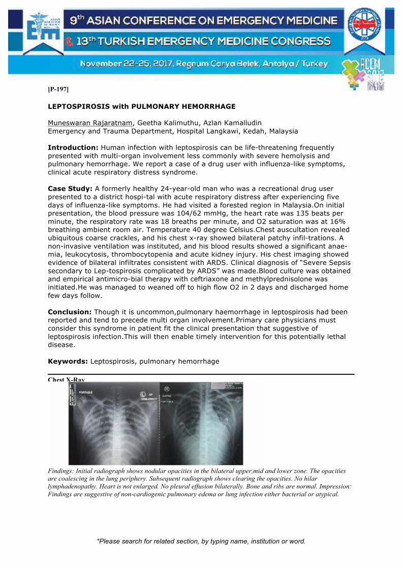
*Please search for related section, by typing name, institution or word.
[P-197]
LEPTOSPIROSIS with PULMONARY HEMORRHAGE Muneswaran Rajaratnam, Geetha Kalimuthu, Azlan Kamalludin Emergency and Trauma Department, Hospital Langkawi, Kedah, Malaysia Introduction: Human infection with leptospirosis can be life-threatening frequently presented with multi-organ involvement less commonly with severe hemolysis and pulmonary hemorrhage. We report a case of a drug user with influenza-like symptoms, clinical acute respiratory distress syndrome. Case Study: A formerly healthy 24-year-old man who was a recreational drug user presented to a district hospi-tal with acute respiratory distress after experiencing five days of influenza-like symptoms. He had visited a forested region in Malaysia.On initial presentation, the blood pressure was 104/62 mmHg, the heart rate was 135 beats per minute, the respiratory rate was 18 breaths per minute, and O2 saturation was at 16% breathing ambient room air. Temperature 40 degree Celsius.Chest auscultation revealed ubiquitous coarse crackles, and his chest x-ray showed bilateral patchy infil-trations. A non-invasive ventilation was instituted, and his blood results showed a significant anae-mia, leukocytosis, thrombocytopenia and acute kidney injury. His chest imaging showed evidence of bilateral infiltrates consistent with ARDS. Clinical diagnosis of “Severe Sepsis secondary to Lep-tospirosis complicated by ARDS” was made.Blood culture was obtained and empirical antimicro-bial therapy with ceftriaxone and methylprednisolone was initiated.He was managed to weaned off to high flow O2 in 2 days and discharged home few days follow. Conclusion: Though it is uncommon,pulmonary haemorrhage in leptospirosis had been reported and tend to precede multi organ involvement.Primary care physicians must consider this syndrome in patient fit the clinical presentation that suggestive of leptospirosis infection.This will then enable timely intervention for this potentially lethal disease. Keywords: Leptospirosis, pulmonary hemorrhage
Chest X-Ray
Findings: Initial radiograph shows nodular opacities in the bilateral upper,mid and lower zone. The opacities
are coalescing in the lung periphery. Subsequent radiograph shows clearing the opacities. No hilar
lymphadenopathy. Heart is not enlarged. No pleural effusion bilaterally. Bone and ribs are normal. Impression:
Findings are suggestive of non-cardiogenic pulmonary edema or lung infection either bacterial or atypical.

*Please search for related section, by typing name, institution or word.
Lab Investigation
Blood/Date 22/05/17 23/05/17 24/05/17
Hemoglobin(g/dl) 9.7 8.1 9.1
MCV(fL) 78 76
MCH(pg) 26.7 26.3
Hematocrit(%) 23.7 23.4
White Cell Count(10^9/L) 19.9 7.97 14.58
Platelet Count(10^9/L) 62 106 214
Sodium(mmol/L) 128 133 136
Potassium(mmol/L) 3.9 3.8 3.7
Urea(mmol/L) 16.2 19.2 21.5
Creatinine(umol/L) 337 284 189
INR 1.2
ALT(U/L) 33
ALP(U/L) 59
AST(U/L) 63
Total Bilirubin(umol/L) 20
Creatine Kinase(U/L) 146
Dengue Serology IgG,IgM,NS1(-ve)
Blood C&S
No Growth
Leptospira(MAT)
Titre 1:200

*Please search for related section, by typing name, institution or word.
[P-198]
Acil Serviste Ağır Sepsis ve Septik Şok Hastalarında İskemi Modifiye Albumin’in Prognostik Değeri Murat Çetin1, Başak Bayram2, Özlem Gürsoy Çalan3, Neşe Çolak Oray2 1Tekirdağ Devlet Hastanesi, Acil Tıp Kliniği, Tekirdağ 2Dokuz Eylül Üniversitesi Tıp Fakültesi, Acil Tıp Ana Bilim Dalı, İzmir 3Dokuz Eylül Üniversitesi Tıp Fakültesi, Biyokimya Ana Bilim Dalı, İzmir Amaç: Çalışmamızda acil serviste ağır sepsis ve septik şok tanısı alan erişkin hastalarda İskemi Modifiye Albumin (İMA) değerlerinin prognostik etkisini araştırmayı amaçladık. Yöntem: 01.01.2015-30.06.2015 tarihleri arasında acil servis hekimi tarafından ağır sepsis ve septik şok düşünülüp dışlama kriterlerini taşımayan hastalar “olgu grubu” olarak çalışmaya alındı. Olgu grubu ile benzer yaş ve cinsiyette olan, sepsis dışında tanı alan ve dışlama kriterlerini taşımayan hastalar “kontrol grubu” olarak kabul edildi. Olgu ve kontrol grubunun acil servis başvurusunda bakılan İMA düzeyleri karşılaştırıldı. Olgu grubunun sosyodemografik verileri, enfeksiyon kaynakları, laboratuvar sonuçları ile acil serviste, ilk 24. saatte ve 28. günde sonlanımları incelendi. Bulgular: Çalışmaya alınan 81 olgu grubu, 72 kontrol grubu hastasının 81’i (%38) kadın ve kadın erkek oranı 1.6 idi. Yaş ortalaması olgu grubunda 71.5 ± 13, kontrol grubunda 62 ±13.7 saptandı(p=0.001). Olgu ve kontrol grubunda İMA ortalamaları sırasıyla 117.7 ± 84.9 IU/g (IQR=59.1) ve 84.7 ± 32 IU/g (IQR=22.6) idi (p=0.002). En sık saptanan enfeksiyon odağı solunum sistemi idi ( n=42, %52). Enfeksiyon odaklarına göre İMA ortalama değerleri incelendiğinde santral sinir sistemi enfeksiyonu olan olguların İMA düzeyleri anlamlı olarak daha yüksekti (p=0.005). Olgu grubundaki 81 hastanın acil serviste sonlanımları incelendiğinde; 51’inin (%58) YBÜ, 23’ünün (%36) öldüğü, 6’sının (%7) servis yatış, bir (%1) hastanın da kendi isteğiyle acil servisten ayrıldığı saptandı. Hastaların acil serviste, ilk 24. saatte ve ilk 28. günde sonlanımlarına bakıldığında; her değerlendirme döneminde sonlanım durumuna göre (taburcu olma, YBÜ yatış, ölüm) İMA ortalama değerleri arasında anlamlı bir fark yoktu (sırasıyla p=0.146, p=0.405, p=0.093). Sonuç: Çalışmamız sonuçlarına göre acil serviste ağır sepsis ve septik şok tanısı alan olguların ölçülen İMA düzeyleri kontrol grubu ile karşılaştırıldığında anlamlı olarak daha yüksektir. İMA ağır sepsis ve septik şok hastalarının tanınmasında yeni bir biyobelirteç olabilir. Ancak İMA düzeyi ile hastaların sonlanımları arasında bir ilişki bulamadık. Bunlar ön sonuçlardır. Çalışmaya kontrol hasta grubuna hasta alımı devam etmektedir. Anahtar Kelimeler: Sepsis, iskemi modifiye albümin, acil servis
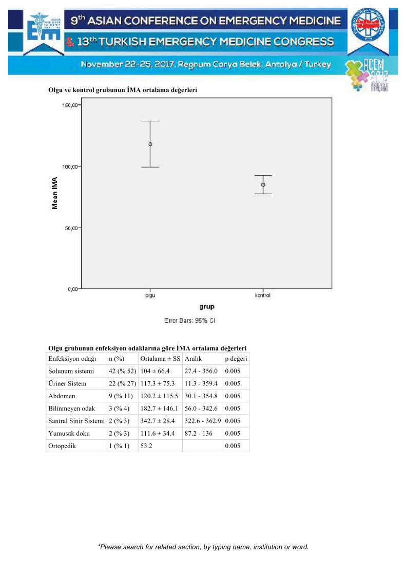
*Please search for related section, by typing name, institution or word.
Olgu ve kontrol grubunun İMA ortalama değerleri
Olgu grubunun enfeksiyon odaklarına göre İMA ortalama değerleri
Enfeksiyon odağı n (%) Ortalama ± SS Aralık p değeri
Solunum sistemi 42 (% 52) 104 ± 66.4 27.4 - 356.0 0.005
Üriner Sistem 22 (% 27) 117.3 ± 75.3 11.3 - 359.4 0.005
Abdomen 9 (% 11) 120.2 ± 115.5 30.1 - 354.8 0.005
Bilinmeyen odak 3 (% 4) 182.7 ± 146.1 56.0 - 342.6 0.005
Santral Sinir Sistemi 2 (% 3) 342.7 ± 28.4 322.6 - 362.9 0.005
Yumusak doku 2 (% 3) 111.6 ± 34.4 87.2 - 136 0.005
Ortopedik 1 (% 1) 53.2
0.005

*Please search for related section, by typing name, institution or word.
[P-200]
Hepatosplenic Candidiasis Ozlem Dikme, Gokce Akgul Karadana, Can Aktas Emergency Department, Koc University Hospital, Istanbul, Turkey Background: Hepatosplenic Candidiasis is a disseminated infection involving spleen and liver. It has observed at increased incidence in immunocompromised patients especially in malignancies after chemotherapy has been administrated. Although the culture results are generally negative, diagnosis is made by showing the pathogen in culture or biopsy. Basic approach in treatment is the use of amphotericin-B. In this case we present a young patient which is diagnosed by radiological. Case presentation: A 29-year-old female was admitted to the emergency department with left axillary and abdominal pain. One year ago, she was hospitalized diagnosis with lung tuberculosis and during the hospitalization time she had complicated with splenic infarct and abcess, infective endocarditis, hypertension and hypoalbuminemia. She lost 10 kg in last month. On admission, general condition was medium and she looked as cachectic. Vital signs found as hypertensive and tachycardia depended on not using any medication. ECG showed that sinus tachycardia, probably left atrial enlargement signs and non-specific T wave abnormalities. 10 mg amlodipin was given orally. Introvenous line was inserted and laboratuary tests showed that neutropenia as 1.17 K/uL, anemia as 9 g/dL, CRP 51 mg/L, LDH 281 U/L, Na 132 mmol/L. Thoracoabdominal CT showed that hepatosplenic candidiazis Keywords: Hepatosplenic candidiasis, emergency department
Figure

*Please search for related section, by typing name, institution or word.
[P-201]
Brucella Infection in the Differential Diagnosis of the Nontraumatic Subcapsular Hematoma Pakize Gözde Gök, Evvah Karakılıç, Nurdan Acar, Kübra Parpucu, Filiz Baloğlu Kaya, Ezgi Akçacı Baran Department of Emergency Medicine, School of Medicine, Eskişehir Osmangazi University, Eskişehir, Turkey Introduction: Brucella can be transmitted with contact to animals or its products. Although hepatomegaly or splenomegaly can be observed, liver abscess or spleen rupture are rarely encountered. Here, we wanted to draw attention to a patient with a diagnosis of brucella and a nontraumatic subcapsular splenic hematoma. Case: A 55-year-old patient was admitted to the emergency with complaints of generalized and abdominal pain and weakness throughout the week. The fever was high. There were night sweating complaints. There was no trauma story. There were no abnormalities except fever which was 39.0 °C. On physical examination, sensitivity on left upper quadrant and defence but no rebound was detected. Laboratory tests were applied on brucellosis. On the control examination, sensitivity to the left upper and hypochondrial area persisted, and the patient had a contrast enhanced abdominal CT scan. 7x2.5 mm big subcapsular hematomas in the upper pole, 7x3 mm in the lower pole of the spleen, hemoperitoneum in the perihepatic coating style, light pollution around that and pelvic fluid were detected. The patient was scheduled for general surgery hospitalisation. The submitted brucellosis agglutination tests were positive. In the follow-up, doxycycline and rifampicin were started for brucellosis treatment. In the control examinations, the patient with regression in liver function tests was discharged on seventh day. Conclusion: Brucellosis prefers locations rich in reticuloendothelial system cells. During the infection, congestion and dilatation can be observed in sinuses and Billroth cords, and thrombosis and focal necrosis can be observed in capillaries and in the splenic pulp. These pathological changes can lead to bleeding in the spleen as well as the rupture of the spleen. If clinical symptoms are compatible with brucellosis, spleen rupture or hematoma should be considered in the differential diagnosis. Keywords: Brusella, abdominal pain, subcapsular hematoma

*Please search for related section, by typing name, institution or word.
CT image of subcapsular hematoma

*Please search for related section, by typing name, institution or word.
[P-202]
Septic Cerebrovasculer Embolism in Intravenous Drug Users Ramazan Sivil, Erkan Göksu, Ertuğ Günsoy Akdeniz University, Department of Emergency Medicine, School of Medicine, Antalya, Turkey Septic cerebrovasculer embolism is a rare disorder generally caused by infective endocarditis. We present a 23-year-old intravenous drug user patient with fatigue, decreased appetite, altered mental status and thrombocytopenia. The patient has GCS of 14, petechial rash on lower extremity, janeway lesions on the palms and soles. The patient's blood test results showed thrombocytopenia, hyponatremia and elevated CRP. Head CT was normal, but the patient's MR imaging showed bilateral multiple acute and subacute infarcts. Also, transthoracic echocardiography showed vegetations on mitral valves. Keywords: Infective endocarditis, stroke, drug abuse

*Please search for related section, by typing name, institution or word.
[P-203]
Thrombophlebitis of the Cerebral Venous Sinus: A Rare Complication of Chronic Otitis Media Rana Toktaş, Mustafa Sever, Serkan Hacar, Enis Yiğitaslan, Gülben Çalışkan Sağlık Bilimleri Üniversitesi Tepecik Eğitim ve Araştırma Hastanesi Acil Tıp Kliniği, Izmir, Turkey CVST is an intracranial complication of otitis media. Eventhough the adequate antibiotherapy, suspicion of CVST must be drawn in all patients with persistent fever, headache and otorrea. Here; we have presented a case diagnosed as CVST which has developed as a complication of chronic otitis media. Case: A male patient aged 23 has presented to the ED with the chief complaint of fever and headache present for 2 days. At the time of application his vital measurements were as follows: BP:100/80 mm/Hg, pulse:110/min, body temp:39C axillary and Sat O2: 99%. In his past medical history knowledge of antibiotherapy initiation for chronic otitis media treatment 2 weeks ago has been gained. In his PE; his mental status was defined as lethargic, but cooperation and orientation could be built via dialogue. In autoscopic examination; a pulsatile serous leakage was noticed. neck stiffness or any sign of meninx irritation were detected to be negative. In laboratory tests; his WBC count was 7200, Urea:38 mg/dL, cr:0.7 mg/dl procalcitonin 41.21 mg/ and plt count was 177.000. A head CT scan was then performed, loss of aeration in the left mastoid cells were found as a result of mastoiditis. A lumbar puncture was then performed no evidence of meningitis was obtained both in direct examination and biochemistrial investigation of CSF. Both MRI & MR Venography were obtained from the patient and acute thrombophlebitis was detected in left transvers sinus partially and total occlusion was seen in left sigmoid sinus including the proximal part of the jugular vein which could be captured in the sequences available. Conclusion: CVST must be considered in all patients who apply to the ED with headache and fever. Antibiotics and surgical intervention is the approved treatment method. Because that the situation is rare, the need for routine anticoagulation is unclear. Anticoagulation therapy is unecessary unless the thrombi is shown in the sagittal sinus or no sign of an increase in ICP has been measured. Keywords: Chronic otitis media, thrombophlebitis of the cerebral venous sinus

*Please search for related section, by typing name, institution or word.
[P-204]
Pathologic Phimosis Associated with Staphylococcal Toxic Shock Syndrome: A Case Report Ryohei Suzuki, Tomoyuki Masuyama, Tsuyoshi Chijiwa, Takashi Moriya Jichi Medical University Saitama Medical Center, Osaka, Japan Introduction: A variety of skin manifestations are seen in toxic shock syndrome (TSS). However, the penile complication has not been reported. Presentation of Case: A 50-year-old man presented to the emergency department with fever, frequent vomiting and watery diarrhea and admitted to the intensive care unit for shock. He suffered a burn injury in the right anterior chest wall two days ago. Physical examination revealed diffuse macular erythroderma on the whole body. We suspected TSS or septic shock of skin and soft tissue infection, and started intensive care management. Methicillin-resistant Staphylococcus aureus was detected from burn wound swab. On day 14, the patient was found to have peeling skin at his fingers. The patient was diagnosed as Staphylococcal TSS. On day 7, penile pain and redness and erosion of the foreskin and scrotum were observed whereas his penis had been unremarkable on admission. The glans penis was easily stored in the foreskin manually. On day 20, the patient could not retract the foreskin and represented dysuria. The tip of foreskin got contracted due to scar and showed pathologic phimosis. On day 35, circumcision and scar-contracture plasty were performed on the foreskin simultaneously at the time of skin grafting for burn injury. Then, he recovered and discharged on day76. Discussion: Late-onset skin manifestations of TSS commonly include desquamation of the palms and soles as seen in this case. In more severe cases, superficial ulcerations occur on the mucous membranes, and petechiae, vesicles, and bullae develop. However, there are few reports of pathologic phimosis requiring surgery. Some patients may not complain because of shamefulness, and just have not be diagnosed. Conclusion: There may be pathologic phimosis in late-onset skin manifestations of Staphylococcal TSS. In patients with TSS, we need long-term observation of skin lesions including penis. Keywords: Phimosis; toxic shock syndrome; staphylococcus aureus

*Please search for related section, by typing name, institution or word.
[P-207]
A Difficult Distinction: A Case of Drug-induced Pneumonitis or a Tick-borne Infection with Lung Involvement? Tatsuo Takama1, Shyogo Soga3, Koki Umeda1, Michiaki Kaku2, Yoshihiko Sakamoto3, Hideaki Yoshihara1 1Emergency And Critical Care Center, Kagoshima City Hospital, Japan 2Emergency And Critical Care Center, Kitakyusyu general hospital, Japan 3Kyusyu Univercity, Department of public health, Japan We report a case of drug-induced pneumonitis that was difficult to distinguish from a tick-borne infection with lung involvement. A 50-year-old man was bitten by a tick in Kyusyu, Western Japan, and minocycline was administered immediately. Two weeks later, he was admitted to our hospital with fever, severe dyspnea, and general fatigue. Chest computed tomography revealed bilateral diffuse ground-glass opacities and an increase in peripheral blood eosinophils was noted on the 3rd hospital day; therefore, we suspected drug-induced pneumonitis. However, this was difficult to differentiate from tick-borne infection with lung involvement on the basis of radiologic findings. We decided to identify the tick species and diagnose the disease based on the vector competence of the tick, in parallel with genetic and serological examinations. The tick was identified as Amblyomma testudinarium, a vector of severe fever with thrombocytopenia syndrome virus (SFTSV) and Rickettsia japonica in Japan. Minocycline-induced pneumonitis was strongly suspected, based on negative results of several examinations for SFTSV and R. japonica. The symptoms and abnormal chest shadows resolved after minocycline was discontinued. Discussion: In treating tick-borne infections, the identification of the tick species and assessment of vector competence are very important for diagnosis, therefore, emergency physician should preserve the removed tick and take careful anamnesis including the bitten situation. Keywords: Vector compitence, tick borne infection, drug induced pneumonitis

*Please search for related section, by typing name, institution or word.
Chest Computed tomography
Chest Computed tomography (CT) on the 1st hospital day revealed bilateral interlobular septal thickening, with
diffuse ground-glass opacities.

*Please search for related section, by typing name, institution or word.
[P-208]
Orf Disease: An Unusual Finger Injury Yasemin Özdamar, Zehra Akçora, Yunus Emre Dündar, İlter Ağaçkıran, Muhammed Sarıer, Mehmet Ali Karaca, Bülent Erbil The Department of Emergency Medicine, Hacettepe University, Ankara, Turkey Orf infection is a rare viral zoonotic infection caused by a dermotropic double stranded DNA parapoxvirus. It typically presents a few weeks after an animal bite as a painless swelling, which is blue-black in color. There is usually minimal cellulitis and no associated lymphadenopathy. We report a case of orf in a patient contracted from a sheep. Case: A 27-year-old female with no medical history presented to the emergency department with a painless swelling over her right index finger. The problem started ten days earlier after she injuried herself while cooking a sheep head’s bone. Then, she noticed a papule on her right index finger that enlarged gradually. She reported no pain, changes in sensation, or reduced range of movement. On clinical examination she was afebrile and hemodynamically stable. Her right index finger exhibited a full range of movement and was neurovascularly intact. The finger was not tender on direct palpation. There was a targetoid lesion on her right index finger. WBC: 11,9x10³/µL. There was no fracture or lytic lesion on her X-ray. The patient was consulted with infectious diseases, dermatology and plastic surgery for diagnose of Orf and discharged with symptomatic treatment. Discussion: Orf also known as ecthyma contagiosum, contagious pustular dermatitis is a viral disease first described in humans in 1934 by Newson and Cross. Orf is caused by a parapox virus, endemic in sheep and goats. Orf can be transmitted to humans by contact with an infected animal or contaminated fomites and most commonly occurs on the hands. Orf is the self-limiting disease typically appears as a solitary red weeping nodule of the hands that reach resolution in approximately four to eight weeks. Symptomatic treatment with antiseptics, moist dressings and finger immobilization help the management of the disease. Routine antibiotics treatment is not recommended. Keywords: Orf, parapoxvirus, ecthyma contagiosum
Clinical picture of orf disease

*Please search for related section, by typing name, institution or word.
A characteristic targetoid lesion of orf disease

*Please search for related section, by typing name, institution or word.
[P-209]
Blastocystis Hominis Infection Causing Acute Hepatitis Ziba Songur Yücel, Baki Doğan, Tuğçe Taşkındere Abbasov, Tugay Aydoğan, Nalan Metin Aksu, Meltem Akkaş Hacettepe University, Shool of Medicine, Department of Emergency, Ankara, Turkey Introduction: Blastocystis hominis is one of the most common human intestinal protozoans that may be found in the stools of healthy people in the developing countries. It transmitted via the fecal-oral route. Blastocystis hominis infection often causes gastrointestinal complaints. Fever, pruritus, weight loss, leucocyte in feces, eosinophilia and anemia can occur also. Case: 33 years old female patient applied to emergecy department with epigastric and back pain for 1 month and nausea for 15 days. She had no medical history and drug usage but eating raw vegetables. She had normal vital signs except fever 37.7⁰ C. Physical examination findings were normal except epigastric tenderness. Her liver function tests were elevated. ALT, AST, ALP, GGT were 98, 70, 942, 443 UL, respectively. She had low hemoglobin value (Hg:9,9) and eosinophilia on her essential laboratory tests. There were extensive hypodens lesions on liver suggesting protozoan infection on abdominal computed tomography. Blastocystis hominis was found in her parasitology examination. She was treated by thiabendazol. Her liver function tests returned to normal levels and her complaints completely disappeared in 6 months. Conclusion: It is difficult to diagnose Blastocystis Hominis infection at emergency department because of non-spesific symptoms and laborotory tests. The history of eating raw vegetables and gastrointestinal complaints with eosinophilia and anemia should remind us protozoan infection such as Blastocystis Hominis. Keywords: Blastocystis hominis, hepatitis

*Please search for related section, by typing name, institution or word.
[P-210]
A Rare Cause of Sepsis Due to Bladder Cancer Treatment with Bacillus Calmette-Guerin Ziba Songur Yücel, Meltem Akkaş, Etkin Keskin, Dilek Sarıoğlu, Özzgür Bayar, Nalan Metin Aksu University of Hacettepe, School of Medicine, Department of Emergency, Ankara, Turkey Introduction: Intravesical administration of Bacillus Calmette-Guerin (BCG) is used as a treatment method in superficial bladder cancer. Systemic mycobacterial infection is rare but serious complication of intravesical BCG therapy. Incidence of systemic infection has been reported as 0.4 to 0.7% in contemporary series. The clinical feature is similar to bacterial sepsis. It has been proposed that BCG sepsis may have a component of delayed-type hypersensitivity. Treatment include antituberculous therapy and glucocorticoids. Case Report: 74 years old male patient was admitted to emergeny department with no oral intake for 1 week. He had stroke, hypertension, prostate and bladder cancer on his past medical history. He was applied intravesical administration of BCG once a week for the five weeks. On admission, vital signs were as following: fever:34.4 °C, blood pressure:74/36 mmHg heart rate:109 /min, respiratory rate:32/min and O2 saturation: 68%. His Glasgow Coma Scale was 7 and he had penis edema. The bedside capillary blood glucose level was measured as 40 mg/dL. High anion gap metabolic acidosis, high liver enzymes, renal failure were detected on his laboratory tests. Firstly, he was intubated, and he was administered fluid replacement theraphy, antibiotherapy and vasopressor agents. His chest radiography was normal. Urine culture was clear. In the absence of another cause, we thought that source of infection causing sepsis was administration of intravesical BCG. Patient was transferred to intensive care and he died within the same day. Conclusion: Intravesical administration of BCG can be source of infection but it rarely causes sepsis. The prognosis of BCG sepsis is very poor. Treatment should be started immediately. Keywords: Bacillus Calmette-Guerin, bladder cancer, sepsis

*Please search for related section, by typing name, institution or word.
[P-211]
Thrombolytics: Friend or Foe? Asiye Muminat Gungor1, Ramazan Sivil1, Salahi Engin1, Refik Emre Altekin2, Yildiray Cete1 1Akdeniz University Faculty of Medicine Department of Emergency Medicine, Antalya, Turkey 2Akdeniz University Faculty of Medicine Department of Cardiology, Antalya, Turkey Coexistence of significant pathologies in a patient is not uncommon. If the clinical symptoms cannot be explained by one condition, the Clinician must have sought to find another coexisting disease. We present a year-old female who was brought in by EMS for altered mental status. Relatives stated she was receiving chemotherapy for breast cancer, but no other comorbidities existed. As per relatives, the patient was found unresponsive in her bed. Upon the arrival, vital sign was: HR 148 BP 92/55 RR 48 Temp 36,7 Sat O2 %64 on room air. Physical examination showed a dyspneic patient with a GCS of 7. Right sided hemiplegia was also present. Patient was incubated with RSI for airway protection. Oxygen saturations failed to increase to normal values despite adequate ventilation and POCUS showed dilated right ventricle. Head CT showed dense Left MCA and CTPA showed a saddle embolus. Since the last time the patient seen was normal was the night before, the patient was deemed ineligible for systemic thrombolytics. Embolectomy for the massive PE was not available at that time at our instruction. Option of local administration of fibrinolytics was discussed with the family. After the family consented, the patient was taken to the catheterization lab. Angiography revealed saddle embolus in main pulmonary arteries, with almost no flow bilaterally. After thrombolytics administration, blood flow restored 20% on the left side. no blood slow was restored on the right side. Unfortunately, during her stay the patient arrested and had a fatal outcome. No autopsy was performed by the request of the family. Our patient did not have any risk factors for coagulopathy except for breast cancer diagnosis and chemotherapy. The patient did not have any known patent foramen ovale. Co-occurrence of PE and CVA is rare and possesses high risk of morbidity and mortality. Keywords: Thrombolytics PE cva

*Please search for related section, by typing name, institution or word.
[P-213]
Superior Mesenteric Artery Syndrome: A Rare Cause of Intestinal Obstruction at Emergency Service Mehmet Ergin1, Selahattin Gürü2, Nihal Saylan1, Ayhan Özhasenekler1, Şervan Gökhan1 1Yıldırım Beyazıt University, Faculty of Medicine, Emergency Medicine Department, Ankara, Turkey 2Ankara Atatürk Training and Research Hospital, Emergency Service, Ankara, Turkey Introduction: Superior Mesenteric Artery (SMA) Syndrome (SMAS) is defined as the clinical symptoms and findings that occur by the compression of the third part of the duodenum between aorta and the superior mesenteric artery. Symptoms such as nausea, bilious vomiting, postprandial epigastric pain, loss of appetite and loss of weight may cause acute or chronic confusion. In this report, we present a case with abdominal pain, nausea and vomiting. Case: A 29-year-old male presented with complaints of abdominal pain, nausea and vomiting, weakness, numbness and constipation in arms and legs. There was a history of stomach cancer and had rigidity on his abdominal examination. Vital findings were in normal range. Second and third parts of duodenum was seen as dilated (diameter of duodenum was 50 mm) and duodenum has collapsed between aorta and SMA in the computerized tomography. General surgery department recommended conservative follow up and he was discharged after his symptoms recovered. Discussion: SMAS is one of the rare cause of obstruction in upper gastrointestinal system. Incidence of duodenal compression in the aortic-SMA region is estimated to be approximately 0.3% in the upper gastrointestinal barium studies, but the clinically significant disease rate is around 0.01-0.08%. Both acute and chronic forms can occur. Chronic form is more common. Abdominal distansion, vomiting and pain are the most common symptoms in the acute form of SMAS. In chronic form, it usually manifests itself as epigastric pain, bloating, nausea, vomiting, anorexia and weight loss after food intake. While conservative treatments are primarily applied in acute cases, surgical treatments are the mainstays in chronic cases. CONCLUSION SMA syndrome is an anatomic, clinical pathology that causes obstructive symptoms. Emergency physicians should keep it in mind even though it is rare. Keywords: Superior mesenteric artery syndrome, emergency department

*Please search for related section, by typing name, institution or word.
[P-214]
A Rare Cause for Pain in the Lateral Neck: External Jugular Vein Thrombosis Ökkeş Yılmaz Çınar, Emir Ünal, Çiğdem Özpolat, Serhad Ömercikoğlu, Kerem Ali Kabaroğlu, Melis Efeoğlu, Sinan Karacabey, Arzu Denizbaşı Marmara University Faculty of Medicine, Department of Emergency Medicine, Istanbul, Turkey Introduction: A thrombus in a jugular vein is an uncommon occurrence. It may occur in the internal jugular vein or in the external jugular vein. Especially if an induration in the region of the big vessels is present, one must keep in mind the differential diagnosis of a spontaneous thrombosis of the internal or external jugular vein (EJV). This diagnosis needs consequent treatment but also consequent search for an underlying pathology. This case report presents such a situation in an exemplary way. Case: A previously healthy, 43-year-old woman presented to the university-based emergency department because of painless swelling in the left anterior side of her neck. Physical examination revealed a painless, soft and immobile mass in the left anterior side of her neck. On ultrasonographic examination, a hyperechogenic mass was visualized in the left external jugular vein, which was suggestive of a thrombus. The patient was administered low-molecular-weight heparin followed by oral coumadin as anticoagulant therapy. Her complaints were relieved within 5 days. She was completely well after 3 months. No underlying reason was found. Discussion: Venous thrombosis generally results from impaired blood flow locally or systemically that leads to activation of coagulation. External jugular vein thrombus is even less clear than others, underlying malignancy must be searched. Also, these superficial vein thrombi most probably occur in relation to neck catheter insertion. Emergency physicians should sustain a high index of suspicion in patients who present with undiagnosed swelling in the neck, or other signs and symptoms attributed to EJV thrombosis. Keywords: Jugular vein, thrombosis
*Please search for related section, by typing name, institution or word.
[P-216]
Paradoks Emboli Salih Kocaoğlu, Abuzer Coşkun, Sedat Özbay, Behnan Gülünay, Osman Mahir Okur, Mustafa Oğuz Cumaoğlu Sivas Numune Hastanesi, Acil Servis, Sivas Giriş: İnme travmatik olmayan nedenlerle ortaya çıkan, beyin damar hastalığına bağlı ani başlangıçlı sıklıkla fokal nörolojik defisitlere yol açan klinik bir tablodur (1). İnme orta ve erişkin yaş grubunun hastalığı olarak kabul edilse de genç erişkinlerde de gözlenebilir (2). 45 yaş altında görülen inmeler genç inme olarak sınıflandırılır. Genç yaş inmelerin etyolojisinde ateroskleroz, kardiyak hastalıklar, gebelik, doğum, oral kontraseptif kullanımı gibi çeşitli nedenler yer almaktadır. Vakamızda tetkiklerinde Patent Foramen Ovale (PFO) saptanan bir hastayı sunduk Olgu: 20 yaşında bayan hasta acil servise yaklaşık 3 saat önce başlayan sol kolda güçsüzlük ve ağzında kayma şikayetiyle başvurdu. Fizik muayene sol üst ekstremitede motor kuvvet 3/5 ve sol nazolabial sulcus silik. Hastanın genel durumu iyi, bilinç açık, GKS:15, tansiyon 110/60 mmHg nabız: 90/dk, Ateş:36.2 C. Hemogram incelemesinde hemogobin 11.6 gr/dl, lökosit 8/mm3, trombosit 237.000 mm3; biyokimya inlemesinde glukoz 92, üre19, kreatinin 0.5, AST 35, ALT11, CK 954, CK-MB 28, NA 138, K 3.7 saptandı. Beyin tomografisinde belirgin iskemi ve hemoraji bulgusu saptanmadı. Diffüzyon MR’da sağ frontoparyetal bölgede 8x4 cm boyutunda akut enfarkt ile uyumlu difüzyon kısıtlılığı saptandı. Nöroloji servisine yatırılıan hastanın yapılan tetkiklerinde karotis ve vertebral doplerde patoloji saptanmadı. Kardiyak değerlendirmesinde Patent Foramen Ovale (PFO) saptandı. Hastanın tedavi ile kuvvet kaybı 7. gününde tamamen düzeldi. Fasyal paralizisi ise taburculuk sonrası 2 ay sürdü. Tartışma ve Sonuç: Genç inmeli hastalarda etyolojik faktörlerin daha ayrıntılı araştırılması ve tedavi edilmesi, rekkürenslerin oluşumunu azaltacaktır. PFO sağlıklı kişilerde % 20-25 oranında görülsede, genç inme vakalarında sık saptandığı görülmektedir. Bu tür hastalarda endovasküler cerrahi düşünülebilir. Bunun medikal tedaviye göre inme riskini azalttığı ileri sürülmektedir. Ancak bu konuda randomize klinik çalışmalara ihtiyaç vardır. Anahtar Kelimeler: Acil, inme, paradoks emboli

*Please search for related section, by typing name, institution or word.
resim1

*Please search for related section, by typing name, institution or word.
[P-217]
Case with Isolated Left Subclavian Artery Occlusion Selahattin Gürü1, Ayhan Özhasenekler2, Alper Gök2, Mehmet Ergin2, Alp Şener2, Gül Pamukçu Günaydın2 1Ankara Atatürk Training and Research Hospital, Emergency Service, Ankara, Turkey 2Yıldırım Beyazıt University, Faculty of Medicine, Emergency Medicine Department, Ankara, Turkey Introduction: The left subclavian artery is an artery that arises from the aortic arch and feeds the left upper extremity. Occlusion is rarely symptomatic and leads to claudication. In this article, we present an elderly case suffering from pain and cold at upper extremity. Case report: A 75-year-old male patient was admitted to the emergency room with a feeling of pain and coldness in the left hand for a few hours. He has hypertension and smoking in his history. First vital signs were in normal range. There was no pathological finding on electrocardiography. Pulses couldn’t be felt on examination of upper extremity. Computed tomographic angiography demonstrated thrombosis on the proximal of left subclavian artery. He was brought to operation room to the cardiovascular surgery service. The patient was monitored by medical treatment with low molecular weight heparin. He was discharged as his symptoms was regressed and distal pulses were taken by examination. Discussion: Isolated left subclavian artery occlusion is a rare and often asymptomatic condition. If symptomatic, usually ischemic signs on the ipsilateral upper limb are recorded like this patient. Atherosclerosis is the most common cause of subclavian artery stenosis. Surgical treatment of symptomatic patients is essential as a basic principle. Patients should be considered for early consultation of cardiovascular surgeon. Best results are said to be taken with angioplasty and stent application in the long term. In the present case, the patient was initially offered surgical intervention, but medical treatment was preferred because he rejected the surgery. Conclusion: Isolated subclavian artery occlusion is an uncommon condition in the emergency room. It should be remembered in patients with pain and cold on upper extremity and signs about deficiency of arterial circulation. Keywords: Subclavian artery occlusion, emergency department

*Please search for related section, by typing name, institution or word.
[P-218]
Göğüs Ağrısının Atlanan Bir Nedeni: Torasik Outlet Sendromu Senem Şaş1, Hacı Mehmet Çalışkan2, Burak Çelik3, Sinan Cemgil Özbek4, Erdoğan Sökmen4 1Ahi Evran Üniversitesi Eğitim ve Araştırma Hastanesi, Fiziksel Tıp ve Rehabilitasyon, Kırşehir 2Ahi Evran Üniversitesi Tıp Fakültesi Eğitim ve Araştırma Hastanesi, Acil Tıp A.D, Kırşehir 3Ahi Evran Üniversitesi Tıp Fakültesi Eğitim ve Araştırma Hastanesi, Acil Servisi, Kırşehir 4Ahi Evran Üniversitesi Tıp Fakültesi Eğitim ve Araştırma Hastanesi, Kardiyoloji A.D, Kırşehir Giriş: Ülkemizde ve dünyada acil servislere göğüs ağrısı nedenli başvurular gittikçe artmaktadır. Bu durum acil doktorlarını koroner iskemik ağrı ile diğer ağrı nedenlerini ayırt etmekte zorlamaktadır. Torasik outlet sendromu kola giden nörovasküler yapıların göğüs kafesinin üst kısmında çeşitli nedenlerle basıya uğraması ve semptom oluşturmasına denir. Bildirimizde kendini koroner iskemik ağrı gibi tarifleyen torasik outlet sendromu tanısı koyduğumuz vakayı sunduk. Olgu: 38 yaşında erkek hasta hastanemiz acil servisine ani başlayan göğüs ağrısı şikayeti ile başvurdu. Hastadan alınan anamnezde ağrısının sıkıştırıcı tarzda ve nefes almakla arttığı, sol koluna ve sırtına yayıldığı ve beraberinde uyuşma ile birlikte karıncalama olduğu öğrenildi. Hastanın buna benzer göğüs ağrısı ile son bir hafta içerisinde 2 defa acile başvurduğu ve kendisine kardiyoloji poliklinik önerildiği ancak gitmediği öğrenildi. Hastanın başvuru anındaki vitalleri: Kan basıncı: 130/90mmHg, nabız 88/dakika, solunum sayısı 22/dakika. Fizik muayenesinde kalp sesleri ritmik olup ek ses ve üfürüm yoktu. Özgeçmişinde 1 paket/gün, 20 yıl sigara içimi dışında bir özellik yoktu. EKG’sinde normal sinus ritmi olup iskemi belirtisi yoktu. Tetkiklerinde keratinkinaz ve troponin negatifti. Kontrol EKG ve kardiak enzimlerinde problem olmayan hasta kardiyoloji polikliniğinde ekokardiyografi ile değerlendirildi. Hastada iskemi bulgusu olmadığı gözlendi. Fizik Tedavi ve Rehabilitasyon polikliniğine yönlendirilen hastanın muayenesinde boyun hareketleri açık ağrılıydı, sol C7,C8 hipoestezikti, motor kayıp yoktu. Solda Adson testi pozitifti. Trapezde yaygın gergin bant tespit edildi. Hastadan iki yönlü servikal grafi istendi. C7 uzun transvers process tespit edildi. Tetik nokta enjeksiyonu yapılarak kaslara germe egzersizleri yapıldı. Akut dönemde ağrısı azalan hastaya ev egzersizleri ve postur eğitimi verildi. Haftalık kontrollere alınan hastanın üç hafta sonra şikayetinin ciddi oranda azaldığı öğrenildi. Sonuç: Biz bu olgumuzda torasik outlet sendromunun iskemik kalp ağrısı gibi acil servise başvurabileceğini sunduk. Acil uzmanları olarak iskemik ağrıyı ekarte ettikten sonra bu hastaların fizik tedavi polikliniğine yönlendirilmelerinin hastalara doğru tedavi alma ve gereksiz hastane başvurularının azalması açısından faydalı olacağını düşünmekteyiz. Anahtar Kelimeler: Gergin bant, göğüs ağrısı, torasik outlet sendromu

*Please search for related section, by typing name, institution or word.
Olguya ait servikal grafi

*Please search for related section, by typing name, institution or word.
[P-219]
Monckebergs's Medial Calcific Sclerosis: An Uncommon Cause of Bilateral Loss of Vision Yeşim Eyler, Necmiye Yalçın Ocak, Turgay Yılmaz Kılıç, Hasan Idil, Özge Duman Atilla, Mebrure Evnur Uyar, Onur Hakoğlu, Rana Toktaş Tepecik Training and Research Hospital, Izmir, Turkey Introduction: Mönckeberg's calcific sclerosis is a vasculopathy characterized by the calcification of the media of small and medium-sized muscular arteries (1). It often affects the coronary arteries, the peripheral arteries of the extremities and the uterine arteries in women, causing clinical manifestations of decreased perfusion (2). We present a case of Mönckeberg's calcific sclerosis who presented to the emergency department with bilateral loss of vision. Case: A 69-year-old female presented to the emergency department with a 3-day history of progressive loss of vision in both eyes. Her past medical history was significant for diabetes, hypertension and ovarian malignancy. She was conscious and oriented, with no sensory and motor deficits apart from the aforementioned bilateral vision loss, and her vital signs were stable. Her eyes were in the primary position, pupils were normoisochoric, eye movements were normal, and pupillary light reflex was delayed. Fundus examination revealed pale and edematous optic discs. Laboratory results were unremarkable except creatinine: 7.7 mg/dl, potassium: 5.7 mg/L, calcium: 8 mg/dl, pH: 7.25 and sedimentation rate 50 mm/hour. The patient's cranial computed tomography and magnetic resonance imaging did not reveal any abnormalities to explain the clinical findings. Renal ultrasonography was unremarkable. The patient received hemodialysis for acute renal failure, after which the serum electrolytes and renal functions normalized, but the loss of vision persisted. The patient was admitted to the neurology ward with suspected temporal arteritis. Temporal artery biopsy revealed findings compatible with Mönckeberg's medial calcification. Discussion: Mönckeberg's medial calcification is a disease which primarily affects the arteries. Though the etiology is still unclear, the disease is often associated with glucose intolerance, age, male gender, autonomic neuropathy, osteoporosis, and chronic renal failure (3,4). This disease should be considered in the differential diagnosis of patients presenting with arterial complaints of unknown origin. Keywords: Loss of vision, vasculopathy

*Please search for related section, by typing name, institution or word.
[P-220]
Calcium levels in Emergency Department Feray Balkan, Ayca Acıkalaın Akpınar, Nezihe Rana Dişel, Zeynep Kekec Cukurova Univerity Medical School, Department of Emergency Medicine, Adana, Turkey Objective: In this study we compare calcium levels with diagnosis patiens in Emergency Department (ED) Methods: In first half of 2017 in ÇÜTF, we have performed the analysis of patients that are applied to our ED which have the values of calcium that is out of our reference range 8.9-10.3 Resuts: In total of 70 patients, 34 were women and 36 were men.11 of the patients have the values over 10.3; 59 of the patients have the value below 8.9; 29 of the patients were over 50 years old;29 of the patients have the complaints as fatigue and myalgia.;60 of the patients have at least one known chronicle illness; 10 of the patients do not have a known illness; 11 of the patients with the high values have a known oncological illness; 9 out of 59 of the patients with the low values have drug intocsication or taken substances that cause addict Conclusion: It is not necessary to perform analysis; if the patient has a known oncological illness; if the patinent is over 50 years old and have fatigue and miyalgia; It is more meaningful to perform the analysis; if the patient is influneced by an drug that cause addiction Keywords: Calcium level, patient diagnosis, emergency department
*Please search for related section, by typing name, institution or word.
[P-221]
Venous Blood Gas Instead of Artery Blood Gas in Emergency Department Yakup Kadri Erdoğan1, Guven Kuvandik2, Ali Karakuş2, Zeynep Kekec3 1Hatay State Hospital Emergency Service- Hatay- Turkey 2Mustafa Kemal University, Faculty of Medicine, Department of Emergency Medicine, Hatay, Turkey 3Cukurova University Faculty of Medicine Emergency Medicine, Adana, Turkey Introduction: Arterial blood gas (ABG) analysis is a laboratory method enables us to have idea about patient's acid-base status and respiratory physiology, and used in the diagnosis of respiratory and metabolic diseases, treatment and follow-up. Arterial blood collection is quite a difficult process as it is required entrepreneurial experience and disrupt the patient's comfort, may cause pain at the place of enterprise, local hematoma, embolism, and ischemia. In addition, has high risk for the medical staff in terms of contagious infections. In the present study, we aimed to determine the relevance of arterial and venous blood gases (VBG) and the venous blood gases usability instead of arterial blood gases in the patients admitted to the emergency department with complaints of respiratory distress, and thought to have metabolic disorders. Result: Totally, 110 patients (35 women and 75 men) participated in our study. Simultaneous arterial and venous blood gas samples were taken and analyzed. PH, PO2, PCO2, HCO3-, SaO2, BEecf, Lac values were recorded. While the strong correlation among PH, PCO2, HCO3-, BEecf and Lac values of ABG and VBG were found, PO2 andSaO2 values found to be moderately correlated. Conclusion: As a result, we think that venous blood gas sampling can be used in patients admitted to the emergency department and venous blood gases can reduce the need for arterial blood gases. Keywords: Arterial blood gas, venous blood gas, emergency department

*Please search for related section, by typing name, institution or word.
[P-222]
Bleeding While Yawning Ahmad Ghaus Mohd Ghouse1, Julina Mohd Noor2, Sabariah Faizah Jamaluddin1 1Emergency & Trauma Department, Hospital Sungai Buloh, Selangor, Malaysia 2Faculty of Medicine, Universiti Teknologi MARA, Malaysia Introduction & Objectives: Yawning is a day to day common behavioural event. However, a not so well-known fact is that excessive yawning, defined as more than 3 yawns every 15 mins has been found to be associated with intracranial pathology. This has been described in stroke, brainstem ischaemia and increased intracranial pressure. We report a case of intracranial bleed who coincidentally found to have this symptom and discuss the importance of recognizing yawning as an important sign of serious brain pathology. Materials & Methods: A 40-year-old gentleman presented to emergency department complaining of lethargy and sleepiness of 1 day duration. He also noticed that he was frequently yawning since last night. He denies any history of head trauma, or symptoms to suggest increase in intracranial pressure. Upon examination, patient was having frequent yawning approximately every 10 minutes. His Glasgow Coma Scale was E3V5M6. His vital signs were: BP 109/58 mmHg, PR 79/min, Temperature 37.5C and RR 17 breath/min. Both pupils were equal and reactive. He was able to stand but he veered towards the left side when walking. Neurological examination of his limbs did not show any weakness, abnormal tone or reflexes. Result: Urgent plain Computed Tomography (CT) brain showed a huge left temporal-parietal intraparenchymal bleed with midline shift. Conclusion: Yawning and neurological disorder has been an area of debate between neuroscientists. Earlier literature has suggested that yawning has thermoregulatory functions in cooling the brain. Not long after, another study discovered association between cortisol level, yawning and thermoregulation. They concluded that yawning has the potential to be a potential diagnostic marker for neurological disease. That being said, symptoms of frequent yawning should alarm the physician and must not to be overlooked. Keywords: Pathological yawning, intracranial bleed

*Please search for related section, by typing name, institution or word.
The patient's CT Brain
Blue Arrow: Perilesional haemorrhage, Red Arrow: Intraparenchymal haemorrhage, Green Arrow: Focal
calcification

*Please search for related section, by typing name, institution or word.
[P-223]
Internal Carotid Artery (ICA) Dissection and Middle Cerebral Artery (MCA) Infarct Ali Barış Şen, Ulaş Karaoğlu, Suphi Bahadırlı, Mehtap Bulut Department of Emergency Medicine, Medipol University Hospital, Istanbul, Turkey Introduction: Carotid artery dissection (CAD) is an important cause of ischemic stroke in all age groups and accounts for a much larger percentage of strokes in young patients. In this case report, the authors reported a 36 year ¬old male patient who admitted to emergency department (ED) with aphasic speech, vertigo and nausea who was diagnosed with left internal carotid artery dissection and left middle cerebral artery (mca) infarct. Case: A 36-year-old man admitted to ED with aphasic speech, vertigo and nausea. He didn’t have any chronic diseases and any trauma history before admission. Patient's vital signs were stable,examination revealed a Glasgow Coma Score (GCS) of 15, aphasic speech, deepened right nasolabial fold, right upper and lower extremity muscle strength defect ( 4/5).The rest of the examination was unremarkable.Brain Computed Tomography (CT) and diffusion weighted MRI was requested.There was a focal increased density of MCA on CT.On patient’s MRI, there were acute thrombosis findings on left ICA and MCA regions and multiple acute ischemic lesions on left insula, posterior watershed, sentinel semiovale and motor cortex areas. An additional contrast enhanced brain and cervical CT was requested. There was an occlusion on the cervical segment of left ICA (Figure -1) and low flow due to dissection. After neurology consultation, the patient was anti-coagulated and taken to intensive care department, hospitalized for 16 days. His right hemiparesis (3/5) and aphasic speech were present during discharge. Conclusion: CAD ‘s are uncommon but not rare. CAD is an identified cause of stroke and accounts for 20% of strokes in the young. Anticoagulation with intravenous (IV) heparin followed by warfarin has generally been accepted as adequate medical management for preventing thromboembolic complications after ruling out the presence of contraindications. Keywords: Stroke, young patient, dissection
*Please search for related section, by typing name, institution or word.
[P-224]
Confusion: Acute Leukosis And Intracranial Hemorrhage Ali Karakus1, Guven Kuvandik1, Iffet Yasaran1, Zeynep Kekec2 1Mustafa Kemal University, Faculty of Medicine, Department of Emergency Medicine, Hatay, Turkey 2Cukurova University, Faculty of Medicine, Department of Emergency Medicine, Adana, Turkey Introduction: Confusion and coma patients are frequently in emergency department. The uncontrolled proliferation myelositer or leukocyte cells in acute leukemia. The cases are brought with fatigue, weight loss, pale skin and general impairment (1). In this article, we present a case of acute leukosis and intracranial hemorrhage. Case: A 24-year-old male patient was brought to the emergency room due to unconsciousness. The general condition of the patient was moderate, Glaskow Coma Scale: 12. Tension arterial: 90/60, 120 / min pulse, 39.5 C fever was detected. The patient was pale, pharynx hyperemic, and there were rashes in his body. At the initial examinations, WBC 97,88 10 ^ 3 / uL (4 - 10), NEU 7.2% (50.0 - 70.0), HGB 6,4 g / dL (11-16), HCT 18 (40 - 54.0), PLT 15 10 3 / µL (100 - 300), GLUCOS 117 mg / dL 70 - 105, SGOT 24 U / L 5 - / L (0-55), BUN 18,6 mg / dL (8,4 -21), CREATININ 1,12 mg / dL (0.57-1.25), Sodium 132.00 mmol / 145-), Potassium 3,80 mmol / L (3,5-5,1), CRP 88 mg / L (0-5), APTT 52,4 (24-35), PT- 1,2), D-Dimer 8367, 72 ng / mL (0-500). pH 7,4, pCO2 41, pO2 172, HCO3 25 were detected in blood gas. Cerebral intraparenchymal hemorrhage in brain tomography. Patient with cerebral intraparenchymal hemorrhage was admitted to for follow-up and treatment in intensive care. Conclusion: Intracranial hemorrhage should be considered as a complication of hematologic malignancies in patients who are brought to emergency services due to general condition disorder, loss of consciousness. Keywords: Hematologic malignancy, confusion, intracranial hemorrhage

*Please search for related section, by typing name, institution or word.
[P-226]
A Rare and Mortal Central Nervous System Infection: Herpes Encephalitis Alp Şener1, Eyüp Karaoğlu2, Alper Gök2, Fatih Tanrıverdi2, Ayhan Özhasenekler2 1Ankara Atatürk Training and Research Hospital, Emergency Service, Ankara, Turkey 2Ankara Yıldırım Beyazıt University, Faculty of Medicine, Emergency Medicine Department, Ankara, Turkey Objective: Herpes encephalitis is the most common type of encephalitis and has the highest mortality rate. If the disease is not treated, it is 70% mortal. We presented a case who was admitted to emergency department with complaint of having trouble with recognizing relatives. Case: A 45-year-old male was admitted after having trouble with recognizing his wife. Cupping procedure was applied to his head and back a day before and a headache began just after the procedure. His vital findings were; blood pressure 150/70 mmHg, pulse 77 bpm, and body temperature 37 °C. In neurological examination he was conscious, fully oriented and cooperating. There were no findings of neck rigidity, facial asymmetry and neurological deficit. Laboratory findings were; 14.7 g/dl hemoglobin (13.5-18), 7100 K / uL white blood cell (4-11), normal liver and kidney function tests and 139 mg / dl blood glucose (74-106). Brain tomography was normal. Lumbar puncture was applied to the patient. There were no pathological findings in analysis of cerebrospinal fluid. The patient was admitted to the neurology clinic. In clinical follow up the patient had fewer and treated with empiric intravenous acyclovir with an initial diagnose of 'herpes encephalitis'. The cranial MRI was compatible with herpes encephalitis. HSV-DNA test was reported positive in CSF sample. The patient was discharged on the 32nd day of follow-up after complaints were improved. Conclusion: Herpes simplex encephalitis is a rare neurological disorder with high mortality and morbidity rates. The virus enters the body from oropharyngeal mucosa, conjunctiva and damaged skin; remains in neurons in latent phase with nonspecific symptoms and causes recurrent infections. If herpes encephalitis is suspected, lumbar puncture should be performed immediately, and HSV-DNA should be investigated. Antiviral treatment should be initiated immediately after the lumbar puncture. Keywords: Encephalitis, herpes simplex, herpes encephalitis

*Please search for related section, by typing name, institution or word.
[P-227]
A Young Man Presenting with An Unusul Cause of Headache: Cerebral Venous Thrombosis Alp Şener1, Eyüp Karaoğlu2, Muhammet Saltuk Deniz2, Tuğba Atmaca Temrel1, Mehmet Ergin2 1Ankara Atatürk Training and Research Hospital, Emergency Service, Ankara, Turkey 2Ankara Yıldırım Beyazıt University, Faculty of Medicine, Emergency Medicine Department, Ankara, Turkey Objective: Cerebral venous thrombosis (CVT) is a rare condition. Clinical presentation is variable and often dramatic. CVT is responsible for only 1-2% of stroke. Although it can be seen in all ages, it is seen more frequently between 20-35 years and clearly in women. Case: A 25-year-old male presented with a headache that started on the left eyebrow line 5 days ago. Patient had normal consciousness, complete orientation, cooperativeness and other neurological examination. The patient's blood pressure was 120/80 mmHg, pulse was 110/min, respiration rate was 18/min and body temperature was 36.5oC. There was no pathology in her background. No abnormal values were found in the laboratory tests. Brain CT was normal. On magnetic resonance and venography imaging studies, occlusion of the left transverse sinus was detected. The patient was admitted to the neurology department and anticoagulation therapy was started. Patient had no additional complaints on follow-up and was discharged on the 7th day without sequelae. Conclusion: Cerebral venous thrombosis is an interesting and difficult disease due to the variety of clinical signs and symptoms. It is difficult to diagnose at the beginning. Systemic inflammatory diseases, hereditary and acquired coagulation disorders are the most common etiologies. Etiology can not be determined in about 30% of cases. The clinical spectrum may vary from headache along with papillary edema to focal deficit, seizure and coma. Magnetic resonance and venography are the first choice for imaging methods. Only computerized tomography may not be sufficient. CVT diagnosis should be considered in patients with a new onset headache or a chronic headache that has changed character and focal neurological findings and rapid radiological investigations should be undertaken and treatment should be initiated. Keywords: Anticoagulation, headache, cerebral venous thrombosis

[P-228]
A Rare Clinical Picture: Limbic Encephalitis
Alp Şener1, Eyüp Karaoğlu2, Selda Kıdak Özkaya2, Gülhan Kurtoğlu Çelik2, Şervan Gökhan2 1Ankara Atatürk Training and Research Hospital, Emergency Service, Ankara, Turkey 2Ankara Yıldırım Beyazıt University, Faculty of Medicine, Emergency Medicine Department, Ankara,
Turkey
Objective: Limbic encephalitis is an inflammatory central nervous system disease that affects the
hippocampus, amygdala, thalamus and the hypothalamus. It is characterized by acute or subacute
onset neuropsychiatric symptoms due to autoimmune damage of limbic system neurons. In this
poster, we presented a case with symptoms of fever and unconsciousness.
Case: A 45-year-old male patient was brought to the emergency room with a generalized tonic-
clonic seizure that started with a numbness in his hand while he was working in the workplace. On
physical examination, blood pressure was measured as 130/70 mmHg, pulse was 75/min and body
temperature was 38 C. The patient had confusion and the cooperation was limited. There wasn’t any neck rigidity and neurological motor deficit. On laboratory; hemoglobin was 15.4 g/dL (13.5-
18), white blood cell count was 12770 K/uL (4-11) and blood glucose was 139 mg / dL (74-106).
Brain CT and diffusion MR imaging showed no pathological findings. The patient underwent lumbar
puncture (LP) procedure. There was no cell in the CSF cell count and no abnormality in the
biochemical parameters; nevertheless, the patient was admitted to the neurology clinic with the
diagnosis of encephalitis. In the cranial MR on T2M and flare sequences, hyperintense signals were
observed in the uncus, hippocampus and para hippocampal gyri of the right temporal lobe till the
level of the insular cortex on the right side. Findings were evaluated as limbic encephalitis. The
general condition improved and on the 9th day of follow-up he left the hospital voluntarily before
the treatment was completed.
Conclusion: Limbic encephalitis is a rare form of encephalitis. Limbic encephalitis patients usually
manifest with rapid progressive memory loss, psychiatric symptoms and seizures. Depending on
some cancers, a clinic called "paraneoplastic limbic encephalitis" can occur. Findings of encephalitis
in this type may be early precursor of cancer.
Keywords: Encephalitis, limbic encephalitis, paraneoplastic syndrome
picture
*Please search for related section, by typing name, institution or word.

*Please search for related section, by typing name, institution or word.
[P-229]
Low-Grade Glial Tumor Arife Erdoğan, Caner Sağlam Department of Emergency Medicine, Izmir Bozyaka Research and Training Hospital, Izmir, Turkey Introduction: Low-grade glial tumors originate from a group of cells called glial cells located within the brain tissue that form supporting tissues in the nerve tissue of the brain. They usually remain in the same region of the brain and do not spread to other regions of the brain. Symptoms of this tumors vary depending on the size of the tumor and the location of tumor in the brain. As the tumor continues to grow, headache, seizure, speech impairment, lack of word, weakness in limbs, inability in distal limb movements, personality and behavioral changes may occur depending on the pressure effect on the normal brain tissue and meninx. Case: A 29-year-old male patient with alcohol was brought to emergency department because of aggression. In the external center, norodol, akineton, largactil and dormicum were administered due to his aggressive behaviours. According to the medical history taken from his brother; it was learned that he suddenly started to attack, had changes in consciousness and spontaneous contractions. Patient was somnolence, pupils were isochoric, PLR was +/+, his vitals were BP 125/75, pulse 85/min, SpO2 99, fever 36,7, BS 114. Blood tests were in normal ranges. In the brain tomography, the appearance of vasogenic edema was observed in the left frontal region, and at this level, the appearance co - exist with brain parenchyma that could be related to lesion was noted. Then, MRI was performed. The lesion which was hypointense in T1-weighted images and hyperintense in T2-weighted images in the left frontal region, and showing no increase in post-contrast signal was observed. Patient was hospitalized in neurosurgery department with the diagnosis of low grade glial tumor. Conclusion: Patients who are admitting to emergency department with rare neurological symptoms such as personality and behavior changes, should be more carefully examined for the possibility of glial tumors. Keywords: Glial tumor, neurological symptoms, personality/behavior changes

*Please search for related section, by typing name, institution or word.
[P-230]
Yürüme Güçlüğü İle Başvuran Hastada Normal Basınçlı Hidrosefali Ataman Köse1, Seyran Bozkurt1, İbrahim Toker1, Secil Kartal1, Arda Yılmaz2 1Mersin Üniversitesi Tıp Fakültesi Acil Tıp Anabilim Dalı, Mersin, Türkiye 2Mersin Üniversitesi Tıp Fakültesi Nöroloji Anabilim Dalı, Mersin, Türkiye Giriş: Yürüyüş bozukluğunun santral nedenler arsında diğer önemli nörolojik nedeni normal basınç hidrosefali olan hastalarda olduğu gibi ventriküler genişlemedir. Bu çalışmamızda 63 yaşında acil servise yürüme güçlülüğü ile başvuran görüntüleme tetkiklerinde ventiküler genişlemenin olduğu normal basınçlı hidrosefali olgusu sunmak istiyoruz. Olgu: 63 yaşında kadın hasta yürüyeme güçlüğü ve baş ağrısı şikayeti ile acil servise başvurdu. Genel durumu orta, şuuru açık, oryante ve koopere idi. Vital bulguları stabil idi. Nörolojik muayenesinde, bilinç açık, bilateral ışık refleksi mevcut, pupiller izokorik, bilateral göz hareketleri her yöne normaldi. Alt ekstrmiteler bilateral parezik (1/5-1/5). Hastanın kognitif etkilenmesi de mevcuttu. İdrar kaçırması şikayeti eşlik ediyordu. Hastanın diğer sistem muayenesi doğaldı. Hastaya acil serviste damar yolu açılarak hidrasyon yapıldı ve kan tetkikleri istendi. Hastanın tetkiklerinde anormal bulguya rastlanılmadı. Bunun üzerine hastaya kraniyal tomografi çekildi. Çekilen BT’sinde ventikülerde belirgin gelişme ve hidrosefaliye sekonder parankim alanlarının inceldiği tespit edildi. Daha sonra lomber patoloji ve stroke ekarte etmek için serebral, servikal ve lomber MRG çekildi. Çekilen hastanın torakal ve servikal mrg'si normal olarak yorumlandı. Serebral MRG’da hidrosefali tespit edilmesi üzerine nöroloji kliniğine konsülte edildi ve nöroloji tarafından normal basınçlı hidrosefali olarak yatırıldı. Hastaya lomber ponkisyon (LP) yapılması planlamış, ancak sırtında dekübit ülseri olması üzerine LP yapılamadı. Hasta yattığı sürece ajitasyonları, konuşma bozukluğu gelişti. Hasta ventriküloperitoneal şant açısından beyin cerrahisine konsülte edildi. Hastaya beyin cerrahisi tarafından şant takıldı. Hastanın yürüme güçlüğünde belirgin değişiklik olmadı, ancak ajitasyonları, konuşma bozukluğunda düzelme görüldü. Hasta yattığı bölümce ajitasyon için psikiyatriye ve dekübit ülserleri için plastik cerrahiye konsülte edildi. Hasta yaklaşık 3 hafta takip ve tedavinin sonunda önerilerle nöroloji polikliniğine gelmek üzere taburcu edildi. Sonuç: Yürüyememe veya yürüme güçlüğü ile acil servise başvuran özellikle yaşlı hastalarda altta yatan sekonder santral patoloji (kafa travması, stroke vb.) olmadığı durumda normal basınçlı hidrosefali akla getirilmeli ve buna göre ileri görüntüleme tetkikleri ve tedavileri planlanmalıdır. Anahtar Kelimeler: Hidrosefali, santral, yürüme güçlüğü

*Please search for related section, by typing name, institution or word.
Hastanın santral BT ve MRI görüntüsü

*Please search for related section, by typing name, institution or word.
[P-231]
A Case of Headache, Double Vision and Ptosis in Emergency Department: Tolosa-Hunt Syndrome Ataman Köse1, Seyran Bozkurt1, Sermin Tok Umay2, Serkan Karakulak1, Arda Yılmaz3 1Mersin University, Faculty of Medicine, Department of Emergency Medicine, Mersin, Turkey 2Mersin University, Faculty of Medicine, Department of Radiology, Mersin, Turkey 3Mersin University, Faculty of Medicine, Department of Neurology, Mersin, Turkey Introduction: In the differential diagnosis of headache and double vision symptoms, problems affecting different systems, especially neurological and optical problems can be seen. We present Tolosa-Hunt syndrome (THS) case in a 34-year-old female patient who presented to the emergency department (ED) with the complaints of unilateral severe headache, double vision and ptosis, presented asymmetric contrast enhancement in the right superior cavernous sinus on MRI and whose pain was reduced with steroids therapy. Case: A 34-year-old female patient presented to the ED with the complaints of headache and double vision. In addition to these complaints, the patient noticed ptosis on the day of admission. There were ptosis in the right eye, double vision in the right eye looking upward and leftward and 3rd cranial nerve paresis. Hypoesthesia was identified in dermatomas of the ophthalmic and maxillary branches of the trigeminal nerve in the right half of her face and corneal reflex could not be detected on the right. On the cerebral MRI of the patient, a focal soft tissue lesion showing asymmetrical contrast enhancement to the posterior tentorial surface and having not clear margins was observed in the right superior cavernous sinus section. Pulse steroid therapy of 1 mg/kg/day was planned based on the patient’s clinical manifestation, MRI findings and presumed diagnosis of THS. It was observed at patients’ follow-up that there were occasional headaches, ptosis and double vision were disappeared and MRI findings were regressed. Discussion and conclusion: In neuro-ophthalmologic emergencies, the emergency physician is confronted consultation with the relevant department, performing imaging studies and determining the need for hospitalization. In the differential diagnosis of patients presenting to the ED with the complaints of headache and double vision, rare pathologies such as THS should be considered in addition of common pathologies such as neurological and ocular diseases. Keywords: Headache, double vision, ptosis

*Please search for related section, by typing name, institution or word.
Figure
Asymmetric enhancement (arrow) extending from the right cavernous sinus supraoposterior toward the
tentorium on contrast enhanced axial MRI examination (arrow).
[P-232]
Spontaneous Intracerebral Pneumocephalus Generated by An Unusual Reason of a Prolonged Yawn Aydın Toksoy, Alev Banu Beköz Antalya Atatürk State Hospital, Emergency Department, Antalya, Turkey Objective and Importance: We report a rare case of spontaneous intracerebral pneumocephalus resulting from an unusual reason: a prolonged yawn. Clinical Presentation: A 29 year - old female presented to the Emergency Department with a complaint of progressive headache. Intervention: Analgesia was the first consideration and administered. It did not respond and within hours, nausea, dizziness and vomiting with gradually declining neurologic status accompanied the symptoms. The patient was reexamined and computerized tomography (CT) of the head revealed air cells in the cranium. The patient was monitorized and along with analgesia, prophylactic cephalosporin was prescribed. No further surgical intervention was applied. Otolaryngology consultation reported clear. In a day long period the air cells were found to have been absorbed by the CT and the patient was discharged. Conclusion: The incidence of pneumatization of the cranium seems quite low. Pneumatization of the cranium, however, may well cause spontaneous intracranial pneumocephalus when occurs. Although the literature is slim, we successfully managed the case without any surgical intervention and suggest that valsalva maneuver is not innocent in especially hyper or hypobaric conditions and may cause hyperpneumatization of the cranium. Keywords: Spontaneous intracerebral pneumocephalus

*Please search for related section, by typing name, institution or word.
Spontaneous intracerebral pneumocephalus
CT scan of the case

*Please search for related section, by typing name, institution or word.
[P-233]
Metoklopramide Bağlı Okülojirik Kriz Behlül Baş, Hakan Topaçoğlu, Özgür Karcıoğlu İstanbul Eğitim ve Araştırma Hastanesi, İstanbul Distoni, istem dışı, aralıklı ya da sürekli kas kasılmalarının neden olduğu döndürücü ve tekrarlayıcı hareketler veya anormal postürle karakterize nörolojik bir tablodur. Akut distonik reaksiyonlar, nigrostriatumdaki dopaminerjik-kolinerjik dengedeki değişiklik nedeniyle meydana gelen ekstrapiramidal yan etkilerdir (1). Antiemetikler, antipsikotikler, antidepresanlar, antimalaryaller vs. ile tedavi sonrası da ilaçla indüklenen akut distonik reaksiyonlar görülebilir (1,2). Akut distonik reaksiyon, özellikle yüz boyun ve sırt kaslarında kontraksiyonlar, opistotonus, tortikollis, okulojirik kriz, dizartri ve trismus ile kendini gösterir (3). Bu yazıda antiemetik kullanımına bağlı çok nadir rastlanan okülojirik reaksiyonla gelen bir olgu rapor edildi. 18 yaşında kadın hasta, acil servise gözlerde yukarı kayma ve boyun hareket kısıtlılığı şikayeti ile başvurdu. Öyküsünde bulantısı nedeniyle tedavi gördüğü bir hastanede oral metoklopramid tedavisi başlandığı ve şikayetlerinin 4 adet ilaç alımı sonrası yaklaşık tedaviden 4 saat sonrası başladığı öğrenildi. Hastanın özgeçmişinde özellik yoktu. Acil servise başvurusundaki vital bulguları normal olarak değerlendirildi. Nörolojik muayenesinde, bilinç açık, koopere ve oryante, pupiller izokorik, IR +/+, kranial sinir ve motor/duyu muayenesi normal olarak saptandı. Hastada göz hareketlerinde istirahat halinde sola yukarıya sabit bakışı, boyunda spazmodik tortikollis izlendi. Diğer sistem muayeneleri doğal olarak değerlendirildi. Hastanın tam kan sayımı, elektrolit değerleri, CRP, karaciğer ve böbrek fonksiyon testleri normaldi. Hastada ilaçla indüklenen akut distonik reaksiyon düşünüldü. Tedavi için 5 mg biperiden uygulandı. Hastanın takiplerinde semptomları geriledi. 6 saatlik semptomsuz takip sonrası önerilerle taburcu edildi. Metoklopramid, dopamin antagonisti etkisiyle bulantı ve kusma şikayetlerinin giderilmesi amacıyla kullanılmaktadır (4). Sıklıkla parenteral tedavi sonrası distonik reaksiyonlar gelişse de oral tedavi sonrası çoklu kullanımda gelişebileceği hatırlanmalıdır ve bu konuda hastaya uygun önerilerde bulunulmalıdır. Anahtar Kelimeler: Distoni, okülojirik kriz

*Please search for related section, by typing name, institution or word.
Okülojirik kriz

*Please search for related section, by typing name, institution or word.
[P-234]
Nadir Görülen, Bir Tüp Torakostomi Komplikasyonu: Hava Embolisi Bensu Pişirici, Mehmet Muzaffer Islam, Serkan Emre Eroğlu, Gökhan İşat Sağlık Bilimleri Üniversitesi, Ümraniye Eğitim ve Araştırma Hastanesi, Acil Tıp Ana Bilim Dalı, İstanbul Giriş: Tüp torakostomi plevral aralıktaki sıvı veya havanın drene edilmesi amacıyla interkostal aralıktan plevral boşluğa kateter yerleştirilmesi ve su altı drenaj sistemine bağlanması işlemidir. Bu cerrahi bir işlem olup hastaya oturur pozisyonda uygulanır. Hava embolisi; akciğer travma ve cerrahisinde, boyun bölgesinde yapılan ameliyatlarda ve derin sulardan hızla su yüzüne çıkan dalgıçlarda görülebilir. Bu olgu sunumunda tüp torakostomi sonrası gelişen venöz hava embolisi komplikasyonunu sunmayı amaçladık. Olgu: 74 yaşında erkek hasta acil servise nefes darlığı şikayetiyle 112 ekipleri tarafından getirildi. Anamnezinde hastanın KOAH olduğu, evinde de nebulizatörü ve oksijen tüpü bulunduğu öğrenildi. Hasta öksürük, balgam veya travma hikayesi olmadığını ve nefes darlığının öncesine göre arttığını ifade etti. Hastanın gelişindeki vital değerleri şu şekilde bulundu; TA 100/60 mmHg, 4lt/dk oksijen desteği altında sPO2: %93, arteryel kan gazında ph 7.47, HCO3 29 mm/L, PCO2 41 mm/Hg. Muayenesinde ekspiryumun uzadığı saptandı. Hastanın çekilen posteroanterior akciğer grafisinde sağ apikalde minimal pnömotoraks saptandı. Sonrasında göğüs cerrahisi konsültasyonu istenerek oksijen desteği ile acil serviste takibe alındı. Hastaya künt diseksiyon ile tüp torakostomi yapıldı. İşlem esnasında bilinç durumunda bozulma farkedilen ve orta hattan verilen ağrılı uyarana yanıt alınamayan hastaya endotrakeal entübasyon yapıldı. Ardından bilinç durumunun kötüleşmesinin nedenini açıklamak amacıyla beyin ve toraks tomografisi çekilen hastanın beyin BT’sinde her iki hemisferde subaraknoid mesafede hava embolileri saptandı. Sonrasında nöroloji, beyin cerrahisi ve göğüs cerrahisi konsültasyonları tamamlanan hasta oksijen ve sıvı desteği altında yoğun bakım ünitesine gönderildi. 2 gün sonra hastanın yoğun bakım ünitesinde hayatını kaybettiği öğrenildi. Sonuç: Bu vakada görülen hava embolisinin mekanizması tam olarak bilinmemekle birlikte distal pulmoner venlerden kaynaklanmış olabileceği düşünülmektedir. Venöz hava embolisi, venlerin açık olduğu ve venöz basıncın atmosfer basıncının altında olduğu durumlarda gelişebilmektedir. Bir tüp torakostomi komplikasyonu olarak venöz hava embolisi oldukça nadir olmakla birlikte toraksta invaziv bir işlem esnasında veya sonrasında bilinç durumunda bozulma veya nörolojik defisit görülen hastalarda akılda tutulması gereken bir durumdur. Anahtar Kelimeler: Tüp torakostomi, hava embolisi, komplikasyon

*Please search for related section, by typing name, institution or word.
Tüp torakostomi sonrasında gelişen hava embolisi

*Please search for related section, by typing name, institution or word.
[P-235]
Guillain-Barre Syndrome Presenting with Bilateral Facial Nerve Paralysis Busra Sapmaz1, Meltem Songur Kodik1, Ayse Guler2, Guclu Selahattin Kiyan1 1Department of Emergency Medicine, Medical School, Ege University, Izmir, Turkey 2Department of Neurology, Medical School, Ege University, Izmir, Turkey Introduction: Bilateral facial nerve palsy is rare with an incidence of 1 per 500000 population. The common causes are Lyme’s disease, GBS, sarcoidosis, diabetes, acute leukemia, porphyria, HIV, multiple sclerosis and idiopathic.GBS is an uncommon cause of facial nerve palsy. In this case, we present an unusual GBS subtype which acute-onset bilateral facial nerve palsy with absence of paresthesia or any other cranial neuropathies. Case: A 46-year-old female presented to ED with headache and vomiting. Patient had difficulty with swallowing and talking. She had no history of infection, medication intake, trauma or vaccination. She was fully oriented with dysarthric speech. On physical examination, her vital parameters were normal. Cranial nerve examination revealed bilateral lower motor neuron type of facial nerve palsy. Motor system power was 5/5 in upper limbs and 4/5 in lower limbs. Deep tendon reflexes were absent in all four limbs. Sensory system and cerebellar examination were normal. Other system examination revealed no abnormality. In her blood tests, sodium level was 118 mEq/lt, CRP 0.69 mg/dl and leukocyte count was 17400/ml. Non-contrast CT scan was clear. Urine sodium test, stool culture and lumbar puncture were performed. LP showed a protein level of 159 mg/dl, glucose of 89 mg/dl and no WBC.She was admitted to the neurologic ICU with a diagnosis of GBS and began receiving IVIg. Discussion: GBS has an incidence ranging from 0.4 to 4 per 100,000 persons annually. It typically presents bilateral ascending weakness, decrease or disappearance of deep tendon reflexes and the exclusion of other causes. Atypical clinical signs and symptoms may lead to delayed diagnosis. GBS was suspected on initial emergency department visit in only 49%. 65% of patients have at least 1 previous physician visit before the correct diagnosis was made. Awareness of atypical clinical presentations and prompt neurology consultation may facilitate timely diagnosis and management of GBS, resulting in better clinical outcomes. Keywords: GBS, facial nerve palsy

*Please search for related section, by typing name, institution or word.
[P-236]
Epidural Anesthesia Related Headache: Pneumocephalus Çağlar Alptekin, Mustafa Silcan, Caner Sağlam, Alper Torun, İsmet Parlak İzmir Bozyaka Eğitim ve Araştırma Hastanesi, Acil Tıp Kliniği, Izmir, Turkey Introduction: Lumbar epidural analgesia has become a common and popular method. Several complications may develop due to epidural anesthesia and analgesia. Headache generally begins in 24hours after dural puncture and nausea-vomiting, dizziness, hearing loss, hyperacusia, tinnitus, photophobia, diplopia, neck stiffness and scapular pain may accompany. Hypotension, bradycardia and cardiac arrest, backache, infective complications, neurological complications, hearing loss and hypothermia, urinary retention and pneumocephalus are other complications. Case: A 28 years old female patient referred to our emergency service with severe headache continuing for two days. It was learned that the patient had lumbar epidural analgesia three days ago. Her headache was migraine without aura or any additional neurological findings and didn't change, increase or decrease in any position. Blood pressure: 130 \ 80, pulse: 85 and fever: 36.6. Systemic examinations of the patient were usual. Cranial nerve examination was normal, no focal and systemic neurological deficits were detected. No central nervous system infection findings were noticed in terms of headache distinctive diagnoses. She didn't have nausea, vomiting, vertigo, ataxia. Analgesic treatment, fluid replacement, caffeine and theophylline (iv) treatment were started as her examinations were normal. Since the patient defined very severe pains, brain tomography was taken to exclude intracranial hemorrhage although her neurological examination was normal. Due to the tomography, millimetric dimensioned air images were observed in left lateral ventricule temporal horn in both ventricular anterior horns. The patient was followed-up by the neurology department. Result: Bedrest in supine position, fluid replacement, caffeine, oxygen treatment and analgesics are recommended as treatment. The disappearance of the symptoms are generally related to the complete absorption of intracranial air. Spinal anesthesia is generally continued to be used due to its advantages and low amount of complications. Patients coming to emergency services would increase everyday after this method and attention should be paid to these complications. Keywords: Epidural anesthesia, headache, pneumocephalus

*Please search for related section, by typing name, institution or word.
Picture-1

*Please search for related section, by typing name, institution or word.
[P-237]
Wernicke’s Encephalopathy After Sleeve Gastrectomy Ece Soykan, Sercan Yalcinli, Yusuf Ali Altunci Ege University Faculty of Medicine, Department of Emergency Medicine, Izmir, Turkey Introduction: Wernicke's encephalopathy is classically related to alcoholism. It can occur with hyperemesis, Crohn's disease, gastrointestinal surgery, bariatric surgery and long-term parenteral nutrition. Oculomotor abnormalities, confusion and ataxia can be seen. Case: 48-year-old female patient who had known hypertension, congestive heart failure and sleeve gastrectomy operation applied to the emergency department with dizziness, diplopia, and ptosis. She had sleeve gastrectomy four months ago and recurrent vomitting. She lost 35 kilograms after surgery. On neurological examination there was opthalmoparesis, diplopia and ptosis on the left side. The Hoffmann test was positive on the left and patient was ataxic. There was no acute radiopathology in the cranial CT scan. Blood tests and urine analysis were normal. Cranial MRI showed non-spesific hyperintens lesions. The patient was treated with high-dose thiamine replacement therapy for three days. The patient’s neurological symptoms were regressed and discharged for follow up. Discussion: Tiamin plays a role as a cofactor for the enzymes involved in pyruvate oxidation. Wernicke's encephalopathy is a condition that occurs in the absence of thiamin. Polyneuropathy, lactic acidosis, cardiovascular system involvement can be seen. Wernicke's encephalopathy can occur with alcoholism, AIDS, bariatric surgery, hyperemesis and long-term parenteral nutrition. Clinically, confusion, ptosis, and ataxia may occur. Cranial MRI may show increased signal in the middle line structures around the 3rd ventricle, such as the periacuaductal gray zone, bilateral medial thalamus, mammilarian bodies and hypothalamus in the T2 and FLAIR series. In non-alcoholic atypical cases like our case atypical lesions in the cerebral cortex, cerebellum, cerebellar vermis, cranial nerve nuclei and splenium can be seen. High-dose IV thiamin therapy is recommended in patients with severe clinical presentation and late diagnosis. Result: Wernicke’s Encephalopathy should be kept in mind on patients with gastrectomy history even if cranial MRI shows no spesific lesions when the clinical findings are present. Keywords: Wernicke’s Encephalopathy, sleeve Gastrectomy, thiamin

*Please search for related section, by typing name, institution or word.
[P-239]
Two in One, Stroke and Gastrointestinal Bleeding Emir Unal, Su Izel Sesigür, Erkman Sanrı, Haldun Akoğlu, Özge Onur, Arzu Denizbaşı Marmara University Faculty of Medicine, Department of Emergency Medicine, Istanbul, Turkey Introduction: Stroke patients are at risk for systemic complications. There is no proven explanation for gastrointestinal bleeding after strokes, the theories include; hospitalization mortality and neurological deterioration. In this case we will share the patient who applied to our ER with the symptom of confusion and developed hematemesis during follow-up. Case: 66-year-old male patient presented with complaints of weakness of the left upper and lower extremities. Vital signs were BP: 190 / 90mmHg blood sugar: 125 temp: 37.2 ECG: sinus GCS: 14 consciousness open, no problem in speech and sense. In motor examination: left lower: 2/5 left upper 1/5, right lower and upper 5/5. Diffusional MRI was planned due to laterality on examination. The MRI showed an acute infarct in the right MCA area. During the follow-up of the patient who was admitted to the neurology service, blood was seen in nasogastric tube (NT). Irrigation of NT collected 300cc of hemorrhage and the patient was consulted to general surgery. General condition of the patient who was followed up due to right MCA infarction and gastric hemorrhage was impaired, and deterioration in consciousness developed, patient admitted to the intensive care unit. Discussion: There are two studies in the literature studying gastrointestinal system bleeding in acute ischemic stroke patients, finding the most common etiologic factor as peptic ulcer. In the studies conducted, the number of patients with both conditions were 17 between 1976-1994 and 89 between 2007 and 2012. The GI bleedings were not considered to be due to peptic ulcer but was thought to be due to neurological deterioration, MCA infarct area, old age and in-hospital mortality, but it results were not definite. We should not forget that patients who are under surveillance for CVE (Cerebro Vascular Event) also have risks of GI bleeding, closer monitorization should be considered. Keywords: Gastrointestinal bleeding, ischemic stroke

*Please search for related section, by typing name, institution or word.
[P-240]
Nadir Görülen Bir Sendrom; AMAN Esra Karaman1, Merve Ekşioğlu1, Bora Çekmen1, Şeref Emre Atış1, Öner Bozan1, Barış Murat Ayvacı2 1Okmeydanı Eğitim Araştirma Hastanesi, Acil Servis, Istanbul 2Yeditepe Üniversitesi, Acil Servis, Istanbul Guillain Barre Sendromu (GBS) periferik sinirleri ve spinal kökleri etkileyen, demiyelinizasyon ile seyreden, muhtemeln otoimmün mekanizmalarla oluşan akut bir nöropatidir (1). Alt ekstremiteden başlayarak, üst ekstremitelere kadar çıkabilen motor güçsüzlük ve arefleksiye eşlik eden duyu kaybı ile birlikte kraniyal nöropati ve otonomik semptomların bir arada olabildiği klinik tablodur. GBS klinik ve elektrofizyolojik olarak alt gruplara ayrılarak incelenir. Ensık görüleni Akut İnflamatuar Demiyelinizan Poliradikülopatidir (AIDP), diğer bir grup sadece motor liflerin tutulduğu Akut Motor Aksonal Nöropatidir (AMAN) ve buna duysal lif tutulumu eklenirse AMSAN sendromu olarak adlandırılır (2,3). Olgu sunumumuzda yutma güçlüğü şikayetiyle başlayıp günler içerinde kollar ve bacaklarda güçsüzlük şikayeti gelişen ve tetkikler sonucunda AMAN tanısı alan hastayı sunmayı amaçladık ve AMAN sendromunun daha ağır ve mortal seyrettiğini vurgulamak istedik. Anahtar Kelimeler: AMAN, guillain barre, subtip

*Please search for related section, by typing name, institution or word.
[P-242]
A Case with Benign Intracranial Hypertension, A Rare Cause of Headache Fatih Tanrıverdi1, Bulut Demırel2, Şeyda Tuba Savrun1, Mehmet Ergın1, Gülhan Kurtoğlu Çelık1 1Ankara Yıldırım Beyazıt University, Faculty of Medicine, Emergency Medicine Department, Ankara, Turkey 2Ankara Atatürk Training and Research Hospital, Emergency Service – Ankara, Turkey Introduction: Benign intracranial hypertension is characterized by increased intracranial pressure with normal cerebrospinal fluid content, without any intracranial pathology and without focal neurological findings. Pupil edema is the most important clue for diagnosis. In this case report, a case with intracranial hypertension is presented. Case: A 38 year - old female patient was admitted to the emergency department with complaints of headache and numbness in the right half of the face about an hour. She had no similar complaints before, and she could not close her eyes. The patient’s blood pressure was 125/60 mmHg, pulse was 85/min, respiratory rate 22/min, fever 36.5 C. The patient was conscious, cooperative, normal orientation to time, place, space. Pupils were isocoric, light reflex intact, and there was papilledema in fundoscopic examination, and the right nasolabial groove was not seen. She could not close his right eye and could not raise his eyebrows. Muscle strength was normal in the extremities and deep tendon reflexes were normal. There was no abnormality in blood tests. No abnormality detected in Cranial CT. Papilledema was detected in Cranial MR The patient received 1 mg / kg intravenous steroid, mannitol and acetazolamide. The patient was hospitalized in the neurology department. The patient was discharged with full recovery after one week treatment. Conclusion: Patients who admitted with headache should be evaluated by detailed anamnesis, neurological examination and laboratory tests. Benign intracranial hypertension should be considered in cases with nonresponsive to treatment for prediagnosis and cases with no pathology despite advanced examination methods, even without papilledema. Keywords: Benign intracranial hypertension, fundoscopy, papillary edema

*Please search for related section, by typing name, institution or word.
[P-243]
Fahr's Disease Presenting with Epileptic Seizure: A Case Reports Fatih Tanrıverdi1, Ferhat İçme2, Melıh Çamcı1, Gülhan Kurtoğlu Çelık1, tuğba Atmaca Temrel2 1Ankara Yıldırım Beyazıt University, Faculty of Medicine, Emergency Medicine Department, Ankara, Turkey 2Ankara Atatürk Training and Research Hospital, Emergency Service, Ankara, Turkey Introduction: Idiopathic bilateral strio-pallido-dentate calcinosis also known as Fahr’s disease(idiopathic basal ganglia calcification) is a rare disorder characterized by symmetrical calcifications.Disease most commonly transmitted as an autosomal dominant trait;but it may also be passed on as an autosomal recessive trait or it may occur sporadically.While idiopathic causes often play a role in etiology, a major part of the causes of secondary causes are disorders of calcium metabolism.It may clinically present with an array of neuropsychiatric symptoms, seizures, dementia, speech disorders, parkinsonism, dystonia,chorea and ataxia. Cranial CT is the most useful imaging method for diagnosing Fahr's disease. Subarachnoid hemorrhage can presented with similar clinical features. In this case report, apatient who was brought to the emergency department with epileptic seizure is presented. Case: A 54-year-old man was brought to the emergency department with complaints of generalized tonic clonic seizure. The patient was in the postictal confusion, extremities were spontaneous moving. He had a 3cm scalp laceration and hematoma.He has been diagnosed with epilepsy for 12years and he has not an epileptic seizure for 2years.He stopped medicines a year ago.The diffuse calcifications in both cerebral hemispheres, basal ganglia and cerebellar hemisphere were detected on Cranial CT (Figure 1).In the laboratory tests, the calcium was7.0 mg/dL(8.5-10.0mg/dL)and phosphor was 3.4mg/dL(2.0-4.5mg/dL).It was thought that the diagnosis of the patient might be Fahr’s Disease with a result of clinical manifest and tests. After IV calcium replacement and phenytoin treatment, the symptoms of the patient were dramatically improved and the seizures were not renewed. Discussion: Fahr’s disease is an uncommon condition that can missed in the clinic. Fahr's disease should be kept in mind, especially in patients with late-onset epileptic seizures, atypical neurological symptoms, and hypocalcemia who are presented in the emergency department. Diagnostic laboratory tests of calcium metabolism and cranial CT and early treatment may improve symptoms, control epileptic seizures, and prevent hypocalcemic adverse outcomes. Keywords: Epileptic seizure, hypocalcemia, fahr’s disease

*Please search for related section, by typing name, institution or word.
The diffuse calcifications in both cerebral hemispheres, basal ganglia and cerebellar hemisphere were
detected on Cranial CT (Figure 1)

*Please search for related section, by typing name, institution or word.
[P-244]
Emergency Service Patients with Seizures: A Retrospective Study Filiz Baloğlu Kaya1, Nazlı Özcan1, Adem Köksal1, Şeyhmus Kaya2, Engin Özakın1, Nurdan Acar1, Evvah Karakılıç1 1Emergency Medicine, Eskisehir Osmangazi University Medical School, Eskisehir, Turkey 2Emergency Service, Eskisehir State Hospital, Eskisehir, Turkey Lifetime prevalence of seizures is between 8 and 10%. 1-2% of Emergency Department (ED) visits occur due to seizures. Purpose: Evaluation of patients who visited the ED with the complaint of seizures. Findings: According to the records of ESOGU Medical School Hospital between 08.01.2011-10.17.2016, of 62603 patients over 17 years admitted to the ED, 2891(4,6%) were diagnosed with seizure/epilepsy. In this study, 291(10%) patients with accessible record details were evaluated. The average age of the patients was 58.7(18-92 years) and 160(55%) were male. The average length of seizure was 5.78 min (1-60 min). 7 patients were treated as status epilepticus. 199 patients (68,3%) had prior epilepsy diagnosis and all were using antiepileptic drugs. Before arrival to the ED, 275 patients (94.5%) and at the ED, 208 patients (71.5%) were not administered any medication for seizure. Low drug level in 54 patients (18.6%), glucose anomaly in 29 patients (10%), and electrolyte anomaly in 24 patients (8.2%) were detected. In 56 patients (19%) tested for lactate, the average level was 3.99 mmol/L (0.03-23.55 mmol/L). Of 167 patients (57.4%), who had cranial CT, 32 had acute ischemia and 5 had acute hemorrhage. During the follow-ups, 252 patients (86.6%) did not have seizure within the first 6 hours. Within the first 24 hours, 106 patients (36.4%) did not have another seizure and 19 patients (6.5%) had seizures again. However, there were no records on the status of 166 patients (57.0%). Within the first 24 hours, there was only one patient reported as deceased. At the ED, 228 patients (78.4%) were discharged, 63 patients (21,6%) were hospitalized. Conclusion: ED physicians should diagnose seizure activity, which might cause serious mortality and morbidity related problems, and accomplish a proper patient administration. At the same time, it is also essential to use the institutional resources for this purpose. Keywords: Emergency, seizure

*Please search for related section, by typing name, institution or word.
[P-245]
Approach to The Prolonged Complaints in Emergency Department: 2 Cases Göksu Bozdereli Berikol1, Gürkan Berikol2 1Department of Emergency Medicine, Faculty of Medicine, Baskent University, Ankara, Turkey 2Department of Neurosurgery, Karaman Public Hospital, Karaman, Turkey
Introduction: Patients with neurological symptoms are not rare in emergency departments. Patients with neurological complaints may lead them to other clinic visits and the imaging techniques sometimes can be insufficient for early diagnose. Case: First case is fifty years old male admitted to emergency department with headache. This complaint lasts for two months. He has a history of smoking and metastatic small cell lung cancer. His vital signs are normal. Two months ago, the tomography was taken and reported normal in neurosurgery department. Neurological examination was normal. Due to nonresponse to analgesics, brain computerized tomography was taken and 24X23mm lesion in right centrum semiovale with vasogenic edema shifted to right lateral ventricule. Second case is seventyone year old female admitted to emergency department with slurred speech and paresia in her left arm. She has these complaints for 3 days, but 6 months ago, she has the same complaints and admitted to neurology department. Her neurological examination was normal except slurred speech. The MRI scan was reported normal. Her vital signs are normal. The computerized tomography of the brain in emergency department was reported as 7mm lesion in left frontal lobe with necrotic center glioblastoma multiforme and lacunar infarct in left of pons. She has hospitalized for operation. Conclusion: Although our cases' complaints are prolonged and investigated in other clinics, the patients have their diagnoses in emergency department. An emergency physician should be alert whether patient has prolonged complaints or normal neurological examination. Keywords: Neurological examination, prolonged complaints

*Please search for related section, by typing name, institution or word.
Brain computerized tomography scans of the first case.

*Please search for related section, by typing name, institution or word.
[P-246]
Isodense Intracranial Subdural Hematoma Göksu Bozdereli Berikol1, Gürkan Berikol2 1Department of Emergency Medicine, Faculty of Medicine, Baskent University, Ankara, Turkey 2Department of Neurosurgery, Karaman Public Hospital, Karaman, Turkey Introduction: Computerized tomography is a sufficient technique for most of the intracranial hematomas. Subdural hematomas can vary in denses due to clot organisation time. Case: Our case is fortyseven year old female has a severe headache for 20 days admitted to emergency department. Glasgow Coma Score is 15, conscious, cooperated, orientated patient has 1/5 in right and 2/5 in left upper and lower extremities respectively. She has bilateral hypoesthesia. She has no history of disease except periodical assault by her husband. Due to her neurological findings, the computerized tomography was taken and reported as isodense subdural hematoma. The patient had been hospitalized and operated by neurosurgery department. Postoperatively, there were no neurological deficits left. Conclusion: Isodense hematomas can be occurred due to the coagulation problems. Emergency physicians should be alert with the patients which has coagulation problems, anemia while reporting the brain computerized tomography. Keywords: Computerized tomography, isolated subdural hematoma

*Please search for related section, by typing name, institution or word.
CT scan of the case

*Please search for related section, by typing name, institution or word.
[P-247]
Bilateral Düşük El; Olgu Sunumu Gönül Akdağ1, Mustafa Çetiner2, Duygu Aras Seyit1, Murat Seyit3, Sibel Canbaz Kabay2 1Dumlupınar Üniversitesi Evliya Çelebi Eğitim ve Araştırma Hastanesi, Nöroloji Kliniği, Kütahya 2Dumlupınar Üniversitesi Tıp Fakültesi Nöroloji AD, Kütahya 3Dumlupınar Üniversitesi Evliya Çelebi Eğitim ve Araştırma Hastanesi, Acil Tıp Kliniği, Kütahya 19 yaşında erkek hasta. Acil servise her iki elde güçsüzlük, idrarını yapamama şikayeti ile başvurdu. Nörolojik muayenesinde; genel durum iyi, bilinç hafif konfüze idi, kısmen koopere oluyordu. Ense sertliği, meningeal irritasyon bulgusu yoktu. Kranial alan intakt, kas gücü muayenesinde bilateral üst ekstremite distallerinde 3/5 kas gücü mevcuttu (Resim 1). Alt ekstremite kas gücü tamdı. Derin tendon refleksleri 4 ekstremitede hipoaktifti. TCR bilateral lakayt yanıtlı idi. İdrar retansiyonu mevcuttu. Özgeçmişinde; 4-5 yıldır adını bilmediği madde kullanım öyküsü olduğu, 3 yıl önce suicid girişimi nedeniyle batında ateşli silah yaralanma öyküsünün olduğu öğrenildi. Soygeçmişinde özellik yoktu. Kranial görüntülemesinde özellik saptanmadı. EMG incelemesinde bilateral median ve ulnar F persistansında azalma saptandı. Hastanın BOS incelemesinde hücre görülmedi, proteini normal sonuçlandı. Kan tetkiklerinde vitamin B12 düzeyi 59 pg/ml olması nedeniyle vitamin replasmanına başlandı. Vitamin B1 düzeyi gönderilemedi. Madde kullanım öyküsü nedeniyle idrar testleri yapıldı, ancak madde tespit edilemedi. Üroloji bölümünce de değerlendirilen hastanın şikayeti takiplerde düzeldi. Hastanın 1 hafta sonra yapılan EMG incelemesinde üst ekstremitede baskın motor lifleri tutan aksonal polinöropati saptandı. Hastanın BOS incelemesi tekrarlandı, patoloji saptanmadı. Etyolojide ciddi düzeyde vitamin B12 eksikliğinin saptanması nedeniyle nutrisyonel ve toksik nedenlere bağlı akut başlangıçlı bir polinöropati tablosu düşünüldü. B12 ve B1 vitamin kompleksi replasmanı ile kliniği belirgin olarak düzeldi. Akut başlangıçlı polinöropati kliniği ile başvuran genç hastalarda ayırıcı tanıda toksik nedenler açısından ileri inceleme yapılmalıdır. Beslenme yetersizliği de gözlenebilen bu olgularda eşlik edebilecek vitamin eksikliği göz ardı edilmemelidir. Anahtar Kelimeler: Düşük el

*Please search for related section, by typing name, institution or word.
Resim 1
her iki elde güçsüzlük

*Please search for related section, by typing name, institution or word.
[P-248]
Baş Ağrısı Ve Nöbet İle Prezente Olan Ensefalit Olgusu Gönül Akdağ1, Mustafa Çetiner2, Duygu Aras Seyit1, Murat Seyit3, Sibel Canbaz Kabay2 1Dumlupınar Üniversitesi Evliya Çelebi Eğitim ve Araştırma Hastanesi, Nöroloji Kliniği, Kütahya 2Dumlupınar Üniversitesi Tıp Fakültesi Nöroloji AD, Kütahya 3Dumlupınar Üniversitesi Evliya Çelebi Eğitim ve Araştırma Hastanesi, Acil Tıp Kliniği, Kütahya 26 yaş kadın hasta, 2-3 gündür baş ağrısı olması nedeniyle acil servise başvurdu. Acil serviste gözlem sırasında jeneralize tonik klonik nöbet geçiren hastanın öyküsünde daha önce bilinen bir hastalığı olmadığı, baş ağrısı şikayeti ile bir gün önce acil servise başvurduğu, analjezik tedavi ve nöroloji poliklinik kontrolü önerildiği öğrenildi. Nörolojik muayenesinde bilinç uykuya meyilli, nonkoopere, ense sertliği mevcuttu. Pupiller izokorik, DI/IR ++/++, fasial asimetri yoktu. Kas gücü değerlendirilebildiği kadarı ile tam idi, taban cildi refleksi bilateral ekstansör yanıtlı idi. Kranial MR görüntülemesinde T2 sekansta sağ temporal lob anteromedialinde hafif hiperintensite saptandı. Lomber ponksiyon yapılamadı. Hastaya asiklovir 3x500mg/gün, seftriakson 2x2gr/gün, levetirasetam 2x750mg/gün başlandı. Etyolojiye yönelik rutin kan tetkikleri, viral ve bakteriyel etyolojilere yönelik tetkikler istendi. Hastaya tedavinin 4. gününde yapılabilen lomber ponksiyonda lenfositoz, protein artışı mevcuttu. Eş zamanlı kan glukozuna göre BOS glukozu düşüktü. Tüberküloz(tbc) açısından göğüs hastalıkları ve enfeksiyon bölümünce tekrar değerlendirilen hastanın PPD ve toraks BT’si istendi. Baş ağrısı artan ve bulantı-kusma şikayeti eklenen hastada kontrol görüntülemesinde sağ temporal lob anteriorunda hemoraji saptandı. Hastaya antiödem(mannitol) tedavi başlandı. Hastanın viral PCR panelinde ve tbc’ye yönelik tetkiklerinde özellik saptanmadı. Hastanın çift görme şikayeti eklenmesi nedeniyle yapılan kranial görüntülemede lezyon boyutlarında değişiklik saptanmadı. Kafa içi basınç artışına bağlı bilateral 6. sinir parezisi olarak değerlendirilen hastanın göz dibi incelemesinde bilateral hafif papilödem saptandı. Görme keskinliği normal olarak değerlendirildi. Hastaya asetozolamid 3x1tb başlandı. Asiklovir ve seftriakson tedavileri 21 güne tamamlanarak kesildi. Takiplerde kliniği düzelen hastanın beyin BT’sinde hemorajide tam rezorpsiyon gözlendi. Hastanın nörolojik muayenesi normal ve levetirasetam 2x500mg/gün kullanmaktadır. Hasta; temporal lob tutulumu, hemorajinin eşlik etmesi, erken tedavi ile prognozunun iyi seyretmesi ile her ne kadar BOS herpes PCR sonucu negatif olsa da (tedavinin 4. gününde örnek alınması nedeniyle) ensefalit etyolojisinde herpes olabileceği düşünüldü. Acil servise baş ağrısı kliniği ile başvuran hastalarda ayırıcı tanıda ensefalit akla gelmeli, eşlik eden nörolojik semptom varlığında mutlaka ayrıntılı inceleme yapılması gerektiğini vurgulamak amacıyla bu olguyu paylaştık. Anahtar Kelimeler: Ensefalit, baş ağrısı
Kranial MR

*Please search for related section, by typing name, institution or word.
Sağ temporal lob anteromedialinde T2 hiperintens görünüm

*Please search for related section, by typing name, institution or word.
[P-249]
A Young Woman Having Takayasu' S Arteritis with Transient Ischemic Attack: A Case Report Gülten Kara1, Meltem Songür Kodik1, Ayşe Güler2, Güçlü Selahattin Kıyan1 1Ege University, Faculty of Medicine, Department of Emergency Medicine, Izmir, Turkey 2Ege University, Faculty of Medicine, Department of Neurology, Izmir, Turkey Introduction: Takayasu’ s arteritis is a potentially life-threatening vasculitis that characterized by granulomatous inflammation of large vessels including aorta, its major branches and pulmonary arteries. It is usually occurs in female gender in middle ages. Case: A 26-year-old female patient was admitted to cardiovascular surgery clinic with sudden onset cyanosis in right hand one year before. The right brachial, radial and ulnar pulses were not palpable in physical examination. Bilateral upper limb arterial Doppler ultrasound revealed minimal flow in right. Peripheral angiography revealed occlusion after the osteal segment of right subclavian artery and total occlusion in abdominal aorta just prior the iliac bifurcation. She treated with Takayasu arteritis prediagnosis. A month ago, she applied to emergency service again, with aphasia and headache. At cranial CT and MR pathology was not observed. Peripheral angiography revealed total occlusion of left subclavian artery, at the level of the arcus. Wall thickening due to Takayasu arteritis, presented in the thoracic aorta, arcus aorta, subclavian artery and main carotid artery. Discussion and Conclusion: Takayasu' arteritis effects the main branches of the aorta and the principal arcus aorta. Its ethology is unknown yet. Of the patients, 80-90% are women and the beginning age of the disease is between 10-30 years old. Angiography is the main diagnostic method of this disease. Corticosteroids are used principally in the treatment. Furthermore, beginning the immunosuppressive treatment at an early stage is vital to prevent the development of vascular complications. The surgical procedures can be performed to the patients who requires additional therapy. In young patients with ischemic stroke or GIA clinic, the pulses and tension arterial control should be questioned on both extremity. Also, detailed systemic examinations should be applied to patients who admitted with micro-circulatory system symptoms. Keywords: Takayasu' s arteritis, transient ischemic attack, woman

*Please search for related section, by typing name, institution or word.
[P-250]
Toksoplazma Ensefaliti: Olgu Sunumu Ibrahim Sarbay, Halil Doğan, Şeyhmus Işık, Esra Günalp, Onur Tosun Bakirköy Dr Sadi Konk Training and Research Hospital Department of Emergency Medicine, Istanbul, Turkey Giriş: Toksoplazmoz, AIDS hastalarında en sık görülen oportunistik merkezi sinir sistemi enfeksiyonudur. HIV pozitif olan hastalarda serebral toksoplazmozun en sık prezantasyonu baş ağrıları, nörolojik defisitler ve nöbetlerdir. Toksoplazma bütün beyin hücrelerini tutabildiğinden toksoplazma ensefalitinin klinik prezantasyonu vakalar arasında değişiklikler göstermektedir. Olgu: 25 yaşında, kadın hasta. Türkmenistan uyruklu. Baş ağrıları ve 1 haftadır sağ bacağında hissizlik olduğunu, 4 gün önce dış merkezde Nöroloji hekimi tarafından görüldüğünü, beyninde tümörler olduğu söylenerek merkezi hastanelere yönlendirildiğini ifade ederek ayaktan Acil Servise başvurdu. Özgeçmişinde; bilinen hastalığı yok. Fizik muayenesinde; bilinci açık, oryante, koopere, hafif uykuya eğilimli. Kan basıncı: 96/61 mmHg, Nabız: 62/dk, Solunum Sayısı: 14/dk, Ateş: 36,70C olarak bulundu. Yer – zaman oryantasyonu var. Yakınlarını tanıyor. Dizartrik konuşması mevcut. Pupiller anizokorik, çift taraflı ışık refleksinin alınamadığı görüldü. Başı sola deviye. Baş fleksiyon ve ekstansiyonu 3/5. Sağ üst ekstremitede kuvvet kaybı mevcut. Desteksiz ayağa kalkamadığı ve yürüyemediği, sağa ataksik olduğu görüldü. Ense sertliği saptanmadı. Kontrastlı Kraniyal MRİ’de; serebellum her iki yarımında, mezensefalonda, sağ talamusta, solda sentrum semiovale düzeyinde postkontrast incelemelerde rim tarzında tutulumlar gösteren çok sayıda kitlesel lezyonlar izlendiği, lezyon komşuluklarında yaygın ödematöz sinyal intensite değişiklikleri görüldüğü raporlandı. Çalışılan ELİSA tetkiklerinde Anti-HIV pozitifliği saptanan hastanın, lezyonlarının ön planda Toksoplazma ile uyumlu değerlendirdi. Tetkiklerinde HIV RNA düzeyi 2.307.778 kopya, CD4 hücresi sayısı 16 hücre/µl olarak saptanan hastaya Trimetoprim/sulfametoksazol ve Steroid tedavisi başlandı. Toxo IgG pozitifliği saptandı. Yatışının 7. Gününde tedaviye Raltegravir ve Tenofovir/Emtrisitabin eklendi. Takiplerinde sol gözde pitozisi ve içe bakış kısıtlılığı azalan, konuşma bozukluğu düzelen, kendi kendine yürüyebilmeye başlayan hasta, yatışının 22. gününde şifa ile taburcu edildi. Sonuç: Toksoplasmik ensefalit HIV pozitif hasta grubunda en sık rastlanan fokal merkezi sinir sistemi lezyonudur. Hemen daima latent infeksiyonun reaktivasyonu sonucu meydana gelir. Ulkemizdeki yüksek toksoplazmoz seroprevalansı, son yıllarda artan HIV/AIDS olguları arasında toksoplazma olgularıyla daha sık karşılaşılabileceğini düşündürmektedir. Anahtar Kelimeler: Toksoplazmozis, baş ağrısı, HIV ensefalit

*Please search for related section, by typing name, institution or word.
[P-251]
Fatal Seyirli Varicella Zoster Ensefaliti: Olgu Sunumu Ibrahim Sarbay, Halil Doğan, Şeyhmus Işık, Fatma Koç Palaz, Huseyin Işık Bakirköy Dr Sadi Konk Training and Research Hospital Department of Emergency Medicine, Istanbul, Turkey Giriş: Varicella-Zoster virus infeksiyonları (suçiçeği) erişkinlerde ve immünitesi sağlam olgularda nadir olup komplikasyonları fatal seyredebilmektedir. Varicella infeksiyonlarına bağlı ciddi nörolojik komplikasyonlar % 1’den daha az oranda görülür. Aseptik meninjit, serebellar ataksi, transvers myelit, ensefalit, Guillain-Barré Sendromu, vaskülitik iskemik inme ve optik nörit olguları görülen komplikasyonlardır. 20 yaşında varicella ensefalitine bağlı fatal seyreden olguyu literatür eşliğinde tartıştık. Olgu: Bilinen hastalığı, alkol ve madde kullanımı hikayesi olmayan, sigara kullanan, 20 yaş, Erkek hasta, “Güçsüzlük” hissi ile acil servise başvurdu. Son 3 gündür hafif ateş yüksekliği ve 1 hafta önce boğaz ağrısı olduğu öğrenildi. Yapılan FM’sinde, Bilinci açık, oryante, koopere. Vital bulgular stabil. Hafif dizartrik, Ense sertliği yok. Sol kolda duyu kaybı tarifliyor. Sol alt ext. 2/5 motor kayıp ve duyu kaybı mevcut. Yüz sol yarımında hemihipoestezi mevcut.CK: 1.560 U/L, WBC: 9.660/µl (Nötrofil hakimiyetinde), CRP: 29,21 mg/dL, Beyin BT’si; “Sağ frontalde enfarkt alanı, Sağ frontalde eksraaksiyal mesafede, hava dansiteleri ve 3,5 mm kalınlığa ulaşan hipodens koleksiyon, paranazal sinüslerde sinüzit lehine efüzyon, mukozal kalınlaşmalar, Diffüzyon MR görüntülemesinde Sağ ACA Alanında diffüzyon kısıtlılığı mevcut. 3. gün, genel durumu bozulan hasta, yoğun bakıma alındı. Entübe edildi. Diffüzyon MR’da Yaygın ödem ve shift gelişti, PLT: 5.290 /µl, Periferik yaymasında, enfeksiyona sekonder olduğu düşünülen Toksik granülasyon görülen hastada, trombosit düşüklüğünün sebat etmesi üzerine, replasman yapıldı. 5. gün, hastanın yüzünde, sol frontal bölümde, cildinde püstüler-büllöz döküntüler başladı. Varicella Zoster düşünülerek Asiklovir 2x750 mg başlandı, yatışının 7. gününde hasta exitus oldu. Sonuc: Suçiçeği geçiren hastalarda ensefalit komplikasyon sıklığı %0.1-0.2 olarak belirlenmiştir. Varicella-Zoster’in neden olduğu ensefalit olgularında mortalite oranları %5 ile %20 arasında değişmektedir. Öncesinde sağlıklı olan, ilk BOS ve kan incelemelerinde özgül bulgu saptanmayan, suçiçeği döküntülerinin görüldüğü olgularda lateralizan bulgular ve suuur bulanıklığı bulgularin varliginda varicella ensefaliti tanısı düşünülmeli ve antiviral tedavi başlanmalidir. Anahtar Kelimeler: Varicella zoster, encephalit, serebro vasküler trombo emboli

*Please search for related section, by typing name, institution or word.
[P-252]
False-Negative Diffusion-Weighted Magnetic Resonance (Mr) Imaging in Acute Ischemic Stroke: A Case Report Ibrahim Toker1, Hüseyin Narcı1, Cüneyt Ayrık1, Güllü Akbaydoğan Dündar2, Gülten Bozali1, Akif Yarkaç1, Serkan Karakulak1 1Department of Emergency Medicine, Mersin University, Faculty of Medicine, Mersin, Turkey 2Emergency service, Mardin State Hospital, Mardin, Turkey Introduction: Diffusion weighted MR imaging (DWI) is an excellent tool for the detection of acute ischemic stroke, but there are many false-negative cases. In one study, false-negative DWI ratio was 5.8% in patients with ischemic stroke, and this ratio was found to be significantly higher in the posterior circulation than in the anterior circulation. In addition, false-negative DWI was detected in 31% of patients with vertebrobasilar ischemic stroke for the first 24 hours. Case: A 87-year-old female patient was admitted to emergency department (ED) with complained an epileptic seizure. She had history of cerebrovascular disease. But she was not taking any medication at home. The patient’s vital signs on admission were a temperature of 36.5∘C, blood pressure of 150/80 mm/Hg, heart rate of 122 beats per minute, and respiratory rate of 24 breaths per minute, and her oxygen saturation was 96% while breathing room air. Her glasgow coma scale (GCS) was 9 and unconscious. On neurologic examination, the patient had 0/5 muscle strength on her right upper extremity, 2/5 muscle strength on her left upper extremity, 4/5 muscle strength on her both lower extremities. A computed tomography revealed no sign of infarction and hemorrhage. The initial MR imaging was performed one hour after the onset of the symptoms, and did not show any diffusion restriction. Acute pons infarction was detected in the control DWI taken three hours after intial DWI. Conclussion: MR latency is defined as the time delay between the onset of symptoms and the initial DWI findings. In acute ischemic stroke, there is no evidence for optimal timing of DWI to prevent false-negative cases. Keywords: Brain stem infarctions, diffusion weighted magnetic resonance

*Please search for related section, by typing name, institution or word.
Figure 1
A and B) DWI and ADC images at one hour after the onset of the symptoms, C and D) DWI and ADC images at
four and a half hours after the onset of the symptoms

*Please search for related section, by typing name, institution or word.
[P-253]
A Young Patient with Sudden Onset of Quadriparesis: Case of Bilateral Medial Medullary Infarction Ibrahim Toker, Refika Şendağ, Hüseyin Narcı, Güllü Akbaydoğan Dündar, Gülten Bozali, Ipek Gülin Ağar, Cüneyt Ayrık Department of Emergency Medicine, Mersin University, Faculty of Medicine, Mersin, Turkey Introduction: Bilateral medial medullary infarction is a very rare, with catastrophic outcomes. We report a young patient with progressive generalized weakness, who developed respiratory failure. Case: A 37-year-old man was admitted to emergency department (ED) with complaints of fatigue. He had rheumatoid arthritis. He was not taking any medication at home. On initial examination, he had stable vital signs. He was conscious and his glasgow coma scale (GCS) was 15. Muscle power was grade 4/5 in his lower limbs and 4/5 in the upper limbs. Shortly after physical examination his condition fastly deteriorated. He became unconscious and developed respiratory failure and he was intubated. A computed tomography revealed no sign of infarction and hemorrhage. The magnetic resonance imaging (MRI) was performed, hyperintensities on Diffusion-weighted (DW) images and hypointensities on apparent diffusion coefficient (ADC) images in the medulla oblongata with the characteristic “heart appearance” sign (Fig. 1). The patient was admitted in intensive care unit with diagnosis of bilateral medial medullary infarction. Conclusion: Early recognition of bilateral medial medullary infarction is important. For this reason, emergency physicians should keep in mind that patients with atypical clinical presentation may have bilateral medial medullary infarction. Keywords: Brain Stem Infarctions, quadriparesis, emergency medicine

*Please search for related section, by typing name, institution or word.
Figure 1
A) DWI and ADC images at presentation, B and C) DWI and ADC images on day 3, DWI images show
characteristic “heart appearance” sign suggestive of bilateral medial medullary infarct.

*Please search for related section, by typing name, institution or word.
[P-254]
Solve the Puzzle! Ismail Kaplan, Kerem Ali Kabaroğlu, Çiğdem Özpolat, Serhad Ömercikoğlu, Sinan Karacabey, Özge Onur, Arzu Denizbaşı Marmara University Faculty of Medicine, Department of Emergency Medicine, Istanbul, Turkey Introduction: Chronic subdural hematoma (SDH) is a frequent disorder in the elderly. Perfusion deficits in chronic subdural hematoma (cSDH)are likely to induce the transient neurologic symptoms or rarely ischemic strokes seen. It was shown that global blood flow after the SDH was reduced by 14% in animal studies. Here, we wish to share our experience of one such patient who presented to us with an unusual combination of the acute cerebral infarction and subdural hematoma. Case: A 84 –year-old woman; known with hypertension, diabetes and alzheimer came to emergency room with complaints of disorientation and weakness on the right side for 8-9 hours. Patient's glasgow coma score was 8, blood pressure: 130/91 mmHg, spo2: %89, Pulse: 80/min., pupillars were anisocoric (right was miotic), right nasolabial sulcus was indistinct, right upper limbs muscular strenght was 2/5, right lower limb was amputed from knee, left upper and lower limb strength was 4/5, respiratory and abdominal examination were evaluated as normal. Laboratory findings were as WBC: 7500 Hgb: 14.7mg/dl Plt: 220.000 INR: 1.1 Na: 164 mEq/ml Cl: 124mEq/ml. For searching an intracranial pathology, CT scan was ordered and a right temporal chronic subdural hematoma was detected. But neurological findings didn’t match the radiologic findings, so a diffusion-weighted imaging ordered. An obvious acute infarction of the left temporal lobe was seen in MRI. Neurosurgeons did not performed an operation to the subdural hematoma. There was no chance for trombolysis. Intensive care monitoring started. Conclusion: Every patient in emergency room is a puzzle. As emergency physicians; we perform laboratory/radiologic evaluations to identify the cause of the patients complaints and our physical findings. Although we can come across incidentally to many pathologies/pitfalls; we should keep searching till we solve the puzzle. Keywords: Subdural, cerebro vascular disease

*Please search for related section, by typing name, institution or word.
[P-255]
Oral Kontraseptif Kullanimina Bağlı Sinüs Ven Trombozu: Olgu Sunumu Izzet Ustaalioğlu, Şebnem Zeynep Eke Kurt, Taygun Baykal, Cemalettin Sakal, Gülbahar Oruçoğlu, Muharrem Doğan, Özden Seven, Ebru Ünal Akoğlu, Tuba Cimilli Öztürk, Özge Ecmel Onur SBU Fatih Sultan Mehmet Training and Research Hospital, Emergency Medicine Department, Istanbul, Turkey Giriş: Sinüs ven trombozu (SVT) beynin arteryel tıkayıcı hastalıklarına oranla daha nadir görülen bir durumdur. Sistemik inflamatuar hastalıklar, kalıtsal ve edinsel koagülasyon bozuklukları, bazı ilaçlar en sık gözlenen etiyolojik nedenlerdir. Olguların %30’unda neden tespit edilemez. Biz oral kontraseptif kullanımına bağlı gelişen bir sinüs ven trombozu olgusundan bahsedeceğiz. Olgu: Depresyon tanısı haricinde bilinen hastalık öyküsü olmayan 36 yaşında bayan hasta, son dört gündür konuşmada ve çevreyle iletişiminde azalma, yemek yememe durumuna, son iki gündür olan inkontinans şikayetinin eklenmesi sonucunda yakınları tarafından acil servisimize getirildi. Hastanın ilaçları arasında oral kontraseptif ve ketiapin saptandı. Hastanın hemodinamik açıdan stabildi. Fizik muayenesinde GKS: 9 (E4M4V), uykuya meyilli, dezoryante ve unkoopere, pupiller izokorik, ağrılı uyaranla dört ekstremitede çekme, Babinski fleksör olarak saptandı. Kranyal Bilgisayarlı Tomografi (BT)’ de belirgin özellik yok ancak difüzyon Manyetik Rezonans (MR)’da bilateral temporo-pariyetal alanda parçalı difüzyon kısıtlılığı saptandı. Sinüs ven trombozu tanısı konan hasta heparinizasyon başlanması için nöroloji yoğun bakımına devredildi. Yoğun bakımda 5 gün heparinize edildikten sonra bilinç açık, oryante, koopere şekilde antikoagülan tedavi ile taburcu edildi. Tartışma: SVT’nin klinik semptom ve belirtileri çok değişken olduğu için tanısı oldukça zordur. En sık başvuru semptomu baş ağrısıdır. Her yaşta görülebilmesine rağmen en sık genç erişkin yaş grubunda izlenir. Bu grupta kadınlarda erkeklere oranla 3 kat sık görülür. Kadınlarda riski artıran durumlar ise gebelik, lohusalık ve oral kontraseptif (OKS) kullanımı gibi hormonal değişiklerdir. Epidemiyolojik çalışmalarda OKS’lerin venöz tromboemboli riskini arttırdığı gösterilmiştir. Tedavinin temelini; antikoagülasyon ve destek tedavisi oluşturmaktadır. Heparin ile yapılan medikal tedavi en önemli basamaktır. Tedaviye oral antikoagülanlar ile 3-6 ay devam edilir. Sonuç: SVT, genç bireylerde görülen önemli inme nedenlerinden biridir. Hiperkoagülabilite hikayesi olmayan kadınlarda da SVT görülebileceği unutulmamalıdır. Genç olgularda OKS kullanımı sorgulanmalı ve OKS kullanımı için başvuran olgular bu risk konusunda bilgilendirilmelidir. Anahtar Kelimeler: Sinos ven trombozu, oral kontraseptif, serebrovasküler olay

*Please search for related section, by typing name, institution or word.
[P-256]
The Clinical Features of Spontaneous Spinal Epidural Hematoma for Past 10 Years in Our Hospital Maho Toyota, Kougaku Umezawa, Hiroshi Yamagami, Hisashi Ofuchi Emergency Department of Shonan Kamakura General Hospital, Japan Learning Objective: Onset time of spontaneous spinal epidural hematoma (SSEH) is midnight to morning in most cases. Introduction: SSEH is a rare disease, whose incidence is reported as 0.1 per 100,000 people, and sometimes misdiagnosed as stroke because of the similarity of symptom. There are some case reports that thrombolytic therapy was performed for SSEH as a result of misdiagnosis. The purpose of this study is to clarify clinical features of SSEH to prevent overlooking this rare and important disease. We report the clinical features of SSEH for past 10 years in our hospital. Cases: We report 11 cases of SSEH who admitted to our hospital between July 2007 and May 2017. The median age was 79 years old (range,59 to 95 years old), and 9 patients were female. All patients complained of sudden-onset neck or back pain and paralysis. Two patients (18%) experienced paralysis before neck or back pain. Three patients (27%) took antithrombotic drugs. Onset time was midnight to morning in 8 patients (73%). Two patients (18%) had surgery. Paralysis was improved spontaneously in 7 patients (64%). Conclusion: Sudden-onset neck or back pain and paralysis are typical findings of SSEH. Most cases of SSEH occurred from midnight to morning in our hospital. Discussion: We found same result in one past report. We will discuss about the possible causes of this result. Keywords: Neurologic emergency, spinal epidural hematoma, paralysis

*Please search for related section, by typing name, institution or word.
[P-259]
Atipik Prezentasyonla Gelen Bir Subaraknoid Hemoraji Olgusu Muhammed Fatih Ciril, Mehmet Koçak, Ebru Ünal Akoğlu, Tuba Cimilli Öztürk, Özge Ecmel Onur SBÜ, Fatih Sultan Mehmet Eğitim ve Araştırma Hastanesi, Istanbul Giriş: Subaraknoid kanama (SAK) kanın subaraknoid mesafeye dağıldığı patolojik bir durumdur ve orta-ileri yaş grubunda önemli bir morbidite ve mortalite nedenidir. Travma sonucu olabileceği gibi spontan olarak da görülebilmektedir. Hipertansiyon, sigara ve alkol kabul edilmiş risk faktörleri arasındadır. Hastalar genelde şiddetli başağrısı şikayeti ile başvurur. Biz olgumuzda atipik prezentasyonla gelen bir SAK hastasından bahsedeceğiz. Olgu: Bilinen kronik bir hastalığı olmayan 58 yaşında erkek hasta aniden başlayan aşırı terleme şikayetiyle acil servisimize başvurdu. Hastanın başvuru anındaki vital parametrelerinde anormal bulguya rastlanmadı. Fizik muayenesinde GKS 15, bilinç açık koopere, oryante idi. Hastanın çekilen elektrografisinde normal sinüs ritmindeydi. Ek sistemik şikayeti olmayan hastanın sistem muayenesinde anormal bir bulguya saptanmadı. Hastadan santral patolojileri ekarte etmek için kontrastsız kranyal Bilgisayarlı Tomografi istendi. BT sonucunda suprasellar sistern ve sol MCA trasesinde subaraknoid kanamayı düşündüren dansite artışları saptandı. Anevrizmatik değişiklikleri araştırmak amacı ile hastaya Kranyal BT anjiografisi çekilmesi planlandı. BT anjiografisinde anevrizma saptanmayan hasta beyin cerrahisi tarafından interne edildi. Tartışma: SAK genelde orta-ileri yaşlarda risk faktörleri olan hastalarda ani şiddetli baş ağrısı şikayeti ile prezente olmaktadır. Erken tanı ve tedaviye rağmen mortalite oranı %50' ye yakındır. Mortalitenin %20-50' si anevrizma rüptürü nedeniyle olmaktadır. Literatürde boyun ve sırt ağrısı ile prezente olan bir SAK olgusu da bulunmaktadır. Bu tip atipik prezentasyonlu olgularda mortalitenin en önemli nedenlerinden biri de tanının geç konması ve müdahalenin gecikmesidir. Şikayetleri ile uyumsuz kliniği olan hastalarda mortalite ve morbiditesi yüksek patolojileri ekarte etmek hasta ve hekim açısından daha doğru olacaktır. Sunulan olguda atipik prezentasyonla başvuran hastada erken dönemde SAK saptanmış olup ileri tetkiklerinde anevrizma saptanmayan hastanın tedavisi sonucunda sekel olmadan iyileşme sağlanmıştır. Anahtar Kelimeler: Subraknoid hemoraji

*Please search for related section, by typing name, institution or word.
[P-260]
The Dark Side of The Moon: Cardioembolic Strokes Related with Infective Endocarditis and Interatrial Septum Murat Çetin1, Başak Bayram2, Cansu Yürek3, Sefa Gül4, Oktay Asım Ergene5 1Department of Emergency Medicine, Ministry of Health Tekirdağ Public Hospital, Tekirdağ 2Department of Emergency Medicine, School of Medicine, Dokuz Eylül University, Izmir 3Department of Psychiatry, School of Medicine, Adnan Menderes University, Aydın 4Department of Cardiology, Ministry of Health Ardahan Public Hospital, Ardahan 5Department of Cardiology, School of Medicine, Dokuz Eylül University, Izmir Cardioembolic strokes related with IE and interatrial septum accounts for 15-20% of ischemic strokes.A 54-year-old man referred to emergency department with the complaint of weakness of left arm.He had hypertension as coexistent disease and, complained of weakness and pain in his right arm one month ago which recovered in a couple of days.He was diagnosed as acute CVD.There was an infarction that showed hemoragical transformation interlaced with the right middle cerebral artery area in the MRI(Case 1).Severe eccentric mitral insufficiency, cord rupture of posterior leaflet, a 0,5x0,5cm lesion on non-coroner cusp which is concordant with vegetation,a 0,9x0,5cm PFO shunt in the interatrial septum which is concordant with IE was seen in TOE.Gentamicin and SAM therapy administered by the reason of MSSA growth detected in blood culture. Infectious diseases physician switched antibiotheraphy to Daptomisin in consequence of persistent fever and MSSA growth in the control blood culture.The patient was transferred to Infectious Disease ward and discharged after an efficacious operation that is conducted by our Cardiovascular Surgery team.For the second case,A 41-year-old woman referred to emergency department with the complaint of dizziness and clouding of consciousness.There was a subacute infarction in the flushing area of right posterior cerebral artery in the brain tomography and restricted diffusion in the flushing area of right PCA in the diffusion MRI(Case 2).There was no trombus and spontaneous echo contrast in the left atrium and left atrial appendage and,PFO was not seen in TOE besides there was an aneurysmal interatrial septum.Right to left color doppler and agitated saline shunt was not determined.Coumadinized patient was discharged after achieving proper effect level of coumadin.Clinical presentations and symptoms of IE are quite variable.Patients who has vegetation in TOE and considered as IE by emergency department physicians should be monitorized particularly for septic embolic complications during diagnostic and therapeutic processes. Keywords: Endocarditis, cardioembolic stroke, emergency department
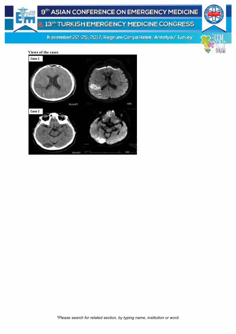
*Please search for related section, by typing name, institution or word.
Views of the cases

*Please search for related section, by typing name, institution or word.
[P-261]
Dissection of Cerebral Arteries due to Neck Cracking
(Boyun kütletmeye bağlı serebellar arter diseksiyonu)
MuratSeyit1,DuyguArasSeyit2,HasanEmreAydın3,AhmetBüber4
1Dumlupınar University Evliya Çelebi Training and Research Hospital, Department of Emergency Medicine, Kütahya, Turkey2Dumlupınar University Evliya Çelebi Training and Research Hospital, Department of Neurology, Kütahya, Turkey3Dumlupınar University Evliya Çelebi Training and Research Hospital, Department of Neurosurgery, Kütahya, Turkey4Dumlupınar University Evliya Çelebi Training and Research Hospital, Department of Radiology, Kütahya, Turkey
This study emphasizes the possibility of vertebrobasilar artery dissections in posterior spinal strokes in young patients and interventional procedures might be needed in such cases.
Case: A 32-year-old male patient presented to emergency with headache, pyotosis, numbness in right side of body and skill deficit due to turning his head for neck cracking 2 days ago. No specifications identified during brain and cervical CT. Diffusion-weighted magnetic resonance imaging revealed hyperintense image in right cerebellar medial (Figure). CT vertebro-basilar angiogram with contrast showed that the image was consistent with superior cerebral artery dissection. Neurology and neurosurgery were consulted. The patient was referred for interventional procedures.
Conclusion: Possibility of dissection should be kept in mind as a result of sudden neck movements.
Keywords: Dissection, cerebral artery, strain

*Please search for related section, by typing name, institution or word.
[P-262]
İskemik İnmede Nadir bir Klinik Bulgu: Pin-Point Pupil Mustafa Cicek, Sinan Paslı, Vildan Özer, Burcu Sağlam, Yunus Karaca, Aynur Şahin, Özgür Tatlı Karadeniz Teknik Üniversitesi, Acil Tıp Anabilim Dalı, Trabzon Pinpoint pupil göz bebeklerinin ileri derecede (<2 mm) küçüldüğü durumlar için kullanılan tanımlamadır. Nadir rastlanan tanısal açıdan değerli bir bulgudur. Genel olarak pin-point pupil yapan sebepler ve kısaltması Tablo-1’de gösterilmiştir. Aklımıza öncelikle pons kanaması ve opiat intoksikasyonu gelse de nadiren farklı problemlerde de pin-point pupil bulgusu karşımıza çıkabilmektedir. Bu vakada 27 yaşında pin-point pupil ve bilinç problemi ile acil servise başvuran iskemik serebrovasküler olay hastası sunulmuştur. 27 yaşında erkek hasta acil servise ani başlayan bilinç bulanıklığı şikayeti ile ailesi tarafından getirildi. Bilinen bilate kardiyomiyopati hastalığı olan ve bu nedenle dabigatran 300mg/gün, karvedilol 12.5mg/gün ve furosenmid 40mg/gün kullanmakta. İlk değerlendirmede genel durum orta, bilinç hafif uykuya meyilli, kısmen oryante koopere, olan hastanın konuşma bozukluğu olması nedeniyle Glaskov koma puanı 10 idi. Baş boyun muayenesinde bilateral pin-point pupil mevcut olup ışık refleksi zayıflamış, sol gözde dışa-yukarı kayma izlendi(Resim-1). Hastanın sağ üst ve alt ekstremitesinde 2/5 kuvveti olan hastaya santral görüntüleme alındı. Alınan Beyin tomografi görüntülemesinde patoloji izlenmedi. Beyin manyetik rezonans görüntülemede sol MCA sulama alanında Diffuzyon sekansında hiperintens, ADC sekansında aynı yere karşılık gelen alanda hipointens geniş enfarkt izlendi. Hasta nöroloji hastalıkları ile konsülte edildi ve yoğun bakıma yatışı yapıldı. Pin-point pupil acil servislere çok sık başvurmayan fakat başvuru anında asla atlanılmaması gereken hastalıkları ihtiva edebilecek bir klinik bulgudur. Bilateral iler derece ola miyosis tablosunda genellikle toksinlere maruziyet sonrası sempatomimetik tonus kaybı, kolinerjik yanıtta artma ve sedatif-hipnotik etkilerin artması ile gerçekleşir. Ön planda opiat zehirlenmesi, pons kanaması ve organofosfat zehirlenmeleri düşünülmekle beraber farklı ilaçlar ve hastalıklara bağlı olabilmektedir. Literatürde farklı maruziyetler veya klinik tablolarda gerçekleşen pin-point vakaları mevcuttur. İnsülin intoksikasyonu, nonketotik hiperglisinemi ve Hashimoto ensefalopatisinde görüldüğü literatürde işlenmiştir. Pinpoint pupil kliniği ile acil servise başvuran hastalarda ön tanılarımız içerisinde olması gereken başlıca tablolar arasında iskemik inme unutulmamalıdır. Anahtar Kelimeler: İnme, SVO, pinpoint

*Please search for related section, by typing name, institution or word.
Pinpoint Pupil

*Please search for related section, by typing name, institution or word.
[P-263]
Why is my patient in deep sleep? Mustafa Emin Çanakçı1, Filiz Baloğlu Kaya2, Şeyhmus Kaya1, Tuğrul Zeytin1, Fatih Alper Ayyıldız1 1Emergency Service, Eskisehir State Hospital, Eskisehir, Turkey 2Department of Emergency Medicine, Eskisehir Osmangazi University Medical School, Eskisehir, Turkey Introduction: Physicians often encounter cases with altered state of consciousness (ASC) at the Emergency Department (ED). The pathologies, which occur in the thalamus, might clinically reveal with ASC. Bilateral thalamic infarcts are rare and accounting for 22 to 35% of all thalamic infarcts. In this case, that bilateral thalamic infarct might be detected in a young patient admitted to the ED with ASC was emphasized. Case: A 37-year-old female patient in deep sleep was brought to the ED. The patient had numbness in her arms and legs for 3 days and was not awake for the last 16 hours. She had no known additional illness and did not use any drugs. In her physical examination, her arterial pressure was 100/70 mmHg, pulse was 65 bpm, respiratory rate was 16 breaths/min, temperature was 36.7 C°, and sPO2 was 99%. Her GCS score was 9 (E2V2M5). IR was +/+ and her pupil size was normal. There was not any pathological findings in her other examinations. No abnormal finding was detected in her EKG, electrolyte levels and blood toxicology test. Her cranial CT revealed bilateral hypodense areas in the thalamic region. Her diffusion MRI showed diffusion restriction in the bilateral thalamic region and the right cerebral hemisphere. She was transferred to a higher level hospital with a stroke center. In her repeated neurological examination, her GCS score was 3 (E1V1M1) and her CT angiography revealed occluded left vertebral artery, left ACA, and bilateral PCA. No clinical improvement was seen and after 45-day follow-up, the patient was declared dead. Conclusion: ED physicians should diagnose the thalamic infarcts being one of the rare and fatal reasons of deep sleep. ED physicians should pay attention to early application of necessary imaging techniques and proper patient management with supporting treatments. Keywords: Thalamic infarct, deep sleep, coma

*Please search for related section, by typing name, institution or word.
Thalamic Infarct MRI
[P-264]
A Rare Cause of Diffusion Restriction Mustafa Emin Çanakçı1, Nurdan Acar2, Emine Karkaç3, Rabia Sedef Üre4, Metin Kıcır1 1Emergency Service, Eskisehir State Hospital, Eskisehir, Turkey 2Department of Emergency Medicine, Eskisehir Osmangazi University Medical School, Eskisehir, Turkey 3Department of Infectious Diseases, Eskisehir State Hospital, Eskisehir, Turkey 4Department of Neurology, Eskisehir State Hospital, Eskisehir, Turkey Objective: We report a rare and stroke-mimicking form of meningitis with complicated with leptomeningeal circulation disorder. Case: A 42 yo male patient presented with ongoing fever, fatigue, headache and sleepiness for two days. No nausea and vomiting. The patient presented the emergency service two times with fever in the last 2 days. When he applied, the patient relaxed and went home. There are no previously known comorbidities. At the time of last admission he had tachycardia (110 bpm) and fever (38.7⁰ C). Pupillary isochoric and light response was normal bilaterally. Right hemiparesis (2/5 muscle strenght). He had no neck stiffness. No rash on the skin. Left parietal hypodens area and no contrast enhencement seen on CT scan. On MR imaging left parietal diffusion restriction was detected. As the patient had altered mental status and fever, we decided and applied lomber puncture. WBC: 15000/ml glucose: 65 mg/dl protein: 0.2 mg/dl seen in laboratory study of cerebrospinal fluid. The diagnosis was made as meningitidis. Existing pathologies were evaluated as leptomeningeal circulation failure related to meningitis. The patient was operated for decompression due to intracranial shift three days later. At the operation, empyema of the patient was evacuated. After medical and surgical therapies, the patient’s clinical condition was fixed and his Modified Rankin’s Scale score

*Please search for related section, by typing name, institution or word.
was 0. Conclusion: Leptomeningeal circulation failure due to meningitis is not common, and CT and MR images provide ischemic stroke-like findings. When similar patients are encountered, it will be important to recognize these images by emergency physicians. Keywords: Diffusion restriction, stroke, subdural empyema
Left Hypodens Area

*Please search for related section, by typing name, institution or word.
[P-265]
Press Sendromu: Olgu Sunumu Neşe Karslıoğlu, Halil Doğan, Esra Günal, Hakan Aydın, Hüseyin Işık Bakırköy Dr. Sadi ̇ Konuk Eği ̇ti ̇m Ve Araştırma Hastanesi ̇ Aci ̇l Tıp Kli ̇ni ̇ği ̇, Istanbul Press Sendromu: Olgu Sunumu: Posterior Reversibi Ensefalopati Sendromu (PRES), bir nöroradyolojik tanı olup ilk kez 1996 yılında Hinchey ve arkadaşlan tarafından tanımlanmıştır. Semptomlar baş ağrısı, mental bozııkluk, görmede ya da görme alanında değişiklikler, kortikal körlük, bulantı, kusma ve epileptik nöbetleri içermektedir. Sıklıkla ani yükselen ve komprese edilemeyen arter basıncına bağlı olarak gelişir ve hipertansij ensefalopati ile karışabilir. Olgu: 87 yaşında bayan hasta bulantı kusma bilinç kaybı nedeni ile acil servise getirildi. Fizik muayenede genel durum kötü, şuur kapalı, GKS: E2M1V1 olan hasta. Kan basıncı: 170/90 mmHg, diğer vital bulgular normal sınırlarda. Diğer sistem muayeneleri doğaldı. Laboratuar tetkiklerinde glukoz:429 keton: negatif asidozu yoktu, osmolaritesi 317 mOsm /L olan hastanın diğer parametreler normaldi. Difüzyon MR ında bilateral serebral hemisferde parietookspital lokalizasyonda verteksten başlayarak okspital lob inferioruna kadar uzanım gösteren ADC serilerinde karşılığı izlenebilen akut sitotoksik ödem ile uyumlu yaygın kısıtlı difüzyon sahası (Resim 1) tespit edilmesiyle PRES sendromu on tanısı ile yogun bakıma yatırıldı, antihipertansif tedaviye başlandı. Tansiyon kontrolu sağlanan şeker regülasyonu düzenlenen hasta yoğun bakım takibinin 5. gününde GKS: E4M4V4 olarak taburcu edildi. Sonuç ve Tartışma: PRES klinikte baş ağrısı, bulantı, kusma görsel ve mental değişiklikler, jeneralize ve fokal nöbetler gibi nonspesifik bulgularla karşımıza çıkabilen,radyolojik olarak parietal ve okspital bölgelerde daha belirgin olmak üzere yaygın serebral ödem ile karakterize geçici bir durumdur.Ancak tanı ve tedavinin gecikmesi durumunda status epileptikus, intrakranial kanama ve masif iskemik enfarkt oluşumu gibi komplikasyonlar sonrası ciddi morbidite ve mortalite ile seyredebilmektedir.Bu nedenle klinik bulgular, altta yatan etkenler ve MRG bulguları birlikte degerlendirilip PRES tanısı konulan hastalarda bu tanının erken teşhisi ve etkene yönelik tedavisi önemlidir. Aksi halde kalıcı beyin hasarına ve kronik epilepsi gibi nörolojik sekellere neden olabilir. Anahtar Kelimeler: Press Sendromu, bilinç kaybı, hipertansiyon

*Please search for related section, by typing name, institution or word.
Resim 1
Difüzyon MR ında bilateral serebral hemisferde parietookspital lokalizasyonda akut sitotoksik ödem ile uyumlu
yaygın kısıtlı difüzyon sahası

*Please search for related section, by typing name, institution or word.
[P-266]
Pneumocephalus After Shunt Laceration Oğuzhan Demir, Murat Doğanay, Eren Onur Karavin, Sinan Karacabey, Haldun Akoğlu, Arzu Denizbaşı Marmara University Faculty of Medicine, Department of Emergency Medicine, Istanbul, Turkey Introduction: Pneumocephalus occurs secondarily to fracture or surgery on the base of the skull, thoracotomy, tumor invasion, or encephalocele. The presence of air inside the skull means that there is a connection between the atmosphere and the intracranial cavity. We present a case of a woman with pneumocephalus who had air through the ventriculoperitoneal shunt she had before, after minor head trauma without skull fracture. Case: An 37-year-old woman admitted to ER after minor head trauma. She stated that she has fallen while walking and hit her head to the floor. She also stated that she had a intracranial surgery history for brain tumor 26-years-ago and had a ventriculoperitoneal (VP) shunt after this operation. She had no history of vomiting or loss of consciousness until then. Her GCS was 15 and vitals were stable. On her physical examination, a scalp laceration was noticed with normal neurological examination. Cranial CT was performed. Air in the ventricles, and a scalp laceration on the path of shunt was present, without any skull fracture. Laceration on the shunt was suspected as the source of the air in the ventricle. The patient was put on analgesics and antibiotics, and admitted to neurosurgery ward. Discussion: Traumatic pneumocephalus usually occurs due to skull fracture. Hyperbaric oxygen therapy, prophylactic antibiotics and analgesia are used for the treatment of pneumocephalus. It usually heals within two weeks. When severe pneumocephalus is present, antiedema therapy and even surgical drainage may be necessary. It is very rare to experience pneumocephalus after shunt laceration. It is important to perform cranial CT even if the neurological examination is normal in patient with VP shunt. Keywords: Ventriculoperitoneal shunt, head trauma, pneumocephaly

*Please search for related section, by typing name, institution or word.
[P-269]
Batın İçine Yabancı Cisim Penetrasyonu Sonrası Görülen Pres Sendromu Özcan Yavaşi1, Mecit Çokluk1, Ensar Durmuş1, Mehmet Oktay1, Mümin Murat Yazıcı1, Ali Demir2, Ömer Lütfi Gündoğgu3 1Recep Tayyip Erdogan University, Faculty of Medicine, Department of Emergency Medicine, Rize 2Recep Tayyip Erdogan University, Faculty of Medicine, Department of General Surgery, Rize 3Recep Tayyip Erdogan University, Faculty of Medicine, Department of Neurology, Rize Giriş: Posterior reversible ensefalopati sendromu (PRES), genellikle geri dönüşümlü beynin posterior bölgesinde vazojenik ödem ile karakterize olan klinik-radyolojik bir sendromdur. Hastalar sıklıkla baş ağrısı, letarji, nörolojik semptomlar görme bozuklukları bulantı gibi nonspesifik şikayetler ile başvurur. Tedavide ilk basamak etyolojik sebebin ortadan kaldırılmasıdır. Hipertansiyonun ve olayı tetiklediği düşünülen faktörlerin engellenmesi anahtar noktadır. Bu olgu ile radyoloji ve nöroloji bölümlerince iyi bilinen ancak sık karşımıza çıkmadığı için acil hekimlerince iyi bilinmeyen bu sendromun ayırıcı tanılarımız arasında olmasını anımsatmak amaçlanmıştır Olgu: 47 yaşında erkek hastanın bilinen hiçbir komorbiditesi olmayan hasta yoktu. Acil servisimize dünden beri başlayan şiddetli baş ağrısı, kusma ve sonrasında genel durumunda kötüleşme ve uyku hali gelişmesi üzerine 112 ambulans ile getirildi. Hastanın genel durumu kötü, bilinç açık, letarjik, oryante ve koopere ve pupilleri izokorikti. Fasiyal asimetrisi yoktu. Sol hemiparezi ve sağ alt ekstremitede monoparezi mevcuttu. Babinski bilateral lakayttı. Tansiyon arteryel 120/90 mmHg, nabız 100 atım/dk, solunum sayısı 25/dk, ateş 36,4 °C ve sPO2:98 idi. Akciğer ve kalp sesleri doğal, batın sağ kadranda 1X0.5 cmlik yara mevcuttu. Hassasiyet yok defans yok rebound yoktu. Difüzyon ağırlıklı manyetik rezonans görüntülemede sağ serebral hemisfer frontal lob, oksipitotemporal bölgede, sol serebral hemisfer frontoparietooksipital bölgede yamalı tarzda kortiko-subkortikal yerleşimli difüzyon kısıtlanma alanları görüldü. Laboratuar tetkiklerinde herhangi bir patolojik bulguya rastlanmadı. Mahkum hastanın gardiyanları hastanın yaklaşık 2 hafta önce karnından şişlendiği şüphelerinin olduğunu söylemesi üzerine dekübit grafi çekildi ve batın içerisinde sivri uçlu radyoopak yabancı cisim görüldü (şiş? tığ?). Hasta genel cerrahi ve nöroloji bölümleri ile konsülte edilip yatışı yapılarak operasyona alındı. Postoperatif çekilen kontrastlı MR venografisinde ise bu difüzyon kısıtlılığı olan bölgelerde yamalı kontrast tutulumu gözlenmiş olup posterior reversible ensefalopati sendromu olarak değerlendirilmiştir. Sonuç: Sonuç olarak PRES sendromu multifaktöriyel nedenlere bağlı ve farklı nonspesifik klinik bulgularla karşımıza çıkabilecek ve radyolojik görüntüleme ile kesinleştirilebilecek bir durumdur. Anahtar Kelimeler: Hypertension, posterior reversible ensefalopati sendromu, PRES sendromu

*Please search for related section, by typing name, institution or word.
[P-270]
Cerebral Sinus Venous Thrombosis After Pregnancy With In Vitro Fertilization Selahattin Gürü1, Ayhan Özhasenekler2, Melih Çamcı2, Fatih Tanrıverdi2, Şervan Gökhan2, Gülhan Kurtoğlu Çelik2 1Ankara Atatürk Training and Research Hospital, Emergency Service, Ankara, Turkey 2Yıldırım Beyazıt University, Faculty of Medicine, Emergency Medicine Department, Ankara, Turkey Introduction: Cerebral sinus venous thrombosis (CSVT) is an acute and life-threatening condition. It has an incidence rate of 0.5-1% in all cerebrovascular diseases. Sinus venous thrombosis, which may occur at every age, is more frequent in women between 20-40 years of age. Because of the variety of symptoms, CSVT is even more important for emergency physicians. In this article, we aimed to discuss the case with unusual cause of headache. Case: A 24-year-old female patient was admitted to the ED with headache starting from the neck for 2 days, on the addition of complaint of double vision in the right eye after pregnancy obtained by in vitro fertilization and 32 days before cesarean section story. She did not have a chronic disease in her background. The patient's vital signs were normal on arrival. Neurological examination revealed no pathological findings. Despite symptomatic treatment, computerized brain tomography was performed because of ongoing headache. Cranial magnetic resonance (MR) imaging and cranial MR venography were performed and the filling defect was detected in left transverse sinus, sigmoid sinus and juguler vein. Low-molecular-weight heparin (enoxaparin) therapy was initiated and admitted to the inpatient bed. After 10 days of follow-up, she was discharged without any sequela. Discussion: CSVT is a pathology that occurs a small proportion among cerebrovascular diseases. All genetic or acquired prothrombotic factor has a major share in the etiology of CSVT. In vitro fertilization treatments, some hormone replacement therapies, and oral contraceptive use, pregnancy and puerperia which are known to be among these factors, have been associated with the more frequent occurrence of CSVT in women. Emergency physicians who frequently encounter headache that the most common symptom, have an important role for early diagnosis of CSVT and for early treatment initiation. Keywords: Sinus Venous Thrombosis, Emergency Medicine

*Please search for related section, by typing name, institution or word.
[P-271]
A Rare Case Present with Headache in The Emergency Medicine; Cerebral Vein and Dural Sinus Thrombosis (Cvst) Selin Bulut, Caner Sağlam, Hayriye Gönüllü, Mustafa Silcan, Özkan Eraslan, İsmet Parlak Department of Emergency Medicine, Bozyaka Eğitim ve Araştırma Hastanesi, Izmir Cerebral vein and dural sinus thrombosis is less common and life-threatening disease which has variable many clinical conditions. It is known that CVST can present different clinical features and has several causes, therefore CVST is a disease that may be encountered not only by neurologist and neurosurgeon, but also by emergency physicians. The aim of this case report is to provide an example of how early suspicion of CVST, even with initial negative imaging, is critical, as any delay in diagnosis and treatment can cause increased morbidity and mortality. A 40-year old female was admitted to the emergency room for onset of severe, on-going headache and left ear ache for the past 1 week. She has no past medical history, with no history of headaches. This is her second visit within the prior 36 hours. On the examination the patient had 15 of glaskow coma score with normal vital signs. Pupils were equal and reactive and there was no papillary edema. Neurological exam was benign with no evidence of meningism or any focal neurological deficit, cranial nerves were intact, Brudzinski and Kernig signs were negative. There was erythama and sensation of mastoid bone with palpation. The ear examination with otoscope were all normal with intact timpanic membrane and no erythema, oedema and exudate at outer ear canal. The laboratory results which included full blood count, urea, electrolytes, sedimentation and CRP were within normal limits. A non-contrast head and temporal CT scans were performed showing subarachnoid hemorrage in the left parietal lobe. In addition, MRI venography was confirmed and showed a thrombosis in the left transverse, sigmoid sinus and jugular vein. Neurology and neurosurgery consultations were requested. She was transfered to the department of neurology further diagnosed and treatment. Keywords: Cerebral vein sinus vein thrombosis, headache, venography

*Please search for related section, by typing name, institution or word.
mrı venography
[P-272]
Unexpected Neurological Emergency for A Child: Transverse Myelitis Sercan Yalçınlı, Mevlüt Özden, Eylem Ulaş Saz, Murat Ersel Ege University School of Medicine, Department of Emergency Medicine, Izmir, Turkey Acute transvers myelitis refers to acute or subacute spinal cord dysfunction characterized by paraplegia, a transverse level of sensory impairment and sphincter disturbance. It is relatively rare, with a reported annual indicence of 1-8 case per 1 million population. It makes two peaks, in 10-19 and 30-39 years old. The pathogenesis is not well described. T cell mediated autoimmunity may be responsible for pathogenesis. Seventy percent of patients have a prior infection history. Fever, neck stiffness, myalgia are observed in a significiant part of the cases. Back pain, leg pain, paresthesia are the first developments to fit the lesion. Nine years-old female admitted with weakness in the legs, difficulty in walking and fatigue which are started after a taekwondo training. Vital sign were noted as normal. At that admittance there was no urinary and fecal incontinence and altered mental status. Lower extremity DTR was ++++/++++, bilateral babinski reflexes were +, lower extremity muscle strength was 1/5, upper extremity muscle strength was 5/5, optic disk view was normal. Abdominal superficial reflexes were not present. Laboratory findings were in normal limits. Serological findings were negative for viral diseases. MR findings were compatible with transverse myelitis. She was admitted to pediatric neurology service with transverse myelitis diagnosis. The patient was treated with pulse steroid from 30 mg/kg for 5 days. At the third day abdominal superficial reflexes are found as positive, urine output and lower muscle strength recovered as 2/5. At the 9th day she recovered completly. Spinal cord pathologies have higher morbidity. We should also consider transverse myelitis at the differential diagnosis of children and young patients who apply with back pain, leg pain, numbness, difficult urination, constipation, difficulty in walking.

*Please search for related section, by typing name, institution or word.
Keywords: Transverse myelitis, emergency, children

*Please search for related section, by typing name, institution or word.
[P-273]
A Case of Reversible Cerebral Vasoconstriction Syndrome Followed by Subarachnoid Hemorrhage Requiring One Week for Diagnosis from The Onset of Thunderclap Headache Toshiyuki Onodera, Ichiro Sekine, Hiroshi Yamagami, Hisashi Ofuchi Emergency Department, Shonan Kamakura General Hospital, Kanagawa, JAPAN Learning Objective 1: Diagnose RCVS when you see recurrent thunderclap headache patient, if appropriate examination doesn’t show any abnormality Learning Objective 2: Recognise RCVS if you find cSAH on head imaging Case: A 51-year-old female with past medical history of cerebellar infarction, came to our hospital with thunderclap headache. On the day of onset, the computed tomography (CT) scan showed no abnormality, and she was diagnosed as migraine. On the next day of onset, thunderclap headache recurred after it’s once improving and she came to our hospital again but head magnetic resonance imaging (MRI) didn’t show any abnormality. 5 days after onset, she felt thunderclap headache again, and came to hospital again. Repeated head CT scan didn’t show any abnormality, then she was suggested lumber puncture to rule out subarachnoid hemorrhage (SAH), but she refused. 8 days after onset, she came to our hospital again with continuous headache, and head MRI showed convexity SAH (cSAH). Finally she was diagnosed as reversible cerebral vasoconstriction syndrome (RCVS) followed by cSAH, which meets International classification of headache disorder-3 beta criteria of RCVS. She was followed up as outpatient with tramadol-acetaminophen combination and calcium channel blocker, and her headache gradually improved. Discussions: 62% patient of cSAH in under 60 patient group, diagnosed as RCVS. RCVS patient found convexity SAH in 30-34%, and cSAH were early complication in the first 10 days (5±5) days of headache onset. The majority of RCVS patients have a self-limited course. There have been no randomized control trials in the treatment of RCVS to guide management. Patients presenting with features of RCVS require early identification and withdrawal of precipitating vasoactive agents, avoidance of headache triggers, analgesia, bed rest, and observation. Empiric treatment with calcium channel antagonists has been instigated in most published series of RCVS. Keywords: Thunderclap headache, SAH, RCVS
*Please search for related section, by typing name, institution or word.
[P-274]
Atipik Başlangıçlı Gullian Barre Sendromu Tuğba Tuksavul, Öner Bozan, Şeref Emre Atiş, Esra Karaman, Merve Ekşioğlu Sağlık Bilimleri Üniversitesi Okmeydanı Eğitim Ve Araştırma Hastanesi Acil Tıp Kliniği, İstanbul Guillain Barre sendromu hızlı progresyon gösteren, simetrik ve aşağıdan yukarı doğru ilerleyen güçsüzlük ve arefleksi ile karakterize akut, demiyelizan, inflamatuar bir polinöropatidir. Görülme sıklığı 2.7/100000/yıldır. Bu olguda solunum güçlüğü ile gelen bir guillain barre sendromunu sunmayı amaçladık. 23 yaşında erkek, bilinen hastalık öyküsü olmayan iki gündür yutma güçlüğü, solunum sıkıntısı ve yeni başlayan kollarda güçsüzlük şikayetleri olan hasta acil servise başvuruyor. Anamnezinde 1 hafta önce üst solunum yolu enfeksiyonu geçirdiğini belirten hastanın vitalleri ta: 127/60, spo2:97 inabız: 75/dk, ateş:36 olan hastanın Glaskow koma skalası 15 olarak hesaplandı. Hastanın yapılan fizik muayenesinde; genel durumu iyi, bilinç açık, koopere, oryante. solunum sesleri dinlemek işe eşit, ral ve ronküs yok. Konuşma spontan, anlama, isimlendirme, tekrarlama doğal, dizartri yok. Solunum sıkıntısına sekonder disfonisi mevcut. Ense sertliği, kerning, brudzinki yok. Pupiller izokorik, ışık refleksleri doğal, göz hareketleri her yöne serbest. Fasial asimetri yok, uvula orta hatta, dil ağız içi ve dışında orta hatta. Hastanın fasial diplejisi mevcut. DTR'ler (derin tendon refleks) sol üst ekstremitede hipoaktif. Solunum sıkıntısı nedeniyle ybü ne transfer edilen ve 5 gün IVIG tedavisi verilen hasta 2 aylık yogun bakım tedavisi sonrası servise alındı. Servise geldiğindeki nörolojik muayenesinde kas gücü sağ üst ekstremitede 5/5, sağ alt ekstremitede 3/5, sol üst ekstremitede prox 1/5, distali 2/5, sol alt ekstremite 3/5 olarak değerlendirildi. Dtr'lerhipoaktif. Anahtar Kelimeler: Guillane barre sendromu, atipik başlangıç

*Please search for related section, by typing name, institution or word.
[P-275]
Acil Serviste Fahr Sendromu Ürke Selen Çöme, Şahin Kahraman, Raziye Sinem Ceylan Dokuz Eylül Üniversitesi Tıp Fakültesi, Acil Tıp Ana Bilim Dalı, izmir Fahr sendromu; kalsiyum ve fosfor metabolizma bozukluğu sonucu gelişen, çift taraflı simetrik intrakranyal kalsifikasyon ile seyreden nörodejeneratif bozukluklarla karakterize nadir görülen bir hastalıktır. Etyolojisi kesin olarak bilinmemektedir. Olgumuzda 55 yaşında kadın,1 gün önce başlayan konuşma bozukluğu şikayeti ile acil servise başvurdu.Total tiroidektomi ve paratiroidektomi öyküsü olan hasta 30 yıldır düzenli olarak Ca replasmanı alıyor.Vital bulguları normal sınırlardaydı.Muayenede bilinç açık oryante koopere konuşma ağır dizartrik ve sağ telem silikti.Beyin tomografisinde Fahr sendromu ile uyumlu görünüm izlenmekte olup bilateral bazal ganglionlar, talamus ve dentate nükleuslarda kalsifikasyonlar mevcut.Diffüzyon manyetik rezonans görüntüsünde sol kapsulointernal posterior bacağında akut difüzyon kısıtlılığı mevcut.Biyokimya parametrelerinde kalsiyum değeri 9,56 olup diğer labaratuar tetkileri olağan.İleri tetkik ve takip amaçlı nöroloji servise yatırıldı.Fahr Sendromu,bilateral ve simetrik olarak bazal ganglionlar,serebellum ve sentrum semiovaleye kalsiyum ve çeşitli minerallerin birikimi ile ortaya çıkar.Klinik olarak genellikle yürümede dengesizlik, konuşma bozukluğu, istemsiz hareketler, ya da kas krampları ile başlayıp tabloya psikoz, kişilik değişiklikleri gibi nöropsikiyatrik semptomlar da eklenebilir.Yazımızda, acil servise konuşma bozukluğu şikayeti ile başvuran BT'de bazal ganglionlarda bilateral yaygın kalsifikasyon saptanan Fahr sendromu olgusu sunulmuştur.Fahr sendromu uzun yıllar önce tanımlanmış olmasına rağmen sık karşılaşılmadığı için gözden kaçabilmektedir. Ani nöropsikiyatrik semptomlar görülen, kalsiyum metabolizma bozukluğu olup, sebebi anlaşılamayan nörolojik semptomlar gelişen hastalarda Fahr sendromu ayırıcı tanıda mutlaka hatırlanması gerekmektedir. Anahtar Kelimeler: Fahr sendromu, kalsifikasyon

*Please search for related section, by typing name, institution or word.
Bilgisayarlı beyin tomografisi
bilateral bazal ganglionlar, talamus ve dentate nükleuslarda kalsifikasyonlar mevcut olup Fahr sendromu ile
uyumlu bilgisayarlı beyin tomografisi görüntüsü

*Please search for related section, by typing name, institution or word.
[P-276]
Percheron Arteri Tıkanıklığına Bağlı Talamusta Enfarkt Vahide Aslıhan Durak, Arif Mesud Kaya, Ahmet Çiftçi, Abdullah Erdem, Anıl Ardıç, Fatma Özdemir, Halil Ibrahim Çıkrıklar, Şule Akköse Aydın Uludağ Üniversitesi Tıp Fakültesi, Acil Tıp Ana Bilim Dalı, Bursa Giriş: Talamus beynin hem ön hem de arka dolaşımından kanlanmaktadır. Arka dolaşımda posterior serebral arterlerin P1 segmentinden çıkan damarlar paramedian talamus ve mezensefalonu, P2 segmentinden çıkanlar ise lateral ve üst yüzlerini beslemektedir. Percheron bu damarsal yapı üzerine çalışmış ve P1’den köken alan 3 sulama şekli tanımlamıştır. Percheron’un tanımladığı 2. tipte, P1’den çıkan tek bir kök bilateral dağılım sağlamaktadır. Bu kökün tıkanması, paramedian talamus ve mezensefalonda enfarktla sonuçlanmaktadır Olgu: 38 yaşında erkek hasta acil servise yeni gelişen jeneralize tonik klonik nöbet geçirme şikayeti ile başvurdu. Hastanın vital bulguları stabil olup fizik muayenesinde ek muayene bulgusu saptanmadı. Kan tetkikleri ve radyolojik görüntülemeleri yapıldı. Beyin tomografisinde acil patoloji saptanmayan hastanın difüzyon MR görüntülemesinde bilateral talamuslarda difüzyon kısıtlayan enfarkt ile uyumlu lezyon saptandı. Hasta nörolojiye konsülte edildi. Asetil salisilik asit, Plavix ve Kepra tedavisi başlanan hastanın yatışı yapıldı. Tartışma ve Sonuç: Talamus önemli nükleusları barındırmaktadır ve bu nükleuslar korteksle bağlantı içinde bulunmaktadırlar. Bu nedenle talamus lezyonlarında çok çeşitli klinik bulgular (davranış bozukluğu, sensoriyel kayıp, demans) ortaya çıkmaktadır. Özellikle paramedian talamik lezyonlarda mental bozukluklar geliştiği bilinmektedir. Percheron arteri, PCA’nın P1 kısmından tek kök halinde çıkıp bilateral talamus medial yüzlerini ve rostral beyin sapını besleyen bir arteriyal varyasyondur. Bu arterin tıkanıklıklarında paramedian talamik ve mezensefalik lezyonlar daha çok simetrik bir görünümdedir. Bilateral paramedain talamik infarkt gözlendiğinde, Percheron arteri tıkanıklığı öncelikle düşünülmelidir. Bu hastalarda genelde klinik şiddetlidir, bilinç kaybı söz konusudur, düzelme de kısmidir ve erken tanı tromboliz açısından önemlidir. Anahtar Kelimeler: Talamus, enfarkt, nöbet

*Please search for related section, by typing name, institution or word.
[P-277]
Gebe Hastada Venöz Tromboz Vahide Aslıhan Durak, Çağlar Karpuz, Habip Emrah Leylek, Nezahat Duruk Kahriman, Mehmet Karamurat, Ziyaettin Uzun, Özlem Köksal, Şahin Aslan Uludağ Üniversitesi Tıp Fakültesi, Acil Tıp Ana Bilim Dalı, Bursa Giriş: Serebral venöz tromboz klinik semptom ve belirtilerin değişkenliğinden dolayı tanı koyması zor olan bir hastalıktır. Klinik tablonun daha yavaş gelişimi ile epilepsi ve hemoraji eğilimi venöz tıkanmayı akla getirmelidir. Sistemik inflamatuar hastalıklar, kalıtsal veya edinilmiş koagulasyon bozuklukları etyolojide rol oynarken, %30 hastada neden bulunamaz. Oral kontraseptifler, gebelik ve lohusalık da ilave risk faktörlerindendir. Olgu: 32 yaşında ve 17 haftalık gebelik öyküsü olan hasta; ani başlayan sırt ağrısı, titreme ve senkop şikayeti ile acil servise başvurdu. Hastanın senkop sonrası yaklaşık 1-2 dakika süren hafıza kaybı şikayeti olmuştu. Dış merkezde çekilen beyin MR görüntülemesinde şüpheli kitle görünümü saptanması üzerine acil servisimize sevk edilmişti. Özgeçmişinde ve soygeçmişinde özellik yoktu. Hastanın gelişinde vital bulguları stabil olup GKS: 15 olarak saptandı. Fizik muayenesinde; sağ üst 2/5 alt früst hemiparezi, duyu muayenesi normal ve derin tendon refleksleri normal olarak saptandı. Yapılan biokimyasal tetkiklerinde patolojik bulgu saptanmayan hastanın beyin ve difüzyon MR tetkikleri istendi. Difüzyon MR görüntülemesinde; sağ trolard kortikal veninde trombüs görünümü ve sağ konveksitede düzeyinde presentral gyrus posteriorunda hemorojik alanın izlendiği enfarkt alanı saptandı. Hastanın ileri tetkik amaçlı nöroloji kliniğine yatışı yapıldı. Tartışma ve Sonuç: Serebral venöz tromboz kadınlarda özellikle de 20-35 yaş arasında, gebelik, lohusalık ve oral kontraseptif kullanımı durumlarında daha sık görülen bir durumdur. İnsidansı Doğu ülkelerinde yüksek olup Avrupa ve Amerika’da daha az olarak saptanmıştır. Hastaların %80’inde predispozan faktörler belirlenebilir. Bunlar arasında travma, enfeksiyon, şiddetli anemi, gebelik, lohusalık, oral kontraseptif kullanımı, hiperkoagülabilite durumları, dehidratasyon, konnektif doku hastalıkları, bazı granülomatöz ve inflamatuar hastalıklar gibi sistemik hastalıklar ve maligniteler bulunur. Tedavide; konvülsiyonları kontrol altına almak anti-konvülzanlar, septik tromboflebit şüphesi varsa antibiotikler başlanır. Anahtar Kelimeler: Gebelik, venöz tromboz

*Please search for related section, by typing name, institution or word.
[P-278]
Tüberküloz Menenjitli Bir Olgu Vahide Aslıhan Durak, Serap Katı, Maruf Beğenen, Yıldız Ongun, Nur Sezin Hacıoğlu, Ayşegül Aktaş, Gizem Gizli, Hilal Aksoy, Özlem Köksal, Erol Armağan Uludağ Üniversitesi Tıp Fakültesi, Acil Tıp Ana Bilim Dalı, Bursa Giriş: Tüberküloz menenjit gelişmiş ve gelişmekte olan ülkelerde ciddi bir halk sağlığı sorunudur. Direkt hematojen yayılmadan ziyade subependimal tüberküllerin subaraknoid alana rüptüre olmaları ile oluşur. Arterlerin tutulumu ile infarkt veya iskemi oluşur. Sıklıkla orta serebral arterin tutulumu ile hemiparezi veya hemipleji görülür. Vakaların büyük çoğunluğundan Mycobacterium tuberculosis, özellikle gelişmekte olan ülkelerde çok küçük bir kısmından ise Bovin tip tüberküloz basili sorumludur. Olgu: 65 yaşında erkek hasta genel durum bozukluğu, halsizlik ve baş ağrısı şikayeti ile dış merkeze başvurmuştu. Potasyum eksikliği tedavisi almış ancak şikayetlerinin geçmemesi üzerine üniversitemize sevk edilmişti. Özgeçmişinde; diabet, hipertansiyon, koroner arter hastalığı ve primer trombositopeni mevcut idi. Fizik muayenesinde; genel durumu orta olan hastanın GKS:13 olarak saptandı. Ateş yüksekliği (38 derece) olan hastanın kan basıncı: 170-80 mmHg olarak ölçüldü. Hastanın şüpheli ense sertliği mevcuttu. Tetkiklerinde CRP: 1.3 mg/ dl olan hastanın lökositozu saptanmadı. Beyin tomografisi ve MR tetkikinde patoloji saptanmayan hasta menenjit ön tanısı ile enfeksiyon hastalıklarına konsülte edildi. Hastanın yapılan lomber ponksiyonunda; Pandy testi pozitif ve mikroskopik incelemede %90 MNL, %10 PNL saptandı. Hücre sayımında eritrosit:1410/mm³ ve lökosit 220/ mm³ ölçüldü. Hastaya tüberküloz menenjit tanısı ile tedavi başlandı. Genel durumunda düzelme olmayan hasta reanimasyon bölümüne konsülte edildi ve yoğun bakıma yatışı yapıldı. Tartışma ve Sonuç: Tüberküloz menenjit genellikle primer infeksiyonun bir komplikasyonudur. Patolojik tablo hastanın yaşı, infeksiyonun ciddiyeti, hastanın immunite veya hipersensitivite durumu, hastalığın süresi ve aldığı tedavi tipine bağlıdır. Hastalık prodrom döneminde halsizlik, başağrısı ve hafif ateş ile başlar, kusma, konfüzyon, meningismus ve fokal nörolojik belirtilerle devam eder. Klinik tablo kronik başağrısı veya hafif bilinç değişikliğinden ciddi menenjit veya komaya kadar gidebilir. INH 5 mg/kg, rifampisin 10 mg/kg, 9- 12 ay süreyle verilir. Tedavinin ilk 2-3 ayına etambutol 25 mg/kg/gün ve pirazinamid 15-30 mg/kg dozunda eklenmelidir. Tedaviye yanıt 2 hafta içinde görülür. Prognoz; yaş, semptomların süresi ve nörolojik tutulum ile ilişkilidir. En önemli prognostik belirti hastanın tedaviye başlandığı andaki bilinç durumudur. Anahtar Kelimeler: Acil servis, tüberküloz menenjit

*Please search for related section, by typing name, institution or word.
[P-279]
Headache in A Patient with Behcet's Disease: Neuro-Behcet Yasin Yıldız1, Ahmet Şenel1, Mine Kayacı Yıldız2, Erdem Çevik1, Serkan Emre Eroğlu2 1University of Health Sciences, Sultan Abdulhamid Han Training and Research Hospital, Emergency Medicine Clinic, Istanbul, Turkey 2University of Health Sciences, Umraniye Training and Research Hospital, Emergency Medicine Clinic, Istanbul, Turkey Introduction: Behcet's Disease (BD) is a multisystemic vasculitic disease with an unknown etiology. It is an inflamatory perivasculitis which characterized with oral and genital ulsers, but any tissue can be affected. Here we aimed to present a case of BD patient who is admitted to the emergency department with headache and hospitilazed with the diagnosis of NBD. Case: A 48-year-old female who had Behcet's disease for 13 years, presented to our emergency department with throbbing and severe headache that located in the left head side. On examination, all vital signs was normal and there was no nuchal rigidity. All neurological examination was normal. Cranial tomography revealed diffuse edemateus changings on the left hemisphere and shift at the left lateral venticule (Figure 1). The patient was reffered to the neurology clinic and hospitalized for advance diagnostic workup with the diagnosis of Neuro-Behcet's Disease. Discussion: Neurological system involvement is the most severe manifestation of BD, and the most frequent neurological symptom is headache. Neuro-Behcet's Disease (NBD) involvement have no any specific diagnostic labratory test. The diagnosis of NBD can be made with clinical and radiological findings. Conclusion: NBD, even though rare, should be kept in mind in the differential diagnosis for the patients who have BD with a complaint of headache. Keywords: Headache, Behcet's disease, Neuro-Behcet's disease
Figure 1
On cranial tomography, edemateus changings on the left hemisphere with evanescence of sulcuses (left parietal
and frontal lobes), and mid-line shift (6mm deviation)

*Please search for related section, by typing name, institution or word.
[P-281]
Intrauterine Exitus Banu Salur, Gülşen Çığşar, Murat Özdemir, Handan Çiftçi Kafkas Üniversitesi Sağlık Araştırma ve Uygulama Merkezi, Kars Intrauterine fetal mortality rates differ from country to country and stillbirth rates are 0.2% in developed countries, 0.7% in developing countries, 2% in southern Africa and some countries in Asia.The definition of intrauterine fetal death is exitus of the fetus older than 20 weeks or whose gestational age is not known, or there is no sign of vitality at birth or after birth of fetus over 500gr. Over90% of intrauterine deaths delivered spontaneously within 3 weeks but there is about 10% risk of developing coagulopathy within 4 weeks.We present a patient with coagulopathy after intrauterine exitus. A 22-year-old woman who was previously healthy was referred from a different health institution to us because she could not feel her fetal movements and the fetal heartbeats could not be detected.This was the first pregnancy of her and she had no history of miscarriage or abortion.She said she did not know her gestational age but she know that she was in term and she was feeling fetal movements at afternoon the day before.She does not remember the name of her medications.On her physical examination the patient was pale,in moderate-good condition but she did not have any pain.Vital signs were;blood pressure:90/60mmHg,pulse:84/min,respiratory rate:20/min,fever:36,6C°,SatO2:98%. A 36 week and 4 days old fetus and oligohydroamnios was detected and the fetal heartbeat was not detected in the fetal ultrasound.Intravenous hydration was initiated in the hypotensive patient after ultrasound and a consultation was requested for the patient from department of obstetrics and gynecology.Her laboratory results were Hgb:14.56,Hct:44.8,WBC:16.5,plt:150,Neu:13.1,%Neu:79.2,Kre:1.94,Üre:31,Ast:202,Alt:177,Alp:506,INR:5.84,PT:12.6,PTT:42.8,pH:7.25,laktat:2.5,ABE:-11.5,HCO3⁻:14.5.After the stillbirth,the mother was taken to the intensive care unit but after about a week later she passed away. Intrauterine fetal mortality rates vary according to the geographical and socioeconomic structures of the countries.There are many risk factors in the etiology of intrauterine fetal deaths,such as obesity, smoking, increased maternal age, maternal diseases. The rate of unknown stillbirths varies between 12% and 50%. Keywords: Intrauterine exitus coagulopathy

*Please search for related section, by typing name, institution or word.
[P-282]
A Case Report: A Shocking Masquerade: A Case of Spontaneous Rupture of Left Uterine Artery Joanah Partible Bithao St. Lukes Medical Center Quezon City, Department of Emergency Medicine, Philippines This is a case of a 38 years-old gravida 3, para 1 (1011) at 31 weeks age of gestation who presented with a 2-day history of right sided abdominal pain and dysuria, which eventually led to hypovolemic shock and fetal compromise. The case was followed from the onset of her symptoms, to the management of hypovolemic shock, to the consideration of placental abruption, then to the exploration of the abdominal cavity where the diagnosis of spontaneous rupture of left uterine artery was made. Conclusions drawn from the case study relates to the recognition and consideration of the rare possibility of an impending rupture of a uterine artery in hemodynamically stable pregnant woman presenting with abdominal pain in the 3rd trimester. If shock ensues in the presence of maternal instability or fetal distress necessitates urgent delivery, exploration of the abdominal cavity and ligation of uterine artery or other vascular bleeders are necessary. Keywords: Spontaneous rupture of uterine artery, obstetrical emergency, pregnancy complication

*Please search for related section, by typing name, institution or word.
[P-283]
A Case of Spontaneous Pneumothorax During Pregnancy Mehmet Esen, Öznur Kelepçe Emergency Department, Medical Faculty, Gaziosmanpasa Universty, Tokat, Türkiye Spontaneous pneumothorax(SP) is rare during pregnancy. SP during pregnancy is potentially serious for both the patient and fetus. But SP in general rarely occurs in women compared with men. A case of SP occurring at 17 weeks’ gestation in a healthy 23-year-old primigravida is described. Diagnosis was made by chest radiograph and treatment was by tube thoracostomy. Case: A 23-year-old primigravida at 17 weeks’ gestation presented with the sudden onset of right-sided pleuritic chest pain and dyspnea. There was no history of fever, chills, trauma, nausea, or vomiting. She was a nonsmoker and had no family history of pulmonary disease. Blood pressure was 120/80 mm Hg, heart rate 110 beats per minute, respiratory rate 16 breaths per minute, and temperature was 36 °C. Chest examination was notable for decreased breath sounds in rihght lung. Chest radiograph showed an almost complete pneumothorax of the right lung (Fig.I). A right tube thoracostomy was then performed in emergency room (Fig. II). Discussion: A SP during pregnancy is a very rare condition, and potentially serious for both the patient and fetus. The typical symptoms of SP, regardless of cause, include pleuritic chest pain associated with dyspnea. Chest radiographs are required for definitive diagnosis. When one suspects pneumothorax in a pregnant patient, it is safe to proceed with the standard chest radiography without placing the fetus at substantial risk from ionizing radiation if the abdomen is shielded. It is a unusual disorder during pregnancy, with approximately only 50 cases in the literature. Diagnosing a SP is not easy since it can be interfused with dyspnea in pregnancy. In addition, the etiology of SP is not well known. In this case report, we describe the diagnosis and treatment of a patient who experienced SP that developed during her pregnancy after the 17th week of gestation. Keywords: Pregnancy, spontaneous pneumothorax, thoracostomy

*Please search for related section, by typing name, institution or word.
Figure I: Chest radiograph image shows an almost complete pneumothorax in the right lung

*Please search for related section, by typing name, institution or word.
[P-284]
Fitz-Hugh-Curtis Sendromu; Bir Sağ Üst Kadran Ağrısı Mehmet Hüsametin Akküçük Başkent Üniversitesi Alanya Uygulama ve Araştırma Merkezi, Alanya, Antalya Başka bir klinik durumu taklit ederek ortaya çıkan hastalıkların tanısal anlamda farklılıklar göstermesi nedeni ile tekrarlayan hastane, özellikle de acil servis ziyaretlerine neden olmaktadır. Bu gibi zor klinik durumların tanısal belirsizliği, konsultasyon konusunda kısır döngüye dönüşür. Vakamız da bu olgulara bir örnek. Vakamız 23 yaşında kadın hasta son 3-4 gündür karın ağrısı ateş nedeniyle birden fazla acil servis ziyareti olmuş. Yapılan muayenesinde sağ üst kadranda ciddi hassasiyeti olan, CRP si 175 ve hafif KCFT yüksekliği olan hastanın kolesistit ön tanısı ile hepatobilier USG istendi. USG normal olan ve 2 hafta önce D&C hikayesi nedeni ile benzer klinik durumu taklit eden Fitz hugh Curtis sendromu olabileceği düşünülerek opaklı abdomen BT planlandı. BT de KC kapsülünde kalınlaşma ve opak tutulumu, ayrıca PID ile uyumlu görünümü olan hasta (Figür 1) kadın doğum servisene yatırıldı. Seftriakson ve metranidazol tedavisi başlandı. Takibinde klinik olarak rahatlayan ve CRP’si gerileyen hasta tedavisi düzenlenerek taburcu edildi. FHC sendromu Pelvik İnflamatuar Hastalığın komplikasyonu olarak karşımıza çıkan, özellikle de salpenjit ile birlikte gelişen karaciğer kapsülünün parankim tutulumu olmadan inflamasyonudur. Transperitoneal yolla yayılarak oluşan bu inflamasyon özellikle sağ üst kadran ağrısı yaparak kolesistit ile karışır. Ancak dikkatli bir öykü bu sendromun varlığından şüphelenilmesini sağlayabilir, bizim olgumuzda da geçirilmiş D&C mevcut. Tanı aşamasında USG daha çok ayırıcı tanıda yardımcı olur. Laparoskopi altın standart görüntüleme tekniğidir ve fibröz bantlarların oluşturduğu keman teli (violin-string) görülmesi ile olur. Arteryel faz BT görüntülerinde karaciğer kapsül üzerindeki kalınlaşmayı görmek FHCS için tanısal anlamda oldukça önemlidir. Genel olarak tedavide Antibiyotik seçiminde C. trachomatis, N. Gonorrhoeae fakültatif gram (-) çomaklar ve anaerob bakteriler hedeflenmelidir. Fakat antibiyotik seçimini izole edilmiş organizmalara yönelik yapmak en uygunu olacaktır. Antibiyoterapiye dirençli kronik vakalarda laparaskopik olarak yapışıklıkların koterize edilmesi klinik rahatlama sağlamaya yardımcı olacaktır. Sonuç olarak fertil çağda sağ üst kadran ağrısı olan kadın hastalarda nedeni açıklayacak bulgular yoksa Fitzh-hugh curtis sendromu akılda tutulmalıdır. Anahtar Kelimeler: Fitz-hugh-curtis sendromu, pelvik inflamatuar hastalık, sağ üst kadran ağrısı
Figure 1

*Please search for related section, by typing name, institution or word.
Karaciğer kapsülde kontrastlanma

*Please search for related section, by typing name, institution or word.
[P-285]
HELLP
MuratSeyit1,IsmailKaya2,DuyguArasSeyit3,HarunYıldırım11DumlupınarUniversityEvliyaÇelebiTrainingandResearchHospital,DepartmentofEmergencyMedicine,Kütahya,Turkey2DumlupınarUniversityEvliyaÇelebiTrainingandResearchHospital,DepartmentofNeurosurgery,Kütahya,Turkey3DumlupınarUniversityEvliyaÇelebiTrainingandResearchHospital,DepartmentofNeurology,Kütahya,Turkey
HELLP is a syndrome defined as Hemolysis, Elevated Liver Enzymes, and Low Platelet count. It is a form of preeclampsia and it may lead to disseminated intravascular coagulation (DIC) which is a very dangerous condition. When HELLP syndromeis diagnosed, the treatment is delivery.
Case:A 28-year-old outpatient being monitored for preeclampsia, delivered her baby in a different health center, intubated due to post natal consciousness deterioration. Brain CT scans showed hemorrhage associated with hypodense oedema 33x22mm in right frontal area and 46x29 mmin left frontal area, subarachnoid hemorrhagein sulcus in both front parietal hemispheres, 2 cm (in diameter) parenchymal hemorrhagearea at left basal ganglia level and 4mm shift in midline structures. Prenatal history of the patient included her outpatient follow-up for hypertension and preeclampsia. Blood test results of the patient were as follows: liver enzymes AST:1935 u/l, ALT:625 and Creatinin:1.7mg/dl; in Hemogram; PLT:39000/uL, Hgb:9,6 g/dl andINR:1,03. Neurology, neurosurgery, and internal medicine services were consulted and the patient was referred to intensive care unit.
Conclusion:It should be noted that HELLP syndrome is a crucial condition; that it may develop more often especially in pregnant patients with preeclampsia and hypertension; that it should be considered for pregnant patients with alterationin consciousness and the treatment is urgent cesarean.

*Please search for related section, by typing name, institution or word.
[P-286]
Rare Complication of Fibroids in Menopause: Spontaneous Rupture of Uterus Taygun Baykal1, Izzet Ustaalioglu1, Tanju Biçer1, Demet Dogan1, Mehmet Koçak1, Tuba Cimilli Öztürk1, Özge Ecmel Onur2 1Fatih Sultan Mehmet Education and Research Hospital, Istanbul, Turkey 2Marmara University Pendik Education and Research Hospital, Istanbul, Turkey Introduction: Uterine leiomyomas (fibroids or myomas) are benign monoclonal tumors arising from the smooth muscle cells of the myometrium. Spontaneous rupture of fibroids is a very rare and a life-threatening event. It’s often a perinatal phenomenon. Clinical signs and symptoms are usually vague and nonspecific, especially in menopausal ages. Case: We report a case of 54-year- old woman presenting with cardiac arrest who was successfully achieved to spontaneous circulation. Computed tomography scanning of the abdomen showed uterine rupture with hemoperitoneum. Emergency laparotomy confirmed massive intra-abdominal hemorrhage and perforated uterus and necrotic ovaries and sigmoid colons. Unfortunately, she died in the intensive care unit second day after laparotomy. Conclusions: Fibroids can rarely cause acute complications like rupture. Therefore, prompt diagnosis and effective management are important. Keywords: Gynecological arrest, postmenopause, spontaneous uterine rupture

*Please search for related section, by typing name, institution or word.
[P-288]
Using Musculoskeletal Ultrasonography as A Diagnostic Tool: A Case Report Ahmet Fatih Kahraman1, Serkan Şahin1, Elif Aslan Taş1, Alp Şener1, Şervan Gökhan2, Gülhan Kurtoğlu Çelik2 1Ankara Ataturk Research and Training Hospital, Department of Emergency Medicine, Ankara 2Ankara Yildirim Beyazit University, Department of Emergency Medicine, Ankara Achilles tendon is the strongest tendon in the body. It can withstand 12 times the body weight and constitutes 20% of all large tendon ruptures. Most of the rupture occurs in the region where blood flow is least at about 6 cm proximal to the calcaneal tendon joint. The most common cause of Achilles tendon rupture is sports trauma. There are many diagnostic tests and signs for the diagnosis of Achilles tendon rupture. Ultrasonography is one of the sensitive diagnostic tool and gives more dynamic images than soft tissue radiography. A 49-year-old male applied to the emergency department with the complaint of crashing his right foot into stairways 2 days ago. He fell after crashing and during the fall, the calf part of his leg hardly struck the edge of the stair. Since swelling and severe pain on his right ankle had continued for two days, he applied to our emergency department. His physical examination revealed swelling, ecchymosis and tenderness on the right ankle and calf part of the leg. (Figure 1) The Thompson test was performed and positive on the right ankle. Firstly, an Achilles tendon rupture was considered. The right leg and ankle X-ray was performed. There was not any pathological fracture in the X-ray. Then, musculoskeletal ultrasonography was performed and Achilles tendon rupture was detected (Figure-2). The patient was referred to the orthopaedic clinic. He was underwent to the operation by orthopaedic surgeons. Although Achilles tendon rupture is mostly seen in sports related injuries, it can also be the result of simple trauma. It may cause serious morbidity especially in athletic people. Early intervention in emergency departments is important to ensure effective treatment. Musculoskeletal ultrasonography is more specific diagnostic tool than direct X-ray to detect soft tissue injuries, beside this, it also provides fast and non-invasive diagnosis. Keywords: Achilles tendon rupture, ultrasonography, trauma

*Please search for related section, by typing name, institution or word.
[P-289]
A Common but Underdiagnosed Lesion: Morel-Lavallée Lesion Buğra Ilhan, Fatih Savran, Şeyhmus Işık, Hüseyin Yüzlü, Halil Doğan Bakirköy Dr Sadi Konuk Training and Research Hospital Emergency Department, Istanbul Morel-Lavallée lesion (MLL) is posttraumatic closed degloving injury between skin and subcutaneous tissue. MLL first described by the French physician Maurice Morel-Lavallée in 1853. MLL most occurs in the region of hip joint after blunt trauma. Rarely occurs other regions of the body. Between skin and subcutaneous tissue there is a serous fluid collection includes blood, lymph or necrotic fat and it have been associated with increased risk of infection. The diagnosis is usually delayed. The treatments of MLL have include elastic compression bandage, percutaneous drainage and surgical debridement. We present a MLL at the knee after blaunt trauma. Case: A 91-year-old woman presented to emergency department complaining of pain and ecchymosis in the right lower extremity. She had no medical history and drug use. Her vital signs were normal and GCS was 15. Before 2 mounths ago she had femur fracture after a fall and her leg put in a plaster cast for 45 days. 15 days ago her plaster cast removed. On physical examination there were a lot of ecchymotic areas, pustuler and ulcere lesions on right lower ekstremity and arterial pulse were not palpable. Lab tests showed WBC: 51900/µL, Hb: 4.4 mg/dl Cre:2.6 BUN: 159 pH: 7.22. No urine output was detected. After X-ray and CT angio we found femur fracture, arterial obstruction and MLL. We began broad spectrum anthibiotics and iv hidration. After a few hours the patient became septic and sent to ICU. 2 days later our patient died because of MOF. Discussion and Conclusion: MLL lesion diagnosis is usually delayed. After ekstremity trauma physicians focused on bones and vessels. MLL is also a severe lesion after blunt trauma and can be presented any region of body. Keywords: Morel-Lavallée Lesion, blaunt trauma, emergency department

*Please search for related section, by typing name, institution or word.
[P-290]
Yüksek Basınçlı Balon Pompası Yaralanmasına Bağlı Subkutan Amfizem ve Kompartman Sendromu Eda Erol1, Fevzi Yılmaz1, Özkan Köse2 1Sağlık Bilimleri Üniversitesi, Antalya Eğitim ve Araştırma Hastanesi, Acil Tıp Kliniği, Antalya, Türkiye 2Sağlık Bilimleri Üniversitesi, Antalya Eğitim ve Araştırma Hastanesi, Ortopedi Kliniği, Antalya, Türkiye Giriş: Ekstremitelerde subkutan amfizem genellikle gaz üreten bakterilerle meydana gelen ciddi enfeksiyonların bir sonucu olarak karşımıza çıkmaktadır. Bunun yanı sıra, basınçlı gaz veya sıvı enjeksiyonlarına bağlı olduğu da literatürde bildirilmiştir. İkisinin ayırıcı tanısı oldukça önemlidir, çünkü tedavileri oldukça farklıdır. Bu olguda Basınçlı Balon Şişirme Makinası kazası sonrasında üst ekstremitede subkutan amfizem ve kompartman sendromu bulguları olan bir vakayı sunuyoruz. Olgu: 35 yaşında erkek hasta sağ üst ekstremitede ağrı, şişlik ve uyuşukluk şikayetiyle acil servise başvurdu. Hastanın hikayesinden bir balon şişirme atölyesinde çalıştığı ve burada basınçlı gaz pompasının patlaması sonucu sağ elini yaraladığı öğrenildi. Fizik muayenesinde, sağ üst ekstremitede ön kol ve elde şişlik, ağrı, uyuşukluk ve başparmak distal falanks bölgesinde solukluk ve 0.5 cm laserasyon mevcuttu. Palpasyonda cilt altı yaygın krepitasyon alınıyordu. Radyolojik incelemelerinde, sağ el baş parmak proksimal falanks’ta fraktür ile birlikte ön kola kadar yumuşak doku gaz infiltrasyonu izlendi (Resim 1). Hastanın baş parmağındaki solukluk, soğukluk ve uyuşukluk şikayetleri üzerine kompartman sendromu düşünülerek cilt altı bölgesine 18 G enjökterle girerek aspirasyon yapıldı. İşlemden sonra şişliğin ve gaz gölgelerinin minimuma indiği görüldü. Semptomları gerileyen hastanın üst ekstremitesi atele alınarak taburcu edildi. Sonuç: ekstremitelerde yüksek basınçlı gaz yaralanmalarıında basınçlı gaz genellikle daha gevşek olan subkütan aralığı tercih ettiği için non operatif takip edilir. Ancak yaralanmanın akut fazında, klinik tablonun kompartman Sendromuna yakın benzerlik göstermesi, hatta komplikasyon olarak kompartman sendromu gelişme olasılığı da göz önünde bulundurulmalıdır Anahtar Kelimeler: Subkutan Amfizem; basınçlı gaz enjeksiyonu; komparman sendromu

*Please search for related section, by typing name, institution or word.
Resim 1
Radyolojik incelemelerinde, sağ el baş parmak proksimal falanks’ta fraktür ile birlikte ön kola kadar yumuşak
doku gaz infiltrasyonu izlendi

*Please search for related section, by typing name, institution or word.
[P-291]
A Lucky Patient, No Neurovascular Injury Despite Deplase Fracture Fatih Tanrıverdi, Şeyda Tuba Savrun, Melıh Çamcı, Mehmet Ergın, Şervan Gökhan Ankara Yıldırım Beyazıt University, Faculty of Medicine, Emergency Medicine Department, Ankara, Turkey Introduction: According to the International Labour Organization (ILO) an occupational accident is an unexpected and unplaannned occurrence, arising out of or in connection with work which results a personal injury, disease or death. In this article, a case with no neurovascular injury despite high angled fracture on humerus and radius presented. Case: A 20 years old male patient was brought to the emergency service by relatives with compound fracture and bleeding. It was learned that the patient had pressed his right arm and right wrist into the work machine at work. The patient’s blood pressure was 110/60 mmHg, pulse was 105/min, and the respiration rate was 22/min. On the physical examination he was conscious and agitated. It was seen that the patient had a compound fracture with a high angulation in the right humerus and a displaced compound fracture on the radius. No neurovascular injuries was detected in the right upper extremity of the patient on upper extremity angiography and examination. The patient was operated by the orthopedics and traumatology department. he was discharged with recommendations, on the 5th day of the hospitalization Conclusion: Patients with angulated and/or compound fractures should be considered for neurovascular injury. Training should be given to persons which working in hazard and risky places about protection methods and be warned about the occupational hazards Keywords: Compound fracture, displaced fracture, neurovascular injury

*Please search for related section, by typing name, institution or word.
No neurovascular injuries was detected in the right upper extremity of the patient on upper extremity angiography

*Please search for related section, by typing name, institution or word.
[P-292]
Basit Bir Burkulmaydı Oysa Mehmet Oktay, Özcan Yavaşi, Enes Güler, Özlem Bilir Recep Tayyip Erdoğan Üniversitesi Tıp fakültesi, Acil Tıp Ana Bilim Dalı, Rize Ortopedik acillerden talotibial eklem kırıklı çıkığını hatırlatmayı amaçladık. 61 yaş kadın hasta öküz tepmesi sonucu ayak bileğim burkuldu diye servisimize getirildi. Genel durumu iyi koopere oryante vitalleri stabil, tansiyon arteriyeli 135/85 mmHg idi. Fizik muayenede sol ayak deforme, eklem açıklığı azalmış, aktif-pasif hareketler ağrılı idi. Nabızları açık, nörolojik defisiti olmayan hastanın çekilen grafilerinde fibula fraktürü ile birlikte talotibial eklemde çıkık mevcuttu. Nabızları açık, nörolojik deformitesi olmayan ayak bilek çıkıklarında redüksiyon işlemi ortopedi hekimlerince, nabızları olmayan hastaların ise acil hekimlerince ivedilikle redükte edilmelerini hatırlatmak istedik. Anahtar Kelimeler: Eklem çıkığı, kırıklı çıkık, talotibial eklem

*Please search for related section, by typing name, institution or word.
[P-294] Akromiyoklaviküler Eklem Çıkığı Özcan Yavaşi, Özlem Bilir, Mecit Çokluk, Ensar Durmuş, Enes Güler Recep Tayyip Erdogan University, Faculty of Medicine, Department of Emergency Medicine, Rize Giriş: Akromiyoklaviküler eklem çıkıkları tüm eklem yaralanmalarının %4’ünü, omuz çevresi çıkıklarının %12’sini oluşturmaktadır. En sık yaralanma omuz üzerine düşme sonucu, omuz üzerine gelen direkt darbe ile olur. Akromiyoklavikular eklem standart omuz grafılerinin ancak üçte birinde görülebildiği için özellikle akromiyoklavikular grafi istenmelidir. Akromiyoklaviküler eklem çıkıkları 6 tipe ayrılmıştır. Tip1 çıkıkta sadece bağlarda gerilme olup radyolojik olarak bir bulgu görülmez. Tip2 çıkıkta akromiyoklaviküler bağlarda yırtılma söz konusu olup korakoklaviküler bağlar sağlamdır. Tip 3 çıkıklarda hem akromiyoklaviküler bağlar hem de korakoklaviküler bağlar yırtılmış olup, klavikulanın distali superiora deplase olmuştur. Tip 4 çıkıklarda klavikulanın distali posteriora doğru yer değiştirmiştir. Tip 5 çıkıkta Tip 3 çıkıktan daha şiddetli bir durum söz konusudur. Klavikulanın distali çevre kas dokularının da tam kat yırtılması ile, eklem mesafesinin %100’ünden fazla yukarı deplase olmuştur. Tip 6 çıkıkta ise klavikulanın distali inferiora deplase olup oldukça nadir görülmektedir. Genellikle Tip 1 ve 2 çıkıklarda konservatif tedavi uygulanır. Tip 4, 5 ve 6 çıkıklarda ise cerrahi tedavi ön plandadır. Tip 3 çıkıklar tartışmalı olup genellikle gençlerde ve aktif sporcularda cerrahi tedavi tercih edilir. Bu olguda trafik kazası sebebiyle acil servisimize getirilen ilk muayenesinde hiçbir şikayeti olmayıp takibinde omuz ağrısı gelişen hastada saptanan akromiyoklaviküler eklem çıkığını sunmayı amaçladık. Olgu: 33 yaşında erkek hasta acil servisimize araç içi trafik kazası sebebiyle 112 ambulansı ile getirildi. Hastanın genel durumu iyi oryante koopere idi. Vitalleri stabil idi. Birincil travma bakısında herhangi bir travmatik lezyona ya da hassasiyete rastlanmadı. Acil serviste takibi sırasında ikincil bakı da sağ omuzda gelişen ağrısı olması üzerine bakılan grafide akromioklaviküler eklem tip 2 çıkığı tespit edildi. Hastaya velpeau bandaj uygulanıp soğuk uygulama yapıldı. Ortopedi bölümüne konsülte edilen hasta ortopedi poliklinik kontrolüne çağırıldı. Sonuç: Bu olgu ile eklem yaralanmalarının daha az sıklıkta görülen ancak tanınması gereken akromioklaviküler eklem çıkıklarınında göz önünde bulundurulmasının altını çizmeyi amaçladık. Anahtar Kelimeler: Akromiyoklaviküler eklem çıkığı, omuz, travma

*Please search for related section, by typing name, institution or word.
[P-296]
Best of Both (Bob) Maneuver in Shoulder Dislocation Vahide Aslıhan Durak, Habip Emrah Leylek, Çağlar Karpuz Emergency Medicine Department, Uludag University Medical Faculty, Bursa, Turkey Introduction: Anterior glenohumeral dislocation is the most common dislocation seen by emergency physicians. Many maneuvers for relocation of these have been described. The Hippocratic, Kocher, Eskimo, Stimson and Milch maneuvers all involve different types of distal traction on the humerus. All of these maneuvers suggests that the angle of pulling does not really make much of a difference. What is likely more important is the amount of force applied on the humerus away from the glenoid fossa. The maneuver described here maximizes this force by using downward motion, with the physician’s body mass and gravity doing the work, and the Emergency Department (ED) gurney providing countertraction. Case: 28 years old male patient admitted to our emergency department. He had shoulder dislocation and said this happened 7 times before. There was no past medical history and he had no trauma applied to his shoulder. In physical examination his vital signs were normal and no positive sign in other body parts. First the Hippocratic maneuver was tried but it was not successful. Then BOB maneuver was applied to the patient and without any analgesic procedure the shoulder was relocated. Following this, bandage was applied and the patient was discharged from emergency department. Conclusion: When done correctly, this maneuver rapidly reduces anterior glenohumeral dislocations with a minimum of analgesia and sedation. Furthermore, the rapidity of reduction minimizes the pain of dislocation for the patient and speeds discharge home. This maneuver may also have a role for athletic trainers or others “in the field” where drugs are unavailable. It is possible that the best of both (BOB) maneuver will serve as a useful tool in the armamentarium of emergency physicians, orthopedic surgeons, and athletic trainers in the future. Keywords: Analgesic, dislocation, shoulder

*Please search for related section, by typing name, institution or word.
[P-297]
Presentation of A Case of Hill-Sachs Lesion Gülin İnan, Birdal Güllüpınar, Buket Eryiğit Department of Emergency Medicine, İzmir Bozyaka Training and Research Hospital, Izmir, Turkey Introduction: The Hill-Sachs lesion is a compression fracture of the posterosuperolateral humeral head that occurs in association with anterior instability or dislocation of the glenohumeral joint. When a trauma takes place, an anterior shoulder dislocation can cause a head impression fracture what we call a Hill sachs lesion. The posterolateral aspect of the humeral head impacts on the anterior glenoid in the dislocated position, what makes the glenohumeral joint unstable. Case Presentation: A 16-year-old female patient applied to the emergency room with a complaint of falling on the left shoulder.Physical examination of the left shoulder pain,tenderness,signs of apolet,movement restriction is present.The patient who was found to have a shoulder protrusion and suspicious fracture was referred to the orthopedic consultation.The patient was treated with KR+Velpau bandage in ER.The MR imaging examination was suggested for the Hill Sachs deformity in the control CT scan.NSAİD therapy has begun.Orthopedic outpatient clinic control is recommended.The patient underwent surgery with Hill-Sachs lesion and Bankart lesion 4 months later. Discussion: A Hill Sachs lesion is an injury that mostly is secondary to a shoulder dislocation. The humeral head ‘collides’ to the anterior part of the cavitas glenoidale, this often causes a lesion, bone loss, defect, deformity, of the humeral head. The incidence of Hill Sachs lesion in patients with anterior shoulder instability approaches 100%. A Hill Sachs lesion is a deformity or a type of fracture that change the shape of the humeral head. This may cause a change in range of motion. Another pathology secondary to an anterior shoulder dislocation is a bankart lesion. This is not located at the humeral head like a hill sachs lesion, but this is an injury of the anterior glenoid labrum of the shoulder. When people got a hill sachs lesion, it is often accompanied by a bankart lesion. Keywords: Hill-Sachs lesion, bankart lesion

*Please search for related section, by typing name, institution or word.
[P-298]
A&E Nurses as Extenders for Splinting and Backslab Application Procedures in A&E Care Centre Ratnasari Yawieriin, Sanjay Patel, Ebreo Eleazar Sarmiento, Audrey Saw, Noribah Bte Abdul Rahman, An Nur Ain Faraliza Bte Razali, Nan Zun Phyu Mie Kyaw Acute & Emergency Care Center, Khoo Teck Puat Hospital, Alexandra Health, Singapore The A&E has seen on average about 240 cases of patients for splitting and backslab per month. The backslab and splinting procedures in A&E are frequently performed by heterogeneous group of Medical officers who are often on their training rotations. The cast is not easy to apply in an effective, comfortable, and safe manner. Some patients returned to A&E complaining of pain under casts, at various stages after injury with symptoms (e.g. burning pain under a cast suggests excess pressure on the skin, constant, severe, progressive pain, unrelieved by simple analgesia or elevation. 13 cases of return visits to A&E for re-application of the splint or backslab were reported from January to June 2016. Waivers of A&E charges were done for this group of patients. The team make up of Emergency physicians and Nursing team reviewed the process. Splinting workshops for nurses are initiated to have A&E Nurses as extenders for splinting and backslab application procedures. The aim is to: 1. Provide positive experience for the patient by preventing A&E return visits for cast issues. 2. Standardised the splitting and backslab application by doctors and nurses. 8 Nurses were shortlisted for the splinting workshop and Standard Work Instructions (SWI) are created to guide nurses and doctors in performing the backslabs and splinting procedure. Total 3 series of Splinting workshops – Splinting for Below elbow, U slab or long slab and Splinting for lower limb, are scheduled from June to November 2016. Collaborate with SOC nurse from clinic to assist the workshop session. From July to November (5 months) 2016, there were a total of 1343 cases with splinting and backslab done in A&E, of which 97 cases were performed by the 8 Nurses who have completed the splinting workshop. Cases for returned visits were dropped to 8 cases. Keywords: A&E nurses splinting backslab procedures

*Please search for related section, by typing name, institution or word.
[P-301]
An Unusual Case with Hemolytic Uremic Syndrome in Postpartum Period Alp Şener1, Eyüp Karaoğlu2, Ayhan Özhasenekler2, Şervan Gökhan2, Fatih Tanrıverdi2 1Emergency Medicine, Ankara Atatürk Training and Research Hospital, Ankara, Turkey 2Emergency Medicine, Ankara Yıldırım Beyazıt University, Faculty of Medicine, Ankara, Turkey Objective: Atypical hemolytic uremic syndrome (aHÜS) is a rare form of thrombotic microangiopathies. There is a 25% mortality risk especially in acute episodes, and 50% of cases result in end-stage renal failure. Case: A 29-year-old female patient was admitted to our emergency department with weakness 10 days after her first delivery. General condition and conscious were normal and her skin was pale. Vital findings were stable. Laboratory findings were hemoglobin 5 g/dl (12-16) (1 week ago 10.5), platelets 27000/mm3 (150000-400000), urea 192 mg/dl (10-48,5), creatin 4.98 mg/dl (<0.7) (urea creatine levels seen in the reference range 1 week ago), albumin 3,1 g/dl (3,5-5,2), lactate dehydrogenase 3645 (<214). The pregnancy period of the patient was normal. The patient was hospitalized in intensive care unit with initial diagnosis of atypical hemolytic uremic syndrome or thrombotic thrombocytopenic purpura. Mechanical ventilation was initiated by orotracheal intubation when the patient's clinical condition was deteriorated. Hemodialysis was performed. Considering aHUS, treatment with plasmapheresis and subsequent treatment with eculizumab was started. Esmolol was started intravenously due to hypertension. Extubation was performed on the fifth day of hospitalization in which clinical findings, vital signs and laboratory results were improved in clinical follow-up. The patient was discharged on the 18th day of hospitalization without any need for dialysis and any additional complications. Conclusion: aHUS is associated with an underlying complement activation disorder. Although it is rare, there is a high risk of mortality and morbidity in the acute phase. There is a possibility of end-stage renal failure in the advanced stage. Today, it is the first option to start plasmapheresis treatment immediately in the treatment of patients presenting with aHUS. Recently, monoclonal C5 inhibitor, eculizumab, is a new treatment option in HUS cases. Keywords: Atypical hemolytic uremic syndrome, plasmapheresis, eculizumab

*Please search for related section, by typing name, institution or word.
[P-302]
Serious Hyponathraphic Case Using Antidepressant Alp Şener1, Serkan Şahin2, Esra Kabadayı Şahin3, Muhammed Saltuk Deniz2, Mehmet Ergin1, Ayhan Özhasenekler1 1Ankara Yildirim Beyazit University, Department of Emergency Medicine, Ankara, Turkey 2Ankara Ataturk Research and Training Hospital, Department of Emergency Medicine, Ankara, Turkey 3Ankara Yildirim Beyazit University, Department of Psychiatry, Ankara, Turkey Hyponatremia is a major electrolyte disorder that causes severe morbidity and mortality in patients. This situation can occur by many reasons. One of them is hyponatremia secondary to antidepressant use. We want to present a case of venlafaxine-associated hyponatremia. A 43-year-old female patient was brought to our emergency department due to loss of consciousness, meaningless speech and agitation. Glascow Coma Scale was determined as 12 points. Blood pressure was 110/60 mmHg, pulse rate 76 beats/min, respiratory rate 18 breaths/min. The ECG was in normal sinus rhythm. According to his medical story, he was followed up due to diagnosis of depression. Acute pathology was not detected in brain computed tomography. The toxicological screening result was negative and the serum sodium level was reported as 119 mmol/L. Pill-dependent hyponatremia was considered. Sodium replacement was planned. After adequate treatment, the serum sodium level was 130 mmol/L, the patient became conscious, irritability declines, the patient was able to communicate. According to the history taken from the patient, venlafaxine 75 mg/day for 1 year has been used but the dose of venlafaxine was increased to 150 mg/day and mirtazapine 15 mg / day was added just 4 days ago since the complaints had been continuing. After the drug dose was raised, meaningless speech, harmful behaviour towards the environment begun. The patient's medication doses were rearranged by psychiatric department before discharging from our emergency department. The confusion and agitation seen in emergency departments have many different causes. Especially in patients with a history of psychiatric illness, such conditions are usually linked primarily to psychiatric illness and possible medical reasons are ignored. Hyponatremia due to the use of antidepressants is an uncommon but known side effect. Serum sodium value should be controlled when the treatment of patients using SSRI or venlafaxine is being regulated. Keywords: Antidepressants, hyponatremia, venlafaxine

*Please search for related section, by typing name, institution or word.
[P-303]
Priapism Secondary to Hematological Emergency Alzamani Mohammad Idrose, Izzah Hazwani Zulkifli Hospital Kuala Lumpur, Kuala Lumpur, Malaysia Introduction: Priapism is more commonly thought as a result of sex-enhancing drugs abuse or in spinal injury. We present such case in an unsuspecting teenager which turns out to be an uncommon manifestation of underlying disease. Case Description: A 16 years old boy with no known medical illness presented to our centre with chief complaint of persistent erection. Patient initially went to Hospital Putrajaya (30 kilometres from our centre) and subsequently referred here. Patient had painful erection for a total period of almost 30 hours by the time we saw him. He denied taking any medications, promiscuity or any recreational drugs abuse. He claimed to only have taken olive oil supplement for many years. Otherwise patient had no abdominal pain or fever and passed urine normally. No history of easy bruising or bleeding tendency or hematological disorder in family. Denied any trauma or spinal injury as well. His vital signs were stable. Upon examination, patient was pale and but had no jaundice. There was large splenomegaly extending to umbilical area. His penis was rigid, enlarged and tender on palpation. Both testes were palpable and normal. His full blood count showed: wbc 421, hb 7.4, hct 24, platelet 957.Patient was immediately referred to the urology team and sent straight to operation theatre for cavernosa aspiration. Diagnosis of Priapism secondary to Chronic Myeloid Leukaemia was made. Lessons Learnt & Conclusion: Priapism can be part of manifestation of chronic myeloid leukaemia apart from pallor, aberrant blood counts and enlarged spleen. In this case priapism is most likely caused by venous obstruction from microemboli or thrombi as well as hyperviscosity caused by the increased number of circulating leukocytes in mature and immature forms. Pallor and sky-high white cell and platelet count should alert managing team to diagnosis of hematological malignancy. Keywords: Priapism hematological malignancy

*Please search for related section, by typing name, institution or word.
[P-304]
A Case with Cellulitis and Cutaneous Necrosis After Sclerotherapy Bulut Demirel, Selda Kıdak, Fatih Tanrıverdi, Ayhan Özhasenekler, Şervan Gökhan Ankara Yıldırım Beyazıt University Atatürk Education and Research Hospital Emergency Department, Ankara, Turkey Introduction: Prominent tortuous veins of the face and hands may result from the process of aging and constitute a source of distress for many patients. Vein sclerotherapy is a commonly performed as cosmetic surgical procedure, which is regarded as a minor, safe procedure, usually performed in a private outpatient clinic. We are presenting a case with two side effects of sclerotherapy; cellulitis and skin necrosis. Case report: 32 years old - female patient admitted to ED with complaints of hair loss and pain of frontal scalp. She had sclerotherapy treatment of facial veins with hypertonic saline eight days ago and didn’t have any medical history. On physical examination of frontoparietal zone, there was hair loss and necrotic skin, cellulitis around necrotic tissue while other findings and vital sings were in normal range. Culture samples were taken and tetanus prophylaxis and wide range antibiotic treatment were begun. After 5 weeks of follow-up at outpatient unit, necrotic tissue was healed with a minor scar. Discussion: Vein sclerotherapy is a procedure, which a sclerosing agent is injected into small veins by using small gauge needles. After the injection, sclerosing agent damages the innermost lining of the vessel, resulting in a clot that blocks the blood circulation in the vein beyond. Sodium tetradecyl sulfate and polidocanol is two FDA-approved sclerosing agents. For small veins, hypertonic saline is used occasionally. Hyperpigmentation, pain, thrombophlebitis, allergic reaction and cutaneous necrosis are common side effects. Conclusion: All medical procedure has a certain rate of complication. Emergency departments may be the first point of admission due to these complications. Emergency physicians should make diagnosis and manage complications related with emergent procedures as well as general procedures.
Keywords: Cellulitis, cutaneous necrosis, sclerotherapy

*Please search for related section, by typing name, institution or word.
[P-305]
Hypokalemic Periodic Paralysis After Dexamethasone Injection Burcu Doğan, Emrah Arı, Seda Özkan Department of Emergency Medicine, University of Health Sciences Diskapi Yildirim Beyazit Research &Educational Hospital, Ankara, Turkey Introduction: Hypokalemic periodic paralysis (HPP) is a genetic disorder with autosomal dominant inheritance, with hypokalemia, reversible muscle weakness and paralysis. Potassium level is normal between attacks. Dexamethasone is a corticosteroid, one of side effects is hypokalemia due to renal excretion. In this article a patient with hypokalemic periodic paralysis after dexamethasone injection was reported. Case Report: 32 years old male patient, with contractions and acute tetraparesis, admitted to emergency department. After dexamethasone injection for his allergy the day before, his complaints began. The patient had giant cell tumor of left femoral bone and no other illness in his history. His GCS was 15, cooperated and oriented,; had isochoric pupillaries and bilateral positive light reflexes. Laboratory tests revealed hypokalemia with potassium level of 2.41meq/L. Level of TSH: 0.43 mIU/mL, fT4: 0.73 ng/dL, fT3: 3.33 pg/mL, cortisol: 0.44 mcg/dL, ACTH: 8.79 pg/mL, vitamin B12: 142 pg/mL, folate: 4.27 ng/ml, ferritine: 28.5 ng/mL, 25 hydroxyvitamin D: 12.65 ng/mL; spot urine albumin creatinine ratio: 8.21 mcg/mg, microalbumin: 0.87 mg/dL, creatinine: 106 mg/dL; aldosterone (upright): 595.98 pg/mL, renin (upright): 74.09 pg/mL. Other parameters were normal. ECG findins: regular sinüs rhythm, ST segment depresssion and U waves (Figure 1). After first replacement for hypokalemia with 40 meq potassium, potassium level was 2.32 meq/L, potassium rich diet started and replacement with 40 meq potassium performed again. After second replacement potassium level of 2.63 meq/L, patient’s complaints regressed, ECG findings persisted. Patient’s follow in emergency department, 120 meq potassium total replaced. Last potassium level of 5.32 meq/L, no physical complaints and ECG findings; hypokalemic periodic paralysis after dexamethasone injection diagnosed, patient discharged with policlinic control. Result: In conclusion for patients with acute paralysis; central nervious system anomalies must be ruled out first and other etiologies analysed. Hypokalemic periodic paralysis should be kept in mind especially for hypokalemic patients. Keywords: Hypokalemia, paralysis, dexamethasone

*Please search for related section, by typing name, institution or word.
ST segment depression and U waves in ECG

*Please search for related section, by typing name, institution or word.
[P-306]
Endoscopic Extraction of Methamphetamine Packet Causing Esophageal Obstruction Christopher Guanzon Manalo, Faith Joan Mesa Gaerlan, Scarlett Mia Tabuñar Department of Emergency Medicine, University of the Philippines-Philippine General Hospital, Manila, Philippines Esophageal obstruction among body stuffers, who haphazardly ingest drug packets to evade police arrest, remain to be rarely reported. Limited studies have been published on its occurrence while conflicting recommendations have been cited on the role of endoscopy as the definitive management. This case report aims to present esophageal obstruction as a rare consequence of body stuffing among prohibited drug users and to recognise the role of endoscopy in its successful management. A case of a 36-year old man, who came in the emergency department due to chest pain after ingesting a methamphetamine packet sealed in a plastic sachet and wrapped with a plumber’s tape resulting to complete esophageal obstruction, is presented in this report. No clinical toxicity was observed. Esophagoscopy with foreign body extraction was done. An intact 3.0 x 3.0 x 3.0-cm spherical dark brown packet containing 300 mg of methamphetamine was successfully extracted without complications. Keywords: Esophageal obstruction, body stuffing, endoscopy
Methamphetamine packet extracted using esophagoscopy

*Please search for related section, by typing name, institution or word.
A 3x3x3 cm spherical foreign body containing 300 mg of methamphetamine was successfully extracted through
esophagoscopy

*Please search for related section, by typing name, institution or word.
[P-307]
Leriche Syndrome Çağdaş Derdiyok, Mustafa Burak Sayhan, Ömer Salt, Osman Kuncan, Tezer Çiftçi, Eray Çeliktürk Trakya University Medical Faculty Department of Emergency Medicine, Edirne, Turkey Leriche syndrome, first described by Leriche is a peripheric vasoocclusive disorder with a high mortality (4.5-5.0%) and morbidity (18-20%), which usually occurs due to bilateral iliac artery bifurcations on an atherosclerotic basis or throm¬bosis at the infrarenal aorta. Physical examination, history and imaging methods in differential diagnosis is very important. We aimed to present a Leriche syndrome in a elderly female admitted to our emergency department with intermittent claudication, numbness, weakness, absent femoral pulses and pain of both lower extremities. Keywords: Leriche syndrome, emergency department

*Please search for related section, by typing name, institution or word.
[P-309]
Ateş Nedenli Başvuruda Ko-Insidental 1.4 Gr/Dl Hemoglobin Deniz Tengerek, Hatice Şeyma Akça, Serkan Emre Eroğlu, Can Özen Sağlık Bilimleri Üniversitesi, Ümraniye Eğitim ve Araştırma Hastanesi, Acil Tıp Kliniği, İstanbul Giriş: Acil servislerde, sıklıkla da kanamayla ilişkili olan akut/subakut anemilere karşılaşılmaktadır. Kronik anemiler ise, seyirleri derinleştikçe semptomatik hale gelip; acil servis başvuruları yaratabilmektedir. Bu bildiri ile acil servise ateş, üşüme, hışıltılı solunum şikayeti ile başvuran ve pnömoni saptanan hastada, farkedilen 1.4 gr/dL hemoglobin düzeyini bildirmek amaçlanmaktadır. Olgu: 19 yaşında erkek,bilinen epilepsi ve serebral palsi tanılı,yatalak,bakım hastası,acil servise ateş, üşüme ve hışıltılı solunum şikayeti getirildi.Tansiyon arteriyel basıncı 81/35 mmHg, kalp tepe atımı 125/dk, SpO2 %100,vucüt sıcaklığı 38.6 ◌֯ olarak ölçülen hastanın solunum muayenesinde sağda daha belirgin olmak üzere kaba raller mevcuttu; ek pozitif muayene bulgusu ise yoktu.Hastanın venöz kan gazında hgb 1.4 gr/dL, laktat 6,9 mmol/L ölçüldü.4 ayrı örneklemede de laboratuar tarafından çok düşük hgb değeri nedeni ile "hemolizli,yanlış numune tüpü,uygunsuz numune, mayi?" gibi açıklamalar eklenerek sonuç verilemeyen hastanın nihai tetkiki de düşük çıkması üzerine mevcut değerler doğru kabul edilerek derin anemi teşhisi konuldu. Prokalsitonin 6.07 olarak saptanan hastanın Toraks bilgisayarlı görüntülemesinde pnömonik infiltrasyonlar görüldü. İnatçı hipotansiyonu olan hastaya Antibiyoterapi, hidrasyon ve ek semptomatik tedavi yanında ivedi kan replasmanı tedavisine başlandı. Vital bulguları stabilize edilen hasta pnömoni, sepsis, kronik derin anemi tanılarıyla takip ve tedavi amaçlı iç hastalıkları servisine interne edildi. Sonuç: Kronik hastalıkları bulunan yatalak ve genel durumu bazalinde kötü olan hastaların acil servis başvuruları sırasında,şikayetleri ile bağımsız insidental farklı patolojiler saptanabilmektedir.Bu tip bakım hastalarında topyekün değerlendirme hastalar için faydalı olabilse de acil servislerde rastlantısal saptanan şikayetten bağımsız ek patolojiler acil servis yoğunluğunu arttırabilmekte, sirkülasyon sekteye uğramakta ve maliyet sorunlarına yol açmaktadır.Kronik hastalıkları olan bakım hastalarının belirli aralıklarla kontrollerini evde sağlık hizmetleri ile yaptırması son derece önem arz etmektedir. Anahtar Kelimeler: Derin anemi, hemoglobin, kronik hastalık

*Please search for related section, by typing name, institution or word.
[P-310]
Hematürili Hastaya Farklı Bir Bakış; Abdominal Aort Anevrizması Eda Yiğit, Yunus Emre Arık, Özgür Çevrim, Mesut Mollaoğlu Şişli Hamidiye Etfal Eğitim ve Araştırma Hastanesi Acil Tıp Kliniği, İstanbul Giriş: Abdominal Aort Anevrizması(AAA) genellikle başka ve ilgisiz bir durum için yapılan rutin sağlık tetkikleri sırasında saptanır. Birçok aort anevrizması, semptomlara neden olacak büyüklüğe gelmeden önce yavaşça büyür. AAA’ların % 75 asemptomatiktir. Semptomlar meydana geldiğinde, anevrizmanın yerine bağlı olarak en yaygın görülenleri; göğüste veya karında ağrıdır; daha az yaygınlıkta görülen hematüridir. Renal arter seviyesinde gelişen anevrizmaların hematüri gibi ürolojik hastalıklar ile başvurabileceği akılda tutulmalıdır. Olgu: Altmış iki yaşında erkek hasta acil servise 3 gündür devam eden hematüri şikayeti ile başvurdu. Özgeçmişinde bilinen hastalığı, ilaç kullanım öyküsü yoktu. Fizik muayenesinde genel durum iyi bilinci açık, oryante, koopere, batın rahat defans rebound yok ele gelen pulsatil kitle mevcut, bilateral kostavertebral açı hasasiyeti yok, periferik nabızlar dört ekstremitede bilateral eşit ve palpabl, diğer sistem muayeneleri doğaldı. Vital bulguları, tansiyon arteriyel sağ kol 135/73 mmHg, sol kol 131/82 mmHg, nabız 92/dk ateş 36,4°C solunum sayısı 20/dk idi. Labratuarında hemoglobin 13,9 g/dl, platelet 144000 uL, INR 0,9, kreatinin 1,09 mg/dl olup, tam idrar tetkikinde lökosit negatif nitrit negatif, her sahada bol eritrosit mevcuttu. Hastanın çekilen tüm abdomen bilgisayarlı tomografi anjiyografisinde inen aortada renal arter seviyesinden başlayan, bilateral iliak arterlere kadar uzanan mesane ile komşu seyirli en geniş yerinde 8,5 cm çapa ulaşan abdominal aort anevrizması tespit edildi. Hasta ileri tetkik ve tedavi amaçlı kalp damar cerrahi kliniğine konsulte edildi. Sonuç: Abdominal Aort Anevrizması 60 yaşın üzerindeki popülasyonda %4-11 sıklıkla görülen, tedavisi çoğunlukla cerrahi olarak yapılan ve tedavisinin zamanında yapılmadığı durumlarda yüksek oranlarda mortaliteye neden olan bir patolojidir. 8 cm den büyük abdominal aort anevrizması varlığında; AAA rüptür riski %30-50 artacağından acil tanı ve tedavi açısından ivedilikle yaklaşılması gerekir. Anahtar Kelimeler: Hematüri, abdominal aort anevrizması
şekil1

*Please search for related section, by typing name, institution or word.
Desenden aortada renal arter seviyesinden başlayan, bilateral iliak arterlere kadar uzanan mesane ile komşu
seyirli en geniş yerinde 8,5 cm çapa ulaşan abdominal aort anevrizması mevcuttur.

*Please search for related section, by typing name, institution or word.
[P-311]
Dermatological Damage Caused by Battery Charcoal Erkut Erol1, Fatih Ahmet Kahraman2, İshak Şan3, Serkan Şahin2 1Health Sciences University Elazığ Training and Research Hospital Emergency Department 2Ankara Ataturk Training and Research Hospital Emergency Service, Ankara, Turkey 3Gölbaşı Hasvak State Hospital Emergency Department, Ankara, Turkey Introduction: Exposure to toxic substances with batteries usually occurs as a result of accidental ingestion or suicide attempt. Exposure to toxic substances with batteries may occur in exceptional ways, such as in our case. An alkaline battery causes tissue damage through four mechanisms:1) Cellular damage due to diffusion of heavy metals;2-Alkaline contents penetrate into the tissue and cause tissue damage occurs due to corrosive substances;3-Low-voltage burns caused by extensive external electrical current in the battery due to potential electricity in it;4-Occurrence of necrosis due to the effect of the pressure. Case Report: A 19-year-old male patient admitted to t emergency department with complaints of swelling and pain in his left arm. He explained that he made himself tattoo by embedding the charcoal which he had extracted from an alkaline battery, which he did not remember the brand and did not know the substances in its content, beneath the skin on the inner and outer sides of his arm, by using a needle, three days ago. The patient was present with tissue edema in his arm, fever was 38C. The patient was recommended to be hospitalized for follow-up in terms of compartment syndrome and heavy metal intoxication Discussion: Although exposure to batteries usually occurs as a result of swallowing, it may rarely occur in different ways, as in our case. The damage caused by the battery chemicals entering the body through the skin, varies depending on the chemical content of the battery, the amount of chemicals entering the body, and the time elapsed. It is necessary to remove the chemicals contained in batteries, as early as possible from the body. Tetanus and antibiotic prophylaxis should be noted. In such cases, the patients should be followed up carefully in terms of corrosive substance damage and heavy metal intoxication and appropriate treatment should be initiated according to the clinical status. Keywords: Battery intoxication, tattoo

*Please search for related section, by typing name, institution or word.
[P-312]
A Delayed Breast Cancer Case Fatih Ahmet Kahraman1, Gülhan Kurtoğlu Çelik2, Serkan Şahin1, Gül Pamukçu Günaydın1, Ayhan Özhasenekler2 1Ankara Ataturk Training and Research Hospital Emergency Service, Ankara, Turkey 2Ankara Yıldırım Beyazıt University Faculty of Medicine Emergency Medicine Department, Ankara, Turkey Introduction: Breast cancer is the most common cancer. It is of note that an insufficient body of information exists for breast cancer even among the young population. Therefore, patients do not present at an early stage and reduce their chance for cure. Breast cancer is not a disease with a single etiology. Breast cancer’s high and gradually increasing prevalence, curative potential if diagnosed at early stage, and high likelihood of diagnosis through screening efforts all increase its importance as a disease. Furthermore, its mortality continues to be reduced by advances in diagnosis and management. Case Report: A 58-years-old woman presented with pain and bleeding from the edge of her left breast. She neither had a history of disease or medication use, nor had she ever presented to any health institution. She started to experience pain at first, and discharge later, from the edge of her left breast 4 months ago; however, she did not present to any health facility as she had doctor phobia.On physical examination, she was found to have a wide base ulcerated area with larvae inside, which started from the mid-chest to envelope the left breast and extended to the left axillary region and the back.Her vital signs were stable.The patient was consulted with the general surgery department and admitted to the same department for debridement. Conclusion: Breast cancer is a common disease especially in developing countries due to several factors such as low educational and/or socioeconomic level. It is observed that the majority of breast cancer cases are delayed by one year or longer. Our patient also had a delayed presentation owing to her low socioeconomic level. In conclusion, the main reason of delayed presentation is the lack of breast cancer awareness in general society. One should remember that newly diagnosed cancer cases are recognized in emergency departments, which should be referred to appropriate disciplines. Keywords: A Delayed breast cancer case

*Please search for related section, by typing name, institution or word.
[P-313]
Superior Mesenteric Artery Thrombosis in a Patient with Vasculitis: Case Report Fatih Tanrıverdi1, Ferhat İçme2, Şeyda Tuba Savrun1, Ayhan Özhasenekler1, Tuğba Atmaca Temrelca Temrel2 1Ankara Yıldırım Beyazıt University, Faculty of Medicine, Emergency Medicine Department, Ankara, Turkey 2Ankara Atatürk Training and Research Hospital, Emergency Service, Ankara, Turkey Introduction: Thrombosis of the superior mesenteric artery(SMA) is often develops as a stenosis caused by atherosclerosis. Patients with acute mesenteric artery thrombosis may be asymptomatic. However, it may also weight loss and diarrhea due to abdominal angina. In this case, a patient with superior mesenteric artery thrombosis and nonspecific abdominal pain is presented Case: A 65-year-old woman presented in our emergency department with abdominal pain for two days and the pain level ever increased. there was no medication use or other disease in medical history. Blood pressure was 120/60mmHg, pulse 7/min, respiration 20/min, fever 36.5C. The patient was conscious, cooperative, normal orientation to time, place, space and Glasgow coma scale was 15. There was common tenderness but was not defense or rebound. No pathological findings were found in other systems. In the laboratory tests; hemoglobin 12.6 g/dl, hematocrit 33.5%, WBC 8750/mm3, platelet 421,000/mm3, PT 14.4 sec(10,9-15,2), aPTT 37.1 seconds(24-37), INR 1.2(0,8-1,2). Abdominal tomography reported as; an image suggestive of a chronic thrombus (which may be vasculitis) that does not cause significant stenosis in the proximal segment of approximately 1 cm from the onset of the superior mesenteric artery, and an increased wall thickness in proximal part of celiac truncus. The patient was consulted to rheumatology clinic, planned to potentiate the dosage of clexane and warfarin treatment, and discharged. Conclusion: Acute superior mesenteric artery thrombus has an uncommon occurrence and it was difficult to diagnose if not clinically suspected. It should be kept in mind that patients with abdominal pain may have an acute superior mesenteric artery thrombus. Patients with thrombosis of SMA should be investigated for etiologic reasons (e.g. immuno-hematologic abnormalities, collagen tissue diseases and vasculitis) for all causes of thrombosis. Keywords: Superior mesenteric artery, thrombus, vasculitis
*Please search for related section, by typing name, institution or word.
[P-314]
Choroidal Osteoma in A Patient with Swelling of The Eye and Vision Loss: A Case Report Fatih Tanrıverdi, Melıh Çamcı, Sueda Zaman, Mehmet Ergın, Şervan Gökhan Ankara Yıldırım Beyazıt University, Faculty of Medicine, Emergency Medicine Department, Ankara, Turkey Introduction: Choroidal osteoma is a benign ossifying tumor, occurs most often in young women, 2 or 3 decades. It may be located in the peripapillary or macula. It can be unilateral or bilateral. The etiology of the tumor is unknown. It may expand, decalcify, or caused choroidal neovascularization over the years. In this case, a choroidal osteoma in a patient with swelling of the eye and vision loss presented. Case: An 81-year-old woman was visited to our clinic with complaints of swelling of the eye and vision loss in her left eye for 3 days. With similar complaints, the patient had been admitted in the the ophthalmologic clinic 5 years ago. Medical follow-up is recommended. The edema and ecchymosis were observed in the left eyelid on the patient's first examination. The edema, ptosis, chemosis in the left eyelid, increased tear secretion and visual acuity was measured as 0.2 in the right eye and 0.05 in the left eye on the patient's ophthalmological examination. Eye movements were limited in all directions on left eye. A bone-density mass was detected in the left frontal sinus that pushed the left globe inferotemporally in orbital tomography. The patient was admitted to the ophthalmologic clinic with the prediagnosis of choroidal osteoma in the left eye. Discussion and Conclusion: Although choroidal osteoma is a benign tumor, it may lead to low visual acuity. If the tumor is located in the macula, it is expected that the low visual acuity. Choroidal osteoma is incurable. However, when choroidal neovascularization develops, laser therapy or photodynamic therapy can be performed. Keywords: Choroidal osteoma, swelling of the eye, vision loss

*Please search for related section, by typing name, institution or word.
[P-315]
Increase in Ck (Creatine Kinase) Level due to Nonsteroid Antiinflammatory Drug Use: A Case Report Fatih Tanrıverdi1, Selahattın Gürü2, Sueda Zaman1, Ayhan Özhasenekler1, Şervan Gökhan1 1Ankara Yıldırım Beyazıt University, Faculty of Medicine, Emergency Medicine Department, Ankara, Turkey 2Ankara Atatürk Training and Research Hospital, Emergency Service, Ankara, Turkey Introduction: Drug-induced myopathy is one of the most common causes of acquired muscle inflammation. The incidence of drug-induced myopathy is not known, although seen common. Drug-induced myopathy extends from mild symptoms such as muscle weakness and myalgia to acute renal failure caused by rhabdomyolysis and to chronic myopathy associated with severe muscle weakness. Case: A 23-year-old male patient. Nonsteroidal antiinflammatory analgesic injected because of the complaint of headache after exercise, 3 days before the application. There was also a swelling on the inner surface of the right forearm one day ago. No pathological findings were found in the physical examination. He had no trauma history. No subcutaneous hematomas, no edema and no free fluid reported in superficial ultrasonography of the right cubital region. The patient’s AST 134 mg / dl, ALT 49 mg / dl, LDH 594 mg / dl and CK 14738 U / L were detected in blood biochemistry. Hydration started to the patient at 300 cc / hour. CK level increased to 17321 U / L in 12 hours despite the hydration treatment. The patient with the pre-diagnosis of drug-induced myopathy was admitted to the internal medicine service. Discussion and Conclusion: Drug-induced myopathies are the result of different mechanisms. Repeated intramuscular injections, muscle contractures after fibrotic reaction occur and results with myopathy. Along with the large number of drugs that cause drug-induced toxic myopathy, frequently used drugs and addictive substances such as alcohol and cocaine should be emphasized. Keywords: Creatine kinase, myopathy, nonsteroidal antiinflammatory drug

*Please search for related section, by typing name, institution or word.
[P-316]
Seizure Due to Hyperventilation Syndrome Fatih Tanrıverdi1, Şeyda Tuba Savrun1, Gül Pamukçu Günaydın2, Ayhan Özhasenekler1, Şervan Gökhan1 1Ankara Yıldırım Beyazıt University, Faculty of Medicine, Emergency Medicine Department, Ankara, Turkey 2Ankara Atatürk Training and Research Hospital, Emergency Service, Ankara, Turkey Introduction: Hyperventilation syndrome is defined as breathing very fast in excess of the body needs, which can be acute or chronic. Hyperventilation syndrome is often caused by intense fear or anxiety. In this case report, seizure due to hyperventilation during cardiopulmonary resuscitation in another patient presented. Case: A 30-year-old male patient was brought to our emergency department with complaints of a seizure and left hemiparesis after the seizure. The patient noticed that the cardiopulmonary arrest of a patient he had followed while he was on duty at the department. He had a one-minute seizure after about fifteen minutes of cardiopulmonary resuscitation. Later he felt weakness in his left arm and left leg. The general condition of patient was good, he was conscious and cooperative. The patient's blood pressure was 90/50 mm/Hg, pulse was 105/min, respiratory rate was 14/min, oxygen saturation was 80%. Neurological examination is significant for 4/5 muscle strength of the left upper and lower extremities. Neck stiffness; babinski's reflex and clonus were not detected bilaterally. Laboratory findings were stabil except the respiratory alkalosis. No abnormality was detected in the patient’s brain CT, ECG, echocardiography and electroencephalogram. The lateralization sign, disappeared during the patient's emergency follow-up. The patient's clinic was supposed to hyperventilation and discharged with full recovery from emergency department. Conclusions: Acute hyperventilation syndrome may caused as a result of excessive stress and anxiety. It should be kept in mind that hyperventilation syndrome may present with neurological symptoms such as, dizziness, weakness, paresthesia, seizures and sudden onset of dyspnea. Keywords: Hyperventilation, respiratory alkalosis, seizure

*Please search for related section, by typing name, institution or word.
[P-317]
A Case of Wegener Granulomatosis with Renal Failure Fatih Tanrıverdi, Şeyda TUba Savrun, Melıh Çamcı, Mehmet Ergın, Şervan Gokhan Ankara Yıldırım Beyazıt University, Faculty of Medicine, Emergency Medicine Department, Ankara, Turkey Introduction: Wegener's granulomatosis(WG) is a rare disorder that restricts the blood flow to several organs, including the kidneys and lower respiratory tract with affecting predominantly small to medium vessels. The cause is unknown. When the disease is diagnosed, only 20% have renal failure. Therefore early diagnosis is important. In our case, a patient who diagnosed WG after an overseas journey presented. Case: A 40 years-old male patient was brought to the emergency department with an 112 ambulance in order to examine the etiology of acute kidney failure from the other hospital. It was learned the patient returned from a five-day overseas journey about a week ago. Hemoptysis and hematuria complaints was started at the second day of the return. Blood pressure was 115/60 mmHg, pulse 80/min, respiration 20/min, saturation 90, fever 36.5 C. The patient was conscious, cooperative, normal orientation to time, place, space and Glasgow coma scale was 15. On the breathing examination breath sounds becomes rough on the left lung. There was suprapubic tenderness, but was not rebound or defense on abdominal examination. There was no abnormalities in other systems. In the laboratory tests; hematocrit 31.5%, WBC 1150/mm3, platelet 270.000/mm3, PT 14.4 sec(10,9-15,2), aPTT 27.1 sec(24-37), INR 1.2(0, 8-1,2), urea: 83 mg/dl, creatinine: 8,6.2?? mg/dl, Na:144 mg/dl, K:3.9 mg/dl. The patient was admitted to the internal medicine service and WG was diagnosed because of the high level of C-ANCA. The patient was discharged on the seventh day of hospitalization after receiving hemodialysis twice and antibiotherapy treatment. Conclusion: WG is a rare disease with unknown etiology. Diagnosis is difficult if there is no clinical suspicion. It should be kept in mind when the patients with renal failure and hemoptysis complaints or with pulmonary involvement. Keywords: Hemoptysis, renal failure, wegener's granulomatosis

*Please search for related section, by typing name, institution or word.
[P-318]
Iliac Vein Thrombosis Due to Behcet’s Disease: A Case Report Fatih Tanrıverdi1, Şeyda Tuba Savrun1, Şervan Gökhan1, Tuğba Atmaca Temrel2, Mehmet Ergın1 1Ankara Yıldırım Beyazıt University, Faculty of Medicine, Emergency Medicine Department, Ankara, Turkey 2Ankara Atatürk Training and Research Hospital, Emergency Service, Ankara, Turkey Introduction: Deep vein thrombosis, is is the formation of blood clot that forms in a vein deep in the body. It occurs most commonly in the leg. The clinical presentation is associated with the size, location, or duration of the thrombus. In this case report, a rare case of iliac venae thrombus is presented. Case: A 45 years old male patient was admitted to the emergency department with abdominal pain. The patient's pain was started three days ago and the pain level ever increased. It was learned that he diagnosed Behçet's disease and had splenic vein thrombosis twice in his medical history. He received warfarin and steroid treatment. The blood pressure was 120/60 mmHg, pulse 85/min, respiration was 2 /min, fever 36.5 C. The patient was conscious, cooperative, normal orientation to time, place, space and Glasgow coma scale was 15. There was rebound tenderness and was not defence tenderness in abdominal palpation. There was no abnormality on other systems. In the laboratory tests; hemoglobin 12.6 g / dl, hematocrit 33.5%, WBC 7750/mm3, PLT 321,000/mm3, PT 14.4 seconds (10,9-15,2), aPTT 37.1 seconds (24-37), INR 2.2 (0.8-1,2). The extensive thrombus starting from bilateral femoral vein to iliac vein was detected on abdominal tomography. The patient was consulted to rheumatology clinic, planned to potentiate the dosage of clexane and warfarin treatment, and hospitalized. Discussion: Bilateral iliac vein thrombosis is a rare disease that is difficult to diagnose if there is no clinical suspicion. It should be kept in mind that it may be in patients with abdominal pain which has risk factors. Keywords: Behcet’s disease, emergency department, iliac vein thrombosis

*Please search for related section, by typing name, institution or word.
[P-319]
Diffuse Alveolar Hemorrhage After Suicide Attempt by Drugs Fatih Tanrıverdi1, Yavuz Otal2, Şeyda Tuba Savrun1, Alp Şener2, Gül Pamukçu Günaydın2 1Ankara Yıldırım Beyazıt University, Faculty of Medicine, Emergency Medicine Department, Ankara, Turkey 2Ankara Atatürk Training and Research Hospital, Emergency Service, Ankara, Turkey Introduction: Bleeding into the alveolar spaces of the lungs characterizes the syndrome of diffuse alveolar hemorrhage. It is related to immunological mechanisms, trauma, toxic agents and many other causes. Dyspnea due to bleeding to alveolar spaces leads to hemoptysis, anemia, and bilateral alveolar consolidations in lung X-ray. In this case, diffuse alveolar hemorrhage after suicide attempt by drug overdose presented. Case: A 60 years old female patient were brought to emergency department by 112 ambulance after suicide attempt. It was learned that the patient received twenty tablets (containing 650 mg paracetamol, 200 mg oxalamine citrate, 4 mg chlorpheniramine maleate, 60 mg pseudoephedrine HCL) approximately one hour ago. The patient’s blood pressure was arterial 100/60 mmHg, pulse was 105/min, respiration rate was 25/min, saturation 80, fever 36.5. In the first examination, general condition was bad, he had drowsiness and glasgow coma scale was 12. No abnormality in other systems except the breath sounds were coarse and crackles on the base of thel lungs. In blood tests; hemoglobin 12.6 g/dl, hematocrit 33.5%, WBC 7750/mm3, PLT 321,000/mm3, PT 14.4sec(10,9-15,2), aPTT 27.1 sec(24-37), INR 1.2(0,8-1,2). Diffuse alveolar hemorrhage was detected in thorax tomography. The patient was treated with 2lt/min oxygen, inhaler treatment and analgesia, gastric lavage, active charcoal and N acetyl cysteine. The patient was admitted intensive care unit. The patient was discharged with full recovery after a week. Discussion: One of the causes that increase mortality and morbidity in cases of suicide by drugs, is the occurrence of diffuse alveolar hemorrhage. It should be kept in mind that diffuse alveolar hemorrhage may be present in patients with hemoptysis, dyspnea, fever, respiratory complications with suicide. Keywords: Diffuse allveolar hemorrhage, drug poisoning overdosage, suicide

*Please search for related section, by typing name, institution or word.
[P-320]
Temporomandibular Joint Dislocation in Emergency Department: A
Retrospective Study Filiz Baloğlu Kaya, Emre Özdemir, Engin Özakın, Nurdan Acar, Muhammed Evvah Karakılıç Emergency Medicine, Eskisehir Osmangazi University Medical School, Eskisehir, Turkey Temporomandibular joint (TMJ) dislocation is an uncommon but debilitating condition. Excessive opening of the mouth during eating, laughing, yawning, vomiting or dentist visit is the most common cause of TMJ dislocation. It might also be the result of trauma, dystonic drug reactions, seizures, or tetanus infection. Likewise, iatrogenic dislocation might occur during anesthesia induction and upper endoscopy. Emergency Department (ED) is where most acute dislocation cases are seen. Patients’ pain is relieved with manual reduction and function of joint is restored. Objective: The aim of this study is to evaluate the patients who came to ED with TMJ dislocation. Results: According to Eskisehir Osmangazi University Medical School patient records between January 1st, 2010 and December 31st, 2016, 63 patients came to ED with TMJ dislocation complaint. The average age of the patients was 42 (range, 12-96). 46 of them were female. There was right joint dislocation in 12, left joint dislocation in 15 and both in 36 cases. In 30 cases dislocation was seen for the first time, in 33 cases it was a recurring incident. In 51 cases, radiological imaging was not done. In 12 cases, radiography of patients were taken due to trauma. During the reduction, sedoanalgesia was used in 49 of the cases. Reductions of 59 cases were done successfully by the ED doctors. Plastic and reconstructive surgery department was consulted in 4 cases. There were no records of complication in these cases. Conclusion: It is essential for ED doctors to diagnose and know how to treat TMJ dislocation, although it is rarely seen in ED, in order to provide patient comfort and restore joint function. Keywords: Temporomandibular joint, dislocation, reduction

*Please search for related section, by typing name, institution or word.
[P-321]
İstisnai Bir Komplikasyon: Ipratropyum Bromid Tedavisi İlişkili Anizokori Haldun Öner, Kamil Kokulu, Serkan Emre Eroglu, Can Özen Sağlık Bilimleri Üniversitesi Ümraniye Eğitim ve Araştırma Hastanesi, Acil Tıp Kliniği, İstanbul Giriş: Her iki pupilla çapı arasında 0,1 mm’ den fazla fark bulunması ile tariflenen Anizokori patolojik sebeplere bağlı olabilse de, fizyolojik de olabilmektedir. Sıklıkla, santral sinir sistemini etkileyen patolojilerde (menejit, intrakranialkanama, beyin tümörü, 3. kranial sinir paralizisi vb.) görülen anizokori, %10-20 fizyolojik görülebildiği gibi bazı ilaçlara bağlı da görülebilmektedir. Bu bildiri ile acil serviste sıkça kullandığımız ipratropium bromide bağlı gelişen anizokorili olguya yer verilmektedir. Olgu: 35 yaşında kadın hasta,acil servise nefes darlığı şikayetiyle başvurdu.Bilinen astım rahatsızlığı olan hastanın geliş vitalleri arteryal tansiyon 110/70 mmHg,SpO2 %98 kalp tepe atımı 80 atım/dk vücut sıcaklığı ise 36.3 C idi.Fizik muayenesinde solunum sisteminde dinlemekle bilateral ronküsleri olan hastanın diğer sistem muayeneleri olağandı.20’şer dakika arayla 3 kez olacak şekilde ipratropium bromid ve steroid nebul uygulaması başlanan hastanın kontrol fizik muayenesinde ronküsleri azaldı, nefes darlığı şikayeti geçti. Taburculuğu yapılan hasta, 1 saat sonra hastanın eşi tarafından fark edilen sol göz pupil çapı farkı nedeniyle acil servise yeniden başvurdu.Hastanın fizik muayenesinde sol göz pupil çapı sağa göre artmış,sol göz ışık refleksi sağa göre zayıf alınmaktaydı.Hastanın görme alanı muayenesi olağandı.Nörolojik muayenesi doğaldı.Çekilen beyin tomografisinde acil patolojiye saptanılmayan hasta göz hastalıklarıyla konsülte edildi.Göz hastalıkları tarafından akut patoloji düşünülmeyen hastanın ilaç öyküsü sorgulandı. Acil serviste aldığı tedavi dışında ilaç kullanmayan hastanın tedavisine bakıldığında ipratropiumbromide bağlı pupil çap farkı gelişebileceği görüldü.Hasta takibe alındı,6 saat aralıklarla kontrol muayenesi yapıldı.24 saat sonra pupil çap farkının kalmadığı,sol gözünde ışık refleksinin geri geldiği görüldü.Anizokorininnebül ilişkili ipratropiumbromid’e bağlı olduğu düşünülen hasta acil servisten taburcu edildi. Sonuç: İpratropium bromid, astım hastalarında sıkça kullanılan bronkodilatatör etkili antikolinerjik bir ilaçtır.Tremor, taşikardi, sinirlilik gibi yan etkilere neden olabilirken olgumuzda olduğu gibi nebül ilişkili anizokoriye de neden olabilmektedir. Maskenin burnu ve ağzı tam içine alacak şekilde burna tam oturacak şekilde olmasının bu yan etkiyi azaltacağı düşünülmektedir. Anahtar Kelimeler: Anizokori, ipratropyum bromid

*Please search for related section, by typing name, institution or word.
[P-322]
Ticagrelor Kullanımına Bağlı Hematüri; Olgu Sunumu Halil Cebec, Halil Doğan Bakırköy Dr. Sadi ̇ Konuk Eği ̇ti ̇m Ve Araştırma Hastanesi ̇ Aci ̇l Tıp Kli ̇ni ̇ği ̇, Istanbul, Turkey Giriş: Antiplatelet tedavi AKS’da re-infarktüsü ve peruktan koroner girişim sonrası stent trombozunu azaltaraktadır. Ticagrelor klinik etkiyi P2Y12 reseptörlerini antagonize ederek yapmar. Antiplatelet tedavi esnasında majör kanama epizodları ortaya çıkabilir. Hematüri bu hastalarda nadir ortaya çıkan komplikasyonlardan birisidir. Ticagrelor ile antiplatelet tedavi almakta olan hastada ortaya çıkan masif hematüri durumlarında ilaçın kesilmesi ve davamı açılarından literatür eşliğinde tartıştık. Olgu: 73 yaşında erkek hasta idrarından kan gelmesi’’ şikayetleriyle başvurdu. Hastanın 10 gündür NSTEMI tanısı nedeniyle ticagrelor kullanmakta. Öncesinde bilinen kronik obstruktif akciğer hastalığı, esansiyel hipertansiyon ve koroner arter hastalığı tanıları bulunmaktadır. Moksefen, vazkor, lasix, concor, ecopirin, brilinta, ventolin, pulmicort ve colastin-l kulanmakta. Fizik muayenesinde genel durumu iyi, bilinci açık, oryante ve kooperedir, kan basıncı 175/67 mm/Hg, nabzı 93/dakika, ateşi 36.5C. Solunum sesleri kaba işitilen hastanın bilateral akciğer orta ve alt zonlarında raller vardır. Batın muayenesinde hassasiyet saptanmayan hastanın kostovertebral açı hassasiyeti bulunmamaktadır. Kardiovasküler sistem muayenesinde s1 ve s2 kalp sesleri doğal ve ritmik işitilmiş, ek ses ve üfürüm duyulmamıştır. Venöz dolgunluğu olmayan hastanın bilateral ++ pretibial ödemi bulunmaktadır. kan tetkiklerinde WBC: 15,8 10e3/uL CRP: 10,94 mg/dl, troponin T: 0,201 ng/ml, laktat: 2,92 mmol/L üre: 62 mg/dl, kreatinin: 1,75 mg/dl olarak tespit edilmiştir Takipte hematokritler stabil. Alınan idrar örneğinde idrar dansitesi 1009, idrar pH’ı 5’tir. İdrar mikroskopisinde her büyütme alanında 610 eritrosit ve 152 lökosit sayılmıştır. Üriner USG doğal.1 gün boyunca ticagrelor u kesilmeden takip edilen hastanın hematürisi geriledi, sondalı ve önerileri ile taburcu edildi. Tartışma ve Sonuç: Ticagrelor kullanan hastalarda majör kanam %10,4 olarak rapor edilmiştir. Bunların %0,2’si hayatı tehdit edici kanamalardır. Hematüri, ticagrelor kullanan vakalarda %0,25 oranında görülmüştür. Bizim takibini yaptığımız, masif hematüri ile gelen bu vakada tedaviye devamın vadettiği faydalar göz önünde bulundurulmuş ve takiplerinde anlamlı hemohlogin ve hematokrit düşüşü olmaması ve hematürisinini gerilemesi üzerine taburcu edilmiştir. Anahtar Kelimeler: Ticagrelor, hematüri, göğüs ağrısı

*Please search for related section, by typing name, institution or word.
[P-323]
Karın Ağrısı ile Gelen Pulmoner Emboli Handan Çiftçi, Elnare Günal, Banu Salur, Gülşen Çığşar, Murat Özdemir Kafkas Üniversitesi Tıp Fakültesi, Acil Anabilim Dalı, Kars Giriş: Pulmoner emboli (PE), farklı klinik tablolarla ortaya çıkabileceği için, tanısı güç kardiyovasküler acil durumdur. Erken tanı ise erken tedavinin son derece etkili olması nedeniyle önemlidir (1, 3). Semptomlar, sıklıkla göğüs ağrısı, dispne, öksürük ve hemoptizi olsa da birçok olgu semptom vermemekte ve ya atipik seyretmektedir. Hastalar karın ağrısı, yüksek ateş, yeni başlamış reaktif hava yolu hastalığı, dissemine intravasküler koagülasyon gibi atipik semptom ve bulgularla başvurabilmektedir (2). Olgumuzda acil servise şiddetli karın ağrısı şikayeti ile başvuran pulmoner emboli tanısı koyduğumuz hastayı sunmayı amaçladık. Olgu: 28 yaşında bayan hasta şiddetli karın ağrısı şikayeti ile acil servise başvurdu. Özellikle epigastrik bölgede olan karın ağrısı sabah saatlerinde başlamış ve gün içinde artarak devam ettiği için acil servise başvurmuş. Özgeçmişinde iki gün önce mesane duvarına invaze olmuş rahim içi araç nedeni ile opere olduğu ve mesane sonda takıldığı öğrenildi. Fizik muayenesinde epigastrik bölgede hassasiyeti mevcut olup akciğer sesleri doğaldı. TA: 80/40 mmHg, Nb: 100 /dk, SPO2: %93, ateş: 36,2°C idi. Kan tetkiklerinde ve batın ultrasonografisinde anormal bulguya rastlanmadı. Acil servis başvurusunun üçüncü saatinde sırt ağrısının olduğunu belirten hastanın ilerleyen saatlerde mide şikayetleri gerilemekle beraber göğüs ağrısı da başladı. Kontrol TA: 80/40 mmHg, Nb:120/dk idi. Elektrokardiyografisi sinüs ritminde olup, S1Q3T3 tespit edildi, arter kan gazında ise pH: 7,39, PO2:64,5 mmHg, PCO2: 28 mmHg idi. Pulmoner arter Bilgisayarlı Tomografi anjiografisinde sağ ana pulmoner arter distalinden başlayıp sağdaki tüm lober ve segmental arterlere uzanan, kontrast geçişine izin veren ancak bazı segmental arterlerde tam oklüzyona neden olan tromboemboli ile uyumlu dolum defekti izlendi, sol alt lober ve segmental arterlerde ve sol üst lob apikoposterior segmental arterinde lümenden kontrast geçişine izin veren tromboemboli ile uyumlu parsiyel dolum defektleri izlendi. Hasta PE tanısı ile yoğun bakım ünitesine yatırıldı. Sonuç: Acil servise başvuran hastaların anamnezleri ile beraber özgeçmişleri de değerlendirilmelidir. Atipik şikayeti olan hastlarda bile risk faktörleri varsa pulmoner emboli düşünülmelidir. Anahtar Kelimeler: Karın ağrısı, pulmoner emboli, acil servis

*Please search for related section, by typing name, institution or word.
torax anjio BT

*Please search for related section, by typing name, institution or word.
[P-324]
Spontan Servikal Spondilolistezis Harun Yıldırım1, Hasan Emre Aydın2, Murat Seyit1 1Dumlupınar Üniversitesi Evliya Çelebi Eğitim Ve Araştırma Hastanesi, Acil Servis, Kütahya 2Dumlupınar Üniversitesi Evliya Çelebi Eğitim Ve Araştırma Hastanesi, Nöroşirurji AD, Kütahya Spontan Servikal Spondilolistezis çok sık görülmeyen bir patoloji olmasından dolayı olgu olarak sunumu planlanmıştır. Olgu: 67 yaşında bayan acil servise boyun ağrısı ve boyunda tutulma şikayeti ile acil servise başvurdu. Hastanın öyküsünde lomber disk hernisi nedeni ile 15 yıl önce opere olduğu, hipertansiyon ve romatoid artrit tanısı olduğu öğrenildi. Hasta kaptopril, deltacortil kullandığı öğrenildi. Hastanın vital bulguları normal olarak ölçüldü. Norolojik muaynesinde belirgin motor ve duyusual defisit olmayan hastanın çekilen servikal tomografisinde Servikal 1. ve 2. vertebrada Spondilolistezis tespit edilince hasta beyin cerrahisi ile konsülte edilerek servise yatırıldı. Spondilolistezis normalde omurların üst üste dik dururken bağlar ile desteklenirken öne doğru kayarak diziliminde değişiklik olmasıdır. Vertebral spondilolistezis alt vertebralarda daha sık görülürken servikal vertebralarda daha az sıklıkta gözükmesine rağmen daha ciddi sıkıntılara sebep olabilmektedir. Bu vakamızı ilginç kılan ise servikal spondilolistezisin spontan olması ve hastada romatoid artrit olması. Özellikle romatoid artirit olan hastalarda osseoz patolojilerin sık olabileceği, ağrısının sadece romatoid artrite bağlı değil de oluşan sekonder osseos patolojilere bağlı olabileceği unutulmamalı. Özellikle de hayati risk oluşturabilecek Servikal Spondilolistezis gibi. Anahtar Kelimeler: Spontan, spondilolistezis, acil
ct

*Please search for related section, by typing name, institution or word.
[P-325]
Fahr's Syndrome Presenting with Muscle Contractions Hayrullah Yönak, Hatice Şeyma Akça, Can Özen, Serkan Emre Eroğlu Sağlık Bilimleri Üniversitesi Ümraniye Eğitim ve Araştırma Hastanesi, Acil Tıp Kliniği, Istanbul, Turkey Introduction: Fahr's syndrome, first described in 1930s, is a calcium and phosphorus metabolism disorder which characterizes with neurodegenerative disorders presenting as movement disorders, dementia and other behavioural disturbances. Calcium and various minerals accumulate in cerebral cortex, basal ganglia, dentate nucleus and subthalamus and can be seen inbrain tomography. Since it has a familial component, all ages can be effected. In this report we present an elderly patients with impaired cognitive function whose CT scan shows this rare condition. Case: A 78 years old woman presented to emergency department with decreased reactions in the last week. Her vital signs and ECG were normal. Physical examination was normal, neurological exam revealed dizziness, involuntary muscle contractions and minor decline in cognitive function. Patient was hospitalized due to pneumonia for 2 times in last 6 months and had undergone total thyroidectomy 30 years ago. Laboratory studies revealed a calcium level of 5.0 mg/dl with a corrected calcium of 5.9 mg/dl. CT scan of brain showed symmetrical calcification in periventricular areas and in brain stem. Intravenous 180 mg calcium gluconate was given as a drip. Patient’s symptoms were relieved and was discharged for follow up. Conclusion: While the etiology of Fahr syndrome is not known in most cases, parathyroid disorders is the most common known cause. Parathyroid disorders (idiopathic or iatrogenic hypoparathyroidism) play an important role in the calcification of various areas in the brain which causes the neurological symptoms. In our patients the calcification was periventricular areas and brain stem which explains the involuntary movement and dizziness. Most of the time loss of neurological funtion is permenant while symptoms of hypocalcemia can be treated with calcium replacement. It is important to recognise this unique radiological finding and address the metobolic disorders of the patients in the emergency department. Keywords: Fahr's syndrome, hypocalcemia

*Please search for related section, by typing name, institution or word.
Cerebral calcium deposition

*Please search for related section, by typing name, institution or word.
[P-326]
Bir Kitlesel Organizasyona Ait Sağlık Çadırı Başvurularının Analizi Hayrullah Yönak1, Serkan Emre Eroğlu1, Emin Pala2, Mehmet Muzaffer İslam1 1Sağlık Bilimleri Üniversitesi, Ümraniye Eğitim ve Araştırma Hastanesi, Acil Tıp Kliniği, İstanbul 2Sağlık Bilimleri Üniversitesi, Ümraniye Eğitim ve Araştırma Hastanesi, Aile Hekimliği Kliniği, İstanbul Giriş: Ülkemizde sıklıkla ve pek çok nedenle yapılan kitlesel toplantılarda sağlık personelleri görev almaktadır. Özellikle açık alan toplantılarında daha çok rastlanılan sağlık problemleri, kalabalıklığın ortaya çıkardığı sorunlardan, çevresel faktörlere uzun süreli maruziyetten ya da bizzat bireyin kendisine ait faktörlerden ortaya çıkabilir. Bu çalışma ile bir sağlık tesisi açılışı için toplanan binlerce kişi arasından Acil Sağlık Çadırına olan başvurular değerlendirilmek amaçlanmaktadır. Materyal ve Metod: Çalışmaya, 10 Nisan 2016 tarihinde yapılan bir sağlık tesisi açılışına katılanlardan Acil sağlık çadırına başvuranlar alındı. Çadırda görevli Acil Tıp hekim ve hemşiresi tarafından yapılan tüm müdahaleler kayıt altına alınarak SPSS v23 (Chicago, Ilinois) istatistik programında analiz edildi. Bulgular: Çalışmamızda 19 hastanın acil sağlık çadırına başvurduğu saptandı. Başvuru yapan hastaların 3’ ü erkek,16’ sı kadın olup yaş ortalaması 50,5±24,1 idi. Hastaların başvuru anı ortalama tansiyon değerleri sistolik 132,6±31,0 mmHg, diyastolik 75,2±12,7 mmHg, ortalama nabız değeri 85,5±20,3 /dk, ortalama vücut ısısı 36,8±0,4 ̊ C, ortalama oksijen saturasyonu 96,8±2,1% olarak saptandı. En sık başvuru nedeninin %57.9 ile fenalık hissi olduğu görüldü. (Tablo 1) Sağlık çadırında değerlendirilen hastaların 9 u tedavisiz olarak alanda izlenirken 10 tanesinin ise acil servise yönlendirildiği saptandı. (Tablo 2) Çalışmaya alınan hastaların gerek geliş şikayetlerinin gerekse de takip sonuçlarının, cinsiyet ile arasında istatistiksel olarak anlamlı fark saptanmadı (p=0,7 ve p=0,4). Yine, geliş şikayeti ve hastaların sonlanımlarının (taburculuk-acil servise yönlendirme) değerlendirilmesinde de istatiksiksel anlamlılık mevcut değildi. (p=0.1) Sonuç: Çalışmamız tek bir kitlesel toplantının verilerini alması sebebi ile kısıtlı bilgi barındırmaktadır. Bununla birlikte, daha kapsamlı çalışmalar için öncü bilgileri ortaya koymaktadır. Bu çalışmaların artması ile acil sağlık çadırları her yönü ile tekrar dizayn edilebilecek ya da koruyucu önlemler çeşitlendirilebilecektir. Anahtar Kelimeler: Acil, kitlesel toplantı, başvuru
Şikayetlerin Cinsiyetlere Göre Dağılımı
n N %
Sıcaklık Hissi Erkek 2 11 57.9
Sıcaklık Hissi Kadın 9
Baş Ağrısı Erkek 1 2 10.5
Baş Ağrısı Kadın 1
Göğüs Ağrısı Erkek 0 2 10.5
Göğüs Ağrısı Kadın 2
Çarpıntı Erkek 0 2 10.5
*Please search for related section, by typing name, institution or word.
Çarpıntı Kadın 2
Ateş Erkek 0 1 5.3
Ateş Kadın 1
Boğaz Ağrısı Erkek 0 1 5.3
Boğaz Ağrısı Kadın 1
TOTAL
19 100
[P-327]
Diffuse Large B Cell Lymphoma Mimicking Adrenal Crisis Secondary to Tuberculosis with Panhypopituitarism: A Report of a Case with Unusual Presentation Hee King Su1, Nin Ern Tan2, Mohammad Fadhyl Bin Yaya1 1Department of Emergency Medicine, Mahkota Medical Centre, Melacca, Malaysia 2Department of Emergency Medicine, Melacca General Hospital, Melacca, Malaysia Background: In this poster, we present a case report of diffuse large B cell lymphoma, misdiagnosed as adrenal insufficiency secondary to tuberculosis with panhypopituitarism. Case presentation: A 54-year-old gentleman presented with fever and body weight loss for one month was found to have adrenal insufficiency and hypopituitarism. On presentation, vital signs were BP of 81/59mm/Hg, Pulse of 110 beat per minute, temperature of 39.1⁰ c and oxygen saturation of 100% under room air. Clinical examination was not significant. Full blood count revealed pancytopenia with haemoglobin 8.9g/L, white cell count 3200/L and platelet 460,000/L. Renal function, liver function test were normal. Hormonal studies revealed random cortisol 11nmol/l, thyroid stimulating hormone (TSH) 0.1mIU/l, free triidothyronine (fT3) 0.10pg/ml, free thyroxine (fT4) 2.46ng/dL, follicular stimulating hormone (FSH) 1.0mIU/ml and lutenizing hormone (LH) 1.2mIU/ml. Infective screening and tumour markers were unremarkable. Bronchial washing for acid fast stain, TB culture and PCR were negative. CT thorax and abdomen revealed multiple segmental consolidation over bilateral upper lobes and presence of a splenic nodule and bilateral adrenal masses. MRI of brain revealed pituitary microadenoma. He was initially treated as adrenal insufficiency secondary to adrenal tuberculosis. Due to failure treatment with anti-tuberculosis agents, bone marrow aspiration was performed and trephine roll pathologically revealed diffuse large B-cell lymphoma (DLBCL) with bone marrow, adrenal and pituitary glands infiltration. He was then started on chemotherapy and discharged with full remission of adrenal and pituitary function after one month of treatment in our cancer centre. Conclusion: Diffuse large B cell lymphoma (DLBCL) is the most common subtype of non-Hodgkin’s lymphoma (NHL), comprising 31% of all NHL in Western Countries. It commonly presents with fever, night sweats, weight loss, lymphadenopathy and splenomegaly which is similar to tuberculosis. DLBCL is an aggressive but potentially curable lymphoma. If left untreated, survival may be measured in weeks to months. Keywords: Diffuse large B cell lymphoma, adrenal crisis; panhypopituitarism

*Please search for related section, by typing name, institution or word.
bilateral upper lobe segmental consolidatoin

*Please search for related section, by typing name, institution or word.
[P-328]
Ramsay Hunt Syndrome Ibrahim Toker1, Akif Yarkaç1, Hüseyin Narcı1, Cüneyt Ayrık1, Giray Altunok1, Mehmet Alioğlu2 1Department of Emergency Medicine, Mersin University, Faculty of Medicine, Mersin, Turkey 2Emergency Service, Mersin Toros State Hospital, Mersin, Turkey Introduction: Ramsay Hunt syndrome describes peripheral facial paralysis with an accompanying ipsilateral vesicular rash on the tongue, palate or ear. Case: A 70-year-old male presented at our emergency department (ED) with complaints of drooping of the mouth on right side. His medical history included metastatic lung cancer, hypertension and chronic hepatitis B. His home medications included candesartan and lamivudine. On neurological examination, the right facial paralysis House-Brackmann grade V associated with vesicular rash on the right pinna (Figure 1). Prednisone and valacyclovir were prescribed. The patient was undertaken to follow-up of otorhinolaryngology. Conclusion: Ramsay Hunt syndrome is similar to Bell's paralysis, but usually more severe and painful. There is also a greater likelihood of permanent sequelae and more than one cranial nerve involvement. Diagnosis is based on history and physical examination findings and antiviral agents and steroids are used in treatment. Keywords: Herpes zoster oticus, facial paralysis
Figure 1
(A) Right-sided facial paralysis. (B) Vesicular rash affecting right external ear.

*Please search for related section, by typing name, institution or word.
[P-330]
Risk Factors Associated with 30-Day Emergency Department Revisits in The Elderly Kailing Rao Khoo Teck Puat Hospital, Emergency Department, Singapore Objective: In Singapore, 30-day readmission and Emergency Department (ED) revisit rates are among the highest in the elderly population. Our objective is to identify risk factors associated with revisits to the ED in the elderly. Material and Method: We conducted a retrospective cohort study of patients aged >= 65 years who visited the ED of a hospital in the Northern Region of Singapore between January 2011 and October 2015. Emergency visits with death (0.9%), discharged against advice or left without being seen (2.5%) and transferred to another hospital (0.1%) were excluded. The final sample was 112267 emergency visits from 47594 unique patients. We studied differences in patient characteristics for revisits to ED using Chi-Square test for categorical variables and independent t-test for continuous variables. Variables with p<0.20 in bivariable logistic regression were selected for backward stepwise multivariable logistic regression. Variables with p<0.05 are retained in the final multivariable model. Statistical analysis was done using STATA 14.0. Results: Overall, the proportion of revisit to ED within 30 days of inpatient discharge was 20.8%. Those who were readmitted tended to be older with age>= 75 years, males, from subsidized class, have more previous inpatient admissions, discharged to nursing home, discharged between 1300 hours and 1700 hours, had longer inpatient length-of-stay and higher Charlson index (p<0.05). Those who were readmitted to the ward from the ED also had poorer function in terms of bathing or dressing, feeding, walking, swallowing and urinary continence (p<0.001) and were more likely to be admitted for congestive heart failure, chronic obstructive lung disease and pneumonia. Conclusion: A high percentage of ED revisits 30 days post-discharge pose a substantial public health challenge. Identification of these factors could aid in tailoring of prevention strategies and intensification of efforts at targeted groups, especially the elderly. Keywords: ED revisits, 30-day readmission, elderly

*Please search for related section, by typing name, institution or word.
[P-331]
Good Medical History and a Completely Physical Examination Are Always Necessity- A Case Report Mustafa Sabak1, Hasan Gümüşboğa1, Mehmet Fatih Özaltun2, Şuanur Cüreoğlu1, Cuma Yıldırım1 1Gaziantep University Medical School Emergency Department, Gaziantep, Turkey 2Gaziantep University Medical School Neurology Department, Gaziantep, Turkey Objective: Early diagnosis and treatment is important in stroke. With the development of stroke units in emergency departments, thrombolytic therapy has been a hope for stroke patients. However, the detection and management of complications and another medical problem are still a problem for both physicians and patients. Case: 63 years old male patient. He had been the weakness and dizziness. Then he fell down. Patient’s family members called 112/911. Nearest state hospital was transported to emergency room. There was loss of strength in the left upper and lower extremities in physical examination. It was performed Computerized tomography. He had no any bleeding findings in the brain computed tomography. The patient was accepted as acute ischemic stroke. Then he was referred to our hospital acute stroke unit. When the patient was admitted to our emergency department, his conscious level was normal, cooperated and oriented. On his neurological examination, there was loss of strength in the left lower limb at 1/5 of the hip and 3/5 at the knee. On the left upper limb, there was a loss of strength of 1/5 in the shoulder and 2/5 in the elbow. The stroke team decided to give tPA to the patient. Before treatment, the patient’s history and physical examination were repeated. There were severe pain and limitation of mevement in the left shoulder and left hip of the patient. On this, it was ordered X-ray graphics his left upper and lower before the thrombolytic treatment. It was detected in the left humerus and left femur neck fracture-ray. Whereupon he was removed from patients with acute stroke protocol. Conclusion: In acute stroke patients, early emergency thrombolytic therapy is life-saving. However, possible complications and additional pathologies before thrombolytic therapy should be well researched and excluded. Especially in patients referred from another hospital must be more careful. There may be some undesirable conditions in these patients. Keywords: Mysterious cases, stroke, fractures

*Please search for related section, by typing name, institution or word.
[P-332]
Rare Cause of Chest Pain: Pneumomediastinum Mustafa Temiz1, Derya Ünlü1, Serhat Örün1, Ali Duman2, Kenan Ahmet Türkdoğan2, Ayhan Aköz2, Hüseyin Şahin3 1Emergency Department of Bandırma State Hospital, Balıkesir, Turkey 2Emergency Department, Medicine Faculty, Adnan Menderes University, Aydın, Turkey 3Emergency Department, Medicine Faculty, Namık Kemal University, Tekirdağ, Turkey Objective: Pneumomediastinum is the localization of air in the mediastinum that cause perforation of the respiratory and/or gastrointestinal system organs. Most cases occur with traumatic causes. Our aim in presenting this case is to emphasize that pneumomediastinum should be evaluated in differential diagnosis in patients who have chest pain. Case: A 15-year-old child patient was admitted to our emergency service due to throat and chest pain. In medical history, the patient had aspire to the cracker and later developed severe coughing, throat and chest pain. Patiens vital signs were stable and there was no evidence other than leukocytosis in the blood tests. Thorax computerized tomography(CT) was performed on the patient because of air views in the mediastinum on lateral chest X-ray. Pneumomediastinum was detected on her CT and she was followed and treated by the pediatric surgery department. The patient was discharged from hospital by healing after 6 days. Conclusion: Patients with pneumomediastinum often have sudden onset of chest pain, respiratory distress, cyanosis, fullness in the neck vein, dysphagia, dysphonia, subcutaneous emphysema and back pain. Physical examination of patients with chest pain should be done in detail and pneumomediastinum should be kept in mind in the differential diagnosis. Keywords: Chest pain, emergency services, pneumomediastinum

*Please search for related section, by typing name, institution or word.
[P-333]
Skrotal Varis Kanaması: Olgu Sunumu Müge Arslan, Halil Doğan, Şeyhmus Işık, Huzeyfe Yeşilkaya, Duygu Kara Bakırköy Dr. Sadi ̇ Konuk Eği ̇ti ̇m Ve Araştırma Hastanesi ̇ Aci ̇l Tıp Kli ̇ni ̇ği ̇, İstanbul Giriş: Siroz, karaciğerin portal hipertansiyon ve hepatik yetmezliğe yol açan hepatoselüler fibroz ile karakterize diffüz bir histolojik anormallisidir. Çoğunlukla alkol ve kronik viral enfeksiyon kaynaklıdır. Asit, hepatorenal sendrom, hepatik ensefalopati ve varis kanamaları kötü prognozla ilişikilidir. Bu olguda siroz kaynaklı varis kanamalarının nadir görülen bir formu olan skrotal varis kanaması olgusunu sunarak skrotal kanamaları literatür eşliğinde tartıştık. Olgu: 67 yaşında erkek hasta, acil servise dış merkezden skrotal kanama şikayetiyle ambulansla sevkedildi. 2 hafta önce hidrosel nedeniyle dış merkezde skrotal aspirasyon yapılan sonrasında skrotumda hematom ve variköz venlerden kanaması olan hastanın acil servise başvurusunda kan basıncı 115/72 mmHg, nabız 102/dk, ateş 36 C,sPO2 99/dk bulundu. Özgeçmişinde kacaciğer sirozu ve koroner arter hastalığı olduğu öğrenildi. Hastanın Desal,aldactone ve coumadin kullandığı öğrenildi. Fizik muayenesinde bilinç açık,oryente,koopereydi.Solunum sesleri doğal,S1 S2 doğal,ek ses yok,üfürüm yoktu.Hastanın alt ekstremite ve skrotumda variköz lezyonları,skrotal ödemi ve skrotal kanaması mevcuttu. Usg de her iki testis boyutları azalmış olup sağ testiste milimetrik kistik açıklıklar izlendi, skrotumda cilt altı 6 cm hipoekoik heretorjen hematomla uyumlu alan izlenmiştir.INR 4.73,aptt 75,hemoglobin 10g/dl dir. Hastaya verilen ES ve TDP sonrası aktif kanaması durmuş,hemodinamisi stabilleşmiştir.Hasta analjezik ve natibiyotik reçetesi ve üroloji poliklinik kontrolü önerisiyle taburcu edilmiştir. Sonuç: Karaciğer sirozunda kötü prognoz göstergesi olan varis kanamalarının nadir bir varyantı olan skrotal kanamalara warfarin kullanan hastalarda görülebilir. Karaciğer sirozu olan hastalarda warfarin kullanımı konusunda dikkatli olunmalı, sıkı INR takipleri yapılmalıdır. Anahtar Kelimeler: Siroz, skrotum, kanama

*Please search for related section, by typing name, institution or word.
[P-334]
Akciğer Kanserinin Nadir Prezentasyonu: Üst Ekstremite Derin Ventrombozu Özcan Yavaşi1, Ensar Durmuş1, Mecit Çokluk1, Gürkan Altuntaş1, Cüneyt Yurdakul2, Serkan Akın3 1Recep Tayyip Erdogan University, Faculty of Medicine, Department of Emergency Medicine, Rize 2Recep Tayyip Erdogan University, Faculty of Medicine, Department of Pathology, Rize 3Recep Tayyip Erdogan University, Faculty of Medicine, Department of Oncology, Rize Giriş: Üst ekstremitelerin venözoklüzyonu oldukça nadirdir ve tüm vücut derin ven tromboz (DVT)’larının yaklaşık %4’ünü oluşturur. Üst ekstremite (DVT)leri genellikle tümör, yabancı cisim, polisitemi, travma, torasik outlet sendromu, trombositoz, kor pulmonale sebebiyle görülmektedir. Burada üst ekstremite DVT bulguları ile başvurup akciğer adenokanse tanısı alan bir olgu bildirilmektedir. Olgu: 81 yaşında erkek hasta, acil servise sol üst ekstremitede şişlik ve ağrı şikayeti ile başvurdu. Hastanın arteryel tansiyonu 130/70 mmHg, nabzı 74 atım/dk, ateşi 36,4 C ve sPO2 %93 idi. Fizik muayenede sol üst ekstremitede çap artışı ve palpasyonla ağrı mevcuttu. Kızarıklık ve ısı artışı yoktu Üst ekstremite distal nabızları palpasyonla alınabiliyordu. Hemogram ve rutin biyokimyasal parametreleri CRP:4.47 mg/dl harici normaldi. Çekilen Doppler ultrasonografide sol subklavien ven, internal juguler ven distali ve brakiosefalik vende total oklüzyona neden olan trombüs materyali izlendi. Öncesinde DVT öyküsü olmayan travma öyküsü olmayan hastaya çekilen Toraks bilgisayarlı tomografide göğüs ön ve yan duvarda çok sayıda kollateral dolaşım geliştiği ve akciğerde kitle saptandı. Hastanın biyopsi randevusu ayarlanarak taburcu edildi. Biyopsi sonucu adenokanser gelen hasta onkoloji tarafından tedavi ve takibe alındı. Sonuç: Üst ekstremite DVT, alt ekstremite DVT’leriyle karşılaştırıldığında oldukça nadirdir. Bunun başlıca nedenleri kol venlerinin daha az yerçekimi etkisine maruz kalması, daha az sayıda kapakçıklara sahip olması, daha yüksek miktarlarda plazminojen aktivatörü üretilmesi, fibrinolitik aktivitenin daha yüksek olmasıdır. Tedavide temel amaç antikoagülasyondur. Antikoagülan tedavi trombolitik etkiye sahip olmasa da venöz kollaterallerin açıklığını sağlayarak, trombüsün ilerlemesini ve embolileri önleyerek önemli yararlar sağlar. Sonuç olarak üst eksremitede nadir de olsa derin ventrombüsü meydana gelebileceği ve bu olgularda altta yatan patolojinin mutlaka araştırılması gerektiği akılda tutulmalıdır. Anahtar Kelimeler: Akciğer adenokanser, derin ven trombozu, üst ekstremite

*Please search for related section, by typing name, institution or word.
Şekil
Akciğerde kitle ve neden olduğu collateral dolaşım.

*Please search for related section, by typing name, institution or word.
[P-335]
Leriche Syndrome in The Differential Diagnosis of Leg Pain Özcan Yavaşi1, Mümin Murat Yazıcı1, Mehmet Altuntaş1, Ozan Karakişi2 1Recep Tayyip Erdogan University, Faculty of Medicine, Department of Emergency Medicine, Rize, Turkey 2Recep Tayyip Erdogan University, Faculty of Medicine, Department of Cardiovascular Surgery, Rize, Turkey Introduction: Leg pain is a common presenting symptom to emergency departments (ED). Both traumatic and non-traumatic conditions can cause leg pain. Leriche Syndrome (LS) is one of the rare non-traumatic etiologies of leg pain. LS, also referred to as aortoiliac occlusive disease, is due to thrombotic occlusion of the abdominal aorta just above the site of its bifurcation. It is a peripheral vascular disease with high mortality rate. The characteristic symptoms include inability to maintain penile erection, fatigue of both lower limbs, intermittent bilateral claudication with ischemic pain, and absent or diminished femoral pulses along with pallor or coldness of both lower extremities. We present a case of Leriche syndrome presenting with leg pain. Case: A 72-year-old man presented to ED with claudication, paleness and leg pain that had started 6 hours ago. He had hypertension and coronary artery disease. His vital signs were: Blood pressure, 120/80 mmHg; body temperature, 36.8 °C and oxygen saturation, 98%. His lower extremity pulses were non-palpable except right femoral pulses. Left popliteal pulses were also absent with Doppler. Computed tomography angiography revealed left femoral artery occlusion (Figure). He underwent emergent thromboembolectomy. Conclusion: Atherosclerosis is the underlying etiology of LS and it is mostly seen at 5.th decade. Our patient had hypertension and atherosclerotic heart disease. Leriche syndrome should be in the differential diagnosis of leg pain especially in patients with a history of atherosclerosis. Keywords: Aorto-iliac occlusive disease, leg pain, leriche syndrome

*Please search for related section, by typing name, institution or word.
Figure
Computed tomography angiography shows left femoral artery occlusion
*Please search for related section, by typing name, institution or word.
[P-336]
Persistent Left Superior Vena Cava in A Dyspneic Geriatric Patient Özcan Yavaşi1, Selim Yurtsever1, Ebru Şimşek2 1Recep Tayyip Erdogan University, Faculty of Medicine, Department of Emergency Medicine, Rize, Turkey 2Rize State Hospital, Department of Anesthesiology and Reanimation, Rize, Turkey Introduction: Persistent left superior vena cava is the most frequent anomaly of the large vessels of the thorax. Persistent left superior vena cava usually occurs in 0.5% of the normal population and 0.47% of patients undergoing pacemaker or implantable cardioverter defibrillator implantation. It is usually diagnosed either as an incidental finding at autopsy or during X-ray imaging for confirming proper position of central venous and pulmonary catheters. We illustrate this rare entity with contrast enhanced computed tomography (CT) in a geriatric patient. Case: A 85-year-old women was transferred to ED with gradual onset dyspnea and altered mental status. She was non-cooperated and non-oriented and able to localize only painful stimuli. She had Alzheimer since 12 years and renal tumor since 5 years. She had rales on all over the thorax. Her vital signs were: Blood pressure, 120/80 mmHg; pulse rate, 95 bpm; respiratory rate, 32 breaths/min; body temperature, 38 °C; and oxygen saturation, 70%. Blood gas analysis yielded a pH of 7.2, pCO2 of 68, pO2 of 42, and HCO3 of 24.9. Other laboratory findings were: WBC, 9.88 103 /uL; hemoglobin, 8.5 g/dL; Na, 124 mmol/L. Chest X-ray yielded widened mediastinum with prominent aortic knob with right lower lobe consolidation. Cranial CT and diffusion-weighted magnetic resonance imaging were non-diagnostic. She was ordered contrast enhanced thorax CT to exclude pulmonary embolism and aortic dissection. On axial (A), coronal (B) and saggital (C) reformatted images double superior vena cava was identified (Figure). In this patient retroaortic right renal vein and hemiazigous continuation of inferior vena cava was also detected (D, E). After supportive treatment patient was hospitalized in intensive care unit but she died 2 weeks later. Conclusion: Although the diagnosis is confirmed before any intervention in this patient, this abnormaly may yield potential clinical complications during invasive thoracic interventions. Keywords: Computed tomography, persistent left superior vena cava, vascular anomaly

*Please search for related section, by typing name, institution or word.
Figure
Persistent left superior vena cava (A, B,C) and hemiazygos continuation of inferior vena cava (D,E).

*Please search for related section, by typing name, institution or word.
[P-337]
Who Moved the Cheese? A&E New Loan and Tracking Form Ratnasari Yawieriin, Corazon Jr Dagsil Cristobal, Gabriel Jomar Alvarado, Wahidah Bte Hassan, Tan Sock Kheng, Noribah Bte Abdul Rahman Acute & Emergency Care center, Khoo Teck Puat Hospital, Alexandra Health, Singapore A&E care centre received an average of 377 patients and about 73 ambulances cases per days. The department has 4 sets of Ferno traction splints which are splinting devices used as a mechanical traction applied onto patients mainly with Femur fracture. The splinting device will be applied onto patient in A&E and loaned to the admitting general ward or ICU for few days. The department keeps a record file of loan chits used for loan items from A&E or other department. There are many different sizes and types of loan chits from different department. Loan items are not promptly returned or reported on missing parts. The team aims: 1.to create a generic A&E Loan chit form 2. to create a pictorial loan chit for Ferno traction splint to ensure all the parts are intact upon return to A&E. 3. standardize a systemic process to track the loan items. 4. to ensure items are returned in a timely manner. Results: Before, the department seen an average 10-12 items on loan per month that were not promptly follow up. After Kaizen implementation in November 2016, the loan chits are cleared daily and average of 2 outstanding items for follow up per week. For Ferno splints, before 4 cases reported of missing parts received from the returning wards. During these periods, the Ferno traction splints are down from 4 to 2 sets while waiting for the replacement. Since the implementation of the pictorial guide loan chit in July 2016, the department has zero cases reported. Conclusion & Impact: With a standardized way in tracking and following up of loan items, it helps the department to ensure that the consumables and equipment are returned timely to their respective department. Findings suggested better resource management. Items used are followed up and returned in timely manner. Keywords: Kaizen resource management equipment loan tracking file

*Please search for related section, by typing name, institution or word.
Table on Outstanding loan item/equipment
Before, the department seen an average 10-12 items on loan per month that were not promptly follow up. After
Kaizen implementation in November 2016, the loan chits are cleared daily and average of 2 outstanding items
for follow up per week.

*Please search for related section, by typing name, institution or word.
[P-338]
Warfarin Induced Spontaneous Rectus Sheath Hematoma Rohat Ak, Fatih Doğanay, Özge Ecmel Onur, Tuba Cimilli Öztürk Sağlık Bilimleri University, Fatih Sultan Mehmet Education and Research Hospital, Department of Emergency Medicine, Istanbul, Turkey Introduction: Rectus sheath hematoma (RSH) is an uncommon cause of acute abdominal pain. It is an accumulation of blood in the sheath of the rectus abdominis, secondary to rupture of an epigastric vessel or muscle tear. It could occur spontaneously or after trauma. Here we report a case of a spontaneous rectus sheath haematoma secondary to oral anticoagulation therapy with warfarin sodium. Case: A 75-years-old woman was referred to our emergency department with abdominal pain. She was under warfarin therapy for atrial fibrillation. Physical examination revealed weakness, severe tenderness and rebound tenderness in the right lower abdomen. A mass was felt in the right side of the abdomen. Laboratory findings were normal except an elevated International Normalization Ratio (INR); 7,1. There was not any predisposing condition (constipation, coughing etc.) that might lead to development of RSH except for being under oral anticoagulation, she did not use any antiplatelet agent beside warfarin sodium. An abdominopelvic computed tomography (CT) scan was performed which showed a large right-sided rectus sheath haematoma. She was diagnosed with rectus sheath hematoma, and anticoagulant treatment was discontinued. Three units of packed cell were transfused and patient was closely monitored for hemodynamic status. Her symptom disappeared in the following days, and rectus sheath hematoma was spontaneously resolved within 8 days. Discussion: Rectus sheath haematoma (RSH) is a known complication of abdominal wall trauma, surgery and excessive strain of abdominal wall. It can also occur spontaneously in patients under oral anticoagulation. The condition has a wide-spectrum of severity depending on its size, aetiology and development of complications. RSH usually runs a benign course but it can present with hypovolemic shock or abdominal compartment syndrome. Treatment is usually conservative and outcome is generally favourable. Keywords: Rectus sheath haematoma, warfarin

*Please search for related section, by typing name, institution or word.
[P-340]
Retinal Hemoraji ile Birliktelik Gösteren Bir Aplastik Anemi: Olgu Sunumu Rumeysa Doğan1, Halil Doğan1, Ozan Sonbahar2, Cem Ayan1, Hüseyin Yüzlü1 1Bakırköy Dr. Sadi ̇ Konuk Eği ̇ti ̇m Ve Araştırma Hastanesi ̇ Aci ̇l Tıp Kli ̇ni ̇ği ̇, Istanbul 2Bakırköy Dr. Sadi ̇ Konuk Eği ̇ti ̇m Ve Araştırma Hastanesi ̇ Göz Kli ̇ni ̇ği ̇, Istanbul Giriş: Aplastik anemi pansitopeni ile giden hayatı tehdit eden halsizlik, solukluk ve ateş gibi nedenlerle acile başvurunun olduğu hayatı tehtid edici bir patolojidir. Görme bulanıklığı nedeniyle acil servise başvuran ve aplastik anaemi ve buna bağlı retinal hemorajili tespit edilen olguyu sunarak aplastik anemi ile retinal hemoraji ilişkisini literatür eşliğinde tartıştık. Olgu: 21 yaşında kadın hasta sol gözde görme bulanıklığı şikayetiyle acil servise başvurdu. Görme bulanıklığının 2 saat saat önce başladığını sol gözünde daha fazla olmak üzere sağ gözdede hafif bir görme bulanıklığı olduğu tarif edilmekteydi. Hasta aynı zamanda son birkaç aydır kendini halsiz hissettiğini, son 1 haftadır ara ara olan burun kanamaları ve adet kanamasının olduğunu ifade etti. Hastanın sigara ve alkol kullanım hikayesi, bilinen bir hastalığı ya da düzenli kullandığı bir ilaç yoktu. Hastanın şu ana kadar görme ile ilgili sorunu bulunmamaktaydı. Yapılan fizik muayenede; genel durum iyi, şuur açık, koopere oryante, kan basıncı: 120/80 mmHg, nabız: 80/dakika, solunum: 13/dakika, hasta soluk görünümde idi. Sol gözde arka kutupta preretinal hemoraji, sağ gözde arka kutupta yaklaşık 5-6 roth spot benzeri retinal hemoraji ve aplastik anemi tespit edildi. Hastanın yapılan tetkiklerinde HGB: 5.38, HCT: 15.9, WBC: 1.29, PLT:17.6, nötrofil:0.2, lenfosit:1.28 saptandı. Demir, demir bağlama kapasitesi ve ferritin normal seviyelerdeydi. Hastaya 3 ünite eritrosit süspansiyonu, 8 ünite ışınlanmış trombosit süspansiyonu verildi. Aplastik anemi ve retinal hemoraji tanıları ile 2 gün yatırılan hasta replasman tedavileri ile şifa ile taburcu edildi. Tartışma Ve Sonuç: Aplastik anemi pansitopeni ile giden hayatı tehdit eden, acil tanı ve müdahele gerekliliği olan bir patolojidir. Hasta kliniğe anemiye bağlı halsizlik ve solukluk, lökopeniye bağlı ateş ve sepsis tablosu, trombositopeniye bağlı hemorajik fenomenlerle gelir. Anemi özellikle ağır ve ani ise retinal iskemiye neden olur. Mansour ve arkadaşlarının 2000 yılında yaptığı bir çalışmada, aplastik anemi tanılı 18 vakanın, %67’ sinde preretinal hemoraji, saptanmıştır. Anahtar Kelimeler: Aplastik anemi, görme kaybı, retinal hemoraji
Şekil 1:
Bilateral preretinal hemorajiler (ok)

*Please search for related section, by typing name, institution or word.
[P-341]
An Allergic Reaction to Eyebrow Contouring Safa Hüseyin Küçük, Yunus Emre Dündar, Yasemin Özdamar, Hatice Gökçe Saraç, Meltem Akkaş, Nalan Metin Aksu Emergency Department, Faculty of Medicine, Hacettepe University, Ankara, Turkey Eyebrow contouring is a permanent makeup technic preferred by people with thin eyebrows or those who want to shape their eyebrows. Serious allergic reactions and infections can be seen in people who have undergone this temporary tattooing process that lasts an hour. In this case we present a patient who applied with a complaint of swelling around the eyelids that developed 5 hours after the eyebrow contouring and her treatment. Keywords: Eyebrow contouring, permanent makeup
The Patient

*Please search for related section, by typing name, institution or word.
[P-342]
Eritema Ab Igne: A Rare Dermatological Finding in Emergency Department Selahattin Gürü1, Gül Pamukçu Günaydın2, Osman Çelik2, Ayhan Özhasenekler2, Mehmet Ergin2, Şervan Gökhan2 1Ankara Atatürk Training and Research Hospital, Emergency Service, Ankara, Turkey 2Yıldırım Beyazıt University, Emergency Department, Ankara, Turkey Background: Erythema ab igne (EAI) is a rare condition caused by exposure to heat such as heating pads, space heaters, heated car seats, and laptop computers. Its incidence has been rising as technology changes. Although this dermatosis is a benign condition, some type of skin cancers has been reported to arise within lesions of erythema ab igne. Case Report: A 55 years - old man presented to emergency department with brownish colored lesions in his left shin. He had not suffered from itchiness or pain. Diameter difference between extremities was not observed and there wasn’t any pulse deficiency. There wasn’t any history for hot exposure but our patient was lived in rural area and used stove for heating. We discharged our patient with diagnose of erythema ab igne and prescribed cream containing betamethasone. We also reffered him to out patient dermatology clinic for follow-up treatment. Conclusion: EAI presents initially as transient, reticular, erythematous to violaceous patches; then hyperpigmentation with epidermal atrophy and telangiectasias progress after continued heat exposure on the same sites. Although it can be seen on any body surface, most cases of EAI are on the back or thighs, correlating with areas where heating products are exposed. EAI is typically asymptomatic whereas if lesions continue to evolve or ulcerate, pain may evolve. Prevention can occur both primarily with patient education and secondarily with early detection and removal of a causative heat source. We present this case because of its rising incidence and make clinicians being familiar. Keywords: Eryhtema ab igne, heat exposure

*Please search for related section, by typing name, institution or word.
[P-343]
Allergic Patiens Who Applied in Emergency Department Sultan Ozselcuk, Ayca Acıkalın Akpınar, Neziha Rana Dişel, Zeynep Kekec Cukurova University Medical School, department of Emergency Medicine, Adana, Turkey Objective: In this study, it was aimed to investigate the causes of allergies, the frequency of allergic agents and the clinical findings of the reactions and to evaluate the results of the patients who applied to ED. Methods: Patients with allergic reactions over 18 years of age who applied to ED between September 2016 and June 2017 were included in the study. Patients with itching, urticarial eruptions, redness and sudden onset of breathlessness were included in the study. Results: A total of 46 patients were included in the study, 19 were male, 27 were female. The most common complaint was 78% itching and deep redness, while the most rare complaint was dyspnea with 8%. It was observed that 39% (18 patients) of the allergy cause was the most frequent allergic disease, while 32% (15 patients) had idiopathic causes. The most common allergic reaction was achieved in nine patients (19%) with antibiotics and the most common antibiotics were beta-lactam antibiotics in four patients (8%) and the most frequent anaphylaxis was in 13% of the drug groups. The most common cause of allergy food was determined as 15%, the most common was 6% flower pollen and 4% black pepper. A patient with pollen allergy was hospitalized due to anaphylactic shock. Attention has been paid to the development of allergies due to urinary tract infection and the allergy peresent of hereditary angioedema. It was noted that 30 patient (65%) of the cases were asthma, allergic urticaria, atopy and allergic diseases, 7 of patients had diabetes mellitus and 6 of them had insulin use. 17% of our patients were hospitalized and 83% were discharged on symptomatic decline. Conclusion: Medications are the most common allergic agents in the emergency care even it causes idiopathic, but unexpected causes can also cause allergies. Keywords: Allergies, emergency medicine

*Please search for related section, by typing name, institution or word.
[P-344]
Abdominal Aort Anevrizmasında Anamnezin Önemi Şimşek Çelik1, Behnan Gülünay1, Salih Kocaoğlu1, Pelin Çelik2 1Sivas Numune Hastanesi, Acil Servis, Sivas 2Sivas Cumhuriyet Üniversitesi Sağlık Hizmetleri Meslek Yüksekokulu, Sivas Giriş: Aort anevrizması acil servisin en önemli hastalıklarından birisidir ve en kısa sürede hastanın tanısının konulup tedavi sürecine geçilmelidir. Hastalar çok farklı semptom ve şikayetlerle acil servise başvurabilmektedirler. Olgu: 67 yaşında erkek hasta acil servise senkop geçirmesi nedeniyle getirildi. Geldiğinde TA: 110/80, nabız: 90 atım/dk, ilk değerlendirmemizde normal fizik muayene bulguları mevcuttu, ancak hafif bir karın ağrısı tarifliyordu. Hastanın ekg’si normal sinüs ritmindeydi. Kan tetkikleri çalışıldı ve monitörize edilerek takibe alındı. Hastanın hemogram, biyokimya ve troponin değerleri normaldi. Gastarointestinal kanamayı dışlamak için yapılan rektal tuşesinde normal vasıflı gayta mevcuttu. Tüm bulguları normal olan hastanın takibinde hasta yakını ile konuşurken bir ifadesi dikkat çekiciydi. Babasının sedyede oturunca kendisini tanımadığını ancak yatırıldığında tanıdığını söylemesi üzerine hastanın TA’ni yatarken 100/70, otururken ve diğer koldan 80/50 aldık bu nedenle aort anevrizması olabileceği ön tanısı ile hastaya hemen kontraslı toraks ve batın tomografisi çekildi, eş zamanlı kontrol hemogram alındı. Hemoglobin değeri: 7.1’e düştü ve tomografisinde rüptüre abdominal aort anevrizması tespit edildi. Hasta hemen kalp damar cerrahisi ile konsulte edildi ve acil ameliyata alındı, ameliyat sonrası 24. saatte hastanın biyokimya değerleri bozulmaya başladı ve postoperatif 40. saatte hasta müdahalelere yanıt vermedi ve eks oldu. Sonuç: Aort anevrizması acil serviste karşılaştığımız en önemli hastalıklardan birisidir. Erken tanı ve tedavi çok önemlidir. Bu hastada olduğu gibi anamnezi tekrar tekrar almak ve hastayı yakın takip önemlidir. Tanı konulsa bile çok zor bir ameliyat ve düşük başarı oranları olsa bile biz acil doktorları olarak en kısa sürede tanımızı koymak zorundayız. Anahtar Kelimeler: Acil Servis, anamnez, aort anevrizması

*Please search for related section, by typing name, institution or word.
[P-345]
Evaluation of Correlatition of Lactate and Forrest Classification in Patients with Diagnosis of Upper Gastrointestinal Bleeding Tuğba Tuksavul, Öner Bozan, Şeref Emre Atiş, Hakan Topaçoğlu University of Health Sciences, Okmeydani Training and Research Hospital, Department of Emergency Medicine, Istanbul, Turkey Aim: It is aimed to evaluate the correlation of the lactate and Forrest classifications in patients applied to emergency services. Material-Method: The patients who agreed to participate in the study at age 18 and above were included, applied to SBU Okmeydanı Training and Research Hospital between May 2017 and July 2017 and who were determined to have upper GI bleeding by endoscopy. Patients who refused to participate in the study, whose automation data were not able to reach, younger than 18 years of age, pregnant and who were arrested were not taken into the study. Demographic characteristics such as gender and age of the patients, blood pressure values measured in emergency department with hemoglobin, hematocrit, platelet counts, lactate level in arterial blood gas, INR levels in platelet testing, endoscopy outcome reports of all patients and amounts of erythrocyte suspension applied at the hospital were recorded in Form 1. The mean, median, interval values of the recorded data were calculated. The demographic characteristics of the patients and the relation of these values were analyzed. The relationship between blood lactate values and Forrest and endoscopic classification and the relationship of patients anticipated application of erythrocyte suspension were analyzed. Results: 35 (77.8%) were male, 10 (22.2%) were female of the 45 patients included in the study. The average age is 61.8 ± 22.5 (Median: 68, range 18-97 years). Blood transfusion was performed in 30 (66.6%) of the patients. There was no significant difference between the lactate and Forrest classification (p>.05). There was no statistically significant correlation between blood transfusions and lactate values (r =.271, p = 0.071). But there is a significant difference between blood transfusions performed and not performed patients (p <0.05). Conclusion: There was no statistically significant correlation between lactate level, forrest classification and blood transfusion. Keywords: Gastrointestinal Bleeding, lactate, forrest classification

*Please search for related section, by typing name, institution or word.
[P-346]
Mirizzi Sendromu Ümre Keskin, Hatice Şeyma Akça, Serkan Emre Eroğlu Sağlık Bilimleri Üniversitesi Ümraniye Eğitim ve Araştırma Hastanesi, Acil Tıp Kliniği, İstanbul Giriş: Safra kesesi boynu ya da sistik kanala impakte olan taşın koledoğa dıştan basısı Mirizzi sendromu adı verilen kliniği oluşturur. Hastalarda biliyer kolik, tıkanma sarılığı, kolanjit ve pankreatit gelişebilirken, Alkalen fosfataz ve bilirubin değerleri, olguların %90’ında artar. Karaciğer enzimleri, GGT ve LDH değerlerinde de artış görülebilir. Kolanjit varlığında lökositoz, pankreatit varlığında amilaz değerleri yüksek saptanabilir. Hastalık tipik olarak biliyer obstrüksiyona sekonder ağrısız sarılık veya kolanjitle kendini gösterir. Olgu: 75 yaşında erkek hasta acil servisimize bulantı kusma ve 2 gündür artan sarılık şikayeti ile başvurdu. Safra kesesinden operasyon hikayesi de mevcut hastanın gelişinde; TA: 140/90 sO2:99, GKS:15, konjuktiva ikterik, vücut yaygın ikterik, batın sağ üst kadranda hassasiyet ve defans mevcut olup rebound yoktu. Diğer sistemik muayneleri doğal olan hastanın ALT: 149U/L, AST:90 U/L bilirübin(total):51.36mg/dl, bilirübin(indirek):25mg/dl, GGT:372U/L, amilaz: 145 U/L, lipaz:117U/L,ınr:1.85 ve crp değeri de 20 idi.Yapılan USGde, safra kesesi cidar kalınlığı 6 mm ile diffüz artmış olup lümende en büyüğü 9 mm çapında kalkül ve seviye veren sludge ile uyumlu görünümü mevcut hastanın çekilen mr kolanjiografisinde, safra kesesinde en büyüğü 1.5x15 mm boyutunda, kese fundusunda en büyüğü 5 mm multiple taşlar izlenirken, kese duvar kalınlığı artmış,koledok 7 mm olarak ölçülmüştü. Sonuç: Mirizzi sendromu, safra kesesinin Hartmann poşunda (infundibulum) ve/veya sistik kanalda yer alan gömülü safra taşları ve bunların neden olduğu enflamasyonun, ortak safra kanalına dıştan bası yaparak obstrüksiyona yol açmasıdır. Normal ortak hepatik kanal ve ortak safra kanalı çapı genelde 7 mm’den küçüktür. Yapılan çalışmalarda safra taşına bağlı nadir olarak ortaya çıkan intestinal obstruksiyonlar bildirilmiştir. Mirizzi sendromunun tipik MR kolanjiopankreotografi özellikleri; kolelitiazis, sistik kanalda taş, ana hepatik kanalda darlık, intrahepatik safra kanallarında ve proksimal ana hepatik kanalda dilatasyon ve normal genişlikte distal ana hepatik kanal gibi bulguları içerir. Bizim vakamızda da benzer bulgular gözlenmiştir. Safra taşı düşünülen olgularda klinik ve labaratuvar bulguları birlikte değerlendirilmeli, mirizzi sendromu da akla gelmelidir. Anahtar Kelimeler: Sarılık, koledokolitiazis

*Please search for related section, by typing name, institution or word.
mirizzi sendromu, safra kesesi taşları

*Please search for related section, by typing name, institution or word.
[P-347]
İki Farklı Acil başvurusu ile Leriche Sendromu'na Yaklaşım Ümre Keskin, Serdar Özdemir, Serkan Emre Eroğlu Sağlık Bilimleri Üniversitesi Ümraniye Eğitim ve Araştırma Hastanesi, Acil Tıp Kliniği, İstanbul Giriş: Leriche sendromu aortada sıklıkla renal arter distalinte oklüzyon ile karakterize bir hastalıktır. Hastalığın klasik triadı alt ekstremite de egzersiz ile ortaya çıkan ağrı, erektil disfonksiyon ve femoral nabızanların azalması veya alınamamasıdır. Dolaşım yetersizliğine bağlı bu semptomlar ile nörojenik ve semptomların ayırıcı tanısı zordur. Sıklıkla 50 yaş erkekleri etkiler. Bu sunumda acil servisimize yürüyememe ile başvuran iki hastaya ait kliniğe yer vermek amaçlandı. Olgu Sunumu: VAKA1: 63yaşında erkek hasta kliniğimize bacaklarda ağrı ve tutmama şikayeti ile getirildi. Bilinen hipertansiyon, hiperlipidemi, epilepsisi olan hastanın muayenesinde TA:90/65 mmHg;O2sat;100;Nabız85/dk, alt extremitede 2/5 motor kayıp periferiknabazanlar palpabl,diğer sistem muayenelerinde özellik yoktu.Femoral nabızlarında azalma tespit edilen hastada anamnez derinleştirildiğinde erektil disfonksiyon olduğunu öğrenildi. Çekilen Trokal, lumbal BT anjio sonucunda abdominal aorttada total oklüzyon saptandı. Kardiyovasküler cerrahi tarafından operasyona alındı. Yoğun bakım takibi sonrası medikal tedavisi düzenlenerek hasta taburcu edildi. VAKA 2: 77yaş bayan hasta, kliniğimize bacaklarda tutmama şikayeti ile başvurdu. Daha önceleri yürüme miktarında azalma başlayan hasta sabah saatlerinden itibaren bacaklarını hissetmemeye ve yürüyememeye başladığı öğrenildi. Bilinen hipertansiyon, diyabet ve periferik damar hastası. Fizikmuayenesinde TA:177/88;O2sat:99;Nbz:73 alt ekstremite parapilejik,babinski bilateral lakayit,alt ekstremite hipoestezik ve DTR alınmıyor,periferik nabazanlar acıktı. Kraniyal BT ve MR sonucunda akut bulgu saptanmadı. Hastaya vasküler patoloji ekartasyonu açısından çekilen BT anjiyosunda lümen içinde daralmaya neden olan her iki ana iliyak artere doğru uzanan plak formasyonu izlendi. Medikal takip ve tedavisi düzenlendi. Tartışma ve Sonuç: Ateroskleroz ilerleyen teknolojiye rağmen çağımızın önemli sorunlarındandır. Aortun oklüzyonu hayatı tehdit edici acil bir sorundur. Kliniği etkilenen bölgeye göre değişir. Egzersizle ortaya çıkan klodikasyo erken bulgudur. Hastalıkta oluşan kollateraller istirahat durumunda bacağı beslemede yeterli gelse de egzersiz de yetersiz kalır. Hastalarda oluşan geniş kollateraller nedeniyle tibialis posterior/dorsalis pedis palbe edilebilir. Vakalarımızda da muhtemelen oluşan gelişmiş kollateraller sebebi ile periferik nabazanlar palpabldı. Leriche sendromu atipik prezentasyonla seyredebilir. Litaratürde erkek vakalarda, erektil disfonksiyon üzerinde yayınlar bulunmaktatır. İkinci vakamızda görüldüğü gibi hastalık bayanlarda gelişebilir ve pubental arter bulgusu vermeyebilir. Ayırıcı tanıda vertebral dejenaratif hastalıklar, diyabetik nöropati, disk hernileri gibi hastalıklar düşünülmelidir. Sonuç olarak, bacak ağrısı, ekstremite duyu/motor kaybı ile acil servise gelen hastalarda vasküler patolojilerin ayrımı yapılmalı. Anahtar Kelimeler: Abdominal aort trombozu, leriche sendromu, ayırıcı tanı

*Please search for related section, by typing name, institution or word.
[P-348]
Epistaksis Akut Koroner Sendrom Bulgusu Olabilir mi? Ümre Keskin, Serdar Özdemir, Serkan Emre Eroğlu Sağlık Bilimleri Üniversitesi Ümraniye Eğitim ve Araştırma Hastanesi, Acil Tıp Kliniği, İstanbul Giriş: Burun kanaması sık rastlanan acil servis başvurularındandır. Genellikle kendini sınırlar ancak nadirde olsa hayatı tehdit edebilir. Akut koroner sendrom, koroner kan akımı ilişkili iskemik olayı işaret eder. Hastalar göğüste ağrı basınç hissi soğuk terleme gibi klasik semptomlarla gelebildiği gibi; özellikle yaşlılarda, kadınlarda, diyabetiklerde atipik semptomlar sıktır. Vakamızda acil servise burun kanaması şikayeti ile gelen ve takibinde NSTEMI tanısı alan hastayı sunmayı amaçladık. Olgu: 55yaşında bayan hasta yaklaşık 5-6 saattir ara ara olup duran yoğun burun kanaması şikayeti ile başvurdu. Özgeçmişinde geçirilmiş AKS, hipertansiyon mevcuttu. Hastanın hiyakesinden; önceki kalp krizinin burun kanamasının ardından olduğu öğrenildi. Hastanın gelişinde aktif göğüs ağrısı yoktu. Muayenesinde GKS:15,TA:108/70 mm/Hg,O2sat:93,nb:75/dk, Ekg'de,atriyal fibrilasyon V5-6 bir küçük kare depreseydi. Hastanın laboratuvar tetkiklerinde hgb:12.2 gr/dl,plt:326 k/uL,APTT:25sn,protrombin zamanı:78sn,INR:1.21,troponin:0.060 ng/dl (<0.0262) saptandı. Takipleri sırasında aktif burun kanaması gözlenmedi, ancak göğüs ağrısı oluşmaya başlayan hastaya troponin enzim kontrolü ve seri ekg takibi planlandı.EKG dinamik değişiklik gözlenmedi. Alınan kontrol enzim değeri troponin I:5.400 ng/dl olması üzerine hasta NSTMI tanısı ile ileri kardiyolojik merkeze sevk edildi. Hastanın yapılan kroner anjiografisinde sirkumfleks arterde %90 kritik darlık saptandı stent yerleştirildi. Tartışma: Epistaksis sık görülen acil servis başvurularındandır.Etyolojiside;enfeksiyonlar,alerjik rinit,travma,koagülapati,hipertansiyon,diyabet, aspirin kullanımı rol almaktadır.NSTMI anstabil anjina ve stemı da içeren klinik bir sendromdur.Hastalık için risk faktörleri;diyabet mellitus,hipertansiyon sigara kullanımıdır.Hastalar göğüste ağrı basınç hissi soğuk terleme gibi klasik semptomlarla gelebildiği gibi özellikle yaşlılarda,kadınlarda,diyabetiklerde atipik semptomlar sıktır.Epistaksis ve AKS arasında litaratürde doğrudan bir ilişki bulunamadı.Ancak hipertansiyon hem AKS hem de epistaksis için önemli risk faktörüdür.Ayrıca hipertansiyon tekrarlayan burun kanamaları içinde risk faktörüdür, hipertansiyonu olan hastalarda kanama kontrolü daha zordur. Şiddetli burun kanamalarında kan kaybı ile alevlenebilecek sistemik hastalıklar (AKS, KOAH) çıkabilir. Vakamızda da hipertansiyonu olan hasta hem epistaksis hem de AKS için risk altındadır. Ayrıca oluşan kanama AKS oluşumunu tetiklemiş olabilir. Sonuç: Bizim vakamızda olduğu gibi koroner arterlerde kritik düzeyde darlık olan hastalar atipik semptomlar ile başvurabilir. Acil tıp hekimi özellikle koroner risk faktörleri bulunan hastalarda atipik semptomlarla acil servis başvurularında AKS olabileceğini akılda tutmalıdır. Anahtar Kelimeler: Epistaksis, NSTEMI, atipik şikayet

*Please search for related section, by typing name, institution or word.
[P-349]
Acil Serviste Nadir Bir Olgu: Sklerozan Mezenterit Vahide Aslıhan Durak, Ahmet Çiftçi, Özlem Köksal Uludağ Üniversitesi Tıp Fakültesi, Acil Tıp Ana Bilim Dalı, Bursa Giriş: Sklerozan mezenterit; mezenterin yağ nekrozu ve fibrozis içeren tümör benzeri kitle ile karakterli, nadir görülen idiopatik, kronik nonspesifik inflamasyonudur. Çoğu vakalarda ince barsak mezenterini tutar. Ancak kolonik mezokolonu da etkileyebilir. Erkeklerde 2-3 kat fazla görülür. Sklerozan mezenteritin ortaya çıkış yaşı 60 yaş civarıdır. Abdominal ağrı ve ele gelen abdominal kitle en yaygın klinik bulgusudur. Diğer semptomlar; kilo kaybı, abdominal şişkinlik, kusma, diyare, kabızlık, etyolojisi bilinmeyen ateştir. Olgu: 77 yaşında bayan hasta acil servise 10 gündür mevcut olan, yemekle ilişkili ve sırta vuran karın ağrısı şikayeti ile başvurdu. Özgeçmişinde hipotiroidi öyküsü mevcuttu. Dış merkezde çekilen abdominal tomografisinde mezenterik pannikülit bulguları saptanmıştı. Hastanın gelişinde vücut sıcaklığı 37 derece, kan basıncı 140-90 mmHg nabzı 100 atım/dakika olarak ölçüldü. Fizik muayenede derin palpasyonla batında ağrı saptandı. Hastanın tetkikleri istendi. Çekilen abdominal tomografide; mezenterik yağ planlarında kirlenme ve reaktif morfolojide lenf nodları (sklerozan mezenterit bulguları) ve mide antropilorik düzeyde duvar kalınlaşması olduğu raporlandı. Hasta takip ve tedavi amaçlı gastroenteroloji kliniğine yatırıldı. Hastanın yapılan endoskopisinde antrum ve postbulber bölgede ülser saptandı ve biopsi alındı. Ön planda vaskülit düşünülen hastanın romatolojik açıdan tetkikleri istendi. Tedavi olarak hastaya prednol başlandı.3 gün sonunda steroid tedavisi azaltılan hasta poliklinik kontrolü önerilerek taburcu edildi. Tartışma ve Sonuç: Sklerozan mezenterit kendini sınırlayıcı ve nispeten iyi prognoza sahip bir hastalıktır. Tanı sadece biyopsi ile konulur ve tanı konduktan sonra asemptomatik sklerozan mezenterit vakaları tedavisiz bırakılıp takip edilebilir. Hafif semptomlu hastalar için bazı hekimler izlemi tavsiye eder, bazıları ise lezyonun progresyonunu önlemek için agresif immuno-supresif tedaviyi (prednisone ve azothioprine) önerirler. Barsak obstrüksiyonu, perforasyon gibi hayatı tehdit eden komplikasyonlarda ise cerrahi rezeksiyon önerilir. Anahtar Kelimeler: Karın ağrısı, sklerozan mezenterit
*Please search for related section, by typing name, institution or word.
[P-350]
Traumatic Deep Vein Thrombosis – An Infant Case Ali Barış Şen, Suphi Bahadırlı, Ulaş Karaoğlu, Elgin Bani, Fatih Koç, Mehtap Bulut Department of Emergency Medicine, Medipol University Hospital, Istanbul, Turkey Introduction: Incidence of deep vein thrombosis (DVT) is significantly low in pediatric patients in comparison to the adults. As there are common differential diagnoses for traumatic extremity pain, clinicians usually have a low index of suspicious of DVT in children. In this case report, the authors reported a male infant of 1 year and 8 months old who admitted to emergency department (ED) with leg pain following a traumatic incident and was diagnosed with DVT. Case: A male infant of 1 year and 8 months old was brought to ED with the complaint of falling down from the bed a few hours ago. He had difficulty of stepping on his left leg and he had common redness and swelling on his left thigh. No additional trauma is described. There was nothing significant in his past medical history. On examination, he was very irritable because of pain. There was obvious swelling of the left thigh as compared to right thigh. The rest of the examination was unremarkable. On investigation with plain X-ray of the thigh is normal. As the swelling of the left thigh had increased in time, left lower extremity arterial and venous doppler ultrasound (VD-USG) were requested. On VD-USG, there were hyperecogenic and heterogenic thrombus findings in left main femoral vein, superficial femoral vein and deep femoral veins (figure -1). Patient was anti-coagulated and consulted to orthopaedics and cardiovascular surgery, hospitalized to cardiovascular surgery department. Conclusion: The incidence of DVT in a pediatric patient is 0.07/10,000 children with age between 1 month and 18 years old and approximately %15 of them caused by trauma. Adequate and early treatment can prevent thromboembolic complications. The available pharmacologic agents include low-dose heparin (LDH), low molecular weight heparin (LMWH), and factor Xa inhibitors. Keywords: DVT, pediatrics

*Please search for related section, by typing name, institution or word.
[P-351]
Late-onsetdystoniaduetooralMetoclopramideuseinAdolescent
Adölesanda metoklopramid oral alıma bağlı geç gelişen distoni
MuratSeyit1,RahmiÖzdemir2,AhmetKapar21DumlupınarUniversityEvliyaÇelebiTrainingandResearchHospital,DepartmentofEmergencyMedicine,Kütahya,Turkey2DumlupınarUniversityEvliyaÇelebiTrainingandResearchHospital,DepartmentofPediatric,Kütahya,
Turkey
Metoclopramideisadopaminereceptorantagonist.Ithasbeenfrequentlyusedasanantiemeticdrug.Itovercomestheblood-brainbarrierandcausesextrapyramidalsymptoms;Parkinsonism,neurolepticmalignantsyndrome,akathisia,tardivedyskinesiaandacutedystonicreaction.
Case:A10-year-oldadolescentpresentedtopediatricspolyclinicwithnauseaandvomiting.Onexamination,althoughtherewasincreaseinintestinalsoundsbuttherewasnogastroenteritisdevelopment,thepatientwasprescribedMetpamit(Metoclopramide)bythepolyclinicandwasadvisedtoapplyagainincaseofgastroenteritis.Thefamilyofthepatientpresentedtoemergencyserviceupon4hour-lastingstiffnessoftheneck,increaseininvoluntarymovements,anddysphagiaduetorightsidedeviationoftheneckaftertakingMetoclopramidetablet.Duringtheurgentexaminationandrecordingofhistory,dystoniawasobserved.Metoclopramideinjectionwasinvestigatedbutthefamilyreportedthatnoinjectionwasmade.UponMetoclopramidetabletinvestigation,thefamilyreportedMetoclopramidetabletintake4hoursago.BecauseofthediagnosisofdystoniaduetoMetoclopramideuse,thepatientwasadministeredintramuscular3mgakineton.Duetodysphagiadevelopment,paediatricsservicewasconsultedforhospitalization.Afterinjection,disappearanceofdystoniawasobservedwithin10mins.Thepatientwashospitalizedinpediatricsintensivecareunitforfollow-up.Thepatientwasdischargedafter
48-hourmonitoringofnosignsofdystoniaandanyothersideeffects.
Conclusion:ItshouldberememberedthatduetooralMetoclopramideusesideeffectsmayalsodevelop.

*Please search for related section, by typing name, institution or word.
[P-352]
Aspirin Kullanımı Ve Kolesterol Değerleri Arasındaki İlişki Bulut Demirel1, Merve Demirbügen2 1Ankara Yıldırım Beyazıt Üniversitesi Atatürk Eğitim ve Araştırma Hastanesi Acil Tıp Kliniği, Ankara 2Ankara Üniversitesi Eczacılık Fakültesi Farmasötik Toksikoloji A.B.D., Ankara Giriş: Asetilsalisilik asit (ASA) veya diğer adıyla aspirin tüm dünyada tromboembolik olayların engellenmesi amacıyla kullanılan bir maddedir. Aspirin kullanan hastaların birçoğunda ek hastalıklar mevcuttur. Bireysel faktörlerin dışında kullanılan ilaçlar da kolesterol değerlerinde değişikliklere neden olabilmektedir. Aspirinin kolesterol değerleri üzerinde etkisini araştırmak için bu çalışma planlandı. Metod: 50 aspirin kullanmayan ve 27 aspirin kullanan toplam 77 hasta katıldı. Aspirin kullanım dozu, süresi, cinsiyet ve kolesterol (yüksek yoğunluklu lipoprotein (HDL), düşük yoğunluklu lipoprotein (LDL), trigliserit (TG) ve total kolesterol (TK) değerleri kayıt altına alındı. Sonuçlar: Kullananların %60’ı kullanmayanların %40,7’si erkekti. Aspirin kullanan ve kullanmayanlar arasında cinsiyet açısından fark yoktur. ASA kullanan ve kullanmayan hastaların HDL, LDL, TG ve TK değerleri benzerdir (p değerleri sırasıyla 0.280, 0.439, 0.298, 0.350). Aynı zamanda HDL, LDL, TG ve TK değerleri ile ASA dozu ve süresi ile korelasyon saptanmadı. Ayrıca regresyon analizi yaptığımızda çalışmadaki yaş haricindeki hiçbir parametrenin HDL, LDL, TG ve TK değerlerine etkisi saptanmamıştır. Tartışma: Tromboembolik olayların etiyolojisinde kolesterol değerlerinin yüksekliği en üst sıralarda yer almaktadır. Kolesterol değerlerinin yüksekliği konusunda genetik ve diyet gibi faktörler rol alabilmekteyken bazı ilaçlar da kolesterol değerlerinde yüksekliğe neden olabilmektedir. Aspirin tromboembolik olayların engellenmesi için kullanılan bir ajan olmasına rağmen kolesterol değerleri üzerindeki etkileri tam bilinmemektedir. Çalışmamızda aspirin dozu ve kullanım süresinin HDL, LDL, TG ve TK üzerinde etkisiz olarak bulunmuştur. Aynı zamanda literatürle uygun olarak kolesterol değerleri yaş ile korele olarak bulunmuştur. Sonuç: Aspirin dozu ve kullanım süresi HDL, LDL, TG ve TK olarak ölçtüğümüz kolesterol değerleri üzerinde etkisi bulunamamıştır.
Anahtar Kelimeler: Aspirin, asetilsalisilik asit, kolesterol

*Please search for related section, by typing name, institution or word.
[P-353]
A Case with Sponatenous Bleeding from Ear Due to Warfarin Overdose Tuğba Atmaca Temrel1, Şervan Gökhan2, Mehmet Ergin2, Ayhan Özhasenekler2, Gülhan Kurtoğlu Çelik2 1Ankara Atatürk Training and Research Hospital, Emergency Service, Ankara, Turkey 2Ankara Yıldırım Beyazıt University, Faculty of Medicine, Emergency Medicine Department, Ankara, Turkey Introduction: Warfarin has anticoagulant activity with inhibiting vitamin-K dependent coagulation factors. It has a narrow therapeutic range, so that it is difficult to maintain within this range. Therefore, over-anticoagulation is common. In this case report, a spontaneous isolated otorrhagia secondary to warfarin use, is presented. Case: A 30-year-old female patient was visited to the emergency service with a sudden onset of hearing loss at home and a complaint of bleeding from ear. There was mitral valve replacement and warfarin use in the history. The auricula, periauricular region and mastoid region were normal in the ear examination of the patient. Her right tympanic membrane was haemorrhagic and intact, and bleeding in external auditory duct was detected by otoscopy. However in the coagulation tests, aPTT was 81.5 sec (NR:25-35 sec), PT 125, 3 sec (NR: 10-14sec), and INR 9.97 (NR: 0,8-1,1) No abnormal signals were detected in the brain parenchyma, but there was an increased density in the right external auditory duct in the computerized brain tomography (Figure 1). The patient was consulted the otorhinolaryngology department, immediate surgical intervention was not considered. The patient's aPTT was 41 sec, PT was 24.7 sec, INR was 2.06 at the 8th hour after the medical treatment. The patient was discharged with recommendations from emergency service after the bledding from ear was stopped and the aural fullness was decreased. Conclusion: The most important side effect of anticoagulation is hemorrhage. Although it is most commonly seen as ecchymosis; hematuria, upper gastrointestinal hemorrhage, gingival hemorrhage are other common bleeding patterns. Intracerebral hemorrhage, which may be fatal but often uncommon, may occur in warfarin-induced hemorrhage. The patients awareness should be provided about side effects and potential risks, because of narrow therapeutic range and life-threatening complications. It should also be recommended not to neglect the INR controls. Keywords: Over-anticoagulation, warfarin, otorrhagia

*Please search for related section, by typing name, institution or word.
Figure 1
Increased density in the right external auditory canal

*Please search for related section, by typing name, institution or word.
[P-355]
Atypical Antipsychotics, Atypical Symptoms: An Unusual Case of Neuroleptic Malignant Syndrome without Fever Busra Temiz1, Adnan Karadas1, Ibrahim Ulas Ozturan1, Nurettin Ozgur Dogan1, Erkan Atak2 1Kocaeli University Medical Faculty, Emergency Department, Kocaeli, Turkey 2Kocaeli State Hospital, Emergency Department, Kocaeli, Turkey Background: Neuroleptic Malignant Syndrome (NMS) is a potentially mortal syndrome characterized by hypertension, hyperthermia, autonomic instability and muscle rigidity. It usually seen two weeks after from the initial antipsychotic treatment. In this case, we aimed to demonstrate an atypical presentation of NMS caused by aripiprazole. Case: A fifty-nine years old female patient was on escitolapram 10mg and aripipirazole 5mg for eight days for the psychotic symptoms. She was presented to emergency department (ED) due to walking difficulty, decreased appetite and fatigue. Physical examination revealed intraoral plaques, dry skin and decreased skin turgor. Her overall condition was poor and GCS score was 12 at the presentation. Vital signs were within normal limit except a temperature of 37.6°C and heart rate of 110 bpm. Laboratory tests revealed a WBC count of 17.475/mm³, serum creatinine of 2.03mg/dL and sodium of 159 mEq/L. Head computed tomography showed no abnormal findings. Acute renal failure and hypernatremia treatment was started. The patient’s mental status was improved during the follow-up in the ED. Additional laboratory tests were ordered upon the development of muscular rigidity. CPK was found to be 592U/L, and the patient was diagnosed with NMS. Patient was admitted to ICU for follow-up and further management. Conclusion: NMS following aripiprazole is a rare condition, which is usually seen after two weeks of initial treatment. Although the duration of treatment is short, the newly started atypical antipsychotics may lead to atypical NMS symptoms. Nonspesific presentation may mask the definitive diagnosis. Keywords: Neuroleptic malignant syndrome, emergency department, atypical antipsychotic

*Please search for related section, by typing name, institution or word.
[P-357]
High Dose Radioactive Iodine Application After Acute Radiation Thyroiditis Serdar Savas Gul1, Mehmet Esen2 1Nuclear Medicine Department, Medical Faculty, Gaziosmanpasa University, Tokat, Turkey 2Emergency Department, Medical Faculty, Gaziosmanpasa University, Tokat, Turkey Introduction: Radioactive iodine (RI) treatment is applied in differentiated thyroid cancers both the removal of residual thyroid gland residues after thyroid surgery and the treatment of thyroid spreads in the body. The RI dose varies according to the nature of thyroid cancer. In this study, a case of acute radiation thyroiditis following RI therapy was presented. Case: A 57-year-old male patient with bilateral total tiridectomy and pathologically proven minimally invasive follicular carcinoma (tumor diameter 2.3 cm) underwent ablation therapy with 100mCi RI. The thyroid function test results were measured as TSH: 18.87µIU/mL, FT4: 0.853ng/dL. The patient was developed common neck pain and swelling in neck during day 2 of the RI therapy applications. Emergency services were taken for radiation safety precautions and the patient was given an infusion of 80mg Methylprednisolone 2x1 IV and Diphenhydramine IV in the Emergency service. The patient was treated and the pain and swelling of the patient's neck were regressed. In whole body scan images after RI, significant residual thyroid tissue was observed in both lobes of the thyroid gland(Figure1). Discussion: Thyroid gland radiation thyroiditis develops after ablation particularly large benign thyroid remnants (neck uptake> 10%) develop during exposure to 50000 rad doses when administered at 2.8-3.7 GBq (>=75-100 mCi). Neck and ear pain, dysphagia, painful swallowing, thyroid sensitivity and airway complaint requiring intubation may begin 2-4 days after post-ablation. Radiation thyroiditis is rare in patients with total or nearly total thyroidectomy (uptake <5%). Although mild symptoms often occur with analgesics, oral prednisone is initially administered 30-60 mg 3-5 days a day and then gradually increased to 7-10 days in painful radiation thyroiditis and painless neck edema. Radiation thyroiditis may develop in the patients with TSH elevation not exceeding 30 µIU/ mL and known to have excess residual tissue during the administration of RI therapy. Keywords: Radioactive iodine, thyroid cancer, radiation thyroiditis
Figure 1

*Please search for related section, by typing name, institution or word.
The neck image of the patient with radiation thyroiditis following before (A) and after steroid treatment (B).
Whole-body scan seen in the significantly residual thyroid tissue after high-dose radioactive iodine therapy

*Please search for related section, by typing name, institution or word.
[P-358]
Bilobar Thyroid Agenesis is Diagnosed in Adulthood: Report of a Case Serdar Savas Gul1, Mehmet Esen2 1Nuclear Medicine Department, Medical Faculty, Gaziosmanpasa University, Tokat, Turkey 2Emergency Department, Medical Faculty, Gaziosmanpasa University, Tokat, Turkey Introduction: Thyroid agenesis may be bilobar, unilateral and isthmic agenesis. Although congenital thyroid abnormalities are well known, they are rarely seen abnormalities. The hemiagenesis of the thyroid gland, which were first described by Handfield-Jones, are the rare abnormalities among the thyroid dysgenesis. Bilobar thyroid agenesis with hypertrophied isthmus as the only functional thyroid tissue was rare. In this study, a case of bilobar thyroid agenesis is diagnosed in adulthood was presented. Case: A 23-year-old female patient had complaints of weakness. There was no remarkable history of prenatal period and childhood of our patient. During the routine controls of the patient, blood TSH level was 55.26µIU/mL (0,27-5,6) and FT4 level was 0.853ng/dL (0,93-1,7). Thyroid USG examination did not show thyroid isthmus, right and left lobe. Thyroid scintigraphy with 99mTcO4 performed and there was no activity associated with functional parenchymal tissue in the thyroid region and it was observed that focal uptake associated with thyroglossal canal enhancement at the level of sublingual salivary glands (Figure 1). Discussion and Conclusion: Thyroid lobe agenesis is an infrequent abnormality. The thyroid gland develops from invagination of the endoderm in the primitive pharynx between the first and second pharyngeal pouches. The prevalence in asymptomatic children is 0.02%. Its true prevalence is not known. Based on the reported data, it occurs among women three times more as seen in our case, and this ratio is uncertain since thyroid hemiagenesis is detected by chance. It is seen more often in the left lobe with a ratio of 4:1 (80%), and absence of isthmus occurs in 40–50 % of the cases. In these cases bilateral thyroid lobe agenesis with diagnosed in adulthood was detected and this is a very rare abnormality. Keywords: Bilateral thyroid lobe agenesis, thyroid scintigraphy

*Please search for related section, by typing name, institution or word.
Figure 1
At the thyroid scintigraphy with 99mTcO4 was not defined uptake in thyroid region and focal uptake was defined
at the level of sublingual salivary glands.

*Please search for related section, by typing name, institution or word.
[P-359]
Graves’ Disease: A Child Case Report Serdar Savas Gul1, Mehmet Esen2 1Nuclear Medicine Department, Medical Faculty, Gaziosmanpasa University, Tokat, Turkey 2Emergency Department, Medical Faculty, Gaziosmanpasa University, Tokat, Turkey Introduction: Graves’ disease, characterized by autoimmune overstimulation of the thyroid gland, is the most common cause of hyperthyroidism in children and adolescents. The overall incidence within the pediatric population is approximately 1 in 10,000 with the highest prevalence seen in adolescent girls. Although the hypermetabolic state in Graves’ disease typically results in weight loss, stable weight or even weight gain due to increased caloric intake can be seen. We are report a child case with Graves’ disease. Case: A 4-year-old girl with a two-and-a-half-year remission of Graves’ disease was found to have recurrence by routine following-up laboratory examinations; including increased free-T4 (2.97 ng/dL) (Normal value range: 0,93-1,7), and low thyroid stimulating hormone (0.005 µIU/mL) (Normal value range: 0,27-5,6). Thyroid USG examination did not show thyroid nodule. Thyroid scintigraphy with 99mTcO4 performed and there is uptake increase in thyroid gland (Figure 1). Discussion and Conclusion: Although Graves' disease has been recognised for more than 100 years, its physiopathological mechanisms are incompletely understood. Treatment strategies today mainly focus on suppression of thyroid hormone production by use of antithyroid drugs or radio-iodine, but neglect the underlying immunological mechanisms. Therapeutic modalities for the treatment of GD include surgery, radioactive iodine ablation and anti-thyroid drugs. Surgery and radioactive iodine ablation are considered definitive therapy and result in permanent hypothyroidism. No consensus has been reached on treatment modalities in childhood Graves’ disease. Antithyroid drugs are primarily used. Radioactive iodine therapy is controversial for the pediatric age group. Keywords: Graves' disease, thyroid scintigraphy

*Please search for related section, by typing name, institution or word.
Figure 1
At the thyroid scintigraphy with 99mTcO4 is uptake increase in thyroid gland.

*Please search for related section, by typing name, institution or word.
[P-360]
Massive Pulmonary Embolism in Ventilation/Perfusion Scintigraphy Serdar Savas Gul1, Mehmet Esen2 1Nuclear Medicine Department, Medical Faculty, Gaziosmanpasa University, Tokat, Turkey 2Emergency Department, Medical Faculty, Gaziosmanpasa University, Tokat, Turkey Symptomatic pulmonary embolism (PE) is a major cause of cardiovascular death and morbidity. PE remains a diagnostic challenge. With the development of modern diagnostic methods, the role of lung scintigraphy in the work up of patients with suspected PE has also changed. In current clinical practice lung scintigraphy is mainly used to exclude PE. Recently developments in scintigraphic methods have been made, as well as modified criteria for the interpretation of scans. In this study massive pulmonary embolism was demonstrated by ventilation/perfusion scintigraphy. Case: A 71-year-old female patient was admitted to the emergency room with complaints of shortness of breath and fatigue. The patient had a coronary stent in cardiology department 3 months ago. The last two months indicate progression in complaints. Blood pressure was 100/55 mmHg, pulse was 128 beats / min. Listen to the right lung bases have rallies. Perfusion scintigraphy of the patient showed significant perfusion limitation in the right lung. Subsequent ventilation scintigraphy revealed hypoventilation of the right upper lobe of the lung (mismatch defect) (Figure 1). The patient started antithrombolytic therapy. Discussion: PE is a frequent disease with a high mortality and a diffucult diagnosis. There are many secondary risk factors that can be escaped notice during early stage of PE. Thrombolytıc therapy was performed for pulmonary embolism which was diagnosed with Ventilation / perfusion scintigraphy. Advances in computed tomographic pulmonary angiography (CTPA) have enabled the direct visualization of PE. This technique has emerged as an important diagnostic tool in the evaluation of patients with suspected PE, almost completely replacing scintigraphy in clinical practice in some hospitals. However, the suitability of CTPA as a primary diagnostic modality is questionable primarly because of the radiation exposure, certain contraindications and significant percentage of false positive results. Keywords: Pulmonary embolism, ventilation/perfusion scintigraphy

*Please search for related section, by typing name, institution or word.
Figure 1

*Please search for related section, by typing name, institution or word.
[P-361]
Red Blood Cell Scintigraphy for Determining the Localization of Upper Gastrointestinal Bleeding: Case Report Serdar Savaş Gül1, Mehmet Esen2 1Nuclear Medicine Department, Medical Faculty, Gaziosmanpasa University, Tokat, Turkey 2Emergency Department, Medical Faculty, Gaziosmanpasa University, Tokat, Turkey Introduction: Gastrointestinal hemorrhage is a common indication for hospital admission. While the great majority of the bleedings are observed to be from the upper gastrointestinal system (GIS), about 20% of them originate from the lower GIS. In the upper GIS bleedings, the source of bleeding can be localized in 80-97% with endoscopy and 75% of these will either stop spontaneously or will respond to the medical therapy, while 10% of them will require urgent operation. In the case of lower GIS bleedings, 80% will cease spontaneously without further intervention, about 25% of them will start to bleed again, and about 10-15% will require urgent operation. We are report clinical experience with red blood cell (RBC) scintigraphy for the case with upper GIS bleeding. Case: A 61-year-old male patient was admitted to the Emergency service with recurrent hematochezia. Endoscopy examination of the patient is normal. Bleeding region can not be localized due to active bleeding in the patient's colonoscopy examination. RBC scintigraphy was done for determining the localization of gastrointestinal bleeding. In-vivo erythrocyte labeling techniques were used: 20 minutes after the injection of stannous agent, 20 millicurie (mCi) Tc-99m injection was performed. The images of the first hour were recorded as dynamic images in 60 frames, each with a duration of 60 seconds(s). In the following hours (first 4-6 hours), images were obtained at the beginning of each hour as static images of 300s. The patient's bleeding point was determined on the upper left abdomen (Figure1). The bleeding region of the terminal ileum was controlled by surgical operation. Discussion and Conclusion: Scintigraphy should be the primary tool for accurate diagnosis of patients with active GIS bleeding. Positive dynamic images obtained within the first hour of imaging may be more accurate for demonstrating bleeding localization and a good predictor of requirement of surgical exploration. Keywords: Gastrointestinal system bleeding, red blood cell scintigraphy
Figure 1

*Please search for related section, by typing name, institution or word.
At the red blood cell scintigraphy seeing the patient's bleeding point was determined on the upper left abdomen.

*Please search for related section, by typing name, institution or word.
[P-362]
Ultrasound Neccesity in Second-Line Emergency Services Serhat Örün1, Mustafa Temiz1, Derya Ünlü1, Ayhan Aköz2, Hüseyin Şahin3, Ali Duman2, Kenan Ahmet Türkdoğan2 1Emergency Department of Bandırma State Hospital, Balıkesir, Turkey 2Emergency Department, Medicine Faculty, Adnan Menderes University, Aydın, Turkey 3Emergency Department, Medicine Faculty, Namık Kemal University, Tekirdağ, Turkey Objective: Ultrasonography(USG) is the first applied examination in the diagnosis of deep vein thrombosis(DVT) because of its high sensitivity and specificity and it is noninvasive. The patient's clinic should be well known for a good USG examination. In our country and the other countries where emergency medicine specialist training is provided, basic and advanced USG training are now accepted and adopted in emergency services. To benefit from the medical knowledge and skills of emergency medicine specialists, emergency services should also have the necessary medical equipment. Our aim in presenting this case; emphasizing the importance of USG in emergency departments and to raise awareness of USG requirement for 2nd level emergency services. Case: A 43-year-old male patient was admitted to the emergency services with complaints of swelling and pain in his right leg. On physical examination, there was a unilateral leg diameter difference and the homans test was positive. D-dimer test was 1973ng/ml. The patients lower extremity venous was examined with USG and radiological USG examination, arterial and venous structures of right lower extremity were reported as normal. After that angiographic tomography examination of right lower extremity was performed and arterio-venous structures of the right lower extremity were reported as normal. Finally, abdominal contrast-enhanced tomographic imaging was performed to patient and thrombus was observed in the external iliac vein lumen. Low molecular weight heparin and pain reliever were applied to the patient along with the extremity elevation. Patient was referred with ambulance to an external center for cardiovascular surgery for further examination and treatment. Conclusion: DVT patients are easily diagnosed with bedside ultrasonography in emergency departments. Emergency medicine specialists have to master the clinical situation of patients in this patient group. Therefore, we think that DVT diagnosis and treatment can be accelerated with diagnostic-focused USG to be implemented by emergency medicine specialists. Keywords: Deep venous thrombus, emergency services, ultrasonography

*Please search for related section, by typing name, institution or word.
[P-363]
Noninvasive Mechanical Ventilation (NIMV) Can Be Lifesaving Ahmet Burak Oguz, Muge Gunalp Eneyli, Onur Polat, Sinan Genc, Serdar Gurler Ankara University School of Medicine, Department of Emergency Medicine, Ankara, Turkey Introduction: For many years, mechanical ventilation has been used for the primary treatment of acute respiratory failure. Intubation and mechanical ventilation have complications such as upper respiratory system trauma, barotrauma and nosocomial infection. And these complications are associated with increased mortality rate. Noninvasive mechanical ventilation (NIMV) applied to a specially selected group of patients has more advantages than invasive ventilation, especially the decreased number of complications. Here, we report a case of acute respiratory failure treated with NIMV. Case: A 42-year-old man presented to the emergency department with sudden onset shortness of breath, fever and hemoptysis. He has no medical history. Patient vital signs were as follows: blood pressure of 130/70 mmHg, core temperature of 38.1oC, heart rate of 110 BPM, and oxygen saturation of 80% in a room air. There was diffuse ralles in bilateral hemithorax. Chest X-ray were normal on admission (Fig 1a). His arterial blood gas was present: pH:7.42, pCO2:40 mmHg, pO2:53 mmHg, sO2:82% and WBC:16820/L, CRP:151 mg/L. IV levofloxacin was started with a pre-diagnosis of pneumonia. The patient showed increased respiratory distress on follow-up. Despite 10 liter/minute oxygen treatment with venturi mask, his oxygen saturation decreased to 65%. Control X-ray on the 6th hour of follow-up showed diffuse consolidation in left hemithorax (Fig 1b). NIMV support was initiated, the patient’s oxygen saturation showed above 95%. NIMV support was performed 5 times a day for 45 minutes. In the 7th day of the treatment, chest X-ray showed normalization (Fig 1c). In the 15th day of follow-up the patient was discharged. Conclusion: NIMV is a useful method in avoiding endotracheal intubation and probably in reducing the morbidity and mortality of patients with respiratory failure. In our case, we found NIMV to be lifesaving. NIMV should be used more in emergency departments in selected cases. Keywords: noninvasive mechanical ventilation, respiratory failure

*Please search for related section, by typing name, institution or word.
Figure 1
Chest X-ray; a) on admission, b) on follow-up, c) after NIMV

*Please search for related section, by typing name, institution or word.
[P-364]
Fırıncı Pnömokonyozu Atakan Savrun1, İbrahim Çaltekin4, Emre Gökçen3, Şeyda Tuba Savrun2, Ali Aygün5, Nihat Danlı6
1Ordu Üniversitsi Tıp Fakültesi Acil Tıp Anabilim Dalı, Ordu 2Atatürk eğitim ve araştırma hastanesi Acil Tıp Anabilim Dalı, Ankara 3Malatya Devlet Hastanesi, Acil Servis, Malatya 4Bozok üniversitesi Tıp Fakültesi Acil Tıp Anabilim Dalı, Yozgat 5Ordu eğitim ve araştırma hastanesi Acil Tıp Anabilim Dalı, Ordu 6 Dr. Nafiz Körez Sincan devlet hastanesi Acil Servis, Ankara
Akciğerlerde inorganik tozların birikmesi ve buna karşı gelişen doku reaksiyonuna pnömokonyoz denir. Kömür, silikoz, asbestoz, siderozis gibi birçok madde bu hastalığa neden olmaktadır. Bizim yazımızda fırın işçisinde görülen pnömokonyozlu olgu sunulmaktadır. Olgu: Kırk yaşındaki erkek hasta nefes darlığı, öksürük ve hırıltılı solunum şikayeti ile acil servise yakınlarınca getirildi. Hastanın öyküsünden yaklaşık bir saat önce başlayan solunum sıkıntısının artması ve evde kullandığı inhaler ilaçlarına rağmen nefes darlığı gerilemeyince acil servise getirildiği öğrenildi. Hastanın öz geçmişinde yirmi yıldır fırında işçi olarak çalıştığı ve beş yıldır KOAH hastalığı tanısı aldığı öğrenildi. Tansiyon arteryel 100/60 mmHg, nabzı 105/dk, solunumu 25/dk, saturasyon 80, ateşi 36.5 C idi. Fizik bakıda genel durumu orta, şuuru açık ve GKS 15 ve ajite idi. Solunum muayenesinde solunum sesi kaba ve her iki hemitoraksta yaygın ronküs duyuluyordu. Diğer sistem muayenelerinde ek özellik saptanmadı. Laboratuar incelemesinde; hemoglobin 12.6 g/dl, hematokrit %33.5, beyaz küre 7750/mm3, trombosit 321.000/mm3, PT 14.4 saniye (10,9-15,2), aPTT 27.1 saniye (24-37), INR 1.2(0,8-1,2) idi. Akciğer grafisinde şüpheli hiperintens alanları olması üzerine torak tomografisi çekildi. Çekilen toraks tomografisinde yaygın pnömokonyozla uyumlu görüntü olduğu saptandı. Hastaya 2lt/ dk oksijen, inhaler tedavi ve intravenöz steroid tedavisi uygulandı. Göğüs hastalıkları kliniği ile konsulte edilen hasta göğüs hastalıkları servisine yatırıldı. Bir hafta serviste tedavi alan hasta şifa ile taburcu edildi Anahtar Kelimeler: Fırın, pnömokonyoz

*Please search for related section, by typing name, institution or word.
[P-365]
Robotik Yardımlı Laparoskopik Prostektomi Sonrasında Gelişen Pnömomediastinum ve Pnömotoraks Berra Kalkavan, Mehmet Muzaffer Islam, Serkan Emre Eroğlu Sağlık Bilimleri Üniversitesi, Ümraniye Eğitim ve Araştırma Hastanesi, Acil Tıp Kliniği, İstanbul Giriş: Robotik yardımlı laparoskopik prostektominin prostat kanseri insidansı ile birlikte artan popülerliği, cerrahi alana uzak birtakım komplikasyonları bildiren literatürü de artırmıştır. Pnömomediastinum solunum sistemi veya sindirim sistemi organlarının perforasyonu sonucunda havanın mediastende lokalize olmasıdır ve laparoskopinin karbondioksit gazı ile infiltrasyon sonucu gelişen seyrek bir komplikasyonudur, ve hayatı tehdit eder. Bu olgu ürolojik laparoskopik işlemlerin nadir görülen bir komplikasyonu olan pnömomediastinumu göstermektedir. Olgu: 65 yaşında erkek hasta karın ağrısı ve 3 gündür bacaklarda ödem şikayeti ile acil servise başvurdu. Hastanın vital değerlendirmesinde tansiyonu 130/70 mmHg, oksijen satürasyonu %95, nabzı 80/dk, EKG'si normal sinüs ritmindeydi. Yapılan fizik muayenesinde solunum seslerinde bilateral kabalaşmaları, ralleri ve cilt altı amfizemi mevcuttu. Batın rahat defans ve rebound negatif olarak değerlendirildi. 2+ periferik tibial ödemi görüldü. Akciğer grafisinde bilateral pnömotoraks ve pnömomediastinum farkedilen hastaya çekilen toraks tomografisinde cilt altı yaygın amfizem, pnömomediastinum, bilateral pnömotoraks ve kesitlere giren üst abdominal bölgede serbest havaya rastlanıldı. Derinleştirilen anamnezde hastaya 10 gün önce prostat kanseri sebebiyle robotik cerrahi yardımlı laporoskopik prostektomi uygulandığı öğrenildi. Sonrasında kontrastlı batın tomografisi de çekilen hastada batın içi serbest hava varlığı haricinde bir patolojiye rastlanılmadı. Nazal kanül ile oksijen başlanarak hastaya künt diseksiyon ile toraks tüpü takıldı ve uygun antibiyoterapi başlandı. Hastanın laboratuvar tetkiklerinde nötrofil hakimiyetinde beyaz küre ve CRP yüksekliği kan gazında azalmış pCO2 ve HCO3 değerleriyle artmış laktat haricinde belirgin bir bozukluk dikkati çekmedi. Yapılan göğüs cerrahisi üroloji ve genel cerrahi konsültasyonları neticesinde pnömotoraks, pnömomediastinum ve batında serbest hava varlığı sebebi ile hastanın yoğun bakım ünitesinde takibi uygun görüldü. Yoğun bakım ünitesinde 29 günlük takibinde hastanın enfeksiyon parametreleri yükseldi, amfizemi arttı, bilateral parapnömonik infiltrasyon gelişti ve hasta hayatını kaybetti. Sonuç: Laparoskopik robotik cerrahi prosedürleri teknoloji geliştikçe popülerliği artan girişimsel yöntemlerdir. Bu yeni girişimsel işlemlerin çok alışık olunmadık, hatta bazen fizik muayenede bulgu vermeyen yeni komplikasyonlarına karşı özellikle acil servis hekimleri dikkatli olmalıdır. Anahtar Kelimeler: Laparoskopi, pnömomediastinum, robotik cerrahi

*Please search for related section, by typing name, institution or word.
[P-366]
Acil Servise Nefes Darliği/Solunum Sikintisi İle Başvuran Hastalarin Arter Kan Gazi Değerlerinin Noninvaziv Yöntemle Ölçülen Kan Gazi Değerleri İle Karşilaştirilmasi Dilek Suveren Erdemli, Cemil Kavalci, Hayati Erdemli, Ümmü Gülsüm Kocalar Başkent Üniversitesi Tıp Fakültesi, Acil Tıp Ana Bilim Dalı, Ankara Çalışmamızda nefes darlığı ile başvuran hastalarda invaziv (arter) ve non-invaziv (transkütan) kan gazı sonuçlarını karşılaştırarak, non invaziv kan gazı ölçümünün klinik kullanıma uygunluğunun belirlenmesi amaçlandı. Çalışmamızda dispne sebebiyle acil servise başvuran ve arteryel kan gazı çalışılan 234 hasta dahil edildi. Hastaların tensor Tip MTX-Matrix cihazı ile parmak ucu sPO2, PO2, PCO2 ve pH değerleri ölçülerek arteryel kan gazı ile karşılaştırıldı. Sürekli verilerin normal dağılımının test edilmesinde Kolmogorov Smirnov testi kullanıldı. Grupların karşılaştırılmasında Mann Whitney U testi kullanıldı. İnvaziv (arter) ve non-invaziv (transkütan) kan gazı sonuçlarının uyumunu test etmede Bland Altman testi kullanıldı. Sonuçlar % 95’lik güven aralığında, anlamlılık p<0.05 düzeyinde değerlendirildi. Çalışmamızda 234 hastanın yaş ortalaması 69,0±18,1 yıl olup, hastaların%42,7’si erkekti. Hastalarda dispne dışında belirlenen en sık semptom göğüs ağrısı, en sık saptanan tanı pnömoniydi. Arter kan gazı (AKG) ölçülen pH yerine, transkütan pH kullanılabileceği saptandı (p>0,05). AKG’de ölçülen PCO2, PO2 ve sPO2’nin yerine, transkütan PCO2, PO2 ve sPO2 kullanılamayacağı saptandı (p<0,05). Ek semptom varlığında ve akciğer/kalp dışı patolojilerde AKG’deki pH ve pO2 yerine, transkütan pH ve pO2 kullanılabilirken (p>0,05); AKG’deki PCO2 ve sPO2’nin yerine ve transkütan PCO2 ve sPO2’nin kullanılamayacağı saptandı (p>0,05). Akciğer patolojisi, kardiyak patolojisi ve enfeksiyon varlığı olan hastalarda; AKG’deki pH yerine transkütan pH kullanılabilirken (p>0,05); arteryel PO2, PCO2 ve sPO2’nin yerine transkütan PO2, PCO2 ve sPO2’nin kullanılamayacağı saptandı (p<0,05). Sonuç olarak transkütan olarak kan gazı parametrelerini değerlendirilen cihazlar dispneik hastalarda pH yerine kullanılabilir. Tüm kan gazı parametrelerinin değerlendirilmesi için cihazların geliştirilmesi gerekmektedir. Anahtar Kelimeler: sPO2, pH, kan gazı

*Please search for related section, by typing name, institution or word.
[P-367]
Idiopathic Bilateral Vocal Cord Paralysis Fadime Güllü Ercan Haydar1, Eyüp Karaoğlu2, Mehmet Ergin2, Ayhan Özhasenekler2, Tuğba Atmaca Temrel1 1Ankara Atatürk Training and Research Hospital, Emergency Service, Ankara, Turkey 2Ankara Yıldırım Beyazıt University, Faculty of Medicine, Emergency Medicine Department, Ankara, Turkey Objective: Bilateral vocal cord paralysis (BVCP) is life-threatening, which causes airway obstruction at various levels. BVCP can be iatrogenic after head and neck trauma, inflammatory events and surgical procedures in the neck region. In this article, there is a case with shortness of breath. Case: 48 years-old male presented with a complaint of shortness of breath that had been continuing for last 1 month. In his history, tracheotomy was performed after appendicitis operation 25 years ago. Physical examination revealed stridor. Arterial pressure was 120/80 mmHg, heart rate 80 beats/min, respiratory rate 25 breaths/min and pulse saturation 70%. In the laboratory tests, no pathological result was present except the creatinine level of 0.84 mg/dL (<0.7). His chest X-ray was evaluated as normal. The patient was consulted to Otorhinolaryngology (ENT) Department, that perform direct laryngoscopy with minimal movement in vocal cords and 2-3 mm rima glottidis opening. The patient was hospitalized in the ENT service with diagnosis of idiopathic vocal cord paralysis. Steroid therapy was initiated and tracheotomy was scheduled. The patient was discharged on the 5th day of admission. The tracheotomy procedure was canceled since there wasn’t any additional complaints, his saturation increased and the general condition improved during the in-patient follow ups. Conclusion: Vocal cord paralysis is a rare disease and most commonly involves dysphonia, dyspnea and stridor in clinic. In diagnosis, radiodiagnostic studies and direct laryngoscopy are useful. Treatment is according to the lesion that causes vocal cord paralysis and the resulting defect. Idiopathic vocal cord paralysis may take 6 to 12 months to heal. Vocal cord paralysis should be considered as differential diagnosis of patients admitted to emergency service with dyspnea and stridor complaints. Keywords: Dyspnea, stridor, vocal cord paralysis

*Please search for related section, by typing name, institution or word.
[P-368]
Thoracoscopic Bronchial Artery Ligation for Treatment of Acute Hemoptysis- A Case Series Hsing Hsien Wu Department of Thoracic Surgery, Tainan Municipal Hospital, Tainan city, Taiwan Objectives: Acute massive hemoptysis is a life-threatening condition. Traditionally, bronchial artery embolization and lung resection are the treatment options. But there is high recurrence rate after bronchial artery embolization and some patients are not suitable for lung resection. We reported a case series to present the results of bronchial artery ligation in treatment of hemoptysis. Materials-Methods: From January 2012 to December 2016, there were 23 patients who had the symptom of hemoptysis and visited emergency department. Standard enhanced Chest CT or axial CT were arranged instead of bronchial angiography in all patients. Then thoracoscopic bronchial artery ligation was arranged after confirmation of diagnosis. Simultaneously definite lung resection was not performed in every patient. The patients with aspergillosis or sepsis were excluded. Total 21 patients and 22 procedures were included in this series. Results: The mean of age was 63.82+-1.98 years. The mean length of postoperative stay was 5.59+-0.56 days. There was no operative death. The complications were one persistent hoarseness, one arterial bleeding, and one bronchus rupture. Eight patients received only thoracoscopic bronchial artery ligation with endoclips and the others received simultaneous definite lung resection. The recurrent rate at one month was zero, and at six months 2/21 (9.5%) respectively. Conclusions: Thoracoscopic bronchial artery ligation is a safe and effective procedure. Instead of transarterial embolization, bronchial artery ligation will become one option in treatment of hemoptysis. Keywords: Hemoptysis, bronchial artery ligation, thoracoscopic surgery

*Please search for related section, by typing name, institution or word.
[P-369]
Pulmonary Embolism Due to AF or AF Due to Pulmonary Embolism? A Case Report Mehmet Esen Emergency Department, Medical Faculty, Gaziosmanpasa Universty, Tokat, Türkiye Symptomatic pulmonary embolism (PE) is a major cause of cardiovascular death and morbidity. Estimated prevalence and incidence of atrial fibrillation (AF) in developed countries are between 388–661 per 100,000, and 90–123 per 100,000 person-years respectively. However, the prevalence and incidence of AF in patients presenting with an acute PE and its predictors are not clear. We will share with you the case of acute pulmonary embolism in a patient who is admitted to emergency department with new diagnosis of high ventricular fast AF. Case: A 60-year-old male patient was admitted to emergency service due to palpitation. Three days ago, antibiotherapy was started with diagnosis of right lower lobe pneumonia. No history of dysrhythmia, cardiac or pulmonary disease on her CV. High-ventricular fast AF was detected in the ECG (Fig.I). Blood pressure was 90/60 mmHg, pulse was 200 beats / min. Listen to the right lung bases have rallies. Despite medical treatment, arterial blood gas was studied when persistent tachycardia persisted. Respiratory alkalosis, hypoxia and hypocarbia were detected in arterial blood gases. Bilateral pulmonary embolism was detected in the pulmonary CT angiography of the patient (Fig.I). Discussion: Nonvalvular AF is the most common cause of cardioembolic stroke. Yasuoka et al noted right atrial spontaneous echo-contrast in patients with nonvalvular AF and concluded that it may be a predictive factor for PE. To date, the prevalence of AF in patients presenting with acute PE, or the subsequent incidence of AF after an acute PE, is unclear. An association between PE and subsequent AF has been hypothesized in the literature. On a pathophysiological level, PE may trigger AF by causing acute right ventricular dilatation with strain. It has been suggested that the relation may also be reversed, that thrombus formation in the right atrium can cause PE in patients with AF. Keywords: Pulmonary embolism, atrial fibrillation
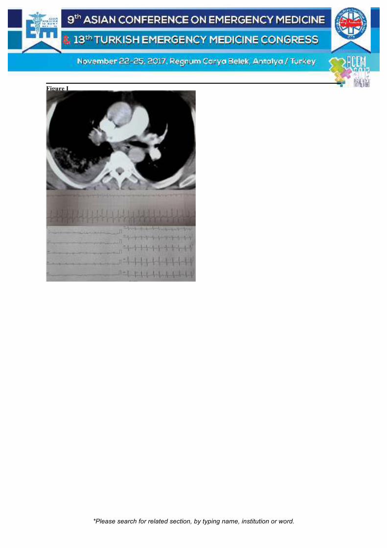
*Please search for related section, by typing name, institution or word.
Figure I

*Please search for related section, by typing name, institution or word.
[P-370]
Atipik Bir Aspirasyon Vakasi: Protez Damak Metin Yadigaroglu1, Elif Ozgenc1, Ali Reza Manevi1, Kübra Nur Seyis2, Ozgur Tatli1 1Karadeniz Teknik Üniversitesi Acil Tıp Anabilim Dalı, Trabzon 2Karadeniz Teknik Üniversitesi Göğüs Cerrahisi Anabilim Dalı, Trabzon Yabancı cisim aspirasyonları (YCA) klinikte ilk olarak 1854 yılında tanımlanmıştır ve halen günümüzde acil servise sık başvuru nedenleri arasında gelir. Daha çok çocuklarda meydana gelen YCA, mental ve psikiyatrik açıdan sağlıklı olan ve alkol tüketim öyküsü olmayan yetişkinlerde genellikle kazayla meydana gelir. Bu durumun klinik ciddiyeti yabancı cismin bulunduğu anatomik lokalizasyona, boyutuna, şekline ve vasfına bağlı olarak değişir. Yabancı cisim (YC)’ler şayet yutulmuşsa %80-90 feçesle atılırken cerrahi girişim gereksinimi %1’ler seviyesindedir. Ancak yabancı cisim aspire edilmişse ve öksürme refleksi ile atılım mümkün değilse hasta için cerrahi girişim olasılığı artık kaçınılmazdır. Bilinen Alzheimer hastalığı öyküsü olan 66 yaşında kadın hasta, yemek yerken üst damak protez dişlerinin boğazına kaçması şikayeti ile getirildi. Hasta pek çok kez öksürmüş ancak boğazındaki cismi atamamış. Genel durumu orta olan hastanın stridoru mevcuttu ve saturasyonu oda havasında %75 idi. X-ray görüntülemede hipofarenks bölgesinde oblik şekilde takılı kalan protez görülmekteydi (Figure-1). Çengelli yapıda olan protez çıkarımı lokalizasyonu ve yapısı nedeniyle senkonder yaralanma ihtimaline karşın acil serviste denemedi. Göğüs cerrahisi ile konsulte edildi. Acil operasyona alınan hastanın hipofarenks bölgesinde vokal kord üst kesiminde yerleşmiş olan protez damağı rijit bronkoskopi yöntemi ile çıkarıldı. Yutulmuş ya da aspire edilmiş YC tespiti için düz grafiler ilk istenecek görüntüleme yöntemi olmakla birlikte radyoopak olmayan YC ‘ler için tomografi tanı için daha aydınlatıcıdır. YCA klinikte yabancı cisim yutulmasına oranla daha seyrek görülmektedir. Çünkü aspire edilen yabancı cisim çoğu zaman kişiyi öksürmeye zorlar ve küçük boyutlu olan yabancı cisimler bu şekilde çoğu zaman atılır. Öksürmeyle atılamayan YC şüphesinde hastaya Heimlich manevrası yapılmalıdır. Ancak vakamızda da olduğu gibi büyük boyutlu ve şekil olarak bulunduğu bölgeye takılma özelliği gösteren YC’ler öksürmekle atılamayabilir veya hasta yapabileceğimiz yardımcı maneveralara uyumsuz olabilir. Aciliyet arzeden bu durum da cerrahi girişimi kaçınılmaz kılar. Anahtar Kelimeler: Aspirasyon, protez, damak, bronkoskopi

*Please search for related section, by typing name, institution or word.
Figure-1: X-Ray'de protez Damak Görünümü AP

*Please search for related section, by typing name, institution or word.
[P-371]
D-Dimer is Negative, Should We Send the Patient Back Home? Mustafa Emin Çanakçı1, Göknur Yıldız1, Şeyhmus Kaya1, Filiz Baloğlu Kaya2, Barış Öztürk1 1Emergency Service, Eskisehir State Hospital, Eskisehir, Turkey 2Department of Emergency Medicine, Eskisehir Osmangazi University Medical School, Eskisehir, Turkey Introduction: Pulmonary thromboembolism (PTE) is a frequently seen, potentially fatal disease. Due to its nonspecific clinical status, the quick diagnosis and treatment might be difficult. The necessity of imaging in these patients is decided considering the scoring and the additional examinations. This paper presents a case with a low clinical risk score, normal D-dimer levels, and a diagnosis of pulmonary embolism. Case: A 76-year-old female patient came to the Emergency Department (ED) with the complaints of dyspnea, which started 12 hours ego. She did not have a history of palpitation, hemoptysis, chest pain, leg pain, immobilization, or recent surgery. In her physical examination, her arterial pressure was 110/70 mmHg, pulse was 107 bpm, respiratory rate was 28 breaths/min, temperature was 37 C°, and sPO2 was 92%. The breath sounds were rough, the expirium was prolonged, and her heath was rhythmic and tachycardic. No pathological findings were detected in her other system examinations. EKG did not reveal any abnormality other than sinus tachycardia. The bedside USG did not detect any pathological findings related to the heart and the lower extremity veins. The posteroanterior chest x-ray did not reveal any pathological findings. The PTE clinical risk score was 1.5 points (Wells: low risk) and 6 points (Genova: medium risk). The D-dimer result was 241 ng/ml (0-243), the troponin level was 6.4 pg/dl (0-34.6). Since the clinical doubt for PTE was still present, the patient underwent a CT angiogram. Bilateral filling defect was detected in the pulmonary arteries. After a consultation with the chest disease specialist, the patient was put into the intensive care unit. Conclusion: Despite its rarity, PTE might be detected in patients with low clinical risk score and negative D-dimer result. As long as the clinical doubt is present, ED physicians should conduct proper patient management to clarify the diagnosis. Keywords: Pulmonary embolism, D-dimer, risk classification

*Please search for related section, by typing name, institution or word.
PTE CT

*Please search for related section, by typing name, institution or word.
[P-372]
Miliary Tuberculosis at Emergency Department Sümeyra Acar Kurtuluş, Can Özen, Serkan Emre Eroğlu Department of Emergency Medicine, University of Health Sciences Umraniye Training and Research Hospital, Istanbul, Turkey Introduction: Tuberculosis is a leading cause of preventable morbidity and mortality due to an infectious agent worldwide. (1) Miliary tuberculosis results from the massive, hematogenous dissemination of tubercle bacilli, from an established focus producing numerous lesions of approximately the same age and size in many organs of the body. (2) Miliary TB can arise as a result of progressive primary infection or via reactivation of a latent focus with subsequent spread via the bloodstream. Case report: 33 years old male patient presented to ED with worsening night fever, weight loss, dyspnea and cough for the last 25 days. On presantation patient was chilly. His vital signs were normal. He had nephrolithiazis in his past medical history. On physical examination, he had diffuse Coarse rales in both lungs. His laboratuary results revealed mildly impaired kidney function (Creatinin=1.58 mg/dl with normal Blood Urea Nitrate, Glomerular Filtration Rate = 57.03 ml/min/1.7 m2), hyponatremia (Na = 121 mEq/L). CRP levels were 5.2 mg/dl and urine dipstick test showed sterile pyruia. A chest X-ray and computed tomography were performed which revealed small diffuse opacities in both lungs (Figure 1 and 2). This patient hospitalized with miliary Tuberculosis. Acid-fast smear and culture belong to patient's expectoration was positive, supporting the diagnosis. Patient was discharged from hospital without any complications. Discussion: In 1700, John Jacob Manget coined the term miliary tuberculosis (derived from the Latin word miliarius, meaning related to millet seed) to describe the resemblance of gross pathological findings to that of innumerable millet seeds in size and appearance (3) Organs with high blood flow e.g the spleen, lungs, liver, bone marrow, kidneys, and adrenals are frequently affected. Although miliary tuberculosis involves almost all organs; Most often the involvement is asymptomatic. Laboratuary abnormalities may help to diagnose. Keywords: Miliary tuberculosis

*Please search for related section, by typing name, institution or word.
Figure 1
*Please search for related section, by typing name, institution or word.
[P-374]
Özefagusta İlginç Bir Yabancı Cisim Olgusu Vahide Aslıhan Durak, Yasin Koçak, Tuğba Sarkı Cander, Özlem Köksal Uludağ Üniversitesi Tıp Fakültesi, Acil Tıp Ana Bilim Dalı, Bursa Giriş: Yabancı cisim aspirasyonları(YCA) çocukluk döneminde daha çok görülmekle birlikte erişkin yaşta da karşılaşılabilen acillerdendir ve hayatı tehdit eden komplikasyonlara yol açabilirler. Asemptomatik olabileceği gibi, çok ciddi solunum yolu komplikasyonlarına hatta boğulma ve ölüme neden olabilmektedirler. Her zaman çok yönlendirici bir hikaye söz konusu olmadığından YCA’nın klinik ve radyolojik bulgularının çok iyi bilinmesi tanının erken dönemde konulması açısından oldukça önemlidir. Bronkoskopik gelişmelere paralel olarak tedavilerinde çok büyük ilerleme olmuş ve mortalite oranları da dramatik olarak düşmüştür. Olgu: 18 yaş bayan hasta, iftardan sonra yemek çatalı ile dişinden besin artığı çıkarmaya çalışırken, söz konusu çatalı yutma sonrası tarafımıza başvurdu. Genel durum iyi, bilinç açık, koopere ve oryante idi. Vital bulguları stabil olan hastanın oral sekresyonlarında artış mevcuttu. Çekilen grafide özefagusta çatal görüntüsü saptandı ve göğüs cerrahisine konsülte edildi. Acil özofagoskopi amaçlı interne edilen hasta, 2 gün post-op takip edildikten sonra taburcu edildi. Tartışma ve Sonuç: Özefagus zayıf peristalsizmi ve anatomik darlıklarından dolayı yabancı cisimlerin sıklıkla takıldığı bir yapıdır. Özefagus yabancı cisimlerinin %10-20’ine endoskopik müdahale gerekirken %1’inden azına cerrahi yapılır. Özefagusta takılma alanları genellikle fizyolojik darlıklarda olur. Bunlar; krikofarengeal kas seviyesinde üst darlık, arkus aorta seviyesinde ikinci darlık ve alt sfinkterde üçüncü darlıktır. Yabancı cisimler özefagusta en sık birinci darlıkta takılırlar. Disfaji, odinofaji, takılma ve batma hissi, retrosternal ağrı, sırt ağrısı, sekresyon artışı ve özafageal obstrüksiyona sekonder trakeal aspirasyon gözlenebilir. Eğer perforasyon yoksa fizik muayenede hiçbir bulguya rastlanmaz. Ancak perforasyon varsa; yüksek ateş, boyunda krepitasyon, boyunda şişme, kızarıklık, wheezing ve stridor gözlenebilir. Şüphelenilen yere göre iki yönlü direk servikal grafiler, iki yönlü akciğer grafisi ve direk batın grafisi çekilebilir Grafilerin normal olması yabancı cismi olmadığı anlamına gelmez. Kontrastlı madde kullanılarak grafi çekilmesi aspirasyon riski yüzünden önerilmez. Nazogastrik sonda kesinlikle takılmamalıdır. Rijid özefagaskopi özellikle keskin kenarlı objeleri çıkarmada ve proksimal özefagusta oldukça etkilidir. Fleksibl endoskopiler hem yabancı cismi hem de özefagus lümenini değerlendirmek için yapılabilir. Anahtar Kelimeler: Yabancı cisim, özefagus, acil servis

*Please search for related section, by typing name, institution or word.
[P-376]
Pneumomediastinum Secondary to Squamous Cell Carcinoma of Tongue Yasemin Özdamar, Yunus Emre Dündar, İlter Ağaçkıran, Zehra Akçora, Huriye Yürük Mısırlıoğlu, Özgür Bayar, Bülent Erbil The Department of Emergency Medicine, Hacettepe University, Ankara, Turkey Pneumomediastinum is first defined as the presence of free air in mediastinum by Hamman in 1939. Its etiology involves head and neck infections, malignancies, trauma, invasive procedures and barotrauma. We present a case of Pneumomediastinum secondary to untreated tongue SCC. Case: A 41-year-old male patient was brought emergency department with dyspnea. He had a diagnosis tongue SCC two years ago, for which he had refused medical treatment and was self-treating himself by herbal remedies. His medical history was otherwise unremarkable. On examination he was hypotensive, hypothermic and his SO2 was 92%. He was cachectic Emphysema was noticeable on his neck and right shoulder. He had a partially necrotized purulent mass on his tongue. His chest X-ray showed linear lucencies on bilateral paratracheal and mediastinal regions. Neck and chest CTs was obtained for the suspicion of pneumomediastinum secondary to purulent and necrotic mass on his tongue. CT scans was remarkable for pneumomediastinum secondary to SCC and the presence of air densities in spinal canal. He was admitted to medical oncology clinic and died of septic shock 48 days later. Conclusion: One of the important cause to consider pneumomediastinum in the etiology of dyspnea. Sparing time for careful evaluation of chest X-ray in busy ED environments may not benefit only diagnostic processes but also hasten the initiation of proper medical treatment. Keywords: Pneumomediastinum, squamous cell carcinoma, dyspnea
Can you spot pneumomediastinum?

*Please search for related section, by typing name, institution or word.
Linear lucencies on bilateral paratracheal and mediastinal regions

*Please search for related section, by typing name, institution or word.
[P-377]
A Rare Pulmonary Infection in The Emergecy Department: Pulmonary Fungus Ball Yasin Yıldız1, Hande Asan1, Mine Kayacı Yıldız2, Erdem Çevik1, Serkan Emre Eroğlu2 1University of Health Sciences, Sultan Abdulhamid Han Training and Research Hospital, Emergency Medicine Clinic, Istanbul, Turkey 2University of Health Sciences, Umraniye Training and Research Hospital, Emergency Medicine Clinic, Istanbul, Turkey Introduction: The incidance of fungal pulmonary infections is rising and this entity is multifactorial. This type of infections can be seen both immuncompetent and immuncompromised patients. Here we aimed to present a debilitated patient that is admitted to the emergency department with complaints of fever, cough and irritability and than hospitilized with the diagnosis of pulmonary fungal infection. Case: A 27-year-old male, presented to our emergency department with complaints of fever, cough and irritability. He had debillated for cerebral palsy and had epilepsy treatment. On admission, his consciousness level was learned that on his basal state. On examination, fever was 37.6 °C, there was no nuchal rigitidy or rushes, and all orther vital signs were normal. On pulmonary oscultation, we revealed that bilaterally rough sounds. On laboratory tests, WBC was 11.3 and all other parameters were normal. Computerized thorax tomography revealed a thin layered cavity with a air-liquid level on the right side, and a pleural effusion on the left side (Figure 1). The patient was refered to the department of chest disease and hospitalized with the diagnosis of pulmonary fungus ball. Discussion: A saprophytical fungal organism that grown in a parenchymal cavity forms pulmonary fungal balls. Radiologically, it can be presented with lung consolidations, hiler or mediastinal adenopaties or nodules. Pleural effusions are rare. Conclusion: Fungus balls, even though rare, should be kept in mind in the differential diagnosis for the patients with a complaint of cough and fever and with cavities on the pulmonary radiological tests. Keywords: Infection, fungus ball, pneumonia

*Please search for related section, by typing name, institution or word.
Figure 1
On thorax tomography, fungus ball image on the right, and plevral effusion on the left.

*Please search for related section, by typing name, institution or word.
[P-378]
How Aeds are Actually Used for Patients in Cardiopulmonary Arrest Before Arrival in Our Center Hayato Yoshioka, Kazushige Inoue, Kouichi Nagasawa, Nobuaki Kiriu, Eiji Hasegawa NHO Disaster Medical Center Critical Care Medicine and Trauma, Japan Purpose: In Japan, over 10 years have elapsed since the use of AED by ordinary citizens was approved. In this study, we investigated how AEDs are actually used and the resuscitation rate therewith. Subjects and Method: We retrospectively investigated the site of occurrence of cardiopulmonary arrest before arrival in our center over the past three years, the number of cases involving the wearing of AEDs as well as the number of defibrillations conducted by general citizens, and the number of defibrillations conducted by ambulance services and the outcomes thereof. Results: Among 1,025 subjects, the sites of occurrence mainly included: general residences: 694 (67.7%); public facilities: 242 (23.6%); and on the street: 72 (7.1%). There were 291 cases involving bystander CPR by ordinary citizens (28.3%), and 57 cases involving the wearing of AEDs (5.5%), with the sites of wearing AEDs being general residences in 5 cases (0.5%) and other areas in 52 cases (5.0%). There were 26 cases involving defibrillation conducted by general citizens (2.5%), with a one-month survival rate of 0.8%. On the other hand, there were 43 cases involving defibrillation conducted by ambulance services (4.2%), with a one-month survival rate of 0.4%. Discussion: The survival rate of patients who underwent defibrillation conducted by citizens was higher than that of patients undergoing defibrillation conducted by ambulance services, suggesting the importance of early CPR including AED. It was considered that the low rate of wearing of AED within residences, where the occurrence rate is highest, highlights the challenges going forward. Conclusion: It is considered important to further educate and disseminate CPR for general citizens, in addition to improving the environment such that AED can be smoothly used in general residences. Keywords: AED, resuscitation

*Please search for related section, by typing name, institution or word.
[P-379]
From the Causes of Reversible Arrest: Tension Pneumothorax Serhat Örün1, Derya Ünlü1, Mustafa Temiz1, Kenan Ahmet Türkdoğan2, Ali Duman2, Hüseyin Şahin3, Ayhan Aköz2 1Emergency Department of Bandırma State Hospital, Balıkesir, Turkey 2Emergency Department, Medicine Faculty, Adnan Menderes University, Aydın, Turkey 3Emergency Department, Medicine Faculty, Namık Kemal University, Tekirdağ, Turkey Objective: The approach to arrest patients is at the top of the cases that all emergency physicians should know and actively implement. Tension pneumothorax is a rare cause of reversible arrest. Our aim in presenting this case is to emphasize the early diagnosis and treatment of tansion pneumothorax, from the causes of reversible arrest. Case: A 56-year-old male patient was brought to our emergency services by 112 ambulance service due to cardiac arrest. Cardiopulmonary resuscitation began to the patient, Simultaneous double-vein path was opened and was monitorizied. No respiratory voices were heard in the left lung after intubation. The location and level of the intubation tube was checked and the tube was fixed. First vital values; Arterial blood pressure was not obtained, heart rate was not, saturate O2 was 49%. Deep tracheal aspiration was applied to the patient. The patient's skin was cyanotic, pupils was dilate. There was distention of abdomen and there was subcutaneous emphysema on the abdomen and chest wall. Also the patient had a 2 cm sized uniformly incision in the left medial axilla, even sutured. it was learned from the patient's medical history that patient have chronic obstructive pulmonary disease, 2 weeks ago tube thoracostomy procedure applied left lung due to pneumothorax and tube was removed 3 days ago. So, tube thoracostomy procedure applied to patient with prediagnosed of tension pneumothorax. After the procedure, the patient's O2 saturation was up to 94% and cyanosis was declined. Spontaneous cardiac pulse occurred after 2 minutes. The patient was hospitalized in intensive care unit. Conclusion: Tension pneumothorax is a rare cause of reversible cardiac arrest. It is the situation that emergency physicians should keep in mind and need to be intervened quickly. Keywords: Cardiac arrest, emergency services, tension pneumothorax

*Please search for related section, by typing name, institution or word.
[P-380]
Septic Shock Secondary to Ruptured Hydatid Cyst of Liver: Case Report Derya Abuşka, Ali Cankut Tatlıparmak, Özgür Karcıoğlu Department of Emergency Medicine, Istanbul Training and Research Hospital, Istanbul, Turkey Background: Hydatid disease is a parasitic infestation by a tapeworm of the genus Echinococcus, which still remains endemic in Turkey. Echinococcus granulosus, causing cystic echinococcosis (CE), is the most common type of Echinococcus. In CE, the prognosis is generally favorable, and complete cure is possible with total surgical excision without spillage. Morbidity is usually secondary to free rupture of the echinococcal cyst, infection of the cyst, or dysfunction of affected organs, while mortality can be seen secondary to anaphylaxis, systemic complications of the cyst or operative complications. Below is report of a patient presented with acute abdominal pain, hypotension, hypotermia and tachycardia diagnosed with ruptured CE. Case: A 44-year old male patient presented with acute-onset epigastric pain was admitted to emergency department. His medical history included hydatid cyst (stage 4) pulmonary hypertension, ventricular septal defect with Eisenmenger syndrome. He was learned to have been performed ERCP four weeks ago. Biliary fistula was seen in ERCP, sphincterotomy and daughter vesicle extraction was performed. His vital signs were blood pressure 90/52 mmHg, pulse 160 bpm, respiratory rate 30 bpm, body temperature 34,50C and SpO2: 97%. On physical examination, diffuse abdominal tenderness with guarding, pulmonary bilateral crepitant rales, cold and pale skin were noted. White blood cell count was 28900 cells uL-1/ml (86% neutrophils). On the bedside ultrasonography there was no free fluid in the abdomen but a heterogeneous lesion was detected in the liver. Aggressive fluid resuscitation, antibiotics and Albendazole was administered. Contrast CT of his abdomen revealed the hydatid cyst rupture. The patient was operated by general surgery and was hospitalized in Intensive Care Unit (ICU). On the 6th day of ICU admission the patient died of cardiac failure. Conclusion: Emergency physicians should not overlook the diagnosis of hydatid cyst rupture and institute expedient treatment in the patient with shock. Keywords: Hydatid cyst, echinococcus, septic shock

*Please search for related section, by typing name, institution or word.
[P-381]
A Rare Reason of Intraabdominal Sepsis Etiology, Superior Mesenteric Vein Thrombosis Duygu Ege1, Bekir Dağlı1, Ömer Yüceer1, Alev Peker2, Mehmet Kıy1 1Emergency Department, Medicine Faculty, Adnan Menderes University, Aydin, Turkey 2Emergency Department, Aydin State Hospital, Aydin, Turkey Acute mesenteric ischemia(AMI) is a life threatening vascular emergency which requires prompt diagnosis and treatment. Mesenteric venous thrombosis(MVT) is an uncommon cause of AMI accounting for 5–15% of the cases. The path to the diagnosis is clinically suspicious. The noninvasive gold standard is computer tomography(CT). In-hospital mortality rate has been around 50-80% from sepsis, which is the most important reason to delay recognition. In this case we would like to emphasize the importance of mesenteric ischemia in a patient who had suffered of abdominal pain for two days. A 63-year-old female patient applied to emergency department(ED) complaint of abdominal pain for two days. She had applied two different hospitals before us and discharged with peptic ulcus prescriptions. Her general condition was poor, blood pressure80/50mmHg, pulse rate123/minute, respiratory rate22/minute, temperature39,1°C. The patient had sinus rhythm, and she had only chronic hypertension was known. Her abdomen was distended and seen caput medusae sign. We provided cardiac monitorization, opened two large vessels to prevent collapse and performed bedside ultrasonographic imaging for shock protocol. We saw intraabdominal free fluid, portal hypertension signs, stepladder sign and to-pro-motion signs for bowel ischemia. We administered appropriate antibiotics and arranged fluid resuscitation in first hour. The patient’s serum parameters were; lactat11.3mmol/L, hemoglobin15.1gr/dL, leukocyte17600/mcrL, thrombocyte185000/mcrL, total bilirubin4mg/dL, direct bilirubin0.59mg/dL. Other parameters were in normal range. After with using CT, we detected superior mesenteric vein(SMV) thrombus, pneumocystis intestinalis, ileus, portal hypertension, we consulted to general surgery and transport to the operation room in 90 minutes after the patient’s arrival. The patient lost her life in postop 10th hour. In this case we examined the patient with differential diagnosis of sepsis and directed to SMV thrombosis with advanced monitoring. We want to remind that mesenteric ischemia diagnosis origins from clinical suspicious, and even early diagnosis has high mortality rates. Keywords: Sepsis, mesenteric ischemia
CT images

*Please search for related section, by typing name, institution or word.
left-up: perihepatic and perisplenic fluid right-up: Intravenous free air left-down: SMV thrombus right-down:
SMV thrombus, contrast in umblical vein
*Please search for related section, by typing name, institution or word.
[P-382]
Efficacy of Recombinant Human Soluble Thrombomodulin in Septic Shock with Urinary Tract Infections Hiromasa Yakushiji, Motohiro Yamada, Wataru Shirasaka, Keitaro Suzuki, Arito Kaji, Masahiro Shinozaki Department of Emergency Medicine, Kishiwada Tokushukai Hospital, Japan Objective: There is limited clinical evidence supporting the use of recombinant human soluble thrombomodulin (rhTM) in patients with septic shock. The purpose of this study was to evaluate survival benefits of rhTM administration in patients with septic shock due to urinary tract infections. Material: This is four-years retrospective, observational study from January 2013 through December 2016. The patients (age ≧15 years) who were transported to our hospital by ambulance and admitted to intensive care units for the treatment of septic shock due to urinary tract infections were analyzed. We diagnosed septic shock based on sepsis-3. Method: The primary outcome measure was in-hospital mortality rate. To evaluated the difference of in-hospital mortality rates between the group treated with rhTM (0.06 mg/kg/day) and treated without rhTM, we used Fisher's exact test. Results: Of 49 patients who are eligible for the analysis, 12 patients (24%) were treated with rhTM. The in-hospital mortality rates were 0.0% (95%CI, 0.0%-22.1%) in the group treated with rhTM and 13.5% (95%CI, 4.5%-28.8%) in the group treated without rhTM. Although the in-hospital mortality rate was lower in the group treated with rhTM, no significant difference was obtained (OR, 0.0; 95%CI, 0.0-3.38, p = 0.31). Conclusion: There was no significant prognostic improvement effect by rhTM administration in patients with septic shock due to urinary tract infections. Keywords: Recombinant human soluble thrombomodulin, sepsis, septic shock

*Please search for related section, by typing name, institution or word.
[P-383]
A Colorful Presentation of Urinary Tract Infection Irfan Abdulrahman Sheth, Faraz Zarisfi, Vetrival Ramar, Mohammad Madeena Faizur Rahman, Sohil Pothiawala Singapore General Hospital, Singapore Introduction: Discoloration of urine is not uncommonly encountered in clinical practice and may indicate a potentially significant underlying pathology. Purple discoloration of the urinary catheter bag is rare and can be concerning to both patients and healthcare workers. Case: An elderly female presented to the ED with dark coloration in the urine bag, while the catheter tube had a normal color. Urine culture from the suprapubic catheter was positive for Pseudomonas aeruginosa and Enterococcus faecalis; urine from the nephrostomy was sterile. She was diagnosed with an E. coli urinary infection and treated with amoxicillin and removal of the indwelling catheter. This case illustrates the typical characteristics of purple urine bag syndrome. Discussion: Purple urine bag syndrome (PUBS) is a rare condition in which purple discoloration of urine occurs inside its collection bag. It can be seen in patients with chronic urinary catheterization and urinary tract infection. Gram-negative bacteria that produce sulfatase and phosphatase are involved in the formation of pigment, indirubin and indigo. Tryptophan metabolism is involved in the pathogenesis. Purple urine bag syndrome is associated with higher morbidity and mortality, compared to urinary tract infection without this phenomenon. Most common bacteria associated with PUBS include Providencia stuartii, Providencia rettgeri, Klebsiella pneumoniae, Proteus species, Escherichia coli, Enterococcus species, Morganella morganii, and Pseudomonas aeruginosa. It has been observed that constipation and intestinal obstruction are strongly associated with PUBS Conclusion: Antibiotic therapy is only indicated in patients with symptomatic urinary infection. The mainstay of preventing purple urine bag syndrome is avoidance of chronic catheterization and prompt removal of urinary catheters once they are no longer needed. Keywords: Chronic urinary catheterization, urinary tract infection, indirubin & indigo pigment, purple bag syndrome, UTI, PBS

*Please search for related section, by typing name, institution or word.
[P-384]
Gelişen Bir Eksternal Otit, Hastanın Hayatını Ne Derece Etkileyebilir? Rıza Gücal1, Gökhan Aksel1, Serkan Emre Eroğlu1, Öner Bozan2 1Sağlık Bilimleri Üniversitesi, Ümraniye Eğitim ve Araştırma Hastanesi, Acil Tıp Kliniği, İstanbul 2Sağlık Bilimleri Üniversitesi, Okmeydanı Eğitim ve Araştırma Hastanesi, Acil Tıp Kliniği, İstanbul Giriş: Nekrotizan external otit(MEO), bilinen adıyla malign external otit; nadir, hayati tehlike yaratan dış kulak yolunun, kafa tabanının ve mastoidin enfeksiyondur. Malign external otit(MEO) daha çok yaşlı, diyabetes mellitus(DM) olan immunsuprese hastaları etkilemektedir. En sık neden olan patojen pseudomonas aeruginosadır, diğer olası patojenler Staphylococcus epidermidis, gram-negative bakteriler, ve fungilerdir. Bu olgumuzda kardiyak arrest durumuna kadar ilerleyen external otitli bir hastayı tartışmayı amaçladık. Olgu: 61 yaşında yabancı uyruklu kadın hasta,3 gündür baş dönmesi, kulak ağrısı şikayetiyle hastanemizin acil servisine başvurdu. Tercüman aracılığıyla alınan bilgiye göre iki aydır kulak akıntısı olduğu, Bilinen hipertansiyon, DM hastalığı olduğu ve insülin kullandığı öğrenildi. Vital bulguları stabil ve sağ pürülan kulak akıntısı olan hasta kulak burun boğaz(KBB) bölümüne konsülte edildi. Konsultan hekim muayenesi sırasında arrest olan hastanın kardiyopulmoner resüsitasyon işleminin üçüncü dakikasında spontan nabız sağlandı. Arteriyel kan gazı tetkikinde PH:7,21 Hb:6gm/dL K:7mmol/dL glukoz 450mg/dL; Bilgisayarlı tomografisinde;temporal tip düzeyinde anterolateralde,bölgesel destrüksiyonel silinmeler ve çevresel yumuşak doku planlarında yoğun natürlü yaygın enflamasyonel ödematöz kalınlaşma,apse formasyonu tespit edildi.Yoğun bakım ünitesine sevk edilen hastadan alınan kulak kültüründe A.baumannii ürediği, 15 gün linezolid (600mgx2),30 gün Meronem(1gmx2) tedavisi aldığı ve 14 günlük takibinden sonra mastoid kemık osteomyeliti tanısıyla kulak burun boğaz servisine alınıp mastoid kemık debritmanı yapıldığı öğrenildi. Tartışma: MEO temporal kemiğin ender görülen, ancak destrüktif seyreden bir hastalığıdır. Sıklıkla yaşlı, diyabetik ve immunsuprese hastalarda görülür. Bu durum external otit olarak başlar daha sonra kafa tabanına Santorini fissürü ve timpanomastoid sutur ile ilerler. Otalji, otore, ateş ve baş ağrısı bazı semptomları olup kafa tabanı osteomyeliti MEO' nun geç komplikasyonlarından biridir. Bizim vakamızda Malign external otite bağlı osteomyelit ve buna bağlı apse formasyonları belirlenmiştir. Sonuç: MEO özellikle diabetik ve immunsuprese hastalarda hayatı tehdit edici bir hastalıktır. Kraniyal sinir felci ve osteomyelit geç dönem komplikasyonlarındandır. Benign karakterli external otitin, DM gibi immünsüprese olan hastalarda hayatı tehdit edici komplikasyonları olabileceğini Acil servis doktorları unutmamalıdır. Bu hastalarda ileri görüntüleme ve değerlendirme gerekliliği olabileceği akılda tutulmalıdır. Anahtar Kelimeler: Malign eksternal otit, osteomyelit, sepsis

*Please search for related section, by typing name, institution or word.
meo-ct

*Please search for related section, by typing name, institution or word.
[P-385]
The Effectiveness of Immunoglobulin for Sepsis Tomohiro Noda, Tetsuro Nishimura, Fumiaki Wakita, Maiko Esaki, Naohiro Hagawa, Tomonori Yamamoto, Shinitiro Kaga, Yasumitsu Mizobata Department of Traumatology and Critical Care Medicine, Osaka City University Hospital, Osaka, Japan In most hospitals in Japan, including our own, sepsis is treated with standard therapies in accordance with the Japanese Clinical Practice Guidelines for Management of Sepsis. Immunoglobulin therapy is weakly recommended by the Japanese guidelines and is sometimes used at this hospital, despite the fact that there is currently little evidence to show that immunoglobulin therapy improves mortality. To determine the significance of immunoglobulin therapy in this context, the present retrospective study was conducted. Ninety-five sepsis patients who were treated at this hospital during the 18 months between February 2014 and August 2015 were included in the study. They were classified into two groups according to the administration or non-administration of immunoglobulin therapy, and the mortality rate was compared between the immunoglobulin-treated (n = 14) and the immunoglobulin non-treated (n = 81) groups. To minimize imbalance in the baseline characteristics of the two groups, propensity score matching was performed. Three factors were matched: age, sex, and the quick sepsis-related organ failure assessment (qSOFA) score; 13 matching pairs of patients were selected for comparison. The mortality rates in the immunoglobulin-treated and immunoglobulin non-treated groups were 30.8% and 7.7%, respectively, and did not differ to a statistically significant extent (Fisher’s exact test, P = 0.322). The results showed that immunoglobulin therapy did not improve the mortality rate. This study indicates the possibility that immunoglobulin therapy does not contribute to the survival of sepsis patients. Keywords: Sepsis, immunoglobulin

*Please search for related section, by typing name, institution or word.
[P-386]
Shock and Coma: Anaphylaxis Case After Bee Sting Ali Karakus1, Guven Kuvandik1, Ali Bucak1, Zeynep Kekec2 1Mustafa Kemal University, Faculty of Medicine, Department of Emergency Medicine, Hatay, Turkey 2çukurova University, Faculty of Medicine, Department of Emergency Medicine, Adana, Turkey Introduction: Anaphylaxis is in the distributional shock group. It can be caused mostly by medicines and environmental reasons. Unconsciousness may develop in the following stages. In this article, we present a patient who developed a shock and coma after bee sting. Case: A 60-year-old male patient was found unconscious 2 hours ago on the field. Emergency departmment were brought in because of impaired consciousness. The general condition of the patient is poor, Glaskow Coma Scale: 11, blood pressure arterial 80/40 mmHg, pulse rate 100 / min, respiration 20 / min, blood sugar 90 mg / dl. Other physical findings and laboratory values were normal. The patient's history was learned that he had hypertension and allergy to bee stings. The patient was treated with 2000 cc saline, 45.5 mg feniramine maleate, and 80 mg prednisolone. He was taken to the follow-up room. The patient was followed for 4 hours and the vitals improved. He was discharged with recommendations and antihistaminic treatment. Conclusion: Death can be seen after anaphylaxis in bee sting cases. It should be considered in coma and shock patients and early treatment should be initiated. Keywords: Bee sting, shock, coma

*Please search for related section, by typing name, institution or word.
[P-387]
Rectal Balloon May Be Useful for Temporary Hemostasis of Lower Rectal Bleeding Hidehiko Nakano1, Kogaku Umezawa1, Hiroshi Yamagami1, Hisashi Ofuchi1, Shinnosuke Tokoro2 1Emergency Department, Shonan Kamakura General Hospital 2Shannon Gastroenterology Medicine Center, Shonan Kamakura General Hospital, Japan Case Presentation: Patient is 49 years old female with cervical cancer stage 4, which was treated with radiation therapy 1 year ago. She took edoxaban for deep venous thrombosis and loxoprofen for loin pain. She had intermittent hematochezia for 2 weeks and came to our hospital by ambulance after massive bleeding. Her blood pressure was 69/48mmHg and heart rate was 103 bpm on arrival. Despite initial fluid resuscitation with 3L of normal saline, 500mL of hydroxyethyl starch and 4 units of red blood cell (RBC) transfusion, she was still unstable, and continuous bleeding from lower rectum was found on anoscopy. Then we decided to intubate her and put the rectal balloon for a temporal hemostasis and gave another 4 units of RBC transfusion and 4 units of fresh frozen plasma. After placement of the rectal balloon, her blood pressure gradually elevated. Once stabilized, we performed upper gastrointestinal endoscopy first because of loxoprofen use and coffee-ground nasogastric aspirate but found no bleeding, then performed colonoscopy and found the rectal invasion of cervical cancer as the cause of bleeding. Transcatheter arterial embolization (TAE) was performed next day and hemostasis was achieved. She was extubated and rectal balloon was removed on day 5 and was transferred to the Hospice without complication on day 12. Discussion: Lower gastrointestinal (GI) bleeding generally has a less severe than upper GI bleeding and the reported mortality rate is 2 to 4%. However, the combination of anticoagulation therapy and bleeding from invasive cancer lead to severe bleeding in this case and we couldn’t perform endoscopy or TAE before enough resuscitation. Rectal balloon was effective to control bleeding in lower rectum, and we could achieve initial resuscitation. Rectal balloon may be a useful and less invasive method for temporal hemostasis of lower rectal bleeding. Keywords: Gastro intestinal bleeding, shock, resuscitation

*Please search for related section, by typing name, institution or word.
[P-388]
Delayed Anaphylactic Shock After Hysterosalpingography Ibrahim Toker1, Gülten Bozali1, Hüseyin Narcı1, Cüneyt Ayrık1, Engin Kara2, Serkan Karakulak1 1Department of Emergency Medicine, Mersin University, Faculty of Medicine, Mersin, Turkey 2Department of Radiology, Mersin University, Faculty of Medicine, Mersin, Turkey Introduction: Hysterosalpingography (HSG), is a fluoroscopic imaging method that uses an iodinated contrast media to evaluate uterine morphology. Case: A 35-year-old female was admitted to emergency department (ED) with complaints of abdominal pain and shortness of breath. She did not have any chronic illness. She had a history of allergies. HSG was applied to the patient for about 32 hours. After HSG, abdominal pain and swelling in the face of the patient were started. She was cold-sweaty and tachypneic. The patient’s vital signs on admission were a blood pressure of 80/40 mm/Hg, heart rate of 140 beats per minute, and respiratory rate of 45 breaths per minute, and her oxygen saturation was 85% on 10 L/min of oxygen with oxygen mask. Her glasgow coma scale (GKS) was 15. There were crackles in all zones in respiratory examination. Because of her oxygen saturation decreased and shallow breathing developed, she was intubated. Laboratory findings on admission were white blood cell count 1180/ mm3, platelet count 35x103/mm3, international normalized ratio (INR) 7.2, serum sodium 144 meq/L, potassium 4.11 meq/L, glucose 69 mg/dl, creatinine 2.8 mg/dl and urea 103 mg/dl. In the blood gas analysis, pH was 7.25, pCO2 was 18 mmHg, pO2 was 66.4 mmHg, HCO3 was 10.9 mmol/L, base excess was -18.3, and oxyhemoglobin rate was 87.4%. A thoracoabdominal CT was performed because of the suspicion of pulmonary embolism and intra-abdominal pathology (intestinal perforation eg.) (Figure 1). The patient was admitted in intensive care unit. After 6 hours admission the patient died. Conclusion: We observed a patient who had a severe anaphylactic shock, multiple organ failure and death after HSG. The initial complaints of abdominal pain and facial swelling could had been due to an early anaphylactic reaction and peritoneal irritation. Keywords: Hysterosalpingography, anaphylaxis, shock
Figure 1

*Please search for related section, by typing name, institution or word.
The CT scan showed multiple fluid-filled and dilated loops of bowel with wall thickening

*Please search for related section, by typing name, institution or word.
[P-389]
Painless Aortic Dissection: An Unusual Case of Nonbloody Diarrhea Nezihat Rana Dişel1, Senem Ertekin1, Buğra Karaaslan2, Tolga Onur Badak3, Ayça Açıkalın Akpınar1, Ahmet Sebe1 1Çukurova University, Faculty of Medicine, Department of Emergency Medicine, Adana, Turkey 2Çukurova University, Faculty of Medicine, Department of Cardiology, Adana, Turkey 3Adana Numune Training and Research Hospital, Cardiovascular Sugery, Adana, Turkey Introduction: Aortic dissection (AD) is a fatal medical disorder. Here we present a patient with De Bakey Type I AD which we believe to be the first case reported presenting with non-bloody diarrhea. Case: A 50-year-old male patient with complaints of fatigue, non-bloody diarrhea and vomiting was admitted to our emergency clinic soon after being discharged from another hospital where he had received fluids and symptomatic medications for the same complaints. This previously healthy patient had experienced a slight chest discomfort in the last night. Initial vitals were: BP: 80/40 mm Hg; PR: 100/min; RR: 20/min; t: 36.7℃; O2 sat: 98%. He had jugular venous distension and diastolic apical murmur without any abnormal lung sounds. He was started saline infusion. ECG revealed sinus tachycardia with biphasic T in V1. RUSH exam revealed 1.98 cm inferior caval ven diameter (no inspiratory collapse), 20 mm pericardial effusion with right ventricular collapse, and a dissection flap in the aortic root. Dynamic contrast CT revealed an AD originating at coronary sinus, affecting brachiocephalic truncus and proximal left subclavian artery and extending to proximal segments of both iliac arteries. Supracoronary tube graft interposition and replacement of ascending aorta was performed. He was discharged healthy on the 17th day. Discussion: The most common symptoms of AD are the sudden onset of severe abdominal or chest pain with a stabbing, tearing or ripping quality. Many other symptoms (like bloody diarrhea up to 5%) arise from decreased blood flow to organ systems. Our patient’s symptoms did not point aortic dissection, and might had been easily overlooked as gastroenteritis causing acute renal injury. In the light of this case, we suggest emergency physicians to be alert in any patient with different symptoms but shock, to keep aortic dissection in mind and rush to perform RUSH. Keywords: Aortic dissection, non-bloody diarrhea, RUSH exam

*Please search for related section, by typing name, institution or word.
Dynamic CT images of the patient.
The laboratory findings of the patient on-admission and pre-discharge.
Parameter (normal values) On- admission Pre-discharge
Glucose (70-105 mg/dL) 190 87
BUN (8-20 mg/dL) 31 16
Cr (0.1.2 mg/dL) 4.48 0.99
K (3.6-5.1 mmol/L) 5.2 4.9
AST (15-41 U/L) 3655 25
ALT (17-63 U/L) - 45
Troponine I (<0.02 ng/mL) 0.59 -
WBC (4.5-10.3 103/uL) 17.6 7.49
Hb (13.6-17.2 g/dL) 15 11.1
Htc (39.5-50.3 %) 43.8 33.7
PLT (156-373 103/uL) 116 313
Cr: Creatinine, K: Potassium, AST: Aspartate aminotransferase, ALT: Alanine aminotransferase, WBC: White
blood cell count, Hb: Haemoglobine, Htc: Hematocrite, PLT: Platelet

*Please search for related section, by typing name, institution or word.
[P-390]
Investigation of Fall Incidents into Irrigation Canal Cases Which Required Emergency Transportation to a Tertiary Medical Institution in Western Japan Motohiro Ichikawa, Tetsunori Ikegami Department of Emergency Medicine, Kurashiki Central Hospital, Japan Objective: Okayama prefecture is located in west Japan. Because of a small annual rainfall, there are many irrigation canals to support agriculture. Kurashiki, one of its major cities, has many canals without safety rail facilities, including in its central city area. We examined cases of falls into irrigation canals which required emergency transport to Kurashiki Central Hospital. Methods: We examined the number of cases, severity, prognosis, vehicle at injury, site of injury and cost by retrospectively examining the medical record of patients falling to an irrigation canal who were transported to the hospital from January to December 2016.
Results: There were 78 cases during the study period. Thirty-five cases required hospitalization, 10 of which were severe cases requiring admission to the intensive care unit. The average age was 59.5 years old, 53% were elderly people aged 65 years or older, and 67% of the patients were male. Of the 35 patients who required hospitalization, 17 patients were able to discharge from the hospital and return home, but 17 were transferred to the other hospitals, and 1 patient died. In total, there were two deaths. In analysis of vehicle type at time of incidence pedestrians were the most frequent, and the limb was the most frequent injury site. The total initial medical expense required for these patients was 25,492,410 yen.
Conclusion: In Kurashiki city, falls into irrigation canals are frequent, with prompt safety measures to prevent falls warranted. Keywords: Fall, cause of injury

*Please search for related section, by typing name, institution or word.
[P-391]
Quadriceps Tendon Rupture Associated with Anabolic Steroids and Growth Hormone: Case Report Engin Özakın1, Cüneyt Çalışır3, Akın Turgut2, Nurdan Acar1, Evvah Karakılıc1 1Eskisehir Osmangazi University, Faculty of Medicine, DepartmentOF Emergency Medicine, Eskisehir, Turkey 2Eskisehir Osmangazi University, Faculty of Medicine, Department of Orthopaedic Surgery, Eskisehir, Turkey 3Eskisehir Osmangazi University, Faculty of Medicine, Department of Radiology, Eskisehir, Turkey Introduction: Quadriceps tendon rupture is a rare condition in the absence of systemic disease. Some athletes use hormonal drugs to improve their performance, personal appearance and to increase their muscle mass. We report partial rupture of quadriceps tendon rupture in a bodybuilder who had taken anabolic steroid and growth hormone for years. Case: Our patient is 50 years old, healthy, white male, previously professional national class bodybuilders. He was admitted to our emergency department with left knee pain following sport training. He experienced sudden severe pain in his right knee after attempting front squat 150 kg of weight. He reported a history combination of androgenic hormones and growth hormone use that he had stopped before 6 months. The substance he took were a combination of gonadotropine and nandrolone. MRI showed swelling, heterogeneity and partial discontinuity of the quadriceps tendon (Figure 1). Partial tear of the quadriceps tendon was diagnosed. Discussion and Conclusion: Use of performance enhancing drugs has become a serious public health problem. Frequently, the anabolic androgenic steroids that bodybuilders use are synthetic modifications of testosterone. There are many adverse effect of anabolic steroids and some can be serious and mortal. Case reports describe sudden death in young athletes who were taking androgens. Some of the side effects that are associated with steroid use are tendinitis and tendon ruptures. There are several case reports regarding tendon ruptures associated to the anabolic steroids but very few of them are in the quadriceps tendon. The risk of tendon rupture is 9.0 times greater in the anabolic androgenic steroid users. Although these drugs is prohibited, their use are very common. Due to increase of abuse today it is possible to encounter more side effects in the future and has to be considered as a growing public health problem. Keywords: Quadriceps tendon rupture, androgenic hormone, growth hormone

*Please search for related section, by typing name, institution or word.
TENDON RUPTURE

*Please search for related section, by typing name, institution or word.
[P-392]
Tüplü Dalış dikkat gerektirir… Profesyonel Dalgıçta Gelişen Dekompresyon Hastalığı Haldun Öner, Deniz Tengerek, Can Özen, Serkan Emre Eroğlu Sağlık Bilimleri Üniversitesi, Ümraniye Eğitim ve Araştırma Hastanesi, Acil Tıp Kliniği, İstanbul Giriş: Dalgıçlık ülkemizde çok yaygın olmasa da, bilimsel, ticari, askeri, sportif amaçlarla ve popülerliği her geçen gün artan yüksek riskli bir aktivitedir. Dalgıçlar su altında karşılaşabilcekleri tüm aksaklıklarla baş edebilecek eğitimi alsalar da, yüksek basınçlı ortamlarda yaşamı tehdit eden sorunlarla karşılaşabilirler. Bu durumlardan biri olan dekompresyon hastalığı (DH), akut arteriyel emboli ve vurgun denilen tablo ile görülebilen bir fenomendir. Yüzeye çıkarken basınç eşitlemek için yeterli zaman harcanmadığında görülür ve kimi zaman ölümcül seyredebilir. Sunumumuzda profesyonel bir dalgıçta görülen akut dekompresyon hastalığı vakasına değineceğiz. Olgu: 42 yaşında erkek hasta, tüplü dalış sırasında 24 metre derinlikten hızlı çıkış sonrası gelişen nefes darlığı, göğüs ağrısı, baş ağrısı, eklem ağrısı ve vucütta yaygın döküntüler nedeniyle ambulans ile acil servise getirildi. Başvuru sırasındaki vitalleri arteriyal tansiyon 90/60mmHg, SpO2 %89, kalp tepe atımı 103 atım/dk olarak ölçüldü. Hastanın EKG'sinde akut bir patoloji saptanmadı. Fizik muayenede göğüste ve ekstemitelerde yaygın purpupik döküntü gözlendi. Nörolojik muayenede tüm ekstemitelerde kas gücü 4/5 olarak saptandı. Hasta salin ile hidrate edildi, analjezi sağlandı ve 10 lt/dk oksijen maske ile başlandı. Rutin tetkiklerinde patolojik bulgu saptanmayan hastanın bilgisayarlı tomografisinde hepatik venlerde hava kesecikleri gözlendi. Takibinde, vital bulguları stabil seyreden hastanın bulgularında ilerleme görülmedi. Dekompresyon hastalığı (tip-2) ön tanısıyla hiperbarik oksijen tedavisi başlandı. Sonuç: Dekompresyon hastalığı vücudun tüm sistemlerini risk altında bırakan bir tabloya neden olur. İlk semptomları etki ettiği dokulara spesifik olabilirken ilerlemesi halinde yaşamı tehdit eden bir hastalık durumu oluşur. Bulguların hızla tanınması ve tedaviye başlanması önem arz eder. İlk semptomları arasında kas iskelet sistemi ağrıları, deri döküntüsü gibi basit tablolar bulunurken (tip 1), merkezi sinir sistemi, iç kulak ve kardiyopulmoner semptomların eşlik ettiği ve ölümcül seyredebilen hastalık durumu da gelişebilir (tip 2). Tanı için spesifik bir yöntem bulunmamakla birlikte öykü ve klinikten şüphelenilmelidir. Tedavide %100 oksijen, rehidrasyon ve hiperbarik oksijenden faydalanılır. Bulguların erken tanınması ve tedavi başlanması hayat kurtarıcıdır Anahtar Kelimeler: Dekompresyon, vurgun, hiperbarik oksijen tedavisi

*Please search for related section, by typing name, institution or word.
[P-393]
Nadir Görülen Akut Batın Nedeni: Spontan Ürinom Ali Aygün1, Atakan Savrun1, Melih İmamoglu2 1Ordu Üniversitesi Eğitim ve Araştırma Hastanesi, Acil Tıp Ana Bilim Dalı, Ordu 2Rize Devlet Hastanesi, Acil Servis, Rize Giriş: Ürinom, idrarın üriner sistem dışındaki dokulara kaçmasıyla batın içerisinde birikerek koleksiyon oluşturması durumudur. Etiyolojide en sık görülen nedenler; taş, cerrahi ligasyon, tümör, posterior üretral valf, künt ya da penetran travmalardır. Renal kolik esnasında görülen spontan ürinom nadirdir. Olgu: 55 yaşında bayan hasta dış merkezden akut batın ön tanısı ile tarafımıza sevk edilmiştir. Hastanın 1 ay önce ürolithiazis nedeniyle sol double J katater takılma öyküsü vardır. Hastanın acil servise başvuru şikayeti bulantı, kusma, karın ağrısı, ateştir. Vital bulguları: TA:100/60mmHg, Ateş:38.5, SS:18/dk, Nabız:110/dk idi. Fizik muayede batında tüm kadranlarda yaygın hassasiyet, defans, sağ kostovertebral açı hassasiyeti mevcuttu. Hastaya kontrastlı batın bilgisayarlı tomografisi çekildi. Tomografisinde sağ böbrekte ödem, sağ perirenal alanda serbest mayi (ürinom), sağ üreteropelvik bileşkede dilatasyon, sağ üreter orta kısmında büyüğü 10mm boyutlu 2 adet obstrüktif taş dansitesi, sağ böbrek alt pol kaliksinde 3.5mm'lik 1 adet taş gözlendi (Resim 1). Üroloji bölümü ile konsulte edilen hasta operasyona alındı. Hasta 3 hafta sonra antibiyoterapi ve polklinik kontrolü önerilerek taburcu edildi. Sonuç olarak biz bu olguda semptomatik bir spontan ürinom olgusunu sunduk, spontan ürinom olgularının akut batın kliniğine neden olabileceğini unutmamalıyız. Anahtar Kelimeler: Ürinom, akut batın

*Please search for related section, by typing name, institution or word.
Resim 1.
sağ böbrekte ödem, sağ perirenal alanda serbest mayi (ürinom), sağ üreteropelvik bileşkede dilatasyon

*Please search for related section, by typing name, institution or word.
[P-394]
Genç Yaşta Görülen Nadir Bir Nefrektomi Nedeni: Ksantogranülomatöz Piyelonefrit Ali Aygün1, Atakan Savrun1, Melih İmamoglu2 1Ordu Üniversitesi Eğitim ve Araştırma Hastanesi, Acil Tıp Ana Bilim Dalı, Ordu 2Rize Devlet Hastanesi, Acil Servis, Rize Giriş: Ksantogranülomatöz piyelonefrit (KP) nadir bir kronik piyelonefrit şeklidir. Erişkinlerdeki tüm piyelonefrit olgularının %0,6-1,4’ünde histopatolojik olarak KP saptanırken, çocuklarda bu oran daha düşüktür. Sıklıkla böbrek malignitesi ile karışmaktadır ve bu nedenle etkilenen böbreğin rezeksiyonu ile sonuçlanmaktadır. Nefrektomi sonrası histopatolojik olarak kesin tanı konulmakla birlikte, ultrason ve tomografi operasyon öncesi tanıya yardımcı olabilmektedir. Bu olgu sunumu ile nadir görülen ve böbrek kaybı ile sonuçlanan ciddi bir hastalık olan KP’ye dikkat çekmek istedik. Olgu: 28 yaşında bayan hasta acil servise 2 gündür olan yüksek ateş, sık idrara çıkma ve yan ağrısı şikayeti ile başvurdu. Aile öyküsünde özellik yoktu. Vital bulguları: TA:100/60mmHg, Ateş:39.5, SS:22/dk, Nabız:110/dk idi. Fizik muayede batında sağ kostovertebral açı hassasiyeti, sağ alt kadranda hassasiyet, defans, sağ derin palpasyonda ele gelen ağrılı kitle mevcuttu. Laboratuvar tetkiklerinde, lökosit 17000/mm3, hemoglobin 11,7 g/dl, MCV 86 fL, trombosit 674x103/L, C- reaktif protein 17.3 mg/L, üre 20 mg/ dl, kreatinin 1,3 mg/dl idi. İdrar tetkikinde lökosit esteraz pozitif; idrar sedimentinde silme lökosit ve 200 morfik eritrosit vardı. Hastaya kontrastsız batın bilgisayarlı tomografisi çekildi. Tomografisinde sağ böbrek ileri hidronefrotik olup parankim kalınlığı belirgin azalmıştır. Sağ böbrekte dilate kaliks içi yerleşimli büyüğü 18 mm çapında taşlar izlenmiştir. Sağ üreterde büyüğü 38x17 mm boyutunda taşlar izlendi. (Resim 1). Üroloji bölümü ile konsulte edilen hasta üroloji servisine yatırıldı ve kültürleri alındıktan sonra antibiyoterapi başlandı. Hastaya 1 ay total sağ nefrektomi yapıldı ve 1hafta sonra antibiyoterapi ve polklinik kontrolü önerilerek taburcu edildi. Sonuç olarak, KP nadir görülen ve ciddi klinik sonuçları olan önemli bir hastalıktır. Ateş, kilo kaybı, yan ağrısı ya da ağrılı batın içi kitle tespit edilen genç hastalarda ayırıcı tanıda akılda tutulması gerekir. Klinik ve radyolojik erken tanı Cerrahi tedavinin planlanmasında önemli yer tutmaktadır. Anahtar Kelimeler: Akut batın, piyelonefrit, renal kolik
Resim 1.

*Please search for related section, by typing name, institution or word.
Sağ böbrekte dilate kaliks içi yerleşimli büyüğü 18 mm çapında taşlar izlenmiştir. Sağ böbrek pelvikalisiyel
yapıları ektaziktir (grade 2-3)

*Please search for related section, by typing name, institution or word.
[P-395]
"Deadly Car": Carbon Monoxide Poisoning from Driving Alzamani Mohammad Idrose, Muhammad Azman Che Abdullah Hospital Kuala Lumpur, Kuala Lumpur, Malaysia Introduction: Carbon monoxide can kill people and had been used in the past for suicidal attempt. We present a case in which such poisoning took place accidentally in a faulty car exhaust system. Case Description: A 43 years old gentleman was found to be unresponsive in his car. Passers-by alerted the ambulance. Upon arrival, patient was unconscious and the GCS was 11. The skin colour was reddish. Pulse oxymetry showed 100% on air. Nevertheless, patient was put on a high flow mask oxygen at 15L/min. His GCS became full on arrival to hospital but still groggy. At the hospital, he complained of feeling lethargic, sleepy and giddy for last 3 days. He drove an old model of a small compact car and for the preceeding 3 days, drove the car more than usual. He admitted smelling exhaust fumes from inside the cabin. Also had a nap in the car twice in those days for half an hour each nap. At the emergency department, vital signs were stable and the SPO2 was 99%. The arterial blood gas showed high carboxyhemoglobin level: 46.5% (normal for non smokers < 3%, for smokers < 10 %). Patient was not a smoker nor had any significant medical illness in the past. Patient was put on a non-rebreathable high flow mask and the level came down to 20.3% and 12% respectively at 30 minutes and 60 minutes later. Patient was admitted for observation and after 2 hours, the level had normalized to less than 3%. He was discharged well the next day. Lessons Learnt: Consider carbon monoxide poisoning in an unconscious person with high oxygen saturation and reddish skin. Accidental poisoning should be suspected in patients with such presentation being unconscious in an old car. Keywords: Carbon monoxide poisoning old car

*Please search for related section, by typing name, institution or word.
[P-396]
Acil Servis Yoğunluğunun Sonucu: Gecikmiş Tanı ve Sinirli Hasta Büşra İhtiyar, Serkan Emre Eroğlu Sağlık Bilimleri Üniversitesi, Ümraniye Eğitim ve Araştırma Hastanesi, Acil Tıp Kliniği, İstanbul Giriş: Psoas apsesi (PA) ender rastlanılan, genellikle tanısı zor ve geç konulan bir antitedir. PA çocuklarda ve gençlerde daha sık, yaşlılarda daha seyrek görülmektedir. Zamanında başlanılan uygun tedavi ile prognoz oldukça iyi olmasına rağmen geciken ve yanlış tanılar ile mortalite oranı yükselmekte ve abse drenajı yapılmayan vakalarda %100'e ulaşmaktadır. Biz bu olgu sunumunda karın ağrısı ile acil servis başvurusu olan ve acil servis yoğunluğu nedeniyle gecikmiş tanı alan hastanın klinik durumunu tartışacağız. Olgu Sunumu: 61 yaşında erkek hasta şiddetli karın ağrısı şikayeti ile acil servisimize başvurdu. Fizik muayenesi sağ inguinal bölgedeki şişlik ve hassasiyet dışında normaldi.Öyküsünde kronik obstruktif akciğer hastalığı, kronik atrial fibrilasyon, kronik anemi ve kardiyak pili olan hasta sorgulandığında ağrısının dört gündür olduğunu, hergün başka bir acil servise başvurduğunu hiç bir tetkikin yapılmadığını, yoğunluk nedeniyle düzgün muayene edilmediğini savunarak, fizik muayene sonrasında ağrı kesiciler aldığını ve rahatlamadığını belirtti.Öyküsündeki hastalıklar nedeni ile varfarin kullanımı olan hastanın yapılan labarotuvar tetkikleri; pt 58 sn,INR 7.17,crp 5.5mg/dL, Hgb 10.5 gr/dL, Hct 31.2 %, RBC 3.57 M /uL olması dışında normaldi.Yapılan yüzeyel doku ultrasonografik incelemesinde; sağ inguinal bölgede cilt altında en büyüğü 16x8 mm boyutlu yakın komşuluklu bir kaç adet reaktif lenf nodu izlenmiş olup çekilen abdominal bilgisyarlı tomogrofisinde ise; sağ ileopsoas kası belirgin volümlü ve heterojen görünümde (kas içi kanama, kitle) izlendi. Hasta varfarine sekonder gelişen psoas hematomu nedeni ile takip amaçlı genel cerrahi servisine interne edildi.3 günlük genel cerrahi servisindeki takibinde varfarin kullanımı kesilip enoksaparin sodyum ile tedavisine devam edilen hastanın kontrol tetkiklerinde tüm laboratuvar değerleri normaldi. Karın ağrısı gerileyen, hematom boyutlarında gerileme gözlenen hasta poliklinik takini önerilerek taburcu edildi. Sonuç: Günümüzde acil servislerin yoğunluğunun neden olduğu dikkat eksiklikleri her hekimde görülebilse de hastaların anamnezinin iyi ve ayrıntılı sorgulanması, özellikle varfarin kullanımı olan hastalarda laboratuvar değerlerinin kontrol edilmesi ve travma olmaksızın da kanama odaklarının olabileceği akılda tutulması gerekmektedir. Anahtar Kelimeler: İnguinal herni, psoas apsesi

*Please search for related section, by typing name, institution or word.
[P-397]
Tension Pneumoperitoneum After Percutaneous Endoscopic Gastrostomy(PEG) Canan Kara Genoğlu1, Ibrahim Toker1, Hüseyin Narcı1, Cüneyt Ayrık1, Güllü Akbaydoğan Dündar2, Esin Apaydın1, Ipek Gülin Ağar1 1Department of Emergency Medicine, Mersin University, Faculty of Medicine, Mersin, Turkey 2Emergency service, Mardin State Hospital, Mardin, Turkey Introduction: Percutaneous endoscopic gastrostomy (PEG) is a nutrition technique used in patients who are unable to feed orally. It was first applied in the 1980s. Complications of PEG include skin infection, hemorrhage, hematoma, aspiration and perforation. PEG-related complications are mild and mortality rate is approximately 0.5%. The pneumoperitoneum after PEG is rare and usually benign. However, pneumoperitoneum may also be a sign of a serious complication, such as perforation. Case: A 69-year-old woman was admitted to the emergency service with complaints of abdominal distension and shortness of breath. On patient’s medical history, she had Parkinson's disease and underwent percutaneous endoscopic gastrostomy four days ago. On initial examination, she had stable vital signs without oxygen saturation was 93% while breathing room air. On abdominal examination, she had massive abdominal distension and diffuse tenderness. Chest and abdomen radiographs are shown in Figure 1. Patients underwent decompressive laparotomy and the leakage of the stomach where the gastrostomy tube enters into the stomach was repaired. She had an eventful recovery and was discharged home. Conclusion: Pneumoperitoneum after PEG, especially in the case of developing tension, may be a potential life-threatening complication. Keywords: Pneumoperitoneum, gastrostomy

*Please search for related section, by typing name, institution or word.
Figure 1
A and B. A large amount of free air under the diaphragm with centralization of the intraabdominal contents

*Please search for related section, by typing name, institution or word.
[P-398]
A Rare Case in Adults: Intussusception Emir Ünal, Su Izel Sesigür, Sinan Karacabey, Haldun Akoğlu, Özge Onur, Arzu Denizbaşı Marmara University Faculty of Medicine, Department of Emergency Medicine, Istanbul, Turkey Introduction: Intussusception is the telescoping of the proximal bowel segment into distal bowel segment in the gastrointestinal tract and is very rare in adults. However, it is the second most common cause of acute abdomen after acute appendicitis in childhood. We present a case of intussusception in a 24 years old male adult diagnosed on CT who was presented to the ER with recurrent abdominal pain. Case: A 24-year-old male patient was admitted to our ED for the complaints of recurrent abdominal pain, nausea, vomiting and constipation. In his medical history, he stated he has dark stool with abdominal pain, without any history for GI bleed, NSAID or anticoagulant drug usage. Patients had no predisposing factors to GI bleeding, such as reflux gastritis. Vitals were as follows: BP 95/74 mmHg, HR 100 rpm, temp 36.5 C, sO2 99%, GCS 15. Physical examination of the abdomen revealed tenderness of the epigastric area. Rectal examination was normal. Nasogastric tube was inserted and gastric lavage revealed normal gastric contents. On abdominal CT, a 4-cm long contrast-enhanced sigmoid mucosal area was seen. Surgical consultation was performed, and the patient was admitted for operation. Discussion: Intussusception is mostly a disease of the pediatric age group, and very rare in adults. The presence of nonspecific clinical findings may lead to delayed diagnosis and treatment. Therefore life-threatening perforation and other important morbidities may occur. This diagnosis should be considered in patients with recurrent abdominal pain. Keywords: Abdominal pain, adult patient, intussusception

*Please search for related section, by typing name, institution or word.
[P-399]
Primary Axillo-subclavian Vein Thrombosis: A Case of Paget-Schroetter Syndrome Erkan Gunay1, Melek Yılmaz2, Mesut Mollaoğlu1 1Department of Emergency Medicine, Şişli Hamidiye Etfal Training and Research Hospital, Istanbul, Turkey 2Department of Cardiovascular Surgery, Şişli Hamidiye Etfal Training and Research Hospital, Istanbul, Turkey Deep venous thrombosis most commonly involves deep veins of the lower extremity and it is rarely seen for the upper extremity. “Paget-Schrotter Syndrome” (PSS) was first described as primary venous thrombosis of axillary-subclavian vein by Paget in 1875 and by Von Schroetter in 1884. It is associated with chronic compression of the subclavian vein at thoracic outlet level. Case series and reports demonstrate that early diagnosis and treatment can improve outcomes. A 40 years old male referred to our emergency department following swollen right upper extremity associated with pain started two days prior. Patient was a carpenter and described repetitive upper extremity trauma during his job. He denied any history of chronic disease or operation. Physical examination revealed uniform edema and erythematic right upper extremity without any neuro-motor deficit or cyanosis. Brachial, radial and ulnar pulses were palpable. A Doppler ultrasound was ordered for the right upper extremity and revealed venous thrombosis at distal subclavien, axillary, brachial veins. Patient was treated with heparin and admitted to the hospital. Primary axillo-subclavian vein thrombosis is a rare condition without an optimal management algorithm. Case series and reports suggest an early recognition and cardiovascular surgery referral for a better outcome. Emergency physicians should be aware of this condition in the differential diagnosis of upper extremity pain and swelling for appropriate management of these patients. Keywords: Deep venous thrombosis, upper extremity pain

*Please search for related section, by typing name, institution or word.
[P-400]
Fournier’s Gangrene: A Deadly Wound Faruk Pehlivanlı1, Gökhan Karaca1, Kuzey Aydınuraz1, Hakan Amioğlu1, Oktay Aydın1, Oğuz Eroğlu2, Sevilay Vural3, Figen Coşkun2 1Kirikkale University Faculty of Medicine, Department of General Surgery, Kırıkkale, Turkey 2Kirikkale University Faculty of Medicine, Department of Emergency Medicine, Kırıkkale, Turkey 3Bozok University Faculty of Medicine, Department of Emergency Medicine, Yozgat, Turkey Introduction: Fournier's gangrene (FG) is characterized by necrotizing fasciitis due to synergistic polymicrobial infection of perineal, genital or perianal regions. Advanced age, diabetes mellitus, chronic liver disease, chronic renal failure, alcoholism, smoking and immunosuppressive conditions are known risk factors for FG. Especially in the patients who applied to the emergency department (ED) with abdominal pain on the lower quadrants, the examination of the genital area is the most important step to diagnosis this disease and not to miss. We present a case admitted to the ED with a complaint of pain in the right groin area diagnosised as FG. Case Report: A 71-year-old diabetic female patient was admitted to the ED with complaints of right groin pain, swelling, redness and fever. On physical examination, there were diffuse erithema of the perineal region, increased fever, swelling, tenderness, and multiple ulcerative lesions that spontaneously draining, containing pus with a maximum size of of 3x3 cm in diameter. Complete blood count values; WBC: 26,770, Hgb: 11,4 Plt: 350,000. The patient was diagnosed as FG. General Surgery and Infectious Diseases consultations were planned. The dual antibiotic therapy was started in the form of meronem 3x1 gr and vancomycin 2x1. Deep debridement operation was performed and the patient admitted to intensive care unit. The computerized tomography of the lower abdomen taken one day later revealed that widespread edematous changes in subcutaneous soft tissue from the anterolateral wall of the abdomen to both the proximal half of the thighs and air values extending from the anterior wall of the abdomen to the pubic region and inwith the obturatorius external muscles. The daily debridement was continued. The primer wound repairment of the patient was started on the 14th day. The follow-up of the patient is still continues. Clinically significant improvement was observed up to now. Keywords: Fournier's gangrene, necrotizing fasciitis

*Please search for related section, by typing name, institution or word.
Fournier’s Gangrene

*Please search for related section, by typing name, institution or word.
[P-401]
Acute Appendicitis or Apendiks Mucoceles: It Makes a Difference! Faruk Pehlivanlı1, Oktay Aydın1, Gökhan Karaca1, Çağatay Erden Daphan1, Oğuz Eroğlu2, Sevilay Vural3, Figen Coşkun2 1Kirikkale University Faculty of Medicine, Department of General Surgery, Kırıkkale, Turkey 2Kirikkale University Faculty of Medicine, Department of Emergency Medicine, Kırıkkale, Turkey 3Bozok University Faculty of Medicine, Department of Emergency Medicine, Yozgat, Turkey Introduction: Most of the appendix vermiformis tumors are detected in laparotomies due to acute appendicitis or other reasons. These tumors are rarely diagnosed before surgery, despite advanced imaging methods. Appendiceal mucoceles occur when there is an abnormal accumulation of mucin causing abnormal distention of the appendix. Abnormal mucin accumulation may be due to retention, mucosal hyperplasia, cystadenoma or cystadenocarcinoma. It is usually seen over 50 years of age and is more common in women. It is a rare clinical entity and reported in 0.2-0.3% of appendectomy materials. We present a case that underwent elective appendectomy due to plastrone appendicitis and diagnosied as mucinous cystadenoma. Case: A 38-year-old male patient admitted to emergency department with an intermittent right lower quadrant pain and dispeptic complaints. Right lower quadrant had sensitivity and defensive but not rebaund during physical examination. The patients had a normal gas output. The blood tests revealed that Hb:13.5 gr/dl, WBC:7250/mm3, CRP:40 mg/dl, AST:12 U/l, ALT:13 U/l, ALP:82 U/l, GGT:70 U/l. The Abdomen USG detected a liquid collection with a content of 85x65 mm, surrounds the 20 mm intestinal segment in the lower right quadrant, which was found to be compatible with the appendix mucocele. Abdomen CT was reported as a thin-walled cystic mass in the lower right quadrant which was approximately 12x9cm in size with calcifications on the wall and within the wall, closely related with cecum and as a compressing lesion of the proximal segment of the ascending colon and the ileal segments. An explorative laparotomy was planned. Appendiceal mucocele structure was observed in the ileocecal region with apparent dilatation and omentum-wrapped plastrone appearance during the laparotomy. The mass was removed from the cecum base and totally resected. Intraoperative frozen study was performed, no additional surgical intervention was performed. The patient was discharged on the 6th postoperative day. Keywords: Acute appendicitis, apendiks mucoceles

*Please search for related section, by typing name, institution or word.
Apendix mucoceles

*Please search for related section, by typing name, institution or word.
[P-402]
Acute Abdomen with High Mortality: Acute Necrotizing Pancreatitis Faruk Pehlivanlı1, Oktay Aydın1, Gökhan Karaca1, Gülçin Aydın3, Ahmet Yıldız1, Oğuz Eroğlu2, Sevilay Vural4, Figen Coşkun2 1Kirikkale University Faculty of Medicine, Department of General Surgery, Kırıkkale, Turkey 2Kirikkale University Faculty of Medicine, Department of Emergency Medicine, Kırıkkale, Turkey 3Kirikkale University Faculty of Medicine, Department of Anesthesia and Reanimation, Kırıkkale, Turkey 4Bozok University Faculty of Medicine, Department of Emergency Medicine, Yozgat, Turkey Introduction: Acute pancreatitis can show a wide range of clinical presentations in emergency departments (ED) that vary from focal, diffuse pancreatic edema or interstitial forms with a milder prognosis to necrotizing type of severe acute pancreatitis. Acute necrotizing pancreatitis is the most severe form with high mortality. Necrotizing pancreatitis is a rare condition but has high mortality rates reported up to 15% and it can be as high as 30% if an infection accompanies. We present a case with acute pancreatitis and want to emphasize the efficacy of antibiotic and surgical treatment in these patients. Case Report: A 60-year-old woman was admitted to the ED with complaints of abdominal pain, nausea, vomiting and fever. Her vitals were blood pressure: 160/90mmHg, Fever: 38.3°C, pulse rate:122/min and respiratory rate:26/min. Physical examination of the patient revealed diffuse tenderness, defense and rebound in the abdomen. The blood tests showed that WBC: 24300/ mm3, Hgb: 9.9 gr/dl, BUN: 106mg/dl, Creatinine: 2.2 mg/dl, Amylase: 1556 U/l, AST: 344U/l, ALT: 267U/l. Hydropic gall bladder with multiple stones in it, necrosis in pancreas almost more than 50% (necrotizing pancreatitis) and widespread free fluid in the abdomen were detected by the abdominal tomography. The patient was consulted to General Surgery and immediately taken to the operation. Cholecystectomy was performed. The necrotic areas of the pancreas were debrided, the surgeons left the patient's abdomen 'open' after abdominal surgery and 4 open drains were placed. The abdomen was washed with 4000 cc daily. The patient received a combination of Meropenem and tigecycline therapy. The abdomen was closed at the postoperative 5th day. The patient who stayed in intensive care unit for 10 days was discharged with recovery at the postoperative 20th day. Keywords: Acute Pancreatitis, high mortality, necrosis

*Please search for related section, by typing name, institution or word.
Acute necrotizing pancreatitis

*Please search for related section, by typing name, institution or word.
[P-403]
Elderly Invagination: A Case Report Fatih Tanrıverdi, Ayhan Özhasenekler, Melıh Çamcı, Sueda Zamn, Mehmet Ergın Ankara Yıldırım Beyazıt University, Faculty of Medicine, Emergency Medicine Department, Ankara, Turkey Introduction: Intestinal invaginations are often seen in children and especially under 2 years old, but rare in adults. The causes of intestinal invagination and the clinical presentation differ between adults and children. In adults, invagination findings may be confused with other intestinal obstruction findings. For this reason, it is difficult to diagnose unlike children in adults, and the final diagnosis is often established during the operation. Case: A 53-year-old male patient was admitted to our emergency department with complaints of intermittent abdominal pain/nausea and bloody stool onset 1day ago. On abdominal examination, the abdomen was tenderness and bloating but no pathological findings were found in other systems. A diameter increase of up to 50mm in the intestine and a decrease in peristaltism were reported in abdominal ultrasound. On abdominal CT, distal ileal segments, mesenteric fatty tissue and mesenteric vascular structures were found to be invaginated to the colon lumen up to the level of the ascending colon and hepatic flexure. An urgent operation was planned for the patient who was referred to general surgery with invagination prediagnosis. Right hemicolectomy was performed and the segemntal small intestine was rejected. The patient's pathology result was compatible with invagination. The patient was discharged with full recovery. Discussion and Conclusion: Intestinal invagination is relatively rare in adults compared to the pediatric group. For this reason, the are no large case series. In adults, intestinal invaginations occur in similar proportions in both sexes, and frequently occur between 6 and 7decades.It has been reported that intestinal invagination can change the findings according to the segment in which it occurs.The combination of abdominal mass, tenderness and rectal bleeding is known as the invagination triad.The first step in diagnosing intestinal invagination in adults, is to keep in mind the possibility of invagination among differential diagnoses in this age group.The usual treatment strategy is resection and anastomosis. Keywords: Abdominal pain, bloody stool, invagination

*Please search for related section, by typing name, institution or word.
[P-404]
Case of Intestınal Obstructıon: A Patient Apply to Emergency Service with The Complaints of Abdomınal Pain Fatih Tanrıverdi1, Gülhan Kurtoğlu Çelık1, Tuğba Atmaca Temrel2, Gül Pamukçu Günaydın2, Şervan Gökhan1 1Ankara Yıldırım Beyazıt University, Faculty of Medicine, Emergency Medicine Department, Ankara, Turkey 2Ankara Atatürk Training and Research Hospital, Emergency Service, Ankara, Turkey Introduction: Acute mechanical intestinal obstruction(AMIO) is a blockage in the intestine due to progression of the contents of the small and large intestinal lumen. The mechanical blockage can be caused by extrinsic pathologies (post-operative adhesions, hernias etc.), intrinsic pathologies (neoplasia etc.), or pathologies in the intestinal lumen (bezoar etc.). Case: A71-year-old male patient was applied to the emergency department with complaints of nausea, vomiting, abdominal pain, constipation for 1 week and inability to defecate for 2days. Increased tympanic sound was assessed in favor of wind in abdominal examination. Glucose:146, Urea:118 and LDH:490 were detected in blood biochemistry. In abdominal ultrasound; the small intestines dilate and the widest diameter is 45mm and a mass with 121x82mm heterogeneous echogenicity with cystic areas within the anterior of the bladder at the pelvic girdle was reported. In abdomidal tomography; a heterogeneous mass with mural nodularity, in the dimensions of 122x92mm, which causes retraction in the surrounding mesenteric tissue and extends inferiorly from the pelvic girdle. It was raported in favor of gastrointestinal stromal tumor or desmoid tumor. The jejunal ans was dilated to the defined mass level and measured as the widest diameter of 55 mm. The patient was admitted to the general surgery department, because of abdominal mass and intestinal obstruction. Discussion and Conclusion: In general terms, peritoneal adhesions due to abdominal surgeries are the most common cause of AMIOs nowadays. However, the etiologic factors in AMIOs show significant differences in terms of quality and quantity compared to the anatomical level of obstruction, that is, small intestinal type or large intestinal type. Complaints and clinical findings may be directive in AMIO diagnosis, or may be insignificant or misleading. In conclusion, AMIO is a complex disorder group with significant morbidity and mortality rates. Clinical evaluation and Abdominal CT, constitute cornerstones in the management of cases. Keywords: Abdominal pain, intraabdominal mass, intestinal obstruction

*Please search for related section, by typing name, institution or word.
[P-405]
Paraduodenal Hernia at the Emergency Department: A Rare Cause of Abdominal Pain Filiz Baloğlu Kaya1, Emre Gönüllü3, Şeyhmus Kaya2, Mustafa Emin Çanakçı2, Barış Öztürk2 1Emergency Medicine, Eskisehir Osmangazi University Medical School, Eskisehir, Turkey 2Emergency Service, Eskisehir State Hospital, Eskisehir, Turkey 3Department of General Surgery, Eskisehir State Hospital, Eskisehir, Turkey Internal abdominal hernias are rare conditions, accounting for 0.9% of all intestinal obstructions. Paraduodenal hernias constitute half of all internal abdominal hernias. This case study emphasizes that the rare and essential conditions such as paraduodenal hernia should always be considered as a cause of abdominal pain. Case: A 20 years old male patient was admitted to the Emergency Department (ED) with an abdominal pain complaint which started suddenly 4 hours before his ED visit. He had pain in the left side of his abdomen, nausea and vomiting complaints. In his anamnesis, there were no additional specific data related to the abdominal pain. In his physical examination, his arterial pressure was 120/80 mmHg, pulse was 86 bpm, respiratory rate was 20 breaths/min, temperature was 36.6 C°, and sPO2 was 97%. When auscultating the abdomen, bowel sounds were normal. His right upper quadrant was tender and there was a lump. No pathological findings were detected during rectal examination. No pathological findings were seen in his erect abdominal radiograph. His abdominal ultrasonography revealed distended views in the intestinal structures and showed scattered liquid smeared within the intestinal loops. Abdominal CT detected a loop formation (approximately 126x86 mm at its largest part) which came of the smoothly outlined ileal and jejunal loops in the left anterior pararenal space. CT scan was interpreted as left paraduodenal hernia. The patient had surgery after the consultation with the general surgeon. There were no post-surgery complications and the patient was discharged. Conclusion: ED physicians might encounter the rare causes of abdominal pain which require surgical operation. In the paraduodenal hernia cases, belated treatment might increase mortality due to the complications. Radiographic evaluation would also help ED physicians in diagnosis. Keywords: Hernia, emergency
*Please search for related section, by typing name, institution or word.
[P-406]
Post-Fall Allergic Reaction: Presentation of a Cystic Hydatid Case Filiz Baloğlu Kaya1, Şeyhmus Kaya2, Mustafa Emin Çanakçı2, Bariş Öztürk2, Tuğrul Zeytin2 1Emergency Medicine, Eskisehir Osmangazi University Medical School, Eskisehir, Turkey 2Emergency Service, Eskisehir State Hospital, Eskisehir, Turkey Cystic hydatid is a parasitic disease which often infects the liver. Most patients are asymptomatic, but the cyst rupture is the most essential and the most frequent complication. In the literature, there are both cases resulted in mortality due to anaphylaxis caused by a rupture and those in which no anaphylaxis developed. The morbidity of the cyst rupture is 35-50% and its mortality is as high as 23%. In this case, it is stated that the underlying cause of an allergic reaction triggered after an abdominal trauma might be hydatid cyst rupture. Case: 7 years old male patient was admitted to the Emergency Depertment (ED) after he fell over his abdomen. The patient’s complaints were abdominal pain, swollen lips, facial rash and itchiness. Dysphagia and dyspnea were not present. During the physical examination, his arterial pressure was 100/70 mmHg; pulse was 100 bpm, respiratory rate was 24 breaths/min; sPO2 was 97%. Facial flushing, angioedema of upper lip, and minimal uvular edema were present. He had widespread abdominal sensitivity with a particular tenderness in the right upper quadrant. There were no additional pathological findings. Abdominal and thoracic CT showed a cystic lesion with light wall calcification (37x34 mm) in the posterior right lobe of the liver, cavitary lesion (17x9 mm) in the right middle lobe of the lung and consolidated areas in the lung parenchyma around the lesions. CT scan was interpreted as ruptured hydatid cyst. After his medical treatment at the ES, the pediatric surgeons provided consultation and evaluation. During the intensive care unit flow-up, no pathological symptoms were detected and an elective surgery was planned. Conclusion: ED physicians encounter cystic hydatid cases, which are still a serious health issue in Turkey. Spontaneous or traumatic hydatid cyst ruptures, which reveal as complications in such cases, might cause lethal allergic reactions. Keywords: Cystic Hydatid, emergency

*Please search for related section, by typing name, institution or word.
[P-407]
Periorbital Emphysema After Nose Blowing: A Rare Complication of Septoplasty Hasan İdil, Turgay Yılmaz Kılıç, Feriyde Çalışkan, Özge Duman Atilla, Murat Yeşilaras, Tarık Doğan Tepecik Training and Research Hospital, Department of Emergency Medicine, Izmir, Turkey Introduction: Nasal septum deviation is a common condition. Some patients with a deviated septum need surgery to relieve symptoms. Nasal septoplasty is the third most common surgery. Postoperative complications such as septal hematoma, hyposmia, chondritis can be seen. We aimed to present a case of atypical complication after septoplasty. Case: A 38-year-old male patient was admitted to the emergency department because of swelling around his right eye, after blowing his nose to clean. It was learned that the patient had undergone septoplasty operation about three months ago and had no additional head trauma. The swelling around the right eye, which was crepitated with palpation, was evaluated as periorbital emphysema (Figure 1). No significant bone defect was observed in the patient's examination and maxillofacial tomography. The patient's condition was thought to be due to air leak in to the orbital space under pressure as a complication of septoplasty. Postoperative bone defects may be present in sizes that can not be distinguished from the tomography. Over time, it was observed that the swelling around the patient's eye decreased. Conclusion: After the nasal surgery, such as the septoplasty, air may leak into the orbital space. In order to avoid this rare complication in the postoperative period, it is necessary to avoid movements that increase intranasal pressure. Keywords: Periorbital emphysema, septoplasty, sneezing
Figure 1. Periorbital emphysema around the right eye.

*Please search for related section, by typing name, institution or word.
[P-408]
Intestinal Invagination: An Atypical Case Hasan İdil, Turgay Yılmaz Kılıç, Murat Yeşilaras, Özge Duman Atilla, Yeşim Eyler, Necmiye Yalçın Ocak Tepecik Training and Research Hospital, Department of Emergency Medicine, Izmir, Turkey Introduction: Intestinal invagination is defined as the ingrowth of a segment of the intestine into the distal segment. It is most common in children under two years old and in small intestines. It is rare in adults and usually has underlying causes such as lymphadenopathy, polyp and cancer. We aimed to present an atypical case. Case: A 25-year-old male patient presented to the Emergency Department with abdominal pain, nausea and vomiting. Vital measurements were within normal limits. There were no features in his medical history. The leukocyte count was 24.6 × 10 3 / µL (4,2-10,6). On physical examination there was moderate distension in the abdomen and tenderness in the lower left quadrant but there was no evidence of acute abdomen. Nothing was detected other than intensive gas in direct abdominal X-ray and ultrasonography. In abdomen tomography, approximately two centimeters of invagination in jejunum and diffuse wall thickening in the small intestine were observed (Figure 1). The patient was admitted to the surgical department and exploratory laparotomy was performed. No additional pathology was found in the area of invagination. After the operation, the complaints of the patient decreased and there were no complications. Conclusion: Intestinal invagination should be considered in the differential diagnosis of abdominal pain in adult patients. Adult invagination cases may not always have additional intestinal abnormalities. Keywords: Abdominal pain, intestinal invagination

*Please search for related section, by typing name, institution or word.
Figure 1. Invagination in jejunum

*Please search for related section, by typing name, institution or word.
[P-409]
Portal Vein Thrombosis After Obesity Surgery Hasan İdil, Turgay Yılmaz Kılıç, Özge Duman Atilla, Murat Yeşilaras, Yeşim Eyler, Necmiye Yalçın Ocak Tepecik Training and Research Hospital, Department of Emergency Medicine, Izmir, Turkey Introduction: Portal vein thrombosis is a rare condition. Patients usually refer to emergency departments (EDs) with non-specific abdominal pain. Portal vein thrombosis should be kept in mind in the differential diagnosis of patients with liver disease and being post-op. With early diagnosis and appropriate treatment, life-threatening outcomes can be avoided. Case: A thirty-five-year-old male patient presented to the ED with abdominal pain. The vital signs of the patient were within normal limits. It was learned that the patient had undergone sleeve gastrectomy for morbid obesity, 35 days ago. There was no other feature in his medical history. On physical examination, there was widespread sensitivity in the abdomen, but there was no evidence of acute abdomen. There were no significant findings in the laboratory results. The direct abdominal x-ray showed massive gas in the intestinal lumen but there was no sign of ileus. No significant results were obtained by abdominal ultrasound. Abdominal angiography revealed thrombosis in portal vein, splenic vein and superior mesenteric vein. In addition, several patch-like hypodense lesions were seen in the liver and diffuse wall thickening and edema were observed in right colon and distal ileum (Figure 1). These findings were thought to be associated with ischemia. The patient was admitted to the surgical service with these findings. Then the anticoagulant treatment was applied. Protein-C deficiency was detected in additional tests. During the follow-up, the patient who did not recur the abdominal pain was discharged 10 days later. Periodically, the coagulation parameters were checked at the outpatient clinic. The patient was not reapplied due to abdominal pain. Conclusion: In the postoperative period, portal vein thrombosis should be considered in the differential diagnosis of patients who are referred to ED with non-specific abdominal pain. Keywords: Abdominal pain, obesity surgery, portal vein thrombosis

*Please search for related section, by typing name, institution or word.
Figure 1. Portal vein thrombosis and ischemic areas in the liver.

*Please search for related section, by typing name, institution or word.
[P-410]
Foreing Body in Rectum; Remowed Under Sedation
MerveŞenkul1,MuratSeyit2,SezginZeren1,AliEgemenTürker1,MustafaCemAlgın11Dumlupınar University Evliya Çelebi Training and Research Hospital, Department of Emergency Medicine, Kütahya, Turkey2Dumlupınar University Evliya Çelebi Training and Research Hospital, Department of General Surgery, Kütahya, Turkey
Introduction: The frequency of foreign bodies seen in the anorectal region is increasing. Management and treatment of these patients is very complex and difficult. Foreign bodies inserted in the rectum through oral intake are mostly seen in mental retardation, low intellectuals, elders, burglars and smugglers; on the other hands foreign bodies that inserted from anus more common in young men. Especially, the rate of complications of penetrating foreign bodies is high. For this reason, approach to this kind of patients is very important.
Case: A 44 years old male patient was admitted to the emergency room with a complaint of bleeding in the rectum. He heard that had a therapeutic effect on hemorrhoids and inserted a bottle to his anal canal. But he couldn’t removed it. He said that about 5-6 hours passed throught the event. His physical examination is normal. There is no signs of aperitoneal irritation. In the lower left quadrant the object that thaught a glass bottle about 4 cm was palpated. There was not any pathology in anal sphincter nd there was a light hyperemicity on the perianal region. There was no free ait on the direct graphy. It was seen that the bottle was in rectum. The patient taken the operating room and glass bottle removed from the anac canal by applying suprapubic pressure under general anesthesia.
Discussion: In the rectum that applied to the emergency service the foreing body diversity is rather high. The objects that taken by mouth are mostly defecated after ileocecal valve. However the objects that inserted from rectum remains in the anal canal and could cause of application to the emergency service. The object may be present in the rectum, decending colon and sigmoid colon. The complications depending on where it is located. In most cases patients are late because they are embarrassed. If they try to pull it out with another foreign object it can cause laceration or perforation at the same time foreign bodies could pushed upwards.
Careful perianal and abdominal examination may provide information abut whether or not perforation is present. Generalise abdominal pain usually refers to perforation. A direct graphy can also be used to inform perforation.
The patient should be assessed from a psychiatric point of view. Should be careful when speaking with patient or relatives for acurrate anamnesis and they shouldn’t be try. We must win the trust of the patient. Psychiatric counselting should be requested if necessary. The approach of foreign bodies in the rectum is quiate diverse. Objects can easily remowed by sedation or local anesthesia. However general anesthesia is needed at the upper levels. The foreign body can be removed transanal manually, by using endoscopic Kocher Clapper, laparoscopic transanally, colostomy by lapatomy. The patient present at an early stage and not presenting with perforation findings and distal to the rectosigmoid region may be tried to remove immediatly under sedoanalgesia. Avoiding surgical intervention as much as possible reduces cmplications.

*Please search for related section, by typing name, institution or word.
Conclusion: The foreign body is close to the anal channel, it is characteristics are known, if the application of emergency service is early and complication in not considered; can be tried to take out under sedation on emergency service. In the opposite case, the general surgery consultation shouldnt be late. The patient should be intervened in the operating room without delay.

*Please search for related section, by typing name, institution or word.
[P-411]
Type B Aort Dissection with Atypical Presentation Mesut Mollaloğlu1, Özgür Çevrim1, Yunus Emre Arık1, Eda Yiğit1, Zeynep Doğan1, Fatih Gökalp2 1Şişli Hamidiye Etfal Education and Research Hospital Emergency Medicine Department, Istanbul, Turkey 2Şişli Hamidiye Etfal Education and Research Hospital Cardiovascular Surgery Department, Istanbul, Turkey Introduction: Aortic dissection is a rare and potentially life-threatening condition and early diagnosis is crucial to prevent mortality and morbidity. Altough acute onset of chest and back pain is the most common presenting symptoms some patients present atypical findings such as paraplegia, stroke, acute renal failure or mezenteric ischemia. In this case we report an aortic dissection that present with atypical symptoms. Case: A 49 years old man presented to emergency department with suddenly onset bilateral leg pain, weakness and lower abdomen pain. There are only hypertension and smoking in his history. His vital signs were; TA: 240/120 mmHg, heart rate 110 beat per minute. Physical examination revealed sensory loss and paraplegia with severe cyanosis of bilateral lower limbs with no pulse of the femoral artery and distal arteries. Blood analysis were; creatinin 1,06 mg/dl, AST 25 U/l, ALT 11 U/l, Na 144 mmol/l, K 5,1 mmol/l, hemogloblin 13,6 g/dl, platellet 143 103/ul. Computed tomography scanning revealed Type B acut aortic dissection with occlusion of superior mezenteric artery, infrarenal abdominal aorta and bilateral iliac arteries with trombus. Intravenous esmolol was administrated to patient for hypertension and tachicardia. Patient consulted to cardiovascular surgery department for intervention. Following multidisciplinary evaluation consensus decided to perform thrombectomy and endovascular thoracic aortic repair. Patient was admitted to cardiovascular intensive care unit for intervention. Conclusion: Aortic dissection and intraaortic trombus revealed by paraplegia is rare and this unusual presentation can lead to misdiagnosis of aortic dissection with fatal complications. The sudden paraplegia as in our patient was result from interruption of blood flow that cause to ischemia of spinal cord. So it is very important to differentiate this condition from other spinal vascular pathologic phenomena that lead to paraplegia with bilateral leg pain and pulseless femoral arteries. Keywords: Aortic dissection, paraplegia

*Please search for related section, by typing name, institution or word.
Figure 1. Type B Aortic dissection and false lumen at celiac trunk level

*Please search for related section, by typing name, institution or word.
[P-412]
Perforation After Non-traumatic SMA Thrombosis and Splenic Infarction Muhammed Yılmaz, Oğuzhan Demir, Serhad Ömercikoğlu, Erkman Sanrı, Çiğdem Özpolat, Sinan Karacabey, Kerem Ali Kabaroğlu, Arzu Denizbaşı Marmara University Faculty of Medicine, Department of Emergency Medicine, Istanbul, Turkey Introduction: Non-traumatic small bowel perforation leading to splenic infarction is a so rare cause of abdominal pain, but is an important cause of morbidity and mortality especially in the elderly patients. In this report, we describe a case of a 72-year-old man who developed a small bowel infarct because of an acute thrombotic occlusion of the superior mesenteric artery, also involving the splenic artery. Case: A 72-year-old patient admitted to our ED with generalized abdominal pain lasting for 10 days. On physical examination, there was no pathological finding. His vital signs were normal. For the differential diagnosis of acute abdomen, computed tomography was ordered. In CT of abdomen, splenic infarction with splenic artery trombosis and a trombosis on middle branch of superior mesenteric artery detected. Patient consulted and hospitalized by surgery team. After 5 days clinical observation, under enoxaparin treatment patient have discharged. After 2 days; patient admitted to emergency department with abdominal pain for a few hours. In the repeated CT; there was free air in abdomen and diagnosed as a perforation. Patient emergently operated and hospitalized in intensive care unit. Discussion: Bowel perforations in adults have multiple causes as tumor, travma, hernia strangulation, ischemia, inflammatory bowel disease, bowel obstruction and also iatrogenic causes. Non-traumatic perforation of the small intestine is rare and have mortality of 15–39%. Especially some diagnosis as mesenteric ischemia need a suspicious approach for saving the patient from perforation. In this report, we have seen occlusion of the superior mesenteric artery, also involving the splenic artery under enoxaparin. So if patient who has abdominal trombosis must follow-up closely. Although the prognosis of acute mesenteric ischemia due to an acute arterial mesenteric thrombosis remains poor, a prompt diagnosis, aggressive surgical treatment and supportive intensive care unit could improve the outcome for patients with this condition. Keywords: Splenic infarction, perforation, SMA

*Please search for related section, by typing name, institution or word.
[P-413]
Primer Aorta-Enterik Fistüle Bağlı Ölüm Mustafa Cicek, Ahmet Uzundurukan, Elif Ozgenc, Ihsan Yıldız, Ozgur Tatli Karadeniz Teknik Üniversitesi Acil Tıp Anabilim Dalı, Trabzon, Türkiye Aortaenterik fistül (AEF) nadir görülen fakat mortalitesi çok yüksek olabilen bir klinik durumdur. Etiyolojine göre primer ve sekonder olmak üzere ikiye ayrılır. Primer AEF’ler daha nadir görülen spontan erozyon ile oluşurken sekonder AEF aort cerrahisine sekonder oluşmaktadır. Enterik fistüllerde klinik durum %80 gastrointestinal hemoraji şeklinde olup minör hematokezya/hematemez’ den masif bir tabloya kadar görülebilmektedir. Seksendört yaşında erkek hasta acil servise ani başlayan ve 3 kez olan hematemez şikayeti ile acil servise getirildi. Hastanın öyküsünde hipertansiyon ve kalp yetmezliği vardı. Hasta acil servise başvurduğunda genel durumu kötü, arteryel tansiyonu 70/45 mmhg seviyesinde idi. hastaya iv kristaloid, inotrop tedavi ve 3 ünite eritrosit replasmanı yapıldı. Takibinde hastanın tansiyon değerlerinde bir iyileşme görülmeyen hastaya yapılan hasta başı ultrasonografik incelemede infrarenal düzeyde 5 cm genişliğe uzanan abdominal aort anevrizması görüldü. Mide irrigasyonunda gelen mayi renginde açılma olmayınca hastaya abdomen tomografi alındı. Alınan tomografide infrarenal düzeyde 58 mm uzunluğunda 55 mm genişliğinde abdominal aort anevrizma görünümü ve komşu barsak segmentinin duvarındaki yağ planında silinme, barsak içinde yoğun hemorajik mayi, aort içerisinde hava değerleri tespit edildi. (Figure 1-2-3) Bunun üzerine genel cerrahi konsültasyonu istendi. Hasta cerrahi açıdan değerlendirildiği sırada kardiyak arrest gelişti. Resusitasyona yanıt alınamayan hasta exitus oldu. Literatürdeki ilk aortaenterik fistül vakası 1843 yılında yayınlanmış olup günümüze kadar yaklaşık 400 adet vaka bildirimi yapılmıştır. Aortaenterik fistül etyolojisinde peptik ülser perforasyonu, aort duvarına invaze gastrointestinal tümörler, tüberküloz, aortit, yabanci cisimler, divertikülit, apandisit ve radyoterapi sayılabilir. Aort anevrizma öyküsü olan hastalarda gastrointestinal hemoraji kliniği olması durumunda aortaenterik fistülü unutmamak gerekir. Tedavisinde yapılması gereken cerrahi olarak barsak rezeksiyonu ve aort duvar tamiridir. Eğer cerrahi tedavi yapılmazsa mortalite %100’e yakındır. Masif gastrointestinal hemorajilerin altında aortaenterik fistüller yer alabilmekte olup erken tanı ve erken cerrahi tedavi yapılmalıdır. Stabil olmayan hastalarda girişimsel olarak tamir bir seçenek olarak düşünülebilir fakat kontaminasyon olasılığı unutulmamalıdır. Cerrahi tedavinin geciktirilmesi durumunda mortalite kaçınılmaz olmaktadır. Anahtar Kelimeler: Aorta enterik fistül, abdominal aort anevrizması, gastrointestinal hemoraji

*Please search for related section, by typing name, institution or word.
Figure-1: Aort içerisinde Hava Değeri

*Please search for related section, by typing name, institution or word.
[P-415]
Faciocranial Injury with A Retained Knife Necati Akkaya, Emre Şancı, Hamza Çıldır Dokuz Eylul University Hospital, Emergency Medicine Department, Izmir, Turkey Introduction: Penetrating cranio-facial trauma patients are rare set of patients, presenting to emergency room with a variety of different injuries. Management of penetrating trauma to the head and neck can be challenging for emergency phycians and surgeons depending the type of the injury. Case Presentation: 20 years old male presented to emergency department with facial stabbing. On admission he was alert and oriented. His vital signs were BP:117/76mmhg HB:92/min So2:%97 and his glasgow coma scale was 15. His examination showed a retained stab wound to his right nasolabial sulcus on the right side of maxillofacial area. He had tenderness on maxillofacial sinus with light palpitation but had no krepitation. Nasal septal hematoma was not present. His other trauma examinations were normal. Penetrating object stabilised and he was given analgesics and antibiotics. On his radiological imaging there was a foreign object starting from his right maxiller sinus frontal wall to ethmoid bone. He had pneumocephalus and subdural hematoma. After imaging surgical consultations was completed and he taken into emergent surgery. Discussion: Penetrating cranio-facial trauma can cause serious neurovascular injury. Even though cranio-facial injuries are rare, because of its complications cranio-facial injuries have high mortality. Diversity of mechanics and type of injury makes it impossible to have a standartised way of treatment. Airway management, stabilization of the object, recognising intracranial and facial extensivity of the injury is the requirement for management of these patients. Learning Points: Intracranial hemorrage should be kept in mind while treating penetrating facial trauma patients If present stabiliziation of the object have utmost importance till radiological imaging completed Removal of the object should be left to operation room Keywords: Faciocranial injury, penetrating facial trauma, retained stab wound

*Please search for related section, by typing name, institution or word.
[P-416]
Spontaneous Splenic Rupture Sefer Özgür, Emre Şancı, Mert Deringöz, Neşe Çolak Oray Dokuz Eylul University Hospital Emergency Department, Izmir, Turkey Splenic rupture is a serious condition with grave consequences. Delayed diagnosis is associated with a poor prognosis. While most common cause being trauma, rarely patients can be presented without any trauma history. This can be caused by infectious, neoplastic, metabolic, vascular and hematologic diseases and drugs such as anticoagulants. We present a case of 46 years - old male admitting to ED with flank pain whom diagnosed with non-traumatic splenic rupture. 46-year-old male presented to the ED with left flank pain radiating to the abdomen with onset 90 minutes. His pain has started in the morning when he lean down to pick something up from garden. His pain was radiating to his back. His vitals were normal. Minimal tenderness was present on abdominal examination. Other physical examinations were normal. Narcotic analgesics were administered with the initial working diagnosis of renal colic.. Urine analysis and BUN, creatinine, electrolytes were normal. CBC showed 11.900 WBC, 12.4 hemoglobin and 147.000 platelets. Because of persistent pain, intravenous contrast enhancement for abdominal spiral CT was performed and revealed grade 4 splenic rupture. Patient was taken into emergent splenectomy. He was discharged on the 6th day of his hospitalisation Spontaneous splenic rupture patients can be presented to ED with non-specific symptoms. and often misdiagnosed as renal colic or nonspecific abdominal pain. This unusual diagnosis should be suspected with patients who had a infectious, hematological, neoplastic, trauma history and patients with unresolved/undiagnosed pain should be further evaluated. Radiological imaging of these patients would be helpful in diagnosis. Any patient presenting to ED with abdominal pain should be meticilously examined and medical history should be acquired throughly. Physicians should be careful not to discharge these set of patients early. Although historically not the criterion standard, contrast-enhanced CT scan is the diagnostic modality most commonly used to diagnose abdominal emergencies. Keywords: Splenic, rupture, non-traumatic

*Please search for related section, by typing name, institution or word.
CT image of nontraumatic splenic rupture

*Please search for related section, by typing name, institution or word.
[P-417]
Recurrent Bilateral Subdural Hematoma Yeşim Eyler, Turgay Yılmaz Kılıç, Necmiye Yalçın Ocak, Hasan İdil, Mustafa Sever, Yahya Kemal Özen, Gamze Çopuroğlu, Olcay Gürsoy Tepecik Training and Research Hospital, Izmir, Turkey Introduction: Chronic subdural hematoma (CSDH) is a common type of intracranial hematoma. In approximately 9.2%-26.5% of the cases treated with burr-hole drainage, the hemorrhage recurs and requires re-intervention (1-7). It is important to recognize the predisposing factors for CSDH. We report a case of recurrent SDH in a patient who was previously treated for CSDH with burr-hole evacuation. Case: An 86-year-old male presented to our emergency department (ED) with dizziness for three days. The patient had no history of head trauma. On physical examination he was conscious, cooperative and oriented. Vital signs were: blood pressure 139/99 mmHg, heart rate 82/min, and body temperature 36.5°C. The physical examination findings were unremarkable except an ataxic gait. The patient had a history of chronic obstructive pulmonary disease, hypertension and diabetes mellitus, however he was not taking any medication on a regular basis. Laboratory results were WBC: 12 K/uL, hemoglobin: 11.6 g/dl, platelet: 266 K/uL, serum glucose: 171 mg/dL, urea: 93 mg/dL, creatinine: 1.5 mg/dl, sodium: 132 mm/L, troponin I: 0.03 ng/mL, INR: 1.1. Brain computerized tomography (CT) revealed bilateral frontoparietal subdural hematoma, with a thickness of 29 mm (Figure 1a). Burr-hole evacuation was performed by neurosurgery and the patient was discharged on day 3, without sequelae. Five weeks later, the patient presented to the ED with dizziness again. Brain CT demonstrated bilateral frontoparietal hematoma with a thickness of 25 mm, with hypodense and hyperdense areas suggesting a recurrent subdural hematoma (Figure 1b). The patient was admitted to the neurosurgery ward for follow-up. Discussion: The recurrence rate of bilateral CSDHs is significantly higher than that of unilateral CSDHs3. Risk factors for recurrence include advanced age, poor clinical status on admission, concomitant disease, bilateral CSDH and coagulopathy (4-7). These risk factors should be considered, and patients must be informed about the risk of recurrence. Keywords: Intracranial hematoma, recurrent, subdural hematoma

*Please search for related section, by typing name, institution or word.
Figure 1.
1a) Bilateral subdural hematoma (asterisk) of brain with brain CT view. 1b) Hyperdense areas (arrows)
suggesting a recurrent subdural hematoma.

*Please search for related section, by typing name, institution or word.
[P-418]
Nadir Rastlanan Nöroşürirjikal Bir Acil: Spontan Spinal Epidural Hematom Yunus Emre Arık, Özgür Çevrim, Eda Yiğit, Mesut Mollaoğlu Şişli Hamidiye Etfal Eğitim ve Araştırma Hastanesi Acil Tıp Kliniği, İstanbul, Turkey Giriş: Spontan spinal epidural hematom nadir görülen nöroşirurjikal bir acildir. Spinal epidural hematom spinal aralıkta yer kaplayan lezyonların %1’inden azını oluşturur. Nedenleri arasında başlıca kanama diatezi, antikoagülan kullanımı gibi spontan nedenler, travma, iyatrojenik, spinal tümor, spinal arteriovenöz malformasyonlar bulunmaktadır. Bulunduğu seviyeye göre hayatı tehdit edebilecek farklı klinik ve nörolojik defisitlerle prezente olabilir. Olgu: Seksen bir yaşında kadın hasta acil servise yaklaşık 3 saat önce başlayan göğüs ve sırt ağrısı; sonrasında kollarda ve bacaklarda güçsüzlük şikayeti ile başvurdu. Öyküsünde yakın zamanda geçirilmiş spinal girişim veya travma öyküsü yoktu. Hastanın özgeçmişinde bilinen kronik böbrek hastalığı, koroner arter hastalığı, atriyal fibrilasyon, intraventriküler trombüs mevcuttu. Kullandığı ilaçlar arasında warfarin, asetilsalisilik asit, metoprolol, atorvastatin bulunmaktaydı. Hastanın yapılan fizik muayenesinde bilinç açık oryante koopere, glaskow koma skoru 15 idi, kalp atım hızı 80/dakika, kalp atımı aritmik olup kalp sesleri doğaldı, periferik nabızları palpabl, her iki alt ve üst ekstremitede bilateral; motor kuvveti 1/5, derin tendon refleksleri azalmış, babiski negatif, duyu kabı mevcut idi. Hastanın diğer sistem muayeneleri normal olarak değerlendirildi. Laboratuar parametreleri; kreatinin 6,51 mg/dl, glukoz 240 mg/dl, AST 214 u/l, ALT 197 u/l, INR 1,33, sodyum 138 mmol/l, potasyum 4,66 mmol/l, beyaz küre 17,54 103/ul, hemoglobin 8,4 g/dl, platelet 190 103 /ul idi. Çekilen spinal manyetik rezonas (MR) T2 ağırlıklı görüntülerinde hipoindens sinyal ağrırlıklı; C5 seviyesinden başlayan spinal epidural hematom tespit edildi. Takiplerinde solunum depresyonu gelişen hastaya endotrakeal entübasyon uygulandı. Acil olarak beyin cerrahi tarafından dekompresyon amaçlı ameliyata alındı. Operasyon sonrası yoğun bakım ünitesine takip altına alınan hasta operasyon sonrası 10. günde eksitus oldu. Sonuç: Kanama diatezi olan ve antikoagülan kullanımı olan hastalarda sırt ağrısına eşlik eden nörolojik defisit varlığında spinal epidural hematom akılda tutulmalıdır. Özellikle servikal epidural hematomu olan hastalarda solunum depresyonu gibi hayatı tehdit eden komplikasyonlar ile nörolojik sekellerin azaltılması amacıyla hastaların erken dönem cerrahi ihtiyacı açısından değerlendirilmeleri önemlidir. Anahtar Kelimeler: Spinal epidural hematom, nöroşirürjikal aciller

*Please search for related section, by typing name, institution or word.
Şekil 1.
T2 ağırlıklı servikal MR’da hipointens görünümlü spinal epidural hematom

*Please search for related section, by typing name, institution or word.
[P-420]
Spontaneous Iliopsoas Hematoma Zeynep Karakaya, Umut Payza, Fatih Esad Topal, mehmet Göktuğ Efgan, Pınar Yeşim Akyol Department of Emergency Medicine, Katip Çelebi University, Atatürk Training and Research Hospital, Izmir, Turkey Introduction: Iliopsoas muscle hematoma is a rare and potentially life-threatening disease. Although the reported iliopsoas muscle hematomes are related to trauma, spontaneous hemorrhages are relatively more common. Spontaneous iliopsoas hematoma usually occurs in patients with coagulopathy due to hemophilia or anticoagülant/antithrombotic theraphy. Case report: A 40 years - old man was brought to the Emergency Depertment with a 5-day history of left leg pain. The patient had been receiving coumadin since was applied mitral valve replasment. There were no fever or trauma events. On physical examinations, he had a blood pressure of 130/90 mmHg and a heart rate of 91 beats/min. The abdomen was normal. He kept the left hip flexed, and the passive mation of his hip aggravated the pain. Laboratory investigation revealed hemoglobin of 12.2 g/dl, platelet of 224x109 and international normalized ratio of 3.34. abdominal computed tomography showed a left iliopsoas muscle hematoma (Fig 1). Discussion: Spontaneous iliopsoas hematoma usually occurs in patients with coagulopathy due to hemophilia or anticoagülant/antithrombotic theraphy. Reflection of the clinic; pain in the lower quadrent, hip and leg pain, pain in the hip flexion and passive extansion, and hypovolemia. Weakness in quadriceps muscle and pressure femoral neuropathy may be present. Computed tomography remains the most common and accessible imaging tool. Iliopsoas muscle hematoma treatment depends on heamodynamic status and neurological deficit. Correction of bleeding abnormalities with small hematomas, inactive bleeds, and mild neurological symptoms is usually adequate and conservative treatment is bed rest. Surgical intervention is necessary for severe motor dysfunction or hemorrhagic shock. Transcatheter arterial embolization provides an alternative approach for patients with hemorrhagic incitability or surgical risk factors. With the widespread use of anticoagulants, iliopsoas muscle hematoma has become more common and should not be missed. Keywords: Iliopsoas hematoma, anticoagülan, emergency medicine

*Please search for related section, by typing name, institution or word.
[P-421]
Transdermal Methanol Intoxication Due to the Alternative Medicine Ahmet Burak Oguz, Muge Gunalp Eneyli, Onur Polat, Sinan Genc, Serdar Gurler Ankara University School of Medicine, Department of Emergency Medicine, Ankara, Turkey Introduction: Methanol is a highly toxic liquid. Methanol poisoning may result from accidental exposure, over consumption of compounds containing methanol with suicidal intent, or following consumption of alcoholic beverages. Presentations of methanol poisoning include headache, nausea, vomiting, weakness, visual disturbance, central nervous system depression leading to respiratory failure and severe metabolic acidosis with a high anion and osmolar gap. Here, we report an unusual case of transdermal methanol intoxication. Case: A 58-year-old woman presented to the emergency department with weakness, diplopia and dizziness. Patient vital signs were on normal range. On the neurological examination only she had diplopia, and the Glasgow Coma Scale score was 15. During physical examination a hyperemic lesion with clear borders was found over the right knee. The patient’s recent medical history was significant: 4 days prior to symptom onset she had covered her knee in a bandage soaked with methanol in an attempt to treat knee pain. The patients metabolic acidosis was present: pH=7.19, bicarbonate=15.3mmol/L, anion gap=17mmol/L. Measured osmolar gap was 7. Methanol intoxication was suspected due to metabolic acidosis with high-anion gap and high-osmolar gap. Serum methanol levels were found 37.9mg/dL. Despite bicarbonate and ethyl alcohol infusion, metabolic acidosis persisted. Hemodialysis was initiated. The patient was treated with IV ethyl alcohol for the next 5 days and 2 sessions of hemodialysis were performed. After treatment, blood gas analysis indicated normalization of metabolic acidosis and methanol levels were measured at 0mg/dL. Patient was discharged with no central nervous system or ophthalmologic sequelae. Conclusion: Exposure to traditional/alternative medical methods is a highly significant component of the patient’s medical history. If the patient is using traditional/alternative medicine, it is important to understand the potential medical implications. Emergency department physicians must be aware of the potential for intoxication in patients using traditional/alternative medicine. Keywords: Methanol, alternative medicine, intoxication

*Please search for related section, by typing name, institution or word.
[P-422]
Skin Lesions in Carbonmonoxide Poisoning; Two Case Reports Ali Kaan Ataman, Başak Bayram, Sefer Özgür Dokuz Eylul University Hospital Emergency Department, Izmir, Turkey Cherry red skin lesions are well known and frequently seen in postmortem skin signs of carbon monoxide poisoning. In addition, it has been reported that erythema, bulla, necrosis can be seen in hands, legs, back and wrists in some cases and it shows the seriousness of toxicity. These types of lesions are usually confused with burns but may also be due to muscle necrosis due to hypoxia. Bullous skin lesions may be seen in patients who are thought to have been exposed to pressure necrosis due to immobilization for a long time, especially after poisoning Case: 38-year-old male patient was found in the hotel room in unconscious state and brought to our emergency department by ambulance. The general condition was moderate, conscious semi-cooperate / semi-oriented. His body and extremities were covered with skin lesions. Lactate level in the arterial blood gas was 3.7mg/dl and carboxyhemoglobin level was found to be 8.7g/dl:; Creatinine: 2.3mg/dl; AST: 390U/L; ALT: 174U/L; K: 6.6mmol/L; CK-MB: 299mg/L and Troponin I: 2.3mg/L. Patient who was thought to be MODS was hospitalized in ICU. Due to unconsciousness; bilateral thalamic diffusion limitation was found in diffusion MRI. On follow-up, faciotomy was performed due to compartment findings on the left flank. He was discharged after 32 days Case: 25-year-old woman was brought unconscious to our emergency department by ambulance.. The general condition was poor; non-cooperate, non-oriented. The patient has bullous lesions in the extremities, decubitus-like lesions in the trunk and hips. The patient had a carboxyhemoglobin level of 16g/dl and was discharged after a 6-day intensive care unit follow up. In carbon monoxide poisoning, cherry red color change in the skin and bulla lesions in various colors can be seen. Skin lesions may not be consistent with blood carbon monoxide levels, but are findings of severe poisoning. Keywords: Carbon monoxide, poisining, skin findings

*Please search for related section, by typing name, institution or word.
skin lesions

*Please search for related section, by typing name, institution or word.
[P-423]
A Rare Case with Drug Eruption Due to Gemifloxacin Alp Şener1, Ismail Erkan Aydın2, Alper Gök2, Tuğba Atmaca Temrel1, Mehmet Ergin2 1Emergency Medicine, Ankara Atatürk Training and Research Hospital, Ankara, Turkey 2Emergency Medicine, Ankara Yıldırım Beyazıt University, Faculty of Medicine, Ankara, Turkey Objective: Gemifloxacin is a broad-spectrum quinolone antibiotic, which is widely used in lower respiratory tract infections. Despite its widespread use, gemifloxacin related allergy is rare in the literature. We are presenting a rare drug allergy and elevated liver function tests simultaneously due to gemifloxacin. Case: A 39 years old male admitted to emergency department with fatigue and body rash on the second day of gemifloxacin therapy. The patient’s vital signs were stable. There were extensive pruritic papular eruptions on physical exam (Figure). Laboratory findings were; WBC: 10.4 K/uL, AST: 89 U/L, ALT: 201 U/L, ALP: 251 U/L, GGT: 420 U/L, LDH: 1179 U/L. There was no known liver disease in her background or any pathology in the abdominal ultrasonography. The patient had intravenous isotonic crystalloid fluid therapy with pheniramine maleate. During her follow-up, the skin findings regressed and the liver function test results were partially improved. After that, the patient was discharged with prescription of antihistaminic drug and recommended outpatient follow-up at gastroenterology. Conclusion: Despite having no history of allergy, our patient had allergic reactions after first usage of gemifloxacin. Drug allergies are rare but clinical manifestations vary so that emergency physicians should consider the risk of allergic reactions, especially for patients with a history of drug allergy. However, drugs can cause allergic reactions and anaphylaxis for patients without any history of allergy even. It also should be known that medications that are known as innocent can cause allergic reactions, even though rare. Keywords: Drug eruption, allergy, gemifloxacin

*Please search for related section, by typing name, institution or word.
Figure. Drug eruption with gemifloxacin

*Please search for related section, by typing name, institution or word.
[P-425]
Hemlock Poisoning: A Case Still Alive Banu Salur, Gülşen Çığşar, Elnare Günal, Handan Çiftçi, Murat Özdemir Kafkas Üniversitesi Sağlık Araştırma ve Uygulama Merkezi, Kars, Turkey Hemlock is a toxic weed,grows in the mountains,forests and wet areas of our country.It is called as Conium macalatum in Latin and in our country it is known as‘’baldıran otu,ağu otu,hırhındilik,körek,şemsiye otu,etc’’.This weed is a member of the Umbelliferae family with white flowers and smells malodorous.It contains some nicotinic alkaloids,such as coniine,N-methylconiine,gama-conicein.It makes the toxic affects by creating nicotine-like effects on autonomic receptors and skeletal muscle.As a result Hemlock alkaloids can create a situation including salivation,fasciculation,mydriasis,tachycardia followed by bradycardia,muscular paralysis,weakness and cardiopulmonary arrest(1,2,3). A 47-year-old previously healthy female presented to the emergency department with ambulance after eating some weed.The patient came to Kars to visit her relatives,they picked up a weed called‘’hidhidik’’from a hillside and ate them before about 2 hours ago.They brought some weed with them.Patient’s relatives ate the weed after plucking its leaves and rinds off,but the patient ate 4or5 approximately 25cm long pieces with leaves and rinds.After 20min later she felt numbness at her hands and feet.In ambulance her initial vital signs were;blood pressure(BP):120/70mmHg,heart rate(HR):92beats/min,respiratory rate(RR):20breaths/min,in emergency department BP:110/70mmHg,HR:60beats/min,RR:20breahts/min,sat O2:%96,temperature:36,5C.On her physical and neurological examination her pupil reflexes was absent,muscle strenght of the 4extremities were5/5,Glaskow Coma Scale was15(Eye4,Motor6,Verbal5).After the arrival of emergency department,she vomited some of the weeds.A nasogastric probe was inserted and gastric lavage was performed with 1000cc normal salin.After gastric lavage,activated coal was given to the patient.Finally she was hospitalised to intensive care unit and later she was discharged from the hospital after 24hours of observation. Hemlock is a poisonous weed for both humans and animals, and fatal cases have been reported. Hemlock may cause respiratory paralysis and death with central nervous system depression and non-depolarizing blockade of neuromuscular junction. The respiratory paralysis can suddenly occur without any other symptoms. So such the patient like this case should be intervened quickly and be observed closely. Keywords: Hemlock poisoning
Hemlock

*Please search for related section, by typing name, institution or word.
Hemlock weed

*Please search for related section, by typing name, institution or word.
[P-426]
Oral Mukozada Bizmut Kullanımına Bağlı Renk Değişikliği Burcu Yılmaz, Can Özen, Serkan Emre Eroğlu Sağlık Bilimleri Üniversitesi Ümraniye Eğitim ve Araştırma Hastanesi Acil Tıp Kliniği, İstanbul, Türkiye Giriş: Oral mukozadaki renk değişimleri endojen ya da ekzojen maddelerin mukozada birikmesine bağlı olabilir. Renk değişimi prensipte yüzeye yakın damarlar içinde dolaşan hemoglobinin cinsine, miktarına veya pigmentlere bağlıdır. Gözlenen bu renk değişiklikleri lokalize veya multifokal olabilir. Özellikle lokalize ve melanin pigmenti içeren lezyonlarda melanomdan şüphelenilmeli ve hastalara biyopsi yapılmalıdır. Renk değişimlerinin başka bir nedeni ise ilaç reaksiyonlarıdır. İlaç reaksiyonlarında ve toksisitelerde görülen lezyonların patofizyolojisinde immünolojik ve non-immünolojik mekanizmalar yer alabilir. Özellikle Bizmut gibi ağır metalleri içeren preparatların dilde yarattığı renk değişiklikleri bildirilmiştir. Bu sunumda gastrik ülser nedeniyle bizmut kullanan hastada dilde gelişen pigment bozukluğuna değinilmiştir. Olgu Sunumu: 61 yaşında kadın hasta acil servise dilde son birkaç günde gelişen renk değişimi şikayeti ile başvurdu. Vital bulguları normal olan hastanın fizik muayenesinde dil üzerinde siyah renkli diffüz renk değişimi gözlendi. Anamnezinde hipertansiyon ve gastrik ülser dışında hastalığı olmayan hastanın 1 ay önce ülser tedavisi için önce bizmut ve sonrasında Helipak® kullanmaya başladığı öğrenildi. Laboratuvar incelemelerinde herhangi bir patolojik bulguya rastlanmayan ve ek şikayeti olmayan hastaya ilacı kullandıktan sonra dilini fırçalaması, ilaca devam etmesi ve poliklinik kontrolü önerilerek taburcu edildi. Tartışma: Bismut subsalisilat gastrik ülser tedavisinde faydalanılan bir ajandır. Çiğneme tableti formunda bulunur. Sıklıkla Bizmut, proton pompa inhibitörü (PPI), metranidazol ve tetrasiklin kombinasyonu rejimi kullanılır. Bu hastalarda Helicobacter pylori tedavisi için kullanılan başka bir rejim 7- 10 günlük ise PPI, klaritromisin, amoksisilin veya metranidazol kombinasyonudur. Vakamızın tedavisinde de her iki rejimden faydalanılmıştır. Eradikasyon için kullanılan ilaçlardan klaritromisin ve bizmut dil renginin siyahlaşmasına neden olduğu bildirilmiştir. Tedavide ilacın kesilmesi ve dilin fırçalanması gibi lokal çözümler önerilir. Acil serviste dilde renk değişimi şikayeti ile başvuran hastalarda sistemik hastalıklar sorgulanmalı ve hastalar ilgili bölümlere yönlendirilmelidir. Akut oluşan renk değişikliklerinde acil servis yönetimini daha yakından ilgilendiren vakalar olan toksisiteler ve ilaca sekonder reaksiyonlar her zaman akılda tutulmalıdır. Anahtar Kelimeler: Bizmut, oral pigmentasyon
Dilde siyah pigmentasyon

*Please search for related section, by typing name, institution or word.
Dil ortasında görülen siyah renk değişimi

*Please search for related section, by typing name, institution or word.
[P-427]
Muscarinic Toxicity After the Consumption of Mushroom from Genus Inocybe- A Case Report and Literature Review Chunchiat Yeoh, Mohd Lotfi Hamzah Emergency and Trauma Department, Hospital Sultanah Nur Zahirah, Kuala Terengganu, Malaysia Introduction: There are over 5,000 species of mushrooms found worldwide and 50-100 of them are poisonous. Most of the mushroom poisoning cases reported were due to misidentification of the poisonous mushroom to an edible mushroom. To our knowledge, there were no similar reports on Inocybe mushroom poisoning in Malaysia. We report this case to increase physicians’ awareness on the muscarinic mushroom poisoning as well as highlight the importance initiating supportive treatment in order to achieve early recovery of the patient. Case presentation: 44 years old rubber tapper accidentally consumed about 20 mushrooms he harvested from his rubber estate, developed increased salivation and multiple episodes of vomiting. He presented to Emergency Room Hospital Sultanah Nur Zahirah, Kuala Terengganu 2 hours later with muscarinic syndrome, intravenous atropine 0.5mg was given. The patient clinically improved. Sample mushrooms were collected from the rubber estate and sent to Malaysia’s National Poison Center for further analysis. Discussion: Mushroom poisoning has become a one of the major health risk especially in rural areas. In most of the cases of ingestion, the type of mushroom is unknown because of difficulties in the exact identification of mushroom species. Therefore, early recognition of the mushroom is not always necessary. The toxicity of Incoybe spp, one of the muscarine-containing mushrooms, is associated with early clinical manisfestation, lower mortality rate as mostly can be treated supportive and the use of atropine will result in full recovery. Lesson learnt: Muscarinic mushroom toxicity has early onset of symptoms and respond well to atropine and symptomatic supportive care. The exact identification of species in establishing accurate diagnosis before initiation of the treatment is unnescessary for patient with mushroom poisoning. Keywords: Muscarinic toxicitity, mushroom poisoning, Inocybe sp

*Please search for related section, by typing name, institution or word.
mushroom

*Please search for related section, by typing name, institution or word.
[P-428]
CO Zehirlenmesi Sonucu Gelişen Senkop'da Farklı Bir Neden: Nargile Deniz Tengerek, Can Özen, Serkan Emre Eroğlu Sağlık Bilimleri Üniversitesi, Ümraniye Eğitim ve Araştırma Hastanesi, Acil Tıp Kliniği, İstanbul, Türkiye Giriş: Nargile içiciliği ülkemizde ve dünyada son yıllarda ergen ve genç erişkin popülasyonda gitgide popülerlik kazanan bir alışkanlıktır. Uzun dönemde kronik etkileri olabileceği gibi akut zehirlenme vakaları ile de acil serviste karşımıza çıkabilir. Bu sunumda acil servise senkop ve kasılma şikayetleri ile başvuran hastada koinsidental olarak farkedilen karbonmonoksit intoksikasyonu vakasına değinmek amaçlandı. Olgu: 58 yaşında erkek hasta sokakta kasılma ve bilinç kaybı şikayetleri olması üzerine ambulans ile acil servise getirildi. Olay yerinde bilinci açık ve vitalleri stabil halde değerlendirilen hastanın acil servise getirildiğinde de aktif şikayeti olmayıp vital bulguları olağandı. Anamnezinde herhangi bir hastalık öyküsü, ilaç kullanımı, operasyon veya allerji öyküsüne rastlanmadı. Fizik muayenesinde ve EKG’sinde de özellik görülmeyen hastanın rutin laboratuvar tetkiklerinde karboksihemoglobin (COHb) düzeyi %21 ölçüldü. Hastanın öyküsü derinleştirildiğinde yoğun sigara ve nargile kullanımı olduğu, hastanın nargile kullanımı ardından kısa bir süre sonra kontrollü bir şekilde yere yığıldığı öğrenildi. Maske ile 10 lt/dk oksijen tedavisi başlandı. Takiplerinde COHb düzeyi normal seviyelere gerileyen, aktif şikâyeti ve laboratuvar parametrelerinde bozukluk olmayan hasta taburcu edildi. Sonuç: Senkop ve kasılma şikâyeti ile acil servise başvuran hastalarda geniş bir ayırıcı tanı yelpazesi bulunur. Bu hastaların değerlendirilmesinde vital parametrelerin yanında, detaylı fizik muayene, EKG ve laboratuvar tetkiklerinden faydalanmak gerekir. CO zehirlenmeleri de ayırıcı tanıda yer almalarına rağmen özellikle sık karşılaşıldıkları sonbahar ve kış mevsimi dışında başvurduklarında gözden kaçabilmektedir. Yüksek CO düzeylerinin yangın, duman maruziyeti, ısınma sistemindeki kaçaklar gibi daha yaygın nedenlerinin yanında tütün ve tütün ürünlerine bağlı olabileceği de unutulmamalıdır. Nargile içiciliği son yıllarda ülkemizde de popülaritesini arttırmış bir alışkanlık olup literatürde nargileye bağlı CO düzeyi artığı ile seyreden vakalar bildirilmiştir. CO zehirlenmesinden şüphelenilen tüm vakalarda tütün ve tütün ürünü kullanım öyküsü, miktarı ve kullanım zamanı sorgulanmalıdır. Anahtar Kelimeler: Nargile, karboksihemoglobin, toksikoloji

*Please search for related section, by typing name, institution or word.
[P-429]
Where is My Father? An Aggressive Approach to a Multi Drug Ingestion Duygu Ege1, Ali Duman1, Yunus Emre Özlüer2, Ferdi Özdemir1, Mehmet Kıy1 1Emergency Department, Medicine Faculty, Adnan Menderes University, Aydin, Turkey 2Emergency Department, Yuksekova State Hospital, Hakkari, Turkey Introduction: Acute drug poisoning is very common in Emergency Departments(ED) and most of the cases are suicidal attempts. Analgesics and antidepressants are the most commonly abused drugs. In this case, we would like to describe our approach to a patient with a multidrug ingestion which could lead to various toxidromes and severe organ failure. Case: A 31-year-old female patient was brought to the ED complaining of malaise after 1.5hours of suicidal multidrug intake. She took Regapen®(4125mg pregabalin), Arveles®(500mg dexketoprofen), Cipralex®(280mg escitalopram), Pyeloseptil®(3000mg nitrofurantoin), Catarin forte® (Chlorpheniramine Maleate 20mg, Oxolamine Citrate 100mg, Paracetamol 6500mg, Pseudoephedrine 600mg), Aferin®(Paracetamol 4500mg, Chlorpheniramine maleate 30mg, Codeine phosphate 150mg), Crebros®(40mg levocetirizine), Etol forte®(11200mg etodolac), Zedprex®(320mg fluoxetine), Urispas®(3000mg flavoxate), a total of 225 pills. Her vitals were blood pressure 120/80mmHg, pulse rate 78/minute, respiratory rate 10/minute, temperature 36.3°C, fingersitck glucose 89 mg/dL with a Glaskow Coma Score(GCS) of 3, dilated pupils. There was no sign of trauma. The patient had sinus rhythm and normal QTinterval. The patient was intubated with rapid sequence to secure the airway during orogastric lavage(OL). Fluid resuscitation, intravenous lipid emulsion therapy(ILE) and N-acetyl cysteine(NAC) protocol was initiated simultaneously. After 100ml of %20 ILE bolus 120cc/hour infusion was administered for 6 hours. OL was carried out with 9000 ml until clean content attained, then activated charcoal was administered. GCS has improved to 7 at 20th minute and after stabilization she was admitted to emergency intensive care unit. On the 6th hour GCS was 15 and she asked for a pen and wrote "where's my father?”. On the 18th hour she was extubated. On the 120th hour, she was discharged following psychiatric evaluation. Conclusion: We obtained a rapid response to aggressive resuscitation in 20 minutes. We wanted to emphasize that decontamination, hydration, ILE and antidote administration are components that must be applied simultaneously in resuscitation. Keywords: Intoxication, lipid, resusitation
the list of preparats with dosages and toxidromes related to

*Please search for related section, by typing name, institution or word.
* the patient took acethaminophen from two different products
PREPARAT DOSE (IN
ONE PILL)
TOTAL DOSE
PATIENT TOOK
t1/2 of
PREPARAT TOXIDROME
PREGABALIN 75 mg 4125 mg 6 h sedative-hypnotic
ESCITALOPRAM 10 mg 280 mg 27-32 h serotonin
syndrome
FLUOXETIN 20 mg 320 mg 24-72 h serotonin
syndrome
DEXKETOPROFEN 25 mg 500 mg 1-2 h -
ETODOLAC 400 mg 11200 mg 7 h -
ACETAMINOPHEN* 650 mg
300 mg 11000 mg 1-4 h -
PSEUDOEPHEDRINE 60 mg 600 mg 9-16 h sympathomimetic
OXOLAMIN SITRAT 100 mg 1000 mg 3-4 h -
CLORPHENIRAMINE
MALEAT 2 mg 50 mg 2-43 h anticholinergic
CODEINE 10 mg 150 mg 3 h opioid
LEVOCETIRIZINE 5 mg 40 mg 8-9 h anticholinergic
NITROFURANTOIN 100 mg 3000 mg 1 h -
FLAVOXATE 200 mg 3000 mg 1-2 h anticholinergic

*Please search for related section, by typing name, institution or word.
[P-430]
A Difficult Case of Digoxin Poisoning Emir Ünal, Oğuzhan Demir, Su Izel Sesigür, Erkman Sanrı, Sinan Karacabey, Haldun Akoğlu, Özge Onur, Arzu Denizbaşı Marmara University Faculty of Medicine, Department of Emergency Medicine, Istanbul, Turkey Introduction: Digoxin poisoning is a common poisoning that can be diagnosed by clinical findings, ECG and digoxin level. We present a case of digoxin poisoning diagnosed according to symptoms, and ECG findings instead of lab findings. Case: A 62-year-old woman was admitted with the complaint of nausea. Her HR was 48 bpm, and glucose was 432 mg/dL. The initial ECG revealed a narrow-QRS bradycardia with a mean velocity of 50 bpm. A nodal rhythm without P waves was noted. Since the medical history revealed that he was on digoxin 5/7, the level of digoxin was ordered, which was reported as 2.63 ng/mL, which was slightly elevated. We diagnosed the patient as digoxin poisoning and proposed admission and IV digifab treatment. Unfortunately, patient has rejected admission and Fab treatment in our hospital, and wanted to be cared by her primary cardiologist We followed-up the patient by telephone which reveled that she was given digifab at another hospital, concluding with decreased digoxin levels and normalisation ECG findings back to sinus rhythm, which confirmes our diagnosis of digoxin intoxication according to Naranjo algorithm. Conclusion: We want to emphasize that in patients on digoxin treatment if symptoms are compatible with digoxin poisoning, slightly elevated or high normal levels of digoxin should not lead physicians to exclude this diagnosis. Keywords: Digoxin, toxicology, bradycardia

*Please search for related section, by typing name, institution or word.
[P-431]
Ibuprofen Induced Acute Hepatic Failure Eren Onur Karavin, Merter Gümüşel, Çiğdem Özpolat, Sinan Karacabey, Erkman Sanrı, Serhad Ömercikoğlu, Haldun Akoğlu, Arzu Denizbaşı Marmara University Faculty of Medicine, Department of Emergency Medicine, Istanbul, Turkey Introduction: Nonsteroidal anti-inflammatory drugs are the most frequently used medications. Despite overall low incidence of hepatotoxicity, due to enormous consumption, they become an important class of drugs those cause hepatotoxicity. Most of them induced injury to liver by an idiosyncratic reaction. There are few cases of hepatotoxicity reported after overdose of ibuprofen, but acute hepatic failure after therapeutic dose is so rare. Here we report an acute hepatic failure acquiring hepatic transplantation in a patient without previous hepatic disorder after 3 dose of ibuprofen. Case: A 18 year of woman presented to emergency room(ER) because of nausea, vomitting and jaundice. She has no fever or abdominal pain. She was prescribed ibuprofen 200 mg for common cold 3 days ago, and used only 3 doses. Her past medical history was unremarkable, there is no alcohol or herbal product usage. Her laboratory data obtained showed normal hemogram, AST 4420 U/L, ALT 3937 U/L,ALP 228 U/L,GGT 64 U/L, total bilirubin 6.04 mg/dl, direct birirubin 3.41 mg/dl, PT 42.3 INR 4.88. Serological sudies rules out viral hepatitis A, B, C, HIV, CMV, Herpes, EBV infection. During the following hours her PT was rised as 55.4 and INR level to 6.93. Hepatic encephalopathy began. She sent to transplantation center for orthotopic liver transplantation. Discussion: Ibuprofen may induce an adverse reaction in the liver in rare cases. This patient had hepatic failure under therapeutic doses suggesting that ibuprofen is inducing an idiosyncratic reaction without immunological and allergic features. Patients with acute liver failure may initially appear relatively well, but can rapidly progress to multi-organ failure. Early organisation for organ transplantation is life saving. Keywords: Ibuprofen, hepatic failure, toxicology

*Please search for related section, by typing name, institution or word.
[P-432]
Opiate Intoxication: Is It Anaphylaxis or Poisoning? Evvah Karakılıç, Eşref Genç, Nurdan Acar, Engin Özakın, Çağlar Kuas, Gözde Gök Department of Emergency Medicine, School of Medicine, Eskişehir Osmangazi University, Eskişehir, Turkey Introduction: Since the signs of opioid intoxication are quite similar to anaphylaxis, it is difficult to distinguish between these two emergencies. We would like to share with you the cases brought with the symptoms of anaphylaxis in the emergency department and the opioid intake is detected. Case: A 42-year-old male patient was brought to the emergency room with 112 after awakening from the sleeping with redness, itching and bad feeling in the body. He had 6 beers and 1 gripin (500 mg paracetamol, 30 mg caffeine) 8 hours ago. He did not describe additional medication and substance use. There were no known common diseases and allergies. Vital signs of emergency hospital arrival: blood pressure could not be measured, pulse 110/min, temperature 34 °C, respiratory rate 24/min and SpO2 was 89%. On physical examination, the general condition of the patient was middle poor. The GCS was 14, the skin was cold, pale and seen diffuse, macular pustules. IV saline, diphenhydramine, prednisolone and adrenaline was administered to the patient for anaphylactic shock. 5 minutes’ post-treatment blood pressure was measured at 90/60 mmHg. He had metabolic acidosis with increased anion gap. Amphetamine level 414 ng / mL (> 500 ng / mL positive), opioid 2000 ng / mL (> 300 ng / mL positive) in the urine. In the follow-up, complaints were regressed after 1 hour and vital signs were normal. After 8 hours, he voluntarily left the emergency service. After 1 week through the contact established on the phone we learned that he did not have any similar problem. Conclusion: In patients with opioid use, it is difficult to determine whether the current situation is due to opioid intoxication or an anaphylactic reaction. Rapid resuscitative interventions are important in these patients. Keywords: Poisoning, opiate, anaphylaxis

*Please search for related section, by typing name, institution or word.
[P-433]
A Violently Swift Case of Impending Doom: Sometimes Its Better to Get High! Merter Gümüşel, Melis Efeoğlu Saçak, Özge Onur, Arzu Denizbaşı Marmara University Faculty of Medicine, Department of Emergency Medicine, Istanbul, Turkey Introduction: In accordance with the increase in synthetic cannabinoid (SC) consumption in Turkey, the number of reports on SC intoxication has exploded in the literature. We report a case with 4-years history of daily SC use, presenting with delirium symptoms after a 3-day disuse of SC. Case: 20-years-old female patient admitted to ED with the complaints of insomnia and delusions. At the initial assessment, GCS was 15, the patient was conscious and oriented, vital signs were stable. Patient's history taken from her relatives revealed a high dose SC consumption on daily basis for the last 4 years, with a 3-day disuse prior to ED admission. Physical exam was unremarkable and routine lab investigations revealed no pathological findings. Haloperidol 10 mg IV was ordered by the consulting psychiatrist for acute insomnia and delusions, and administered in the ER, and the patient was admitted to observation area of the ED. 3 hour later, disorientation, increase in motor activity and intensified visual delusions were detected. Repeat lab examinations revealed laboratory findings of rhabdomyolysis. In the 6th hour, symptoms of rhabdomyolysis and MODS was emerged, which rapidly followed by systemic deterioration and cardiac arrest of the patient. Discussion: Severity of the withdrawal symptoms depends on the amount of SC daily consumed. There are reports of symptoms presenting in as rapid as 15 minutes, where it’s challenging to conclude whether if it is an intoxication or withdrawal. Rhabdomyolysis is known to occur in the setting of antipsychotic use, but symptom onset is not expected before 24 hours after drug administration. As rhabdomyolysis develop, acute renal failure was reported in between %10 to %40 of cases. Conclusion: Not only intoxication, yet also addiction and withdrawal of SC may have lethal consequences. Emergency physicians shall have particular knowledge on the effects of this popular drug family. Keywords: Synthetic cannabinoids; intoxication; MODS

*Please search for related section, by typing name, institution or word.
[P-434]
What a Mushroom, True Sheep or Wolf? Feriyde Caliskan1, Handan Arlı2, Gamze Çelik3, Hasan Idil1, Mustafa Sever1 1Izmir Tepecik Eğitim ve Araştırma Hastanesi, Acil Tıp Kliniği, Uzman Hekim, Izmir, Turkey 2Izmir Tepecik Eğitim ve Araştırma Hastanesi, Aile Hekimliği, Asistan Hekim, Izmir, Turkey 3Izmir Tepecik Eğitim ve Araştırma Hastanesi, Acil Tıp Kliniği, Asistan Hekim, Izmir, Turkey Objective: Although fungi are a popular source of food, some species are potentially dangerous at high risk due to the presence of potential toxins. In this case, a rare fungal poisoning is mentioned, in which a pregnant woman and her family are poisoned by a mantel known as false morel. Case: Twenty-five-year-old pregnant woman was admitted with complaints of diarrhea and vomiting, who was suspected of poisoning with mushroom from her obstetrician's hospital. Her vital findings were stable. She has stated that vomiting and non-bloody diarrhea started 5 hours after eating fungus and similar complaints were found in his wife, too. She express that the fungus has a wrinkled face and black color, called false moral and was collected from the mountain villages of Manisa, rutinly in April-May. The patient's initial laboratory results did not deteriorate. However, decision was to stay in the internal medicine service. The pregnant patient was discharged after 5 days without any complications, whereas his wife was discharged immediately without any complaints. Conclusion: Symptoms of false morel toxicity occur within 6–12 hours of consumption. But severe poisoning may present sooner—as little as 2 hours after ingestion. Significant symptoms of severe toxicity are including kidney and liver damage, neurological dysfunction including seizures and coma. Individual responses may vary and people who have ingested similar amounts may develop anything from minimal to severe toxicity. False moral have toxin named Gyromitrin. This is a colorless, volatile, highly toxic, carcinogenic compound, first discovered and used by NASA as rocket fuel. Volatility is why gyromitrin has a low boiling point, it can have a puzzling non-effect on some, whereas others are severely poisoned. Therefore our cases may have non-effected. We are keep on observing what will happened in the pregnancy. Until now, both fetus (34th week) and the mother are good surveyed. Keywords: False moral, gyrometra toxicity, pregnancy
kuzugöbeği ebesi (false mural/gyrometra esculenta)

*Please search for related section, by typing name, institution or word.
The muschroom toxicity from the gyrometra esculenta has the gyrometrin toxin, which have a volatile compound
and make the symptoms different from patients.

*Please search for related section, by typing name, institution or word.
[P-435]
Cultivated Mushroom Poisoning Fırat Ekin1, Songül Araç2, Mustafa İpek1, Mehmet Üstündağ1, Murat Orak1, Cahfer Güloğlu1 1Emergency Medicine Department, Medical School, Dicle University, Diyarbakir, Turkey 2Department of Internal Medicine, Diyarbakir Training and Research Hospital, Diyarbakir, Turkey Introduction: There are more than 10.000 in the world. About 100 of them are toxic. Besides poisonous mushroom, cultivated mushroom which are not stored under suitable conditions can also be toxic. Clinical 4 phases are separated in mushroom poisoning. Phase I: The first 6-12 hour is latent period. Phase II: Gastrointestinal symptoms are present. Phase III: Clinic improves at this phase but laboratory parameters distortion begins. Phase IV: Fulminant hepatic failure period. In this period mortality is between 10-60%. In this case we aimed to present the patient who developed multiple organ failure (MOF) after eating cultivated mushroom. Case: Fortysix years old male patient was admitted to emergency department due to nausea, vomiting, worsening in general condition. The patient had eaten the cultivated mushrooms 2 days ago. The general condition of the patient was moderate, conscious open, cooperative and orientated. Vital findings were normal. In laboratory tests, Troponin I: 13.17 ng/mL, Ck-mb: 128.4 ng/mL, Urea: 115 mg/dL, Creatinine: 3.44 mg/dL, ALT 1616 U/L, AST 2040 U/L, LDH 1932 U/L, CK 2773 U/L, GGT 67 U/L, Amylase 2297 U/L, INR: 1.22, Lactat: 3.1 mmol/L, cHCO3: 17.3 mmol/L, pH:7.24. No positive parameter observed in the narcotic panel in the urine. There were no significant changes in other parameters. Echocardiography showed normal wall motion, ejection fraction: 60%. Ischemic toxic hepatitis and fungal intoxication were considered as the preliminary diagnosis. Treatment was started with 100cc/hour 5% dextrose solution. Silibinin 5 mg/kg Intravenous (IV) loading, 30 mg/kg IV continuous treatment (going in 48 hours) was started. N acetylcysteine infusion was given. The patient was discharged with cure on the 6th day of admitted to the hospital. Result: It should be kept in mind that patients who are exposed to MOF or liver enzyme elevation after acute mushroom ingestion may rarely be cultivated mushroom intoxication. Keywords: Mushroom, poisoning
*Please search for related section, by typing name, institution or word.
[P-436]
Yüksek Doz Asetil Salisilik Asit Alımı İle Ortaya Çıkan Şikayet: Ani İşitme Kaybı Haldun Öner, Serkan Emre Eroğlu, Can Özen Sağlık Bilimleri Üniversitesi Ümraniye Eğitim ve Araştırma Hastanesi, Acil Tıp Kliniği, İstanbul, Türkiye Giriş: Aspirin©'in ham maddesi olan salisilik asit ve türevleri, söğüt ağacından elde edilir. 4000 yıl önce Sümerler baş ağrısı ve ateşli hastalıklarda söğüdün iç kabuğunu çiğnemenin iyileştici etkisini keşfetmişlerdi.1899 yılındaysa ilk sentetik asetil salisilik asit (ASA) satışa sunuldu.ASA, analjezi, antipiretik, ve antinflamatuar etkinlik gösteren dünyadaki en popüler ajanlardan biridir. Düşük dozlarda antiagregan etki yapar, miyokart enfarktüsünde mortaliteyi azaltır. Kronik süreçte gastrointestinal sisteme ait, renal sisteme ait yan etkileri raporlanmıştır. Akut salisilat intoksikasyonundaysa, tinnitus, işitme kaybı hipertermi, hiperventilasyon, solunum depresyonu hatta ölüme kadar gidebilen yan etkileri mevcuttur. Bu sunum ile intihar amaçlı aspirin© alan hastada gelişen tinnitus ve işitme kaybına değinmek amaçlanmıştır. Olgu: 23 yaşında kadın hasta, sabah 09.30 sularında 20 adet 300 mg aspirin© içmiş. Alımdan 3 saat sonra baş dönmesi, halsizlik, kulakta uğultu çınlama ve iki taraflı işitme kaybı başlayan hasta, bu şikayetlerinin 12. saatinde acil servise mevcut şikayetlerinin gerilemeden devam etmesi üzerine başvurdu. Başvuru sırasındaki arteryal tansiyonu 120/65 mmHg, SpO2'si %99, kalp tepe atımı 76 atım/dk olarak ölçüldü. Hastanın EKG’sinde akut bir patoloji saptanmadı. Rutin tetkiklerinde patolojik bulgu olmayan hastanın işitme kaybı tespiti üzerine kulak-burun-boğaz hastalıklarına gösterildi. İlk muayenesinde her iki dış kulak yolunu ve kulak zarını doğal izleyen hekim işitme testi istedi. İşitme testinde hafif işitme kaybı saptanan hastanın bu kaybının aspirin ile ilintili olabileceği düşünüldü. Acil servis takiplerinde vital bulguları stabil seyreden hastanın kontrol fizik muayeneleri doğaldı. İşitme kaybının asetil salisilik asite bağlı olduğu söylendi, 1 hafta sonra kulak-burun-boğaz poliklinik kontrolü önerilerek taburcu edildi.1 hafta sonra yeniden yapılan işitme testinde hafif miktarda olan işitme kaybının kaybolduğu görüldü. Ayaktan polklinik takibine alındı. Sonuç: ASA dünyanın en eski ve en popüler ajanlarından biridir. Ne var ki, çoğu hastalıkta kullanılmasına rağmen doz aşımı olduğunda her ilaç gibi yaşamı tehdit edecek kadar yan etkileri mevcuttur. Bu kadar popüler kullanılan bir ilacın yan etkilerinin bilinmesi ve bu yan etkilere bağlı gelişen semptomların acil serviste hızlıca tanınıp tedavi edilmesi gerekmektedir. Anahtar Kelimeler: Asetil salisilik asit, aspirin©, işitme kaybı

*Please search for related section, by typing name, institution or word.
[P-437]
Uyuşturucu Madde; Gençlerin Ölüm Sebebi Harun Yıldırım1, Murat Seyit1, Özlem Öner2 1Dumlupınar Üniversitesi Evliya Çelebi Eğitim Ve Araştırma Hastanesi, Acil Servis, Kütahya, Türkiye 2Dumlupınar Üniversitesi Evliya Çelebi Eğitim Ve Araştırma Hastanesi, Anestezi ve Reanimasyon, Kütahya, Türkiye 19 yaşında hasta 112 ekipleri tarafından acil servise genel durum bozukluğu, taşikardi ve hipotansiyon ile getirildi. Hasta yakınından alınan bilgiye göre geyik şeklinde 2 tablet hap içimi sonrası olmuş. Hastanın geliş tansiyonu:90/50 mmHg; geliş GKS 9(E2,M5,V2). Hastanın EKG ritmik taşikardik (sinüs taşikardisi, 160vuru/dakika). Hastanın biliç kapalı, gözler kızarık, ışık refleksi -/-, gözler bilateral miyotik hiperemik ve hipersalivasyonu mevcut. Hastanın kan parametreleri gönderildekten sonra entübasyon yapmak için hazırlık yapılırken hastada kardiak arrest gelişmesi sonucu kardio pulmoner resüsitasyona (KPR) başlanmış, 15 dakika sonrası hasta yanıt vermiştir. Hastanın çıkan labaratuar sonuçlarında WBC:14500, Hb:18,4/ul; Kreatin:2,25mg/dl, AST:63u/l, Etanol:0.01 promil, Troponın:0.58ng/ml (normal değer:0.04), CK-MB: 42ng/dl (normal değer:0.6-6.3). Hastanın kan gazında PH:7.33, Po2:34 mmHg, Pco2:34 mmHg HCO:18.1mmol/l olduğu görülmüştür. KPR sonrası hiptansif seyreden hastaya dopamin ve dobutamin başlanmış ve anestezi yoğun bakım ile görüşülerek hasta yoğun bakım ünitesi yatırılmıştır. Yoğun bakım ünitesinde hastanın bakılan kontrol kan gazında PH:6.99, Po2:361 mmHg, Pco2:54.4 mmHg HCO:11.4 mmol/l; Kreatin:3.16mg/dl, k:5.8 olması üzerine acil dializ planlanmış ancak hipotansif olması ve tekrar kardiak arrest gelişmesi nedeni ile diyalize alınamamış; hasta yapılan KPR'a yanıt vermeyince exitus kabul edilmiştir. Son yıllarda özellikle artan sentetik uyuşturucuların kullanım, satış ve dağıtımın artmasının dünya ve ülke genelinde ciddi problem oluşturmaktadır. Özellikle acil servise başvuran uyuşturucu kullanımlarının ilaç alımı şeklinde değil de uyuşturucu-muhtemel uyuşturucu ilaç kullanımı şekline adli raporunda belirtilmesi; hastanın narkotik ekiplerince sorgulanması ve uyuşturucu trafiğinin takibi açısından önemlidir. Anahtar Kelimeler: Uyuşturucu, acil servis, yoğun bakım

*Please search for related section, by typing name, institution or word.
[P-438]
Ends One's Own Life with Nitric Acid Hayrullah Yönak, Sümeyra Acar Kurtuluş, Gökhan Aksel, Serkan Emre Eroğlu Department of Emergency Medicine, University of Health Sciences Umraniye Training and Research Hospital, Istanbul, Turkey Introduction: Methemoglobinemia is formed by the reduction of ferric iron in the hemoglobin which contents ferrous iron. Normally, about 1% of hemoglobin in human blood is in the form of methemoglobin. At elevated levels it’s a potentially fetal disorder that require prompt recognization and treatment. However it has a treatment with methylene blue, ascorbic acid. This reports a case of an old woman who ingested a glass of household cleaner which contains 40-50% nitric acid for suicide and resulting methemoglobinemia and stomach bleeding. Case Report: A 68 year old female patient presented to Emergency department with abdominal pain, a half hour after oral ingestion of household cleaner that contain nitric acide with suicidal intent. She had only hypertension in her past medical history. On presentation patient was alert, had a pulse rate of 91/ minute, blood pressure 140/90 mmHg, respiratory rate 20/rate and spO2 94%. On physical examination, she had hyperemia on her tongue and her abdomen was sensitive but had no defence and rebound. Laboratuary results were pH; 7.350 pCO2;40.6 mmHg HCO3; 20.3 mmol/L, lactat; 2.9 mmol/L,methemiglobin 1.4%, base excess; -3.1 mmol/L. Rest of lab tests were normal interval. We had consultation with gastroenterology and general surgery. Fifteen hours later at appointments of patient, lab results were; pH; 6.96, pCO2; 30.5 mmHg HCO3; 7.6 mmol/L, lactat; 4.4 mmol/L, methemiglobin 24.1 %, base excess -25.4 mmol/L.Because of methemoglobin level was 24.1% we administrated ascorbic acide and methylen blue to this patient.At her physical exam,her botton had tenderness so we had consultation with general surgery second time and got abdominal tomography.At her imagination, there was stomach perforation so she was taken to urgent operation. Conclusion: Methemoglobinemia should be kept in mind in the different diagonisis of patients who present with refractroy cyanosis. Keywords: Suicide, methemoglobinemia
*Please search for related section, by typing name, institution or word.
[P-439]
Exstazi Kullanımına Bağlı ST Elevasyonlu MI: Olgu Sunumu Ibrahim Sarbay, Halil Doğan, Şeyhmus Işık, Cem Ayan, Emine Ayça Şahin Bakırköy Dr Sadi Konk Training and Research Hospital Department of Emergency Medicine, Istanbul Giriş: Ekstazi (3,4-metilenedioksimetamfetamin) amfetamin türevi bir madde olup son yıllarda genç nüfusta oldukça yaygın olarak kullanılmaktadır. Maddenin bağımlılık yapıcı etkisinin yanı sıra yaygın kötüye kullanımı sistemik etkilere neden olmaktadır, ST segment elevasyonu meydana gelmiş olan bu olguyu sunarak ekstasi kullanımına bağlı myocardial infarktusu literatür eşliğinde tartıştık. Olgu: 29 yaşında yaş erkek hasta, ekstazi aldiktan 2 saat sonra çarpıntı, sıcaklık hissi olması şikayetleriyle, Acil Servise başvurdu. Fizik muayenesinde; bilinci açık, oryante, koopere, hafif ajite. Kan basıncı: 200/110 mmHg, nabız: 110/dk, solunum sayısı: 14/dk, sPO2: %98, EKG’si sinüs taşikardisi ile uyumlu bulundu. Özgeçmişinde 2-3 aydır Extacy kullandığı öğrenildi. Cilt rengi flushing ile uyumlu, solunum sesleri doğal, S1, S2 doğal, ek ses ve üfürüm yok. Pretibial ödem yok. Diğer fizik muayenesi doğal bulundu. 2 saat sonra “göğüste baskı” ve “nefes alamama” şikayetleri başlayan hastanin çekilen EKG’sinde D1, aVL, V2-6 derivasyonlarında 1’er milimetre ST elevasyonu görüldü (Resim 1). Tetkiklerinde Troponin T: 0,981 ng/ml, CK: 378 U/L, CK-MB: 70 U/L, AST: 42 U/L, LDH: 315 U/L, CRP: 9,26 saptandı. 3 saat sonra alınan ikinci kan tetkiklerinde Troponin T: 1,440 ng/ml, CK: 869 U/L, CK-MB: 117 U/L, AST: 80 U/L görüldü. Koroner Anjiyografide patoloji saptanmadı. Semptomatik tedavi başlanılan hasta 2. gün de şifa ile taburcu edildi. Tartışma ve Sonuç: Ekstazi kullanımına sonucu vazoaktif aminler ve enflamatuvar mediyatörlere bağlı reaksiyonlar sonucu akut koroner sendrom gelişmektedir. Olgumuz normal koroner anatominin olduğu endotelyal disfonksiyona sekonder vazospazmın sonucunda ST elevasyonun geliştiği düşünüldü. Ekstazi kullanımı sonrasında ciddi kardiyak yan etkilerin gelişebileceğini ve herhangi bir kardiyovasküler risk faktörü olmayan, akut göğüs ağrılı genç hastalarda bu durum mutlaka sorgulanıp akılda tutulmalıdır. Anahtar Kelimeler: Exstazi, MI, gögüs ağrısı
*Please search for related section, by typing name, institution or word.
[P-440]
Lead-Free or Die! Izzet Ustaalioğlu, Ecem Deniz Kırkpantur, Ferhat Yıldız, Ahmet Aykut, Erdem Kurt, Ali Göksu, Abdullah Taşçı, Onur Yeşil, Tuba Cimilli Öztürk, Özge Ecmel Onur SBU Fatih Sultan Mehmet Training and Research Hospital, Emergency Medicine Department, Istanbul, Turkey Introductions: Lead poisoning can be seen in people working in mine processing, battery, paint, typography, plastic, ceramic, glass and leather industries. It can be acute or chronic. In our case presentation, we will talk about a patient who had drunk a lead solution. Case: A 31 years old male patient who was previously diagnosed with Down Syndrome was brought to emergency room, 3 hours passed after drinking of a solution named ‘Eau de Goulard’ (125 ml) which contains 2,5 grams of lead and 122,5 grams of water. He drunk 5 ml of the solution, which was calculated as 100 mg of lead in it. Hemodynamically stable patient doesn’t have any complaints. Lead levels were found to be toxic (25 mcg/dL) and the routines were in normal range. Patient was admitted to intensive care unit and antidote therapy was given. He was discharged after three days of admission without any deficit. Discussion: While lead absorption is low through the skin, the oral absorption is high inversely proportional to the size of the lead particles. Lead poisoning can affect the whole-body systems but mostly the central nervous system. And it affects kidneys most. Lead poisoning may impair cognitive development; it can cause behavioral and learning problems. In acute poisonings, encephalopathies, severe abdominal pains, nausea and vomiting, diarrheas, comas and contractions can be seen. In chronic poisonings, weakness, long term abdominal pains, anemia, nausea, loss of weight, fatigue, headache and loss of cognitive functions can be seen. Toxicity seen in lead levels below 10 mcg/dL is rare. Results: In lead poisonings, it is important to stop exposure first. In acute poisonings, stomach lavage should be done. Blood lead levels above 45 mcg/dL should be treated with dimercaprol and disodium acetate in both adults and pediatric cases. Keywords: Lead poisoning, toxicity

*Please search for related section, by typing name, institution or word.
[P-441]
Cigarette Smoking Can Kill; What About Using Varenicline? Mehmet Hüsametin Akküçük Department of Emergency, Alanya Training and Medical Research Center, Başkent University Faculty of Medicine, Antalya, Turkey It is globally accepted that cigarette smoking is the most important cause of early mortality. For this reason, treatment that aid quitting and reduce withdrawal symptoms are the subject of focused. One of those treatment option is Varenicline, (champix®) a newly introduced α4β2 nicotinic acetylcholine receptors (nAChR) partial agonist. Recently publications it has been reported to increase neuropsychiatric symptoms such as seizure, suicide attempt and depression. We present a patient who used 2x1 mg Varenicline (champix®) and experienced seizures at the 4th week. 23 years old male patient was found during a seizure episode at about 06:00 am in front of his house by his father. He was postictal and inclined to sleep when he was brought to the emergency service by his parents. In the arterial blood gas obtained from the patient; PH was 6,950, Oxygen saturation: 78% and BP was 90/50 mm/hg. The patient was immediately given oxygen using manual aeration (ambu) support. The patient airway was secured after which he began to reply to verbal stimulations during follow up. The patient had no previous history of epilepsy. It was learned that he has been using champix® (Varenicline) for 4 weeks as a measure to quit smoking. The patient was taken to the intensive care unit for close monitorization. During the follow up the patient did not experience any seizures, and was discharged. In the case reports published by The Australian Bulletin of adverse drug reactions in 2008; epilepsy seizures induced by Varenicline was reported in 15 cases. Consequently, we believe that there is a relationship between Varenicline and epilepsy seizures. We believe that the patients should be evaluated in terms of epilepsy before initiating Varenicline treatment; it should not be used by in people whose worker quires so much attention and jobs that can cause accidents and injuries. Keywords: Varenicline, seizure

*Please search for related section, by typing name, institution or word.
[P-442]
Sık Kullanılan, Allerjik Yan Etkisi Sık Görülmesine Rağmen Sık Gözden Kaçan Topikal Ajan: Nitrofurazon Mine Kayacı Yıldız1, Yasin Yıldız2, Rıza Gücal1, Serkan Emre Eroğlu1, Kamil Kokulu1 1Sağlık Bilimleri Üniversitesi, Ümraniye Eğitim ve Araştırma Hastanesi, Acil Tıp Kliniği, İstanbul 2Sağlık Bilimleri Üniversitesi, Sultan Abdülhamid Han Eğitim ve Araştırma Hastanesi, Acil Tıp Kliniği, İstanbul Giriş: Allerjik kontakt dermatit (AKD),geç tip hipersensitivite reaksiyonudur,deriye temas eden maddelere karşı gelişir(1).Kontakt allerjiye önceleri en sık endüstriyel madde allerjenleri neden olurken,günümüzde topikal tedaviler ve kozmetik ürünler suçlanmaktadır(1).Olgumuzda,sık kullanılan nitrofurazona bağlı kontakt dermatiti sunmayı amaçladık. Olgu: 49 yaşında erkek hasta, bir gece öncesinde akut gelişen tüm vücutta yaygın döküntü ve göz kapaklarında ödem şikayeti ile acil servisimize başvurdu.Vital bulguları stabil olan hastanın fizik muayenesinde,sol el 3.parmakta,yaklaşık bir hafta kadar önce bıçak kesisi nedenli sütürasyon sonrası gelişen yüzeyel cilt enfeksiyonu mevcuttu.Yüz bölgesinde şişlik ve bal rengi krutlanma,ekstremitelerde ve gövdede makülopapüler görünümde,yer yer basmakla solan lezyonlar mevcuttu(Resim1).Skleralar bilateral minimal hiperemikti.Ön tanıda akut başlangıç ve yaygın tutulum nedeniyle Steven-Johnson Sendromu düşünülen hasta,Dermatolojiyle konsülte edildi.Anamnezin derinleştirilmesi sonrası,parmaktaki enfeksiyonu için hastanın topikal nitrofurazon kullandığı öğrenildi.Son tanıda bu ajana bağlı kontakt dermatit düşünülerek tedavisi başlandı. Tartışma: Deri bariyerinin bozulması,bu hasta grubunda AKD gelişme riskini arttırmaktadır(1).Bir çalışmada,travmatik lezyonları olan ve deri bütünlüğünün bozulduğu hastalarda duyarlılık sıklığı %70.2 saptanmıştır(2).Başka bir çalışmada ise,cerrahi kesi ve posttravmatik egzema gibi cilt bütünlüğünün bozulduğu hastalıklarda topikal ilaçlara karşı duyarlanma sıklığı %70.8, deri bütünlüğünün korunduğu hastalarda ise %39.2 olarak bulunmuştur(1).Nitrofurazon,ülser,yanık ve deri enfeksiyonlarının tedavisinde topikal olarak kullanılan geniş spektrumlu bir antibakteriyel ajandır(3).Kligman tarafından nitrofurazon,güçlü bir duyarlandırıcı olarak değerlendirilmiştir(4).Kontakt duyarlılık insidansındaki yükseklik nedeniyle nitrofurazon kullanımı batı ülkelerinde çoğunlukla terkedilmiştir(5).1987'de,Hindistan'da,390 hasta ile yapılan bir çalışmada nitrofurazon, %36.2 ile en sık duyarlandırıcı olarak saptanmıştır(6).Aynı ekip tarafından 2007'de yapılan 1000 vakalık başka bir çalışmada ise, oranın %6'ya gerilediği saptanmış ve bu durumun güçlü bir duyarlandırıcı olduğu farkedilen nitrofurazonun,Hindistan'daki kullanımının azalmasına bağlı olduğu düşünülmüştür(7).2013 yılında,ülkemizde yapılan bir çalışmada ise %18.6 oranı ile nitrofurazon en sık duyarlandırıcı topikal ajan olarak saptanmıştır(1). Sonuç: Kontakt dermatit şüphesi ile değerlendirilen hastalarda, topikal ilaçlara bağlı kontakt duyarlılık ihtimali de mutlaka akılda tutulmalı ve sorgulanmalıdır. Ayrıca, acil servislerde sıkça kullanılan bir topikal ajan olan nitrofurazonun da bu sık görülen yan etkisi unutulmamalıdır. Anahtar Kelimeler: Allerjik kontakt dermatit, nitrofurazon, topikal ajanlar
Resim 1

*Please search for related section, by typing name, institution or word.
Parmaktaki enfeksiyon ve yüzdeki deri döküntüleri.

*Please search for related section, by typing name, institution or word.
[P-443]
DITP Secondary to Ibuprofen and Chlorzoxazone Murat Doğanay, Emir Ünal, Oğuzhan Demir, Su Izel Sesigür, Kerem Ali Kabaroğlu, Haldun Akoğlu, Özge Onur, Arzu Denizbaşı Marmara University Faculty of Medicine, Department of Emergency Medicine, Istanbul, Turkey Introduction: Immune thrombocytopenia is also called as idiopathic thrombocytopenic purpura. Drugs are the part of etiology and called as drug induced immune thrombocytopenia (DITP). In DITP bleeding risk is grater than ITP. Clinical findings of DITP was related to thrombocytopenia or bleeding. Case: A 23-year-old woman presented to ER with petechiae and thrombocytopenia. She stated that she took a single dose of ibuprofen 600 mg and chlorzoxazone 300mg orally for her knee pain on the previous day. She has no history of any hematological diseases. On examination, petechiae were seen all over her body including oral mucosa and tongue. Her vital signs were normal. On lab test WBC was 9.100/L, Hgb was 13.7 mg/dL and PLT was 9000/L. Examination of a peripheral blood smear demonstrated a markedly decreased platelet count, with platelets of normal size and no clumping. Fibrinogen, fibrin degradation product, and lactate dehydrogenase levels were all normal, ruling out disseminated intravascular coagulopathy. Prednisolon 1 mg/kg/day was started and the patient was admitted to hematology ward. After administration of prednisol, the platelet count started to increase. No decrease in hemoglobin levels was present in the follow-up. The patient was diagnosed with an immune thrombocytopenia secondary to ibuprofen or chlorzoxazone, and was discharged with the recommendations. Conclusion: Actually DITP is a rare and dangerous adverse event. It can be life threatening. Identification and discontinuation of drug is the cornerstone of treatment. Any study about the etiologic role of chlorzoxazone in DITP has not been reported yet, however, ibuprofen is a usual suspect. Keywords: Immune thrombocytopenia, ibuprofen, chlorzoxazone

*Please search for related section, by typing name, institution or word.
[P-445]
Datura Stramonium Poisoning Mustafa İpek1, Ramazan Yiğit1, Songül Araç2, Murat Orak1, Mehmet Üstündağ1, Cahfer Güloğlu1 1Emergency Medicine Department, Medical School, Dicle University, Diyarbakir, Turkey 2Department of Internal Medicine, Diyarbakir Training and Research Hospital, Diyarbakir, Turkey Introduction: Datura stramonium is an annual plant containing alkaloids namely hyoscyamine, atropine and scopolamine, and it is 20-100cm tall with an uprightstem having green fruits with brown and black seeds as big as 3-4 cm, and large white tubuler flowers. Anticholinergic symptoms and signs may occur due to the substances contained. We aimed to present datura stramonium poisoning which is rare and hallucination complaint with this case. Case: A fortysix years old male patient applied to the emergency department with dryness and dullness on the tongue, worsening of consciousness, and hallucination after a vegetative meal for which he did not know his name. The general condition of the patient was medium-conscious open co-operative and oriental. In the physical examination, pupils were isokoric and mydriatic, the tongue was dry. The patient had occasional nonsense talks. The patient’s narcotic panels were negative. There was no significant pathology in the other laboratory panels. Computerized brain tomography revealed a hypodense lesion leading to destruction of the internal tabula 16 mm in diameter in the right occipital parafalcine. Tachycardic sinus rhythm was present in the electrocardiogram of the patient. The patient's relatives provided with the plant that the patient had eaten. It was understood that the plant was a datura stramonium. Activated carbon was given in the treatment. Symptomatic treatment was given against anticholinergic effects. Physiological saline was started for hydration. Proton pump inhibitor was given. After one day of observation, the patient was discharged with cure when his symptoms were regressed. Result: It should be kept in mind that datura stramonium poisoning may be present in patients who present emergency department with unconsciousness, dry mouth, hallucination and tachycardia after eating plant. Keywords: Datura stramonium

*Please search for related section, by typing name, institution or word.
[P-448]
Parakuat Zehirlenmesi Olgu Sunumu Nezihat Rana Dişel, Burak Koyutürk, Ayça Açıkalın Akpınar, Ahmet Sebe, Zeynep Kekeç Çukurova University, Faculty of Medicine, Department of Emergency Medicine, Adana, Türkiye Giriş-Amaç: Az miktar alımla mortal seyreden bir zehirlenmeye dikkat çekmek amacı ile, özkıyım amaçlı parakuat alımı sonrası akut solunum sıkıntısı sendromu (ARDS) ve multiorgan yetmezliği (MOY) nedeni ile kaybedilen bir olgu tartışılacaktır. Olgu: Ondokuz yaşında kadın hasta acil servisimize özkıyım amaçlı çoklu ilaç alımı nedeniyle sevkedildi. Öyküde yaklaşık 70 mL parakuat ve çok sayıda tablet (analjezik, antibiyotik, kasgevşetici) aldığı belirtilen hastaya, dış merkezde mide lavajı ile aktif kömür, ardından NaHCO3 (5 amp puşe, 5 amp inf), kasılmaları için 20 mL kalsiyum glukonat ve 30 mg ketamin verilmişti. Alımın 8. saatinde acil servisimize kabulünde, ağız boğazda yanma ve ağrı şikayetine mevcuttu. Vital bulguları stabil, fizik muayenesi ve EKG’si doğaldı. Hidrasyon başlanan hastanın aynı gün hipoksemisi gelişti, idrar miktarı azaldı. Kreatinin yükselmesi ve idrar çıkışında azalma nedeniyle hemodiyaliz uygulanan hastanın yatışının 3. günü hipoksemisi belirginleşti. Pankreatit, hepatik yetmezlik ve ARDS kliniği nedeni ile mekanik ventilatörde izlendi. Plazmaferez uygulandı. Destek tedaviye rağmen yatışının 14. günü MOY nedeniyle kaybedildi. Tartışma: Korozif ve irritan parakuat, en çok akciğer ve böbrekte birikir. Bilinen en toksik pestisittir ve 30 mg/kg dozu (%20 lik sulu çözeltisinin oral yolla yetişkinde 10-20 ml, çocukta 4-5 ml alımı bile) ölümcüldür. Kazara alımda mortalitesi %50-90, özkıyımlarda %100 olabilir. İdrarda metaboliti tespit edilerek tanı alabilir ancak test rutin kullanımda değildir. Yaşayan olgularda akciğer fibrozisi kaçınılmazdır. Parakuat reaktif oksijen radikalleri üreterek selüler nekrozu indükler. Hücre içine nötrofil ve makrofaj akışını takiben fibrozis gelişir. Hipoksik olan hastaya verilen oksijen desteği, reaktif oksijen türlerinin yapımını dolayısıyla parakuatın alveoler toksisitesini artırır. Hepatoselüler nekroz, pankreatit, sürrenal yetmezliğe de sebep olabilir. Erken dönemde korozif etkilenime bağlı şikayetler, kreatinin yüksekliği ve hipoksemi ile kendini gösterir. Henüz antidotu ve kanıtlanmış etkin tedavisi olmadığı için erken dekontaminasyon en hayati yaklaşımdır. Tedavide immünsüpresanlar, antioksidanlar, hemodiyaliz ve hemoperfüzyon, lipid emülsiyonları ve ağrı kontrolü önerilmektedir. Şüpheli tüm alımlar ciddi zehirlenme kabul edilip erken agresif yaklaşılmalıdır. Anahtar Kelimeler: Parakuat zehirlenmesi, özkıyım, ARDS

*Please search for related section, by typing name, institution or word.
Hastanın kreatinin ve PO2 değerlerinin özeti ile akciğer hasarlanmasının görünümü.

*Please search for related section, by typing name, institution or word.
[P-449]
Etilen Glikol Zehirlenmesi Olgu Sunumu Nezihat Rana Dişel1, Mustafa Oğuz Tuğcan1, Emre Karakoç2, Ayça Açıkalın Akpınar1, Zeynep Kekeç1, Ahmet Sebe1 1Çukurova University, Faculty of Medicine, Department of Emergency Medicine, Adana, Türkiye 2Çukurova University, Faculty of Medicine, Department of Medical ICU, Adana, Türkiye Giriş-Amaç: Acil serviste etilen glikol (EG) zehirlenmesi metil alkole göre nadir görülür ancak önce akut böbrek hasarlanması (ABH) sonra kronik böbrek yetmezliği yapabilir. Burada bir olgu eşliğinde EG zehirlenmesine dikkat çekmeyi amaçlamaktayız. Olgu: Kazara antifriz içme ve bilinç bulanıklığı nedeni ile kabul edilen 54 yaşındaki erkek hastanın vitalleri; TA:140/80 mmHg, Nb:109/dk, SS:26/dk, O2 sat: %96, ateş:36,2oC idi. Fizik bakıda, genel durum kötü, bilinç konfü, GKS:11(E2V4M5), asidotik soluyordu. Kalp ritmik taşikardikti. Diğer sistem muayeneleri doğaldı. Kan gazında yüksek anyon açıklı metabolik asidoz olan hastanın serebral BT'sinde akut patoloji yoktu. Toksik alkol-olası EG zehirlenmesi düşünülerek nazogastrikten (NG) 0,8 g/kg etanol ve 50 mg Folik asit, intravenöz 100 mg tiamin verildi. Hidrasyon ve NGden 0,15 mg/kg/sa idame etanol başlanarak hemodiyalize (HD) alındı. Sonradan temin edilen fomepizol 15 mg/kg yükleme, 4 saat arayla iki doz 10 mg/kg verildi, etanol kesildi. Sekiz saat diyaliz sonrası çıkış pH:7.27, HCO3:16.8 mmol/L idi. İki doz daha 10 mg/kg fomepizol (12 saat arayla) alıp bilinci düzelen hastanın kreatinin (Cr) değerleri arttı. Üçüncü gün EGye bağlı ABH olarak değerlendirilerek HD uygulandı. İki gün yoğunbakım ve ardından serviste izlendi. Toplam 6 seans HD uygulandı, 23. günde Cr:1.94 mg/dL ile taburcu edildi. Takiplerinde Cr normal izlendi. Tartışma: Etilen glikol otomotivde soğutucu ve antifiriz, organik çözücü, hidrolik fren sıvısı ve köpük sabitleyici olarak kullanılan renksiz, kokusuz ancak tatlı bir sıvıdır. Oral alımı takiben alkol ve aldehid dehidrogenaz ile toksik metaboliti glikolik asite, sonra glioksilik ve oksalik asite çevrilir. Klinik bulguları glikolik asitin hücresel toksisitesi, kalsiyum oksalat kristallerinin dokularda birikmesi ve hipokalsemik aritmiye bağlıdır. Santral sinir sistemi depresyonu, yüksek anyon ve osmolar açıklı metabolik asidoz ve ABH ile seyreder. Oral alımın ilk 30 dk-72 saatinde semptomatik olur ancak etil alkolle birlikte alımı bulguları geciktirebilir. Yönetiminde toksik metabolitlerin oluşması engellenip hızla atılması, asidozun tedavisi esastır. Bilinç bozukluğu, >50 mg/dL kan EG seviyesi, Cr yüksekliği iv/oral antidotlara ilaveten HD gerektirir. Anahtar Kelimeler: Etilen glikol, zehirlenme, hemodiyaliz
Metabolik asidoz ve akut böbrek hasarlanmasını gösteren laboratuvar sonuçları
Parametre(normal değerler) Giriş HD sonrası 2. gün 3. gün 7. gün 10. gün 15. gün 20. gün 23. gün
pH (7,35-7,45) 7,11 7,27 7,34 7,37 7,40 7,41 7,39 7,38 7,36
HCO3 (21-28 mmol/L) 4,8 16,8 17,2 21,0 18,0 20,0 19,0 26,0 27,0
AA (8-16 mEq/L) 49,2 15,2 12,9 8,2 15,1 10,6 14,4 8,9 13,2
BUN (8-25 mg/dL) 7 8 31 71 85 55 43 28 24
Cr (0,6-1,1 mg/dL) 1,28 1,74 4,04 8,56 9,67 7,66 4,29 2,18 1,94
AA: Anyon açık, BUN: kan üre azotu, Cr: kreatinin, HD: Hemodiyaliz. Tüm testler venöz kanla çalışılmıştır.
AA=Sodyum-(klor+bikarbonat) şeklinde hesaplanmıştır. Kan etilen glikol seviyesi çalışılamamıştır.

*Please search for related section, by typing name, institution or word.
[P-450]
Unusual ECG Presentation and Better Outcome in The Extremely Levetiracetam Overdose Oğuz Eroğlu, Sinan Oğuzhan Özsan, Hüseyin Baltacıoğlu, Turgut Deniz, Figen Coşkun Kirikkale University Faculty of Medicine, Department of Emergency Medicine, Kirikkkale, Turkey Introduction: Levetiracetam(LEV) is currently being used for partial seizures and as an adjunctive therapy for generalized tonic-clonic convulsions. LEV has a favorable pharmacokinetic profile with quick absorption through oral intake, and minimal plasma protein binding. Its half-life is 6-8 hours. The recommended adult dose is 1 g/day and can be increased to 3 g/day at 2 weeks. Case Report: A 22-year-old male (68 kg) presented 2 h after an overdose of 75 tablets Keppra (1000 mg levetiracetam). His past medical history included epilepsy on levetiracetam 1000 mg twice a day over the last five years. On arrival he had a Glasgow Coma Scale:13(E:3,M:6,V:4), heart rate:52 beats/min, blood pressure:118/87 mmHg, respiratory rate:16/min, and oxygen saturations of 98%. His pupils were normal in size and nystagmus was not present. His neurologic examination was significant for symmetrically decreased deep tendon reflexes in all extremities and his muscle tone was decreased globally. No drowsiness or difficulty breathing was observed. There were no complaints of any abdominal discomfort, nausea, or vomiting. Patient was consulted the poison control center. Orogastric lavage was performed and activated charcoal(50gr) was administered. His serum electrolytes, glucose, BUN, and creatinine were within normal limits as was his complete blood cell count. A urine screen for drugs of abuse was negative for ethanol, benzodiazepines, barbiturates, cocaine, opiates, methadone, cannabinoids, amphetamines, propoxyphene, phencyclidine salicylates, and acetaminophen. On admission 8 h post ingestion LEV concentration was 277mg/l. His ECG showed a sinus brady cardia (42 beats/min), and multifocal ventricular extrasystole (Figure 1). The man was admitted to the ICU and closely monitored and observed. Deep tendon reflexes and muscle tone spontaneously recovered after 12 hours of drug ingestion and the heart rhythm returned to normal sinus rhythm admitted after 24th hours without any additional medication. The patient was discharged with full healing after 48 hours of drug ingestion. Keywords: ECG findings, levetiracetam

*Please search for related section, by typing name, institution or word.
ECG with Levetiracetam overdose

*Please search for related section, by typing name, institution or word.
[P-451]
A Rare Cause of Trauma: Air-Conditioner Gas (Freon) Intoxication Özcan Yavaşi1, Süleyman Kalcan2, Gökçen Sevilgen3, Mehmet Altuntaş1, Mümin Murat Yazıcı1, Enes Güler1 1Recep Tayyip Erdogan University, Faculty of Medicine, Department of Emergency Medicine, Rize, Turkey 2Recep Tayyip Erdogan University, Faculty of Medicine, Department of General Surgery, Rize, Turkey 3Recep Tayyip Erdogan University, Faculty of Medicine, Department of Thoracic Surgery, Rize, Turkey Introduction: “Freon” refers to chlorinated and fluoinated hydrocarbon solvents. Depending on the room temperature they could be clear, colorless liquids or gases. These chlorofluorocarbons (CFCs) are widely used as a refrigerant in air-conditioners and freezers. Fluorocarbon compounds are lipid-soluble and thus are generally well absorbed through lung. There are so many types of refrigerant gases used in the air-conditioners. R134a (1,1,1,2-Tetrafluoroethane; Norflurane; HFC-134a) is the most widely used gas in air-conditioners of the cars. In their toxicity, epinephrine should not be given because of the tendency of fluorohydrocarbon to induce cardiac arrhythmias. We present a case of motor wehicle accident after Freon gas inhalation. Case: A 42-year-old manarrivedtoemergencydepartmentby 112 ambulance due to wehicle accident. Upon arrival, the Glasgow Coma Score was E3M4V2. She was non-cooperated and non-oriented. Her vital signs were: Blood pressure, 100/70 mmHg; pulse rate, 110 bpm; respiratory rate, 25 breaths/min; and oxygen saturation, 90%. Sonographic examination was entirely normal. An airway was placed, ventilated by bag-valve-mask and two intravenous lines were inserted. Whole body computed tomography examination was normal except that a suspicious sternal fracture. The rescue team stated that he had crashed an electricity pylon and the car was locked. When they opened the door, they felt an intense air current from inside to outside of the car. Laboratory findings were almost normal except hemoglobin of 10.9 mg/dL and hematocrit of 32.8 %). The patient became conscious about 45 minuteslater. He stated dyspne and epigastric pain. Arterial blood gas analysis was: pH:7.41, pCO2: 42.2, pO2: 76.8 and HCO3: 26.4. Electrocardiogram and troponin were normal. He has no tenderness on sternum. He was hospitalized for observation and discharged two days later. Conclusion: While driving car, we recommend to clear the inside-air with frequent intervals to avoid air conditioner gas intoxication. Keywords: Chlorofluorocarbons (CFCs), freon, intoxication

*Please search for related section, by typing name, institution or word.
[P-453]
A Hazardous Complication of Anticoagulation: Warfarin Induced Skin Necrosis Pınar Kır, Sinan Genç, Müge Günalp Eneyli, Ahmet Burak Oğuz, Onur Polat Department of Emergency Medicine, School of Medicine, Ankara University, Ankara, Turkey Introduction: Warfarin-induced skin necrosis (WISN) is a complication of warfarin therapy in which the patient develops demarcated areas of purpura and necrosis due to vascular occlusion. Obesity, perimenopausal age, viral infections, hepatic disease, and drug interactions are other predisposing factors. Case presentation: A 58-year old female with a past medical history of obesity, hypertension, diabetes mellitus, chronic obstructive pulmonary disease, coronary artery disease and recently diagnosed metastatic pancreatic cancer presented to the emergency medicine with dyspnea and palpitation. She was tachycardic, tachypneic and her vital signs were; blood pressure: 111/76 mmHg, heart rate: 160/bpm and oxygen saturation: 93%. Physical examination showed pretibial edema and other examination findings were unremarkable. A 12 derivated ECG revealed atrial fibrilation with rapid ventricular response. Repeated ECG showed sinus rythym. She was diagnosed paroxysmal atrial fibrillation and anticoagualtion with enoxoparine 4.000 iu was initiated. On the second day warfarin 5 mg/per day was initiated. Two days after the warfarin treatment, extensive ecchymoses and hemorrhagic bullae were developed in both lower extremities. WISN was considered and warfarin treatment was discontinued. Vitamin K and fresh frozen plasma was administered. On the 8th day of admission, the patient died from cardiopulmonary arrest. Conclusion: The mechanism of WISN involves a transient hypercoagulable state during initial warfarin administration that in turn leads to vascular occlusion and tissue infarction followed by extravasation of blood. The differential diagnosis includes hematoma, disseminated intravascular coagulation, purpura fulminans, necrotizing fasciitis, cellulitis, venous gangrene and calciphylaxis. Cessation of warfarin treatment is the mainstay of treatment. Early recognition, rapid treatment and continued wound care are essential to prevent further complications. Keywords: Necrosis, skin, warfarin

*Please search for related section, by typing name, institution or word.
[P-454]
Özkıyım Amaçlı İlaç İntoksikasyonu: İntravenöz Lipid Emülsiyonu Rana Toktaş, Gökçe Özbay Yenice, Mustafa Sever, Hüseyin Aydın, Feriyde Çalışkan, Gamze Çopuroğlu Sağlık Bilimleri Üniversitesi Tepecik Eğitim ve Araştırma Hastanesi Acil Tıp Kliniği, İzmir, Türkiye Giriş: İntravenöz lipid emülsiyonu (İLE) ilk olarak lokal anestezik ilaçların toksisitesinde ortaya çıkan kardiyovaskuler kollapsın tedavisinde kullanılmıştır. Ancak acil servislerde lipofilik ilaç toksisitesinde (beta blokerler, kalsiyum kanal blokerleri, antidepresanlar vb) İLE' nin başarılı bir şekilde kullanılmasına ilişkin veriler ortaya çıkmaya devam etmektedir. Burada acil servisimize bilinci kapalı olarak getirilen özkıyım amaçlı çoklu ilaç alımı olan bir olgu sunulmuştur. Olgu: 52 yaşında kadın hasta evde bilinci kapalı halde bulunması nedeniyle getirildi. Hastanın kan basıncı 96/40mmHg, nabız 104/dk, ateş 36.4oC, SaO2 %99 idi. Hastanın evinde çok sayıda boş ilaç blisterleri bulunmuş. Blisterler sayıldığında perindopril 10mg (60tb), olanzapin 5mg (19tb), naproksen (20tb), metoklopramid (9tb), ketiapin 25mg(12tb), venlafaksin 75mg (34tb), sertralin 50mg(6tb), amlodipine 5mg (6tb), deksketoprofen 25mg (20tb), ornidazol 250mg (20tb) eksikti. Fizik muayenesinde Glasgow koma skalası (GKS) 3, pupilleri myotikti, ışık refleksi mevcuttu. Hastaya beyin BT çekildi, görüntüleme sonucunda patoloji saptanmadı. Hastanın eşinden alınan öyküde 2 gün önce de özkıyım girişimi olduğu öğrenildi. GKS 3 olması nedeniyle entübe edildi, mekanik ventilatörde izlenmeye başlandı. Hastaya nazogastrik sonda takılarak aktif kömür verildi. İzleminde hipotansif seyretmesi nedeniyle vasopressor tedavi başlandı. Hemodinamik açıdan instabil olan hastaya 1.5ml/kg dan İLE verildi. Hemodinamik instabilitesinin devam ettiğinden bolus doz tekrarlandı. Hastanın bilincinin açılması ve hemodinamik stabilitenin sağlanması üzerine ekstübe edildi. izleminin 20. saatinde vasopressor tedavi kesildi. İzleminin 3. gününde yoğun bakıma yatırıldı. Tartışma: Tavsiye edilen İLE dozu 1.5 mL / kg bolus, ardından saatte 15 mL / kg intravenöz infüzyon uygulanır. Hastanın hemodinamik instabilitesi devam ederse bolus doz tekrarlanabilir. Lipofilik ilaçlara bağlı intoksikasyonlarda kardiyovasküler yetersizlik veya kardiyak arrest geliştiğinde İLE kullanımı erken dönemde resusitasyonda kurtarıcı tedavi olarak düşünülmelidir. Ancak nadir de olsa labarotuvar değerlerinde karışıklık, pankreatit, ARDS ve vasopresörlerle etkileşim riski olduğu unutulmamalıdır. İLE tedavisi lipofilik ilaç intoksikasyonlarında hayat kurtaran bir ajandır, ancak kardiyak ve/veya nörolojik semptomları olan, spesifik bir ilaç alımı öyküsü olmayan bilinci kapalı hastalarda da kullanılabilir. Anahtar Kelimeler: İntoksikasyon, İntravenöz lipid emülsiyonu, intralipid tedavisi
*Please search for related section, by typing name, institution or word.
[P-455]
Koroziv Madde Alımı Sonrası Hızla Gelişen Mortalite Rıdvan Çevlik1, Pakize Gözde Gök1, Kübra Parpucu1, Engin Özakın1, Fatma Didem Duman2, Nurdan Acar1 1Eskişehir Osmangazi Üniveristsi, Acil Tıp Anabilim Dalı, Eskişehir, Türkiye 2Eskişehir Osmangazi Üniversitesi Acil Tıp Anabilim Dalı, Eskişehir, Türkiye Giriş: Koroziv maddeler asit ve alkali olarak ikiye ayrılır. Koroziv madde zehirlenmeleri çocuklarda en sık ev kazaları, erişkinlerde en sık intihar amaçlı alımlar sonucu meydana gelir. Asit maddeler pH<2 olduğunda genellikle daha korozivdir. Güçlü asit alımları daha az doku hasarına yol açmakla birlikte daha mortaldir. Yüksek mortalite güçlü asidin sistemik emiliminin komplikasyonlarına bağlıdır. Bunlar; metabolik asidoz, intravasküler hemoliz, böbrek yetmezliği, solunum yetmezliğidir. Akut ölümlerin çoğu ise mediastinit, peritonit, sepsis ve multiorgan yetmezliğine bağlı görülür. Olgu: İntihar amaçlı pH: 2 olan yüzey temizleyiciden yaklaşık 300 ml içen hasta dış merkezden sevk ile acil servise getirildi. Başvurusunda genel durum orta-kötü, koopere oryante ve ajite idi. Şikayeti midede yanma ve karın ağrısı idi. Fizik muayenesinde GKS:15, kan basıncı 140/80 mmHg, nabzı 120/dk, solunum sayısı 28/dk, oksijen saturasyonu 82 olarak kaydedildi. Solunum sesleri kaba, batında yaygın hassasiyet mevcut idi. Erken dönemde hava yolu güvenliğini korumak amacıyla hasta hızlı seri entübe edidi. Arter kan gazında; pH: 6.78, HCO3: 5,4mmol/L, laktat: 2mmol/L, Baz defisiti: -25.9 mmol/L saptandı. Biyokimyasında K: 7.31mEq/L dışında patoloji saptanmadı. Batın ve toraks bilgisayarlı tomografilerinde, üst solunum yollarında ödem, akciğerde yaygın buzlu cam görünümleri ve konsolidasyon alanları ve pnömonitis intestinalis saptandı. Takibinde nodal ritm gelişen hastaya iv 30 ml %10 kalsiyum glukonat ve 10R insülin 25 g glukoz ile birlikte olacak şekilde tamponize mayi verildi. Ancak tüm müdahalelere rağmen hasta da kardiyak arrest gelişti ve CPR sonucu cevap vermemesi üzerine exitus kabul edildi. Sonuç: Derin asidoz ile seyreden koroziv madde alımlarında ani ve hızla mortalite gelişebileceği acil hekimleri tarafından unutulmamalıdır. Anahtar Kelimeler: Asidik, koroziv madde, perforasyon

*Please search for related section, by typing name, institution or word.
Abdomen BT

*Please search for related section, by typing name, institution or word.
[P-456]
Kısa Süreli Nargile İçimi Sonrası Karbonmonoksit İntoksikasyonu: Bir Olgu Sunumu Selim Yurtsever, Mehmet Altuntaş, Gürkan Altuntaş, Ercan Nalbant, Ensar Topaloğlu, Özlem Bilir Rize Recep Tayyip Erdoğan Üniversitesi Eğitim Ve Araştirma Hastanesi, Rize, Türkiye Giriş: Karbonmonoksit zehirlenmeleri ciddi morbidite ve mortaliteye yol açabilen ve nonspesifik semptomlarla başvuru sebebiyle gözden kaçırılmaması gereken bir klinik tablodur. Eskiden sigara kadar zararı olmadığı düşünülen nargile içiminin toksik etkileriyle ilgili son yıllarda çokça çalışma yapılmıştır. Bu bildiride 10 dakika nargile içen hastada gelişen karbonmonoksit intoksikasyonu vakasını sunmayı amaçladık. Olgu: 29 yaşında erkek hasta başağrısı, göğüs ağrısı, çarpıntı, ellerde uyuşma, huzursuzluk ve bulantı şikayetiyle acil servise başvurdu. Anamnezinde hastanın oruçlu olduğu, iftardan sonra yaklaşık 10 dakika kadar açık havada nargile içtiği normalde de 2 paket/gün sigara içtiği öğrenildi. Hastanın GKS:15 ti. Hasta oryante ve koopere idi. TA:130/90 mmHg, Nabız:144 sO2:98 Ateş:36.4 °C idi. EKG si sinüs taşikardisi idi. Alınan arter kangazında FCOHb: % 28.5 görülmesi üzerine hasta karbonmonoksit zehirlenmesi olarak kabul edildi. Hastaya %100 normobarik oksijen tedavisi başlandı. 8 saatlik takip sonrası COHb seviyesi normale dönen ve klinik bulguları tamamen ortadan kalkan hasta şifa ile taburcu edildi. Tartışma: Nargile içimine bağlı karbonmonoksit zehirlenmeleri genelde kapalı ortamlarda ve minimum 30 dakika ile 1 saat arasında süren uzun süreli içimlerde ortaya çıkmaktadır. Bu vakada hastanın hem açık havada olması, hem 10 dakika gibi kısa bir süre nargile içmesi hemde oruçlu olup öncesinde başka bir karbonmonoksit kaynağına maruz kalmamış olması dikkat çekici olmuştur. Anahtar Kelimeler: Nargile, karbonmonoksit zehirlenmesi
*Please search for related section, by typing name, institution or word.
[P-458]
Acı Kelek Komplikasyonları Serdar Süha Dönmez1, Hacı Mehmet Çalışkan2, Burak Çelik1, Zamir Kemal Ertürk1 1Ahi Evran Üniversitesi Eğitim ve Araştırma Hastanesi, Acil Servisi, Kırşehir, Türkiye 2Ahi Evran Üniversitesi Tıp Fakültesi Eğitim ve Araştırma Hastanesi, Acil Tıp A.D, Kırşehir, Türkiye Giriş: Halk arasında acı kelek olarak adlandırılan Datura Stramonium adlı bitki birçok yan etkilere sahiptir. Güney asya ülkelerinde ve Akdenize kıyı ülkelerde yaygın olarak bulunan bu bitki bazı hastalıklara (sinüzit, hemoroid vs.) iyi geldiği düşüncesi ile insanlar tarafından sık olarak kullanılmakta ve birçok yan etkilere sebep olmaktadır. Bildirimizde acı kelek tohumlarının neden olduğu antikolinerjik semptomlarla acil servise başvuran 58 yaşındaki olguyu sunduk. Olgu: 58 yaş erkek hasta idrar yapamama şikayetiyle acil servisimize başvurdu. Şikayeti yaklaşık 1 saat önce başlamıştı ve 10 gün önce prostat hipertrofisi nedeniyle biyopsi öyküsü olduğu öğrenildi. Özgeçmiş ve soygeçmişinde başka özellik yoktu. Başvuru anındaki vital bulguları; kan basıncı:130/80mmHg, nabız sayısı:94/dk, ateş:36.2 C’, solunum sayısı:22/dk idi. Batın alt kadranda hassasiyet ve şişlik mevcut olup diğer sistem muayeneleri doğaldı. Glob vesikale olarak değerlendirilen hastaya foley sonda takıldı ve 850cc idrar çıkışı oldu. Tetkikleri normal olan hasta üroloji poliklinik kontrolü önerilerek taburcu edildi. Yaklaşık bir buçuk saat sonra bilinç bulanıklığı ve ajitasyon gelişmesi üzerine hasta, yakınları tarafından tekrar getirildi. Hastanın akşam yemeğinde hemoroid için aktardan aldığı siyah renkli bitki tohumları yediği ve eve gidince tekrar bu tohumlardan aldığı öğrenildi. Hastanın cebinde de bu tohumlardan bulundu. Aktara sorulduğunda tohumların "acı kelek" bitkisine ait olduğu öğrenildi. Muayenesinde hasta ajite, nonoryante ve nonkoopere idi. GKS:11 olup kuru görünüme sahipti. Kan basıncı:140/70mmHg, nabız sayısı:126/dk, ateş:39.1C, O2 SAT: %98'di. Nöromuskuler muayenesinde: pupiller izokorik, midriyatik, ışık reflexi +/+ olup belirgin defisiti yoktu. Diğer sistem muayeneleri normaldi. Yapılan tetkik ve kranial görüntülemelerinde patolojik bulgu saptanmadı. Mevcut bulgularla hasta antikolinerjik zehirlenme olarak kabul edildi ve 2 mg fizostigmin tedavisi yapılarak yoğun bakıma yatışı yapıldı. Takibinde semptomları gerileyen hasta 2 gün sonra şifa ile taburcu edildi. Sonuç: Acı kelek gibi birçok bitki tedavi amaçlı kullanılmaktadır. Bu bitkiler birçok farklı yan etkilere sebep olabilmektedir. Bu nedenle acil uzmanları her zaman anormal şikayetleri olan vakalarda bu tür madde kullanımını sorgulamalıdır. Anahtar Kelimeler: Acı kelek tohumu, antikolinerjik semptom, datura stramonium

*Please search for related section, by typing name, institution or word.
datura stramonium

*Please search for related section, by typing name, institution or word.
[P-459]
A Rare Case in Emergency Department: Disulfiram Ethanol Reaction Suphi Bahadırlı1, Öncü Sancak2, Ulaş Karaoğlu1, Ali Barış Şen1, Elgin Bani1, Mehtap Bulut1 1Department of Emergency Medicine, Medipol University Hospital, Istanbul, Turkey 2Department of Emergency Medicine, Koc University Hospital, Istanbul, Turkey Introduction: Disulfiram (tetraethylthiuram disulfide) has been used for almost 60 years in the treatment of alcohol addiction. The unpleasant reaction occurs after ethanol intake during disulfiram treatment. We want to present this case because it’s rare and has a difference in the way of ethanol intake. Case Report: A 37-year-old male patient was brought to the emergency room with complaints of progressive vomiting. The patient stated that he was using disulfiram 500 mg tablet per day for alcohol abstinence about 1 week and worked in the boiler where alcohol had to be inhaled for last two days. Vital signs were normal except blood pressure: 70/55mmHg. General condition was moderate, he was anxious, cooperate and well-oriented with Glasgow Coma Score of 15. The rest of the examination was unremarkable. There’s a minimal V4-5-6 ST depression on electrocardiogram(ECG). Arterial blood gas showed a pH: 7.51, PaO2: 73mmHg, PaCO2: 25 mmHg. Other laboratory tests were normal. Brain computed tomography (CT), abdominal CT and pulmonary angiography were performed for differential diagnosis and they were in normal limits. Patient thought to have a disulfiram ethanol reaction(DER) and discussed with the National Poison Consultation Center. Supportive treatment has started. Internal medicine, cardiology and anesthesia consultations were done. There was no change in the control ECG. The patient became stable after the treatment and was admitted to the internal medicine service. After 2 days of follow-up complaints regressed and patient was discharged. Conclusion: The classical DER includes flushing, sweating, tremor, nausea, vomiting, increased heart rate, moderate decrease in blood pressure. Rare cases of life-threatening reactions like myocardial infarction, brain hemorrhage, convulsions and even death have been reported. DER is an urgent situation that can interfere with other diagnoses like miyocardial infarction, neurological/psychiatric disorders and the treatment should be started quickly after correct anamnesis. Keywords: Disulfiram, ethanol, alcohol

*Please search for related section, by typing name, institution or word.
[P-460]
CT-Scan Transition of Pulmonary Edema Due to Water-Soluble Paint Inhalation Takaaki Nakano, Masashi Kanazawa, Ito Toshitaka, Tomonori Imamura, Mamiko Sugimura, Masaaki Takemoto Department of Emergency Medicine, Shinyurigaoka General Hospital, Kawasaki, Japan Introduction: We experienced a massive disaster due to inhalation of water-soluble paint. Sixteen patients were brought to our emergency room, and pulmonary edema was revealed on the CT images of 12 cases. Purpose: Transition of chest CT-scan findings in cases with pulmonary edema was examined.
Method: CT-scans were performed on the 1st, 2nd, 5th, and 19th days after the inhalation event. Patients whose pulmonary edema showed amelioration or exacerbation were classified into the improvement or the exacerbation group, respectively. Those with lung edema findings appearing at different sites after the second day were classified into the changing group.
Results: Eight, one and 3 patients were in the improvement, exacerbation and changing groups, respectively. In all cases, the pulmonary edema had disappeared from CT images on the 19th day after the inhalation event.
Conclusion: Inhalation of water-soluble paints is considered to be relatively safe. However, our observations in these emergency cases suggest that, even if pulmonary edema is not severe immediately after the exposure, new lesions may appear later and existing lesions may worsen. Follow-up imaging is thus necessary for about two weeks. Keywords: Water-soluble paint, pulmonary edema, computed tomography

*Please search for related section, by typing name, institution or word.
[P-461]
Rapid Regretion of Acute Synthetic Cannabinoid Intoxication Presented as Loss of Conscious After Intravenous Lipid Emulsion Therapy Tayfun Anıl Demir, Fevzi Yılmaz, Yavuz Yılmaz, Mehmet Akif Karadaş Sağlık Bilimleri University, Antalya Education and Research Hospital, Emergency Department, Antalya, Turkey Introduction: Synthetic cannabinoids (SK) are becoming increasingly popular as substances of abuse. Synthetic Cannabinoid intoxication can present in several ways with the most common emergency room presentations to be of neurologic and psychiatric manifestation. Although Synthetic cannabinoids are sold in colourful packages under various names, including ‘spice’, ‘K2’, ‘crazy monkey’ and ‘chill out’, “bonzai” is new and the most commonly preferred definition in Turkey Case: We report a 22-year-old male who became deeply comatose and was admitted to the emergency department after abuse of an initially unknown drug. His medical history revealed that he became unresponsive after smoking bonzai. His Arterial blood pressure (ABP) was 140/100 mmHg, pulse rate was 30 beats/minute, body temperature was 37,1 C, saturation was 91%. His initial Glasgow Coma Scale (GCS) was 3, pupils were fixed dilated and there was no pupillary light response (Figure 1). The ECG showed a sinus tachycardia at a rate of 130 bpm. As soon as the patient was intubated, activated charcoal was given and a 1,5 mL/kg bolus of 20% lipid was administered intravenously, followed by an infusion of a 0,25 mL/kg/minute for 60 minutes (total dose of 1155 mL). When the infusion finished, the tachycardia had completely resolved, ABP was measured 110/70 mmHg, and the patient’s GCS improved to 15 three hours after infusion. He was discharged in good health after 24 hours of observation with no complications. Conclusion: Based on the fact that ILE is beneficial to patients intoxicated with lipophilic drugs, unstable patients presented to the ED with acute SC intoxication may be candidates for ILE treatment. Keywords: Synthetic cannabinoid intoxication; bonzai; lipid emulsion therapy

*Please search for related section, by typing name, institution or word.
Figure 1
Pupils were fixed dilated and there was no pupillary light response.

*Please search for related section, by typing name, institution or word.
[P-462]
Five Cases of Caffeine Intoxication Due to Taking More Than the Lethal Dose Tomonori Hattori, Takayuki Goshima, Hiroshi Sasano, Asako Matsushima, Yoda Yamagishi, Toshiyasu Miura, Yusuke Shigemasa Department of Emergency Medicine, Nagoya City University Hospital, Japan Objective: In Japan, Caffeine poisoning among young people can be social issue. Because ‘Sleepiness removing drugs’ including much caffeine can be easily bought at even convenience store other than pharmacy, these are introduced as the over-the-counter drugs which can commit suicide by the Internet. Caffeine poisoning will be increased more in future. We, emergency physicians, should be well-acquainted with caffeine intoxication. Materials and Methods: We examined the 5 cases of caffeine overdose for two years (2015-2017) in our hospital. Results: All cases took more caffeine than lethal dose for suicide (9-19.6g). Symptoms at the emergency room were restlessness, nausea, vomitting, tachycardia and tachypnea. Furthermore, leukocytosis, hypokalemia, lactic acidosis and ketonuria were presented. On the second day after admission, rabdomyolysis (elevation of Creatine Phosphokinase) was also complicated. Fortunately, all patients can fully recover within one week, due to mild sedation, fluid therapy (3000ml of crystalloid/day) and correction of hypokalemia. Conclusion: The lethal dose of Caffeine (5-10g) induced various critical symptoms. Because caffeine are rapidly resolved in liver within 5 hours, the caffeine poisoning can be ameliorated by the proper initial conservative therapy. Keywords: Caffeine, overdose, intoxication

*Please search for related section, by typing name, institution or word.
[P-463]
Dangerous Couple: Energy Drinks and Alcohol Tuğba Atmaca Temrel1, Mehmet Ergin2, Fatih Tanrıverdi2, Şervan Gökhan2, Gülhan Kurtoğlu Çelik2 1Ankara Atatürk Training and Research Hospital, Emergency Service, Ankara, Turkey 2Ankara Yıldırım Beyazıt University, Faculty of Medicine, Emergency Medicine Department, Ankara, Turkey Objective: Energy drinks have places in diets, especially to increase the mental and physical performances of adolescents and young adults. There are various substances in energy drinks, but the main ingredient is caffeine. The basic-effect of caffeine is sympathomimetic insult. In this case, ST segment elevation myocardial infarction after using energy drink is presented. Case: A 32-year-old man visited our emergency service with continuous chest pain described like pressure that is radiating to the left arm for one hours. The pain did not change with changing position. It was learned that the patient was drinking a can of caffeinated energy drink and a bottle of vodka. The patient was hospitalized in resuscitation room. The patient's blood pressure was 110/70 mmHg, body temperature was 36,5 C, pulse was 102 / min. ST segment elevation in leads DII-DIII-aVF and ST segment depression in leads V1-V2 were detected in electrocardiography (Figure 1) The patient received 300 mg aspirin orally and primer percutaneous coronary invervention was performed immediately with initial diagnosis of inferior myocardial infarction. Normal coronary structure is observed in angiography. Conclusion: Caffeine inhibits phosphodiesterase enzyme, blocks adenosine receptors and increases the amount of catecholamine. Caffeine overdose may include tachycardia, supraventricular and ventricular tachycardia, myocardial infarction, aortic dissection, sudden death. Alcohol have an effect on electrolyte metabolism and produces proarrhythmic effects. Alcohol ingestion with energy drink may produce undesirable effects. The public awareness should be provided, especially young population, about the energy drink consumption and should be warned about substance abuse. Keywords: Energy Drinks, Myocardial infarction, Alcohol
Figure 1

*Please search for related section, by typing name, institution or word.
Electrocardiography of the patient

*Please search for related section, by typing name, institution or word.
[P-464]
Geç Başvuru İhmale Gelmez: Metanol Intoksikasyonu Ümre Keskin, Sümeyra Acar Kurtuluş, Hatice Şeyma Akça, Serkan Emre Eroğlu Sağlık Bilimleri Üniversitesi, Ümraniye Eğitim ve Araştırma Hastanesi, Acil Tıp Kliniği, İstanbul Giriş: Metanol; oral, inhalasyon ve dermal yolla çok az miktarda alımlarda bile ciddi zehirlenmelere yol açabilen oldukça toksik bir alkoldür (1) Alım sonrası kanda en yüksek seviyeye yaklaşık 30-90 dakika sonra ulaşır. Erken tanı ve tıbbi müdahale prognozda etkindir. Bu bildiri ile metanol alımı olan "geç başvuru" olgusu ve ona ait hızla değişen klinik konu edilmek istenmektedir. Olgu: 41 yaşında erkek hasta görmede bulanıklık, çevreye bakarken mavi yeşil noktalanma şikayetiyle hastanemize başvurdu.1 gün önce alkol aldığı öğrenilen hastanın bilinen hipertansiyon ve ritm bozukluğu hastalıkları vardı. Beta bloker kullandığı bilinen hastanın başvuru anı TA:170/100 mmHg, nabız:100/dk ve O2 satürasyonu: 95 idi. Kan şekeri 43 ölçülen hastaya %30 dexroz ıv puşe verilirken, kan gazı analizinde PH:6.6, PCO2:38, HCO3:4.9, Laktat:10.6, K:7 olarak saptandı. İntravenöz sıvı ve antipotasyum tedavi başlanan hastanın takipleri sırasında nöbet geçirdi ve entübe edildi. Yaklaşık 80 kg olarak düşünülen hasta ya 800ml etanol 30 dk da infüzyon ile verildi. İdamesi, 80ml/h'dan infüzyon ile devam edilen hastanın Göz hastalıkları ve iç hastalıkları konsultasyonlarında ek tedavi önerilmedi. Osmolar açığı 10, anyon açığı 40 olan hasta, diyaliz ihtiyacı nedeniyle diyaliz ünitesi bulunan başka bir merkeze sevk edildi. Sonuç: Metanolün oral yolla alınabilecek öldürücü dozunun 30- 240ml (20-150 gr) olduğu tahmin edilmektedir. (1,2,3). %3’ü kadarı değişmeden renal yolla ve %10- 20’den daha azı da pulmoner yolla elimine olur. Ciddi anyon gaplı metabolik asidoz, görsel bozukluklar, körlük, nöbetler, koma, myoglobinüriyle birlikte seyreden akut renal yetmezlik ve ölüm görülebilir. (1,4,5) Alkol dehidrogenaz ve aldehit dehidrogenaz ile formik asite dönüşür. Formik asit ise, görme bulanıklığı, göz önünde kar tanesi uçuşmaları şikayetlerine neden olur. Göz dibi muayenesinde papilödem rastlanan diğer bir bulgudur. Sadece görme bulanıklığı şikayetiyle gelen hastamızda da göz dibi muayenesinde papilödem mevcuttu. Metanol etil alkolle birlikte alındığında; latent dönemin uzayabileceği ve hastaların sadece görme bulanıklığı ile de gelebileceği unutulmamalıdır. Anahtar Kelimeler: Metanol intoksikasyonu, yeşil-mavi görme bozukluğu

*Please search for related section, by typing name, institution or word.
[P-465]
Acil Serviste Antikolinerjik Zehirlenme Ürke Selen Çöme, Burcu Poyraz, Raziye Sinem Ceylan Dokuz Eylül Üniversitesi Tıp Fakültesi, Acil tıp Ana Bilim Dalı, İzmir, Türkiye Sinapis Alba (Hardal otu) Brassica familyasına ait bir bitkidir. Bu familyaya ait hint hardalı, çin hardalı, kara hardal otu, kahverengi hardal, yabani hardal otu gibi çok çeşitli türleri mevcuttur.Biz Acil Servise halk arasında yemek için kullanılan Sinapis Alba(hardal otu) bitkisini yedikten sonra konvülziyon,deliryum,midriazis şikayetleri ile başvuran ve takiplerinde antikolinerjik sendrom bulguları tespit edilen 55 yaşındaki bir olguyu sunmayı amaçladık.55 yaşında erkek,bahçeden topladığı sinapis alba(hardal otu) yedikten sonra ani gelişen bilinç kaybı,bulantı kusma,terleme,nöbet yakınmalarıyla başvurdu.Hastanın Sinapis Alba(hardal otu) dışında başka bitkisel ürün, ilaç veya alkol kullanmadığını belirtti.Mevcut şikayetleri antikolinerjik semptomlarla ile uyumlu idi.Vital bulguları normal sınırlardaydı. Muayenede; bilinç durumu kötü, non-koopere non-oryante, Glaskow Koma Skoru:8(GKS:E2M4V2) olup, pupiller midriyatik olarak saptandı. Işık refleksi vardı.Biyokimyasal parametrelerinde anormallik saptanmadı.Çekilen beyin ve toraks bilgisayarlı tomografisinde akut patoloji saptanmadı.Intravenöz olarak tek doz Fizostigmin tedavisi uygulandıktan sonra hastanın bilinç durumu dramatik şekilde düzeldi.Hasta 2 gün acil serviste izlendi ve şikayetleri ortadan kaybolan hasta taburcu edildi.Hasta 24 saat sonra kontrole çağrıldığında tüm şikayetleri tamamen gerilemişti..Bu familyaya ait bitkilerle zehirlenmelerde türlere bağlı olmakla birlikte çoğunlukla gastrointestinal sisteme ait bulgular; karın ağrısı, bulantı, kusma ve salivasyon gözlemlenmektedir. Ayrıca bu bitkilerin nitrat içerebileceği; zehirlenmelerinde ortostatik hipotansiyon, taşikardi, diaforez, flushing, methemoglobinemi, bilinç kaybı, aritmi, nöbet ve metabolik asidoz gelişebileceği bildirilmiştir. Bu familyaya ait bitki zehirlenmelerinde antikolinerjik semptomlar görülmesi beklenmez. Ancak bizim olgumuzda görülmüş olup sonuç olarak, acil servise konvülziyon, deliryum, gibi nonspesifik semptomlarla başvuran olgularda ayrıcı tanıda antikolinerjik toksitite düşünülmelidir. Acil servise başvuran tüm olgulardan iyi anamnez alınmalı ve detaylı bir şekilde muayene edilmeli ve yakın zamanda tükettikleri besinler ve ilaçlar detaylı bir şekilde sorgulanmalıdır. Anahtar Kelimeler: Sinapis alba, antikolinerjik sendrom

*Please search for related section, by typing name, institution or word.
Sinapis Alba
Sinapis Alba(hardal otu)
*Please search for related section, by typing name, institution or word.
[P-466]
Toxic Hepatitis Caused by Tragopogon Ziya Aygun, Muge Gunalp Eneyli, Ahmet Burak Oguz, Onur Polat, Sinan Genc, Serdar Gurler Ankara University School of Medicine, Department of Emergency Medicine, Ankara, Turkey Introduction: Tragopogon oligolepis is a herbal that grows in spring in northeast and eastern of Turkey. It is eaten fresh in salads or in cooked form. Tragopogon oligolepis contains glutathione (GSH), β-carotene, vitamin C, B2, B3 and B6. GSH is a major antioxidant and cell signaling regulator. Glutathione homeostasis is a highly complex process, which is predominantly regulated by liver, lung and kidney. Mitochondrial GSH depletion causes mitochondrial reactive oxygen species (ROS) exposure which is critical for cell death. Depending on the extent of mitocondrial damage, hepatoscytes may go under different modes of cell death. We report a case of toxic hepatitis associated with eating the herbal tragopogon. Case: A 52-year-old male presented with dark urine and acute diffuse abdominal pain in colic form. His complaints started after ten days eating Tragopogon Oligolepis. He had no medical history, no blood transfusion and did not take any other drugs or toxins. On physical examination, he had scleral jaundice. Blood pressure was 110/70 mmHg, the pulse was 82 bpm and body tempature was 36,7 C. His laboratory tests showed elevated serum Alanine aminotransferase (1569 IU/L), Aspartate aminotransferase (611 IU/L) gama-glutamyltransferase (311 IU/L) and also high bilirubin level both conjugate and unconjugated. Additional neurological examination were normal. Hepatobiliary ultrasonography showed any pathology. Serological markers for hepatitis A, B and C were negative. Toxic hepatitis was suspected, IV hydration and ursodeoxycholic acid treatment initiated. In the 6th day of the treatment laboratory tests results showed normalization. In the 7th day of follow-up the patient was discharged. Discussion: Toxic hepatitis is an emergency situation. Patients’ eating habits especially herbal products can cause toxic hepatitis. Frequency of using herbal products increases day by day. So, in cases of toxic hepatitis, patients’ history should be learned carefully. Keywords: Toxic hepatitis, tragopogon

*Please search for related section, by typing name, institution or word.
[P-467]
Single Rib Fracture Leading to Delayed Massive Hemothorax Ahmad Ghaus Mohd Ghouse, Sarah Shaikh Abdul Karim, Shamila Ali, Sabariah Faizah Jamaluddin Emergency & Trauma Department, Hospital Sungai Buloh, Selangor, Malaysia Introduction & Objectives: Rib fractures are common in blunt chest trauma. In a high contact sport such as rugby, diaphragmatic injury can be a complication if it involves lower rib fractures. The infrequency of this encounter may delay diagnosis and lead to an increase in morbidity. Material & Methods: A 22 years old male presented to emergency department complaining of right sided pleuritic chest pain with worsening shortness of breath after an injury sustained during a rugby game three days previously. Initial examination revealed local tenderness over the right lower chest with reduced air entry. Chest radiography showed a 1 cm right sided pneumothorax with 7th rib fracture. In addition, a 3.5 cm of right-sided pleural effusion with absent sliding sign over the right lung was detected from bedside ultrasound. He was diagnosed with right 7th rib fracture and minimal right pneumohemothorax. He was given oxygen supplement and analgesia. Five hours later, his condition deteriorated and he became more tachypneic. Repeated chest radiography revealed increasing amount of right pleural effusion. Results: A right chest tube was inserted and 1.2 liters of blood was drained. Clinical diagnosis of massive hemothorax was made and the patient, subsequently, underwent three series of thoracotomies and one embolisation procedure. Intraoperatively, he was having right diaphragm injury and 7th intercostal vessel injury. The diaphragm was likely injured by edges of the right 7th rib penetrating through it. Conclusion: This case illustrated a rare presentation of a single rib fracture leading to delayed massive hemothorax due to diaphragmatic injury consequent to contact with sharp edges of the fractured rib. Although this presentation is rare, it caused significant morbidity to this patient. Keywords: Rib fracture, diaphragm injury, delayed hemothorax

*Please search for related section, by typing name, institution or word.

*Please search for related section, by typing name, institution or word.
[P-472]
Yemek Yerken Boğaza Takılan Kemik Atakan Savrun1, Ali Aygün5, Emre Gökçen3, İbrahim Çaltekin4, Şeyda Tuba Savrun2, Nihat Danlı6
1Ordu Üniversitsi Tıp Fakültesi Acil Tıp Anabilim Dalı, Ordu 2Atatürk eğitim ve araştırma hastanesi Acil Tıp Anabilim Dalı, Ankara 3Malatya Devlet Hastanesi, Acil Servis, Malatya 4Bozok üniversitesi Tıp Fakültesi Acil Tıp Anabilim Dalı, Yozgat 5Ordu eğitim ve araştırma hastanesi Acil Tıp Anabilim Dalı, Ordu 6 Dr. Nafiz Körez Sincan devlet hastanesi Acil Servis, Ankara
Giriş: Yabancı cisim aspirasyonları (YCA) çocukluk döneminde daha çok görülmekle birlikte erişkin yaşta da karşılaşılabilen acillerdendir. Hayatı tehdit eden komplikasyonlara yol açabilirler. Asemptomatik olabileceği gibi, çok ciddi solunum yolu komplikasyonlarına hatta boğulma ve ölüme neden olabilmektedirler. Bu yazıda yemek yerken boğazına kemik takılan olgu sunulmaktadır. Olgu: Altmış beş yaşındaki bayan hasta yemek yedikten sonra boğazında yabancı cisim takılma hissi ve yutma güçlüğü şikayeti ile acil servise başvurdu. Hastanın hikâyesinde akşam etli yemek yedikten sonra ani başlayan takılma hissi ve yutma güçlüğü olduğu öğrenildi. Tansiyon arteryel 110/60 mmHg, nabzı 85/dk, solunumu 20/dk, oksijen satürasyonu %80 idi. Fizik muayenesinde palpasyonla servikal 5 hizasında hassasiyet olduğu saptandı. Hastanın çekilen servikal iki yönlü direk grafisinde yabancı cisimle uyumlu görüntü saptandı. Hastaya yapılan Rijit özefagoskopi ile özefagusuna takılan 1x2cmlik kemik parçası çıkartıldı ve şifa ile taburcu edildi. Sonuç: Ani başlangıçlı boğazda yabancı cisim hissi ve yutma güçlüğü yakınmasıyla acil servise başvura hastada yabancı cisim olacağı akılda tutulmalıdır. Bu nedenle ayrıntılı anamnez, fizik muayene ve görüntüleme yöntemleri yapılmalıdır. Komplikasyonları önlemek için çıkartma işlemi deneyimli kişilerce uygulanmalıdır. Anahtar Kelimeler: Kemik, yemek, boğaz

*Please search for related section, by typing name, institution or word.
[P-473]
İp Ve Sabuna Dokunun Atakan Savrun1, Emre Gökçen3, Seyda Tuba Savrun2, İbrahim Çaltekin4, Ali Aygün5, Nihat Danlı6
1Ordu Üniversitsi Tıp Fakültesi Acil Tıp Anabilim Dalı, Ordu 2Atatürk eğitim ve araştırma hastanesi Acil Tıp Anabilim Dalı, Ankara 3Malatya Devlet Hastanesi, Acil Servis, Malatya 4Bozok üniversitesi Tıp Fakültesi Acil Tıp Anabilim Dalı, Yozgat 5Ordu eğitim ve araştırma hastanesi Acil Tıp Anabilim Dalı, Ordu 6 Dr. Nafiz Körez Sincan devlet hastanesi Acil Servis, Ankara
Çocukluk çağında özelikle el parmaklarının sıkışması sonucu oluşan yaralanmalar; acil servise sık başvurular arasında yer almaktadır. Parmak sıkışması acil girişim gerektiren ve erken müdahale edilmediği takdirde amputasyona kadar ilerleyebilen önemli bir klinik tablodur. Bizim yazımızda el parmağını metal bir kapağa sıkıştıran olguyu ip ve sabun kullanaraktan nasıl çıkarılabileceği sunulmaktadır. Olgu: On yaşındaki erkek hasta sağ el ikinci parmağının sokakta bulduğu metal kapak ortasındaki deliğe sıkışması ve çıkaramamaları üzerine yakınlarınca acil servise getirildi. Öyküsünde iki saat önce sağ el ikinci parmağını oynarken bulduğu metal kapağın ortasındaki deliğe soktuğu ve çıkaramadığı öğrenidi. Yakınlarının evde çıkarmak için deneme yaptıkları ancak başarılı olamadıkları ve parmağın daha da şiştiği öğrenildi. Fizik bakıda sağ el ikinci parmağın proksimal falankstan başlayıp distale kadar uzanan ödemli ve ağrılı bir durum aldığı saptandı. Kapiller dolumu 3 saniye kadar uzadığı saptandı.Hastanın parmağı kapak deliğinin distalinden itibaren yaklaşık 1cm iple ödemi daha distale ilerletecek şekilde sıkı sarıldı ve üzeri sabun köpüğü ile kayganlaştırılması sağlandı.Parmak ödemi azaltılınca kalınlığı azalan falanks üzerinden metal kapak ilerletildi.Bu işlem yaklaşık beş kez ardışık yapıldıktan sonra metal kapak çıkartıldı.Hastaya bactroban krem 3x1, parasetamol 3 x1 ve soğuk uygulaması önerisi ile taburcu edildi. Sonuç: Parmağın yabancı cisme sıkıştırıldığı olgularda hızlı davranılmadığı durumlarda daha da ödemli ve ağrılı bir hal alıp dolaşım bozukluğuna neden olabilmektedir. İp ve sabun gibi günlük yaşantımızda kolay ulaşılabilen materyallerle bu işlem kolaylıkla yapılabileceği akılda tutulmalıdır. Anahtar Kelimeler: İp, sabun, parmak travması

*Please search for related section, by typing name, institution or word.
[P-474]
Bayram Şekeri Yerken Gelişen Retrofarengeal Laserasyon Atakan Savrun1, Şeyda Tuba Savrun2, Emre Gökçen3, İbrahim Çaltekin4, Ali Aygün5, Nihat Danlı6
1Ordu Üniversitsi Tıp Fakültesi Acil Tıp Anabilim Dalı, Ordu 2Atatürk eğitim ve araştırma hastanesi Acil Tıp Anabilim Dalı, Ankara 3Malatya Devlet Hastanesi, Acil Servis, Malatya 4Bozok üniversitesi Tıp Fakültesi Acil Tıp Anabilim Dalı, Yozgat 5Ordu eğitim ve araştırma hastanesi Acil Tıp Anabilim Dalı, Ordu 6 Dr. Nafiz Körez Sincan devlet hastanesi Acil Servis, Ankara
Giriş: Yabancı cisim aspirasyonları (YCA) ve yabancı cisimlere bağlı retrofaringeal yaralanmalar çocukluk döneminde çok karşılaşılabilen acillerdendir. Asemptomatik olabileceği gibi, retrofaringeal kanama, laserasyon, çok ciddi solunum yolu komplikasyonlarına ve ölüme neden olabilmektedirler. Bu yazıda bayram şekeri yerken retrofarengeal laserasyon gelişen olgu sunulmaktadır. Olgu: Sekiz yaşındaki bayan hasta boğazında ağrı ve kanama şikayeti ile acil servise başvurdu. Hastanın hikâyesinde bayram şekeri yerken şekeri ağzında kırması sonucu ani başlayan yutma güçlüğü ve boğazında kanama şikayeti olduğu öğrenildi. Tansiyon arteryel 100/60 mmHg, nabzı 85/dk, solunumu 18/dk, oksijen satürasyonu %95 idi. Fizik muayenesinde retrofarengeal bölgede yaklaşık 1x1 cm genişlikte laserasyon olduğu tespit edildi. Hastanın çekilen servikal iki yönlü direk grafisinde yabancı cisim yada ek patolojik görüntü saptanmadı. Hasta kulak burun boğaz doktoruna konsulte edildi. Hasta genel anestezi altında yapılan süturasyon sonucunda bir gün müşahade edildi ve antibiyoterapi verilerek şifa ile taburcu edildi. Sonuç: Çocukluk çağında yabancı cisimlere bağlı retrofarengeal yaralanmalar sık görülmektedir. Bu dönemde komplikasyonları önlemek için ayrıntılı anamnez, fizik muayene yapılmalıdır. Anahtar Kelimeler: Şeker, retrofarengeal, laserasyon

*Please search for related section, by typing name, institution or word.
[P-475]
Dikkat At Isırabilir Atakan Savrun1, Şeyda Tuba Savrun2, Emre Gökçen3, İbrahim Çaltekin4, Ali Aygün5, Nihat Danlı6
1Ordu Üniversitsi Tıp Fakültesi Acil Tıp Anabilim Dalı, Ordu 2Atatürk eğitim ve araştırma hastanesi Acil Tıp Anabilim Dalı, Ankara 3Malatya Devlet Hastanesi, Acil servis, Malatya 4Bozok üniversitesi Tıp Fakültesi Acil Tıp Anabilim Dalı, Yozgat 5Ordu eğitim ve araştırma hastanesi Acil Tıp Anabilim Dalı, Ordu 6 Dr. Nafiz Körez Sincan devlet hastanesi Acil Servis, Ankara
Giriş: Hayvan ısırıkları hafif yaralanmalardan ölümcül komplikasyonlara kadar değişik klinik tablolarda karşımıza çıkmaktadır. Daha çok köpekler, kediler ve kemirgenler tarafından gerçekleştirilmektedir. Bu yazıda çok nadir görülen at ısırığı ile kopan alt çenesinin plastik cerrahi tarafından rekonstrüksiyon yapılan olgu sunulmaktadır. Olgu: Elli yaşındaki erkek hasta acil servise alt çene derisinin kopması şikayeti ile yakınlarınca getirildi. Hastanın öyküsünde; kendi beslediği atına sabah saatlerinde ot vermek için girdiğinde, hayvanın birden gerginleştiğini, sakinleşmek için yaklaştığı esnada ise aniden at tarafından alt çenesini ısırıldığını ve buna bağlı olarak alt çene derisinin koptuğunu bildirmiştir. Tansiyon arteryel 115/60 mmHg, nabzı 85/dk, solunumu 20/dk ateşi 36.5 C idi. Fizik muayenesinde genel durumu iyi, şuuru açık ve koopere idi. Alt çenede yaklaşık 3x3 doku kaybı olduğu saptanmıştır. Hastanın yara yeri temizlenip, tetanoz, kuduz aşısı, antibiyotik ve analjezi yapıldıktan sonra plastik cerrahi doktoru ile konsulte edildi. Hasta plastik cerrahisi servisine yatırıldı ve rekonstruktif cerrahi işlemi yapıldı. Yaklaşık beş gün sonra şifa ile taburcu edildi. Sonuç: Hayvan önemli bir halk sağlığı sorunu olmaya devam etmektedir. Bu ısırıklar ciddi enfeksiyon ve komplikasyonlara neden olabilmektedir. Ayrıntılı değerlendirme, yaranın zamanında usulüne uygun temizlenmesi ve riskli hastalara uygulanacak proflaktik yaklaşımlarla ciddi seyirli komplikasyonlar önlenebilir. Anahtar Kelimeler: At, hayvan ısırığı
*Please search for related section, by typing name, institution or word.
[P-476]
Mandibular Condyle Fracture Ayça Çalbay, Atıf Bayramoğlu, Sibel Güçlü, Nazım Onur Can Emergency Department, Atatürk University, Erzurum, Turkey Mandibular Condyle Fracture Introduction: In facial traumatic patients, mandible fractures constitute 40%–62% of all facial bone fractures. Mandibular condyle fractures are important injuries that can cause functional and anatomical defects after maxillofacial trauma. Case: A 13 years old boy admitted to emergency service after bicycle accident, he was falled down and took a trauma from his left face. There was not any malformation on his face and the mouth openness did not decrease. He had pain on affected site. His vital signs were in normal range and also his laboratuary tests were normal too. Because of his maxillafacial trauma and coexintance of spine and mandibular fractures computarized tomography performed to the patient. Mandibular condyle fracture seen on the scans (Figure 1). We consulted him to the Plastic and reconstructive surgery clinic and discharged with suggestions. Discussion: The repair or treatment of the mandibular condyle fracture is important for the continuation of the anatomical shape of face, normal function of the chewing and mimic muscles. The emergency physician keep in mind this region fractures with maxillofacial trauma patients. Keywords: Mandybula fracture, condyle fracture, maxillofacial trauma

*Please search for related section, by typing name, institution or word.
Figure 1

*Please search for related section, by typing name, institution or word.
[P-478]
Bir Motorsiklet Görevi Kazası ve Gelişen İş Gücü Kaybı Berra Kalkavan, Hatice Şeyma Akça, Serkan Emre Eroğlu Sağlık Bilimleri Üniversitesi Ümraniye Eğitim ve Araştırma Hastanesi, Acil Tıp Kliniği, İstanbul Giriş: Ülkemizde görev sırasında gelişen yaralanmalara sıklıkla rastlanabilmektedir. Motorsiklet kullanımın olduğu görevler esnasında gelişen kazalarda travmaya bağlı kırık gelişme riski yüksektir. Bu olgu sunumunda, işi gereği motorsiklet kullanan bir polis memurunda kaza sonrası uzun süreli iş kaybına yol açan bilateral radius kırığı gelişmesi ve buna ait süreç konu edilmiştir. Olgu: 23 yaşındaki erkek hasta yunus polis olarak görev yaptığı iş yerinde kaza yapması sebebi ile 112 ambulans ile acil servis kliniğimize getirildi. Motorsikletten el bileklerinin üstüne düştüğü ve başka travmasının olmadığı öğrenilen hastanın başvuru anı tansiyon arteriyel basıncı 123/70 mm/Hg, nabız 78/dk ve oksijen satürasyon 100 idi. Yapılan fizik muayenede sağ ve sol el bileklerde hassasiyet, rom kısıtlı idi. Nöromotor ve nörovasküler muayenede patoloji saptanmayan, diğer sistem muayene bulguları doğal olan hastanın çekilen el bileği grafilerinde sağ sadius şaft ve sol radiusta distal uç kırığı (galeazzi) saptanan hastaya bilateral uzun kol atel yapıldı. Hasta ortopedi servisine opere edilmek amacı ile interne edildi. Taburculuk sonrası şikayetlerinin gerilememesi üzerine 4 ay içinde tekrar opere edildiği ve toplamda 209 gün iş görmezlik raporu aldığı öğrenildi. Tartışma ve Sonuç: İzole ulna ve radius kırıkları yanısıra Monteggia (ulna üst uç kırığı ve distal radius çıkığı) ve Galeazzi (radius alt uç kırığı ve radioulnar eklem çıkığı) kırıklı çıkıkları da sık görülen kırıklardır. Nondeplase kırıklar uzun kol atele alınarak immobilize edilebilir Ancak, Monteggia ve Galeazzi kırıklı çıkıkları ve deplase kırıklar genellikle açık redüksiyon ve internal fiksasyon gerektirir. Vincent L ve arkadaşları genç erişkinlerde bilateral distal radius ve ulna kırıklarının sekelsiz iyileştiklerini saptamışlardır. Ülkemizin iş gücü potansiyeli ve geleceğe yönelik hedefleri düşünüldüğünde, iş gücü kayıplarımızın en aza inmesi önemlidir. İş gücü kaybını en aza indirmek için ise çalışma kollarında daha donanımlı koruyucu ekipmanların tercih edilmesi ve çalışanların daha fazla bilinçlendirilmesine ihtiyaç vardır. Anahtar Kelimeler: İş gücü kaybı, radius kırığı, motorsiklet yaralanması

*Please search for related section, by typing name, institution or word.
[P-480]
A Case with Post-Coital Recto Vaginal Perforation Bulut Demirel, Melih Çamcı, Gül Pamukçu Günaydın, Ayhan Özhasenekler, Mehmet Ergin Ankara Yıldırım Beyazıt University Atatürk Education and Research Hospital Emergency Department, Ankara, Turkey Introduction: Consensual vaginal coitus is a normal physiologic act and a basic human need. It normally does not lead to any significant trauma. Cases with minor hymenal or vaginal tears sustained during vaginal penetration for the first time may be observed. There are also some reports with major vulval hematomas, vaginal lacerations, ruptured ectopic pregnancies, and even hemoperitoneums. Recto vaginal fistulas (RVF) are commonly caused by obstetric injuries, infections, inflammatory bowel disease, malignancy, and radiations and rarely after sexual abuse and rape. Consenting sexual event is one of the rarest causes of recto vaginal perforation and RVF. The case is to highlight a rare presentation with RVF after consistent vaginal coitus and its management in ED. Case Report: 28 year-old female admitted to ED with post-coital abdominal pain and vaginal bleeding.There wasn’t any significant medical history.On physical examination, there was an abdominal tenderness.Vaginal examination showed active bleeding in posterior wall of vagina.On rectal examination, there was a perforation on the wall of lower rectum and upper anal canal towards posterior wall of vagina.Tetanus prophylaxis and wide range antibiotics were begun in ED.Abdominal computed tomography was taken and showed free air in abdomen and perforation in vagina and rectum.She underwent repairmen with a diverting colostomy.RVF was repaired transvaginally, she became continent to both feces and flatus and colostomy was closed.After 9 days of hospitalization, the patient was discharged without any complication. Discussion: Coitus related injuries are mostly self-limited and minor.Lack of education about sex,vaginopenile disproportion,virginity,faulty position during intercourse especially dorsal decubitus,influence of drug or alcohol, use of sex-enhancing drugs and rape are the most common causes of coital injuries.History and physical examination are sufficient for diagnosis however patient may give false history because of social and cultural reasons.Surgical repairmen should be performed properly for normal daily life and sexual function.
Keywords: Recto vaginal perforation, post coital, consistent vaginal coitus

*Please search for related section, by typing name, institution or word.
[P-481]
Evaluation of The Relationship of Vertebral Injuries Due to Traffic Accidents to Other Additional Injuries Celil Alkan1, Mehmet Necdet Yıldız2, İbrahim Halil Erkal Çetin1, Barış Alper1, Serkan Emre Eroğlu1 1Universtity of Health Sciences Ümraniye Training and Research Hospital, Istanbul, Turkey 2T.E.V Sultanbeyli State Hospital, Istanbul, Turkey Aim: We aimed to contribute to the literature by evaluating the relation of vertebral injuries due to traffic accidents to other additional injuries. Material-Method: In our study; age, gender, trauma cause, spinal injury level, joint organ injury, GCS, neurological deficit, intensive care hospitalization frequency and mortality status of patients were investigated who were brought up with traffic accidents and who had spinal trauma between 01/01/2015 - 31/12/2015. The cause of trauma in patients with spinal trauma and association of spinal injury levels with additional organ injury were researched. Findings: The mean age of the 166 spinal trauma patients included in the study was 36.7 ± 15.4 years and 65.7% of the patients were male. 59.6% of the patients were in-car traffic accidents, 30.1% were pedestrian injuries and 10.3% were motorcycle accidents. In 10,8% of the patients cervical, 28,3% thoracal, 56% lumbar, 16,3% sacral and 1,8% coccyx injuries were detected. Additional injuries were detected in 36% of the patients and the most common injury was the lower extremity. In patients with sacral injury, the incidence of abdominal injury and pelvic injury was significantly higher. The incidence of neurological deficits was 3% and it was not associated with cause of trauma and spinal trauma level. Patients' intensive care unit admission rate was 8.4%. While cause of trauma and spinal trauma level were not related to intensive care admission, the presence of additional injury increased the frequency. Mortality rate in emergency department was 0.6%, mortality frequency was not associated with additional organ injury, trauma cause and spinal level. As a result: Because of spinal injuries are the result of high-energy traumas, additional organ injuries are common pathologies. For this reason, patients with spinal trauma should be evaluated for additional injuries. Keywords: Spinal trauma, motor vehicle accidents, organ injuries

*Please search for related section, by typing name, institution or word.
[P-482]
Early Predictive Factors of Functional Outcome in Surgically Treated Traumatic Brain Injury: A Single Centre Analysis Cherng Kae Liew, Raja Nurul Izni Raja Mohd Nordin, Xiao Yi Lim, Hafidahwati Hamad, Rahaizah Rani Emergency and Trauma Department, Queen Elizabeth II Hospital, Kota Kinabalu, Sabah, Malaysia Introduction: Traumatic brain injury (TBI) is associated with high mortality and morbidity. Early intervention in the emergency department will reduce the secondary brain damage which may affect the functional outcome in TBI patients. The aim of this study is to identify the early predictive factors affecting the functional outcome in surgically treated patients with TBI. Methods: This is an observational, cross sectional study performed in retrospect by reviewing case notes and relevant documentations of all surgically treated TBI patients presented to the Emergency and Trauma Department (ETD), Queen Elizabeth II Hospital, Kota Kinabalu over a one-year period (January 2016 to December 2016). Data on patients’ socio-demographic, initial presentation and management as well as functional outcome upon discharge were extracted and analysed descriptively. Univariate analysis was conducted to study the association between variables. Results: There were a total of 48 patients with the mean age of 35 (±16.87) years. The majority of them were Malaysian male (81.3%) and the commonest mode of injury is from motor vehicle accidents (58.3%). Almost half of these patients (47.9%) had an initial Glasgow Coma Scale (GCS) of 3 to 8. Most of the cases demonstrated extra-dural hemorrhage on Computed Tomography (CT) imaging (45.8%). Functional outcome measured using Glasgow Outcome Scale shows 64.5% patients discharged with a good recovery and most patients required rehabilitation upon discharge (64.6%). The median duration of stay was 7 (3-13) days. Mortality rate was 6.3%. There are significant associations between initial GCS presentation, pupillary light reactivity and equality (p-value<0.001), airway intubation (p-value<0.001), fluid resuscitation (p-value = 0.019) and administration of mannitol 20% (p-value = 0.002) with the functional outcome. Conclusion: Functional outcomes among surgically treated TBI patients are significantly associated with initial GCS presentation, pupillary light reactivity and equality, airway intubation, fluid resuscitation and administration of mannitol 20% in ETD. Keywords: Brain injury, outcome

*Please search for related section, by typing name, institution or word.
Glasgow Outcome Scale upon discharge
Significant associations between early presentation and management in emergency department and functional outcome at discharge
Variables n (N=48)
Good functional outcome n (%)
Poor functional outcome n (%)
X2 statistics (df)[a]
p value[a]
GCS on presentation Non Severe (9-15) Severe (3-8)
25 23
2 (8.0) 15 (65.2)
23(92.0) 8 (34.8)
17.15 (1) <0.001
Pupillary reactivity Yes No
35 13
7 (20.0) 10 (76.9)
28 (80.0) 3 (23.1)
- <0.001[b]
Pupillary inequality Yes No
35 13
7 (20.0) 10 (76.9)
28 (80.0) 3 (23.1)
- <0.001[b]
Airway intubation Yes No
23 25
16 (69.6) 1 (4.0)
7 (30.4) 24 (96.0)
22.51 (1) <0.001
Fluid resuscitation Yes No
32 16
15 (46.9) 2 (12.5)
17 (53.1) 14 (87.5)
5.51(1) 0.019
Mannitol 20% infusion Yes No
14 34
10 (71.4) 7 (20.6)
4 (28..6) 27 (79.4)
- 0.002[b]

*Please search for related section, by typing name, institution or word.
[a]Chi-square for independence [b]Fisher’s exact test

*Please search for related section, by typing name, institution or word.
[P-485]
Work-Related Injuries Sustained by Foreign Workers in Singapore: A Decade Later Daniel Yong Jing Quek1, Aishwarya Narayanan2, Shilpa Vijayasrinivasan3, Kum Ying Tham1 1Tan Tock Seng Hospital, Singapore 2Lee Kong Chian School of Medicine, Singapore 3Yong Loo Lin School of Medicine, Singapore Background: The Singapore Workplace Health and Safety report 2016 showed decreasing major injuries, but increasing minor injuries since 2015. A study in 2004 on foreign workers in Singapore found that they had similar injury patterns and severity as local workers. This study aims to compare work-related injuries sustained by foreign workers today to those a decade ago. Methods: Data was collected from medical records of foreign workers, with work-related injuries, presenting to an urban public hospital from 1 April to 30 June 2015. This included demographics, injury descriptions, incident details and disposition. Results: There were 1094 foreign workers. Mean age was 32.8 years [standard deviation (SD) 7.8] and 89.9% were male. ED attendance was lowest on Sundays. Mechanism of injury was predominantly blunt (78.2%), followed by penetrating (19.2%) and burns (2.6%). Compared to 2004, 5% of foreign workers required admission (vs 19.6% in 2004, p = <0.0001), 7.9% underwent day or inpatient surgical procedures (vs 13.4% in 2004, p = < 0.0001), 40.9% were referred to Specialist Outpatient Clinics (SOC) (vs 27.6% in 2004, p = <0.0001), 12.5% were referred to primary care follow up (vs 29.9% in 2004, p = <0.0001), mean duration of sick days was 4.5 (vs 5.1 in 2004, p = <0.0001). Of admitted patients, 49.1% had extremity injuries and 36.3% had head and neck injuries. Of discharged patients, 46.2% had extremity injuries and 46.2% had external injuries. There was no death. Conclusion: Compared to 2004, there were fewer major/fatal work-related injuries and an increase in proportion of minor injuries with implications for resource requirements for Specialist Outpatient Clinics. Keywords: Foreign worker, injuries

*Please search for related section, by typing name, institution or word.
[P-486]
Pediatric Stroke with Trauma. Which One is the Reason? Duygu Ege, Ayhan Aköz, Mücahit Kapçı, Selçuk Eren Çanakçı, Mehmet Kıy, Talha Tatlı Emergency Department, Medicine Faculty, Adnan Menderes University, Aydin, Turkey Pediatric stroke is very rare situation in worldwide. It can be seen with childhood disease, viral infections or hematologic diseases as sickle cell anemia. We want to present a 11-year-old male patient who developed middle cerebral artery(MCA) infarct after 12th hour of head trauma. 11-year-old male patient was brought with head trauma from bicycle accident. His vital signs were normal and glaskow Coma Score(GCS) was 15. His general condition was good and neurological examine was normal. When we asked to him how the accident happened, he answered he had seen a horrible thing, got frightened and fallen from bicycle. He looked as he was still in shocked from that. Brain Computed tomography(CT) results were also normal. We decided to observe him until he shaken off this shocked mood. After 12 hours in his neurological examine was normal but in cognitive tests we realized that he couldnt repeat longer than 3 words. When repeated his brain CT we saw an 6cm hypodens area and when we corrected it with brain and diffusion magnetic resonance(MR) we saw an acute MCA infarct. We used CT angiography to exclude the patient's carotis artery dissection, and hopitalize him to pediatric neurology ward. His advanced tests are still continuing to investigate the etiology of infarct. Untill now his peripheral blood smear was normal and the genetic test results have not yet been finalized. In this case we emphasize the benefit of extending the observation period especially in trauma patients. Keywords: Head trauma, pediatric stroke

*Please search for related section, by typing name, institution or word.
11-yo. MCA infarct
first CT 12th hour CT MR

*Please search for related section, by typing name, institution or word.
[P-487]
A Rarely Visible Blunt Abdominal Trauma in Istanbul: A Cow Tortion Before Sacrifice Feast Duygu Korkmaz, Serkan Dogan, Melis Dörter, Utku Murat Kalafat, Doganay Can, Dogac Niyazi Özücelik Istanbul Kanuni Sultan Süleyman Education and Research Hospital, Department of Emergency Medicine, Istanbul, Turkey Introduction: Abdominal injuries can occur as blunt, penetrant or gunshot wounds.Blunt abdominal injuries may be caused by injury to the internal organs of the abdomen without disturbing the integrity of the abdomen.The most common causes are blunt abdominal injuries, traffic accidents, injuries from high to low, and beating injuries.The most frequently injured organs in blunt abdominal injuries are spleen, liver and kidneys.Half of the deaths are due to liver trauma and 72% of them die in the first 24 hours. The most important cause of death is blood loss. For this reason, they are the ones that need to be evaluated and taken seriously. We evaluated the blunt abdominal trauma and emergency management that occurred witht he head of a cow brought to Istanbul for the sacrifice feast. Case: A 51-year-old male patient was admitted to our emergency department with trauma (cow-kicking). There is no known disease. On his physical examination he had sensitivity on the right upper quadrant. Respiratory voices were bilaterally natural. There was no abrasion or ecchymosis in the skin of the abdomen. Patient laboratory tests and radiological images (USG, Tomography) were requested. Minimal fluid was detected in morrison pouch (hepatorenal area) in bedside USG.In the tomography of the abdomen, a hypodense appearance with peripheral nodular contrast enhancement was observed in 20x15 mm in liver segment 6). There was no pathology in the patient's laboratory examinations. After blunt abdominal trauma the patient was consulted with general surgery with preliminary diagnosis of hemorrhage in the liver. As a result of the consultation, the patient was admitted to the hospital without emergency operation. Conclusion: Although there is no significant trauma in the blunt abdominal injuries, very serious injuries may develop in the abdomen. For this reason, the possibility of injury to the internal organs in the high-energy injured person should always be considered. Keywords: Sacrifice feast, emergency, trauma
*Please search for related section, by typing name, institution or word.
[P-489]
Lucky Day! An Unusual Case of Stabbing on the Right Back Emir Ünal, Burak Özcan, Serhad Ömercikoğlu, Haldun Akoğlu, Özge Onur, Arzu Denizbaşı Marmara University Faculty of Medicine, Department of Emergency Medicine, Istanbul, Turkey Introduction: Penetrating trauma of the thorax is frequently seen in metropolitan areas and is a rapidly mortal entity which needs imminent care. In this case, we present a 33-year-old man presented with penetrating trauma to the upper back of torso survived with no imminent threats to major vessels or heart. Case: A 33-years-old patient was presented with a stab wound injury from his back at the right paraspinal region of the T10 level where the foreign body was still inside. He was hemodynamically stable. His all examinations were within normal limits. Spinal, and thoracoabdominal CTs and CT angiography were performed. A metallic foreign body artifact was present at the right paravertebral area of the dorsal region, which advances to the spinal canal, vertebral body and left paraaortic area. The patient was taken to the operating room and the foreign body was removed. The patient was discharged without any problem in follow-up and good overall condition. Discussion: Large vascular structures are usually located at the posterior part of the thorax. Therefore, in any penetrating injury from the back, the risk of injury to these vascular structures is higher, so the risk of death is higher as well. Even the smallest injuries to the great vessels such as aorta and inferior vena cava can cause significant blood loss. In conclusion, in patients with thoracic trauma, especially with stab injuries, all patients must undergo a CT examination to exclude major vessel injury. Keywords: Thorax, computerized tomography, penetrating trauma

*Please search for related section, by typing name, institution or word.
[P-491]
Unilateral Adrenal Hematoma After a Work Accident Fadime Güllü Ercan Haydar1, Mehmet Ergin2, Eyüp Karaoğlu2, Özcan Sağlam2, Şervan Gökhan2 1Ankara Atatürk Training and Research Hospital, Emergency Service, Ankara 2Ankara Yıldırım Beyazıt University, Faculty of Medicine, Emergency Medicine Department, Ankara Objective: Adrenal hemorrhage following blunt abdominal trauma is very rare. Most lesions are unilateral and right-sided. Along with generally being asymptomatic, life threatening adrenal insufficiency can occur when bilateral adrenal gland hemorrhage occurs. Isolated adrenal injuries are very rare. They are usually associated with other organ injuries. In this article, we are presenting a case admitted to our emergency department after a work accident. Case: A 34-year-old male patient was brought to the emergency department with the complaint of being thrown away by a device that uses high pressure. Blood pressure was 110/70 mmHg, pulse was 72 beats/min, respiration rate was 22 breaths min. In the physical exam, he was consciousness and alert. There was no tenderness in the examination of the abdomen, no defense or rebound. In the laboratory work up of the patient; hemoglobin was 14.8 g/dl (13.5-18) hematocrit 44.7% (40-50), white blood cell count 13500 K/uL (4-11), urea 31 mg/dL (10-48.5), creatine 1.17 mg/dl (<0.7), aspartate transaminase 174 U/L (0-40), alanine transaminase was 174 U/L (0-41). In abdominal contrast-enhanced computed tomography, hematoma in the right surrenal region, dense hemorrhagic fluid in the Morison’s pouch, and laceration in the upper pole parenchyma of the right kidney were seen. The patient was hospitalized in the service by urology department for close follow-up. The patient was discharged with recommendations on the 7th day of admission who had stabilized vital findings and regression in the fluid in Morison pouch in control abdominal CT and regression in laceration area in the right kidney. Conclusion: Unilateral adrenal hemorrhage in adults is very rare but is a serious condition. For this reason, adrenal glands should always be carefully evaluated with imaging methods in patients with trauma history. Keywords: Blunt trauma, adrenal hematoma, adrenal insufficiency

*Please search for related section, by typing name, institution or word.
picture2

*Please search for related section, by typing name, institution or word.
[P-492]
The Retroperitoneal Hematoma in A Patient with Low Back Pain: A Case Report Fatih Tanrıverdi1, Selçuk Çoşkun2, Sueda Zaman1, Mehmet Ergın1, Ayhan Özhasenekler1 1Ankara Yıldırım Beyazıt University, Faculty of Medicine, Emergency Medicine Department, Ankara, Turkey 2Ankara Atatürk Training and Research Hospital, Emergency Service, Ankara, Turkey Introduction: Retroperitoneal hematoma frequently occurs as a result of trauma, vascular lesions, tumors, surgical interventions and bleeding from the retroperitoneal area due to anticoagulation treatment. In blunt trauma, it is difficult to diagnose and suspected patients with signs and symptoms of hemorrhagic shock without a significant source of bleeding. Case: A 34-year-old male patient fell down the stairs at his office one day ago and hurts his back. The patient was evaluated in another hospital and discharged previously. He visited to our emergency department because of the aggravation of back pain. Traumatic solid organ injury was not observed in abdominal ultrasound. The left psoas and iliopsoas were heterogeneous and thicker than normal and it was evaluated as traumatic hemorrhage. There was no free fluid in the abdominal cavity. It was reported that a 94x83 mm hematoma progressed along the iliopsoas muscle in the left iliacus muscle in the lower abdominal tomography. Urgent surgical intervention was not considered to the patient and following with conservative treatment was recommended in the general surgery department. The patient’s hemoglobin values were reduced from 13.9 mg / dl to 12.3 mg / dl, he was admitted to the general surgery service. Discussion and Conclusion: Haemorrhage within the retroperitoneal area may be massive. Patients may have abdominal pain, back pain, local or diffuse sensitivity, and shock. Hemoglobin and hematocrit values may decrease and hematuria may occur. The treatment of retroperitoneal hematomas depends on the anatomical location of the hematoma, the mode of the injury and the size of the hematoma. Keywords: Back pain, hemorrhagic shock, retroperitoneal hematoma

*Please search for related section, by typing name, institution or word.
[P-493]
Thoracic Dislocation Due to An Object Which Falls Down Height: A Case Report Fatih Tanrıverdi1, Şeyda Tuba Savrun1, Ayhan Özhasenekler1, Alp Şener2, Gül Pamukçu Günaydın2 1Ankara Yıldırım Beyazıt University, Faculty of Medicine, Emergency Medicine Department, Ankara, Turkey 2Ankara Atatürk Training and Research Hospital, Emergency Service, Ankara, Turkey Introduction: The construction sector ranks first among the business lines in Turkey in terms of the number and severe consequences of accidents of occupational accident. In this case report, thoracic injury due to an object which falls down height, is presented. Case: A 32 years old male patient was brought to our emergency department by 112 ambulance due to injuries caused by heavy object falling down from high place. It is learned that about fifty kilograms of heavy objects fall on the patient, because there was not taken sufficient safety means in construction area. The patient's blood pressure was 100/60 mmHg, pulse rate 105/min, respiration rate 22/min. A hematoma about 3x3 cm at a level of 6th cervical vertebra was determined in physical examination. It was determined that the patient was paraplegic on the neurological examination. There was no abnormality on other systems examinations. It was detected unstable fracture and depletion in T4 and T5 in thoracic tomography. The patient was operated by the neurosurgery department urgently. The patient was discharged paraplegically with medical follow-up recommendation on the tenth day of hospitalization. Conclusion: As a consequence of work accidents in the construction sector, many construction workers are losing their lives or crippling every year. However, important social and economic problems and losses are on the agenda. Care must be taken for sufficient safety means. Emergency physicians should not avoid taking precautions to injure the spine during risky injuries. Keywords: Thoracic injuries, paraplegia, work accident

*Please search for related section, by typing name, institution or word.
It was detected unstable fracture and depletion in T3 and T4 in thoracic tomography

*Please search for related section, by typing name, institution or word.
[P-494]
Acil Serviste Çıkartılan Rektumda Yabancı Cisim Hacı Mehmet Çalışkan1, Zamir Kemal Ertürk2, Burak Çelik2, Serdar Süha Dönmez2 1Ahi Evran Üniversitesi Tıp Fakültesi Eğitim ve Araştırma Hastanesi, Acil Tıp A.D, Kırşehir 2Ahi Evran Üniversitesi Tıp Fakültesi Eğitim ve Araştırma Hastanesi, Acil Servisi, Kırşehir Giriş: Son yıllarda rektumda yabancı cisim vakaları acil servislerde sık görülmektedir. Rektuma yabancı cisim farklı nedenlerle karşımıza çıkabilmektedir. Vakaların çoğu orta yaş ve homoseksüel erkeklerdir. Bu hastalarda çoğunlukla bu durum seksüel amaçlıdır. Bunun yanında özellikle yaşlı hastalarda rektal kaşıntıyı azaltmak, hemoroid veya rektal prolapsusu tedavi etmek amacıyla anal kanala yerleştirilen yabancı cisim vakaları görülmektedir. Daha nadir olarak düşme gibi çeşitli kazalar sonucunda rektumda yabancı cisim görülebilmektedir. Biz bu bildirimizde kaza sonucu rektumuna plastik parça kaçan ve acil serviste başarılı bir şekilde çıkartılan olguyu sunmak istedik. Olgu: 66 yaşında erkek hasta makatında şiddetli ağrı ve kanama şikayeti ile acil servisimize başvurdu. öyküsünde hasta duş alırken oturduğu taburenin kırılması sonucu bir parçasının makatına girmiş olabileceğini belirtti. Yapılan muayenesinde anal dış bakıda yabancı cisim görülemedi. Rektal tuşede anüsten yaklaşık 3 cm içeride sivri uçlu sert bir cisim hissedildi. Hastanın çekilen batın ve pelvis tomografisinde rektumda yabancı cisim görüldü. Batın içi serbest sıvı ve peroforasyon lehine bulgu saptanmadı. Hasta genel cerrrahiye konsülte edildi. Yabancı cisim genel cerrahi tarafından anal kanaldan portegü yardımı ile çıkartıldı. Hastanın acil serviste yaklaşık 4 saatlik gözlemi sonrası herhangi şikayet ve semptomu olmadan antibiyotik ve analjezik reçete edilerek ve genel cerrahi poliklinik kontrolü önerilerek taburcu edildi. Sonuç: Görüntüleme yöntemleri iyi kullanılarak komplike olmayan rektal yabancı cisimlerin ilk aşamada acil serviste çıkartılması düşünülebilir, fakat komplike veya çıkartılması zor yabancı cisimler cerrahi konsultasyonu ile ameliyathane şartlarında çıkartılmalıdır. Anahtar Kelimeler: Rektumda yabancı cisim
çıkarılan yabancı cisim ve olguya ait pelvis tomografisi

*Please search for related section, by typing name, institution or word.
[P-495]
Tracheal Rupture and Pneumomediastinum Without Aspiration with a Metal Foreign Body: A Case Report Hakan Cileli1, Seda Guney1, Ibrahim Ulas Ozturan1, Duygu Ferek Emir1, Serkan Yilmaz1, Erkan Atak2 1Kocaeli University Medical Faculty, Emergency Department, Kocaeli, Turkey 2Kocaeli State Hospital, Emergency Department, Kocaeli, Turkey Purpose: Penetrating laryngotracheal injuries rarely occur, but can result in severe mortality and morbidity. Regardless of the type of injury, providing airway and haemodynamic control and not to miss accompanying tissue damage is the main concern. Case: 28-year-old male patient was referred to our hospital with swelling on his neck after splattering with a 1 cm diameter metal foreign body. He stated that he was able to remove the foreign body with coughing without shortness of breath. His vital signs were within normal limit at presentation and oxygen saturation was measured 97% in room air. In the physical examination, there was 1 cm incision and extensive subcutaneous crepitation were present under the cricoid cartilage. He had no signs of hoarseness, hemoptysis or active bleeding. There was no leakage in the contrast-enhanced esophagogram and his endoscopic nasopharynx and larynx examination were evaluated normal except for subglottic mucosal hemorrhagic area. The patient was admitted to the ward with the diagnosis of tracheal rupture and pneumomediastinum without esophageal rupture and was discharged without complication. Conclusion: Although there is a single wound in laryngotracheal injuries, injury can involve more than one region. In order to minimize morbidity and mortality, emergency physicians should provide safe and comprehensive management by assessing the necessity of operations such as tomography, contrast-enhanced radiography, laryngoscopy and bronchoscopy to ensure the safety of surrounding structures and airway. Keywords: Neck trauma, tracheal rupture, emergency department

*Please search for related section, by typing name, institution or word.
[P-496]
A Case of Effective Early Steroid Pulse Therapy for Traumatic Optic Neuropathy Hiromi Ihoriya, Kanae Inoue, Yoshinori Kosaki, Hirotsugu Yamamoto, Tetsuya Yumoto, Atsuyoshi Iida, Taihei Yamada, Kouhei Tsukahara, Takaaki Osako, Hiromichi Naitou, Atsunori Nakao Emergency and Critical Care Center, Okayama University Hospital, Okayama, Japan Introduction: Because of differences between institutions in practices, treatment of traumatic optic neuropathy has not yet reached widespread acceptance. Here, we report our experience with a case where conservative treatment by administration of early corticosteroids was effective for traumatic optic neuropathy, along with some considerations from the literature. Case: An 18-year-old male was struck and injured by a motor vehicle while riding a bicycle. Upon arrival, his vital signs were stable. He had a contused left forehead wound and computed tomography showed a left superior mandible fracture and a left orbital fracture. Immediately after injury, left eye light perception and left horizontal lower half blindness was recognized. He tested positive for relative afferent pupillary defect, the flicker value in his left eye was ↑ 27 ↓ 23, and the visual acuity in his left eye was 0.05, leading to the diagnosis of left traumatic optic neuropathy. Conservative treatment was selected, and methylprednisolone1,000 mg/day × three days was administered starting on the first day of hospitalization. As post-treatment, prednisolone 30 mg/day, acetazolamide 750 mg/day, and methylcobalamin 1500 µg / day were administered. On the 12th day of hospitalization, the patient’s left horizontal lower half blindness recovered and his left eye vision improved to 1.0. Discussion: Immediately after injury, it is extremely difficult to restore visual acuity with light perception as well as optic nerve damage without reflection. Administration of corticosteroids is generally started as soon as possible. Opinions vary on dosage, timing, duration, etc. In this case, we aimed at reducing edema of the optic nerve and its surrounding tissues; it can be inferred that a good prognosis was obtained as steroid pulse therapy could be performed as early as about six hours after injury. Conclusion: Early steroid pulse therapy for traumatic optic neuropathy is a very effective treatment. Keywords: Traumatic optic neuropathy, steroid pulse therapy

*Please search for related section, by typing name, institution or word.
[P-497]
Spontan Sternum Fraktürü: Olgu sunumu Ibrahim Sarbay, Halil Doğan, Sinem Çelik, Onur Tosun, Esra Günalp Bakirköy Dr Sadi Konk Training and Research Hospital Department of Emergency Medicine, İstanbul, Türkiye Giriş: Sternum fraktürleri göğüs travması nedeniyle acil servise başvuran hastaların % 3-8 inde görülmektedir. Spontan sternum frakturu multiple myeloma, osteoporos gibi durumlarda görülebilen oldukça nadir bir durumdur. Ancak, kardiyak yaralanmanın habercisi olabilmeleri nedeniyle tanı ve izlemleri önem kazanmaktadır. Acil servise göğüs ağrısı nedeniyle başvuran hastalarda spontan sternum fraktürü görülebileceği ve nedenlerini literatür eşliğinde tartıştık. Olgu: 80 yaşında kadın hasta, göğüs ağrısı olması şikayetiyle ayaktan acil servise başvurdu. Hasta, göğüs ağrısının göğüs ortasından yayılır tarzda olduğunu ifade etti. Fizik muamelesinde; kan basıncı: 135/75 mmHg, nabız: 85/dk, solunum sayısı: 13/dk, sPO2: %99, ateş: yok. EKG’si normal sinüs ritminde bulundu. Özgeçmişinde, Koroner Arter Hastalığı ve Hipertansiyonu olduğu öğrenildi. Alkol, sigara kullanmıyor. Anjiyografi hikayesi yok. Göğüs cildi doğal görünümde. Solunum sesleri doğal, S1,S2 doğal, ek ses ve üfürüm yok. Palpasyonla sternum üzerinde hassasiyeti mevcuttu. Hemogram, Troponin, INR değerleri çalışıldı. İki yönlü Akciğer Grafisi istendi. Lateral Akciğer Grafisi’nde sternum alt bölümde deplase fraktür görüldü. Çalışılan kan tetkiklerinde özellik görülmedi. Troponin T: <0,003 ölçüldü. acil girişim düşünülmedi, poliklinik kontrolü önerisi ve analjezik reçetesi ile taburcu edildi. Sonuç: Etyolojisi bilinmeyen sternum fraktürü ile, göğüs ağrısının nadir bir sebebi olarak karşılaşılabileceğine unutulmamalıdır. Tanıda lateral göğüs grafisi yeterli iken, göğüs ağrısı olguların ilk değerlendirilmesi sırasında bazen bu inceleme ihmal edilmekte veya ağrılı hastaya pozisyon verilememesi nedeniyle çekilemediği için sternum fraktürü gözden kaçabilmektedir. Anahtar Kelimeler: Spontan sternum fraktürü, göğüs ağrısı, geriatri

*Please search for related section, by typing name, institution or word.
[P-498]
Embolizasyon İle Tedavi Edilen Renal Kitleye Bağlı İntraabdominal Hemoraji: Olgu Sunumu İbrahim Sarbay, Halil Doğan, Cem Ayan, Sinem Gülden Çelik, Hüseyin Yüzlü Bakırköy Dr. Sadi ̇ Konuk Eği ̇ti ̇m Ve Araştırma Hastanesi ̇ Aci ̇l Tıp Kli ̇ni ̇ği ̇, İstanbul, Türkiye Giriş: 40 yaş altı ölümlerin en sık nedeni travmalardır. Renal travmalar, bütün yaralanmaların yaklaşık olarak %1-5’inde görülür. Böbrek, üriner sistem içerisinde travmadan en çok etkilenen organdır. Renal arter embolizasyonu endikasyonları; renal travma, renal tümörler, iyatrojenik komplikasyonlar ve tıbbi renal hastalıkları da içerecek şekilde genişletilmiştir. Minör travma sonrası renal kitleye bağlı gelişen ve embolizasyonu ile tedavi edilen intraabdominal hemoraji olgusunu literatür eşliğinde tartıştık. Olgu: 19 yaşında mental retarde kadın hasta, 2 saat önce evde koşarak karnının üst bölümünü sandalyeye çarpma şikayetiyle getirildi. Fizik muayenesinde: Genel durum orta şuur açık, oryante ve koopere Kan basıncı: 130/75 mmHg, Nabız: 80/dk, Solunum Sayısı: 13/dk olarak görüldü. Batınının serbest olduğu, defans ve rebound hassasiyetinin olmadığı görüldü. Hemogram, biyokimya, koagülasyon, tam idrar tetkiki çalışıldı. Toraks BT ve batın ultasonografisi istendi. Toraks BT’sinde patolojik özellik görülmedi. Batın görüntülemesinde serbest sıvı veya organ yaralanması görülmemesi üzerine, hasta izlenmek üzere gözleme alındı. İlk kan tetkiklerinde Hb: 10,16 g/dL, Hct: %35,25, WBC: 20.270/ul, AST: 157 U/L, CK: 1.448 U/L saptandı. 2 saat sonra gerçekleştirilen kontrol muayenesinde, batında yaygın hassasiyetinin olduğu görüldü. gerçekleştirilen kontrol muayenesinde, Kan basıncı: 75/50 mmHg, Nabız: 120/dk idi. Hastanın ajitasyonunun yeniden başladığı ve gözlem altındayken ailesi tarafından hasta yatağından kaldırılırken yere düştüğü öğrenildi. Hemoglobin düzeyinin 5,42 g/dL’ye kadar düşen hastaya çekilen IV Kontrastlı Batın BT’de sağ böbrek alt polden kaynaklı anjiyomiyolipoma sekonder böbrek parenkiminde laserasyon ve kontrast ekstravazasyonunun olduğu aktif hemoraji saptandı. 4 Ü ES replasmanı sonrası hemoglobin değeri 10,8 g/dL e yükseltildi. Anjiyogramlarda, sağ renal arter alt segment dallarından ve sağ renal arter orifisi komşuluğundan başlayıp süperiora, laterale ve daha sonra inferiora uzanan vasküler yapıdan hemoraji görüldü. Mevcut kanamaya embolizasyon ile durduruldu, 10 gün sonra önerileri ile şifa ile taburcu edildi. Sonuç: Renal Arter Embolizasyonu, renal anjiyomyolipomların tedavisinde önemli tedavi yöntemidir. Yaşamı tehdit eden renal travmalara bağlı hemorajilerde embolizasyon tedavi seçenekleri arasında düşünülmelidir. Anahtar Kelimeler: Renal travma, embolizasyon, anjiyomiyolipom

*Please search for related section, by typing name, institution or word.
[P-503]
Travmaya Sekonder Asemptomatik Pnömosefali Olgusu Murad Yunusov, Büşra Ihtiyar, Can Özen, Serkan Emre Eroğlu Sağlık Bilimleri Üniversitesi Ümraniye Eğitim ve Araştırma Hastanesi, Acil Tıp Kliniği, İstanbul Giriş: Pnömosefali intrakraniyal boşlukta hava birikmesidir. Bu durum nöroşirurjik girişimlere, tümörlere, enfeksiyonlara veya travmaya bağlı oluşabilir. Bu sebeplerin yanında nitröz oksid anestezisi, pozitif basınçlı ventilasyon, hiperbarik oksijen tedavisi, barotravma, spinal anestezi girişimleri gibi bazı iyatrojenik sebepler de tanımlanmıştır. Semptomların ciddiyeti ve süresi de kafa içindeki havanın miktarına ve gelişme hızına bağlıdır. Pnömosefali bulantı, kusma, baş ağrısı, bilinç bulanıklığı ve epileptik nöbetler gibi kafa içi basınç artışı semptomları ile bulgu verir. Pnömosefali ile ilgili baş ağrısı direkt olarak subaraknoid aralığa giren hava ile ilişkilidir. Intrakranial havanın dağılımına göre çeşitli nörolojik semptomlar olabilir. Hastaların başvuruda asemptomatik olduğu nadiren bildirilmiştir. Bu sunumda travma sonrası gelişen ve asemptomatik seyir sonrası komplikasyonsuz taburcu edilen yaygın bir pnömosefali olgusunu paylaşmayı amaçladık. Olgu: 15 yaşında erkek hasta araç dışı trafik kazası nedeniyle acil servise getirildi. Gelişinde tüm vital bulguları doğal olan hastanın aktif epistaksisi mevcuttu. Fizik muayenesinde frontal bölgede orta hatta hassasiyet, nazal kemik sırtında ödem ve hassasiyet üst dudak medialinde 1,5 cm'lik laserasyon, üst kesici dişlerde kırıklar, sağ mandibular ark üzerinde 1 cm'lik laserasyon tespit edildi. Kanama kontrolü yapılan hastanın çekilen kraniyal tomografisinde basal sisternalarda ve her iki serebral hemisferde, kortikal sulkuslarda yaygın pnömosefali ile uyumlu olan hava dansiteleri, nazal kemikte her iki tarafa çok sayıda parçalı, deplase fraktür hattı izlendi. Her iki maxilla anterior, medial ve posterior duvarlarda minimal deplase fraktür hatları, hemoraji lehine seviye veren yüksek dansiteli aerasyon kaybı görüldü. Hastanın laboratuvar tetkiklerinde patolojik bulgu saptanmadı. Hastaya nöbet ve menenjit profilaksisi başlandı. Bilinç durumunda değişiklik olmayan hasta takip amaçlı Nöroşirurji servisine yatırıldı. 4 gün takip edilen hastanın bilinç durumunun stabil seyretmesi ve intrakraniyal havanın rezorbe olması sonucunda poliklinik kontrolü önerisiyle taburcu edildi. Sonuç: Pnömosefali en sık travmaya sekonder olarak gelişen ve mortal seyredebilen bir olgudur. Nadiren vakalar semptomsuz seyredebilir. Hastalar asemptomatik olsa da özellikle baziler fraktür düşünülen travma hastalarında pnömosefali unutulmaması gereken bir komplikasyondur. Anahtar Kelimeler: Pnömosefali, travma, posttravmatik

*Please search for related section, by typing name, institution or word.
Şekil 1

*Please search for related section, by typing name, institution or word.
[P-505]
A Rare Complication That Should Not Be Forgotten in Stab Wounds: Pericarditis Mustafa Temiz1, Serhat Örün1, Derya Ünlü1, Hüseyin Şahin3, Ayhan Aköz2, Kenan Ahmet Türkdoğan2, Ali Duman2 1Emergency Department of Bandırma State Hospital, Balıkesir, Turkey 2Emergency Department, Medicine Faculty, Adnan Menderes University, Aydın, Turkey 3Emergency Department, Medicine Faculty, Namık Kemal University, Tekirdağ, Turkey Objective: Although pericardial damage due to puncture stab wounds may be encountered in emergency departments, the development of pericarditis due to this is very rare. Our aim in presenting this case recall that complications such as pericarditis may also develop in stab wound and to raise awareness in this regard Case: A 37-year-old male patient was admitted to the emergency services with chest pain and shortness of breath. According to his anamnesis; he was injured with a stab wound between the left nipple and the sternum 2 days ago. But he have not applied to any health center. The vital findings of the patient were stable and Glasgow coma score was 15. There were common ST elevations and PR segment depression in the patient's lectrocardiograpy. In computed tomography scan of thorax; pericardium was evident and there were minimal fluid in the pericardial space with pericardial injury. The patient was operated by cardiovascular surgery urgently. Pericardial repair was performed on the patient and he was discharged after the intensive care followed by healing. Conclusion: Mediastinal structures should be assessed in penetrating injuries of the chest wall and pericardial injury should be kept in mind. Keywords: Emergency services, pericarditis, stab wound

*Please search for related section, by typing name, institution or word.
[P-507]
Transient Hemiplegia Due to A Gunshot Injury Oğuzhan Demir, Melis Efeoğlu Saçak, Haldun Akoğlu, Özge Onur, Arzu Denizbaşı Marmara University Faculty of Medicine, Department of Emergency Medicine, Istanbul, Turkey Introduction: The number of arm or bullet inflicted injuries have multiplied in parallel to the increase in the number of armed conflicts and criminal activities, as well as the increase in the use of firearms by individuals worldwide. A case presenting with left transient hemiplegia after a gunshot injury but without any evidence of spinal injury is discussed in this presentation. Case: A male 26-years-old male was referred to ER with gunshot injuries to the upper and lower extremities. Initial GCS was as 15 with stable vital findings [Fever:36.6C, TA:135/75 mmHg, HR:97/dk, Saturation: %99]. Physical examination revealed bullet entrance and exit points located at the left lateral and posterior neck, left lateral thigh and posterior thigh, and left anterolateral crus. Motor weakness was evident on left-sided extremities. However, CT scans denied any vascular or spinal pathology or edema. At the 4th hour of the follow-up, weakness was diminished with the recovery of full motor activity at all four extremities. Discussion: Extent of the gunshot inflicted tissue injury is in accordance with the kinetic energy of the ammunition, which is influenced by the velocity, mass and the distance. Injury is also related to the resistance of the tissue affected by the ammunition. Bullet passing through the tissues causes tissue disruption and injury in forms of radially expansive waves. In this case, transient hemiplegia may possibly be caused by a thermal wave rather than a direct disruptive insult. Conclusion: The most decisive element for the extent of tissue injury is the amount of energy transferred to the tissues. Secondary thermal or inflammatory effects of gun shot injuries may be taken into consideration as well as the disruptive consequences of gun shot injuries. Keywords: Hemiplegia, gunshot injury

*Please search for related section, by typing name, institution or word.
[P-511]
A Throbbing Headache: Traumatic Pseudoaneurysm of Superficial Temporal Artery Sabariah Faizah Jamaluddin1, Aaron Chris Luis1, Julina Mohammad Noor2 1Department of Emergency and Trauma, Sungai Buloh Hospital, Malaysia 2Faculty of Medicine, University Technology MARA, Malaysia Introduction & Objectives: A pseudo aneurysm from the superficial temporal artery is uncommon and can occur as a result to direct trauma. However, it is easily mistaken for cyst, dermal lesions and other various aneurysms Material & Methods: A five years old child fell in the bathroom and hit her forehead on a screw jutting out from the door one week prior. She sustained a wound which stopped bleeding on compression. 3 days later, she started to notice swelling at the site of injury which progressively increased in size with pulsation. The mass is firm and non-mobile with a size of 1cm x1 cm and normal overlying skin. Upon occlusion of the temporal artery, pulsation vanished and the size reduced. Results: Ultrasound confirmed the suspicion and the diagnosis were supported by MR angiogram. Conclusion: Traumatic pseudoaneurysm of the superficial temporal artery, although rare, can potentially have dire consequences if unrecognized and not treated. Most aneurysms manifest within 2 to 6 weeks of injury, and may present asymptomatically or as nontender, expanding, pulsatile masses found along the course of an artery. Complications reported include cranial nerve palsies, paresthesias, and vascular compromise if the pseudoaneurysm leaks or ruptures. Imaging studies in the assessment of probable STA pseudo aneurysm include sonography, CT, MR imaging. Conservative measures are not recommended as the mass may continue to cause discomfort, headaches, disfigurement, and potentially lethal hemorrhage. Keywords: Pseudoaneurysm, superficial temporal artery

*Please search for related section, by typing name, institution or word.
[P-512]
A Case of Subclavian Artery Injury: Delayed Atypical Presentation with Haemothorax Sabariah Faizah Jamaluddin, Muhammad Hafeez Ahmad Kamal, Nurul Azlean Norzan, Julina Mohammad Noor Department of Emergency and Trauma, Sungai Buloh Hospital, Malaysia Introduction and Objectives: Posterior first rib fracture carries a risk of major vessel injury. Common presentations include diminished or absent distal pulses, palpable supra-clavicular hematoma, widened mediastinum, pulsatile hematoma and brachial plexus palsy. We report a case of delayed diagnosis of subclavian artery injury following posterior left first rib fracture who presented atypically with massive haemothorax. Materials and Methods: We present the case of a 17 years old male motorcyclist brought in to the Emergency Department after crashing into a car complaining of chest pain. Initial primary survey was unremarkable. The vital signs were: BP 104/57, PR 84/min, RR 18/min and SpO2 100% under room air. Distal radial pulse had good volume and there was no radio-radial delay. Initial E-FAST was negative. Chest x-ray showed left posterior first rib fracture, apical pneumothorax and no widened mediastinum. Within 2 hours, he became haemodynamically unstable. Repeat E-FAST showed left haemothorax. Chest tube drained 2000mls of blood. CT thorax showed left tension haemopneumothorax with active bleeding from the proximal portion of left subclavian artery, at its posterior-inferior aspect. Emergency thoracostomy was performed and showed active bleeding from proximal part of the posterior aspect of the artery. The left subclavian artery was repaired. Results and discussion: Hemothorax is not an uncommon presentation, but it’s presence, together with first rib fracture should raise high suspicion of major vessel injury. Failure to recognise this will lead to inadequate surgical intervention. Conclusion: Major vessel injury may have delayed atypical presentation without initial vascular compromise. Diligent and repeat examination is mandatory. Keywords: Subclavian artery injury, first rib fracture, haemothorax
*Please search for related section, by typing name, institution or word.
[P-514]
Yaşlı Bir Kadında SCIWORA Sendromu Senem Şaş1, Hacı Mehmet Çalışkan2, Ziya Asan3, Burak Çelik4 1Ahi Evran Üniversitesi Eğitim ve Araştırma Hastanesi, Fiziksel Tıp ve Rehabilitasyon, Kırşehir 2Ahi Evran Üniversitesi Tıp Fakültesi Eğitim ve Araştırma Hastanesi, Acil Tıp A.D, Kırşehir 3Ahi Evran Üniversitesi Tıp Fakültesi Eğitim ve Araştırma Hastanesi, Beyin Cerrahi A.D, Kırşehir 4Ahi Evran Üniversitesi Tıp Fakültesi Eğitim ve Araştırma Hastanesi, Acil Servisi, Kırşehir Giriş: Tomografide belirgin radyolojik bulgunun eşlik etmediği Spinal kord Yaralanması (SCIWORA), spinal kord yaralanmasının (SKY) nedenlerinden biri olmakla birlikte çoğu vakada manyetik rezonans görüntüleme (MRG) ile minör spinal kord hasarları saptanabilir. Biz bu bildirimizde omurgasının herhangi bir düzeyinde spesifik bir bulgu saptayamadığımız travma sonrası tetraplejik bir SCIWORA sendromu gelişen 73 yaşındaki olgumuzu sunduk. Olgu: 73 yaşındaki bayan hasta trafik kazası sonrası ekstremitelerinde güçsüzlük ve nefes darlığı şikayeti ile acil servisimize 112 tarafından getirildi. Yapılan incelemede toraks travması ağırlıkta olup hemotoraks ve multiple kosta kırığı tespit edildi. Hastanın ekstremitelerindeki güçsüzlük nedeniyle kranial, servikal, torakal ve lomber tomografileri çekildi fakat disklerde herniasyon dışında bir anormallik saptanmadı. Hastanın göğüs tüpü takıldıktan sonra yoğun bakıma yatışı yapıldı yoğun bakımda takibi esnasında hastanın extremitelerindeki güçsüzlüğün devam etmesi nedeni ile hastaya tekrar beyin cerrahi konsültasyonu istendi ve tekrar değerlendirilen hastanın görüntülenebilen bir spinal injurisinin olmadığı SCIWORA sendromu olabileceği düşünüldü. Beyin cerrahı uzmanının hastanın fizik tedaviden fayda görebileceğini önermesi üzerine FTR ile konsulte edildi daha sonra hasta FTR servisine rehabilitasyon amaçlı nakil edildi. Hastaya servikal torakal ve lomber MR’ lar çekildi ve sadece servikal MR’ında (C7) minimal dejenerasyonlar gözlendi. Hasta fizik tedavi servisine alındığında oturma dengesi ve ayakta dengesi yoktu. Hastanın muayenesinde C7 altında hipoestezi, C5-6 5/5, C7 4/5, C8-T1 1/5, L2-S1 1/5 tespit edildi. Anal kontraksiyon mevcuttu. Foley kateteri mevcuttu. Hofmann ve klonus pozitifti. ASIA (American Spinal Cord Injury Association) B olarak değerlendirildi. Takiplerinde kalça addüktörlerinde ve aşilde grade 2 spastisite gelişti. 8 haftalık rehabilitasyon sonucunda hasta tekerlekli sandalye düzeyinde ambule hale geldi. Sonuç: SCIWORA sendromunda tedavi erken immobilizasyon ve multidisipiler yaklaşım ile yapılmalıdır. Literatürde ciddi başarılar elde edilen vakalar vardır. Bizim olgumuz belkide ileri yaşı nedeni ile tekerlekli sandalye ile ambule olmuştur. Spinal kord yaralanmalarının nadir nedenlerinden biri olan SCIWORA sendromu'nda erken tanı ve tedavi önem arz etmektedir. Anahtar Kelimeler: SCIWORA, spinal kord yaralanması, tetrapleji

*Please search for related section, by typing name, institution or word.
Olguya ait Servikal ve Torakolomber MR

*Please search for related section, by typing name, institution or word.
[P-516]
Evaluation of Traumatic Patients with Alcohol Who Applied to Emergency Department Sibel Senem Baş, Ömer Salt, Mustafa Burak Sayhan Trakya University Medical Faculty Department of Emergency Medicine, Edirne, Turkey The patients who applied to Trakya University Emergency Department due to accidents caused by alcohol and traumas caused by these accidents were reviewed in our study. The data of patients who applied to our ED in two-year period (2015-2017).230 patients met the inclusion criteria in our study. The patients included in the study were examined according to their demographic characteristics, distribution by seasons, time of entry, length of stay, trauma mechanisms, requested consultations, tomography and ending. The obtained data were evaluated statistically. 210 of 230 patients were male,20 were female and the mean age was 34,5±12,6 in our study. When the patients were categorized according to their blood alcohol levels, most of the application to our emergency department was made by the patients with mild alcohol level (0-150 mg/dl). When length of stay was evaluated, the patients with mild alcohol level stayed 1,5 hours longer compared to the patients with other alcohol levels. When trauma mechanisms were evaluated, most frequent admission was due to accident in vehicle(n=88,%38,3) followed by motorcycle accidents(n=47,%20,4). The highest number of injuries was observed in the head and neck region (47.9%). This was followed by extremity injuries (31,9%). Overall,567 computed tomography examinations were requested for the patients. The most requested CT by body parts and their ratios were as follows:brain(30%),cervical(16,9%),thorax(15,9%).After initial treatment, the highest number of consultation was requested from neurosurgery(n=47),followed by orthopedics(n =45).The patients(44,3%) who applied to emergency department rejected the treatment, 23% were hospitalized,14,3% were operated,5% were sent to the external intensive care units and 37% were discharged after cure.There was a statistically significant relationship between the blood alcohol level and the number of treatment rejection.As alcohol level increased, the number of patients who were operated decreased.Similarly, as the alcohol level increased, the number of patients who refused the treatment decreased. Keywords: Trauma, alcohol, emergency department

*Please search for related section, by typing name, institution or word.
[P-517]
Pneumomediastenium Due to Dental Treatment Sinan Görgülü, Muhammed Sarıer, Zehra Akcora, Ilter Ağaçkıran, Meltem Akkaş, Mehmet Mahir Kunt The Department of Emergency Medicine, Hacettepe University, Ankara, Türkiye Pneumomediastinum is defined as existence of air in mediastinum. In the literature, there are a few cases of pneumomediastenium as a result of dental extraction. Case: A 31-year-old woman was admitted to the emergency department with a complaint of swelling in the right half of her face. In dental hospital, during a tooth filling to one of her right lower teeth, a swelling on the right side of her face had been noticed. Antihistamines and corticosteroids had been administered considering allergic reaction to hydrogen peroxide. Since the patient’s complaints didn’t regressed, he was directed to our center. At the presentation to in our ED, her vital signs were normal, swelling of the right side of her face and there were crepitus extending from the base of the mouth to the sternum. There were edema in the right upper and lower eyelids. Computed tomography results showed subcutaneous emphysema in the right superior periorbital lateral superior wall, between facial and masticar distances. Intravenous sulbactam-ampicillin as a prophylactic antibiotic for mediastinitis was started. She was admitted by Cardiovascular Surgery for conservative follow up. She discharged after antibiotherapy. Conclusion: Pneumomediastinum may occur after dental treatments using an air turbine drill. It may rarely cause life-treatening conditions such as tension pneumothorax, mediastinitis, cardiac tamponade and airway obstruction. Air forced under pressure and a communication between the oral cavity and the subcutaneous tissue are held responsible as an etiology. Misdiagnosis as allergic reaction may lead to errors in treatment and adverse outcome. In order to prevent these complications, the compressed air should be used carefully and when it is necessary. Emergency physcian should be aware of this complication. Keywords: Pneumomediastenium dental treatment

*Please search for related section, by typing name, institution or word.
1

*Please search for related section, by typing name, institution or word.
[P-518]
Elevation of the St Segment During the Follow-Up of a Patient with Penetrating Cardiac Trauma Su Izel Sesigür, Emir Ünal, Erkman Sanrı, Haldun Akoğlu, Özge Onur, Arzu Denizbaşı Marmara University Faculty of Medicine, Department of Emergency Medicine, Istanbul, Turkey Introduction: Penetrating cardiac injuries are associated with mortality and severe morbidity. In the late period after pericardial injuries, post-cardiac injury syndromes may also occur. For this reason, it is of vital importance to follow-up and evaluate new symptoms and findings in cases with cardiac injury. We report a case with a pericardial effusion due to knife injury whom developed ST segment elevation during the follow-up. Case: A 28-year-old male was brought to the ED after a penetrating injury. Vital signs were normal. There was a 4-cm incision at the left MCL 1st ICS. Respiratory sounds were decreased on the left. ECG was normal without any ischemic changes. Echocardiogram revealed a pericardial effusion of 10 mm. A left-sided hemopneumothorax was also noted. Patient was admitted to ICU for follow-up, where he complained of chest pain with diffuse ST-segment elevation. Echocardiography and angiography were normal. With supportive measures and NSAIIs, hemopneumothorax resolved, cardiac markers decreased and ST-elevation was normalized. This attack was named as posttraumatic pericarditis, and the patient was discharged without any sequelae. Discussion: The post-cardiac injury syndrome, including posttraumatic pericarditis, initiated by the damage to mesothelial pericardial cells and blood in the pericardial space. During the initial injury, cardiac antigens were released and an immune response is stimulated ending with the inflammation. This syndrome includes: 1) a discrete latent period from injury to onset, 2) coexistent pleural effusion, 3) excellent response to anti-inflammatory therapy, and occasional relapses after steroid withdrawal. In patients with normal findings after penetrating trauma, close monitorization for pericarditis should be performed. This case proves that after appropiate evaluation in the ER, and close monitorisation should be maintained for complications – even though there are no major effects of the trauma such as cardiac vessel or muscle injury. Keywords: ST segment elevation, penetran cardiac injury, pericardial effusion

*Please search for related section, by typing name, institution or word.
[P-519]
Two Cases of Pulmonary Artery Injury Successfully Rescued by Trauma Interventional Radiology Taihei Yamada, Hiromi Ihoriya, Kanae Inoue, Yoshinori Kosaki, Hirotsugu Yamamoto, Tetsuya Yumoto, Atsuyoshi Iida, Kouhei Tsukahara, Takaaki Osako, Hiromichi Naitou, Atsunori Nakao Emergency and Critical Care Center, Okayama University Hospital, Okayama, Japan Introduction: In recent years, reports on the utility of transarterial embolization (TAE) for hemothorax due to arterial bleeding with extravasation have been increasing. However, few reports have found TAE useful for pulmonary artery injury. Here, we describe our experience with two cases of pulmonary artery injury that could be saved with trauma interventional radiology (IVR). Case 1: A 24-year-old man was struck by a motor vehicle while walking. Upon arrival, he was in shock with multiple right rib fractures, right pneumothorax and bleeding, and so on. After performing thoracic drainage, positive pressure ventilation was started, but his respiratory condition did not improve and bleeding from the thoracic drain continued. Computed tomography (CT) showed right pulmonary artery damage and a large right intercostal artery injury. Hemostasis was restored to administer emergency TAE. After that, intensive care management was carried out, tracheotomy was performed on hospital day 14, and the patient was transferred to the hospital on day 65. Case 2: A 69-year-old man fell down the stairs. He was in shock at the time of arrival with cervical spinal cord injury, right chest contusion, etc. He underwent emergency spinal fusion surgery and then entered the intensive care unit. On the third day of hospitalization, his respiratory condition suddenly worsened and respiratory bleeding appeared. CT showed right inferior pulmonary artery injury and emergency TAE was performed to restore hemostasis. He was transferred to the hospital on the thirteenth day. Discussion: Open thoracotomy is a common choice for pulmonary artery injury. However, considering time and treatment priorities, trauma IVR is relatively safe, minimally invasive, rapid, can preserve organ function, and can resuscitate the patient if they cannot survive surgery. Trauma IVR should be positively incorporated into the treatment strategy. Conclusion: Trauma IVR is an effective therapeutic tactic for pulmonary artery injury. Keywords: Pulmonary artery injury, trauma interventional radiology

*Please search for related section, by typing name, institution or word.
[P-520]
Patella Fractures Diagnosed with Bedside Ultrasonography Turgay Yılmaz Kılıç, Yeşim Eyler, Necmiye Yalçın Ocak, Hasan Idil, Murat Yeşilaras, Tarık Doğan, Olcay Gürsoy, Gülben Erkuş Tepecik Training and Research Hospital, Izmir, Turkey Introduction: Patella fracture is seen especially at a young age and constitutes 1% of all skeletal fractures (1). Standard anteroposterior and lateral knee radiographs are being used as a first imaging modality in patients with a suspected patella fractures. Recently, bedside ultrasonography in the diagnosis of fractures is frequently used by emergency medicine physicians (2). Case: A 57-year-old male patient admitted to the emergency department (ED) complaining of knee pain after falling. Vital signs of the patient were normal on admission. He had difficulty walking. Physical examination revealed tenderness on the right patella and limitation of motion of the knee. Neurovascular examination was within normal limits. Ultrasonographic examination (Mindray® M5, color Diagnostic Ultrasound System, China) was performed before knee radiographs (Figure 1). Orthopedic physician planned conservative treatment for the patient. The patient's knee was immobilized with a cylinder cast from the groin to the ankle to keep the knee in extension. Discussion: In patients presenting to the ED because of a knee injury are initially evaluated with x-rays. Hovewer it may not always shown the fractures especially patients with no depletion. Bedside ultrasonograhy is an alternative option in patients with a normal x-ray but ongoing patella fracture suspicion becausse of due to its superficial location (3). Keywords: Patella fracture, knee, ultrasonography

*Please search for related section, by typing name, institution or word.
Figure 1.
A) Cortical disruption (arrow) and hematoma (asteriks) of patella with longitudinal ultrasonography view. B)
Anteroposterior and lateral knee radiography shows transverse fracture of the patella (arrow head).

*Please search for related section, by typing name, institution or word.
[P-522]
Traumatic Renal Artery Thrombosis Yasemin Özdamar, Özgür Bayar, Ahmet Aksan, Yunus Emre Dündar, İlter Ağaçkıran, Bülent Erbil The Department of Emergency Medicine, Hacettepe University, Ankara, Turkey Blunt abdominal trauma is one of the main causes of morbidity and mortality amongst the population below the age of 35 years worldwide. Renal artery injury secondary to blunt abdominal trauma however, is a rare occurrence. We present a case of renal artery thrombosis secondary to blunt abdominal trauma. Case: A 21-years-old male patient presented to the emergency department following after 24 hours a motor vehicle accident. The patient came from another hospital with diagnosed traumatic renal artery trombosis. He had no medical history. On physical examination, there was tenderness with deep palpation in the left upper quadrant. FAST: no free fluid in the abdomen, no pneumothorax. Urinary catheterization showed minimal hematuria. The patient's contrast enhanced abdomen bt was found to be secondary to left renal artery thrombosis as the lower ¾ section was not infused, and the upper ¼ section contained a different artery. The patient was consigned to the department of Urology and the department of interventional radiology. Conservative follow-up was taken, considering that it was irreversible parenchymal damage due to more than 24 hours after the injury, and policlinic control was planned and discharged by additional proposals. Conclusion: Renal artery thrombosis a rare complication of blunt abdominal trauma. Renal vascular injury has been reported in %1-4 of patients who sustain blunt abdominal trauma and associated with a high mortality rate of %19 to %44. Renal ishemic changes start to appear around 60-120 minutes after injury. Conversion therapy, surgical treatment and endovascular stent applications are included in the management of these patients. The type of injury and the time that passes over the damage are the determinants of treatment success. Keywords: Renal artery thrombosis, blunt trauma

*Please search for related section, by typing name, institution or word.
[P-523]
Acute Inferior ST Segment Elevated Myocardial Infarction Due to Re-Stenosis of Right Coronary Artery Stent Following Blunt Chest Injury Yunus Emre Özlüer1, Gökhan Alıcı2, Mehmet Ali Sakallı3, Şevket Tuna Türkkolu4, Duygu Ege5 1Yüksekova State Hospital Department of Emergency Medicine, Hakkari, Turkey 2Yüksekova State Hospital Department of Cardiology, Hakkari, Turkey 3Yüksekova State Hospital Department of Thoracic Surgery, Hakkari, Turkey 4Yüksekova State Hospital Department of Cardiovascular Surgery, Hakkari, Turkey 5Adnan Menderes University Hospital Department of Emergency Medicine, Aydin, Turkey Objective: Acute myocardial infarction following blunt chest injury is rarely reported in the literature. Recent autopsy series revealed that the incidence of coronary artery injury secondary to blunt chest trauma is approximately 2%. However, re-stenosis of a previously placed stent secondary to blunt chest trauma is not previously reported. Results: 54 years old male presented to our emergency department as a victim of car accident. He had coronary stent in right coronary artery (RCA) secondary to non-st segment elevated myocardial infarction 1 year ago. He was complaining of pressure-like chest pain. His vitals were as follows: heart rate 50/min, blood pressure 110/70 mmHg, oxygen saturation 94% in room air. Initial physical examination revealed abrasions and tenderness with palpation on sternum. With suspicion of cardiac injury, ECG showed significant st segment elevation in inferior derivations with resiprocal changes in lateral derivations. After initiation of 300 mg of acetyl salisilic acid, the patient was scanned with computed tomography (CT) to rule out injuries causing major bleeding which might be a contraindication for thrombolytic theraphy. A CT scan was normal but, minimally depressed fractures in the right parasternal reigon of 3rd, 4th and 5th ribs. Following thrombolysis therapy protocol with reteplase, the patient was transferred to tertiary medical center for rescue percutaneous coronary angiography (PCA). We have been acknowledged that total occlusion of RCA stent could not be recanalised and the patient died. Conclusion: Resuscitation of trauma patients requires a wide perspective. Also, emergency physicians should always keep in mind that medical issues such as coronary artery disease may associate with traumatic injuries. Keywords: Coronary restenosis, blunt chest injury, acute inferior myocardial infarction

*Please search for related section, by typing name, institution or word.
[P-524]
Intussusception Following Blunt Abdominal Trauma in an Adult Zeynep Toprak, Aslıhan Yürüktümen Ünal, Ertuğ Günsoy Akdeniz University, Antalya, Turkey Intussusception following blunt abdominal trauma is extremely rare. We present a 32 years old patient have abdominal pain started 6 days after in car accident. The patient had abdominal tenderness. The patient's utrasound was normal. Abdominal CT showed jejuno-jejunal Intussusception about 3 cantimeters. We recommended hospitalization. Patient refused treatment. The patient voluntarily left the hospital. Keywords: Intussusception, trauma

*Please search for related section, by typing name, institution or word.
[P-525]
Why Non-Urgent Adult Patients Prefer the Ed During Regular Office Hours in Turkey Hasan İdil, Turgay Yılmaz Kılıç, İbrahim Toker, Kadriye Dura Turan, Murat Yeşilaras Tepecik Training and Research Hospital, Department of Emergency Medicine, Izmir, Turkey Objective: Non-urgent patients are one of the important causes of emergency department (ED) overcrowding. Many studies have determined that non-urgent cases account for more than half of ED visits. In this study, it is aimed to identify the characteristics of non-urgent patients and the reasons why they prefer the ED. Methods: This study was conducted in the ED of a tertiary hospital during regular office hours. Non-urgent adult patients that agreed to participate were included in the study. The characteristics of the patients, their complaints, the frequency of preferring their family physicians, the frequency of using the Central Hospital Appointment System (CHAS) and reasons for preferring the ED over alternative health units were questioned. Results: This study was conducted with 624 patients that completed the questionnaire. Among them, 326 (52.2%) were male. The mean age was 38.4 years (SD ± 14.4). It was identified that 80.3% of the patients did not have any chronic diseases and that 97.4% had health insurance. The most common presentation complaints were musculoskeletal system pain (25.2%) and upper respiratory tract infections symptoms (19.7%). It was identified that 28.7% of the patients did not prefer visiting their family physicians and that 48.6% did not use the CHAS. The reasons why patients preferred the ED were being able to get examined quicker in the ED (36.4%), not being able to book an early appointment with alternate units (30.9%), being close (12.8%) and being at the hospital for a different reason (12.3%). Among the patients, 20.2% did not express any particular reason. Conclusions: Non-urgent patients often visit the ED for preventable reasons. Promoting the use of primary health care services and polyclinics could reduce the number of inappropriate visits. To achieve this goal, it should be ensured that the CHAS is used more often. Keywords: Emergency department, non-urgent, overcrowding

*Please search for related section, by typing name, institution or word.
[P-526]
Investigation of the Demographic Characteristics of the Patients Who Apply to the Emergency Service Because of Carbon Monoxide Poisoning Mehmet Necdet Yıldız1, Serkan Emre Eroğlu2, Can Özen2, Handan Yıldız4, Bilge Kağan Sektioğlu3, Celil Alkan2 1T.E.V Sultanbeyli State Hospital, Istanbul, Turkey 2Universtity of Health Sciences Ümraniye Training and Research Hospital, Istanbul, Turkey 3Universtity of Health Sciences Fatih Sultan Mehmet Training and Research Hospital, Istanbul, Turkey 4Universtity of Health Sciences Erenköy Mental Health Training and Research Hospital, Istanbul, Turkey Purpose: In this study, it is aimed to determine therelation among the demographical attributes the levels of carboxyhemoglobin, lactate and troponin and hospitalizationduration of the patients who apply to the Emergency Roomdue to CO intoxication for the past three years. Material-Method: This study was carried out retrospectively by examining 450 patients who applied to Emergency Service due to the CO intoxication between 01/01/2013 and 01/01/2016. The ages, sexes, manners of application, etiology of intoxication, clinical findings, levels of blood carboxyhemoglobin (COHb), lactate and troponin, requirement of cranial computed tomography, applied treatments and the results of the patients are evaluated. The data analysis is done by Shapiro-Wilk, Student t test, Mann Whitney u and Ki-Kare. Findings: Age range of the patients is between 37,5±14,0 and%59,1 of them are women. It is detected that patients’ levelsof COHb are 14,01±11,77, levels of lactate are 2,11±1,90 mmol/L and levelsof troponin are 0,07±0,06 ng/ml. Observations show that in the patients whosecomputed tomography scans of the head is done and who receive hyperbaric oxygen treatment, levels of COHb statistically are high (p<0,05). the levels of lactate is detected statistically high in the patients with syncope, the patients whose computed tomography scans of the head is done, the patients who receive hyperbaric oxygen treatment, and the inpatients (p<0,05). Levels of lactate is detected statistically high in thepatients who has been brought with ambulance, who is withsyncope, whose computed tomography scans of the head is done, who receive hyperbaric oxygen treatment and who is hospitalized (p<0,05). The significant treshold value which is as high in specifity and affinity as to provide an insight to the clinician about hyperbaric oxygen treatment. (p<0,05) Conclusion: The levels of COHb, lactate and troponin can provide an insight to the clinician about hospitalization and the type of treatment. Keywords: CO intoxication, hyperbaric oxygen treatment, normobaric oxygentreatment

*Please search for related section, by typing name, institution or word.
[P-530]
Acil Serviste Nadir Karşılaşılan Bir Olgu Belkıs Şimşek, Himmet Dündar Akdeniz Üniversitesi Hastanesi Acil Servis/Antalya Y.Y. 62 yaşında bayan hasta. Acil servisimize baş dönmesi şikayeti ile başvurmuştur. Bilinen Koroner Arter Hastalığı ve yapılmış Koroner Anjiografi bulunmamaktadır. Baş dönmesine ek olarak başında ağırlık hissi de olan hastanın şikayetleri acil servis başvurusundan bir gün önce başlamıştır. Bir gün önce denize gitmiş, güneş altında kaldıktan sonra halsizlik, mide bulantısı, baş dönmesi başlamış, başka bir acil servise başvurmuş, medikasyonla taburcu edilmiş. Taburcu olduktan kısa bir süre sonra şikayetlerinin tekrarladığını, sabah ise şikayetlerinin şiddetlendiğini ifade etmiştir. Başvuru yaptığı gün net olarak baş dönmesi tariflememiş ve ek kardiyak yakınması olmamıştır. Hastanın tanı almış hastalıkları: romatoid artrit, vertigo. Kullandığı ilaçlar: yok Vital bulguları: TA (mm Hg):140/80 Nb(dk):40 SS(dk). 24 A(C): 36,5 Oksijen saturasyonu(%): 97 Çekilen tomografi sonucu hastada pnömosefali, sol mastoid kemik defekti, mevcut hava dansitelerinin muhtemel juguler ven aracılığıyla sağ atrial apendikse de yayıldığı saptanmıştır. Bu hasta aşağıdaki bölümlerle görüşülmüştür; Kalp-Damar Cerrahi bölümü; mastoide bağlı juguler ven aracılığıyla sağ atrial apendikse hava yayılımının mevcut olması dolayısıyla hastayı değerlendirmiştir. Vasküler cerrahi düşünülmemiştir. Kulak Burun Boğaz bölümü; mastoid kemik defekti ve pnemosefali sebebiyle hastayı değerlendirmiştir. Hastanın travma öyküsünün ve geçirilmiş operasyon öyküsünün olmaması, gözlemlenen kesitlerde BT ön raporunda tariflenen kemik defekte ait belirgin bulgu izlenmemiş olması nedeniyle hasta için ek öneride bulunulmamıştır. Beyin cerrahi bölümü; pnömosefali sebebiyle hastayı değerlendirmiştir. Acilen yatırılarak tedavi edilmesi gerekli görülmemiş, poliklinik kontrolü önerilmiştir. Kardiyoloji bölümü; nabız: 48/dk olması dolayısyla nodal ritim (?) açısından hastayı değerlendirmiştir. Yapılan eko sonucu; lvef normal eser my eser ay 2 ty izlendi sağ boşluklar normal izlendi ty max vel 2,6 m/ sn olarak izlenmiştir. Koroner Yoğun Bakım yatışı uygun bulunmuş olup, hasta izlem amacıyla yatırılmıştır. Bu hastada görülen tablo dünya üzerinde çok az sayıda görülmüş olup tarafımızdan olgu sunumu olarak değerlendirilmek istenmiştir. Anahtar Kelimeler: Pnömosefali, nontravmatik, mastoideus

*Please search for related section, by typing name, institution or word.
Hastaya ait elektrokardiyografi
Hastaya ait elektrokardiyografi

*Please search for related section, by typing name, institution or word.
[P-531] Reversal of vitamine K antagonists overdose in the emergency department Hanène Ghazali, Ines Chermiti, Mohamed Mguidiche, Bessem Chatbri, Ihsène Henane, Mahbouba Chkir, Monia Ngach, Sami Souissi Emergency department, Regional hospital of Ben Arous, Medecine School of Tunis, Tunias el Manar University, Tunisia Objectives: Vitamin k antagonist (VKA) is the only oral anticoagulation currently available in our country. VKA overdose can be asymptomatic or associated with bleeding. It is a serious situation leading to complications and even death. Management is codified and based on international guidelines. The aim of our study was to evaluate the reversal treatment in VKA overdose patients admitted to the ED. Material and methods Retrospective observational study over 2 years. Inclusion of adult patients admitted to the ED for VKA overdose. Both asymptomatic and symptomatic overdose patients were included. Available reversal treatments were: Vitamine K or prothrombin complex concentrate (PCC). Results: Inclusion of 123 patients. Mean age: 66 +/- 11 years. There was not gender predominance. Indications of VKA n(%): atrial fibrillation 76(62), valvular diseases 24(19), pulmonary embolism 5(4), deep venous thrombosis 4(3). Comorbidities n(%): hypertension 68(55),diabetes 48(39), cardiac failure 9(7), renal failure 7(6) and ischemic stroke 11(9). Five patients (4%) had a history of VKA overdose with major bleeding. Chief complaint n(%): external bleeding 33(26), ecchymosis 8(6), neurological signs 5(4) and systematic monitoring 47(38). Ten patients (8%) were haemodinamically instable with systolic arterial pressure less than 90mmHg. International normalized ratio (INR) was out the measuring range in 21% of cases (n=26). Causes of overdose n(%): drug interactions 15(12), lack of INR monitoring 35(28), dosage error 22(18) and recent change doage 17(14). Vitamine K was prescribed in 87 patients (70%). Four patients had PCC. Two patients required vasoactives drugs, transfusion and urgent haemostatic interventions. Only 24 patients (19%) had early discharge from the ED within 6 hours of admission. VKA were definitively stopped in 14 patients (11%). Conclusion: The therapeutic ojective was obtained only in 19% of patients who had early discharge.. The remaining patients need repeating doses of vitamine K or PCC. Keywords: Vitamine K overdose, management

*Please search for related section, by typing name, institution or word.
[P-532] Interest of evaluation of guidelines in syncope study (egsys) Score in prediction of cardiogenic syncope Hanen Ghazali, Ahmed Souyah, Mohamed Mguidich, Marwa Mabrouk, Mahbouba Chkir, Monia Ngach, Saoussen Chiboub, Sami Souissi Emergency department of regional hospital of ben arous, tunisia Introduction: Syncope is a common reason for emergency department (ED) consultation. The management of these patients is not codified. Evaluation of Guidelines in Syncope Study (EGSYS) is designed to differentiate between cardiac and noncardiac causes of syncope. According to the European Society of Cardiology, an EGSYS score >= 3 predicts a cardiac origin. The present study aimed to evaluate the accuracy of this predictive model. Patients and Methods: In this prospective study over four years, we included adult patients admitted in the ED with a diagnosis of syncope. Exclusion criteria: no consent, neurological deficit suggestive of stroke, previous recruitment into the study, collapse related to alcohol consumption, trauma, or seizure activity. All patients underwent complete physical examination and electrocardiogram (ECG). Orthostatic hypotension has been investigated. The EGSYS score was calculated. All patients were explored in the cardiac unit. The diagnosis of cardiac syncope was based on the results of cardiological exploration. The specificity, sensitivity, positive predictive value and negative predictive value of the EGSYS score were calculated. Results: Inclusion of 158 patients. Mean age = 52 ± 20 years. Sex-ratio = 1.62. No medical history in 65 (39%) of patients. ECG was normal in 89 (53%) patients. EGSYS score was >= 3 in 74 (44%) patients. Fifty two (33%) patients were diagnosed with cardiac syncope. Specificity, sensitivity, positive predictive value and negative predictive value of the EGSYS score were (%): 73, 70, 55, and 84 respectively. The likelihood ratio (LR-) was 0.38. Conclusion: The results of this study demonstrated the acceptable accuracy of EGSYS score in predicting cardiogenic causes of syncope at a score >= 3. It seems that using this prediction model in daily practice can help physicians in the selection of at risk patients and proper triage of them for further evaluations. Keywords: syncope, cardiac cause, emergency department

*Please search for related section, by typing name, institution or word.
[P-533] The prognostic value of intrahospital hemorrhagic complications for acute coronary syndrome without st elevation patients in the emergency department Ahmed Souayah, Mouna Gammoudi, Mohamed Mguidich, Wided Daroucih, Mahbouba Chkir, Amel Ben Garfa, Sami Souissi, Ines Chermiti Emergency department of regional hospital of ben arous, tunisia Objective: Hemorrhagic complications were associated with short-term mortality in patients with acute coronary syndrome without ST elevation (NSTEMI). The CRUSADE score was used to assess the short-term mortality. But its predictive value for the midterm mortality has been poorly studied. We propose to determine the predictive value of the six month mortality of the CRUSADE score for NSTEMI patients seen in the emergency department (ED). Material-Method: A prospective observational study was conducted over two years. Patients with NSTEMI diagnosis were included. Anamnestic, clinical, electrocardiographic, biological and therapeutic criteria were collected. The CRUSADE score was calculated. The prognosis was based on the six-month mortality. Multivariate analysis by multiple logistic regression was performed. Results: Three hundred and ninety patients were included. Mean age 61 + -11 years. Sex ratio to 1.34. Comorbidities N (%): HTA 235 (60), Diabetes 208 (53), dyslipidemia 112 (29), tobacco 101 (26), Coronary Artery Disease 112 (31). Overall mortality at six months was 5%. The mean CRUSADE score was higher in the non survivors patients comparing to the survivors: 45± 11 vs. 27 ±11 respectively, P <0.001. The CRUSADE score was predictive of six months mortality with a cut-off at 45 with an area under the curve at 0.82, 95% CI [0.74 to 0.90]. Sensitivity = 53%, specificity = 86%, Liklehood ratio (LR) += 3.78, LR- = 0.54. Conclusion: Hemorrhagic complications have a major impact on the short and midterm mortality prognosis of NSTEMI patients. Including the CRUSADE score in the management and risk stratification of NSTEMI patients could reduce the midterm mortality. Keywords: acute coronary syndrome, CRUSADE score, mortality

*Please search for related section, by typing name, institution or word.
[P-534] Predictors of stroke outcome in the emergency department Hanen Ghazali, Sirine Jaouani, Mohamed Mguidich, Ahmed Souayah, Sawsen Chiboub, Amel Ben Garfa, Sami Souissi, Ines Chermiti Emergency department of regional hospital of ben arous, tunisia Objective: Stroke is a frequent pathology which becomes a public health priority because of mortality and disability that generates. The purpose of this study was to elucidate the factors that correlate with unfavorable outcomes and to develop a simple validated model for assessing its risk in patients with cerebrovascular events. Material-Method: Prospective, monocentric, observational study conducted over 3 years. The study included a community-based cohort of acute stroke patients (age>=18 years). We examined the clinical data and scanner criteria of patients who admitted for acute stroke. Stroke severity was evaluated with the National Institutes of Health Stroke Scale (NIHSS). Unfavorable outcome was defined as modified Rankin Scale (mRS) >3 considered as severe disability) or death (mRS=6) at 90 days. Multivariate regression analysis was performed to identify factors predictive of unfavorable outcome. Results: Inclusion of 248 patients. Mean age was 66 ± 14 years. Sex ratio = 1.36. The risk factors for stroke were dominated by n (%): hypertension 156 (63), diabetes 87 (35), history of valvular heart disease 19 (8) and previous stroke 61 (25). Ischemic stroke accounted for 63.7%. Unfavorable outcome was found in 88 patients (35.5%) The mortality rate was 23%, a mRS>3 were found in 34.7% of patients. In adjusted multiple regression models, age >70 years (adjusted OR = 1.91, 95% CI [1.45- 2.02], p=0.03), haemorrhagic stroke (adjusted OR= 2.06, 95%CI [1.33-3.2], p<0.001), NIHSS> 13 (adjusted OR= 2.84, 95% CI[1.84-4.36], p<0.001) and Glasgow coma scale (GCS ) <11 (adjusted OR= 1.94, 95%CI[ 1,26- 2.97], p=0.002) were independently associated with unfavorable outcome. Conclusion: In this study, onset stroke severity, hemorrhagic lesion and old age were predictors for poor prognosis after stroke. These factors should be taken into consideration when planning treatment and rehabilitation after stroke. Keywords: stroke, outcome, emergency department

*Please search for related section, by typing name, institution or word.
[P-535] Epidemiology of septic shock in patients admitted to the emergency department for sepsis Hanen Ghazali, Chiraz Ben Slimen, Mohamed Mguidich, Bassem Chatbri, Wided Bousselmi, Monia Ngach, Ahlem Azouzi, Sami Souissi Emergency department of regional hospital of ben arous, tunisia Objective: The mortality with septic shock remains a priority to clinicians worldwide and is deserving of greater public health attention. Despite an early management its incidence remains elevated. The objective of this study was to determine the epidemiological, clinical, and therapeutic and outcome features in patient presenting to the emergency department (ED) for septic shock. Material-Method: Prospective study over 12 months. Inclusion: patients (age >= 18 years) with septic (suspected infection associated with two or more criteria of the systemic inflammatory response syndrome (temperature >= 38 ° or <= 36 °C, heart rate> 90 bpm, respiratory rate> 20 / min, or Blood pressure in CO2 < 32 mmHg or White blood cells> 12,000 cel / mm3, or <4,000 / mm3) with persistent hypotension (SBP < 90 mmHg ) or signs of hypoperfusion despite fluid resuscituation and/or lactate > 4 mmol/l). APACHE 2 and SOFA scores were calculated. Prognosis was evaluated on intra-hospital mortality. Results: Inclusion of 247 patients. Mean age = 61 ±17 years. Sex ratio = 0.81. Comorbidities (%): hypertension (45), diabetes (43), dyslipidemia (11), chronic obstructive pulmonary disease (8). Clinical manifestations (%): fever (77), altered general state (38), dyspnea (20), digestive signs as abdominal pain (34), vomiting (19), diarrhea (13), neurological signs (7). Site of Infection (%): renal (40), pulmonary (34), cutaneous (12), digestive (12). Organ failure was identified in 53 % of patients: renal (26), cardiac (18), respiratory (17), hepatic (9) and hematological (6) events. Median APACHE 2 score = 12. Median SOFA score =1. Isotonic saline was used for all patients’.Intra-hospital mortality: 1.6 % and 7-day: 1.6 %. Conclusion: This study showed that septic shock is common in ED occurring more in females.The renal and pulmonary infection are the first incriminated. Its prognosis has improved but still associated with an important mortality. Keywords: septic shock, epidemiology, emergency department

*Please search for related section, by typing name, institution or word.
[P-536] Predictor factors of major adverse cardio-vascular events in patients with unstable angina Hanen Ghazali, Asma Aloui, Mohamed Mguidich, Mouna Gammoudi, Aymen Zoubli, Ahlem Azouzi, Mahbouba Chkir, Sami Souissi, Ines Chermiti Emergency department of regional hospital of ben arous, tunisia Objective: The incidence of unstable angina is commonly increasing. However, its potential severity is underestimated. An identification of predictor factors of major adverse cardio-vascular events (MACE) defined by the occurrence of myocardial infarction, stroke and death is necessary to improve its prognosis. The purpose of this study was to identify predictor factors of MACE at six-months in patients with unstable angina. Material-Method: A prospective observational study was conducted over six years. Patients were eligible for inclusion if the diagnosis of unstable angina was made (based on medical history, clinical symptoms, electrocardiogram, and troponin asses). The demographics, co-morbidities, clinical, biological data and in-hospital procedures were collected. The incidence of MACE was evaluated at six-months. Multivariate analysis by multiple logistic regressions was performed to identify predictor factors of MACE. Results: Inclusion of 297 patients. Mean age = 61+/- 11 years. Sex-ratio: 1.18. Co-morbidities n (%): hypertension 40 (13), diabetes mellitus 23(8), Coronary disease 27(9), dyslipidemia 22(7), current smoker 22(7).MACE rate were 19.5%. In multiple logistic regression, age > 70 years (Odds ratio (OR) adjusted= 1.69; p=0.022; 95% confidence interval (CI) (1.080-2.647)),male sex ( OR adjusted = 1.994; p<0.001; 95% CI (1.319-3.015)) and a TIMI score > 3 ( OR adjusted= 1.57; p=0.032; 95%CI (1,039-2,385)) were independent predictors of MACE. Conclusion: This study showed that age > 70 years, male sex, and TIMI score > 3 were independently associated with a higher MACE at six months in patients with unstable angina. Keywords: unstable angina, outcome, emergency department

*Please search for related section, by typing name, institution or word.
[P-537] Drug anaphylaxis in an emergency department: epidemiology, clinical features and management Sirine Jaouani, Maroua Mabrouk, Mohamed Mguidich, Bassem Chatbri, Najla El Heni, Monia Ngach, Amal Ben Garfa, Sami Souissi Emergency department of regional hospital of ben arous, tunisia Objective: Anaphylaxis is a serious allergic reaction that is rapid in onset and may cause death. There are few data on the incidence, clinical features and management of patients with acute anaphylaxis presenting to emergency department (ED). The objective of this study was to describe the epidemiology, clinical features, management and outcome of patients with drug anaphylaxis. Material-Method: Prospective study over four years. Inclusion criteria: adults patients presenting consecutively to ED with the diagnosis of drug anaphylaxis. Collection of epidemiological, clinical and therapeutic parameters. Results: Inclusion of 147 patients. Mean age = 40±14 years. Sex-ratio=0, 8. The median time to consult the ED was 90 minutes, with extremes ranging from 5 minutes to 26 hours. Antibiotics were the most represented drug class among drug allergens (63%), the most incriminated were the penicillins (83%). Cutaneous features were present in 95% of patients. Cardiovascular, respiratory, gastrointestinal and neurologic features were found respectively in 64, 45, 21 and 2,7%. An anaphylactic shock was recorded in 56 patients (38%). Anaphylaxis was moderate grade in 136 patients (92%). Adrenaline was used in 86%of patients, intravenously in 38%. The total median dose of adrenaline was 40 micrograms with extreme dose ranging from 10 to 1400 micrograms. Fluid resuscitation was given to half of the patients. One hundred and twenty one patients (51%) received histamine H1 antagonist, 98% corticosteroids. Nighty-nine percent of patients were discharged directly from ED after a mean period of observation as 6 hours. Biphasic reactions were reported in two patients. There was no death cases registered. Patients were all referred to the allergy clinic. Conclusion: Identifying the characteristics of drug anaphylaxis presentation to ED and its risk factors helps to improve the diagnosis of this medical emergency and suggest the necessity of a standardized guideline for anaphylaxis management in ED. Keywords: anaphylaxis, drug, emergency department

*Please search for related section, by typing name, institution or word.
[P-538] Cost of Erroneous Injection: Tetanus Alert! Ceral Efe Araci, Aysenur Dasdibi, Hanife Yenigun, Salahi Engin, Oktay Eray Akdeniz University Faculty of Medicine Department of Emergency Medicine, Antalya, Turkey Tetanus is a rare but catastrophic disease with an annual incidence of 0.10 cases/million population overall in the United States. Clostiridium tetani is a gram negative bacillus which causes the disease. Differential diagnoses for tetanus is extensive and the disease itself is uncommon in the developed world. Despite the well –known opisthotonus, the presentation is usually subtle. As a result, tetanus is underdiagnosed and the diagnosis is usually late. We present a case of tetanus in a 65 year old female patient. The patient presented with a chief complaint of trismus after fifteen days of an IM injection. Our case is unique since tetanus was also complicated by cardiac rhythm disturbance. The patient was treated with human immunoglobulins. Keywords: opisthotonus, injection, tetanus

*Please search for related section, by typing name, institution or word.
[P-539]
Kaçırılan Hemşirelik Bakımı Serap Bingöl1, Serpil İnce2 1Akdeniz Üniversitesi Tıp fakültesi, Acil Tıp Anabilim Dalı, Antalya 2Akdeniz Üniversitesi Hemşirelik Fakültesi, Hemşirelik Esasları Anabilim Dalı, Antalya Sağlık kurumlarında hastalara verilen hizmetin büyük bir kısmını oluşturması nedeniyle hemşirelik bakımı, hastanın iyileşmesinde önemli katkılar sağlayan, hastaların sağlık bakımına ilişkin genel memnuniyetlerini etkileyen ve hastanenin sağlık-bakım kalitesini arttıran önemli bir faktördür. Dünya genelinde, kaliteli hemşirelik bakımı ve hasta güvenliğinin sağlanması büyük bir zorluktur. Hemşireler planladıkları hemşirelik bakımını istendik şekilde gerçekleştirememektedirler. Kalisch; 2006 yılında hemşirelerle yaptığı odak görüşmeler sonucunda, rutin olarak atlanmış bir dizi kaçırılmış hemşirelik bakımı olduğunu keşfetmiştir. Kaçırılan bakım; ‘‘özürlü bakım’’ hasta için gereken bakımın atlanması, gecikmesi ya da ertelenmesidir. Diğer bir deyişle; ihmal edilmiş ve hatalı yapılmış bakımdır. Çalışmalar, kaçırılan hemşirelik bakımlarının; ambulasyon, pozisyon verme, besleme, aldığı-çıkardığı takibi, taburculuk planlaması, hasta eğitimi, duygusal destek, gözetim, hijyen, ilaçların etkinliğini değerlendirmek, ağız bakımı, planlanan ilaçların zamanından önce ya da sonra verilmesi olduğunu göstermektedir. Vital bulguların takibi ve glikoz izleminin ise nadiren kaçırıldığı belirlenmiştir. Ülkemizde ise durum, Amerika Birleşik Devletleri ile benzer şekildedir. Kalisch, Terzioğlu & Duygulu (2012)’ nun yaptıkları çalışmada kaçırılan hemşirelik bakımlarının; ambulasyon, pozisyon verme, hastanın sıcak gıda ile beslenmesi, planlanan zamanda ilaç uygulanması ve vital bulguların takibi olduğu belirlenmiştir. Kaçırılan bakımın nedenleri; personel sayısının yetersiz olması, bakım için yeterli zamanın olmaması, mevcut personel kaynaklarının kötü kullanılması, ‘‘bu benim işim değil’’ sendromu, alışkanlık, maliyet sınırlaması, hemşirenin bilgi ve becerisi, daha az deneyimli personelle çalışma, denetimin az olması ve iletişim olarak gösterilmektedir. Bakımın kaçırılması; hastaların enfeksiyona yakalanma, hastane kalış sürelerini uzatma hatta ölüme varan sonuçlara neden olmaktadır. Bu; hasta memnuniyetini olumsuz etkilerken, aynı zamanda kuruma ek bir maliyet getirmektedir. Mesleki memnuniyeti ortaya koyan en önemli faktör hasta memnuniyeti olduğundan, neden ne olursa olsun personelin suçluluk, çaresizlik duyguları yaşamasına neden olmaktadır. Bu da hemşirelerin iş doyumunu ve memnuniyetini olumsuz yönde etkilemektedir. Sonuç olarak; kaçırılan hemşirelik bakımının olumsuz sonuçlarını en aza indirgemek için; kurumun maddi kaynaklarının yeterliliği, hemşirelerin bireysel özellikleri ve iletişimin göz önünde bulundurulması büyük önem taşımaktadır. Anahtar Kelimeler: Bakım; Kaçırılan bakım; Hemşirelik

*Please search for related section, by typing name, institution or word.
[P-540] Allopregnanolon; Migren Tanısında Yeni Bir Umut Nesrin Gökben Beceren, Makpere Delikanlı, Hamit Hakan Armağan Süleyman Demirel Üniversitesi Tıp Fakültesi Amaç: İnsanları etkileyen tüm ağrıların içinde baş ağrısı en sık olanıdır. Migrende kesin bir tanı yönetim yoktur. Laboratuvar tetkikleri, görüntüleme yöntemleri ciddi baş ağrısı nedenlerini dışlamak için kullanılır. Allopregnanolon insan beyninde denovo üretilen progesteron kaynaklı bir steroidtir. GABAa reseptör aktivitesi ile nöron eksitabilitesini düzenleyen etkili, seçici endojen regülatördür. Yapılan çalışmalar alzehimer hastalığında, depresyonda, epilepsi gibi organik beyin hastalıklarında allopregnanolun beyinde etkinliğinin artmasının hastalıkları olumlu yönde etkilediğini göstermiştir. Burdan yola çıkarak endojen nörosteroidlerin stimülasyonu ile sentetik steroid analoglarının sentezi migren tanısının konmasında ve tedavi için umut verici yeni stratejiler oluşturabilir Çalısmamızın amacı akut atakdaki migrenli hastalarla, sağlıklı kontrol grubu arasında serum allopregnanolon düzeylerinde farklılık olup olmadığını incelenmek; allopregnanolonun migrende tanı koydurucu bir madde olup olmadığını belirlemektir. Gereç Yöntem: SDÜ (Süleyman Demirel Üniversitesi)Tıp Fakültesi acil servisine başvuran önceden migren tanısı konulmuş, atak döneminde 50 hasta ile 50 sağlıklı kontrol grubuyla çalışma yapılmıştır. Katılımcılardan bilgilendirilmiş onam formu alınmış, 5er cc kan, düz biyokimya tüpüne konularak, serumu ayrıştırılıp ELİSA yöntemi ile allopregnanolon düzeyi incelenmiştir. Bulgular: Migrenli hastaların demografik özellikler kaydedildi. 35'i kadın, 15'i erkek olan hastaların, 32'si auralı ve 18'i aurasızdı. Migrenli hastalarda allopregnanolon seviyesi sağlıklı kontrol grubuna kıyasla anlamlı olarak düşük (p<0,001), migren ile allopregnanolon düzeyi arasında negatif yönlü anlamlı bir korelasyon (r:0.576) tespit edilmiştir. Sonuç: Allopregnanolonun migren patogenezinde rol oynayan bir hormon olduğu, migrenin tanı ve prognozuna anlamlı katkı sağlanacağı düşünüldü Anahtar Kelimeler: Migren, allopregnanolon, GABA

*Please search for related section, by typing name, institution or word.
[P-541] Development of a novel therapy for acute kidney injury Koichiro Homma, Junichi Sasaki Department of Emergency and Critical Care Medicine, Keio University School of Medicine Background and Objective: Acute kidney injury (AKI) is known to occur in 30-40% of intensive care unit patients. AKI is associated with higher mortality rates when compared to myocardial and cerebral infarctions. However, currently there is no specific therapy for AKI, and conservative treatment is the only available option. We reported an incidence rate of 10% for contrast-induced acute kidney injury (CI-AKI) following administration of a contrast material among hospital patients in emergency settings, in the absence of preventive strategies. Hydrogen gas inhalation was previously investigated for its efficacy in preventing AKI in a laboratory setting. Methods: A CI-AKI model was induced and subjected to hydrogen gas inhalation. Blood and urine samples, and kidneys, were collected for analyses. Results: Hydrogen gas inhalation-treated models were found to have the following Results: contrast-induced creatinine-clearance impairment was significantly inhibited; there was amelioration of casts and tubular necrosis on renal histopathology; expression of the oxidative stress marker, 8-hydroxy-2’-deoxyguanosine, on immunostaining was reduced. Conclusion: Hydrogen gas inhalation may inhibit the deterioration of renal function in CI-AKI. Keywords: Contrast-induced acute kidney injury; Hydrogen gas

[ P- 5 4 2 ]
Pulm oner trom boem bolism :Return to life after throm bolyt ic therapy
Sercan Bıçakçı, Hüseyin Şahin, Mustafa Numan Erdem, Oğuzhan Bol, Rahime Merve Yanıker, Gizem Yıldırım
Namık Kemal Üniversitesi Acil Tıp Anabilim Dalı
Pulm onary em bolism is a blockage in the pulm onary artery and it s branches. The second m ost
frequent cause of sudden, unexpected, non- t raum at ic deaths. Pulm onary em bolism should be
considered in any pat ient with unexplained tachycardia, acute respiratory dist ress, chest pain, and
hypoxem ia.
Presentat ion Of Case : 40 years old fem ale pat ient adm it ted to external center with dyspnea
com plaint . She was discharged by sym ptom at ic t reatm ent . She applied to our hospital with an
am bulance by calling 112 because of the increase of com plaints.
Pat ient seem ed agitated and anxious. Exam inat ion of her; Both lungs part icipate equally in
breathing, no addit ional voice, ral and rhynchus. No signs of acute deep vein throm bosis. No
pret ibial edem a.On arr ival, her blood pressure was 80/ 50, pulse rate was 120, respiratory rate was
30, oxygen free saturat ion was 80,under oxygen saturat ion was 96. The ECG is present in the V1-4
derivat ions with T-negat ive. Chest x- ray is naturel. Pat ient suspected to have pulm onary em bolism
was rapidly scanned with pulm onary CT angiography. At the t im e of the scanning, throm bus were
seen t runcus pulm onalis and both pulm onary arter ies.
The pat ient taken to the resuscitat ion room developed cardiac arrest after respiratory arrest . The
pat ient was intubated quickly and started CPR. During the CPR, the pat ient was adm inistered
int ravenous bolus of 50 m g of act ilyse. 50 m g act ilyse, whereas 25 m g/ hour infusion is opened.
Return of spontaneous circullat ion after the act ilyse. After 10 m in CPR, pat ient was t ransferred to
intensive care unit . Pat ient was extubated after 3 hours. After 3 days, the pat ient t ransferred to the
service was discharged by adjust ing the coum eadin dose.
Result :Throm bolyt ic therapy has been reported to reduce m ortalit y and recurrence in these
pat ients.I n recent studies, and as we have seen in our case, rapid 50 m g tPA adm inist rat ion in
arrest pat ients with pulm onary em bolism has a pulm onary hypertension- reducing and life-saving
prescript ion.
Keyw ords: Cardiac arrest , Pulm onary em bolism , Throm bolyt ic therapy
pulm oner em boli1

*Please search for related section, by typing name, institution or word.
ORAL ABSTRACT

*Please search for related section, by typing name, institution or word.
[S-001]
Simultaneous Synchronous and Metacron Multiple Primer Malignant Cases and MR Findings Arzu Turan1, Hatice Beyazal Polat3, Fatma Beyazal Çeliker1, Meryem Demirtaş2 1Recep Tayyip Erdogan University Radiology Department, Rize, Turkey 2Medipol University Health Economy, Istanbul, Turkey 3Recep Tayyip Erdogan University Medicine Faculty, Rize, Turkey Multiple primer malignancies (MPM) are neoplasms that develop in different foci or in multiple organs in the same organs but are different or successive in time. In the course of a malignancy, it is reported that the rate of second cancer development is 0.7-11%. In recent years, increasing life span, increased carcinogens in daily life, early diagnosis and improvement of treatment methods have increased the incidence of MPM.The number of patients with MPM is rapidly increasing and the National Cancer Institute (NCI) 's 2003 report is responsible for 16% of cancer events. Genetics is an important factor in MPM etiology. Chromosomal aberrations were significantly detected in a study in which the genetic equilibrium state was assessed in MPM cases that were under the age of 45 and family history of cancer. Brownson et al grouped MPMs on the basis of the time interval between the two malignancies. Tumors diagnosed at the same time are called simultaneous PM, a new tumor synchronized PM diagnosed within six months, and a tumor diagnosed after six months is called metacronized PM. Data on clinical features and survival comparison between synchronic and metacron MPM have been published more than simulations. It has been reported that simultaneous single or multiple breast cancers are associated with colon, vulva, lung, larynx, liver, uterus and kidney but it is rarely observed in the literature in association with lymphoma. Increase in the rate of MPM in NHL has been reported. We evaluated the MR and PET findings of 5 patients with simultaneous, synchronone and metacrone MPM. The frequency of MPMs is increasing. Due to the widespread use of MR imaging and PET imaging, studies confirming the new trends of MPM are increasing. Regulation of appropriate treatment and control plans for this disease reduces mortality.MPM should be considered if there are multiple masses. Keywords: Multiple primer malignancy, mri, PET

*Please search for related section, by typing name, institution or word.
Figure 1
Prostat and bladder tumour

*Please search for related section, by typing name, institution or word.
[S-002]
Differential Diagnosis of Hematuria: Nutcracker Syndrome Ecem Deniz Kırkpantur1, Demet Devrimsel Dogan1, Tuba Cimilli Ozturk1, Burcu Azapoglu Kaymak1, Ozge Ecmel Onur2 1Fatih Sultan Mehmet Research and Education Hospital, Emergency Medicine, Istanbul, Turkey 2Marmara University Hospital, Emergency Medicine, Istanbul, Turkey The Nutcracker Syndrome (NCS) occurs due to the compression of the left renal vein between the aorta and the superior mesenteric artery, thereby reducing the outflow of blood from the renal vein. The diagnosis of the syndrome is often underdiagnosed [1]. NCS may be suggested by any combination of the following manifestations: hematuria, proteinuria, varicocele, infertility [2]. The exact prevalence of NCS is unknown, partly because of an absence of definitive diagnostic criteria and partly because of the variability in symptomatic presentation [3] A 26 years old female patient admitted to emergency room with severe left lower abdominal pain and hematuria. The patient does not have nausea, vomiting, pain in urination, urge to urinate, gynecologic and defecation problems and also does not have any medical condition like diabetes, nephrology disease or hematuria in the family. In her physical examination, abdominal tenderness was seen in left lower quadrant with deep palpation. There were no signs of peritoneal irritation. Laboratory results were within normal limits. In her urine analysis there was proteinuria (+1). Her imaging studies were done, in abdominal ultrasound no pathology was found but patient’s pain became intractable, so abdominal computed tomography (CT) was planned. Her CT result was as follows: Left renal vein is dilated and suppressed between Superior mesenteric artery (SMA) and Aorta; this is compatible with the NCS. Patient was consulted to general surgery, and hospitalized for pain control and evaluation of vascular surgery. Despite the rarity of NCS, its recognition and management are important [3]. The main clinical manifestations of the NCS are hematuria and pelvic or back pain and it affects mostly females between ages 20 and 40 years old. The advancement of imaging exams has helped better identify NCS, and also treatment with major developments in endovascular surgery, making it less invasive [1]. Keywords: Nutcracker syndrome, abdominal pain, differential diagnosis

*Please search for related section, by typing name, institution or word.
[S-003]
Discussion of the Computed Tomography Signs of Hypotensive Shock Complex: A Case Report Maksude Esra Kadioğlu1, Arzu Turan1, Ertan Zengin1, Eda Beykoz Çetin1, Gürkan Altuntaş2, Ensar Topaloğlu2 1Recep Tayyip Erdogan University Department of Radiology, Rize, Turkey 2Recep Tayyip Erdogan University Department of Emergency Medicine, Rize, Turkey Shock is an acute state of circulatory dysfunction. Although, the effects of shock are initially reversible, but rapidly become irreversible and result in multiorgan failure and death. Therfore rapid recognision and immediately initiate therapy is significantly important to ensure patient survival. In a state of shock, variable perfusion abnormalities are seen in abdominal organs due to the splanchnic vasoconstriction. CT findings can be divied into two parts; vascular and visceral findings. Constellation of vascular and visceral CT signs seen in case of hypovolemic shock. This findings can be confused with other conditions such as bowel ischemia and traumatic bowel injury. Hence, radiologist and clinicians must be aware of this findings and correlate them with clinical situations. We would like to discuss computed tomography (CT) signs of hypovolemic shock complex with a hypotension patient who hypotensive due to myocardial infarction. Keywords: Hypotensıve shock complex, abdominal CT
Fıgure 1
Axial contrast enhanced abdominal computed tomography shows hyperdens adrenal glands (short white
arrows), flattened inferior vena cava (long white arrow)

*Please search for related section, by typing name, institution or word.
[S-004]
Hydatid Cyst Ruptured in The Biliary Duct: An Unusual Cause of Acute Pancreatitis Yasemin Özdamar1, Yunus Emre Dündar1, Buğra İlhan2, Ahmet Aksan1, Bülent Erbil1, Mehmet Ali Karaca1 1The Department of Emergency Medicine, Hacettepe University, Ankara, Turkey 2Bakırkoy Dr. Sadi Konuk Training and Research Hospital, Istanbul, Turkey Hydatid cyst is a parasitic infection caused by Echinococcus granulosus. Although hydatid cysts may remain asymptomatic for long periods of time; they can become symptomatic secondary to its complications such as cyst rupture or pyogenic infections. Cystic echinococcosis represents more than 95% of the hydatidosis cases registered annually. Intrabiliary rupture is one of the most uncommon and serious complications of the liver hydatid cyst. We present a case of acute pancreatitis secondary to intrabiliary rupture of a hydatid cyst. Case: A 21 years old man presented to the emergency department complaining of abdominal pain. He had no medical history and no alcoholism or drug consumption. The pain started with vomiting 12 hours earlier. At the presentation to in our department, his vital signs were stabile with pain in his upper abdominal quadrants. He was diagnosed with type III hydatid cyst ruptured in the biliary duct. The patient was admitted to general surgery department. ERCP was performed and a biliary stent was inserted. Following the resolution of acute pancreatitis, the patient underwent partial cystectomy and cholecystectomy. He was discharged to follow-up with albendazole prescription. Discussion and Conclusion: Hydatid cyst of liver is a zoonosis caused by Echinococcus granulosus, endemic in Turkey. Acute pancreatitis secondary to intrabiliary cyst rupture is a very uncommon complication. Management of these cases requires long term use of antiparasitic medications and close follow-up. Keywords: Hydatid cyst, pancreatitis, echinococcus granulosus

*Please search for related section, by typing name, institution or word.
[S-005]
The Diagnostic Value of Irisin in Patients with Acute Abdominal Pain: A Preliminary Study Selman Yeniocak1, Ozgur Karcioglu2, Asım Kalkan1, Fatma Saraç3, Gökçe Akgül Karadana4, Zehra Zeynep Keklikkıran5, Alper Gümüs6, Macit Koldas6 1University of Health Sciences, Haseki Training and Research Hospital, Emergency Department, Istanbul, Turkey 2University of Health Sciences, Istanbul Training and Research Hospital, Emergency Department, Istanbul, Turkey 3University of Health Sciences, Haseki Training and Research Hospital, Clinic of Pediatric Surgery, Istanbul, Turkey 4Koc University Faculty of Medicine, Department of Emergency Medicine, Istanbul, Turkey. 5University of Health Sciences, Haseki Training and Research Hospital, Clinic of General Surgery, Istanbul, Turkey 6University of Health Sciences, Haseki Training and Research Hospital, Department of Clinical Chemistry, Istanbul, Turkey Aim: The objective of this study was to investigate the prognostic value of smooth muscle protein irisin by examining serine levels in patients presenting to the ED with acute abdominal pain. Methods: This research was performed as a single center, prospective and cross-sectional study. 353 adult patients presenting to the ED with non-traumatic acute abdominal pain were included in the study. Serine irisin levels were studied and were correlated with leukocyte, CRP, amylase and creatine kinase (CK) values. Irisin levels were compared between groups with regard to admission vs discharge and surgical vs. medical treatment. Results: Mean irisin value of the patient group (n=213, 119 males) was 6,81±3,17 mcg/ml. Mean irisin value of 140 control patients, 74 male and 66 female, was 5.69±2.08 mcg/ml. Mean irisin values of the hospitalized subjects were significantly higher than those of the discharged subjects and of the control group (p=0,001). Mean irisin values of the hospitalized patients (7,98±3,11 mcg/ml) were significantly higher than those of the discharged (6,38±3,09 mcg/ml) and the control group (control vs. discharged p=0,202, control vs. hospitalized p<0,001, discharged vs. hospitalized p=0,001). When compared with those of the control group, irisin levels were significantly higher in patients with gall bladder diseases, urolithiasis and acute appendicitis (p=0,001, p=0,007, p=0,007). Conclusions: Serum irisin levels in patients with abdominal pain may serve as a guide in the diagnostic decisionmaking and prognostication of acute abdominal pain involving luminal obstruction in tubular intraabdominal organs with a smooth muscle structure. Keywords: Irisin, acute abdominal pain
*Please search for related section, by typing name, institution or word.
[S-007]
The Effectiveness of The Use of Intravenous Tranexamic Acid in Patients with Gastrointestinal Bleeding Admitted to Emergency Department İlker Yeşilkaya1, Müge Günalp Eneyli2, Sinan Genç2, Ahmet Burak Oğuz2, Serdar Gürler2, Onur Polat2, Ali Türkeli1, Ayça Koca Tanrıverdi3, Emre Öner4 1Kepez State Hospital, Department of Emergency Medicine, Antalya, Turkey 2Ankara University, School of Medicine, Department of Emergency Medicine, Ankara, Turkey 3Yıldırım Beyazıt University, Yenimahalle Training and Research Hospital, Department of Emergency Medicine, Ankara, Turkey 4Adana State Hospital, Department of Emergency Medicine, Adana, Turkey Objective: The aim of this study was to evaluate the effect of tranexamic acid, an antifibrinolytic agent, on endoscopic treatment, need of blood transfussions, lenght of stay in hospital and mortality in patients with gastrointestinal bleeding admitted to emergency medicine. Materials-Methods: The trial was a prospective, cross-sectional randomized study held at emergency department of an academic research and training hospital. Patients presented to emergency department with gastrointestinal bleeding between January 01, 2016 to June 01, 2016 and who fulfill the inclusion criteria were enrolled to the study. Patients with gastrointestinal bleeding were randomized to treatment group and control group. Patient demographics, chief complaints (hematemesis, melena, hematochezia), suspicion of variceal hemorhage, Glascow-Blatchford score, endoscopic findings, need for endoscopic treatment, need for blood transfusion and mortality rates were compared. Mann-Whitney test was used to compare numerical variables, the Chi-square test and the Fisher's chi-square test were used to compare categorical variables. P value <0.05 was considered significant. Results: We included 81 patients with gastrointestinal bleeding admitted to emergency medicine. The mean age was 65.1±17.7 and 70.4 % of patients were male. There was no significant difference between treatment group and control group for need for blood transfusion, need for endoscopic treatment and mortality rates. The lenght of hospital stay was 3.8±2.7 days for treatment group and 2.4±1.7 days for control group, significant difference was found between two groups (P:0.003). Conclusion: Intravenous tranexamic acid treatment has no beneficial effect on need for endoscopic treatment, need for blood transfusion and mortality in patients admitted to emergency medicine with gastrointestinal bleeding. Reduction at length of hospital stay approximately 1 day has been found favourable effect of tranexamic acid treatment. Keywords: Emergency medicine, gastrointestinal bleeding, tranexamic acid

*Please search for related section, by typing name, institution or word.
[S-008]
Evaluation of The Relationship Between D-Dimer Levels and Endoscopic Findings of Patients Followed in Er Due to Upper Gastrointestinal Hemorrhage Ali Saglik, Dogac Niyazi Özücelik, Utku Murat Kalafat, Serkan Dogan, Rabia Birsen Tapkan, Alp Yilmaz Emergency Medicine Department of MoH Health Sciences University Kanuni Sultan Suleyman Education and Research Hospital, Istanbul, Turkey Objective: The aim of our study is to evaluate the relationship between the endoscopic findings of patients with upper GIS hemorrhage and D-Dimer. Material-Method: All 90 patients with GIS hemorrhage fulfilling the inclusion criteria who presented six months were included. The patients were assessed for demographic characteristics, histories, D-Dimer, and all requested tests including endoscopy. Significance was evaluated at p <0.05. Results: Of the 90 patients included in the study, 61.1% were male and 35% were female. 11.1% of cases had syncope before admission. 32.2% of the cases had a hemorrhage in the nasogastric tube and 68.9% had melena on digital rectal examination. PPI and hydration were administered to 100% of cases. While there was no statistically significant difference between the systolic blood pressure measurements and the stages according to the Forrest classification (p = 0.020), no difference was found between the other vital findings and the stages. For co-morbid diseases, no difference related to the liver disease (8.9%) was observed, whereas there was a statistically significant difference between the groups according to the presence of congestive heart disease(p=0.009). Considering the use of NSAID (34.4%), while there was a statistically significant difference between the groups (p = 0.010), no difference was seen for the use of ASA (27.8%) and coumadin (13.3%). There was no statistically significant difference in mean D-Dimer levels or D-Dimer limit values between the patients with and without a previous history of GIS hemorrhage or hospital admission(p> 0.05). No statistical difference was found between D-Dimer mean values and limit values and the stages according to Forrest classification(p>0.05). Conclusion: In study, there was no relationship between the endoscopic findings (Forrest classification stage I, II, III) and the increased D-Dimer levels. Keywords: D-Dimer, endoscopy, GIS hemorrhage

*Please search for related section, by typing name, institution or word.
[S-009]
Bladder Rupture Due to Emphysematous Cystitis Mehmet Ergin1, I. Erkan Aydın1, Melih Çamcı1, Gül Pamukçu Günaydın2, Tuğba Atmaca Temrel2 1Ankara Yıldırım Beyazıt University, Faculty of Medicine, Emergency Medicine Department, Ankara, Turkey 2Ankara Atatürk Training and Research Hospital, Emergency Service, Ankara, Turkey Introduction: Emphysematous cystitis is a rare clinical condition and seen more often in the geriatric female patients. Predisposing factors include diabetes mellitus, urinary retention, steroids and other immunosuppressive treatments. The most common causative agents are E. coli, K. pneumoniae. Treatment includes various antibiotic regimens and surgical interventions. We present a case with bladder rupture due to emphysematous cystitis. Case Presentation: An 87-year-old female patient presented with fatigue and unconsciousness to emergency department. Her history included poorly controlled diabetes mellitus, hypertension, Alzheimer’s disease, bullous pemphigoid. The patient had use oral antidiabetic and steroid. She had blood pressure 185/80 mm/Hg, pulse 155/min, SpO2 92%, fever 36.2 °C. She was sleepy, her cooperation and orientation is limited. There was widespread tenderness in the abdomen, but no guarding and rebound tenderness. Laboratory results showed glucose 510 mg/dL, urea 97 mg/dL, creatinine 1.27 mg/dL, procalcitonin 0.265 mg/mL, WBC 10,9 k/uL, lactate 2.1 mmol/L in blood and leukocyte 175 HPF and yeast cell 148 HPF in urine. Emphysematous cystitis and rupture of the bladder were detected on contrast-enhanced abdominal computed tomography (Figure-1). Empirical antibiotics were started with piperacillin-tazobactam and clarithromycin. The patient was admitted to intensive care unit. The urology clinic does not plan urgent surgical intervention for the patient, and the patient died of septic shock on the fourth day of follow-up. Conclusion: Although emphysematous cystitis is a rare clinical condition, it may occur with serious symptoms including bladder rupture. If it is diagnosed in late phase, it may even cause septic shock. Early diagnosis and antibiotic treatment in the emergency department is very important for mortality and morbidity. Keywords: Emphysematous cystitis, bladder rupture
Figure 1.

*Please search for related section, by typing name, institution or word.
Contrast enhanced abdominal computed tomography scan shows perforated bladder with emphysema of the
bladder wall and endoluminal air bubbles (arrow).

*Please search for related section, by typing name, institution or word.
[S-010]
Use of Serum Cortisol Level for Surgery Prediction in Acute Abdominal Pain Ozgur Dikme1, Ozlem Dikme2 1Emergency Department, Istanbul Training and Research Hospital, Istanbul, Turkey 2Emergency Department, Koc University Hospital, Istanbul, Turkey Introduction: The variability of symptoms and clinical features of many serious causes of abdominal pain has led to doctors relying heavily on diagnostic tests. Currently, there isn’t any recently defined a clinical guideline for predicting emergency department (ED) patients at risk for a surgical or emergent cause of their symptoms who might require further tests. The aim of our study was to evaluate the association between serum cortisol level and operation in ED patients with acute abdominal pain. Method: This prospective cross-sectional preliminary study included patients 18+ who were admitted to the ED with short-term acute abdominal pain in a week. The primary end-point was defined as the serum cortisol level differences between operated and non-operated ED patients with acute abdominal pain. The secondary objective was to compare the surgery predictability of cortisol level and CBC variables. Results: Total 203 consecutive subject were admitted with abdominal pain and 92 (45%) of them were excluded. Thirty patients (27%) were hospitalized and 22 of them (73.3%) were operated. The median cortisol levels were 16.63 µg/dl (IQR:9.65 to 23.92) in all patients, 23.13 µg/dl (IQR:22.5-50.2) in operated patients and 13.94 µg/dl (IQR:13.7-29.7) in non-operated patients (p<0.001). When evaluated the CBC variables -WBC, Neutrophil count, Neutrophil ratio and Netrophil-to-Lymphosyte Ratio- all of the variables were statistically significantly higher in operated patients than non-operated patients (p=0.003, p=0.001, p=0.007, p=0.011 respectively). ROC curve analyze was performed to determine the accuracy of serum cortisol level in surgical prediction, and calculated as 0.750 (95%CI, 0.659-0.827) and it was found higher than the other variables. Conclusion: Cortisol is a helpful quantitative variable for treatment planning. It can be valuable for decision-making on the surgery of ED patients with acute abdominal pain. High serum cortisol level of patients can be used as a marker for the prediction to surgery. Keywords: Cortisol, acute abdominal pain, emergency department
Table1. The ROC curve analyzes for surgery.
Test Result Variable AUC CI Sensitivity Specificity cut off
WBC 0.706 0.611-0.789 %54.6 %87.4 12.50
Neutrophil 0.721 0.627-0.803 %59.1 %88.5 9.99
Neutrophil, % 0.683 0.587-0.769 %90.9 %44.8 64.8
NLR 0.677 0.580-0.763 %86,4 %44.8 2.87
Cortisol 0.750 0.659-0.827 %77.3 %67.4 17.98

*Please search for related section, by typing name, institution or word.
[S-011]
Difficult Airway, A Complete Cervical Trachea Penetrating Injury Serkan Dogan, Utku Murat Kalafat, Alp Yilmaz, Ramiz Yazici, Bensu Bulut, Busra Bildik, Dogac Niyazi Ozucelik Deparment of Emergency Medicine, Kanuni Sultan Suleyman Education and Research Hospital, Istanbul, Turkey Introduction: Penetrating cervical trachea injuries are rarely seen. A research on penetrating cervical injuries indicates that the frequency of penetrating trachea injuries is %7.6. Tracheobronchial injuries cre-ates a life threatening clinical entity. Respiratory distress is observed in 2/3 of the patients. In these patients emergency airway management and appropriate surgery are needed. In this case, we discussed the emergency airway management of a patient that was transferred to our emer-gency service with a complete cervical trachea transaction. Case: A 35-years-old male patient was transferred to our emergency department by 112 Emergency Health Service Unit with a complete of penetrating injury. The anamnesis of the patient who had a penetrating stab wound in his neck received from the paramedic unit. According to the pa-tient’s physical examination there was a deep cutting that starts from the throat and extend over to carotid artery such that the cutting is between the upper part of the epiglottis and posterior pharyngeal wall (Figure-1). The patient has been intubated directly from the cervical trachea which is overlap with the stab wound since the airway of the patient was not reliable (Figure-2). We observed that the patient did not have active vascular bleeding, the arytenoid cartilages and vocal folds were normal as well. The patient was consulted the ENT physician after necessary dressing applied. Result: The frequency of the penetrating injuries for the cervical trachea is %75-80 among the all pene-trating injuries on trachea because of the shallowing structure of cervical trachea. On the other hand there are also cervical trachea injuries from blunt traumas that caused mostly during the motor vehicle accidents. In all trachea injuries the priority is the airway management and provide the surgery immediately in case it is needed. Keywords: Airway, cervical trachea, penetrating injury

*Please search for related section, by typing name, institution or word.
[S-012]
If Urgent and Non-Urgent Patients Treated by Different Emergent Physicians Can Facilitate Patient Flow in the Emergency Department Flora Fei Fei Yau, Chao Jui Li Department of Emergency Medicine, Kaohsiung Chang Gung Memorial Hospital, Chang Gung University College of Medicine, Kaohsiung, Taiwan Objective: Emergency Department (ED) overcrowding is a worldwide problem and one of the reasons may be prolonged patient stay in the ED. This study tried to analyze whether different clinical practice model can facilitate patient flow. Materials-Methods: A retrospective, one-year cohort study was conducted across two EDs in the largest healthcare system in Taiwan. A total of 37,580 adult non-trauma patients were involved in this study. Different clinical practice models of the two ED were compared. The separated model, which urgent and non-urgent patients are separated and treated by different emergency physicians (EPs), is adapted in the ED located in the northern part of Taiwan. The merged model used in southern part of Taiwan is that EPs treated all patients assigned randomly in spite of level of disease severity. The ED length of stay (LOS), diagnostic tool use (including laboratory examinations and computed tomography scans), and patient dispositions (including discharge, general ward admission, intensive care unit admissions, and ED mortality) were selected as outcome indicators. Result: Patients who were eventually discharged from ED had 0.4 hours shorter ED LOS in the separated model. After adjusting for potential confounding factors through regression model, there is no difference in patient disposition of the two practice models. However, separated model show a less use in laboratory examinations (adjusted odds ratio, 0.9; 95% confidence interval, 0.83~0.96) compared with the merged model. Conclusion: Separated model may have a better patient flow compared to the merged model. It decreases ED LOS in discharged patients and uses less laboratory examinations. Keywords: Emergency department, crowding, practice model

*Please search for related section, by typing name, institution or word.
[S-013]
Social Workers Improve Patient Flow in the Emergency Department Funda Karbek Akarca1, Elif Dilek Cakal2, Oktay Aslan3, Murat Ersel1 1Department of Emergency Medicine, Medical School, Ege University, Izmir, Turkey 2Department of Emergency Medicine, Mersin State Hospital, Mersin, Turkey 3Medical Social Services Unit, Ege University, Izmir, Turkey Objective: Patients with low socioeconomic status and socially vulnerable individuals prefer emergency departments (ED) as the gateway to healthcare services. Our study aims to evaluate the data on the social workers’ contribution to ED flow regarding socially dysfunctional patients. Method: A retrospective descriptive study of all patients seen by daytime available social workers in the ED was held in Ege University Hospital from November 2012 to February 2016. Results: A total of 100 patients were included. The majority were male (63%). The median (range) age was 60 (17-94). Ambulances brought 74% of the cases, and 65% had relatives accompanying them at the presentation. Medical reasons were the top presentation (62%). The median (range) duration of ED stay was 1 (0-13) nights. Accommodation request, transportation, and counseling are the top three reasons for social worker consultations (35%, 25%, 18% respectively). While 79% of the patients had a place to live, 14% were homeless. Ninety percent of the patients had a type of social security. The end results of social worker consultations were the referral to a state institution (43%), the handover of patients to the relatives (18%), accommodation to a state institution (15%), secure transportation (14%) and discharge from the ED (10%). As a result of these services, 73% of the patients were able to be discharged, and a total of 90% were safely disposed from the ED. Conclusion: Social workers not only provide potentially longer-lasting solutions to socially vulnerable patients’ problems but also help to improve ED flow. Social workers determined to ED and available 24/7 may increase the impact on the both the quality and efficiency. Keywords: Social worker, emergency department, ED management

*Please search for related section, by typing name, institution or word.
[S-014]
Relationship Between Emergency Department Length of Stay, Hospital Occupancy and Short-Term Mortality for Admitted Patients Elizabeth Ming Jing Tan1, Nan Liu2, Xuan Jing2, Marcus Eng Hock Ong1 1Department of Emergency Medicine, Singapore General Hospital, Singapore 2Singhealth Health Services Research Centre, Singapore Objective: Emergency Department (ED) crowding and access block is an international healthcare problem. This study aims to examine the relationship between waiting times in an ED in Singapore and short-term (7-day) mortality in patients admitted from the ED. Materials/Methods: Retrospective study of patients admitted through the Singapore General Hospital ED from 1st January 2013 to 31st December 2013. Data was obtained from the hospital electronic database and bed management unit database. Patients admitted from the emergency observation ward were excluded. We evaluated ED length of stay, age, triage acuity, co-morbidities and their effect on short term mortality. Statistical analysis was performed using the Chi-square test for categorical variables and logistic regression to determine the adjusted odds ratios. Results: 46,726 cases were included of whom 886 patients died within 7 days of admission. Age >65, male gender, P1 triage acuity, and increased number of co-morbidities were variables predicting 7-day mortality (p<0.001). For patients with P1 triage acuity versus P2 triage acuity, the odds ratio for 7-day mortality was 4.59 (95% confidence interval, 3.96-5.33). For all patients, even when analysed by subgroups of triage acuity, ED length of stay >8 hours was not associated with a higher rate of 7-day mortality. Hospital occupancy rate >=90% on admission date was also not associated with higher short-term mortality. Conclusion: ED length of stay >8 hours not being associated with a higher mortality rate could have been due to appropriate triage and earlier identification of sicker patients as well as expedited disposition, resulting in reduced length of ED stay. This is supported by the findings of triage acuity and increased comorbidities being among the factors predicting 7-day mortality. Keywords: Emergency department, length of stay, mortality

*Please search for related section, by typing name, institution or word.
[S-016]
Non-Rheumatic Giant Left Atrium Causing Central Airway Obstruction: A Case Report Christopher Guanzon Manalo, Faith Joan Mesa Gaerlan Department of Emergency Medicine, University of the Philippines-Philippine General Hospital, Manila, Philippines A giant left atrium is a rare condition. While it predominantly develops from a rheumatic mitral heart disease, a non-rheumatic valvular involvement is exceptionally uncommon. Airway and pulmonary effects are infrequently described in literature unlike the typically reported cardiac complications such as worsening heart failure and persistent atrial fibrillation. This paper aims to present a rare case of a non-rheumatic giant left atrium causing central airway obstruction in a 71-year old Filipino woman. Transthoracic two-dimensional echocardiogram and contrast-enhanced chest computed tomography revealed a giant left atrium measuring 102 mm x 99 mm in maximum axial dimensions compressing the left main stem bronchus. Spectral color flow Doppler findings showed severe aortic stenosis with secondary severe mitral regurgitation. Keywords: Non-rheumatic giant left atrium, aortic stenosis, mitral regurgitation
Transthoracic two-dimensional echocardiogram in apical four-chamber view
A giant left atrium with increased volume indexed to body surface area of 211 mL/m2 (normal value of 16-28
mL/m2)

*Please search for related section, by typing name, institution or word.
[S-017]
Gastrointestinal Bleeding After Berry Extract Ingestion Bulut Demirel, Melih Çamcı, Fatih Tanrıverdi, Mehmet Ergin, Şervan Gökhan Ankara Yıldırım Beyazıt University Atatürk Education and Research Hospital Emergency Department, Ankara Introduction: Herbal supplement products are increasingly being used. Some of these products have an ingredient list but most just have the name of herb or fruit. These supplements may be abused due to the belief that herbal products are harmless. We are presenting a case who took a mixture of berry extract and had gastrointestinal bleeding. Case report: 56 years - old male patient admitted to ED with black colored stool and vomiting. He had no medical history and used a mixture of berry extract formula three times per day for five days. His vital signs were in normal range. On the patient’s physical examination, there was tenderness in abdomen and melena via rectal exam. Laboratory findings showed no abnormality. 80 mg esomeprazole bolus and 40 mg infusion in eight hours were given. Upper gastrointestinal tract endoscopy was planned, which showed four superficial ulcers smaller than 0.5 cm diameter on antrum and 5 mm linear erosion in distal esophagus. The patient admitted to hospital bed and discharged after four days without a complication. Discussion: Herbal supplement are being used widely as alternative medicine. These products are sold over the counter. Mostly patients use them without any medical consultation and have a blind faith that herbal products are harmless. In this case, patient consumed blueberry extract that contains methyl salicylate and took three times more than suggested dose. Conclusion: Herbal products must be under control of ministry of health and must be consumed with a supervision of healthcare professional to prevent side effects. Keywords: Berry extract, gastrointestinal bleeding

*Please search for related section, by typing name, institution or word.
[S-018]
Astaxanthin Pretreatment Prevents Burn-Induced Gut Translocation Shiro Mishima, Kentaro Morinaga, Kentaro Kawai, Jun Oda Department of Emergency and Critical Care Medicine, Tokyo Medical University Hospital, Tokyo, Japan Objective: Major burn injury results in impairment of gut mucosal barrier function. There is convincing evidence to support the acute oxidative stress is involved in the development of burn-related gut mucosal barrier dysfunction. Astaxanthin (ASX) is the strongest antioxidant in nature that shows preventive and therapeutic properties. The aim of present study was to determine whether ASX pretreatment provides protection against burn-induced gut barrier failure. Materials-Methods: Balb/c mice were randomly divided into four groups, including sham burn, sham burn+ASX, burn and burn+ASX groups. 30 mg/kg/day of ASX were administered for seven days. The burn and burn+ASX groups were given 20% of total body surface area thermal injury under general anesthesia. The animals were sacrificed to evaluate gut translocation to the mesenteric lymph node complex (MLNs) at 24 hours post-burn. The MLNs was harvested using sterile technique and homogenized. Aliquot of the homogenized MLNs was placed onto sheep blood agar and examined after 48 hours of aerobic incubation at 37˚C for bacterial growth. The cecal population level of bacteria was also examined. Results: The incidence of gut translocation to the MLNs in the burn group was significantly higher than that of in the sham burn and sham burn+ASX groups (p<0.01). Treatment with ASX prevented the gut translocation although animals burn+ASX group had same level of the cecal bacterial population. Conclusions: The ASX pretreatment prevented gut translocation after the burn injury. The bacterial population was not decreased with the ASX administration. Therefore, the mechanism of ASX should be improvement of gut mucosal barrier function. Keywords: Burn, antioxidants, gut translocation

*Please search for related section, by typing name, institution or word.
[S-021]
Predictors of Mortality in ST-Elevation MI Patients: A Prospective Study Onur Zorbozan1, Arif Alper Cevik2, Nurdan Acar1, Engin Ozakin1, Hamit Ozcelik1, Alparslan Birdane3, Fikri Abu Zidan4 1Department of Emergency Medicine, Eskisehir Osmangazi University, College of Medicine and Health Sciences, Eskisehir, Turkey 2Departments of Internal Medicine, College of Medicine and Health Sciences, United Arab Emirates University, Al Ain, United Arab Emirates 3Department of Cardiology, Eskisehir Osmangazi University, College of Medicine and Health Sciences, Eskisehir, Turkey 4Department of Surgery, College of Medicine and Health Sciences, United Arab Emirates University, Al Ain, United Arab Emirates Objective: To define factors predicting mortality in patients having STEMI had primary percutaneous coronary intervention (PCI). Methods: This is a prospective study on patients presenting to the ED with ST elevation myocardial infarction (STEMI) and who underwent PCI during a 12-month period. Physiological parameters were calculated by using the vital signs and age of patients. Time-based factors in the institutional protocol were collected. Univariate analysis was performed to define significant factors that affected mortality. Significant factors were then entered into a logistic regression model. Factors significantly affecting mortality were defined. Receiving operating characteristic curve was applied to define the best predictors of mortality. Results: 167 consecutive patients were studied. 128 (76.6%) were males. The mean (SD) age of the patients was 61.9 (12.8) years. The logistic regression model showed that were age (p = 0.002), modified shock index (p = 0.028), systolic blood pressure (p = 0.028), and time between consultation and activation of catheter laboratory (p = 0.047). These cut-off points which highly significant to predict mortality were age of 71.5, systolic blood pressure of 95 mmHg, modified shock index of 0.85, and more than 3.5 minutes between consultation and activation of catheter laboratory. Conclusion: Our study shows that significant predictors of 30-days mortality of STEMI were age, systolic blood pressure, modified shock index, and the time between consultation and catheter laboratory activation. Improving prehospital resuscitation and activation of the catheter laboratory may reduce mortality in our setting. Keywords: STEMI, mortality, modified shock index

*Please search for related section, by typing name, institution or word.
Backward logistic regression model defining significant predictors of mortality for patients with STEMI had primary percutaneous coronary intervention (n = 167)

*Please search for related section, by typing name, institution or word.
[S-023]
Evaluation and Clinical Correlation of Dyspnea Scales in Patients with Isolated Heart Failure Who Present to Emergency Department with Dyspnea Canan Karagenoğlu1, Ataman Köse1, Ahmet Celik2, Seyran Bozkurt1, İbrahim Toker1, Semra Erdogan3, Necati Muslu4 1Mersin University, Faculty of Medicine, Department of Emergency Medicine, Mersin, Turkey 2Mersin University, Faculty of Medicine, Department of Cardiology, Mersin, Turkey 3Mersin University Medical Faculty, Biostatistics and Medical Informatics Department Mersin, Turkey 4Mersin University Medical Faculty, Biochemistry Department, Mersin, Turkey In this study, we aimed to evaluate dyspnea severity by dyspnea scales at admission and after standard heart failure (HF) treatment and its impact on decisions about hospitalization and discharge in patients with isolated HF who presented to emergency department with dyspnea. This study prospectively included a total of 114 patients older than 18 years who were diagnosed with isolated systolic HF with an ejection fraction of <40% after presenting with dyspnea to Mersin University Research and Application Hospital’s Emergency Department. Dypsnea severity was mostly reduced after treatment at emergency department when assessed with each of Likert scale, visual analog scale (VAS), and numeric rating scale (NRS). The scales were positively correlated with one another between 0th and 4th hours, and this correlation was valid for both hospitalized and discharged patients. While there occurred significant increases in finger tip oxygen saturation, troponin, NT-pro-BNP, partial oxygen pressure, oxygen saturation, pH, and base deficit at 4th hour compared to baseline, significant drops were observed in respiratory rate, systolic blood pressure, diastolic blood pressure, and heart rate. VAS and NRS levels at fourth hour were significantly greater in hospitalized patients than the discharged ones. In all three dyspnea scales the fourth hour measurements were lower in both hospitalized and discharged patients. Seventy-four percent of patients whose dyspnea improved according to dyspnea scales were discharged. The likelihood of dyspnea not being improved was 2 times greater in the hospitalized ones compared to the discharged ones. In patients with HF who present to emergency department with dyspnea, dyspnea scales are consistent with an improvement in dyspnea and clinical status, although this improvement was not correlated to an expected drop in NT-proBNP in the acute setting. However, dyspnea scales and an improvement in dyspnea should not be used alone for decisions about patients’ hospitalization or discharge. Keywords: Dyspnea scales, heart failure, proBNP

*Please search for related section, by typing name, institution or word.
[S-024]
Kounis Syndrome Sedanur Bağdigen1, Kurtuluş Açıksarı1, Onur İncealtın1, Cihan Büyüksütçü2, Kenan Demircioğlu1, Merve Altay1, Gönül Açıksarı1 1Göztepe Eğitim ve Araştırma Hastanesi, Istanbul, Turkey 2Mus Devlet Hastanesi, Mus, Turkey Introduction: Kounis syndrome is a clinical situation that thinking secondarily allergic reaction with acute coronary seyndrome semptoms. It was first described by Kounis and Zavras in 1991 as an allergic angina syndrome. This syndrome is not rare but is often misdiagnosed. We report a case about this syndrome on this paper. Case: A 57-year-old male patient have coronary artery disase and hypertension previously. He presented to emergency department with chest pain, rash, itch and dyspnea. He reported this symptoms onset were later antibiotic drug that amoxicilline. His physical examination was normal and vital signs were stable. An electrocardiogram showed sinus rythm and no signs about myocardial injury or ischemia. He was administered medication that prednisolone (intravenously), adrenaline (intramuscular), acetylsalicylic acid (peroral) at emergency department. His first troponin level was 0.09 ng/ml and second was 0.38 ng/ml. Heart catheterization revealed his proximal coronary cx was 20% patent with stent, his LAD was patent with stent (before stent 30-40% obstruction) and his RCA was 30% obstruction Conclusion: Kounis syndrome is the association of ACS with an anaphylactic reaction. Emergency physicians must be aware of this entity and suspect it. Kounis syndrome should be born in mind when diagnosing patient with no cardiovascular risk factors who experience acute coronary syndrome and who report accidental ingestion of a drug or food accompanied by symptom of anaphylaxis. Keywords: Allergy, angina, coronary

*Please search for related section, by typing name, institution or word.
[S-025]
Evaluation of Electrocardyographic and Ecocardiographic Properties in Syncope Cases Fatih Guneysu, Ayhan Saritas, Harun Gunes, Feruza Turan Sonmez, Semih Guneysu Department of Emergency Medicine, School of Medicine, Duzce University, Duzce, Turkey Introduction: Syncope is a sudden loss of consciousness and postural tone, which develops instantly as a result of temporary impairment of cerebral perfusion and ends up in spontaneous recovery. Syncope is a frequently seen medical problem in emergency department and accounts for 3-5% of patients. There are many diagnostic tests for the evaluation of syncope cases. Nowadays, electrocardiography and echocardiography are used to investigate the syncope patients with cardiac etiology. These devices provide etiologic information for some of the syncope patients and provide diagnosis. The purpose of this study is to investigate the cause of syncope, to distinguish cardiac syncope patients, to diagnose the disease causing syncope, and to identify the group of patients at risk among patients who refer to emergency department with syncope. Methods: A total of 90 patients aged >18 years old who were admitted to our emergency department in 2016 with complaint of syncope were included in the study. Patient electrocardiography and echocardiography examinations were performed and the results were examined. Results: A negative correlation was found between age and ejection fraction (EF) in our study group. There was a low positive correlation between age and duration of syncope (SD). There was a positive correlation between age and pulmonary artery pressure as well as age and QT and QT6. There was a low negative correlation between the SD and the admission heart rate of the patient. Positive correlation was found between SS and ECHO pressure measurements. There was a negative correlation between hospitalization and EF, and a positive correlation with PAP. Conclusion: Among patients who are admitted to the emergency department due to syncope, high risk patients must be identified by emergency physicians. The use of ECG and ECHO in these patients is very useful in distinguishing cardiac syncope patients, diagnosing syncope, and identifying at-risk patients. Keywords: Syncope, electrocardiography, echocardiography

*Please search for related section, by typing name, institution or word.
[S-026]
An Unexpected Cause of Hemodynamic Stroke: Aortic Dissection Harun Gunes, Ayhan Saritas, Feruza Turan Sonmez Department of Emergency Medicine, School of Medicine, Duzce University, Duzce, Turkey Introduction: Hemodynamic stroke is a type of ischemic stroke that is caused by hypo-perfusion rather than by embolism or local vasculopathy (1). It can be caused by systemic diseases such as heart failure or hypotension, but also by severe obstruction of the carotid or vertebral arteries (1). Hemodynamic strokes account for nearly 10% of stroke incidents (2). As far as we know, no patients with hemodynamic stroke secondary to aortic dissection have been reported up to now. Case: A 45-year-old male was brought to the emergency department after having a syncope attack. It was learned that he had had chest pain before fainting. His arterial blood pressure and pulse rate were low (90/60 mmHg; 31 beats/min, respectively). Electrocardiogram showed sinus bradycardia without any ischemic findings; 0.5 mg atropine was given intravenously after which pulse rate reached 77 beats/min. He was confused on admission but no other abnormal findings were found on neurological examination. Head computed tomography scan (CT), and diffusion-weighted magnetic resonance imaging (MRI) were performed. Head CT was normal. Diffusion-weighted MRI showed ischemic infarction areas in both cerebral hemispheres (Figure 1), and the patient was diagnosed with hemodynamic cerebral infarction secondary to hypotension and bradycardia. Because the patient had had chest pain before the syncope, intravenous contrast-enhanced CT of the chest and abdomen were performed, and Type 1 aortic dissection, which was a good explanation for the patients hypotension, was diagnosed (Figure 1). Then the patient was referred to another center for surgical repair of the dissection. Conclusion: Emergency physicians should be familiar with the appearance of hemodynamic stroke because the treatment of this unique type of stroke differs from those of the other ischemic stroke subtypes. The underlying reason of hypo-perfusion should be found, and treatment aimed at increasing cerebral blood flow might be considered in selected patients. Keywords: Aortic dissection, hemodynamic stroke, hypo-perfusion
Figure 1

*Please search for related section, by typing name, institution or word.
Diffusion-weighted MRI images showing hemodynamic stroke areas in both cerebral hemisheres (a and b);
appearance of the type 1 aortic dissection on CT angiographic and 3-dimensional reconstruction (c and d).

*Please search for related section, by typing name, institution or word.
[S-027]
The Benefit of Leukocyte and Platelet Counts and Neutrophil/Lymphocyte Ratio for Predicting Elevated Troponin Levels in Emergency Department Yusuf Uğurlu, Ertan Sönmez, Bedia Gulen Emergency Department, Bezmialem Vakif Univercity, Istanbul, Turkey Objective: We aimed to find out whether the number of leukocyte, platelet and neutrophil / lymphocyte ratio of the patients, who were admitted to the emergency department with the complaints suggestive of acute coronary syndrome (ACS) and who requires troponin monitorization, at admission had positive predictive value in identifying the patients in whom troponin value becomes positive and who are considered as non-st-segment elevation myocardial infarction (NSTEMI). Materials-Methods: Three hundred twenty-eight patients who were admitted to the emergency department with chest pain and the complaints suggestive of ACS and whose electrocardiograms did not reveal ST elevation were examined retrospectively. A total of 163 patients who was diagnosed with NSTEMI were included as the study group while 165 patients in whom ACS was excluded and discharged from the emergency department as the control group. The leukocyte count, platelet count, neutrophil / lymphocyte ratio and routine tests were compared among groups by the SPSS for Windows 22.0 program. Results: The leukocyte value and neutrophil / lymphocyte ratio were found to be significantly higher in the study group compared to the control group (p <0.001) while the platelet count was found to be significantly lower in the study group compared to the control group (p <0.001). In the Receiver operating curve (ROC) analyses, the cutoff value, sensitivity and specificity were determined to be 9.10 * 3 / uL, 74%, 70.8% for leukocyte, 235.10 * 3 / uL, 71.3%, 72% for platelet and 2.5, 84.8%, 71.9% for the neutrophil / lymphocyte ratio, respectively. Conclusion: Increased leukocyte value and neutrophil / lymphocyte ratio and degreased platelet value in the initial blood samples of the ACS patients’ may predict the troponin increase. We suggest that these parameters may be used in conjunction with other cardiac parameters for the diagnosis of NSTEMI patients. Keywords: Neutrophil / lymphocyte ratio, platelet, troponin

*Please search for related section, by typing name, institution or word.
[S-028]
Aortocaval Fistula Resulting from Rupture of Abdominal Aortic Dissecting Aneurysm Çağlar Alptekin, Hayriye Gönüllü, Arife Erdoğan, İsmet Parlak İzmir Bozyaka Training and Research Hospital, Izmir, Turkey Fistulization of abdominal aortic aneurysm into the inferior vena cava is a quite rare condition associated with high mortality. Findings may vary in a wide range. In this study, we present a case of Abdominal Aortic Aneurysm associated with Aortocaval Fistula accompanied by mortality. Keywords: Aorto-caval fistulization, abdominal aortic aneurysm, emergency department
Figure

*Please search for related section, by typing name, institution or word.
[S-029]
Kounis Syndrome Utku Murat Kalafat, Serkan Dogan, Ramiz Yazici, Doganay Can, Melis Dorter, Dogac Niyazi Ozucelik Department of Emergency Medicine, Istanbul Kanuni Sultan Süleyman Education and Research Hospital, Istanbul, Turkey Introduction: Kounis syndrome is the development of acute coronary syndrome associated with allergies, hypersensitivity, anaphylactic or anaphylactoid reactions with the activation of mast cells. Placing medications, foods, environmental factors (such as insect bites, bee stings, pollen, latex-related) and intracoronary stents may cause an allergic reaction. We presented that the patient who diagnosed kounis syndrome presenting with itching and chest pain. Case: A 57-year-old man with no known history of chronic illness was admitted to our emergency department with redness, itching, nausea and chest pain. According to the patient's anamnesis, he started taking sexual enhancement medication 1 hour before he applied to the emergency service and the symptoms started after that. In the patient's examination the consciousness is open, no uvula edema breathing examination is natural, cardiac sounds are natural, body upper half has redness. The ECG was planned because the patient described chest pain in a print style. The acquired ECG was interpreted as normal sinus rhythm (Figure-1) and acute pathology was not considered and the patient was followed up with cardiac enzyme. The cardiac enzyme results of the patient were: troponin T: 0.021 ccMb: 31. When the control troponin taken at 6th hour of the patient was T: 0.122 ccMb: 46.2, the patient was referred to the angiography center. (Figure-2) As a result of the angiography performed, RCA:95% stenosis in the distal. This was followed by an RCA stent. Conclusion: The most important step in the diagnosis of Kounis syndrome is suspicion of this diagnosis in patients with allergic symptoms accompanying chest pain. We describe the myocardial infarction associated with an allergic reaction following drug intake in this case of type-2 kounis syndrome. In patients presenting with cardiac symptoms in addition to emergency allergic symptoms, kounis syndrome should be considered among the differential diagnoses. Keywords: Allergic reaction, chest pain, kounis syndrome

*Please search for related section, by typing name, institution or word.
[S-030]
Wolff-Parkinson-White Syndrome in Atrial Fibrillation in Rapid Ventricular Response with Diffuse Toxic Goiter Not in Storm Ramon Anthony Broñola Miñoza, Eduardo Aguilar Fronda, Pat Rowena Namnama Gahol Department of Emergency Medicine, Vicente Sotto Memorial Medical Center, Cebu City, Philippines Wolff-Parkinson-White (WPW) syndrome is a condition in which there is presence of an abnormal connection between the atrium to the ventricle other than the normal atrioventricular node. Impulses can travel this accessory pathway and may lead to arrhtyhmias which are mostly supraventricular tachycardias. Hyperthyroidism which predisposes to tachycardia and atrial fibrillation increase atrial impulse which aggravates the pre-excitation of the ventricles via the accessory pathway which may leading to irregular rhythms with a wide QRS complexes. This irregular rhythm has higher risk to convert to a more lethal ventricular fibrillation. A case of Wolff-Parkinson-White (WPW) Syndrome in atrial fibrillation (AF) is reported in a hyperthyroid 35yo male patient who presented with on and off palpitations of 2 days duration. Primary survey revealed conscious and coherent patient with HR: 180-220 beats/minute and Blood pressure of 110/70mmHg. No symptoms or signs of instability. Cardiac monitor shows rapid irregular wide polymorphic QRS tachycardia without QRS twisting around the isoelectric baseline, diagnostic of AF in Wolff-Parkinson-White Syndrome. Pharmacologic management in the emergency department was opted but choices of anti-arrhythmic drugs were limited to availability and to the patient’s co-morbid conditions. Lidocaine boluses were given with note of transient response. Cardioversion was advised but was refused. Upon availability, Flecainide tablet was given with subsequent conversion to sinus rhythm. Objectives: To present a case of therapeutic dilemma in managing arrhythmia with concomitant hyperthyroidism where common anti-arrhythmic drug (Amidarone) is contraindicated. Material: Lidocaine, Flecainide Method: Pharmacologic cardioversion Results: Lidocaine boluses (Max:3mg/kg) was given which noted transient response. Upon availability, Flecainide 100mg tablet was given per orem and conversion to sinus rhythm was noted 30 minutes after. Conclusion: WPW in AF in rapid ventricular response with concomitant hyperthyroidism is extremely rare and its management is challenging may deviate from the normal guidelines. Keywords: WPW syndrome, atrial fibrilation, hyperthyroidism

*Please search for related section, by typing name, institution or word.
Wolff-Parkinson-White Syndrome in atrial fibrillation in rapid ventricular response
12-Lead ECG demonstrating a rapid, wide, irregular QRS complex tachycardia with varying QRS complex
morphologies. Diagnostic Criteria for Wolff-Parkinson-White Syndrome
Short PR Interval
Presence of Delta waves
Widened QRS complex
Accessory pathway transmits atrial impulse directly to the ventricles bypassing atrioventricular node and its
normal physiologic delay thus exhibiting the classic ECG finding of Wolff-Parkinson-White Syndrome

*Please search for related section, by typing name, institution or word.
[S-031]
Peptic Ulcus Bilinç Kaybı İle Gelirmi? Burak Çelik1, Hacı Mehmet Çalışkan2, Serdar Süha Dönmez1, Zamir Kemal Ertürk1, Hatice Saray1 1Ahi Evran Üniversitesi Tıp Fakültesi Eğitim ve Araştırma Hastanesi, Acil Servisi, Kırşehir 2Ahi Evran Üniversitesi Tıp Fakültesi Eğitim ve Araştırma Hastanesi, Acil Tıp A.D, Kırşehir Giriş: Midenin pilor bölgesindeki peptik ulcus ülserasyonun derecesine göre obstrüksiyon belirtileri ile gelebilir. Biz bu bildirimizde iki olguda bilinç kapalılığına kadar yol açabilen pilor bölgesindeki peptik ulcus vakasını sunduk. Olgu 1: 57yaşında erkek hasta senkop nedeniyle acil servisimize getirildi. Hikayesinde hastanın yaklaşık bir haftadır bulantı, kusma ve halsizlik şikayetlerinin olduğu öğrenildi. Özgeçmişinde koroner arter hastalığı, By-Pass operasyonu geçirdiği ve kalp yetmezliği nedeni ile ICD takıldığı öğrenildi. Fizik muayenesinde bilinci açık, oryante, koopere GKS:15 idi. Kan basıncı 100/70 mmHg, nabızı 98/dakika saptandı. Batın muayenesinde epigastrik bölgede hassasiyeti mevcuttu. Diğer sistemik muayene bulgularında özellik yoktu. Laboratuar incelemelerinde akut böbrek yetmezliği ve hipokloremik metabolik alkaloz saptandı(Tablo1). Batın ultrasonunda midede aşırı genişleme ve mide antrumda kalınlaşma gözlendi. Senkop nedeni ile çekilen kranial BT normal. Batın BT tetkikinde çok geniş bir mide ve kalınlaşmış pilor mevcuttu. Olgu 2: 58yaşında erkek hasta acil servise bilinç değişikliği, bulantı, kusma, halsizlik şikayetleri ile getirildi. Hikayesinde bir haftadır karında ağrı ve şişliği olan hastanın 3 gündür sürekli kustuğu, son bir saattir de bilinç bulanıklığı başladığı öğrenildi. Özgeçmişinde koroner arter hastalığı ve konjestif kalp yetmezliği ve 4yıl öncede pacemaker takıldığı öğrenildi. Muayenede kan basıncı 90/60 mmHg, kalp hızı 108/dakika idi. Hasta letarjik, bilinci konfüze, batın distandü ve barsak sesleri azalmış. Hastanın laboratuar değerleri Tablo 1’de gösterilmiştir. EKG: sol dal bloğu ve pace ritmi mevcut. Batın ustrasonunda dalak lojunda ileri derecede büyüklükteki mide izlendi gaz ve sıvı ile doluydu. Batın tomografisinde mide ileri derecede dilate ve mide çıkışında obstruksiyon şuphesi mevcuttu. Her iki hastada önce nazogastrik dekompresyon yapıldı ve sonra endoskopi yapıldı, pilor stenozuna peptik ulcusun neden olduğu görüldü daralmış pilor endoskopi ile dilate edilerek tedavisi gerçekleştirildi. Yoğun bakımda sıvı resusitasyonu ve proton pompa inhibitörleri ile tedavi edilip şifa ile taburcu edildi. Sonuç: Peptik ulcusun pilor stenozuna ve inatçı kusmalara yol açarak; metabolik alkaloz, böbrek yetmezliği, senkop ve konfüzyon ile gelebileceğini akılda bulundurmalıdır. Anahtar Kelimeler: Alkaloz, konfüzyon, peptic ulcus

*Please search for related section, by typing name, institution or word.
Olguların Laboratuar Değerleri
Değişken 1. Olgu 2. Olgu
Yaş (yıl) 57 58
Glukoz (mg/dl) 157 112
Kan üre nitrojen (mg/dl) 123 65
Kreatinin (mg/dl) 3,03 4,8
ALT(U/L) 22 83
AST(U/L) 12 398
Sodyum (mmol/L) 133 132
Klor (mmol/L) 64 84
Potasyum (mmol/L) 3 2,7
Kalsium (mg/dl) 8,6 7,9
Albumin (gr/dl) 3,6 2,4
CRP(mg/dl) 1,95 2,1
pH 7,656 7,61
HCO3 (mmol/L) 94 68
PaCO2 (mmHg) 81 58
myoglobin 440 1000
WBC(10-3/uL) 18,31 19,5
Hematokrit (%) 42 46,4
Hemoglobin (g/dL) 12,8 15,7

*Please search for related section, by typing name, institution or word.
[S-032]
Acil Serviste Ranson Kriterleri ve BT Ağırlık İndeksinin Akut Pankreatit Şiddetini Öngörmedi Gücünün Değerlerilmesi Emre Onur Tosun1, Ebru Ünal Akoğlu1, Tuba Cimilli Öztürk1, Özge Ecmel Onur2 1Sağlık Bilimleri Üniversitesi Fatih Sultan Mehmet Eğitim ve Araştırma Hastanesi Acil Tıp Kliniği, İstanbul 2Marmara Üniversitesi Tıp Fakültesi Acil Tıp Anabilim Dalı, İstanbul Akut pankreatit (AP), inaktif haldeki sindirim enzimlerinin çeşitli sebeplerle aktif hale geçerek pankreas dokularını sindirmesi ve buna karşı yaygın bakteriyel olmayan bir inflamasyon gelişmesi ile karekterizedir. AP’nin klinik bulgularının geniş bir yelpazede seyretmesinden dolayı bulguların hastalığın şiddetinin derecelendirilmesinde nesnel olarak değerlendirilmesi zordur. Günümüze kadar birçok skorlama sistemi bu amaçla geliştirilmiştir. Bu çalışmada da klinikte kullanılmakta olan Ranson ve Balthazar skorlama sistemlerinin hastalığın ciddiyetini öngörmedeki değerliliklerinin kıyaslanması ve acil serviste AP tanısında ilk başvuru anında sınırlı role sahip olan skorlama sistemlerine prokalsitonin ve CRP gibi inflamatuar belirteçlerin eklenmesinin AP prognozunun belirlenmesine olan rolünü göstermek amaçlanmıştır. Bu araştırma için 8 ay boyunca Sağlık Bilimleri Üniversitesi Fatih Sultan Mehmet Eğitim ve Araştırma Hastanesi Acil Servisi’nde Amerikan Gastroenteroloji Kolejinin Akut Pankreatit klavuzundaki kriterlere göre AP tanısı alan hastaların demografik, klinik, radyolojik ve laboratuvar verileri prospektif olarak analiz edilmiştir. Çalışma sonunda hastaların “ilk 24 ve 48. saat Ranson” ve “CTSI” skorları hastaların klinik bulguları, laboratuvar değerleri ve prospektif olarak hastanenin PACS hasta takip sistemine Radyoloji Anabilim Dalı uzmanlarınca işlenen resmi BT raporları kullanılarak hesaplanmıştır. Çalışmamızın sonuçlarına göre; prognostik skorlama yöntemleri incelendiğinde Akut pankreatit prognozunun belirlenmesinde başvuru anındaki Ranson skorunun >2 olduğu değerlerde mortaliteyi öngörme gücü %82; sensitivitesi %85,7 ve spesifisitesi %78,9 olarak saptanmıştır. Hastaların başvuru BTSİ değerlerinin mortalite üzerine olan etkisi incelendiğinde ise; başvuru anındaki BTSİ skorunun >3 olduğu değerlerde mortaliteyi öngörme gücü %66; sensitivitesi %42,9 ve spesifisitesi %88,7 olup; istatistiksel olarak yüksek anlamlılıkta değildir. Ciddi pankreatit kliniği olgularında daha yüksek BUN ve D-dimer değerleri, daha uzun yatış süreleri, daha yüksek Ranson skoru ve psödokist ve abse gelişimi, daha yüksek mortalite oranları saptanmıştır. Prokalsitonin değerlerinin prognoz göstergesi öngörme açısından pek başarılı olmadığı bulunmuştur. Anahtar Kelimeler: CTSI, prokalsitonin, ranson
Hastaların Akış Diyagramı

*Please search for related section, by typing name, institution or word.
Çalışmamıza dahil edilen ve çıkarılan hastaların akış şeması

*Please search for related section, by typing name, institution or word.
[S-033]
Acil Tıp Kliniği'ne Başvuran Akut Koroner Sendromu Hastalarında Nötrofil/Lenfosit Oranının Değerliliği Buket Ayvacı1, Barış Murat Ayvacı2, Halil Doğan1, Mustafa Yazıcıoğlu2, Sezgin Sarıkaya2 1Sağlık Bilimleri Üniversitesi Bakırköy Dr. Sadi Konuk Eğitim ve Araştırma Hastanesi Acil Tıp Kliniği, İstanbul 2Yeditepe Üniversitesi Acil Tıp Ana Bilim Dalı, İstanbul Giriş: Amerika Birleşik Devletleri’nde koroner arter hastalığı halen 35 yaş ve üzeri ölümlerin yaklaşık üçte birinden sorumludur. Avrupa’da ise yıllık 4,3 milyon ölümün % 48’i başta koroner arter hastalığı ve inme olmak üzere kardiyovasküler hastalıklara bağlı olmaktadır. Akut koroner sendrom tanım itibariyle koroner arter kan akımının azalması sonucu miyokart iskemisinin neden olduğu klinik tabloların tamamını ifade etmektedir. Çalışmamızda; Bakırköy Dr. Sadi Konuk Eğitim ve Araştırma Hastanesi Acil Servisine başvurusu sonrası akut koroner sendrom tanısı alarak kardiyoloji kliniğine yatırılan hastalarda nötrofil/lenfosit oranının, koroner arter tutulumu, yaş, ral mevcudiyeti, kısa dönem mortalite ve tanı olarak NSTEMI ve STEMI hastalarındaki değerliliği incelendi. Gereç-Yöntem: Acil servise 01/01/2014 ile 01/01/2016 tarihleri arasında başvuran akut koroner sendrom tanısı ile kardiyoloji kliniğine yatırılan 1762 hasta geriye dönük olarak çalışmaya alındı. Akut koroner sendrom dışında tanısı olan ve dosyalarına ulaşılamayan hastalar çalışma dışında bırakıldı. Çalışmaya alınan hastalar yaş, cinsiyet, başvuru tarihi, başvuru saati, başvuru şikayeti, özgeçmiş, EF(%), ral varlığı, WBC, nötrofil, lenfosit sayıları, nötrofil/lenfosit oranı, troponin, tanı, koroner anjiografi sonuçları ve sonlanım açılarından incelendi. Bulgular: Acil Tıp Kliniğinden kardiyoloji kliniğine yatırılan 1762 hasta incelendi. Bu hastaların 498’si (% 28,2) kadın, 1266’sı (%71, 8) erkekti. Tüm hastaların yaş ortalamaları 59,67 ±13,646 idi. Hastalar acile en sık göğüs ağrısı şikayeti ile başvurdu (%89,2). Hastaların %42’si STEMI, %57’si NSTEMI tanısı aldı. Çalışmada ex olan hastaların EF ortalamalarının taburcu olanlara göre düşük, nötrofit/lenfosit oranlarının anlamlı yükseklikte olduğu görüldü. STEMI hastalarında NLO (nötrofil/lenfosit oranı) ’nun NSTEMI hastalarına göre anlamlı yükseklikte olduğu tespit edildi. Sonuç: Nötrofil/lenfosit oranı literatürde koroner kollateral gelişimi, akut koroner sendrom sırasında tutulan infarkt alanı, hastane içi mortalite, koroner anjiografi sonuçları ile ilişkilendirilmiştir. Çalışma sonuçlarımız özellikle kısa dönem mortalite anlamından NLO’nun kullanlabileceğini göstermektedir. Anahtar Kelimeler: Akut koroner sendrom, nötrofil/lenfosit oranı, mortalite

*Please search for related section, by typing name, institution or word.
Nötrofil /Lenfosit Oranı Aralıklarının Karşılaştırılması
Ortalama Değerler
Erkek (ortalama) Kadın (ortalama)
Yaş 57,29±13,18 65,71 ±13,3
Beyaz küre 11,38±4,15 10,8±3,84
Nötrofil Sayısı 7,9±3,91 7,48±3,61
Lenfosit Sayısı 2,32 ±0,97 2,29 ±1,3
Ortalama değerleri cinsiyetlere göre sınıflandırılması

*Please search for related section, by typing name, institution or word.
[S-034]
Akut ST Yükselmeli Miyokard Infarktüsü Ile Başvuran Hastalarda Reperfüzyon Tedavi Öncesi Dönemin Değerlendirilmesi Aylin Telefarlı, Okhan Akdur, Murat Das, Canan Akman Çanakkale Onsekiz Mart Üniversitesi Acil Tıp Ana Bilim Dalı, Çanakkale Giriş: Akut miyokard infarktüsünde (AMI)erken reperfüzyon, morbidite ve mortalitenin azaltılması için önemlidir. Bu çalışmada, ST yükselmeli miyokard infarktüsü tanısı alan hastaların prospektif olarak, reperfüzyon öncesi döneminin değerlendirmesi amaçlanmıştır. Materyal-Metod: Çalışma hastalar ile yüz yüze görüşme yöntemi ile prospektif olarak yapıldı. Çalışmaya Çanakkale Onsekiz Mart Üniversitesi Acil Servisine ST yükselmeli MI tanısı alan ayaktan ve sevk ile başvuran hastalar dahil edildi. Bulgular: Çalışmamıza 70 olgu alındı. Hastaların %72,9’u erkekti. Olguların yaş ortalaması 67,20±13,02 yıl idi. Hastaların %85,7’sinde göğüs ağrısı şikayeti olmuştu. Hastaların şikayetler başladıktan sonra en sık başvurdukları zaman aralığı 2-4 saat idi. Hastaların %61,4’ü ambulans ile hastaneye başvurmuştur. Başvurudaki gecikmenin nedeni en sık (%75,7) hastaların acil servise başvurmada kararsızlık yaşamaları ve semptomların geçeceğini düşünmeleri olduğu görüldü. Tipik ağrısı olan hastaların daha kısa sürede hastaneye başvurduğu görüldü. Çalışmaya alınan olguların elektrokardiyografisinde en sık inferior derivasyonlarda (%36,4), daha sonra anterior derivasyonlarda (%25,8) ST yükselmesi olduğu görüldü. Hastalar kendi araçları ile hastaneye en kısa sürede (ortalama: 120,00±106,49 dk) başvurduğu görülmüştür. Hastaların %58,5 oranında miyokard infarktüsü hakkında bilgi sahibi olmadıkları görüldü. Hastaların hastaneye başvurmadaki gecikme nedeninin dağılımında; acil servise gitmekte kararsızlık cevabını veren 11(%16,2) kişi, semptomları ciddiye almama cevabını veren 7(%10,3) kişi, semptomların geçeceğini umut etme cevabını veren 6(%8,8) kişi, bilgi eksikliği cevabını veren 1(%1,5) kişi, semptomu ciddiye almama+semtomun geçeceğine dair umut+acil servise gitmede kararsızlık cevabını veren 26(%38,2) kişi, bilgi eksikliği ve kararsızlık cevabını veren 10(%14,7) kişi, semptomu ciddiye almama ve kararsızlık cevabını veren 6(%8,8) kişi oldu. Sonuç: Sonuç olarak acil servise ST segment yükselmeli miyokard infarktüsü tanısı ile başvuran olguların verileri değerlendirildiğinde ciddi sonuçlara neden olabilen miyokard infarktüsü gibi önemli bir hastalığın semptomları ve bu semptomlar ile karşılaşıldığında hastaların nasıl bir yol izlemeleri gerektiği konusunda eğitim ve bilgilendirme yapılması toplumda farkındalık yaratacağını düşünmekteyiz. Anahtar Kelimeler: Erken reperfüzyon, ST yükselmeli MI

*Please search for related section, by typing name, institution or word.
[S-035]
Acil Servise Başvuran Göğüs Ağrılı Hastalarda Akut Koroner Sendrom Tanısında ve Mortalite Tahmininde Değiştirilebilir Risk Faktörleri ile Dolaşımdaki Kan Hücre Tiplerinin Korelasyonunun Değerlendirilmesi Habip Emrah Leylek, Özlem Köksal Uludağ Üniversitesi Tıp Fakültesi Acil Tıp AD, Bursa Amaç: İnflamatuar mekanizmaların akut koroner sendrom(AKS) oluşumunda ve prognozunda önemli bir rol oynadığı güncel çalışmalarla gösterilmeye başlanmıştır. Bu amaçla lökosit sayısı(WBC) - Nötrofil / Lenfosit oranı(NLR)- Platelet / Lenfosit oranı(PLR) - Trombosit Dağılım Genişliği(PDW) ve C Reaktif Protein(CRP) gibi birçok inflamasyon mediatörü, iskemik vasküler olayların öngörülmesinde kullanılmaktadır. Bu çalışmada AKS tanısı ve mortalite tahmininde inflamatuar mediatörler ile değiştirilebilir risk faktörleri arasındaki ilişki incelenmiştir. Gereç-Yöntem: Çalışmada Uludağ Üniversitesi Tıp Fakültesi Hastanesi Acil Servisi (AS)’ne göğüs ağrısıyla başvuran hastalarda, AKS tanısında ve mortalite tahmininde WBC, nötrofil, trombosit, NLR, PLR ve yüksek duyarlılıklı CRP(hsCRP) düzeyleri gibi kan değerleri ile birlikte iskemik kardiyovasküler olaylar için değiştirilebilir risk faktörleri (diyabetes mellitus (DM), hipertansiyon (HT), sigara vb) ilgili veri formuna kaydedilmiştir. Ayrıca kısa dönem (4 hafta) mortalite tayini için hastalar takip edilmiştir. Bulgular: Çalışmaya alınan toplam 100 hastanın %18’i kadın, %82’si erkek ve yaş ortalaması 58.83±11.36 idi. Hastaların %52‘sinde HT, %24 ‘ünde DM mevcut iken, %16’sında aile öyküsü mevcut ve %43’ü sigara içmekteydi. Değerlendirilen hastaların %48’ine STEMI, %52’sine ise NSTEMI tanısı kondu. Bu hastalardan %24’ü hastanemizde yer olmadığından dış merkezlere sevk olurken, %76’sı koroner yoğun bakım ünitesine yatırıldı. Çalışmaya alınan hastaların kısa dönem mortalite oranı %7 olarak saptandı. Sonuç: Halen devam etmekte olan bu çalışmanın ön sonuçlarına göre, AS’de AKS tanısı ve mortalite tahmininde söz konusu bazı inflamatuar mediatörler ve kan hücre tiplerinin kullanışlı olabileceğini söyleyebiliriz. Anahtar Kelimeler: Acil servis, akut koroner sendrom, inflamatuar belirteçler

*Please search for related section, by typing name, institution or word.
[S-036]
Türkiye’de Acil Tıp Asistanlarının İstifa Nedenlerinin Değerlendirilmesi; Neden Ayrılıyorlar, Nereye Gidiyorlar? Anket Çalışması Murat Çetin1, Sercan Bıçakçı2, Mustafa Emin Çanakcı3, Mevlüt Okan Aydın4, Başak Bayram5 1Tekirdağ Devlet Hastanesi, Acil Tıp Kliniği, Tekirdağ 2Namık Kemal Üniversitesi Tıp Fakültesi, Acil Tıp Anabilim Dalı, Tekirdağ 3Eskişehir Devlet Hastanesi, Acil Tıp Kliniği, Eskişehir 4Hakkari Devlet Hastanesi, Acil Tıp Kliniği, Hakkari 5Dokuz Eylül Üniversitesi Tıp Fakültesi, Acil Tıp Anabilim Dalı, İzmir Tıpta uzmanlık eğitimi yoğun, yorucu ve yıpratıcı bir periyottur. Tıpta uzmanlık öğrencisi eğitimi süresince hasta bakımı hizmetleri ve eğitim uygulamalarını birlikte geçirmektedir. Çalışma koşulları, mesleki tatminsizlik, sosyal yaşam koşulları ve kişisel nedenlerle uzmanlık eğitiminden vazgeçilmesi ya da başka bir branşın seçilmesi söz konusu olabilmektedir. Bu prospektif kesitsel araştırmada iki yıllık dönemde (2014-2015) herhangi bir acil tıp kliniğinde eğitime başlayıp sonrasında istifa eden hekimlere ulaşılması hedeflenmiştir. Çalışma için 25 sorudan oluşan bir anket hazırlanmıştır. Hazırlanan anket formu bir web sitesine (www.surveey.com) yüklenmiştir.2014-2015 yıllarında 1193 kadro açılmış ve bunlardan 395’i (%33.1) başvuru olmaması nedeniyle boş kalmıştır. Bu dönemde acil tıp tıpta uzmanlık eğitimine başladıktan sonra en az 1 ay acil serviste çalışan ve sonrasında istifa eden 41 hekim çalışmamıza alınmıştır. Hekimlerin 26’sı (%63,4) erkekti), 27’si 30 yaş altındaydı (%65.9). Hekimlerin 20’si (%48.8) Acil Tıp’ın ilk tercihi olduğunu bildirdi ve 19’ü (%46.3) altı aydan daha kısa süre Acil Tıp kliniğinde çalıştığını bildirdi. Çalışmaya katılan hekimlerin 21’i (%51.2) çalıştıkları Acil Tıp kliniğinde 20’den fazla Acil Tıp için tıpta uzmanlık öğrencisi bulunduğunu bildirdi. Hekimlerin %78’i (n=32) istifa ettikleri klinikte düzenli eğitim yapıldığını ve 33’ü (%80.5) klinikte eğitimde eksiklikler olduğunu bildirdi. Hekimlerin büyük çoğunluğu (n=35; % 85.4) acil tıp kliniğinde çalışırken fiziksel ya da sözel şiddete maruz kaldığını bildirdi. İstifa nedenleri değerlendirildiğinde şiddet/güvenlik kaygısı en sık istifa nedeni olarak bildirildi (n=26; %63.7). Yoğun çalışma ortamı (n=22; %53.7) ikinci en sık nedendi. 30 yaş üstü hekimlerin (n=14; %34,1) %71.4’ü yetersiz eğitim nedeniyle istifa ettiği bildirdi ve bu oran 30 yaş altına göre oranı anlamlı olarak yüksekti (p=0.02) (tablo 1). Hekimlerin hepsi (n=41; %100) istifa sonrası herhangi bir pişmanlık duymadıklarını bildirdi. Acil tıp uzmanlık eğitiminden istifa eden hekimlerin istifa nedenlerinin değerlendirildiği bu çalışmada hekimlerin en sık şiddet/güvenlik endişesi, yoğun çalışma ortamı ve asistanlık eğitiminde yetersizlik nedeniyle istifa ettikleri saptandı. 30 yaş üstünde ve 6 aydan uzun süredir acil serviste çalışan, kliniğinde düzenli rotasyon yapılmayan ve bilimsel yayınlara ulaşılamayan hekimlerin eğitim eksikliği nedeniyle istifaları daha sıktır. Anahtar Kelimeler: Asistan istifasi, tipta uzmanlik eğitimi, acil tip asistani

*Please search for related section, by typing name, institution or word.
Eğitim eksikliği nedeniyle istifa nedenlerinin değerlendirilmesi
Değişken Kategori Hayır Evet Toplam P değeri
Yaş < 30 yaş 18 (66.7)
9 (33.5) 27 (65.9)
0.020
Yaş >= 30 yaş 4 (28.6) 10 (71.4)
14 (24.1)
0.020
Çalıştığınız klinik Üniversite 15 (68.2)
14 (48.3)
29 (70.7)
0.945
Çalıştığınız klinik EAH 6 (27.3) 5 (45.5) 11 (26.8)
0.945
Çalıştığınız klinik Her ikisi de
1 (4.5) 0 (0) 1 (2.4) 0.945
ATA olarak çalışma süreniz >12 ay 6 (27,3) 6 (31,6) 12 (29,3)
< 0.001
ATA olarak çalışma süreniz 7-12 ay 2 (9.1) 8 (42.1) 10 (24.4)
< 0.001
ATA olarak çalışma süreniz 1-6 ay 14 (63.6)
5 (26.3) 19 (46.3)
< 0.001
Çalıştığınız klinikte düzenli eğitim yapılıyor muydu? Evet 20 (90.9)
12 (63.2)
32 (78) 0.057
Çalıştığınız klinikte düzenli eğitim yapılıyor muydu? Hayır 2 (9.1) 7 (36.8) 9 (22) 0.057
Klinikte düzenli rotasyon yapılıyor muydu? Evet 15 (68.2)
4 (21.1) 19 (46.3)
0.003
Klinikte düzenli rotasyon yapılıyor muydu? Hayır 7 (31.8) 15 (78.9)
22 (53.7)
0.003
Çalıştığınız klinikte bilimsel makalelerin tam metnine "full text" ulaşım sağlanıyor muydu?
Evet 15 (68.2)
7 (31.8) 22 (53.7)
0.045
Çalıştığınız klinikte bilimsel makalelerin tam metnine "full text" ulaşım sağlanıyor muydu?
Hayır 7 (36.8) 12 (63.2)
19 (46.3)
0.045
Asistanlığınız boyunca bilimsel etkinliğe (kurs/kongre vb.) katıldınız mı?
Evet 10 (45.5)
10 (52.6)
20 (48.8)
0.647
Asistanlığınız boyunca bilimsel etkinliğe (kurs/kongre vb.) katıldınız mı?
Hayır 12 (54.5)
9 (47.4) 21 (51.2)
0.647
Çalıştığınız klinikte eğitim eksikliği olduğunu düşünüyor musunuz?
Evet 14 (42.4)
19 (57.6))
33 (80.5)
0.004
Çalıştığınız klinikte eğitim eksikliği olduğunu düşünüyor musunuz?
Hayır 8 (47.6) 0 (0) 8 (19.5) 0.004

*Please search for related section, by typing name, institution or word.
[S-037]
Eğitimin Batın Bilgisayarlı Tomografi Değerlendirilmesi Üzerine Etkisi Emre Sevim1, Gülden Özerk2, Caner Sağlam3, Türker Acar3, İsmet Parlak3 1Mersin Şehir Hastanesi, Mersin 2SBÜ Sultan Abdülhamid Han Eğitim ve Araştırma Hastanesi, İstanbul 3SBÜ Bozyaka Eğitim ve Araştırma Hastanesi, İzmir Giriş ve Amaç: Teknolojideki ilerlemelere paralel olarak gelişen görüntüleme tetkiklerinden biri de bilgisayarlı tomografidir. Acil servislerde karın ağrılı hastaların değerlendirilmesinde sensitivitesi ve spesifitesi oldukça yüksektir. Kullanılan batın bilgisayarlı tomografisinin doğru yorumlanması çok önemlidir. Çalışmamızda batın bilgisayarlı tomografinin acil tıp uzmanlık öğrencileri tarafından doğru yorumlanmasına eğitimin katkısını araştırmayı amaçladık. Gereç ve Yöntem: Çalışmamızda İzmir Bozyaka Eğitim ve Araştırma Hastanesi acil servisinde çekilen batın BT görüntülemeleri geriye dönük olarak incelendi. Batın BT görüntülemelerinden acil patoloji içerenler radyoloji eğitim kliniği işbirliği ile belirlenerek toplam 50 BT‘nin olduğu vaka havuzu oluşturuldu. Her sınavdan önce randomize olarak seçilen 30 vakanın, 1 yılın altında, radyoloji rotasyonunu yapmamış acil tıp asistanlarından eğitim öncesinde, eğitim sonrasında ve eğitimden 1 ay sonra değerlendirilmesi istendi. Radyoloji hekimi raporu altın standart kabul edilerek yapılan değerlendirmeler istatistiksel olarak analiz edildi. Bulgular: Çalışmamızda 9 acil tıp asistanı 30‘ar vakalık 3 sınav değerlendirmişlerdir. Uyumlu yanıtların oranı eğitim öncesi %40.7 iken, eğitim sonrası %55.6 ve eğitimden 1 ay sonraki sınavda %57.2‘dir. Yanlış negatif yanıtların oranları sırasıyla; %41.6, %30.9 ve %36.2 dir. . ve . sınavlar arasında (p=0,005), . ve . sınavlar arasında (p<0,001) ayrıca . ve . sınavlar arasında (p=0,03) istatistiksel olarak anlamlı farklılık görülmüştür. Sonuç: Acil tıp asistanlarının 2 saatlik eğitimle batın tomografisi yorumlama becerilerinde anlamlı artış bulunmuştur. Acil tıp asistanlık eğitimi müfredatında özellikle batın BT değerlendirmesine daha fazla yer ayrılması gerektiğini düşünüyoruz. Anahtar Kelimeler: Karın ağrısı, batın BT yorumlama, eğitimin etkisi

*Please search for related section, by typing name, institution or word.
[S-038]
Acil Serviste Rectal Yabanci Cisim Ve Yönetimi Mustafa Keşaplı Antalya Eğitim Ve Araştırma Hastanesi, Antalya Acil servise rektal yabancı cisim şikayeti ile başvuran hastalarda çeşitli cisimler etken olabilir. (plastik-cam şişe, çeşitli meyveler) Bu maddeler genellikle cinsel tatmin yada taciz amaçlı olarak kullanılırlar. Homoseksüel eğilimli erkeklerde daha sıklıkta izlenirler. Öykü başlangıçta net belirlenemese de genellikle rectum yada alt batında ağrı, dolgunluk hissi, gaita çıkaramama, tenezm, kanama gibi semptomlarla başvuru yapılır. Hastalar, sosyal durumlarından dolayı cisimleri kendileri çıkarmaya çalıştıklarından dolayı rektal yaralanma olasılığı artar. İçerdeki cismin fiziksel özelliklerinden dolayı rektal muayene yapılırken dikkatli olunmalıdır. Keskin-delici bir yabancı cisim varlığı şüphesinde, gerekirse önce tanı yöntemleri kullanılmalıdır. (Düz grafi, USG, X-Ray gibi) Belirlenen cismin hacim ve rektosigmoid bileşkenin altında olup olmadığına göre, sedasyon eşiliğinde, anal yoldan acil serviste çıkarılması düşünülebilir. Diğer olgularda cerrahi çıkarım planlanmalıdır. Bizim olgumuzda, 47 yaşında erkek hasta, cam şişe kullanarak (250 ml), self-erotizm amaçlı rektal yabancı cisim gelişmesine sebep olmuştur. Cisim, hacmi, keskin vasıflı ve yaralanma olasığılından dolayı genel cerrahi tarafından opere edilerek çıkarılmıştır. Sonuç: Rektal yabancı cisim vakalarında, cismin nitelik ve hacmine göre acil serviste çıkarılması denenebilir. Anahtar Kelimeler: Rektal yabancı cisim, acil servis

*Please search for related section, by typing name, institution or word.
rektal yabancı cisim

*Please search for related section, by typing name, institution or word.
[S-039]
Ani Başlayan Göğüs Ağrısında BT Anjiyografi ile Saptanan Akut Aortik Patolojiler ve Eşlik Eden Ekstraaortik Patolojilerin Sıklığı Betül Tiryaki Baştuğ1, Saffet Öztürk1, Berkay Subaşı1, Pakize Gözde Gök2, Engin Özakın2, Nurdan Acar2 1Eskişehir Osmangazi Üniversitesi Tıp fakültesi Radyoloji Anabilim Dalı, Eskişehir 2Eskişehir Osmangazi Üniversitesi Tıp fakültesi Acil Tıp Anabilim Dalı, Eskişehir Amaç: Bu çalışmada amaç acil servise ani başlayan göğüs ağrısı şikayeti ile başvuran ve akut aortik patoloji şüphesi bulunan hastaların bilgisayarlı tomografi anjiyografi (BTA) sonuçlarının değerlendirilmesidir. Gereç-Yöntem: Hastanemiz radyolojik görüntüleme arşivi retrospektif olarak taranmıştır. Ocak 2017- Mayıs 2017 tarihleri arasında, acil servise ani başlayan göğüs ağrısı şikayeti ile başvuran ve acil aort patolojisi şüphesi olan hastaların çekilen BTA incelemeleri çalışmaya dahil edilmiştir. Acil aortik patolojiler ve benzer ağrıya neden olabilecek ekstraortik patolojiler kaydedilmiştir. Bulgular: Toplam 67 BTA incelemesinin 15 (%22.3)'nde aort patolojisi saptanmıştır. 67 hastanın 4'nde (%5.9) aort diseksiyonu, 1 'nde (%1.4) aort anevrizma rüptürü ve 10'nda aort anevrizması görülmüştür. Anevrizma saptanan grupta 1 hastada ileus, 1 hastada renal arter ve inferior mesenterik arter trombozu, 2 hastada akciğer kitlesi, 1 hastada vaskülitik akciğer tutulumu, 1 hastada surrenal kitle,2 hastada hidropik safra kesesi ve kolesistit, 1 hastada ventrikül içi trombüs, 1 hastada tromboze sakküler anevrizma ve 1 hastada da ekstravazasyon saptandı. Disseksiyon saptanan hastaların 1'nde çölyak arter diseksiyonu ve 1'nde psödoanevrizma eşlik etmekteydi. Sonuç: Birbirinden farklı torakoabdominal acil patolojilerin ağrısı aortik patolojiler ile karışabildiği gibi birlikteliklerine de sıkça rastlanmaktadır. Anahtar Kelimeler: Akut aortik patolojiler, ekstraaortik patolojiler, çok kesitli bilgisayarlı tomografi anjiyografi

*Please search for related section, by typing name, institution or word.
[S-040]
Primer Başağrısı Nedenli Acil Servis Başvurularının, Meteorolojik Parametrelerle İlişkisinin Değerlendirilmesi Hatice Şeyma Akça, Serkan Emre Eroğlu, Sümeyra Acar Kurtuluş, İbrahim Altunok, Can Özen Sağlık Bilimleri Üniversitesi Ümraniye Eğitim ve Araştırma Hastanesi, Acil Tıp Kliniği, İstanbul Amaç: İklim, insanoğlunun yaşama gereksinimlerini karşılayan kaynağın kendisidir (1). Hava ve iklim, insan etkinliklerini, insanın refahını ve sağlığını çok değişik yollardan etkiler. Doğrudan baş ağrısı kliniği ve başka hastalıklarla ilişkili olmayan primer baş ağrıları, tüm baş ağrılarının %90' ını oluşturur. Bu bildiri ile, Acil servise baş ağrısı şikayetiyle başvuran hastaların mevcut şikayetlerinin, başvuru tarihindeki kısmi meteorolojik olaylarla ve bu olaylara ait alt parametrelerle ilişkisinin analizi amaçlanmaktadır. Materyal-Metod: Retrospektif olarak planlanan çalışmada, 1.01.2016-31.12.2016 tarihleri arasında Acil servise primer baş ağrısı şikayeti ile başvuran 18 yaş üstü hastalar araştırıldı. Hastalara ait yaş, cinsiyet vb. demografik veriler ile başvuru günlerine ait ortalama sıcaklık, nem, rüzgar hızı ve lodos varlığı gibi gerçekleşmiş meteorolojik parametreler SPSS v16 (SPSS Inc, Chicago, IL) programı yardımı ile analiz edildi. Parametrelerin normal dağılıma uygunluğu için kolmogorov-smirnov ve shapiro wilk testleri kullanılırken, diğer analizler için ki kare ve t-test’ leri kullanıldı. İstatistiksel anlamlılık için %95 güven aralığı ve p< 0.05 değerleri kabul edildi. Bulgular: Çalışma dahil edilme kriterlerine uygun toplam 6451 vaka saptandı. Verilerin normallik dağılımı açısından yapılan analizde, normal dağılıma uyulmadı saptandı. Çalışmaya dahil edilen tüm hastalar arasında kadınların sayısının 4317 (%66.9) ve tüm hastalara ait yaş ortanca değerinin 37 (IQR 18-94) olduğu gözlendi. Lodos rüzgarlarına maruz kalınan günlerde kadınların başvuru oranı ise %63.74 idi. Çalışmamızdan elde edilen veriler doğrultusunda, yaş ile sıcaklık arasında çok zayıf negatif ilişki olduğu ve bunun istatistiksel olarak anlamlı olduğu saptandı. (r= -0,044; p=0,00) Yapılan kısmi korelasyon analizi ile lodos durumunun yaş ve sıcaklık değişkenleri üzerinde etkisi olduğu saptandı. Aynı etki, cinsiyet durumu ile de olmakta idi. Yaş ile rüzgar hızı ya da nem düzeyi arasında ise istatistiksel anlamlı korelasyon saptanmadı. Sonuç: Çalışmamız; nem, sıcaklık ve rüzgar hızı açısından cinsiyet ve yaşın önem arzetmemesi çeşitli iş kollarında, bu açılar gözetilerek cinsiyet ayrımcılığına gidilemeyeceğine dair fikir oluşturmuştur. Başağrısı haricindeki farklı hastalıkların da araştırılması ile bu fikrin alt yapısını güçlendirebilir. Anahtar Kelimeler: Lodos, sıcaklık, baş ağrısı

*Please search for related section, by typing name, institution or word.
[S-041]
Comparising AIMS65, Early Rockall Score and Glascow Blatchford Score Effects on Predicting Discharge and Mortality in Emergency Department Patients with Upper Gastrointestinal Bleeding; A Prospective Study Nihat Bulandere, Yusuf Ali Altunci, Sercan Yalçınlı, Funda Karbek Akarca, Selahattin Kıyan Emergency Department, Faculty of Medicine, University of Ege, Izmir, Turkey Upper gastrointestinal system hemorrhages are a common emergency to a value of approximately 10% mortality. Our aim is to evaluate GBS, early Rockall and Aims 65 scores on decision making or hospitalization, erythrocytes transfusion requirement, rebleeding and mortality in emergency department patients. In our prospective observational study, 181 cases were evaluated. All scores were calculated separately for each patient. For prediction of high risk patient the GBS score system (AUC: 0,824) ERS (AUC: 0.691) proved to be superior AİMS65 (AUC: 0,612) (p <= 0.05). The success of ERS in the predict of mortality (0,725 ± 0.46) proved to be more successful with the AUC value. ERS’s cut-off value of 2 for the sensitive 48.3 and % 83.3 specifics. In AIMS 65 scores (0,687 ± 0.58) cut-off value was received 1of sensitive was 60%, specificity was74.8%. GBS score for predicting mortality of at least proved to be successful. (AUC: 0,628 ± 0.52). A cut-off value of 11 sensitivity % 86.7% and specificity rate %40.4 was shown. Predicting the re-bleeding all the results were statistically significant. (p<= 0.05) but the AUC value very close to each other, although most successful AUC score: 0,699 ± 0.48 with ERS. Predicting need of erythrocyte transfusion GBS (AUC: 0,833 ± 0,30) was the most successful score system. GBS’s cutoff value for 11, sensitivity was % 54.4 and specificity was % 39.2 (p<= 0.05). In this study, for predicting of high-risk patients, erythrocyte transfusion need and hospitalization, most successful score was GBS. ERS predicted mortality best but had no serious superiority according to AİMS65 scoring. Risk scores in generally had no good cutoff values, individually the outcome some of the cut-off values had high sensitivity, low specificity, and the high negative predictive value was determined. Keywords: AIMS65, early rockall score, glascow blatchford score

*Please search for related section, by typing name, institution or word.
Analysis of score systems' different cut off values on outcomes

*Please search for related section, by typing name, institution or word.
[S-042]
Akut Gastrointestinal Sistem Kanamalı Hastalarda Başvuru Zamanı ile Endoskopi Zamanı Arasında Geçen Sürenin Toplam Hastane Maliyeti ile İlişkisi İbrahim Altunok, Serkan Emre Eroğlu Sağlık Bilimleri Üniversitesi, Ümraniye Eğitim ve Araştırma Hastanesi, Acil Tıp Kliniği, İstanbul Giriş Ve Amaç: Akut gastrointestinal sistem (GİS) kanaması hastane yatışlarının ve sağlık harcamalarının yaygın bir nedenidir ve potansiyel olarak hayatı tehdit edici bir abdominal acil olmakla birlikte morbidite ile mortalitenin belirgin bir nedenidir. Biz bu çalışmamızda GİS kanaması ile acil servise başvuran hastaların endoskopiye alınma süreleri ile toplam hastane maliyetleri arasındaki ilişkinin incelenmesi amaçladık. Materyal ve Metod: SBÜ Ümraniye EAH Acil Tıp Kliniğine 01.07.2016 ve 01.07.2017 tarihleri arasında GİS kanaması şikayetiyle başvuran ve endoskopiye alınmış olan hastalar Hasta Bilgi Yönetim Sistemi üzerinden retrospektif olarak taranıp hastaların demografik özellikleri (yaş, cinsiyet), başvuru zamanı, başvuru ve endoskopi zamanı arasında geçen süre ve toplam hastane maliyetleri incelendi. Bulgular: SBÜ Ümraniye EAH Acil Tıp Kliniğine 01.07.2016 ve 01.07.2017 tarihleri arasında GİS kanama ile başvurup endoskopi yapılmış olan 138 hasta çalışmaya dahil edildi. Hastaların %55’i erkek (n:76), %45’i kadındı (n:62). Hastaların başvuru anında yaş ortalaması 64,52 ± 19,65 (%95 GA 61,21 – 67,84) idi. Başvuru ile endoskopi yapılma zamanı arasında geçen sürenin ortancası 1345 dakika (99 – 21.308) idi. Toplam hastane maliyeti ortancası 1788 TL (174 – 50499 TL) idi. Ancak hastanemizde yoğun bakım ünitesine alınmış olan 18 hasta ayrıca incelendiğinde toplam hastane maliyeti ortancası 8.828 TL (3.630 TL – 50.499 TL) olduğu görüldü. Çalışmamızda acil servis başvurusundan endoskopi uygulamasına kadar olan sürenin toplam hastane maliyeti üzerinde çok zayıf pozitif doğrusal ilişkisi olduğu saptandı. Bu ilişki istatistiksel olarak da anlamlıydı (r = 0,03; p = 0,042) Sonuç: Çalışmamızda erken endoskopi uygulamasının toplam hastane maliyetlerini azalttığı tespit edilmiştir. Anahtar Kelimeler: Gastrointestinal sistem kanaması, endoskopi süresi, hastane maliyeti

*Please search for related section, by typing name, institution or word.
[S-043]
The Characteristics Analysis of Japanese Emergency Department Frequent Ambulance Users 〜Under Japanese Universal Health Insurance Coverage〜 Tomohiro Sonoo, Kei Kira, Hiromu Naraba, Yuji Takahashi, Hideki Hashimoto, Kensuke Nakamura Hitachi General Hospital, Japan Objective: In many countries, emergency department (ED) frequent users’ characteristics analyses have been published and their results have influenced on social policy making. In addition, these characteristics vary according to the countries’ medical environments, insurance systems, and social cultures. In Japan, there is universal health coverage and access to medical care is easy, but ED frequent visitors’ characteristics research from Japan is limited. We conducted a community-based frequent ED ambulance users’ research. Materials and methods: 2-year ambulance registry of the Hitachi city, Ibaraki, Japan was used. Frequent ED ambulance users were defined as more than 4 times ambulance use during 2 years. Control patients were extracted from non-frequent users by random sampling. The patients’ basic information, chief complaints and diagnoses, dispositions, comorbidities, and insurance information were collected from hospitals in the city and compared between two groups. Results: Final results would be shown in the conference. Pilot result of multiple variables analysis suggests that the patients’ age, several comorbidities (malignancy and psychiatric condition) differed significantly between frequent and non-frequent users. But, there was no significant between-group difference about the kinds of insurance. By this result suggest that ED frequent ambulance users call ambulance frequently mainly because of their unstable clinical condition rather than relatively lower payment rate at Japanese ED. This tendency is generally the same as seen in previous frequent users’ studies in other countries. This research is quite insightful to worldwide ED physicians when discussing about the appropriate policy making related to the ED and the value of the universal health insurance coverage. Conclusion: Frequent ED ambulance users were generally sicker than non-frequent users, and this tendency seemed not to be related to individual financial factors. This is one of quite limited community based study that focuses on frequent ED users’ characteristics. Keywords: Community-based study, registry

*Please search for related section, by typing name, institution or word.
[S-045]
Cut-Off Values of High Sensitive Troponin T in Predicting Major Adverse Cardiac Events in Patients Presenting with Chest Pain to the Emergency Department Ziwei Lin1, Swee Han Lim2, Yiong Huak Chan3, Terrance Chua4, E Shyong Tai5, Arthur Mark Richards6 1Emergency Medicine Department, National University Hospital, National University Health System, Singapore 2Department of Emergency Medicine, Singapore General Hospital, Singapore 3Biostatistics Unit, Yong Loo Lin School of Medicine, National University of Singapore, National University Health System, Singapore 4National Heart Centre, Singapore General Hospital, Singapore 5Division of Endocrinology, National University Hospital, National University Health System, Singapore 6Cardiovascular Research Institute, Department of Medicine, Yong Loo Lin School of Medicine, National University of Singapore; Cardiac Department, National University Hospital, National University Health System, Singapore; Christchurch Heart Institute, Department of Medicine, University of Otago, New Zealand Objective: This study aims to investigate cut-off values for high sensitivity Troponin T (hsTnT) to risk stratify patients presenting with symptoms suggestive of acute coronary syndrome (ACS) to the emergency department (ED) for major adverse cardiac events (MACE). Materials and Methods: Adult patients presenting to Singapore General Hospital ED from March 2010 to April 2013 with symptoms suggestive of ACS were recruited. hsTnT (Roche diagnostics) levels were taken at 0, 2 and 7 hours from presentation. Patients were followed up for a year. Patients with ST-elevation myocardial infarction at ED, end-stage renal failure, and with unknown outcomes, were excluded. The primary outcome was MACE at 1 month. Results: Out of 2546 patients recruited, 2445 patients were included in the final analysis. Median age was 55 years (IQR 47-64) and 66.9% (n=1635) were male. A total of 230 patients (9.4%) had MACE 1 month post index visit. The sensitivity, specificity, positive predictive value (PPV), and negative predictive value (NPV) of hsTnT>=10Ng/L at presentation for 1-month MACE was 65.7%, 30.6%, 84.5%, 96.0% respectively. The sensitivity, specificity, PPV and NPV of 2-hour hsTnT>=10Ng/L for 1-month MACE was 71.4%, 25.1%, 86.5%, 98.0% respectively. Out of 494 patients with 0-hour hsTnT>=10Ng/L and 259 patients with 2-hour hsTnT>=10Ng/L, 94 (19.0%) and 46 (17.8%) had acute myocardial infarction (AMI) at index visit respectively (p<0.001). Cut-off of 0-2 hour Delta hsTNT>=100Ng/L for 1-month MACE had a PPV and NPV of 100% and 94.6% respectively. The following were independently associated with 1-month MACE (p<0.05): male gender, hsTnT at presentation, electrocardiogram changes, exertional chest pain, chest pain relieved by sublingual nitroglycerin, radiation to neck/jaw, radiation to both arms, diaphoresis, and history of ischemic heart disease. Conclusion: hsTnT should be used concurrently with clinical factors to determine risk of MACE in patients who present with symptoms suggestive of ACS to ED. Keywords: Troponin, acute coronary syndrome, MACE

*Please search for related section, by typing name, institution or word.
[S-046]
Comparison of the Effectiveness of Valsalsa, Modified Valsalva and Carotis Massage (Vagal Manoeuvres) for the Termination of Supraventricular Tachycardias: A Randomised Clinical Study Ezgi Ceylan, Çiğdem Özpolat, Haldun Akoğlu, Arzu Denizbaşı Marmara University Faculty of Medicine, Department of Emergency Medicine, Istanbul, Turkey Introduction: Paroxysmal supraventricular tachycardia (pSVT) is an episodic condition with an abrupt onset and termination. For acute termination of SVT, vagal manoeuvres, including Valsalva(V), modified valsalva manoeuvre(MV) and carotid sinus massage(C), can be performed quickly and should be the first line of treatment. The aim of this study was to compare the success rates of different vagal maneuvres for the termination of SVT in the emergency department(ED). Methods: This prospective, a randomised controlled study conducted in the ED. We randomly allocated (in a 1:1:1 ratio) adults presenting with SVT to undergo a one of the 3 vagal maneuvres (MV, V or C massage). The primary outcome was the response rate (return to sinus rhythm) at 1st min after intervention, determined by the admitting physician and electrocardiogram findings. Also recurrence of SVT was recorded. Results: To date, 57 patients were recruited to this study, and this is the result of the first planned interim analysis. The immediate, 1-minute and 5-minute response rates by manoeuvres were 36.8% (7/19), 31.6% (6/19) and 31.6% (6/19) for MV group, 27.8% (5/18), 11.1% (2/18) and 5.6% (1/18) for V group, and 10.0% (2/10), 5.0% (1/20) and 0.0% (0/20) in C group. There were no statistically significant differences among response rates at the immediate moment, and at the 1st minute after the manoeuvres. However, a statistically significant difference was observed between the response rates at the 5th minute. Discussion: It is suggested that use of vagal maneuvers will decrease the number of patients with complications of drug therapy. Vagal maneuvers are safe and efficacious in terminating about one third of spontaneous SVTs if the maneuvers are properly performed. The efficacy of the V, VM, C manoeuvres in terminating SVT are same for first minutes, but VM is more efficacious in maintaining NSR with lower recurrence rates. Keywords: Supraventricular tachycardia, vagal manoeuvres

*Please search for related section, by typing name, institution or word.
[S-047]
Lower Rates of Bystander Cardiopulmonary Resuscitation and Other Resuscitative Efforts are Associated with Poorer Outcomes for Out-Of-Hospital Cardiac Arrest Occurring at Night Andrew Fu Wah Ho1, Ying Hao2, Pin Pin Pek3, Shahidah Nur3, Susan Yap3, Yih Yng Ng4, Kwanhathai Darin Wong5, Eui Jung Lee6, Pairoj Khruekarnchana7, Win Wah8, Nan Liu9, Marcus Eng Hock Ong3 1SingHealth Emergency Medicine Residency Programme, Singapore Health Services, Singapore 2Division of Medicine, Singapore General Hospital, Singapore 3Department of Emergency Medicine, Singapore General Hospital, Singapore 4Medical Department, Singapore Civil Defence Force, Singapore 5Emergency Department, Hospital Pulau Pinang, Georgetown, Pulau Pinang, Malaysia 6Department of Emergency Medicine, Seoul National University Hospital, Seoul, Korea 7Department of Emergency Medicine, Rajavithi Hospital, Bangkok, Thailand 8Unit for Prehospital Emergency Care, Singapore General Hospital, Singapore 9Health Services Research Centre, Singapore Health Services, Singapore Background: Studies are divided on the effect of day-night temporal differences on clinical outcomes in out-of-hospital cardiac arrest (OHCA). Temporal differences in modifiable resuscitative efforts are largely uninvestigated. Methods: This was a prospective, observational study of OHCA cases across multinational Pan-Asian sites. We excluded traumatic cases, less than 18 years old, cases where resuscitation was not attempted or started but terminated before arrival at Emergency Department, and missing time-of-call-received data. Cases were divided according to time call received by dispatch centers into day (0700H-1900H) and night (1900H-0659H). Primary outcome was 30-day survival. Secondary outcomes were prehospital and hospital modifiable resuscitative characteristics. Results: 22501 out of 55881 cases (40.3%) occurred at night. Incidence was lower at night (p<0.001), with a trough at 0300H. There was a large increase from 0700H-0900H, and a smaller increase from 1700H-1900H. Compared to day cases, night cases were less likely to be witnessed (40.2% versus 43.1%, p<0.001), more likely to occur at home (32.5% versus 29%, p<0.001), have non-shockable initial rhythms (90.8% versus 89.4%, p<0.001), lower bystander CPR rates (36.2 versus 37.6%, p=0.001), lower bystander AED application rate (0.3% versus 0.7%, p<0.001), lower rates of prehospital defibrillation (13% versus 14.4%, p<0.001) and less likely to receive prehospital adrenaline (9.8% versus 11%, p<0.001). Odds of 30-day survival at night was lower with an adjusted odds ratio of 0.79 (95% Confidence Interval, CI 0.73-0.86, p<0.001). Night cases had lower 30-day survival with a trough at midnight. However this difference diminished when considering only unwitnessed cases. On multivariate logistic regression, occurrence at night was associated with decreased provision of bystander CPR, AED application and prehospital adrenaline. Conclusion: 30-day survival was worse in OHCA occurring at night, with circadian patterns in incidence. Bystander CPR and AED application were significantly lower at night, which would at least partially explain the decreased survival. Keywords: Out-of-hospital cardiac arrest, bystander CPR, automated external defibrillator

*Please search for related section, by typing name, institution or word.

*Please search for related section, by typing name, institution or word.
Incidence of out-of-hospital cardiac arrest cases by time of call receipt by dispatch center separated by witnessed and unwitnessed cases.

*Please search for related section, by typing name, institution or word.
[S-048]
Comparison of Common Carotis Artery Flow Changes by Passive Leg Raising Test with Inferior Vena Cava Diameter Measurements for Predicting Volume Status at Emergency Service Critical Patients Nur Zafer1, Murat Yesilaras2, Yesim Eyler2, Ibrahim Toker3, Yahya Kemal Ozen2, Gamze Copuroglu2, Ahmet Ergin Capar2 1Department of Emergency Medicine, Mersin State Hospital, Mersin, Turkey 2Department of Emergency Medicine, Health Sciences University, Tepecik Research and Educational Hospital, Izmir, Turkey 3Department of Emergency Medicine, School of Medicine, Mersin University, Mersin, Turkey Objective: We compared common caroid artery(CCA) flow measurements before and after passive leg raising(PLR) test with clinician’s volume evaluation and vena cava inferior(VCI)’s passive respiratory dynamical examination at emergency department(ED) patients. Methods: Six practitioner measured VCI and CCA at ED patients. CCA doppler USG measurements were done while patients were sitting 45°up-right. Patients taken into supine position for IVC measurements. Diameter and collapse ratio were measured. Carotid artery measurements repeated after PLR test. CCA peak systolic velocity (PS cm/s), end diastolic velocity(ED cm/s), time averaged maximum velocity(TAmax cm/s), time averaged mean velocity(TAmean cm/s), velocity time integral(VTI cm) measurements were done. Physician’s considerations regarding volume status evaluation were noted. Mann-Whitney U, chi-square, Spearman correlation test were used. Predictive volume status of VCI measurements with CCA measurements were analised with Receiver Operating Characteristics(ROC) analysis. Results: After excluding 25% of 191 patients due to exclusion criteria, analysis completed with 150 patients. There was significant relation between VTI and TAmax values appeared by physician’s volume status consideration and CCA measurements (p<001, p=0.019). There was no significant relation between VCI collapsibility below/above 50% and CCA measurements. There was a significant but weak correlation between ΔVTI, ΔTAmax and ΔTAmean values with physician’s volume status evaluation, VCI diameter and cathegorical volume status decided by collapsibility below/above 50%. When we analyzed predicting volume status of VCI’s maximum diameter and below/above 50% collapsibility with respiration for increase of VTI, TAmax, TAmean values, area under the ROC curve for hypovolemic patients were 0.64, 0.64, 0.63 and decreasing prediction power for hypervolemic patients were 0.65, 0.63, 0.63. Conclusion: We detected weak but significant relation between variation in VTI and TAmax values before and after passive leg raise test and patient’s volume status. But, it necessitates further investigations to decide patient's volume status and determining a cut-off value for intravenous fluid resusitation. Keywords: Emergency department, velocity time integral, volume status

*Please search for related section, by typing name, institution or word.
[S-049]
Significant Improvement in Thiol/Disulphide Homeostasis Resulting from Erythrocyte Transfusion in Humans Selcuk Coskun1, Ferhat Icme1, Yucel Yuzbasioglu1, Fatih Tanriverdi1, Gul Pamukcu Gunaydin1, Cagri Serdar Elgormus1, Ozcan Erel2, Salim Neselioglu2 1Department of Emergency Medicine, Ankara Ataturk Training and Research Hospital, Ankara, Turkey 2Department of Clinical Biochemistry, Yildirim Beyazit University, Ankara Ataturk Training and Research Hospital, Ankara, Turkey Background: It is already well recognized that oxidative stress is increased in anemic patients and eventually several organ systems are subjected to this increased oxidative stress. We aimed to study the effect of erythrocyte transfusion on thiol disulfide homeostasis in anemic patiens who need transfusion Methods: A total of 60 anemic patients who need replacement were enrolled to this study. During the study period we included all patients who were admitted to emergency department due to any acute condition and had anemia that needed erythrocyte transfusion in the emergency department. Blood samples were drawn before and after erythrocyte transfusion to the patients. A recently defined method was used for the measurement of plasma native-total thiol and disulphide levels. Results: The concentrations of total thiol (374,24±94,84 to 344,6±88,5) and native thiol (338,91±90,51to 304,91±90,95) were decreased significantly after erythrocyte suspension treatment compared with baseline (P<0.0001). However, disulfide level was increased (17,75±6,63to 20,97±7,25; p =0,009). The statistics provided after the removal of a total of 13 patients who had acute or subacute hemorrhage (12 patients), and aplastic anemia (did not cause coagulation disorder) (1 patients) were much more specific. Conclusions: To our knowledge, this is the first study in the literature exploring the change of thiol/disulphide balance in anemic patiens after erythrosite tranfusion. We found that thiol/disulphide balance significantly improved after transfusion of erythrocytes. Keywords: Anemia, thiol/disulphide, erythrocyte replacement
Total thiol, native thiol and dynamic disulphide concentrations of group 1before and after the administration of erythrocyte suspensions and paired sample T testresults (n:60)
Baseline After Replacement p value
Total Thiol 374,24±94,84 344,6±88,5 <0,001
Native Thiol 338,91±90,51 304,91±90,95 <0,001
Dynamic Disulphide 17,75±6,63 20,97±7,25 0,009
Values are means±SD; All differences are statistically significant

*Please search for related section, by typing name, institution or word.
[S-050]
Predictive Value of Glasgow Coma Score and Full Outline of Unresponsiveness Score on the Outcome of Multiple Trauma Patients Admitted to the Emergency Department Alireza Baratloo1, Sahar Mirbaha2 1Department of Emergency Medicine, Sina Hospital, Tehran University of Medical Sciences, Tehran, Iran. 2Department of Emergency Medicine, Imam Hossein Hospital, Shahid Beheshti University of Medical Sciences, Tehran, Iran Introduction: The Full Outline of Unresponsiveness (FOUR) score was developed to compensate for the limitations of Glasgow coma score (GCS) in recent years. This study aimed to assess the predictive value of GCS and FOUR score on the outcome of multiple trauma patients admitted to the emergency department. Materials-Methods: The present prospective cross-sectional study was conducted on multiple trauma patients admitted to the emergency department. GCS and Four score were evaluated at the time of admission and at the sixth and twelfth hours after admission. Then receiver operating characteristic (ROC) curve, sensitivity, specificity and positive and negative predictive value of GCS and FOUR score were evaluated to predict patients’ outcome. Patients’ outcome was divided into discharge without medical injury and with a medical injury (motor deficit, coma or death). Results: Finally, 89 patients were studied. Sensitivity and specificity of GCS in predicting adverse outcome (motor deficit, coma or death) were 84.2% and 88.6% at the time of admission, 89.5% and 95.4% at the sixth hour and 89.5% and 91.5% at the twelfth hour, respectively. These values for Four score were 86.9% and 88.4% at the time of admission, 89.5% and 100% at the sixth hour and 89.5% and 94.4% at the twelfth hour, respectively. Conclusion: The findings of this study indicate that the predictive value of FOUR score and GCS on the outcome of multiple trauma patients admitted to the emergency department is similar. Keywords: Glasgow coma scale, patient outcome assessment, multiple trauma
GCS and 4S predictive value in predicting disease’s outcome
Variable The time of admission Sixth hour Twelfth hour
GCS
Sensitivity 84.2 (68.1-93.4) 89.5 (74.3-96.6) 89.5 (74.3-96.6)
Specificity 88.6 (74.6-96.0) 95.4 (82.9-99.2) 91.5 (76.4-97.8)
Positive predictive value 86.5 (70.4-94.9) 94.4 (80.0-99.0) 91.9 (77.0-97.9)
Negative predictive value 86.7 (72.5-94.5) 91.1 (77.9-97.1) 89.2 (73.6-96.5)
FOUR coma score
Sensitivity 86.9 (71.1-95.0) 89.5 (74.26-96.57) 89.5 (74.3-96.6)
Specificity 88.4 (74.6-95.6) 100.0 (89.79-100.0) 94.4 (80.0-99.0)
Positive predictive value 86.8 (71.1-95.0) 100.0 (87.36-100.0) 94.4 (80.0-99.0)

*Please search for related section, by typing name, institution or word.
Negative predictive value 88.4 (74.6-95.6) 91.5 (78.73-97.24) 89.5 (74.3-96.6)

*Please search for related section, by typing name, institution or word.
[S-051]
Accuracy of Lung Ultrasound Versus Chest Radiography for the Diagnosis of Pneumonia in Adults in the Emergency Department Canbahar Sezgin1, Muge Gunalp Eneyli2, Nurdan Acar3, Evren Ustuner4, Ahmet Burak Oguz2, Onur Polat2, Sinan Genc2 1Hitit University Erol Olcok Education and Research Hospital, Department of Emergency Medicine, Corum, Turkey 2Ankara University School of Medicine, Department of Emergency Medicine, Ankara, Turkey 3Eskisehir Osmangazi University School of Medicine, Department of Emergency Medicine, Eskisehir, Turkey 4Ankara University School of Medicine, Department of Radiology, Ankara, Turkey Objective: Community acquired pneumonia is very common life-threatening condition with high hospitalization rate. Therefore, accurate and fast diagnosis is very important. Lung radiography is suggested for the first diagnostic test. Computerized tomography(CT) is the golden standard for diagnosis, but due to high radiation rate and cost related reasons it is not chosen for the first stage. Ultrasonography(US) can be the recommended in emergency services for evaluating pneumonia, because it is an inexpensive, irradiation-free bedside imaging method. The aim of this study was to determine the efficiency of bedside lung ultrasound in the diagnosis of acute pneumonia. Material and Methods: Between July 2015 and March 2016 who admits to emergency service and thought of pre-diagnosis of pneumonia and presenting the inclusion criteria were included in the study. All patients underwent ultrasonography and chest radiography. Some patients underwent chest tomography if indicated for the differentiating diagnosis. The sensitivity and specificity values for US in evaluating pneumonia were calculated. Results: 125 patients were included in the study. When the last diagnose of the patient at discharge is taken as the gold standard of diagnosing pneumonia, the sensitivity of the US was 98,0%, specificity 95,8% and the sensitivity of the radiography 88,1%, specificity 91,7%. When CT was taken as the gold standard of diagnosing pneumonia the sensitivity of the US was 98,0%, specificity 91,7% and the sensitivity of the radiography 89,8%, specificity 83,3%. Conclusion: The bedside ultrasonography of the lungs in the emergency department has higher sensitivity and specificity in diagnosing pneumonia when compared to chest X-ray. It can be chosen as the first diagnostic test especially for the patients whose clinical situation is unstable, also the patients whose are risky for exposure to radiation like pregnants and children. Keywords: Ultrasonography, pneumonia, emergency department

*Please search for related section, by typing name, institution or word.
[S-052]
CT Interpretations in Multiply Injured Patients: Comparison of Emergency Physicians and On-Call Radiologists Zeynep Aslı Kartal1, Nalan Kozacı2, Bülent Çekiç3, İnan Beydilli2, Mehmet Akçimen2, Dilek Soydam Güven2, İclal Erdem Toslak3 1Department of Emergency Medicine, Gaziantep Education and Research Hospital, Gaziantep, Turkey 2Department of Emergency Medicine, Antalya Education and Research Hospital, Antalya, Turkey 3Department of Radiology, Antalya Education and Research Hospital, Antalya, Turkey Objective: In this study, emergency physicians and on-call radiologistswere compared regarding identification of fatal injuries on computed tomographic (CT) scans in patients with trauma. Materials andMethods: Multiply injured patientswhowere older than 18 years and underwent CT scanning were included in the study. The CT scans were interpreted by the responsible emergency physician. At the same time, these imageswere also evaluated by the on-call radiologist. Final evaluationwas done 1week later by a radiology instructor who knew the clinical follow-up of the patient. Results: The study included 156 patients. The mean age of the patients included in the study was found to be 41.6 years. Less than half (33.5%) of the patients were female and 86.5% were male. A total of 482 CT scans were performed in the patients. Regarding brain CTs, the concordance rate for emergency physicians was 98%, whereas it was 94% for on-call radiologists. Regarding thoracic CTs, the concordance rate for emergency physicians was 91%, whereas was 93% for on-call radiologists. There was a perfect concordance (κ value N 0.75) for on-call radiologists and emergency physicians in terms of brain and thoracic CTs. Regarding abdominal-pelvic CTs, the concordance rate for emergency physicians was 97%, whereas it was 98% for on-call radiologists. Moderate concordance (κ range = 0.40-0.75) was detected for emergency physicians in terms of identification of liver, spleen, kidney, and intra-abdominal/retroperitoneal hemorrhages. There was a perfect concordance (κ value N 0.75) for pelvic fractures. Conclusion: In this study, itwas shown that emergency physicians were successful in identifying fatal injuries on trauma CT images after a short-term training on interpretation of trauma CTs Keywords: Emergency medicine, trauma, computed tomography
The interpretation success of the emergency physicians on brain CT
spes. sens. NPV PPV PLR NLR Area
Brain edema 97 81 96 95 27 0,19 0,89
Parenchymal hemorrhage/contusion 96 100 100 91 25 0,0 0,98
SDH/EDH 99 88 98 93 88 0,12 0,93
SAH 98 94 99 89 47 0,16 0,96
Skull fractures 99 93 99 93 99 0,17 0,96

*Please search for related section, by typing name, institution or word.
Herniation 100 88 99 100 88 0,01 0,94
Presence of pathological finding 98 100 100 94 45 0,0 0,99
Spinal vertebral fractures 94 96 99 64 16 0,04 0,93

*Please search for related section, by typing name, institution or word.
[S-053]
Yılan Isırması Vakalarında Prognozu Etkileyen Faktörler Mehmet Özbulat, Ayça Açıkalın, Ufuk Akday, Nezihat Rana Dişel, Ahmet Sebe, Zeynep Kekeç Çukurova Üniversitesi Tıp fakültesi, Acil Tıp AD, Adana Amaç: Bölgemizdeki yılan ısırmalarında, hastane öncesi alanda, acil serviste ve yatışı sırasında, hastanede kalış süresini uzatan, hastanın taburculuğunda prognozu etkileyen faktörleri tespit etmeyi amaçladık. Gereç-Yöntem: Acil Tıp AD’na, Mayıs 2013- Ağustos 2016 tarihleri arasında, yılan ısırması nedeniyle başvuran, ardışık 38 hasta çalışmaya alındı. Tam kan sayımı, kanama diatezi parametreleri, biyokimyasal testlerine bakıldı. Hastalar klinik olarak evrelendirildi. Isırık-iğne ve hastanede kalış süresi, antivenom dozu, sonlanım şekli kaydedildi. Bulgular: Çalışmadaki 38 hastanın 21’i (% 55,3) erkek, 17’si (%44,7) kadındı. Hastaların 35’i yanlış ilk yardıma maruz kalırken, 30 hastaya antibiyoterapi gerekti. Hastaların 9’unda profilaktik antibiyotik tedavisine rağmen yumuşak doku enfeksiyonu gelişti. Hastaların 20’sinde ısırık bölgesi alt ekstremite, 17’sinde üst ekstremite, 1’inde toraks bölgesiydi. Hastalarımızın 9’u Evre 1, 24’ü Evre 2, 5’i Evre 3 idi. Evre 2’ye 3,33±1,29, Evre 3’e 4,40±1,14 vial antivenom verildi. Hastaların ısırık-iğne (Antivenom alma) süresinin ortalaması 80,92±47,57dakika bulunurken, ısırık iğne süresi kısa olan hastaların, hastanede kalış süresinin anlamlı derecede kısa olduğu görüldü. (P=0,000) Sadece bir hastada antivenoma karşı alerjik reaksiyon gelişti. VKİ’ne göre 18 hasta normal (BMI=25-29,9 kg/m2, 20 hasta fazla kilolu (30-39,9 kg/m2) sınıfındaydı. Fazla kilolu hastaların hastanede kalış süresinin daha uzun olduğu tespit edildi. (p=0,027) Biyokimyasal parametrelere bakıldığında ALT değeri ile evreleme arasında ilişki olduğu(p=0,032), hemoglobin ve platelet değeri düşük, kreatinin kinaz değeri yüksek hastaların yatış süresinin uzun olduğu tespit edildi. (p<0.05) Tartışma: Bu hastalarda yanlış ilk yardım uygulaması oldukça yüksek olup, halkın bilinçlendirilmesi önem taşımaktadır. Geçmiş yıllara bakıldığında gelişen üretim teknolojisiyle, antivenom reaksiyon sıklığının oldukça azaldığı tespit edilmiştir. Isırık iğne saati düşük olup, erken antivenom tedavi alan hastalarda yatış süresinin kısalığı, antivenom tedavinin önemini göstermektedir. Fazla kilolu hastaların yatış süresinin uzunluğu bu hastalarda komorbidite yaratan faktörlerin yara iyileşmesini yavaşlattığını düşündürmüştür. Platelet ve hemoglobini düşük, kreatinin kinaz değeri yüksek olan hastaların yatış süresinin uzun olması, ALT değerinin klinik ciddiyetle olan ilişkisi kötü prognostik belirteçler olarak değerlendirilebilir. Anahtar Kelimeler: Yılan ısırması, prognoz, acil servis

*Please search for related section, by typing name, institution or word.
[S-054]
Upgrading and Impact of the Geriatric Trauma Outcom Score by Japan Trauma Data Bank Takashi Fujita, Kaori Ito, Shinji Nakahara, Kahoko Nakazawa, Hiroto Chiba, Takeshi Nagao, Taichiro Tsunoyama, Maki Kitamura, Tomohide Koyama, Masami Onai, Yasufumi Miyake, Tetsuya Sakamoto Department of Emergency Medicine, School of Medicine, Teikyo University, Tokyo, Japan Introduction: We have already validated the Geriatric Trauma Outcome Score (GTOS) with Japan Trauma Data Bank (JTDB). The purpose of this study was to update this with the sample of JTDB and to evaluate the impact of updating score. Methods: We extracted all subjects >65 years of age from the JTDB2016. Using the same logistic regression model as the GTOS, we estimated new coefficients for the independent variables to create jGTOS. The original GTOS predicts probabilities of mortality based on age, Injury Severity Score (ISS), and PRBCs transfused in the first 24 hours being the predictors: GTOS = [age] + [ISS×2.5] + [22 if transfused]. We randomly extracted 50% of the subjects to estimate the jGTOS and used the remnant 50% as validation sample. The area under the curve (AUC) was used to evaluate the performance of the jGTOS and the original GTOS formula, when applied to the validation sample. Results: From the JTDB sample, 62786 subjects were extracted with a median age of 78(interquartile range [IQR] 71-78). By 24 hours, 9173 (14.6%) were transfused PRBC. The median ISS was 10(IQR 9-19) and the crude mortality was 11.4%. The mortality was estimated by logistic regression as Logit (probability)= 6.43 -0.031Age-0.092ISS-0.841[if transfused]. The jGTOS was developed as; jGTOS = [age] + [ISSx3] + 27[if transfused]. The AUCs for validation sample were 0.846 by jGTOS and 0.842 by original GTOS. Conclusions: The jGTOS was more acceptably predicts the mortality than original GTOS. International external validation and implementation should be needed for jGTOS. Keywords: Geriatric trauma outcome score, japan trauma data bank, updating score

*Please search for related section, by typing name, institution or word.
[S-055]
Intravenous Metoclopramide in the Treatment of Acute Migraines: A Double-Blind, Randomized, Placebo-Controlled Trial and Meta-Analysis of Literature Nurettin Özgür Doğan1, Murat Pekdemir1, Serkan Yılmaz1, Elif Yaka1, Adnan Karadaş1, Uğur Durmuş1, Nazire Avcu2, Esra Koçkan1 1Kocaeli University, Faculty of Medicine, Department of Emergency Medicine, Kocaeli, Turkey 2Bitlis State Hospital, Department of Emergency Medicine, Bitlis, Turkey Objective: The present study aimed to evaluate the efficacy and safety of intravenous metoclopramide for acute migraine treatment. Methods: A double-blind, randomized, parallel-group, placebo-controlled trial was carried out in an academic emergency department (ED). After the patients were assessed for eligibility via the International Headache Society criteria for migraines, they were randomized into 10 mg intravenous metoclopramide and normal saline groups. The headache intensity was evaluated using an 11-point numeric rating scale (NRS) score. The primary outcome measure was determined as the median between-group change in the NRS score at the 30th minute. A meta-analysis was also conducted using the previous literature comparing parenteral metoclopramide with a placebo. Results: A total of 148 patients were randomized into two equal groups with similar baseline characteristics, including the baseline NRS scores (8 points). The median reduction in the NRS scores at the 30th minute was 4 (IQR:2 to 6) in the metoclopramide group and 3 (IQR:1 to 4) in the normal saline group [median difference: -1.0, 95% confidence interval (CI): -2.1 to 0.1]. No serious adverse events were observed and the ED revisit rates were similar for both groups. In the meta-analysis of the four trials, including ours, the metoclopramide was found to be superior to the placebo (odds ratio:2.72, 95% CI:1.71 to 4.34) in the patients with migraines, the NNT was 5 (95% CI:2.8 to 7.5). Conclusion: Intravenous metoclopramide is an effective and safe treatment for patients with acute migraines. Keywords: Migraine, metoclopramide, emergency department

*Please search for related section, by typing name, institution or word.
Meta-analysis of the studies comparing intravenous metoclopramide against placebo
Meta-analysis with current investigation for adjusted odds ratios
Weights Metoclopramide Normal saline OR (95% CI)
(Treatment benefit/total) (Treatment benefit/total)
Tek et al. 13.4% 16 / 24 5 / 26 8.40 (2.31 to 30.60)
Coppola et al. 15.8% 12 / 24 7 / 24 2.43 (0.74 to 8.00)
Jones et al. 20.1% 14 / 29 10 / 29 1.77 (0.62 to 5.10)
Doğan et al. 50.7% 49 / 74 33 / 74 2.44 (1.25 to 4.74)
Overall - fixed effects 100.0% 91 / 151 55 / 153 2.72 (1.71 to 4.34)

*Please search for related section, by typing name, institution or word.
[S-056]
The Effectiveness of Greater Occipital Nerve Blockade in the Acute Migraine Treatment in Emergency Department Osman Korucu1, Seda Dağar2, Şeref Kerem Çorbacıoğlu2, Emine Emektar2, Yunsur Çevik2 1Keçiören Training and Research Hospital, Department of Neurology, Ankara, Turkey 2Keçiören Training and Research Hospital, Department of Emergency Medicine, Ankara, Turkey Objective: This study aimed to evaluate the effectiveness of gerater occipital nevre (GON) blockade against placebo and classical treatment (NSAIDs and metoclopramide) among patients who admitted to emergency department (ED) due to acute-migraine-attack-related headache. Method: The prospective, double-blind and randomized controlled study was conducted in patients with acute migraine-related headache between January and December 2016. Patients were randomized into three treatment groups; GON blockade group (nerve blockade with bupivacaine) or placebo group (normal saline injection in the nerve area) or intravenous treatment group (IV dexketoprofen and metoclopramide). Severity of pain was assessed at 5th, 15th, 30th, and 45thminutes with an 11-point visual analog scale (VAS). Results: Sixty acute migraine attack patients were randomized into three groups of 20 patients each. Mean decrease in the 5th, 15th, 30th and 45th min VAS scores were observed to be higher in the GON blockade group than in the dexketoprofen and placebo groups. When comparing the 30th and 45th min VAS changes, a statistically significant difference was found between the three groups (p=0.03, p=0.03, respectively). This difference was due to the decrease of VAS score in the GON blockade group compared to the placebo group (p=0.04 for 30thmin; p=0.03 for 45thmin). Conclusion: GON blockade is as effective as the IV dexketoprofen and metoklopromide treatment and superior to placebo in patients with acute migraine attack. Despite being an invasive procedure, GON blockade might be an effective option for the acute migraine treatment in ED due to its rapid, easy and safe application. Keywords: Acute migraine, headache, greater occipital nerve blockade

*Please search for related section, by typing name, institution or word.
[S-057]
Comprasion of Efficacy of Intravenous Paracetamol and Dexketoprofen for Acute Non-Traumatic Musculoskeletal Pain in The Emergency Department Ezgi Demirözoğul1, İbrahim Türkçüer2, Atakan Yılmaz2, Cüneyt Arıkan2, Mert Özen2, Aykut Uyanık2, Bülent Erdur2, Ahmet Sarohan3, Zarife Selvan4, Gülgün Battal5 125 Aralık State Hospital, Gaziantep, Turkey 2Department of Emergency Medicine, Pamukkale University, Denizli, Turkey 3Hatay State Hospital, Hatay, Turkey 4Kars Harakani State Hospital, Kars, Turkey 5Department of Emergency Medicine, SBU Fatih Sultan Mehmet Training and Research Hospital, Istanbul, Turkey Study Objective: The goal of this study was to compare the efficacy of intravenous dexketoprofen with paracetamol in the treatment of acute non-traumatic musculoskeletal pain. Methods: This double-blinded, randomized, controlled study was conducted in the emergency department of an urban tertiary-care hospital from August 2015 to September 2016. Patients who presented with non-traumatic musculoskeletal pain were randomized to iv paracetamol and dexketoprofen group. 10-cm visual analog scale (VAS), numeric rating scale (NRS) and verbal rating scale (VRS) pain scores were recorded. Results: Two hundred patients were included in the final analysis. Of these patients, 100 (%50) received paracetamol; 100 (%50) dexketoprofen. The mean age was 32,6 years and 49% (n=96) were female. Median reduction in VAS score at 60 min was 6,44 ± 1,71 cm (mean±SD) for the paracetamol group and 7,09 ± 1,44 cm for the dexketoprofen group (p=0,001). Conclusion: As a result our study it is found that dexketoprofen and paracetamol statisticly diminish non-traumatic acute musculoskeletal system pains besides dexketoprofen has a superior analgesic efficacy compared to paracetamol. In addition, paracetamol equivalent dexketopofen in neck, shoulder, back and hip-knee pain. Keywords: Paracetamol, dexketoprofen, musculoskeletal pain

*Please search for related section, by typing name, institution or word.
[S-058]
Five Years Experience with C1-INH Replacement Treatment in Anjioedema Cases Nezihat Rana Dişel1, Sultan Özselçuk1, Ayça Açıkalın Akpınar1, Ahmet Sebe1, Mustafa Yılmaz2 1Çukurova University, Faculty of Medicine, Department of Emergency Medicine, Adana, Türkiye 2Çukurova University, Faculty of Medicine, Department of Pediatrics, Division of Pediatric Allergy and Immunology, Adana, Türkiye Introduction: C1 esterase inhibitor deficiency (C1-INHd) is one form of bradykinin-mediated angioedema (AE) and may be herediatry (HAE) or acquired. Diagnose and treatment of HAE differs from allergic AE. Aim: We aim to present our treatment experience with C1-INHd cases to raise awareness of the disease among ED physicians. Materials-Methods: Adult patients who were treated between May 1st, 2012 and May 31th, 2016 in the ED for nonallergic AE in Çukurova Univeristy were analyzed retrospectively. Results: A total of 71 ED admissions were evaluated. Fifty-seven cases received only C1-INH (Cetor®, Purified human C1-INH, CenturionPharma, İstanbul, Türkiye), 7 cases received fresh frozen plasma (FFP) first, then Cetor®, and 7 received only FFP. The most common complaint was swelling of lips (21/71, 29.5%). Eighteen (25.4%) admissions were with life threatening sweeling of the tongue and throat. Three cases had admitted for prophylaxis before dental procedures. Fifty cases had a history of at least one previous swelling attack. Of the 38 individuals (14 (36.8%) females) in this admissions, 11 known HAE patients had repetitive (a total of 44) admissions. Total number of known HAE patients was 15. The cases received 2000 U, 1500 U, 1000 U and 500 U of Cetor® are one (1,6%), one (1.6%), 47 (73.4%) and 15 (23.4%), respectively. Given dose of FFP was two packs for each case. Discussion: All study patients were hospitalized (worldwide ratio is 45–50% for HAE) to infuse Cetor® according to national health insurance directions. The real number of patients needed for hospitalization is unknown. Desicion to start C1-INH in new cases depends on clinicians’ choices and clinics’ protocols because enzyme level measurement is out of ED facilities. Conclusions: Symptoms and history of local swelling without urticeria and itching have to make any ED physician to think C1-INHd, and to use replacement products. Keywords: C1-INH enzyme deficiency, hereditary angioedema, emergency

*Please search for related section, by typing name, institution or word.
[S-059]
A Retrospective Analysis of Nebulized Versus Intravenous Fentanyl for Renal Colic Melih İmamoğlu, Ali Aygün, Ömer Bekar, Erkan Erdem, Mustafa Çiçek, Özgür Tatlı, Yunus Karaca, Aynur Şahin, Süha Türkmen, Süleyman Türedi Karadeniz Technical University, Faculty of Medicine, Department of Emergency Medicine, Trabzon, Turkey Study Objective: To assess the effectiveness of nebulized fentanyl used for analgesia in renal colic. Materials/Methods: This research was planned as a randomized, blinded study in which prospectively collected data were analyzed retrospectively to compare nebulized and intravenous (iv) fentanyl therapies. Patients with renal colic with‘moderate’ or worse pain on a four-point verbal pain score (VPS) or with pain of 20 mm or above on a 100-mm visual analogue score (VAS) at time of presentation were randomized into iv fentanyl (n = 62) or nebulized fentanyl (n = 53) study groups. Decreases in VAS and VPS scores at 15 and 30 min compared to baseline, rescue analgesia requirements and side-effects between the groups were compared. Results: Both iv fentanyl and nebulized fentanyl provided effective analgesia in renal colic patients at the end of 30 min. However, iv fentanyl provided more rapid and more effective analgesia than nebulized fentanyl. Patients receiving iv fentanyl had lower rescue analgesia requirements than those receiving nebulized fentanyl (37.1% vs 54.7%), although the difference was not statistically significant (p = 0.058). In addition, side-effects were more common in the iv fentanyl group compared to the nebulized fentanyl group (22.1% vs 9.4%), although the difference was also not significant (p = 0.058). Conclusion: Nebulized fentanyl provides effective analgesia in patients with renal colic. However, iv fentanyl exhibits more rapid and more powerful analgesic effects than nebulized fentanyl. Nonetheless, due to its ease of use and few potential risks and side-effects the nebulized form can be used as an alternative in renal colic. Keywords: Analgesics, fentanyl, renal colic
*Please search for related section, by typing name, institution or word.
[S-060]
Diagnostic Accuracy of Ultrasonography in Rib Fracture Ali Çelik1, Sinan Karacabey1, Haldun Akoğlu1, Kerem Ali Kabaroğlu1, Onur Buğdaycı2, Ökkeş Yılmaz Çınar1, Özge Onur1, Arzu Denizbaşı1 1Marmara University Faculty of Medicine, Department of Emergency Medicine, Istanbul, Turkey 2Marmara University Faculty of Medicine, Department of Radiology, Istanbul, Turkey Introduction and Aim: Rib fractures are the most common injuries in blunt thoracic trauma. Computed tomography (CT) is considered as the gold standard for the diagnosis, however, accessibility, radiation exposure and high cost limits its use. Ultrasonography (US) is fast and cheap, and it is a promising diagnostic utility for this indication. In this study, we aimed to determine the diagnostic utility of US for rib fracture compared to CT. Material-Methods: This was a prospective, diagnostic accuracy study performed between February 2016 and July 2017 at the Emergency Department (ED) of a University Hospital with convenience sampling. Patients demographics, vital signs, physical examination, US and CT findings were recorded. In all patients with thorax trauma, a standardized point-of-care US was performed by the researcher emergency physicians (EP). Diagnosis was confirmed by CT in all patients. All CTs were analyzed by an academic radiology specialist who was blind to the US results. STARD guideline was followed in the preparation of this report. Results and Conclusion: In this study, a total of 484 ribs were examined in 122 patients. The diagnostic utility metrics of US for the diagnosis of rib fracture was as follows: sensitivity 87.4% (95% CI: 80.4 - 92.3), specificity 86.3% (95% CI: 82.2 - 89.7), AUC 0.87 (95% CI: 0.83 - 0.90), +LR 6.37 (95% CI: 4.87 – 8.33), -LR 0.15 (95% CI: 0.09 – 0.23). US is a valuable point-of-care test which can be performed at the bedside in patients with the suspicion of rib fracture, especially if the patient is unstable to leave the ED for CT. Keywords: Ultrasound, thorax trauma, fracture

*Please search for related section, by typing name, institution or word.
[S-061]
Karbon Monoksit Zehirlenmesi Laktat Ve Kardiyak Belirteç Ilişkisi Serdar Özdemir, Serkan Emre Eroğlu, İbrahim Altunok Sağlık Bilimleri Üniversitesi Ümraniye Eğitim ve Araştırma Hastanesi, Acil Tıp Kliniği, İstanbul Giriş ve Amaç: Karbon monoksit hemoglobine bağlanarak, kanın oksijen taşıma kapasitesini düşürür ve doku hipoksisine neden olur. Beyin, kalp ve böbrekler oksijen ihtiyacının yüksek olması nedeniyle karbon monoksit zehirlenmelerinde ilk etkilenen organlardır. Biz bu çalışmada karbon monoksit zehirlenmesi olgularında miyokart hasar belirteçlerinden Troponin I ve doku hipoksisi belirteçlerinden laktat ile karboksihemoglobin düzeyi arasındaki ilişkiyi değerlendirmeyi amaçladık. Materyal ve Metot: SBÜ Ümraniye EAH Acil Tıp Kliniğine 01.01.2015 ve 01.01.2017 tarihleri arasında başvuran karboksihemoglobin düzeyi %15 üzerinde olan hastalar hastane bilgisayar tabanlı veri sisteminden retrospektif olarak taranarak hastaların klinik ve demografik özellikleri (yaş, cinsiyet), laktat düzeyleri troponin I düzeyleri ve bunlar arasındaki ilişkiler değerlendirilmiştir. Bulgular: SBÜ Ümraniye EAH Acil Tıp Kliniğine 01.01.2015 ve 01.01.2017 tarihleri arasında başvuran karboksihemoglobin düzeyi % 15 üzerinde olan 135 hasta çalışmaya dâhil edildi. Hastaların %59,4’ü kadın (n:82), %40,6’sı erkektir. Hastaların başvuru anında ortalama yaşı 37,9±16,7 yıldır. Hastaların %65,9’u (n:91) kış mevsiminde, %20,3’ü (n:28) ilkbaharda, %2,2’si (n:59) yaz ve %11,6‘sı (n:16) sonbahar mevsiminde başvurmuştur. Çalışmaya alınan hastaların çok büyük oranda Kış aylarında geldiği saptandı. %65,9 hastanın geldiği Kış aylarına ait bu değerlendirmemiz istatistiksel olarak da anlamlı idi. (p= 0,00 Ki kare test) Hastaların ortalama karboksihemoglobin değeri (%)25,35 ± 8,1‘dir. Ortalama hemoglobin değeri 13,4±1,6 gr/dL, ortalama lökosit 10,5±3,6K/uL, trombosit ortalama 256,3±62,1K/uL olarak hesaplandı. Hastaların ortanca troponin I 0,001 ng/mL (IQR:0,24), ortanca laktat 2,3 mmol/L (IQR: 26,5), ortanca pH değeri 7,39 (IQR: 0,87) olarak hesaplandı. Çalışmamızda yaş ile Troponin I değeri arasında istatistiksel anlamlı zayıf doğrusal ilişkisi bulunmakta idi. (r=0,269; p=0,008) Diğer bir istatistiksel anlamlı korelasyon da karboksihemoglobinin laktat ve Troponin I değeri arasında olan korelasyondu. Buna göre karboksihemoglobin, troponin ile çok zayıf (r=0,239; p=0,019); laktat ile zayıf (r= 0,361; p=0,00) pozitif doğrusal ilişkisi mevcuttu. Sonuç: Çalışmamızda kan laktat düzeyleri ile troponin-I yüksekliği ile anlamlı korelasyon tespit edilmiştir. Anahtar Kelimeler: Karbon monoksit, laktat, troponin I

*Please search for related section, by typing name, institution or word.
karbonmonoksit

*Please search for related section, by typing name, institution or word.
[S-062]
Acil Serviste Taze Donmuş Plazma Verilen Hastaların Retrospektif Analizi Mehmet Karamurat, Özlem Köksal Uludağ Üniversitesi Tıp Fakültesi Acil Tıp AD, Bursa Amaç: Taze Donmuş Plazma (TDP) endikasyonuna uygun kullanıldığında hayat kurtarıcı olabilmektedir. Gereksiz kullanım sonucu ekonomik yük getirdiği gibi, hasta için de volüm yükü, anaflaksi, transfüzyonla bulaşan hastalıklar gibi potansiyel komplikasyonlara yol açabilmektedir. Bu çalışmada Uludağ Üniversitesi Tıp Fakültesi Hastanesi (UÜTF) Acil Servis (AS)’inde TDP verilen tüm hastaların irdelenerek uygun endikasyonla TDP verilip-verilmediğinin analiz edilmesi planlanmıştır. Gereç-Yöntem: Retrospektif olarak planlanan çalışma için; hastalar AS dosyalarından ve kan merkezi kayıtlarından taranarak, 01/02/2014- 01/02/2017 tarihleri arasında bulunduğu sürece TDP transfüzyonu verilen tüm hastalar çalışmaya alınmıştır. Çalışmaya 18 yaş üstü hastalar dahil edilmiş olup, gebeler çalışma dışı bırakılmıştır. Hastalara ait demografik veriler, transfüzyon öncesi ve sonrası laboratuar değerleri, hastalara hangi endikasyon ile TDP transfüze edildiği, verilen TDP miktarı ve herhangi bir komplikasyon gelişip-gelişmediği gibi bilgileri kaydedilecektir. TDP kullanımının endikasyonuna uygunluğunu denetlemek için; “British Committee for Standarts in Hematology” ye ait 2004 yılında yayımlanan kılavuzu kullanılmıştır. Bulgular: Halen devam etmekte olan bu çalışmaya şimdiye kadar alınan 40 olgunun yapılan değerlendirmesinde; ortalama yaş 61.9 olup, hastaların 24’ü erkek (%60), 16’s kadın (%40) idi. TDP verilen 40 hastanın 13’ ü AS’den taburcu edilmiş, 20 hasta yatırılmış, 4 hasta sevk edilmiş ve 3 hasta AS’de eksitus olmuştur. TDP verilme endikasyonları ise şöyledir; Kumadin overdozu nedenli 8 hasta, GİS kanama nedenli 9 hasta, TTP nedenli 1 hasta, KC hastalığı nedenli 8 hasta, invazif girişim nedenli (kateter takılma, torasentez amaçlı vb) 9 hasta, intrakranial kanama nedenli 1 hasta, hemoptizi nedenli 2 hasta, MODS nedenli 1 hasta, travma nedenli 2 hasta ve masif transfüzyon nedenli 3 hastaya TDP verilmiştir. Sonuç: Bu çalışmanın ön sonuçlarına göre; AS’de TDP verilen hastaların büyük çoğunluğunu İnvazif işlem öncesi koagülasyon parametrelerini yükseltmek amacıyla TDP verilen hastalar oluşturmaktadır. Bunun dışında GİS kanama ve kumadin overdoz nedenli AS’de sıklıkla TDP verilmektedir. Anahtar Kelimeler: Acil servis, taze donmuş plazma, transfüzyon

*Please search for related section, by typing name, institution or word.
[S-063]
Tekrarlayan bir senkop sebebi, karotis sinüs hipersensivitesi Sefer Özgür, Ali Kaan Ataman, İrem Olcay Gürünlüoğlu, Sedat Yanturalı Dokuz Eylül Üniversitesi Acil Tıp Ana Bilim Dalı, İzmir Karotis sinüs aşırı duyarlılığı, karotid sinüs baroreseptör stimülasyonuna abartılı bir yanıttır. Geçici azalmış serebral perfüzyondan baş dönmesi veya senkop ile sonuçlanabilir. Bu olgu sunumunda acil servisimize tekrarlayan senkop sebebiyle başvuran bir karotis sinüs hipersensivitesi hastası anlatılmıştır. Olgu: 75 yaşında kadın hasta, 4 kez olan bayılma ve kusma nedeniyle acil servise başvurdu. Son 1 haftadır evde, genelde otururken olan,10 saniyede kısa süren bilinç kaybı ve sonrasında kusma öyküsü tarifleniyor. Hastanın özellikle son 2-3 aydır baş dönmesinden dolayı, 4-5 yastıkla başını yüksek tutarak yatmaya başladığı öğrenildi. Hastanın vitallerinde bir anormallik gözlenmedi. Fizik muayenesinde soluk görünüm dışında anormal bulgu saptanmadı. EKG’de hastanın eski öyküsünde de var olan sol dal bloğu izlendi. Hastanın hemogram ve acil biyokimya testleri olağan görüldü. Hastanın acil serviste izlendiği dönemde, PAAC istendiği sırada hastanın 4-5 saniye süren senkop olduğu öğrenildi. Daha sonra hasta sedyesi monitorıze alana alındı. Hasta defibrilatör ile monitorize iken kontrollü bir şekilde yatar pozisyona getirildi. Bu pozisyonda iken hastanın yaklaşık 7-8 saniye sürebilen asistoli atakları yaşadığı gözlendi. (video) Bu özellikleriyle hasta, karotis sinüs hipersensitivite sendromu ön tanısıyla kardiyolojiyle konsülte edildi ve Koroner Yoğun Bakım’a yatışı yapıldı. Koroner yoğun bakımda pacemaker önerilen hasta, tüm riskleri alarak kendi isteği ile hastaneden ayrıldı. Tartışma: Karotis sinüs gerilime 2 tür yanıt verir. Yanıtlardan ilki, %80’e yakın hastada görülen kardiyoinhibitör yanıttır. Bu yanıtta izole olarak kalp yavaşlar ve asistoli zamanları görülebilir. İkinci yanıt tipi vazodepressör reaksiyon olup, olguların %10’unda gelişir. Bu hastalarda bradikardi olmaz. İzole hipotansiyon yanıtı vardır. Sistolik kan basıncının 50 mmHg düşmesi veya nörolojik semptomların eşlik ettiği 30 mmHg’lık düşüşle tanınabilir. 65 yaş üstü tekrarlayan senkop hastalarında %30 oranla karotis sinus hipersensivitesi bulunmuştur. Yine sürekli düşen ve senkop öyküsü olan yaşlıhastalarda yapılan bir randomize kontrollü çalışmada, bu hastalarda pozisyonla değişen kalp hızı olabileceği vurgulanmıştır. Sonuç: Acil servise tekrarlayan düşme veya senkop sebebiyle başvuran yaşlı hastalarda karotis sinus hipersensitivesi sık görülen ve akılda tutulması gereken bir tanıdır. Anahtar Kelimeler: Yaşlı, senkop, karotis sinüs hipersensivitesi
*Please search for related section, by typing name, institution or word.
[S-064]
Acil Serviste Sık İdrar Yolu Enfeksiyonu Tanısı Alan 65 Yaş Ve Üzeri Hastalarda Predispozan Faktörlerin İncelenmesi Gülden Özerk1, Emre Sevim3, Bahadır Çağlar4, Caner Sağlam2, İsmet Parlak2 1SBÜ Sultan Abdülhamid Han Eğitim ve Araştırma Hastanesi, İstanbul 2SBÜ Bozyaka Eğitim ve Araştırma Hastanesi, İzmir 3Mersin Şehir Hastanesi, Mersin 4Elazığ Eğitim ve Araştırma Hastanesi, Elazığ Giriş ve Amaç: İdrar yolu enfeksiyonu 65 yaş ve üzeri hastalarda sepsise bağlı mortalitenin en önemli nedenlerinden biridir. Bu çalışmanın amacı sık idrar yolu enfeksiyonu geçiren 65 yaş ve üzeri geriatrik hastaların predispozan risk faktörlerini araştırmaktır. Materyal ve Metod: 20.05.2016-01.07.2016 tarihleri arasında acil servise başvuran 65 yaş ve üzeri idrar yolu enfeksiyonu tanısı alan hastaların geliş şikayetleri, ek hastalıkları, immobilite, kalıcı sonda kullanımı, son 15 günde antibiyotik kullanımı, nefrolitiazis, üriner sistem cerrahisi, kadınlarda doğum şekli, erkeklerde BPH ve prostat cerrahisi öyküleri veri kayıt formuna kaydedildi. Son bir yıl içinde 3 veya daha çok; ya da son altı ay içinde 2 veya daha çok idrar yolu enfeksiyonu geçirmiş hastaları sık idrar yolu enfeksiyonu (İYE) geçirenler olarak, bunun dışındaki hastalar ise nadir idrar yolu enfeksiyonu geçirenler olarak tanımladık. Sık ve nadir idrar yolu enfeksiyonu grupları arasında bağımsız değişkenler açısından anlamlı fark olup olmadığı araştırıldı. Bulgular: Çalışmaya katılan 99 hastanın 57’si (%57,6) kadın, 42’si (%42,4) erkektir. Ortalama yaş kadınlarda 77,6±7,80, erkeklerde 76,5±7,6 dır. Çalışmamızda sık İYE geçiren hasta grubunda 26 (%26,3), nadir İYE geçiren hasta grubunda 73 (%73,7) hasta mevcuttur. Sık ve nadir İYE grubundaki hastalar demografik verileri, vital bulguları, acile geliş şikayetleri, fizik muayene bulguları, ek hastalıkları, kolaylaştırıcı faktörler ve idrar analizleri açısından karşılaştırılmıştır. Kalıcı sonda kullanımı nadir İYE grubunda 13 (%17,8) hastada varken sık İYE grubunda 16 (%61,5) hastada saptanmıştır (p<0,001, odds ratio 7,4). Son 15 günde antibiyotik kullanım öyküsü nadir İYE grubunun %21,9’unda, sık İYE grubunun %42,3’ünde vardır (p=0045, odds ratio 2,6). Sonuç: Sonuç olarak iki kolaylaştırıcı faktör olan kalıcı sonda kullanımı ve son 15 günde antibiyotik kulanımının sık İYE geçirme açısından istatistiksel olarak anlamlı olduğu bulunmuştur. Anahtar Kelimeler: Geriatri, sık idrar yolu enfeksiyonu, predispozan faktörler

*Please search for related section, by typing name, institution or word.
[S-065]
Mental Durum Değişikliği Olan Hastalarda Prediktif Faktörlerin Tanısal Değerliliği Mehmet Ali Aslaner1, Necmi Baykan1, Nurettin Özgür Doğan2, Murat Ziyan1 1Nevşehir Devlet Hastanesi, Acil Servis, Nevşehir 2Kocaeli Üniversitesi Tıp Fakültesi, Acil Tıp, Kocaeli Giriş: Mental durum değişikliği (MDD), geriatrik hasta grubunda hayatı tehdit eden bir durum olmasına karşın, bu kavramın literatürde sınırları net değildir. Bu çalışmada, geriatrik hastalarda MDD tanısının konmasında, klinisyenler için prediktif olan faktörlerin tanısal değerliliği araştırılmıştır. Metod: Bu araştırma, ikinci basamak bir acil servise başvuran 65 yaş üstü hastaların ardışık olarak alınmasıyla, Ocak 2017-Haziran 2017 tarihleri arasında prospektif ve gözlemsel olarak gerçekleştirildi. Hastalar öyküsünde ve acil servis başvurusunda MDD olup olmamasına göre değerlendirildi. Hastaların bilinç seviyeleri Richmond Agitation and Sedation Scale (RASS) ölçeği; bilinç içerikleri ise dikkatsizlik, algı bozukluğu, disorganize düşünceler, bellek ve oryantasyon bozukluğunu içeren 5 kriterle, üç acil tıp uzmanı tarafından değerlendirildi. Bulgular: Başvuran 966 hastanın; %9,1’inde acil servis (AS) başvurusunda MDD mevcuttu, %3,5’inde sadece öyküde vardı ve %87,4’ünde MDD şikâyeti ve öyküsü yoktu. AS başvurusunda MDD olan hastalarda yeni gelişen dikkatsizlik için sensitivite %78,9 (%95 GA; %62,7-%90,4), spesifite %81,7 (%95 GA; %79,0-%84,2); yeni gelişen disorganize düşünceler sensitivite %31,6 (%95 GA; %17,5-%48,6), spesifite %99,8 (%95 GA; %99,3-%100,0); yeni gelişen algı bozukluğu sensitivite %39,5 (%95 GA; %24,0-%56,6), spesifite %99,9 (%95 GA; %99,3-%100,0); yeni gelişen hafıza bozukluğu sensitivite %50,0 (%95 GA; %33,4-%66,6), spesifite %63,2 (%95 GA; %59,9-%66,4); yeni gelişen oryantasyon bozukluğu sensitivite %78,9 (%95 GA; %62,7-%90,4), spesifite %83,1 (%95 GA; %80,4-%85,5) ve RASS skorundaki 2 birimlik değişimin sensitivitesi %45,6 (%95 GA; %34,8-%56,4), spesifitesi %100,0 (%95 GA; %99,6-%100,0) olduğu görüldü. AS başvurusunda MDD olan hastaların 1 ve 3 aylık mortalite oranları diğer iki gruba göre anlamlı olarak daha yüksekti (p<0.05). Sonuç: Acil serviste konulan MDD tanısında en önemli faktör, MDD’nin kısa süreli olmaması veya hekim tanıklı olmasıdır. RASS skorunda 2 birimden fazla değişim, yeni gelişen disorganize düşünceler ve algı bozukluğu tanı koymada oldukça yüksek spesifite değerlerine sahiptir. Anahtar Kelimeler: Mental durum değişikliği, geriatri, tanısal değerlilik

*Please search for related section, by typing name, institution or word.
[S-066]
Acil Serviste Akut Pankreatit Tanısı Konan Hastalarda 0. Saat Ve 48. Saat Prokalsitonin Düzeyinin Mortalite Üzerine Etkisi Zeynep Karakaya1, Pınar Yeşim Akyol1, Firdevs Topal2, Fatih Esad Topal1, Mehmet Coşkun Aykaç3 1İzmir Katip Çelebi Üniversitesi Atatürk Eğitim Araştırma Hastanesi, Acil Tıp Kliniği, İzmir 2İzmir Katip Çelebi Üniversitesi Atatürk Eğitim Araştırma Hastanesi, Gastroenteroloji Kliniği, İzmir 3Erciş Devlet Hastanesi, Acil Tıp Kliniği, Van Amaç: Akut pankreatit pankreas dokusunun değişik derecelerde etkilendiği lokal doku veya organ sistemlerinin iştirak ettiği mortalitenin % 50 lere ulaşabildiği inflamatuar bir süreçtir. Prokalsitonin (PCT) de bakteriyel enfeksiyon belirteci olarak son zamanlarda tanımlanmış bir akut faz proteinidir. Biz bu çalışma ile başvuru ve 48. Saatte alınan prokalsitonin seviyesinin mortalite ve yatış süresi üzerine etkisini araştırmayı planladık. Gereç-Yöntem: Kesitsel retrospektif olan çalışmamızda 3 ay içerisinde acil servise başvuran ve pankreatit tanısı ile servise yatırılan hastalar çalışmaya alındı. Çalışmaya dahil edilen tüm hastaların yaş, cinsiyet, kronik hastalık varlığı, alkol alımı, taş varlığı, ranson geliş değeri, ranson 48. saat değeri, CTSI indexi, lökosit, platelet, lenfosit, nötrofil sayıları, NLR, PLR değerleri, CRP, Prokalsitonin 0. saat, Prokalsitonin 48. saat düzeyleri ve bunların 6 aylık mortalite ve morbidite üzerine etkisi değerlendirildi. Bulgular: Çalışmaya alınan 39 hastanın %48.7 si (n=19)’u erkek %51.3’ü (n=20) kadındı. Pankreatit tanısı alan hastalardan %10.3 hastada (n=4) alkol kullanımı, %53.8 hastada(n=21) taş varlığı tesbit edildi. Olguların yatış süreleri ile CRP değerleri arasında pozitif yönde ve istatistiksel olarak anlamlı korelasyon bulundu (p<.05). 6 aylık mortalite (+) olguların Ranson 48. Saat değerleri 6 aylık mortalite (-) olguların Ranson 48. saat değerlerinden istatistiksel olarak anlamlı yüksek bulundu (p<.05). Prokalsitonin 0. saat, Prokalsitonin 48. saat düzeyleri ve bunların 6 aylık mortalite ve yatış süreleri üzerine istatistiksel anlamlı etkisi bulunmadı. Sonuç: Pankreatit hastalarında acil serviste CRP değerlerine bakılması yatış süresinin tahmininde kullanılabilir. Prokalsitonin 0. saat ve 48. saat değerleri acil serviste yatış süresi ve mortalitenin değerlendirilmesinde kullanımı uygun değildir. Ranson 48. Saat değerlendirmesi mortalite tahmininde kullanılabilir. Anahtar Kelimeler: Mortalite, pankreatit, prokalsitonin

*Please search for related section, by typing name, institution or word.
[S-067]
Antalya Eğitim ve Araştirma Hastanesi Acil Servisi’ne Başvuran Kuduz Şüpheli Hayvan Temaslı Hastaların Değerlendirilmesi Mehmet Akif Karadaş1, Fevzi Yılmaz1, Resmiye Nur Okudan1, Tayfun Anıl Demir1, Gülsüm Çalışkan1, Olgun Keskin2 1Sağlık Bilimleri Üniversitesi, Antalya Eğitim ve Araştırma Hastanesi, Acil Tıp Kliniği, Antalya, Türkiye 2Sağlık Bilimleri Üniversitesi, Antalya Eğitim ve Araştırma Hastanesi, Göğüs Hastalıkları, Antalya, Türkiye Amaç: Kuduz tüm dünyada yaygın bir enfeksiyon hastalığıdır. Özellikle geri kalmış ve gelişmekte olan ülkelerde önemli mortalite nedenlerinden biridir. Bu çalışmada, kuduz riskli temas olgularının ülkemiz için önemli bir halk sağlığı sorunu olduğunu ve Kuduz hastalığının temas öncesi ve sonrası profilaksinin önemini belirtmek amaçlanmıştır. Gereç-Yöntem: Bu çalışmada acil servise 2016 yılı içerisinde Antalya Eğitim Ve Araştırma Hastanesi’ne kuduz şüpheli hayvan teması nedeniyle gelen 1455 hastanın demografik özelliği retrospektif olarak incelenmiştir. Bu çalışmada elde edilen bulgular değerlendirilirken, istatistiksel analizler için SPSS (Statistical Package for Social Sciences) for Windows 20.0 programı kullanılmıştır. Çalışma verileri değerlendirilirken niteliksel verilerin karşılaştırılmasında Pearson Chi- Square, One samples ve İndepentend samples T testinde yararlanılmıştır. Tanımlayıcı veriler frekans ve % ile ifade edildi p<0.05 değeri istatiksel olarak anlamlı Kabul edildi Bulgular: Toplam 3455 hastanın 1454’ ü (%42,1) kadın, 2001’ i (%57,9) erkek idi. Yaş dağılım tablosu incelendiğinde Bebeklik dönemi (0-2) 168 hasta ile % 3,9, Çocukluk dönemi (3-11) 867 hasta ile 25,1, Ergenlik dönemi (12-18) 413, hasta ile % 12, Gençlik dönemi (19-30) 663 hasta ile %19,2, Yetişkinlik dönemi (31-65) 1210 hasta ile %35 ve Yaşlılık dönemi (65-+) hasta ile % 3,9 olarak dağılım göstermiştir. 84 (%2.4) hasta profilaksi gerektirmediği için aşı takvimine alınmamıştır. 780 (%22.5) hastaya2-1-1 şeması, 2059 (%59.5) hastaya4 doz şeması, 235(%6.8) hastaya 5 doz aşı şeması uygulanmıştır. 1693(%49) hastaya immunoglobulin(IG) yapılmıştır. 1848(%53.4) hastaya Tetanoz profilaksisi yapılmıştır. Kuduza karşı bağışıklanma ihtiyacı olup hastaneye başvuru yapan hastaların mevsimsel dağılım tablosu incelendiğinde, Kış mevsimde %19,4 ile 671 hasta, Sonbahar mevsiminde % 25,4 ile 878 hasta, İlkbahar mevsiminde %25,5 ile 880 hasta, Yaz mevsiminde ise %29,7 ile 1026 hasta başvuru yapmıştır. Sonuç: Bölgemizde kuduz riskli temas önemli bir halk sağlığı sorunudur. Kuduz şüpheli hayvan teması acil bir durumdur. Kuduz uygun yara bakımı ve uygun profilaksi ile %100 önlenebilir. Anahtar Kelimeler: Kuduz; temas sonrasi proflaksi; toplum sağliği
Grafik 1

*Please search for related section, by typing name, institution or word.
Kuduz Profilaksisinin Aylara Göre dağılım grafiği

*Please search for related section, by typing name, institution or word.
[S-068]
Künt Travma Sonrası Gelişen Akut Arter Greft Trombozu Rezan Tahtacı, Zeynep Karakaya, Umut Payza, Fatih Esad Topal, Agit Akgül, Erdinç Doğan İzmir Katip Çelebi Üniversitesi Atatürk Eğitim Ve Araştırma Hastanesi, İzmir Tüm dünyada, özellikle genç-erişkin yaş gruplarında, ölümlerin en sık nedenlerinin başında travmalar gelmektedir. Travmaya bağlı ölümlerin dörtte birine vasküler yaralanmalar neden olmaktadır. Künt travmalara bağlı vasküler yaralanma insidansı %1.6’dır. Bu yaralanmalar genelde major vasküler yapılarda izlenir, mortalitesi ve morbiditesi oldukça yüksek yaralanmalardır. Ektremitelere ait yaralanmalarsa daha nadir görülür. Bu yaralanmaların büyük bir kısmını penetran yaralanmalardır. Künt travmalara bağlı vasküler yaralanmalar daha az görülmektedir. Özellikle ekstremilerde, non-penetran yaralanmalara bağlı vasküler olaylara rastlamak zordur. Darp nedenli başvuran olguda görülen arteriyel tromboz ise oldukça nadirdir. 31 yaşında, erkek hasta, darp nedeniyle acil servise getirildi. Vital bulguları normal ve sistemik muayenesi olağandı. Hastanı özgeçmişinde madde bağımlığı ve 5 yıl önce ateşli silah yaralanması sonrası sağ bacakta vasküler yaralanma nedenli greft işlemi öyküsü vardı. Yüz bölgesinde yaygın ödem ve ekimoz mevcuttu. Tüm ekstremitelerine de darbe aldığını ve özellikle sağ alt ekstermite boyunca ağrısı olduğunu ifade etti. Bilateral duyu-motor defisit yoktu, nabızlar elle değerlendirildiğinde sağda popliteal arter ve tibialis posteriorda zayıf alındı. Çekilen direkt grafide sağ femarol arter grefti görüldü. diğer grafiler normaldi. Takiplerinde hastanın sağ alt ekstremite ağrısı arttı. Sağ ayak çevresinde ve periferik alanlarında soğukluk ve solukluk izlendi. Sağ alt ekstremite nabızları alınamadı. Doppler ultrasound da sağ femoral arterde lümeninde ekojen trombüs izlendi. Trombüs, distalde, hunter kanalına uzanmaktaydı. Popliteal arter, posterior tibial ve dorsal pedal arterde akım alınmadı. Hasta akut greft trombozu ön tanısıyla kardiyovasküler cerrahiye(KDC) konsülte edildi ve trombektomi yapıldı. Arteriyel trombüs ve spazmlar tehlikeli tablolardır. Özellikle künt ve fraktürlerin eşilik etmediği yaralanmalarda tanı gecikebilir. Genç hastalarda var olan kollateral dolaşımlar, periferal ve ana vasküler yatağın sağlam oluşu nedeniyle bulgular daha geç ve sessiz seyredebilir. Hem arteyal hem venöz dolaşım dikkatli bir şekilde değerlendirilmelidir. Arterlerde ve venlerde oluşabilecek trombüsler nedeniyle gelebilecek uzuv kayıpları, pulmoner emboli, serebrovasküler hadiseler gibi mortalite ve morbidite içeren durumlar engellenmelidir. Ancak teşhisin temelini yüksek klinik şüphe oluşturmaya devam etmektedir. Anahtar Kelimeler: Künt travma, arterial greft trombüsü, alt ekstremite

*Please search for related section, by typing name, institution or word.
arterial greft trombüsü
çekilen direkt grafide görülen endovasküler greft

*Please search for related section, by typing name, institution or word.
[S-069]
Anaflaksi Nedeniyle Başvuran Hastada Intramusküler Adrenalin Sonrası Gelişen Akut Iskemik Inme Ataman Köse, Akif Yarkaç, Gülten Bozali, Seyran Bozkurt, Ersin Altınsoy Mersin Üniversitesi Tıp Fakültesi Acil Tıp Anabilim Dalı, Mersin, Türkiye Giriş: Anaflakside ölüm solunum ya da kardiyak arreste bağlı olduğu için tedavinin kritik önemi vardır. Bu çalışmada anafilaksi tanısı konulan ve İM adrenalin uygulaması sonrası kardiyovasküler komplikasyonlar ve stroke gelişen bir olgu sunmak istiyoruz Olgu: 69 yaşında kadın hasta bilinen KAH, koroner bypass öyküsü, DM olan yumuşak doku enfeksiyonu nedeniyle amoksisilin-klavulonik asit kullanımı sonrası acil servise nefes darlığı, dilde ve dudakta şişme, yaygın kaşıntı, bulantı şikayetleriyle getirildi. Hastanın geliş vital bulguları Kan basıncı 90/60, nabız 130/dk, solunum sayısı 30/dk, O2 sat 90 idi. Fizik muayenede hasta soluk, terli, takipneik görünümde, akciğerlerde dinlemekle bilateral bronkospazmı mevcut. Dil ve dudakta şiş ve ödemli idi. İlk EKG:100/dk normal sinüs ritminde, akut iskemik değişiklik yoktu. Hastada anaflaksi düşünüldü. Hasta damar yolu açılarak monitorize edildi. İntravenöz hidrasyon, 0,5 mg intramüsküler (İM) adrenalin yapıldı. Oksijen tedavisi başlandı. Hastaya ventolin inahaler, antihistaminik ve steroid 80 mg İV verildi. Kontrollerinde kan basıncı düzeldi. Karın ağrısı, yaygın kaşıntısı, takipnesi devam etmesi üzerine 0,3 mg adrenalin İM yeniden uygulandı. Acil serviste EKG takibinde hızı 140/dk, ritim atrial fibrilasyon olarak değerlendirildi. D2-D3-aVF, V5-V6’da nonspesifik ST-T değişikliği gelişmesi üzerine allerjik reaksiyona bağlı koroner vazospazm ön tanısı ile hasta kardiyolojiye konsülte edildi. EKO’sunda akut patoloji tespit edilmedi. Hasta kan tetkiklerinde ciddi bir anormal bulguya rastlanılmadı. Hastanın takibinde sağ üst ekstremitede güçsüzlük ve konuşma bozukluğu gelişti. Fasiyal asimetrisi olan hastanın, sağda üst ekstremite 1/5 motor defisiti vardı. Hastanın takibinde yaklaşık 15-30 dakika sonraki kontrol muayenede hastada sağ üst ekstremitede 4/5 kayıp tespit edildi. Çekilen serebral difüzyon MR'da sol MCA ve PCA da akut enfarkt ile uyumlu lezyon mevcut olan hastaya nöroloji tarafından yoğun bakım ihtiyacı nedeniyle dış merkeze sevk edildi. Sonuç: Anafilakside hayatı tehdit edici duruma ilerlemeyi önlemek için epinefrinin erken uygulanması son derece önemlidir. Ancak yaşlılarda ve altta kardiyak problemleri olan hastalarda adrenalin kullanımına dikkat edilmeli ve gelişebilecek komplikasyonlara hazırlıklı olunmalıdır. Anahtar Kelimeler: Adrenalin, anafilaksi, inme

*Please search for related section, by typing name, institution or word.
Hastanın diffüzyon ve ADC MRI görüntüsü

*Please search for related section, by typing name, institution or word.
[S-070]
Malign Nöroleptik Sendrom ve Sertralin Hatice Şeyma Akça, Mehmet Muzaffer İslam, Deniz Tengerek, Serkan Emre Eroğlu Sağlık Bilimleri Üniversitesi Ümraniye Eğitim ve Araştırma Hastanesi, Acil Tıp Kliniği, İstanbul Giriş: Nöroleptik malign benzeri sendrom (NMbS) hipertermi, ekstrapiramidal semptomlar, otonom sinir sistemi bozuklukları ve bilinç bulanıklığı bulgularını içeren ölümcül potansiyeli olan bir hastalıktır. Bu sunuda sertraline bağlı olarak gelişen malign nöroleptik sendrom olgusunun anlatılması amaçlanmıştır. Olgu: 72 Yaşında kadın hasta ateş ve şuur bulanıklığı şikayetiyle acil servise yakınları tarafından getirildi. Tansiyon arteryel basıncının 204/88 mmHg ve ateşin 38.5°C olması haricindeki vital bulguları normal sınırlarda olan hastada; Ense sertliği ve sağ alt ve üst extremitede güç kaybı mevcuttu. Perlinganit infüzyon tedavisi başlanan hastanın tetkikinde lökosit sayısı 14500 K/uL ve Creatin Kinaz 238 idi. Beyin BT ve difüzyon MR da ise intrakraniyal kanama ve infarktla uyumlu görüntü saptanmadı. Yine yapılan lumbar ponksiyonda patolojik BOS bulgalarına da rastlanılmadı. Hastanın takipleri sırasında hasta yakınlarından, hastanın son 1 haftadır 100 mg sertralinden günde birkaç doz aldığı bilgisine ulaşıldı. Ön planda malign nöroleptik sendrom düşünülen, hipertansif durumu ve ateşi tedavi edilen, intravenöz sıvı tedavisi başlanılan hastanın klinik seyri düzeldi. Dahiliye ve Psikiyatri tarafından konsülte edilen hasta, Acil servis sonrası takibi için yoğun bakıma yatırıldı. Tartışma ve Sonuç: Nöroleptik malign benzeri sendrom nörolojik acil durumlardan biri olup ölümcül seyredebilir. Literatürde, 51 Yaşında erkek şizofreni hastasında aylık 100 mg alınan haloperidol tedavisi sonrası kreatinin kinaz yüksekliği ile birlikte olan malign nöroleptik sendrom geliştiğine yer verilmektedir. Bununla birlikte yapılan son çalışmalarda olanzapin, aripiprazol ve valproata bağlı ortaya çıkan malign nöroleptik sendrom olgu sunumları mevcuttur. Yine, sertralinle antipsikotik kullanımına bağlı MNS de bildirilmiştir. Yaptığımız literatür taramalarında, yalnızca SSRI kullanımına bağlı olarak ortaya çıkan malign nöroleptik sendrom çalışmasına rastlamamış olmamız nedeni ile bldirimiz önemlidir. Bu sebeple, malign nöroleptik sendroma farklı ilaçların da yol açabileceği unutulmamalı ve sıvı/destek tedavisine erken başlanılmalıdır. Anahtar Kelimeler: Sertralin, malign nöroleptik sendrom, SSRI

*Please search for related section, by typing name, institution or word.
[S-071]
Hava Kirliliği ile Baş Ağrısı Nedenli Acil Servis Başvuruları Arasındaki İlişkinin Araştırılması Gülşah Ildır1, Serkan Emre Eroğlu2, Emin Pala1, Ibrahim Altınok1, Sema Basat3 1Sağlık Bilimleri Üniversitesi Ümraniye Eğitim ve Araştırma Hastanesi, Aile Hekimliği, İstanbul 2Sağlık Bilimleri Üniversitesi, Ümraniye Eğitim ve Araştırma Hastanesi, Acil Tıp Kliniği, İstanbul 3Sağlık Bilimleri Üniversitesi, Ümraniye Eğitim ve Araştırma Hastanesi, İç Hastalıkları Kliniği, İstanbul Giriş ve Amaç: Yapılan araştırmalar Acil servis başvurularının yaklaşık %4'ünde geliş nedeninin baş ağrıları olduğunu bildirmektedir. Baş ağrı nedenleri arasında en sık görüleni Primer baş ağrıları olup hayati risk barındırmazlar. Buna karşın, bazı durumlarda; baş ağrısı, pek çok hayatı tehdit edici hastalığın habercisi de olabilmektedir. Bu çalışma ile; tek şikayeti baş ağrısı olan ve nedeni/tanısı konusunda karar verilemeyen acil hastaların başvurularında, hava kirliliğinin etkisi araştırılmak istenmektedir. Metod: Retrospektif olarak planlanan çalışma, 01.01.2015-30.12.2016 tarihleri arasında SBÜ Ümraniye Eğitim ve Araştırma Hastanesi Acil servis yeşil alan polikliniklerinde bakılan izole baş ağrısı şikayetli hastaları kapsamaktadır. Nonspesifik bulgulara sahip, bilinen başka hastalığı düşündürmeyen ve mevcut şikayeti tek başına baş ağrısı olan hastalardan; ESI triyaj kategorisi 4 ve 5 olan 18 yaş üstü erişkinler çalışmaya dahil edildi. Bu hastaların başvurduğu günlere ait hava kirliliği parametreleri (PM10, PM2.5,SO2,CO,NO2 vb.) kaydedildi.Tıbbi sürecinde herhangi bir patolojik bulgu olmayan, verileri eksiksiz olan hastalara ait bilgiler SPSS v22(İlinois,Chicago) programı ile analiz edildi. Bulgular: Çalışmamıza alınan toplam 5224 hastanın yaş ortancası 38 (IQR 18- 93) olup, 3324'ü (%63,6) kadın idi. Çalışma süresi boyunca CO ve PM10 haricindeki (SO2, CO ve NO2) değerlerinin sınır değerlerin üstüne çıkmadığı görüldü. PM10 ortanca değeri 47µg/m3 (IQR 0-339µg/m3) idi. PM10 düzeyi ne olursa olsun, tüm başvuru yaşları içinde en çok saptanan yaş aralığı 26-55 yaşları idi. Ancak bu sıklık, istatistiksel olarak anlamlı değildi. (p=0631, Ki kare test) PM10 açısından incelenen tüm başvurularda, hastaların cinsiyetlerinin Kadın ağırlıklı olduğu saptandı. Ancak kadın cinsiyetin daha sık gözlenmiş olması istatistiksel anlamlılık taşımamakta idi. (p=0,514, Ki kare test) Tartışma ve Sonuç: Çalışmamız hava kirliliği parametrelerinin tek başına baş ağrısı başvurusunda etkili olduğunu göstermemiştir. Bununla birlikte, PM10'un temel alındığı hava kirliliğinde acil servise başvuran baş ağrısı şikayetli hastaların temel özellikleri büyük çoğunluğunun 26-55 yaş grubunda olmasının nedeni, bu yaş grubundaki hastaların çalışma hayatına daha çok katılıyor olması ve bu sebeple de hava kirliliğinden daha fazla etkilenebilmeleri olabilir. Anahtar Kelimeler: Hava kirliliği, baş ağrısı, PM10

*Please search for related section, by typing name, institution or word.
Hastaların başvuru tarihlerine göre cinsiyet dağılımı

*Please search for related section, by typing name, institution or word.
[S-072]
Diyabetik Ketoasidoz ile Komplike Olan Ektopik Gebelik Rüptürü Canan Akman1, Murat Das1, Okhan Akdur1, Orcun Ertan2 1Çanakkale Onsekiz Mart Üniversitesi Acil Tıp Ana Bilim Dalı, Çanakkale 2Ardahan Devlet Hastanesi Acil Servisi, Ardahan Giriş: Ektopik gebelik fertilize ovumun endometriyal kavite dışında bir alana yerleşmesidir. İlk trimesterde görülen ölümlerin en önemli nedenidir. Ultrasonografik görüntülemede pelvik kitle veya pelvik serbest ekojenik sıvı ile birlikte boş uterusun olması ektopik gebelik riskini artırmaktadır. Bu vaka ile acil servise komatöz halde başvuran dış merkezden diyabetik ketoasidoz tanısı ile sevk edilen, acil serviste ektopik gebelik rüptürü ve buna bağlı hemorajik şok bulguları olan hastanın yönetimini sunmayı amaçladık. Olgu: Dış merkezden diyabetik ketoasidoz tanısı konularak, entübe bir şekilde yoğun bakım gerekliliği için hastanemize sevk edilen 46 yaşındaki bayan hasta, 112 ambulansı ile acil servisimize getirildi. Gelişinde genel durumu kötü, glaskov koma skalası skoru (GKS) 3’tü. Spontan solunumu yoktu. Vital bulguları; TA=70/30mmHg, Nabız= 109/dk, Ateş=36.9°∁, kan şekeri=496 mg/dL, SO2=%100 olarak ölçüldü. Karın muayenesi tam değerlendirilemeyen hastaya acil servis hekimi tarafından yapılan yatak başı ultrasonografide (USG) serbest sıvı görüldü. Anamnez derinleştirildiğinde hastanın adetlerinin düzensiz olduğu, karın ağrısı olduğu fakat gebelik ihtimali olmadığı 3-4 gündür adet kanaması olduğu öğrenildi. Başvuruda alınan laboratuar sonuçlarında venöz kan gazı: pH: 6,63, PCO2:55mmHg, HCO3: 6 mmol/L, lökosit: 43000/uL hemoglobin 6,0 g/dL Üre: 41 mg/dL, kreatinin 1,93 mg/dL, AST:43 U/L, ALT:34 U/L, sodyum 142mmol/L, potasyum 5,18mmol/L, beta HCG 2105mlU/ml olarak ölçüldü. Tam idrar tetkikinde; keton 2(+), glikoz 4 (+) diğer parametreler normal saptandı. Hastaya ciddi metabolik asidoz nedeni ile sodyum bikarbonat tedavisi başlandı. Diyabetik ketoasidoz için sıvı ve insülin başlandı. Sıvı tedavisine rağmen hipotansif seyreden hastaya dopamin infüzyonu açıldı. Karında serbest sıvı ve hipotansiyon nedeni ile genel cerrahi ve kadın doğum konsültasyonu istendikten sonra hasta yoğun bakıma transfer edildi. Sonuç: Acil servise karın ağrısı ile başvuran 15 - 49 yaş arası kadın hastalarda ayırıcı tanıda ektopik gebelik düşünülmelidir. Karın ağrısı ve hipotansiyonu olan hastalarda yatak başı yapılan USG’ de pelvik bölgede serbest sıvı görülmesi tanıyı hem destekler hem de erken tanı ve tedavi yönetimine katkıda bulunabilir. Anahtar Kelimeler: Diyabetik ketoasidoz, ektopik gebelik, karın ağrısı

*Please search for related section, by typing name, institution or word.
[S-073]
Travmatik Bilateral Kalça Çıkığı Hacı Mehmet Çalışkan1, Mehmet Yetiş2, Serdar Süha Dönmez3, Zafer Ünveren4, Burak Çelik3 1Ahi Evran Üniversitesi Tıp Fakültesi Eğitim ve Araştırma Hastanesi, Acil Tıp A.D, Kırşehir 2Ahi Evran Üniversitesi Tıp Fakültesi Eğitim ve Araştırma Hastanesi, Ortopedi ve Travmatoloji A.D, Kırşehir 3Ahi Evran Üniversitesi Tıp Fakültesi Eğitim ve Araştırma Hastanesi, Acil Servisi, Kırşehir 4Ahi Evran Üniversitesi Tıp Fakültesi Eğitim ve Araştırma Hastanesi, Ortopedi ve Travmatoloji Servisi, Kırşehir Giriş: Kalça çıkıkları travmatik ve nontravmatik olarak ikiye ayrılır. Nontravmatik kalça çıkıkları genellikle bebeklik çağda ve bilateral olarak gelişir. Travmatik kalça çıkıkları ise çoğunlukla unilateral olarak gelişir bilateral kalça çıkığı ise travmaya bağlı çok nadiren gelişen çıkıklardır. Travmatik bilateral kalça çıkıklarına genellikle femur kırığıda eşlik eder. Biz bu bildirimizde araç içi trafik kazası nedeni ile nadir bir çıkık türü olan bilateral kalça çıkığı vakasını sunduk. Olgu: 42 yaş bayan hasta araç içi trafik kazası nedeni ile 112 tarafından acil servisimize getirildi. İlk acil kabulunde hastanın genel durumu orta, bilinci açık, oryante, koopere ve GKS:15 idi. Vital bulguları arteryel kan basıncı: 140/80 mmHg, nabız sayısı:118/dk, ateş:36.2 ‘C, O2 SAT:%97 idi. Hastanın baş boyun muayenesinde paryetal bölgede saçlı deri içerisinde 5 cm cilt, cilt altı doku kesisi mevcuttu fakat servikal, torakal ve batın muayeneleri doğaldı. Sol femoral bölgesi dışa deviye olup kalça muayenesi aşırı derece ağrılı idi. Her iki kalça alt kısımlarında şiddetli hassasiyet mevcut olup sol femur proximal kısmı ödemli ve deforme görünümlü idi. Alt extremiteler şiddetli ağrılı idi. Hastanın çekilen pelvis grafisinde bilateral femur başı çıkığı ve sol femur subtrokhanterik kırık mevcuttu. Alınan kan tahlilleri normal olarak geldi. Hastaya Ortopedi konsültasyonu istendi ve sağ kalça çıkığı acil serviste sedo-analjezi altında Bigelow manevrasıyla kapalı olarak redükte edildi. Sol kalça çıkığı parçalı femur kırığından dolayı kapalı redükte edilemedi. Açlık süresi dolunca hasta ameliyathaneye alındı. Spinal anestezi altında supin pozisyonda sol kalçaya Watson-Jones insizyonuyla girildi. Katlar geçildi. Öncelikle kalça eklemi redükte edildi. Takiben subtrokanterik parçalı kırıklar redükte edilip proksimal anatomik plak+vida ile fikse edildi. Postoperatif 5. gün hasta taburcu edilip takibe alındı. Sonuç: Kalça çıkıkları acil redüksiyon gerektiren durumlardır. Fraktürün eşlik etmediği travmatik unilateral kalça çıkıkları acilde sedoanaljezi ile kolaylıkla redükte edilebilirken, beraberinde femur fraktürü olan çıkıkların anestezi altında ameliyathanede redükte edilmesi daha uygundur. Anahtar Kelimeler: Bigelow manevrası, kalça çıkığı, redüksiyon

*Please search for related section, by typing name, institution or word.
Bilateral kalça çıkığı, sol femur fraktürü ve postoperatif grafiler

*Please search for related section, by typing name, institution or word.
[S-074]
Acil Serviste, Çocukta Okült Ateşli Silah Yaralanmaları Özlem Karasu Şanlıurfa Siverek Devlet Hastanesi, Acil Tıp Kliniği, Şanlıurfa Ateşli silah yaralanmaları Amerika birleşik devletlerinde hastalık dışı ölümlerde ilk sıralarda yer almaktadır. Tüm ölüm nedenleri arasında ise sekizinci sırada yer alır. Ülkemizde TÜİK 2016 verilerine göre; doğal hastalık dışı dışsal yaralanma nedenleri ve zehirlenmeler dolayısıyla ölüm sayısı; 18136'dür. Nedenlerle ilgili detaylı veri bulunmamakla birlikte, Türkiye'de özel bir vakfın yaptığı çalışmada 2016 yılında 2720 bireysel silahlı olayın ulusal ve yöresel medyaya yansıdığı; bu olaylarda, 2056 kişinin öldüğünü, 1961 kişinin de yaralandığını ortaya koymuştur. Çocuklarla ilgili çalışma yapılmadığından ateşli silah yaralanmaları nedenli etkilenen çocuk sayısı ile ilgili geçerli veri yoktur. Ailesi tarafından acil servise getirilen 3 yaş çocuk, ilk getirildiğinde düşmüş kolu kesilmiş anamnezi verilmiştir, sol üst ekstremite humerus fleksör yüzde 2 cmlik deri kesisi mevcut olan, kol ve el hareketleri kısıtlı olan hastaya primer suturasyon uygulanmıştır. Ertesi gün yeniden yakınları tarafından kolda şişlik nedenli getirilen hastaya grafi çekildiğinde sol ön kolda metalik röfle veren yabancı cisim tespit edilmiştir. Yabancı cisim ön kolda palpasyonla farkedilmemektedir. Arteriyel nabızlar elle palpabldır. Duyu muayenesi yapılamamıştır. Ortopedi doktoru değerlendirilmesi sonrasında ön kol fleksör yüzde deri, derialtı yağ doku alt kısmında olduğu yabancı cisim ile ilgili; kalp damar cerrahisi, plastik cerrahi ve ortopedi tarafından beraber değerlendirilebileceği merkeze sevk edilmiştir. Çocuklarda nedeni bilinmeyen veya mekanizması tam anlaşılamamış yaralanmalarda ateşli silah yaralanması kaynaklı olabileceği ve ek görüntüleme yapılma gereksinimi akılda bulundurulmalıdır. Anahtar Kelimeler: Ateşli silah yaralanması, çocuk, ortopedik yaralanmalar

*Please search for related section, by typing name, institution or word.
Vaka

*Please search for related section, by typing name, institution or word.
[S-075]
Acil Servise Başvuran İzole El ve El Bileği Yaralanmalı Hastaların Maliyet Analizinin İncelenmesi Onur Polat, Ahmet Burak Oguz, Muge Gunalp Eneyli, Ziya Aygun, Sinan Genc Ankara Üniversitesi Tıp Fakültesi, Acil Tıp Anabilim Dalı, Ankara Giriş: El ve el bileği yaralanmaları yaygın olarak görülen bir sağlık problemidir. Acil servis başvurularının yaklaşık %10-20’sini ve tüm yaralanmaların yaklaşık %6.6-28.6’sını oluşturmaktadır. Yaralanma çeşitleri, yumuşak doku yaralanması ve basit kesilerden, komplike kesiler (tendon sinir yaralanması), kırıklar ve amputasyonlara kadar geniş bir yelpazede görülebilmektedir. Bu çalışmanın amacı acil servise izole el ve/veya el bileği yaralanması ile başvuran hastaların tedavi maliyetlerinin araştırılmasıdır. Metod: Retrospektif olarak dizayn edilen bu çalışmaya Ankara Üniversitesi Tıp Fakültesi Acil Tıp Anabilim Dalı’na 1 Ocak 2016–31 Aralık 2016 tarihleri arasında izole el ve/veya el bileği travması ile başvuran tüm hastalar dahil edilmiştir. Çalışmaya dahil edilmeme kriterleri arasında trafik kazası sonrası başvuru, eşlik eden başka sistem veya ekstremite yaralanması ve tıbbi kayıtlardan yeterli veriye ulaşılamaması yer almaktadır. Dahil edilmeme kriterlerini karşılayan hastaların maliyet hesaplamasını etkileyeceği düşünüldüğünden bu hastalar çalışmaya dahil edilmemiştir. Bulgular: 712 hasta çalışmaya dahil edildi. Hasta başına düşen total maliyet medyan 45,50₺ (16-5131₺ ), toplam maliyet ise 178678,17₺ idi. Hastaların 440’ı erkek, 272’si kadın idi(p<0.05). Yaş ile maliyet arasında aynı yönlü 0,136lık bir ilişki saptandı(p<0.001). Spor yaralanmaları en az maliyet oluşturan grup iken iş kazaları en yüksek maliyet oluşturan grup olarak bulundu(p<0.001). Penetran yaralanmalar, künt yaralanmalara göre daha maliyetli bulundu (p<0.001). Cerrahi uygulanan hastalarda zon 1 yaralanmalar en düşük maliyete sahipken, zon 5 yaralanmalar en yüksek maliyete sahip olduğu görüldü(p<0.001). Saat 00:00-08:00 arası başvuran hastaların maliyetinin daha yüksek olduğu bulundu(p<0.001). Skafoid yaralanmaları ortalama 836,15₺ ile en yüksek maliyete sahip yapı olarak saptandı. Toplamda 32 hastaya bilgisayarlı tomografi çekildiği, bunların 23ünün skafoid yaralanması olan hastalar olduğu görüldü(p<0.001). Tüm hastaların toplamda 212 gün hastane yatışı ve 783 rapor gününe sahip olduğu görüldü. Sonuç: Maliyetler hakkında bilgi, sağlık ekonomisinin en önemli belirleyicilerinden biridir. Çalışmamız basit bir yaralanma türü gibi algılanabilen el/el bileği yaralanmalarının direkt maliyetinin ve yüksek hastane yatış ve rapor gün sayısı ile, indirekt maliyetinin yüksek olduğunu ortaya koymuştur. Anahtar Kelimeler: Maliyet analizi, travma

*Please search for related section, by typing name, institution or word.
[S-077]
Relationship Between Neutrophil Lymphocyte Ratio and Outcome of Patients Who Were Diagnosed with Lower Respiratory Tract Infection in the Emergency Service İbrahim Altunok1, Serkan Emre Eroğlu1, Can Özen1, Mehmet Necdet Yıldız2 1Department of Emergency Medicine, University of Health Sciences Umraniye Training and Research Hospital, Istanbul, Turkeyaining and Research Hospital, Istanbul, Turkey 2Department of Emergency Medicine, Sultanbeyli State Hospital, Istanbul, Turkey Introduction: In our study we aim to determine whether neutrophile lymphocyte ratio (NLR) can be used to predict the outcome of patients (discharge, hospital admission ot intensive care unit admission) who were diagnosed with lower respiratory tract infection (LRTI) in the emergency department (ED). Material-Method: This retrospective study was held in University of Health Sciences Umraniye Education and Research Hospital Department of Emergency Medicine between 01/01/2013 and 31/12/2015. 830 patients who were diagnosed with lower respiratory tract infection were included. Patients who were under 18 years of age, patients with missing data and who had other comorbities were excluded. Patients' age, sex, laboratory results and outcomes were recorded. Findings: Of the 830 patients who are included in the study, 495 (59.64%) were male. The mean age of the patients was 71.24 ± 14.96. 240 patients (28.92%) were discharged, 420 patients (50.60%) were admitted to ward and 170 patients (20.48%) were admitted to intensive care unit. (ICU). Mean NLR of discharged patients were 8.62 ± 7.82, while it is 9.75 ± 9.76 for patients who were admitted to ward, and 12.17 ± 10.00 for patietns who were admitted to ICU which was significantly higher. (p < 0.000). Mean leukocyte level, neutrophil percentage, lymphocyte percentage, blood urea nitrogen(BUN) and creatinine levels were significantly higher in patients who were admitted to ICU (p values <0.005, <0.005, <0.005, <0.005 respectively). Admission to ward and admission to intensive care unit groups were combined and one single admission group is formed. Mean NLO level, neutrophile percentage, lymphocyte percentage, BUN and creatinine levels were significantly higher in admission group (p values 0.013, 0.011, <0.005, <0.005 respectively). Conclusion: Increased NLR indicates clinical severity of patients with pneumonia and other LRTIs and may be a useful parameter to predict the outcome of those patients in the ED. Keywords: Pneumonia, neutrophil lymphocyte ratio, emergency medicine

*Please search for related section, by typing name, institution or word.
[S-078]
Resuscitation Quality of Rotating Chest Compression Providers at One Minute vs Two Minutes Intervals: A manikin Study Erkan Goksu1, Deniz Kılıç2, Taylan Kılıç3, Derya Yılmaz4, Çağrı Sefa Buyurgan1 1Akdeniz University School of Medicine Department of Emergency Medicine, Antalya, Turkey 2Kepez State Hospital, Emergency Service Antalya, Turkey 3Antalya Education and Research Hospital, Clinics of Emergency Medicine Antalya, Turkey 4Mediterranean Health Foundation Life Hospital, Emergency Service Antalya, Turkey Objective: The aim of this randomized cross-over study was to compare one minute and two minutes continuous chest compressions in terms of chest compression only CPR quality metrics on a manikin model in the ED. Materials-Methods: Thirty-six emergency medicine residents were participated into this study. In the 1-min group, there was no statistically significant difference in mean compression rate (p=0.83), mean compression depth (p=0.61), good compressions (p=0.31), the percentage of complete release (p=0.07), adequate compression depth (p=0.11) and the percentage of good rate (p=51) over the four minutes time period. Only flow time was statistically significant among the 1-minute intervals (p<0.001). In the 2-minute group, mean compression depth (p=0.19), good compression (p=0.92), the percentage of complete release (p=0.28), adequate compression depth (p=0.96), the percentage of good rate (p=0.09) was not statistically significant over time. In this group, number of compressions (248±31 vs 253±33, p=0.01) and mean compression rates (123±15 vs 126±17, p=0.01) and flow time (p=0.001) were statistically significant along the two-minute intervals. There was no statistically significant difference in the mean number of chest compressions per minute, mean chest compression depth, the percentage of good compressions, complete release, and adequate chest compression depth and percentage of good compression between the 1 minute and 2 minutes groups Conclusion: There was no statistically significant difference in the quality metrics of chest compressions between 1 and 2 min chest compression only groups Keywords: Chest compression only CPR, resuscitation, simulation
*Please search for related section, by typing name, institution or word.
[S-080]
The Value of Lactate Clearance in Admission Decision of Patients with Acute Exacerbation of COPD Uğur Durmuş, Nurettin Özgür Doğan, Murat Pekdemir, Serkan Yılmaz, Elif Yaka, Adnan Karadaş, Seda Güney Pınar Kocaeli University, Faculty of Medicine, Department of Emergency Medicine, Kocaeli, Turkey Aim: Lactate and lactate clearance is being used as a biomarker in several critical conditions. The aim of this study was to examine the clinical utility of 6th hour lactate clearance in the emergency department (ED) in management of patients with COPD exacerbations. Methods: This single-center, cross-sectional study was conducted in a tertiary ED in patients presented with acute exacerbation of COPD. All patients were evaluated with standard laboratory or imaging tests and received appropriate treatments. Discharge and admission decisions were specified according to GOLD criteria and clinician’s own decision. In the study, lactate clearance was defined as the percent decrease in lactate from admission to ED to 6th hour of follow up. Results: A total of 118 patients were evaluated and 20 patient was excluded for several reasons. Among included patients, 53 (54.1%) were admitted and 45 (45.9%) were discharged. Between two groups, there were no statistically significant difference by means of age, gender, comorbid diseases, and vital parameters. Whereas median delta lactate levels in admitted patients was –1 mg/dL (95% CI: -6 to 5), discharged patients had a median of 3 mg/dl (95% CI: -1 to 9) after six hours. Median lactate clearance was found -11.8% (95% CI: -50.0 to 34.5) in admitted group and 14.7% (95% CI: -11.3 to 42.3) in discharged group. Median difference between two groups regarding lactate clearance was found 26.5% (95% CI: 0.6 to 52.4). Multivariate logistic regression analysis revealed that, delta lactate value can determine the indication of hospitalization (OR:0.91, 95% CI: 0.85-0.97). Conclusion: Lactate clearance can be evaluated as a useful marker in patients with COPD exacerbations. This study suggests that lactate monitoring in the ED course has clinical benefits additional to GOLD guidelines to make discharge or admission decision. Keywords: Chronic obstructive pulmonary disease, lactic acid, emergency department

*Please search for related section, by typing name, institution or word.
Distribution of delta lactate levels in admitted and discharged COPD exacerbations
Red columns: Discharged patiens Black columns: Admitted patients First and second measurements of arterial lactate levels and lactate clearance
Admitted patients (Median, IQR)
Discharged patients (Median, IQR)
Median difference (95% CI)
p value
(n=53) (n=45)
First lactate measurement (mg/dL) 13 (9-18) 16 (11-22) 3.0 (-1.1-7.1) 0.088
Second lactate measurement (mg/dL)
13 (9-19) 13 (9-16) 0.0 (-3.5-3.5) 0.610
Delta lactate (mg/dL) -1 (-6-5) 3 (-1-9) 4.0 (0.2-7.8) 0.015
Lactate clearance, % -11.8% (-50.0-34.5)
14.7% (-11.3-42.3)
26.5% (0.6-52.4) 0.023

*Please search for related section, by typing name, institution or word.
[S-081]
Emergency Department Laparotomy for a Patient in Cardiac Arrest with Abdominal Compartment Syndrome Atasi Bhattacharjee, Fabith Moideen Emergency Department, Baby Memorial Hospital, Calicut, Kerala, India Objective: Abdominal compartment syndrome(ACS) is defined as sustained intra-abdominal pressure associated with new organ dysfunction/failure. It may occur after intra-abdominal events for which emergency laparotomy can be considered for abdominal decompression. This is the first known case of abdominal compartment syndrome due to perforation of sigmoid volvulus in a patient which led to cardiac arrest. Materials-Method: Patient's medical charts were reviewed thoroughly. Results: 21-year-old mentally challenged male presented with complaints of abdominal pain, distension and constipation for 1 week which aggravated since 1 day. Constipation was treated with enema at home. On arrival, patient was restless with decreased air entry and grossly distended hard abdomen. His vitals were: HR: 132, SpO2: 80%, BP: systolic 70mmHg. He became unresponsive with no pulse within minutes of arrival. CPR was initiated as per ACLS protocol. Despite aggressive measures, he continued to be in cardiac arrest. Bedside ultrasound revealed dirty gas shadow with absent free fluid. Obstructive shock, a feature of ACS, was taken into account for which emergency laparotomy was done in the emergency department(ED) with the help of a gastro surgeon. Loads of fecal matter was expelled following which return of spontaneous circulation was attained. He was diagnosed to have sigmoid volvulus on further exploration in the operation theatre and shifted to ICU for further management. Conclusion: ACS causes adverse effects on various physiological systems in the body for which timely recognition and management is essential. While evaluating for reversible causes of cardiac arrest, we should also consider the possibility of tamponade effect from ACS with the above clinical findings. In this case, we based the diagnosis solely on the history and clinical examination for which timely intervention was possible in the ED itself which saved the life of our patient. Keywords: Abdominal compartment syndrome, ED laparotomy, cardiac arrest

*Please search for related section, by typing name, institution or word.
[S-082]
Cheap Portable Pulse Oxymeters: Are They Accurate and Reliable? Alzamani Mohammad Idrose1, Norsham Juliana2, Azmani Sahar2, Syed Amirfaiz2, Nuriqmaliza Akmal2, Ainul Huda Sulaiman2, Nur Azma Amin3, Hayati Abd Rahman2 1Emergency Department, Hospital Kuala Lumpur, Malaysia 2Faculty of Medicine and Health Sciences, Universiti Sains Islam Malaysia 3Institute of Medical Science Technology (UniKL MESTECH), Malaysia Introduction: In the past, pulse oxymeters are rather costly and price ranges around 250-500 US Dollars. In recent times, cheap and smaller pulse oxymeters had been introduced in the market and they are available for as cheap as US 12-20 Dollars. We aim to evaluate its accuracy and reliability. Methodology: We purchased 10 cheapest pulse oxymeters via Lazada online store. Each cost RM50 (US 15 Dollars). Twenty-three healthy male volunteers aged 20 to 40 years old were recruited in this study. The test was done with subjects lying recumbent in a temperature-regulated room (25°C + 0.5°C). 10 of these pulse oxymeters (Brand OLED SPO2) were placed on all volunteers’ fingers on both hands simultaneously. The best fingers for each hand with highest readings were determined from here. Oxygen saturation from each of the pulse oxymeter was monitored and recorded and compared with ear probe of fixed-gain hospital based pulse oximeter (Nellcor N-560) as the standard. This is a good quality but expensive pulse oxymeter. After recording was done, cold pressor test was done by dipping the right hand into ice-water for 30 seconds while the recordings continued on the contralateral hand.. Results: The results were analyzed using ANOVA with p < 0.05 was considered to be significant. We found no significant difference upon comparison with standard for pulse oxymeter placement on 1st, 3rd and 5th fingers of both hands. The 2nd and the 4th fingers of both hands showed statistically significant differences. There was no significant difference between the ‘cheap’ pulse oxymeters and the standard probe upon comparison. All results indicate low bias between SpO2 level measurement via portable finger pulse oximeter and the standard. Conclusion: Cheap portable finger pulse oximeter oxygen saturation estimation using our tested model is accurate and reliable when used on 1st,3rd and 5th fingers. Keywords: Pulse oxymeter cheap accurate reliable

*Please search for related section, by typing name, institution or word.
[S-083]
Searching for Mortality Predictors in Trauma Patients: A Challenging Task Arif Alper Cevik1, Fikri M Abu Zidan2 1UAEU CMHS, Internal Med, Tawam Hosp-Emergency Medicine, ESOGU Emerg. Med., UAE 2UAEU CMHS, Surgery, UAE Background: We aimed to compare new physiological predictors for trauma mortality with ISS and GCS in a high-income developing country having a young population. Methods: Al-Ain City Road Traffic Collision Registry was used to retrieve data. 1008 consecutive trauma patients were recorded in the registry. Demography of patients, systolic blood pressure, heart rate, shock index, simple age based predictors including shock index age (SIA) and blood pressure age index (BPAI), Glasgow Coma Scale (GCS) on arrival, injury severity score (ISS), and in-hospital mortality were analyzed. Univariate analysis was used to compare those who died and those who survived. Significant factors were then entered into a backward logistic regression model to define factors significantly affecting mortality. Results: 80.3% of the patients were males, and the median age (range) of patients was 26 (1-78) years. The most significant predictor for mortality was GCS (p < 0.0001). Other significant predictors were SIA (p = 0.003); ISS (p = 0.007), and BPAI (p = 0.022). Conclusions: Our study shows that important factors that predict mortality will differ in different settings. A well validated model in one setting may not be similarly useful in another. A large global multi-centric study may address these problems. It would possibly define an accurate formula that is useful globally and that uses both anatomical and physiological predictors. Keywords: Shock index age, glasgow coma scale, injury severity score

*Please search for related section, by typing name, institution or word.
Receiver Operating Characteristic (ROC) curve for the best four variables that predicted mortality in the logistic regression model

*Please search for related section, by typing name, institution or word.
[S-085]
The Comparison of the PECARN and CATCH Scales in Identifying Children at High Risk for Traumatic Brain Injury After Minor Head Trauma Muhittin Işsever1, Fatih Savran2, Ulaş Karaoğlu1, Hasan Aydın1, Mehtap Bulut1 1Acil Tıp Kliniği, Medipol Üniversite Hastanesi, İstanbul, Türkiye 2Acil Tıp Kliniği, Dr. Sadi Konuk Eğitim ve Araştırma Hastanesi, İstanbul, Türkiye Objective: This study aims to compare the detection efficiency and cost effectivity of the ‘Pediatric Emergency Care Applied Research Network’ (PECARN) and the ‘Canadian Assessment of Tomography for Childhood Head injury’ (CATCH) scales on identifying clinically important traumatic brain injury (ciTBI) in children aged 0-18 who presented to the Emergency Room (ED) with blunt minor head trauma (MHT). Materials-Methods: We conducted a monocenter prospective cohort study. We enrolled children aged 0-18 who presented to the emergency department (ED) within the first 24 hours after a blunt minor head trauma (MHT) with an initial GCS >=14. Results: 250 children were included in the study and 3 of them had ciTBI. Both PECARN and CATCH identified all clinically important traumatic brain injuries. The sensitivity of the PECARN for identifying ciTBI in children with MHT was 100%, the specificity was 84,2%, the PPV value was 7,14% and the NPV was 100%. The sensitivity of the CATCH was 100%, the specificity was 83,4 %, the PPV value was 6,82% and the NPV was 100%. The AUC value for predicting ciTBI was found to be 0.810 (%95CI 0.679-0.941) for PECARN, and 0.798 (%95CI 0.659-0.936) for CATCH. Compared to the CT scans that the doctor ordered, there is a 7,6% decrease on CT orders if the patients are managed according to the PECARN scale and a 10% decrease if managed according to the CATCH Scale. Conclusion: Our study has found the PECARN scale to be superior to the CATCH scale for identifyingciTBI in high risk MHT patients.In conclusion, the routine usage of both these scales on patients who present to the ED with MHT will reduce unnecessary CT scans thus making them cost-effective scales. Keywords: Pecarn, catch, minor head trauma

*Please search for related section, by typing name, institution or word.
[S-087]
Evaluation of Focused Assessment with Sonography in Trauma (FAST) in Pediatric Trauma Patients’ Managment Özlem Inci, Yusuf Ali Altunci, Özge Can, Funda Karbek Akarca, Murat Ersel Emergency Department, Faculty of Medicine, University of Ege, Izmir, Turkey Introduction: Blunt torso trauma frequency and the unreliability of physical examination findings cause in the need for additional laboratories or imaging methods in pediatric patients. The physical examination findings and the patient's diagnosis may show inconsistencies. FAST is a standard examination in the trauma approach. Adult FAST effectiveness has been shown in many studies and there are not enough studies in pediatric patients Our aim is to evaluate FAST examination coherence with torso computerized tomography(CT) imaging and effect on hospitalization decision in pediatric trauma patients retrospectively Results: Under 19 years of age 255 patients who were referred to the emergency department in the last 3 years with the blunt torso trauma was evaluated retrospectively. The age range was 0-18. The mean age was 8.9 ± 5.2 and 172 of the patients (67.5%) were male, and 83 (32.5%) were female. The most common trauma mechanism was found to be a falls(37.3%). FAST was normal in 214 (83.9%) patients and positive in 41 (16.1%) patients. We accepted Torso CT as a gold standard. In CT, 108 (42.4%) patients had pathologic findings, although 147 (57.66%) patients were normal. Comparision of FAST and CT was shown in Table 1. FAST indication on pathologic finding sensitivity was 20 % (13-29 %95 CI) and specificity was 87% (80-91 %95 CI) Conclusion: FAST examination has not studied well in pediatric trauma group. FAST not significantly but seems to be useful to rule out torso injury in blunt pediatric trauma patients. Keywords: Trauma, pediatric, FAST
Table 1
TORSO CT
FAST Pathologic Normal TOTAL p
Negative 86 128 214 0.077
Positive 22 19 41
TOTAL 108 147 255
FAST examination coherence with torso computerized tomography (CT) imaging

*Please search for related section, by typing name, institution or word.
[S-088]
Ultrasonography in Evaluation of Foot and Ankle Injury at ED Çağrı Serdar Elgörmüş, Ayhan Özhasenekler, Mehmet Ergin, Gülhan Kurtoğlu Çelik, Erdem Haytaç, Şervan Gökhan Ankara Yıldırım Beyazıt University Medicine Faculty Emergency Medicine Department, Ankara Introduction: The aim was to evaluate the efficiency of ultrasonography (US) in identifying fractures for patients admitted to the ED with foot and ankle injury. We had a hypothesis that US in the diagnosis of fractures in ankle and foot may make x-ray unnecessary in the ED. Material-Methods: There were 136 patients included in the study. The investigators examined patients for fracture via US in longitudinal and transverse plane just before anteroposterior radiographs of ankle of the patients were obtained. Foot and ankle radiographs reviewed by emergency physicians were assumed to be the gold standard diagnostic tool. The dempgraphic features and mechanism of injury were recorded in the study forms. Results: Fracture was detected in 62 of the 136 patients according to results of x-ray imaging. Sixty of these fractures were visualized by US. So that, we calculated that sensitivity of was 96.7%, specificity was 100%, a positive predictive value was 100%, and negative predictive value was 97.3% US in the diagnosis of fractures at foot and ankle. Conclusion: In light of our findings and the characteristics of US such as being repeatable, not having ionizing radiation, transportable, and safely available in pregnant women and children; we suggest that US can be used safely and effectively in the diagnosis of foot and ankle fractures in the EDs and X-rays can be avoided. Keywords: Foot and ankle injury, ultrasonography

*Please search for related section, by typing name, institution or word.
[S-089]
Relationship of Injury Severity Score (ISS) and New Injury Severity Score (NISS) Trauma Scores with 28-Day Mortality in Patients Admitted to Emergency Service with Chest Trauma Eyüp Karaoğlu1, Mehmet Ergin1, Adnan Karaibrahimoğlu2, Fatih Tanrıverdi1, Ayhan Özhasenekler1, Gülhan Kurtoğlu Çelik1, Şervan Gökhan1 1Ankara Yıldırım Beyazıt University, Faculty of Medicine, Emergency Medicine Department, Ankara 2Konya Necmettin Erbakan University, Faculty of Medicine, Medical Education and Informatics Department, Konya Objective: Among all trauma cases, chest traumas rank as the third entity after head-neck and extremity traumas and they cause 25% of trauma associated deaths. Scoring systems enable us to have information about the treatment and outcomes of trauma. The aim of this study was to demonstrate the relationship of Injury Severity Score (ISS) ve New Injury Severity Score (NISS trauma scores with 28-day mortality in the patients admitted to emergency services due to thoracic trauma. Methods-Materials: In our study, the patients who were admitted to our emergency service with thoracic trauma for one year were evaluated retrospectively. These patients were examined in terms of age, gender, type of emergency admission, length of hospital stay and mortality. Also, the shock index, Glasgow Coma Score (GCS), ISS and NISS scores were calculated for each patient. Results: A total of 449 cases with thoracic trauma were included in the study. In the ROC analysis for 28-day mortality, AUC was determined to be 0.902 (p <0.001) for ISS and 0.892 (p <0.001) for NISS. The cut-off values were 31.5 for ISS and 38.5 for NISS while the accuracy rates were calculated to be 93.7% for ISS and 93.9% for NISS. The sensitivity was determined to be 73.3 for ISS and 70% for NISS while the specificity values were found to be 95.2 for ISS and 95.7 for NISS. Conclusion: Trauma scores play an important role in proper triage, distinguishing high-risk patients, determining treatment priorities and designing the treatment. Both ISS and NISS systems can be used effectively in thoracic trauma. A statistically significant relationship was found between the outcomes of ISS and NISS and 28-day mortality in our study. Further studies should be conducted in order to determine the appropriate use of ISS and NISS for specific patient status and trauma. Keywords: Trauma, chest traumas, trauma scores

*Please search for related section, by typing name, institution or word.
[S-090]
Antiemetics to Control Pediatric Vomiting: Double-Blind Randomised Placebo-Controlled Trial of Efficacy and Safety Onur Karakayalı, Serkan Yılmaz, Yavuz Selim Divriklioğlu, Yavuz Yiğit, Hüseyin Cahit Halhallı Emergency Medicine, Derince Training and Research Hospital, Kocaeli, Turkey Introduction: Vomiting is a nonspecific sign of a number of childhood diseases. Although the use of antiemetic drugs is proposed in the literature, it is limited by clinicians due to the frequency of side effects. The frequency of extrapyramidal symptoms with single doses and slow infusions in the emergency department was not clearly defined. Aim: The goal of this investigation was to prospectively compare the efficacy and adverse effects of common intravenous antiemetic agents with a placebo. Materials-Methods: In this randomised double-blind trial, participants age <18 years who presented with vomiting were enrolled. Participants were randomly allocated to receive treatment with 0.15 mg/kg intravenous ondansetrone,0.2 mg/kg intravenous metamizole with 100 mL normal saline and 100 mL normal saline as a placebo. Adverse effects, vomiting that continued after discharge, readmission to emergency department due to vomiting, side effects and continuance of existing complaints were recorded. Primary Outcome: Recurrence of vomiting after 60 minutes subsequent to antiemetic therapy. Results: A total of 270 patients were included in the study and randomised into treatment groups. The median age of the patients was 68.27 ± 39.97 months, and 49.1% of the patients were male. The continuation of vomiting in the 30th minute was statistically significant among the treatment groups (p = 0.002), but there was no significant difference between ondansetron and metoclopramide (p = 0.357). After 60 minutes the difference among the treatment groups was statistically significant (p = 0.001), but there was no significant difference between the ondansetron and metoclopramide groups (p = 0.557). An allergic reaction was detected in 1 patient (1.3%) in the ondansetron group, and extrapyramidal symptoms were found in 1 patient in the metoclopramide group (1.3%) in terms of treatment complications. Conclusions: Intravenous single-dose slow infusion of antiemetics is effective in controlling vomiting in comparison with a placebo, and they reduce the length of hospital stays and hospitalisation rates Keywords: Pediatrics, emergency medicine, vomiting

*Please search for related section, by typing name, institution or word.
[S-091]
Development of Polymorphic Ventricular Tachycardia with Acute Gastroenteritis Vermi Değerli, Serhat Akay, Ozkan Erarslan, Nesli Belen, Bora Karabağ Bozyaka Training and Research Hospital, Izmir Introduction: Hypokalemia is defined as measurement a potassium level of <3.5 mEq/L. Lower gastrointestinal losses in the form of persistent diarrhea can also result in hypokalemia and may be accompanied by hyperchloremic acidosis. Atrial and ventricular tachyarrhythmias, atrioventricular dissociation, Torsades de Pointes (TdP), and ventricular fibrillation (VF) may develop in severe hypokalemia (K <2.5 mEq/L). Case: 35 years old male presented to the emergency department with syncope after persistent diarrhea 10 days ongoing. He had a diarrhea six times a day with no blood and mucus. On the examination was patient was confusion state of consciousness with normal vital sign. After a little while, the patient become unconsciousness, and cardiac monitor showed torsades de pointes (TdP) sign. The initial emegency medical treatment was included 2 grams of intravenous (IV) magnesium sulfate. Measurement of blood gas was included PH 7.28, HCO3 14.1 mmol/L, K 1.7 mmol/L. He was replate promptly with 40 mmol of potassium chloride in 1 liter salin throught peripheral lines for his several hypokalemia. TdP sign was managed with unsynchronized biphasic shock for the 30 times because of the arrhythmia was persistent. Electrocardiography (ECG) obtained after stabilization was performed showing U wave which is characteristic ECG finding of hypokalemia. The patient was transferred to the coronary care unit after stabilization for the further investigation and treatment. Conclusion: Treatment of hypokalemia aims to prevent or correct cardiac electrical disturbances and serious neuromuscular weakness. Recommended modalities of treatment in TdP include IV magnesium sulfate as an initial therapy of choice regardless of serum level. If sustained hemodynamically unstable polymorphic ventricular tachycardia or VF develops, immediate nonsynchronized defibrillation is indicated. Serum potassium should be maintained in the high-normal range. Keywords: Acute gastroenteritis, hypokalemia, torsades de pointes

*Please search for related section, by typing name, institution or word.
[S-092]
Neutrophil/ Lymphosyte Ratio and Echocardiographic Parameters in Suspected Acute Coronary Syndrome Tuğba Atmaca Temrel1, Gül Pamukçu Günaydın1, Şervan Gökhan2, Ayhan Özhasenekler2, Gülhan Kurtoğlu Çelik2, Selçuk Coşkun1, Hüseyin Ayhan2 1Ankara Atatürk Training and Research Hospital, Ankara, Turkey 2Yıldırım Beyazıt University, Ankara, Turkey Objectives: The aim of this study is to evaluate the relationship between neutrophil-lymphocyte ratio and echocardiographic parameters and role of these in predicting critical coronary stenosis requiring urgent intervention in patients with suspected acute coronary syndrome. Methods: This is a prospective observational analytical clinical study. A total of 88 patients who were hospitalized in the cardiology department with suspicion of ACS were included in the study. All off the patients underwent echocardiography and angiography. 24 of those patients had no critical stenosis requiring coronary intervention and they were assigned to the control group. Results: The ratio of patients with critical coronary stenosis on angiography was 72.7% (n = 64). The median NLR value was 2.78 in the patient group and 2.42 in the control group; the difference was not statistically significant (P = 0.38). There were no significant differences between the systolic/diastolic septum and posterior wall thicknesses, systolic and diastolic septum/posterior wall thicknesses of the patients depending on whether critical stenosis was present or not in LAD, Cx, RCA and LMCA. Conclusions: We did not find any statistically significant relation between neutrophil/ lymphosyte ratio and echocardiographic parameters and critical stenosis. This may be because in our study rather than people with completely normal coronaries the control group consisted of the patients who underwent angiography due to suspected acute coronary syndrome, and no critical stenosis was identified. Although no critical stenosis that requires intervention were identified in these patients, coronary stenosis and plaque in varying degrees were present. Keywords: Acute coronary syndrome, neutrophil / lymphosyte ratio, echocardiography

[S-093]
Diagnostic Accuracy of ECG Findings for Left Ventricular Hypertrophy in Hypertensive Patients
Nurseli Bayram, Melis Efeoğlu Saçak, Erkman Sanrı, Haldun Akoğlu, Özge Onur, Arzu Denizbaşı Marmara University Faculty of Medicine, Department of Emergency Medicine, Istanbul, Turkey
Objectives: Our primary aim was to investigate the diagnostic accuracy of the ECG criteria used to diagnose Left ventricular hypertrophy (LVH) compared to the findings in Echocardiography. Our secondary aim was to assess the kappa of cardiologists and emergency physicians for the diagnosis of LVH in echocardiography.
Materials-Methods: This was a prospective, diagnostic accuracy study performed between January 2016 and January 2017. This study was completed with 165 patients. Patient with the age of 18-65 with a blood pressure of 135/85 mmHg and above at the time of admission, were enrolled if they met the inclusion criteria. During resting, ECG and echocardiography was performed. Sokolow-Lyon index, Cornell voltage index and RomHilt Estes scores were calculated on the ECG, and the diagnostic value of each criteria was calculated according to echocardiography, which was accepted as the gold standard in left ventricular hypertrophy. Transthoracic echocardiography was performed by both a cardiology and an emergency physician in 85 patients included in the study. All echocardiographic reports are written by a cardiologist. The findings of the EP wa also compared to cardiologist.
Results: The sensitivity of the Romhilt Estes score was found to be higher than the Sokolow-Lyon index and Cornell voltage index, in this study. However, the diagnostic value of these three criteria does not correlate with the diagnostic value of echocardiographic left ventricular hypertrophy. In the evaluation of the correlation between the cardiologist and the emergency doctor, which is the secondary aim of our study, we found high agreement between the measurements. Conclusion: The value of ECG parameters in the diagnosis or exclusion of LVH is low, and should not be trusted alone as a test.
Keywords: Left ventricular hypertrophy, electrocardiography, hypertension
*Please search for related section, by typing name, institution or word.

*Please search for related section, by typing name, institution or word.
[S-094]
Correlation of Central Venous Pressure with Venous Blood Gas Analysis Parameters; a Diagnostic Study Alireza Baratloo1, Sahar Mirbaha2 1Department of Emergency Medicine, Sina Hospital, Tehran University of Medical Sciences, Tehran, Iran 2Department of Emergency Medicine, Imam Hossein Hospital, Shahid Beheshti University of Medical Sciences, Tehran, Iran Introduction: This study was conducted to assess the correlation between central venous pressure (CVP) and venous blood gas (VBG) analysis parameters, to facilitate management of severe sepsis and septic shock in emergency department. Methods: This diagnostic study was conducted from January 2014 until June 2015 in three major educational medical centers, Tehran, Iran. For patients selected with diagnosis of septic shock, peripheral blood sample was taken for testing the VBG parameters and the anion gap (AG) was calculated. All the mentioned parameters were measured again after infusion of 20 cc/kg normal saline 0.9% in 30 minutes. Results: Totally, 93 patients with septic shock were enrolled, 63 male and 30 female. The mean age was 72.53±13.03 and the mean Shock Index (SI) before fluid therapy was 0.79±0.30. AG and pH showed significant reverse correlations with CVP, While HCO3 showed a significant straight correlation with CVP. These relations can be affected by the treatment modalities used in shock management such as fluid therapy, mechanical ventilation and vasopressor treatment. Conclusion: It is likely that there is a significant statistical correlation between VBG parameters and AG with CVP, but further research is needed before implementation of the results of this study. Keywords: Septic shock, central venous pressure, blood gas analysis

*Please search for related section, by typing name, institution or word.
[S-095]
An Unusual Cause of Traffic Accident: Atypical Brugada Syndrome Demet Devrimsel Doğan1, Muharrem Doğan1, Mehmet Koçak1, Tuba Cimilli Öztürk1, Halil Doğan3, Özge Onur2, Doğaç Niyazi Özüçelik4 1University of health Sciences, Fatih Sultan Mehmet Training and Research Hospital, Emergency Medicine Department, Istanbul, Turkey 2Marmara University, Emergency Medicine Department, Istanbul, Turkey 3University of Health Sciences, Dr. Sadi Konuk Training and Research Hospital, Emergency Medicine Department, Istanbul, Turkey 4Istanbul University, Faculty of Health Sciences, Istanbul, Turkey Background: Arrhythmias have generally benign characteristics in adulthood but sometimes they can result in fatal consequences such as sudden death. This result is generally attributed to genetic disorders like channelopathies. In this case, we present you a young patient presented to emergency department with syncope causing motor vehicle accident and diagnosed as atypical Brugada syndrome. Study Design: Case Report Patient Sample: One patient Outcome Measures: Electrocardiographic and functional Case: An 18-year-old male driver was admitted to emergency department after motor vehicle accident due to syncope. He was suffering from amnesia and headache and had no other additional symptom. His vital signs were within the normal limits and physical examination revealed minor dermal abrasion in the scalp. Imaging modalities and blood tests showed no abnormality. As a syncope patient, he was monitored since admission; he had an episode of sinus pause greater than 10 seconds and fainted. His admission electrocardiography (ECG) showed ST abnormalities in V1-V2 compatible with Brugada type 2. Cardiology consultation resulted in normal echocardiography but abnormal electrophysiological work up causing patient bradicardia and atrial fibrillation episodes. The patient was diagnosed as atypical Brugada and had an ICD. Discussion: Brugada syndrome is described by the presence of an atypical right bundle branch block pattern with a characteristic cove-shaped ST elevation in leads V1 to V3, in the absence of obvious structural heart disease, electrolyte disturbances or ischemia. Clinical history with special emphasis on syncope, palpitations, nocturnal agonal respirations, and family history. Spontaneous atrial fibrillation (AF) has been reported to appear in 10–53% of patients with Brugada syndrome and to be associated with a higher incidence of syncopal episodes. The only effective strategy that prevents sudden cardiac death in Brugada Syndrome is ICD. Its diagnostic importance emphasis on preventing patients and their relatives from sudden death. Keywords: Atypical brugada syndrome, syncope, ECG
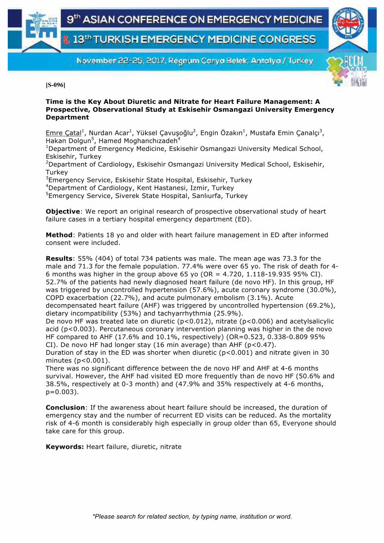
*Please search for related section, by typing name, institution or word.
[S-096]
Time is the Key About Diuretic and Nitrate for Heart Failure Management: A Prospective, Observational Study at Eskisehir Osmangazi University Emergency Department Emre Çatal1, Nurdan Acar1, Yüksel Çavuşoğlu2, Engin Özakın1, Mustafa Emin Çanalçı3, Hakan Dolgun5, Hamed Moghanchızadeh4 1Department of Emergency Medicine, Eskisehir Osmangazi University Medical School, Eskisehir, Turkey 2Department of Cardiology, Eskisehir Osmangazi University Medical School, Eskisehir, Turkey 3Emergency Service, Eskisehir State Hospital, Eskisehir, Turkey 4Department of Cardiology, Kent Hastanesi, Izmir, Turkey 5Emergency Service, Siverek State Hospital, Sanlıurfa, Turkey Objective: We report an original research of prospective observational study of heart failure cases in a tertiary hospital emergency department (ED). Method: Patients 18 yo and older with heart failure management in ED after informed consent were included. Results: 55% (404) of total 734 patients was male. The mean age was 73.3 for the male and 71.3 for the female population. 77.4% were over 65 yo. The risk of death for 4-6 months was higher in the group above 65 yo (OR = 4.720, 1.118-19.935 95% CI). 52.7% of the patients had newly diagnosed heart failure (de novo HF). In this group, HF was triggered by uncontrolled hypertension (57.6%), acute coronary syndrome (30.0%), COPD exacerbation (22.7%), and acute pulmonary embolism (3.1%). Acute decompensated heart failure (AHF) was triggered by uncontrolled hypertension (69.2%), dietary incompatibility (53%) and tachyarrhythmia (25.9%). De novo HF was treated late on diuretic (p<0.012), nitrate (p<0.006) and acetylsalicylic acid (p<0.003). Percutaneous coronary intervention planning was higher in the de novo HF compared to AHF (17.6% and 10.1%, respectively) (OR=0.523, 0.338-0.809 95% CI). De novo HF had longer stay (16 min average) than AHF (p<0.47). Duration of stay in the ED was shorter when diuretic (p<0.001) and nitrate given in 30 minutes (p<0.001). There was no significant difference between the de novo HF and AHF at 4-6 months survival. However, the AHF had visited ED more frequently than de novo HF (50.6% and 38.5%, respectively at 0-3 month) and (47.9% and 35% respectively at 4-6 months, p=0.003). Conclusion: If the awareness about heart failure should be increased, the duration of emergency stay and the number of recurrent ED visits can be reduced. As the mortality risk of 4-6 month is considerably high especially in group older than 65, Everyone should take care for this group. Keywords: Heart failure, diuretic, nitrate

*Please search for related section, by typing name, institution or word.
[S-097]
VT Managemant Patient with LVAD Özge Can, Yusuf Ali Altuncı, Funda Karbek Akarca Department of Emergency Medicine, Ege University, Izmir, Turkey Introduction: Patients with VADs are inclined to ventricular tachyarrhythmias due to underlying cardiomyopathy, electrolyte abnormalities, myocardium ischemia, RV failure. We present the cases admitted to ED with VT. Case 1: A 70-year-old male patient admitted to the emergency department with reduced oral intake who have LVAD due to CHF. VT was determined and in the follow-up, VF was developed. After defibrillation and CPR patient had the pulse. Amiadorone infusion started. ECO showed, the volume was depletion and hyperpotassemia was detected in the laboratory tests. Patients were stabilized with an isotonic solution and antipotassium treatment. Case 2: A 38-year-old male patient with LVAD due to cardiomyopathy was admitted with near syncope. On ECG, VT was detected and amiodarone infusion was started. Patient hospitalized at ICU. Case 3: A 52-year-old female patient was admitted to ER who had recurrent VT episodes in. Due to her history of thyroiditis associated with amiodarone patient hospitalized ICU for cardioversion Discussion: VT etiology is unknown with patient LVAD. Dysrhythmias can cause to RV dysfunction, thrombus, suction events, and poor perfusion, that electrolyte abnormalities (1,2) The incidence has been reported to be 30% to 50% (1,2). The patient can tolerate and has minimal symptoms of dysrhythmias. Electrolytes, myocardial ischemia or acute infarction should be checked Echocardiographic evaluation should be performed and suction dysfunction or blood flow. Malign arrhythmias should be treated immediately with cardioversion or antiarrhythmic medications. Amiodarone is often used as a first-line agent, although the use of b-blockers can also be considered (3). Keywords: VT, LVAD, emergency management

*Please search for related section, by typing name, institution or word.
[S-098]
Ischemic Stroke Due to Infective Endocarditis in a Young Patient Fatih Ahmet Kahraman1, Gülhan Kurtoğlu Çelik2, Osman Çelik1, İsmail Erkan Aydın1, Mehmet Ergin2 1Ankara Ataturk Training and Research Hospital Emergency Service, Ankara, Turkey 2Ankara Yıldırım Beyazıt University Faculty of Medicine Emergency Medicine Department, Ankara, Turkey Introduction: Infective endocarditis is an infectious disease primarily caused by bacteria, which is characterized by the involvement of heart valves. Due to its grace prognosis and high mortality, its diagnosis and treatment in emergency department is of paramount importance. Embolic events are common and life-threatening complications of infective endocarditis and their differential diagnosis remains difficult. It is more common among patients with heart valve disorders, intravenous drug users, elderly patients with valvular sclerosis, patients with valvular prosthesis, and dialysis patients. In this case report, we report a young patient with ischemic stroke due to infective endocarditis. Case Report: A 42-year-old man presented to emergency department with headache, dizziness, and intermittent speech disturbance, forgetfulness, and altered consciousness for 4 days. His past history was not notable for any condition except for suspected drug use. His vital signs were stable. Laboratory tests included a platelet count of 107 K/uL (NORMAL RANGE), CRP: 12 mg/L (0-5), and hsTroponin T: 249 pg/ml (0 -14). A cranial CT demonstrated no acute infarct. A diffusion MRI revealed bilateral hemispheric multiple diffusion limitations. He was examined with echocardiography with the initial diagnosis of infective endocarditis. His EF was 65%; there was aortic valvular thickening, aortic insufficiency of grade 2-3, and normal left ventricular function. He was admitted to the infectious diseases department with the diagnosis of ischemic stroke due to infective endocarditis. Conclusion: Some studies have revealed cardioembolism as the most common cause of ischemic stroke in the young. In patients with infective endocarditis, ischemic CVA is either present at admission or occurs within 2 weeks after the diagnosis of endocarditis. An appropriate antibiotic regimen should be immediately started. Recurrent embolism indicates the failure of antibiotic regimen. Presentation with stroke is rare among young population. In emergency department, infective endocarditis should be considered as a cause of ischemic stroke in young patients. Keywords: Infective endocarditis, ıschemic CVA

*Please search for related section, by typing name, institution or word.
[S-099]
Cardiac Contusion and Sudden Cardiac Ischemia Serkan Dogan, Utku Murat Kalafat, Melis Dorter, Doganay Can, Ramiz Yazici, Dogac Niyazi Ozucelik Department of Emergency Medicine, Istanbul Kanuni Sultan Süleyman Education and Research Hospital, Istanbul, Turkey Introduction: Blunted heart failure occurs in a wide spectrum, from clinically silent transient arrhythmias to lethal heart wall rupture. The most common type is the "cardiac contusion". Contusion is difficult to diagnose because there is no gold standard test. Attention should be paid to blunt cardiac trauma, arrhythmias, cardiac wall motion abnormalities, cardiogenic shock progressive clinical signs, rupture of valves, septum or heart walls. Compression between the sternum and the vertebrae of the heart, sudden pressure changes within the chest or abdomen, blast effect are other mechanisms. Blunt heart damage is rare in childhood groups, the most common cause is traffic accidents. Case: A 1-year-old girl without a known disease story was brought to our emergency department due to a fall from about 2.5 meters. On physical examination, AVPU-allert, there are no sensory defects, left lower limb movement defects and deformities present, light reflex + / +, respiratory sounds natural, chest area sensitivity present, heart sounds natural, pulses are palpable, no abdomen defense and rebaund on examination. In the laboratory tests performed on the patient, troponin T: 1.060 ck-mb: 105.3 patient was followed up with cardiac contusion pre-diagnosis. ST depression was detected in leads v1, 2, 3 and 4 of the electrocardiogram. No pathology was detected in ECO. The patient was followed up for cardiac enzyme due to sudden cardiac ischemia and cardiac contusion. The patient, who was kept under observation for 24 hours and regressed with cardiac enzymes, was discharged. The outpatient clinic control of the child was recommended. Conclusion: The chest wall of children is more compliant, so the amount of kinetic energy acting on the internal organs is greater. Therefore, the findings of external examination of children with blunt heart damage may be inconsistent. Cardiac contusion should be considered if external examination is present, thoracic sensitivity, cardiac examination, or abnormality in heart rate. Keywords: Contusion, ischemia, trauma

*Please search for related section, by typing name, institution or word.
[S-100]
Bilateral Anterior Omuz Çıkığı Mustafa Oğuz Cumaoğlu, Abuzer Coşkun, Salih Kocaoğlu, Osman Mahir Okur Sivas Numune Hastanesi, Acil Servis, Sivas Giriş: Acil servise eklem çıkığı şikayeti ile başvuran hastalarda; omuz ekleminin dislokasyonu önemli bir yer tutmaktadır. Omuz ekleminin bilateral çıkığı oldukça nadir görülen bir tablodur. Bu durum jeneralize epileptik nöbetler sonrası, eş zamanlı meydana gelen travmalarda, elektrik çarpmalarında, eklem gevşekliği olup istemli olarak omuz eklemini çıkartanlarda görülebilmektedir. Genellikle bilateral çıkıklar; posteriora doğru olurken, anteriora doğru gerçekleşenler daha az görülür. Olgu: 40 yaşında bayan hasta evde perde takmak için merdivene çıktığı esnada, dengesi kaybederek arkaya doğru omuzlarının üzerine yaklaşık 2 metre yükseklikten yere düşmüş. Her iki omuzda şidddetli ağrı ve omuzlarını oynatamama şikayeti ile ilçe devlet hastanesine başvurmuş. Periferde çekilen posteroanterior akciğer grafisinde her iki omuzda humerus başının anteriora doğru yer değiştirdiğinin görülmesi üzerine (resim 1) bilateral anterior omuz çıkığı ön tanısı ile merkeze sevki sağlanmış. Hasta acil servisimize getirildiğinde şiddetli ağrıya bağlı anksiyöz haldeydi. Muayenesinde her iki omuzda çok şiddetli ağrı olup diğer sistem muayeneleri doğaldı. Hasta kollarını hareket ettiremediği için sağ ayak sırtından damaryolu açıldı. Fentanyl (1 mikrogram/kg)– midazolam (0.1 mg/kg) kombinasyonu ile sedoanaljezi sağlandı. Sırası ile önce sağ, sonra sol omuz Milch yöntemi ile redükte edildi. Kontrol sağ-sol omuz grafileri alındıktan sonra (resim 2-3) her iki omuza velpau bandaj uygulandı. 2 saat acil servis gözlemi sonrası vitalleri stabil durumda olan hastaya non-steroid antiinflamatuar ilaç başlandı. Ortopedi poliklinik kontrol önerileriyle şifa ile taburcu edildi. Sonuç: Eş zamanlı olarak gerçekleşen bilateral anteriora omuz çıkığı nadir olarak görülse de önemli bir ortopedik acildir. Vücudun arka tarafından uygulanan yüksek enerjili travmalarda meydana gelebileceği akılda tutulmalıdır. Omuz çıkıklarında sedo-analjezi uygulamak hem hasta konforu, hem de uygulanan redüksiyon işleminin daha az zamanda yapılabilmesi açısından önemlidir. Anahtar Kelimeler: Acil, bilateral omuz çıkığı

*Please search for related section, by typing name, institution or word.
[S-101]
Üç Vaka Eşliğinde Anterior Diz Çıkığı Ve Komplikasyonları Özcan Yavaşi1, Gürkan Altuntaş1, Ozan Karakişi2, Mehmet Sabri Balık3, Mümin Murat Yazıcı1 1Recep Tayyip Erdogan University, Faculty of Medicine, Department of Emergency Medicine, Rize 2Recep Tayyip Erdogan University, Faculty of Medicine, Department of Cardiovascular Surgery, Rize 3Recep Tayyip Erdogan University, Faculty of Medicine, Department of Orthopedics, Rize Giriş: Travmatik diz çıkığı tüm ortopedik yaralanmaların %0,2’sinden daha azını oluşturur. Femurun tibia ile ilişkisine göre diz çıkıkları temel olarak anterior, posterior, medial, lateral ve rotasyonel olmak üzere beşe ayrılır. Burada acil servise, izole anterior diz çıkığı nedeniyle getirilen üç hasta, görüntüleme ve klinik sonlanımları açısından tartışılmıştır. Olgular Olgu 1: 65 yaşında kadın hasta sağ dizine kütük çarpması sonucu acil servise getirildi. Yapılan muayene ve çekilen grafilerde anterior diz çıkığı tespit edildi. Distal nabızlar palpasyonla alınıyordu. Redüksiyon sonrası bilgisayarlı tomografi (BT) anjiografisi çekildi patoloji düşünülmedi. Hasta atele alınıp immobilize edilip ortopedi servisine yatırıldı. 4 gün sonunda komplikasyonsuz taburcu edildi. Olgu 2: 62 yaşında bayan hasta, evde merdivenlerden dizinin üzerine düşme sonrası acil servise başvurdu. Fizik muayenede diz eklem hareketlerinde kısıtlılık ve postür bozukluğu mevcuttu, dorsalis pedis nabzı elle alınamıyordu. Yapılan radyolojik görüntülemeler sonrası anterior diz çıkığı tespit edilen hastanın dizi redükte edildi. Sağ alt ekstremite BT anjiografisi çekildi patoloji düşünülmedi. Ortopedi tarafından immobilizasyon ve antikoagulan tedavi ile taburcu edildi. İki gün sonra acil servise bacakta morarma ve ağrı şikayetiyle tekrardan başvuran hasta tekrar değerlendirildi. Muayenede femoral, popliteal, dorsal nabızlar elle alınıyordu, diz ve çevresinde ekimoz mevcuttu. Hasta hemartoz ön tanısı ile ortopedi servisine yatırıldı. Takipleri sonrası kliniği düzelen hasta şifa ile taburcu edildi. Olgu 3: 56 yaşında kadın düşme sonrası acil servise getirildi. Sol anterior diz çıkığı tespit edilip redükte edildi. Distal nabızları alınamayan hastaya çekilen BT anjiyografide sol popliteal artere bası yapan hematom saptandı. İlerleyen saatlerde kompartman sendromuna doğru ilerleyen vaka operasyona alınıp popliteal arter onarımı ve fasyatomi uygulandı. Sonuç: Diz çıkıkları en kısa sürede redükte edilmelidir. Anterior çıkıklar arterde traksiyon lezyonu yaparken, posterior çıkıklar genellikle komplet yırtıklara neden olurlar. Popliteal damarların görüntülemesi muhakkak yapılmalıdır. Kanama, antikoagülan tedavinin en önemli komplikasyonudur. Sonuç olarak diz yaralanmalarında olası vasküler ve sinir yaralanmaları açısından mümkün olduğunca yatırılarak takip edilmelidir. Anahtar Kelimeler: Anterior diz çıkığı, kompartman sendromu, popliteal arter yaralanması

*Please search for related section, by typing name, institution or word.
[S-102]
Öksürük Atağının Tetiklediği Periorbital Ekimoz Murat Das, Okhan Akdur, Erşan Yurtseven, Canan Akman Çanakkale Onsekiz Mart Üniversitesi Acil Tıp Ana Bilim Dalı, Çanakkale Giriş: ''Rakun Gözleri'' veya ''Panda İşareti'' olarak da adlandırılan periorbital ekimoz, periorbital subkutan dokulara kan sızmasına bağlı oluşur. Periorbital ekimozda, orbital septum bu sızmanın tarsal plakanın ötesine geçmesini engeller. Periorbital ekimoz orbital venöz basıncı artıran intrakranial patolojiler, antikoagülan ilaçlar ve valsalva manevraları ile tetiklenmektedir. Yemek aspirasyonu sonrası öküsürük atağı ile periorbital ekimoz gelişen hastayı sunmayı amaçladık. Olgu: Yemek aspirasyonu sonrası şiddetli öksürük atağı gelişen 80 yaşında erkek hasta, her iki göz çevresinde morarma şikayeti ile sağlık merkezine başvurmuş. Dış merkezden ayırıcı tanı yapılmak üzere acil servisimize gönderilen hastanın değerlendirme sırasında arteriyal tansiyon 130/90mmHg, vücut ısısı 36.7 °C, periferik nabızlar bilateral eşit ve 80 atım/dakika, solunumu düzenli ve 14/dakika olarak saptandı. Fizik muayenede inspeksiyonda bilateral periorbital ekimoz (resim 1) mevcuttu; travma izine rastlanmadı. Palpasyon ile hassasiyet yoktu. Tam kan sayımı, karaciğer ve böbrek testleri, elektrolitler ve koagülasyon parametreleri normal sonuçlandı. Ayırıcı tanı için bilgisayarlı beyin (BBT) tomografi ve difüzyon manyetik rezonans görüntüleme (MRG) alındı. BBT normal olarak sonuçlandı. Tesadüfi olarak difüzyon MRG de laküner enfarkt saptandı. Hastanın göz içi değerlendirilmesi için göz hastalıkları bölümüyle, tesadüfi saptanan laküner enfarkt için nöroloji bölümü ile konsültasyon yapıldı. Laküner enfarktı olan hasta takip ve tedavi düzenlenmesi açısından nöroloji bölümüne yatırıldı. Hastanın ekimozu iki hafta sonra geriledi. Tartışma: İntakranial venöz basıncı artıran, sinüs trombozları, nöroblastom, idiopatik intrakranial hipertansiyon, migren, trigeminal otonomik sefalji, rinore, nazal konjesyon, kontrolsüz hipertansiyon gibi nedenler periorbital ekimoza neden olabilir. Valsalva manevrası inrakranial venöz basıncı artırarak periorbital ekimozu tetikleyebilir. Bu mekanizma ile şiddetli hapşırık ve şiddetli kusma sonrası periorbital ekimoz vakaları bildirilmiştir. Öksürük refleksi de intrakranial basıncı artıran bir diğer valsalva manevrası örneğidir. Bizim olgumuzda da öksürük sonrası tetiklenen intrakranial basınç artışına sekonder periorbital ekimoz gelişmiştir. Valsalva manevrası mekanizması ile gelişen durumlarda hastalık bening seyreder. Ancak altta yatan hayatı tehdit edici diğer nedenler ayırıcı tanıda mutlaka dışlanmalıdır. Anahtar Kelimeler: Öksürük, periorbital ekimoz

*Please search for related section, by typing name, institution or word.
Resim 1
Periorbital bilateral ekimoz görüntüsü

*Please search for related section, by typing name, institution or word.
[S-103]
Acil Servise Başvuran Erişkin Akut Gastrointestinal Sistem Kanamali Hastalarda Kan Laktat Düzeyinin Tanısal Ve Prognostik Değeri Hande Akbal Kahraman1, Gökhan Aksel2, Öner Bozan3, Serkan Emre Eroğlu2 1University of Health Sciences, Fatih Sultan Mehmet Training and Research Hospital, Department of Emergency Medicine, Istanbul, Turkey 2University of Health Sciences, Umraniye Training and Research Hospital, Department of Emergency Medicine, Istanbul, Turkey 3University of Health Sciences, Okmeydani Training and Research Hospital, Department of Emergency Medicine, Istanbul, Turkey Çalışmamızda acil servise akut gastrointestinal sistem kanaması ile başvuran hastaların kan laktat düzeyi tespit edilerek bunun sağ kalıma ve prognoza etkisinin belirlenmesi amaçlanmıştır. Çalışmamız Ümraniye Eğitim ve Araştırma Hastanesi Acil Tıp Kliniği’nde 01.01.2014 ile 31.05.2016 arasında akut gastrointestinal kanama nedeniyle takip edilen 506 hasta ile retrospektif olarak yapıldı. 18 yaş altı hastalar, gastrointestinal kanama tanısı şüpheli olan hastalar, fizik muayene, öykü ve laboratuvar verileri eksik olan hastalar çalışmadan dışlandı. Hastaların yaşı, cinsiyeti, başvuru tarihi, başvuru şikayeti, bilinen hastalıkları, kullandıkları ilaçlar, fizik muayene semptom ve bulguları, laboratuvar sonuçları, uygulanan tedavi, prognoz ve mortalite durumları kaydedildi. Çalışmaya dahil edilen 506 hastanın 310’u (%61.3) erkek, 196’sı (%38.7) kadındı. Hastaların%77.67’si 50 yaş üzeriydi. Erkek ve kadınlarda alt veya üst gastrointestinal kanama görülmesi açısından anlamlı fark saptanmadı (p=0.122). Tüm gastrointestinal sistem kanamalı hastalarda yaşınmortaliteye etkisine bakıldığında, yaşın artmasıyla birlikte mortalitenin de istatiksel olarak anlamlı şekilde arttığı tespit edildi (p<0.001). Cinsiyet açısından gruplar arasında istatiksel olarak anlamlı bir mortalite farkı bulunmadı (p=0.051). Laktat düzeyi ise ölen hastalarda istatistiksel anlamlı şekilde daha yüksek saptandı (p<0.001). Yine tüm gastrointestinal sistem kanamalı hastalarda sistolik ve diyastolik kan basıncı değerlerinin düşük olmasının mortaliteyi arttırdığı tespit edildi (p değerleri sırasıyla <0.001 ve <0.001). Gastrointestinal sistem kanamalı hastalar üst ve alt gastrointestinal sistem kanaması olarak ayrı ayrı ele alındığında; üst gastrointestinal sistem kanamalı hastalarda hematokezya varlığı, malignite öyküsü, karaciğer hastalığı, replase edilen eritrosit süspansiyonu miktarı ve laktat yüksekliğinin (p değerleri sırasıyla 0.001, 0.000, 0.029, 0.009, 0.000), alt gastrointestinal sistem kanamalı hastalarda ise ileri yaş ve malignite varlığının mortaliteyi arttırdığı belirlendi (p değerleri sırasıyla 0.030, 0.013). İleri yaşla birlikte ek hastalık varlığı, unstabilite belirteçlerinden olan düşük arteriyel tansiyon bulunması, fazla eritrosit süspansiyonu replasmanı gerektirecek hızlı kanama bulguları, hemodinamik strese eşlik eden yüksek laktat, hastane içi mortaliteyi arttırır ve klinisyene prognoz hakkında önemli bilgiler vererek tedavi sürecini belirlemede yardımcı olur. Anahtar Kelimeler: Gastrointestinal kanama, laktat, prognoz

*Please search for related section, by typing name, institution or word.
[S-105]
Ekstazi Kullanımına Bağlı Spontan Pnomomediastenum: Olgu sunumu Neşe Karslıoğlu, Halil Doğan, Fatma Oruç, Efken Toptop, Onur Tosun Bakırköy Dr. Sadi ̇ Konuk Eği ̇ti ̇m Ve Araştırma Hastanesi ̇ Aci ̇l Tıp Kli ̇ni ̇ği ̇, İzmir Giriş: Pnömomediastinum, solunum sistemi veya sindirim sistemi organlarının perforasyonu sonucunda havanın mediastende lokalize olmasıdır. Spontan veya travmatik olarak oluşabilmektedir. Spontan pnömomediastinum gelişimi nadirdir ve etiyolojisinde sıklıkla toraks içi basıncı artıran nedenler söz konusudur Spontan pnömomediastinum gelişiminde, predispozan faktör olarak astım gibi altta yatan bir akciğer hastalığının yanı sıra şiddetli bir öksürük atağı, kusma, yüksek sesle ve şiddetle bağırma, ağır egzersiz ve yük kaldırma, vajinal doğum gibi Valsalva manevrasına benzer şekilde intrapulmoner basıncı yükselten tetikleyici bir neden veya madde bağımlılığı (kokain, ekstazi) bulunur. Olgu: 21 yas erkek hasta acil servisimize nefes darlığı ve gögüs agrısı şikayetı ile ayaktan basvurdu. Herhangi bir travma öyküsü olamayan hasta sorgulamasında nefes darlığı ve gögüs ağrısı basladıgı gün 2-3 saat öncesinde hayatında ilk defa 3 adet extacy ve bir paket sigara aldığını belirtti. Fizik muayenesinde genel durum iyi oryante ve koopere, vital bulgular stabil, boyun ve göğüste bilateral yaygın krepitasyon mevcuttu. PA Ak ciğer grafisinde ve toraks tomografisinde pnömomediastinum ve pnomoperikardium ile uyumlu yaygın hava imajları mevcuttu (Resin 1). Laboratuar tetkikleri normal sınırlarda idi. Gögüs cerrahisi servisine yatırılan hasta 4 günlük takibi sonrası radyolojik ve klinik olarak gerileme saptanması ile taburcu edildi. Tartışma: Spontan pnömomediastinum özellikle genç erkeklerde görülen, tanının ardından semptomların genellıkle kendiliğinden gerilediği nadir bir klinik durumdur. Görülme sıklığı değişik hastane kayıtlarında 1/7000 ile 1/320000 arasında bildirilmiştir. Spontan alveolar rüptür genellikle öksürme, kusma ıkınma gibi alveol basıncında artışa yol açan durumlarda ortaya çıkar Astım, KOAH, Diyabetik Ketoasidoz, aşırı egzersiz esrar kokain ekstazi gibi madde içimi ve Diffüz interstisyel fibrozis öncülük eden hastalıklardandır. Tanı da altın standart toraks tomografisi olup tedavi ise nedene yöneliktir. Genellikle 48 saat içinde kendiliğinden gerileme gösteren bu klinik durumda mediastinit gelişmesi durumunda antibiyotik başlanır. Sonuç olarak madde kullanımı öyküsü olan ve nefes darlığı ve göğüs ağrısı şikayetinin varlığında, nadir görülen bir klinik durum olmasına rağmen, ayırıcı tanıda spontan pnömomediastinum mutlaka akla gelmelidir. Anahtar Kelimeler: Pnömomediastinum, ekstazi, gögüs agrısı
Resim 1

*Please search for related section, by typing name, institution or word.
Toraks tomografide pnömomediastinum ile uyumlu yaygın hava imajları

*Please search for related section, by typing name, institution or word.
[S-106]
Türkiye’de 3. Basamak Sağlık Kuruluşu Erişkin Acil Servisine Başvuran Kulak Burun Boğaz Hastalarının Epidemiyolojik Özellikleri Murat Dogan1, Ali Duman2 1Adnan Menderes Üniversitesi, Tıp Fakültesi, Kulak Burun ve Boğaz Anabilimdalı, Aydın, Türkiye 2Adnan Menderes Üniversitesi, Tıp Fakültesi, Acil Servis, Aydın, Türkiye Giriş: Son yıllarda acil servislere başvuran hasta sayıları giderek artmakta ve artan başvurulara etkin tedavi politikası geliştirebilmek için başvuruların epidemiyolojik analizlerinin yapılması gereği duyulmaktadır. Bu çalışmamızda, üçüncü basamak sağlık kuruluşuna ait erişkin acil servisine bir yıl içerisinde başvuran Kulak Burun Boğaz (KBB) hastalarını inceledik. Materyal-Metod: Bir Ocak 2016 ile 31 Aralık 2016 tarihleri arasında Aydın Adnan Menderes Üniversitesi Tıp Fakültesi erişkin acil servisi kliniğine başvurmuş 57500 hasta retrospektif olarak incelendi. Hastaların uluslar arası hastalık kodları (ICD) ve dermografik özellikleri, başvuru zamanları ve sosyal güvenlik kurumu maliyetleri incelendi. Kulak burun boğaz kliniği ile ilişkili 7028 hasta çalışmaya dahil edildi. Hastaların gruplandırılması ICD kodları üzerinden yapılarak analizler gerçekleştirildi. Sonuçlar: 7028 hasta KBB ile ilgili tanılar nedeniyle acil servise başvurdular. Başvuran hastaların erkek/kadın oranı 1:1.3 idi. Ortalama yaş 36.0 ± 16.3 yıldı. Başvuran hastaların %73.5’i Akut rinit (üst solunum yolu enfeksiyonları), Akut farenjit ve akut tonsillit tanılarından oluşmaktadır. KBB hastalarının tüm acil servise başvuran hastalar arasındaki oranı Aralık, Ocak ve Şubat aylarında en yüksek oranlara ulaşırken (%17.3, %22.8 ve %16.3, sırasıyla), haziran ayında en düşük seviyelere ulaşmaktadır (%6.0). Hastalar hafta içi yada hafta sonu olmasından ve mesai saatlerinden bağımsız olarak, öğleden önce 8’den gün sonu 24:00’a kadar olan vakit aralığında çoğunlukla başvururken (%93.8), gece saatlerinde başvuru oldukça az görülmektedir (%6.2). Hasta başına düşen tedavi maliyeti tüm KBB hastalarında 12.7 ± 16.3 dolar olarak hesaplandı. Tonsillit, rinit ve farenjit tanısı olan hastaların tedavi maliyetleri ortalama 7.0 ± 5.9 dolar olduğu görüldü. Sonuç: KBB ile ilgili hastalıklardan dolayı acil servise başvuran hastalar önemli bir oran oluşturmaktadır. Bu hastaların nitelikleri incelendiğinde büyük çoğunluğunun üst solunum yolunu ilgilendiren enfeksiyonlar ve gerçekte acil olmayan niteliğe sahip olduğu görülmektedir. Dolayısıyla, çalışmamızdan elde edilen veriler ışığında KBB hastalarına yönelik izlenecek politikalar belirlenebilir ve gereksiz yere acil servise başvurular azaltılabilir. Anahtar Kelimeler: Acil servis, kulak burun boğaz

*Please search for related section, by typing name, institution or word.
[S-107]
Bu Kez Ürtiker Değil Müge Arslan, Buğra Ilhan, Halil Doğan, Attila Beştemir, Ökkeş Taha Küçükdağlı Bakırköy Dr Sadi Konuk Eğitim ve Araştırma Hastanesi Acil Tıp Kliniği, İstanbul Lökositoklastik vaskülit sıklıkla deri ve eklem tutulumlarıyla giden küçük damarların inflamatuar bir hastalığıdır. Lökositoklastik vaskülitlerde etiyolojide ilaçlar, enfeksiyonlar, kimyasallar, konnektif doku hastalıkları, gluten intoleransları görülebileceği gibi idiopatik de olabilir. En sık klinik prezentasyonu alt ekstremite yerleşimli palpable purpuralardır. Lezyonlar nadiren üst ekstremite ve gövde yerleşimli olabilir. Fizik muayenede palpable purpura dışında nekroz, vezikül ve bül görünümü de olabilir. Lökositoklastik vaskülit esas olarak deriyi ilgilendirmekle beraber renal, gastrointestinal, kas ve iskelet sisteminde de tutulum yapabilir. Tanıda etiyolojiye yönelik enfeksiyöz parametreler, cilt biyopsisi ve sistemik tutuluma yönelik testler kullanılır. Tedavide ise iyi seyirli cilt lezyonları için elevasyon, yatak istirahati antihistaminik ve analjezikler kullanılırken sistemik veya rekürren olgular için sistemik steroid, dapson, immünsupresif ajanlar (azotiyopürin, mikofenolat mofetil, siklosporin), metotreksat, IVIG ve plazmaferez seçenekler arasındadır. Vaka: 77 yaşında kadın hasta acil tıp kliniğimize 1 hafta önce başlayan ayaklarda ödem ve kızarıklık şikayetleriyle başvurdu. Gelişinde ölçülen vital değerleri normal olarak tespit edildi. Özgeçmişinde 10 yıldır hipertansiyonu ve anksiyete bozukluğu olduğu öğrenildi ve bu hastalıklar için zofenopril-hidroklorotiazid ve paroksetin kullanımaktaydı. 20 gün önce bacaklarda şişlik nedeniyle furosemid tablet başlanmış olan hastanın lezyonlarının bacaklardan başlayıp gövdeye yayıldığı öğrenildi (Resim 1). İlk olarak dış merkezde değerlendirilen hastanın lezyonları ürtiker olarak değerlendirilip antihistaminik reçete edilmiş. Hastanın kullandığı antihistaminiğe rağmen lezyonlarında gerileme olmadığı öğrenildi. Hastanın kliniğimizde yapılan tetkiklerinde karaciğer fonksiyon testlerinde, üre, kreatin ve CK değerlerinde artış tespit edildi. Yapılan cilt biyopsileri de lökositoklastik vaskülit ile uyumlu olarak raporlandı. Hastaya immünsupresif tedavi başlandı ve poliklinik takibine alındı. Tartışma: Lökositoklastik vaskülit nadir görülen fakat sistemik tutulum nedeniyle morbiditesi ve mortalitesi fazla olan bir hastalıktır. 24 saati aşan, alt ekstremiteden başlayıp gövdeye yayılan ve antihistaminiklere yanıtsız lezyonlarda acil tıp hekimlerinin ayırıcı tanıda mutlaka düşünmesi gerekmektedir. Anahtar Kelimeler: lökositoklastik vaskülit, döküntü, ürtiker
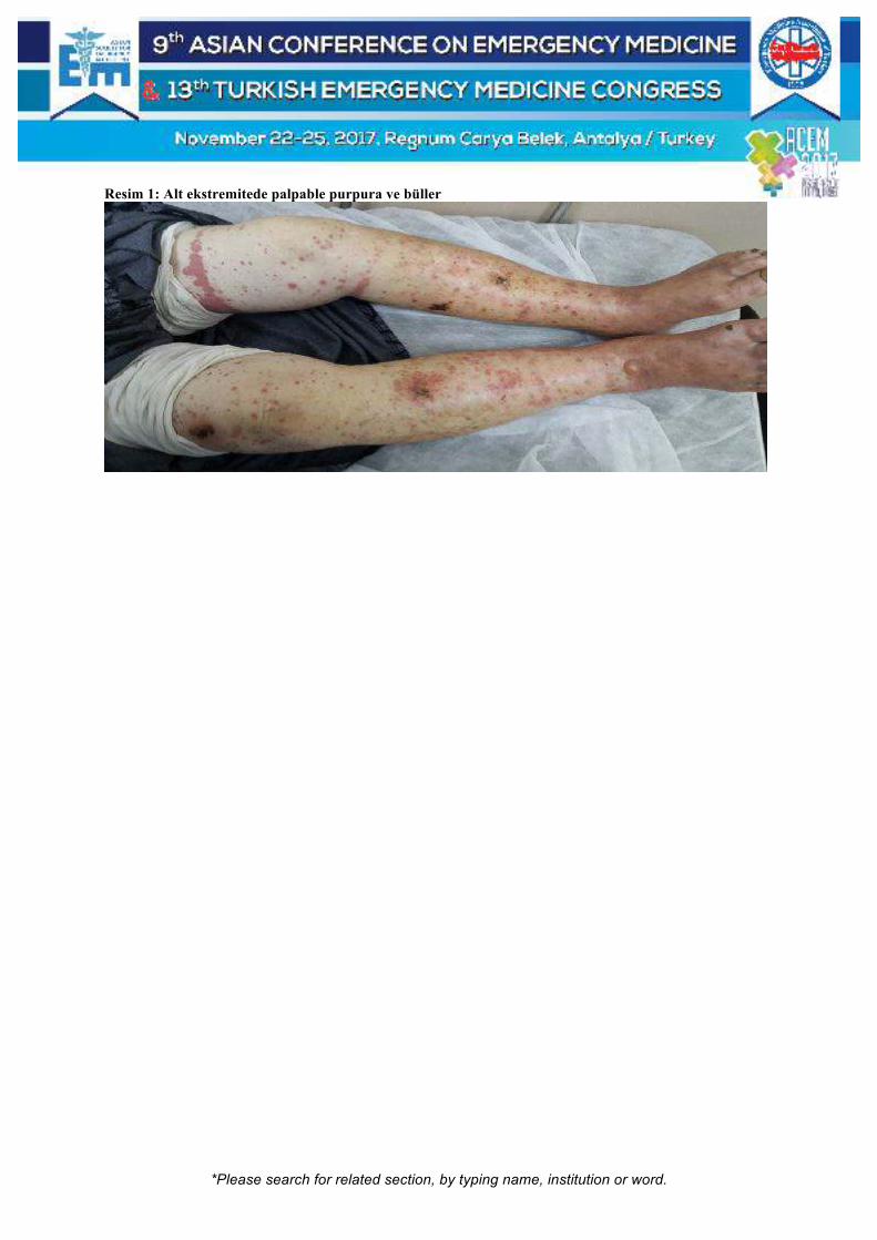
*Please search for related section, by typing name, institution or word.
Resim 1: Alt ekstremitede palpable purpura ve büller

*Please search for related section, by typing name, institution or word.
[S-108]
Fenitoin Kullanımına Bağlı Toksik Epidermal Nekroliz Rezan Tahtacı, Umut Payza, Ecem Ermete, Fatih Esad Topal İzmir Katip Çelebi Üniversitesi Atatürk Eğitim Araştırma Hastanesi Acil Tıp Anabilim Dalı, İzmir Toksik epidermal nekroliz sıklıkla ilaçlara bağlı bazen de enfeksiyonlar sonrası gelişen nadir görülen ancak mortalitesi yüksek bir durumdur. yaygın eritem, nekroz ve epidermisin büllöz dekolmanı ile karakterize olup vücut yüzeyinin % 30u ndan fazlası etkilenir. ilaçlardan nonsteroid antiinflamatuvar ilaçlar, fenobarbital, fenitoin, karbamazepin, valproik asit, lamotrigine, antibiyotikler suçlanırken, enfeksiyoz ajanlardan mycoplazma, E.coli, varisella ve kızamık neden olabilmektedir. Dünya genelinde yıllık ten insidansı % 0,41.3 tür. Olgu: 42 yaşında erkek hasta 3 ay önce trafik kazasına bağlı intrakrniyal hemoraji nedeniyle dış merkez beyin cerrahi yoğun bakımda takip edilmiş, taburculuğunda laroxyl, epdantoin, nörodol başlanmıştır. Tedavinin birinci ayında avuç içi ve ayak tabanlarında kaşıntı sonrasında kızarıklıklar ve soyulmalar baş göstermiştir.39 dereceyi bulan ateş yükselmesi ve ödemde artma nedeniyle dış merkeze başvuran hasta karaciğer fonksiyo testlerinde yükselme saptanması üzerine hastanemize yönlendirilmiştir hastanın öz ve soygeçmişnde bir özellik yok. Vital bulgular normaldi. Tüm vücutta yaygın makülopapüler eritematöz lezyonlar, ekstremitelerde, oral mukozada, auriküler, pubik, anal ve ingiunal bölgede büller mevcuttu. Laboratuarda Hb:13.7 beyaz küre 11700 %54 PNML,%46 lenfosit,PLT 36300, CRP 18.02, aspartattransaminaz 242, alaninaminotransferaz 418,total bilirubin 6.67 indirekt bilirubin 2 mg/dl ölçüldü.göz muayenesi normaldi. Kan, yara ve idrar kültürleri alındı, dahiliye ve dermatoloji konsültasyonları istendi. Dermatolojinin yaptığı deri biyopsi sonucu dermoepidermal bileşkede ayrışma ve keratinosit nekrozu saptandı. Hasta yatırıldığı dermatoloji servisinde sistemik steroid, IVIG, antibiyoterapi, uygun yara bakımı tedavisi almaktayken ikinci günde oral mukoza lezyonlarında artış, büllöz alanlarda enfeksiyon, ve konjoktivit gelişti. Takip eden haftada hasta sepsise girdi. Yoğun bakım takibinin ardından sepsis tablosu geriledi. hasta halen dermatoloji servisinde yatmaktadır. Sonuç: TEN yaşamı tehdit eden dermatolojik acil durumdur. Basit allerjik bulgularla başvurabilir. Mortalitenin yüksek olması nedeniyle acil tıp hekiminin daima aklında bulunması gereken tanılar arasında yer almalıdır. Tedavide ilk olarak etken öngörülerek maruziyetin derhal ortadan kaldırılması ve hızla destek tedavinin başlanması gerektiğinden ilaç alımı mutlaka sorgulanmalıdır. Anahtar Kelimeler: Toksik epidermal nekroliz, fenitoin
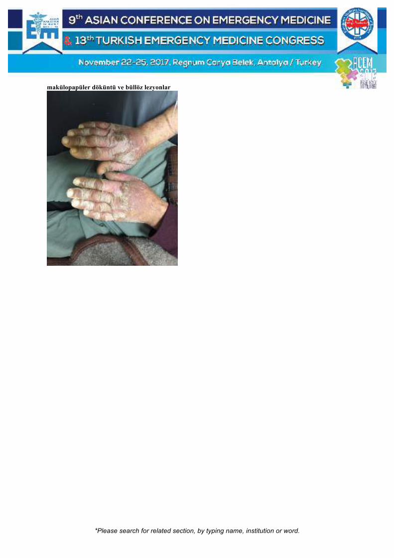
*Please search for related section, by typing name, institution or word.
makülopapüler döküntü ve büllöz lezyonlar

*Please search for related section, by typing name, institution or word.
[S-109]
Kolonoskopi Sonrası Gaz Distansiyonu Neye Bağlı? Ahmet Burak Erdem1, Muhittin Serkan Yılmaz1, Bahattin Işık2 1Ankara Numune Eğitim ve Araştırma Hastanesi, Acil Tıp Kliniği, Ankara 2Ankara Dışkapı Yıldırım Beyazıt Eğitim ve Araştırma Hastanesi, Acil Tıp Kliniği, Ankara Giriş: Kolonoskopi; rektumdan terminal ileuma kadar bağırsak hastalıklarının endoskopik tanısında kullanılan en iyi tanı yöntemlerinden biridir. Nadirde olsa kanama, perforasyon gibi komplikasyonları vardır. (1) Kolonik perforasyon mekanik, terapötik veya hava basıncı sonucu gelişebilir. (2) Bu hastalar erken dönem gelebildiği gibi literatürde 9. haftada bile görülebilen vakalar mevcuttur. Tanıda yaygın karın ağrısı, distandü bir batın, defans ve rebaund görülebilir. (2) Direkt grafi ile serbest hava görüntülemesi ile tanı konulur. Buna rağmen direkt grafide bulgu vermeyen vakalara, bilgisayarlı tomografi (BT) ile perforasyon tanısı konulur. Tedavide konservatif ve cerrahi yaklaşım uygulanır. (2) Vaka: 62 yaşında erkek hasta acil servise karın ağrısı şikayeti ile başvurdu. Yaklaşık 1 saat önce kolonoskopi uygulanan hastanın işlem öncesi karın ağrısı, bulantı ve kusma şikayetleri mevcutmuş. İşlemden sonra ağrı şikayeti artan hasta subileus ön tanısı ile acil servise yönlendirilmiş. Hastanın fizik muayenesinde vital bulguları stabildi. Batın distandü, defans ve rebaund mevcuttu. Diğer sistem bulguları normaldi. WBC değeri 10,8 uL– HGB ise 8 g/Dl diğer biyokimyasal parametreleri normaldi. Direkt grafide diyafram altı hava ve yoğun gaz görünümü mevcuttu. Batın BT’de tüm reseslerde serbest hava izlendi ve perforasyon lehine değerlendirildi. Genel Cerrahi konsültasyonu yapılan hasta opere edildi. Rektosigmoid bileşkedeki perforasyon tamir edildi. Ayrıca çekumda tespit edilen kitleye sağ hemikolektomi uygulandı. Sonrasında düzelen hasta taburcu edildi. Sonuç: Kolonoskopi inzaviz bir işlem olduğu için perforasyon %0,08-0,19 oranında görülebilir. (1) Terapötik işlemlerde bu oran daha da artar. (2) Perforasyon anında bulgular asemptomatik olabilir. Genelde gecikmiş bulgular ilk 72 saatte görülür. Perforasyonu işlemi yapan doktor endoskopik görüntüleme esnasında görebilir. Geciken vakalarda direkt grafi ve BT tanı koydurucudur. Cerrahi tedavi olarak perfore alanın onarımı, bağırsak rezeksiyonu gibi yöntemler uygulanır. (2) Kolonoskopi sonrası gaz distansiyonu sıklıkla karşılaşılan bir durumdur. Bazen bu tür hastalarda mortalitesi yüksek komplikasyonlar görülebilir. Acil hekimleri bu tür hastalarda perforasyon olabileceğini düşünerek mortalitenin azalmasına büyük katkı sağlayacaktır. Anahtar Kelimeler: Kolonoskopi, perforasyon, serbest hava
Direkt Grafi Perforasyon

*Please search for related section, by typing name, institution or word.
Batında gaz distansiyonu ve sağda diafram altı serbest hava

*Please search for related section, by typing name, institution or word.
[S-111]
Are Markedly Elevated Nt-proBNP Levels Always Reliable to Diagnose Acute Decompansated Heart Failure in Haemotologic Malignancy Patients? Murat Ersel, Sercan Yalçınlı, Yusuf Ali Altunci Ege University School of Medicine, Department of Emergency Medicine, Izmir, Turkey The association of NT-proBNP levels in haematologic malignancy patients are complicated for determination of acute decompansated heart failure. Burjonroppa et al. was found no significant association with markedly elevated BNP values and clinical evidence of volume overload or LV dysfunction. The exact mechanism of that extreme elevation of NPs in these population is unclear. A 55 years old woman with history of multipl myeloma was admitted to the ED with compliant of diarrhea, nausea, anuria and altered mental status. She was on bortezomib, a novel first-in-class proteasome inhibitor, which has gastrointestinal side effects such as nausea, vomiting, diarrhea, constipation. Her vital signs were as folllows; BP:64/40 mmHg, pulse 102/bpm, fever: 37 C and SatO2: 88%. On the physical examination there were no significant signs of pulmonary and systemic congestion (no rales and ronchi, bilateral 1+ lower extremity edema). Chest X ray obtained in PA position showed a mildly enlarged cardiac silhouette and pulmonary vascular congestion. Her NT-proBNP was 58.209 pg ⁄mL (eGFR 8.58 mL⁄min). Bedside USG revaled a 2.2 VCI diameter and less collable than 50%. The patient treated with hemodialysis + ultrafiltration. However echocardiography demonstrated EF of 60%, normal pulmonary artery pressure and mild tricuspid regurgitation. Despite no signs of acute decompansated heart failure, NT-proBNP levels were found as extremely elavated. Several factors, such as age, sex, obesity, and renal function have all been known to affect NT-proBNP levels. In case series, levels of serum NT-proBNP of volume overloaded haematologic malignancy patients were observed far exceeding than expected values. Even in euvolemic patients high levels of NT-proBNP were recorded, also, in those patients, NT-proBNP levels might not always be associated with acute decompansated heart failure, emergency physician should be aware of these phenomenon. Keywords: Haematologic malignities, heart failure, nt-probnp

*Please search for related section, by typing name, institution or word.
[S-112]
Transient Vocal Cord Paralysis Following Central Venous Hemodialysis Catheter Insertion Gül Pamukçu Günaydın1, Selahattin Gürü1, Elif Aslan Taş2, Fatih Tanrıverdi2, Gülhan Kurtoğlu Çelik2 1Department of Emergency Medicine, Ankara Atatürk Training and Research Hospital, Ankara, Turkey 2Department of Emergency Medicine, Faculty of Medicine, Yıldırım Beyazıt University, Ankara, Turkey Introduction: In this article, we present a case of recurrent laryngeal nerve palsy not caused by nerve injury but due to local anesthetic infiltration that was applied prior to central venous catheterization. Case: A 47-year-old female patient was admitted to emergency room with fatigue and nausea and was diagnosed with acute renal failure. Right jugular venous catheterization was performed for emergency hemodialysis with Seldinger technique using middle approach. Within minutes and immediately after the procedure the patient complained of hoarseness and shortness of breath, and she had stridor in her physical exam. Awake flexible fibreoptic laryngoscopy revealed unilateral right-sided vocal cord paralysis with no edema. The patient was asked to remain nil per os and observed in ER with nasal oxygen. At the 3rd hour of follow-up without any other intervention, her symptoms resolved. Discussion: Due to its proximity to the internal jugular vein injury to the recurrent laryngeal nerve while attempting to insert a central venous line can occur, particularly with difficult and repeated attempts. Local anesthesia led temporary ipsilateral vocal cord paralysis in patients undergoing carotid endarterectomy is described in literature. We think temporary vocal cord palsy in our case was due to local anesthetic infiltration rather than nerve injury, since it resolved spontaneously within only hours. Expectant treatment is a good choice ensuring the patient’s airway is safe. Emergency physicians should be aware of this rare complication and its right management. Keywords: Central venous catheterization, local anesthesia, and vocal cord paralysis

*Please search for related section, by typing name, institution or word.
[S-113]
Effect of C1 Esterase Inhibitor in Angiotensin-Converting Enzyme Inhibitor Induced Angioedema Funda Karbek Akarca, Sercan Yalcinli, Güçlü Selahattin Kıyan Ege University Faculty of Medicine, Department of Emergency Medicine, Izmir, Turkey Introduction: Angiotensin converting enzyme inhibitor induced angioedema (ACEI-IAE) constitutes 4 / 10.000 visits to emergency departments per year. Unlike hereditary angioedema, findings in ACEI-IAE tend to occur in the face region instead of the abdomen. This situation may lead to life-threatening clinical conditions associated with upper airway obstruction during an attack. Method: Patients who visited Ege University Emergency Department from March 2011 to April 2017 and who received C-1 esterase inhibitor treatment were retrospectively investigated. Totally, 82 applications were found in 27 different patients. Six emergency department appeals were detected in five different patients due to ACEI-IAE. Patients' age, sex, complaints, physical examination findings, treatment protocols, airway intervention requirements, intensive care and hospital admission requirements were investigated. Results: Eighty percent of the patients were female (n: 4). The mean age was 65.6 ± 13 years. The most common complaint was tongue swelling (n: 3, 50%). On physical examination, tongue edema in three patients, tongue and uvula edema in one patient, lip edema in one patient and skin redness in one patient were observed. As initial treatment, antihistamines were used in 66.7% of patients (n: 4), corticosteroids in 83.3% (n: 5) and adrenaline in 33.3% (n: 2). It was observed that C-1 esterase inhibitor treatment was started on the whole of the patients after the antihistaminic and / or corticosteroid treatment failed to obtain adequate response. Patient with advanced airway intervention need was not detected. 83.3% (n: 5) of the patients were discharged after the emergency observation. The mean length of stay time was 1749 ± 1362 minutes in emergency department. Conclusion: Despite limited data in the literature, it may be appropriate to use plasma C1 esterase inhibitor therapy in ACEI-IAE. C1 esterase inhibitor therapy in ACEI-IAE may reduce the need for advanced airway intervention. Keywords: C1 Esterase Inhibitor, Angioedema, Angiotensin-Converting Enzyme

*Please search for related section, by typing name, institution or word.
[S-114]
Comparison of Hgb, Htc, Na, and K Levels Measured by Blood Gases Analyzer and Laboratory Auto-Analyzer in Different pH Stages Şeref Kerem Çorbacıoğlu, Emine Emektar, Yunsur Çevik, Hikmet Şencanlar Çetiner, Mehmet Ali Özbek, Sedat Akkan Keçiören Training and Research Hospital, Department of Emergency Medicine, Ankara, Turkey Purpose: In this study, primary aim was detecting whether blood gases analyzer (BGA) is reliable or not in daily practice by comparing of Na, K, Hgb, and Htc levels measured by BGA and laboratory auto-analyzer (LAA). Secondary aim was whether BGA is reliable or not in daily practice by comparing of Na, K, Hgb, and Htc levels measured by BGA and LAA in different pH stages. Material-Methods: This study screened electronic data and file records of all patients who admitted to ED with any complaint during the study period, retrospectively. Patients who had results of venous blood gases and routine laboratory taken at the same time were included this study. For each parameters, agreements and correlations between results of BGA and LAA were evaluated Bland-Altman test and Spearmen Correlation test, respectively and r-value higher than 0.80 was considered a strong correlation. Results: Finally, laboratory results of 1374 patients were evaluated for statistically analyses. When evaluated correlations between results of BGA and LAA, it was found that there was only strong correlation for K (p<0.001, r=0.83). When assessed agreements between results of BGA and LAA, the mean difference were found as 0.02±6.1 for sodium, 0.3±0.44 for potassium, -0.5±1.6 for hemoglobin, and -0.6±5 for hematocrit. Conclusion: Whereas there is strong correlation and relatively acceptable-good agreement for K measurement, there is no strong correlation and good agreement for other measurements – including Na, Hgb, and Htc. In addition, we found that these results did not change according the different pH stages Keywords: Blood gases analysis; venous blood gases; potassium; sodium; hemoglobin; hematocrit

*Please search for related section, by typing name, institution or word.
[S-115]
Evaluation of Potential Drug-Drug Interactions in Critical Care Area of the Emergency Department Ali Kaan Ataman1, Başak Bayram1, Nil Hocaoğlu2, Engin Yıldıztepe3, Şule Kalkan2 1Department of Emergency Medicine, Dokuz Eylül University School of Medicine, Izmir, Turkey 2Department of Medical Pharmacology, Dokuz Eylül University School of Medicine, Izmir, Turkey 3Department of Statistics, Dokuz Eylül University Faculty of Science, Izmir, Turkey Introduction and Objective: Drug-drug interactions (DDI) are one of the most important variables of the treatment. Potential DDIs in ED are often not noticed by physcians. The aim of this study was to determine the rate, severity, and frequency of DDIs among patients treated in the critical care area of the emergency department (ED). Materials & Methods: The first hundred patients of the four months in 2016 (a total of 400), who were given more than one medication in critical care area of our ED, were enrolled to this retrospective cross-sectional descriptive study. The medications that given in ED, patient demographics and outcomes were investigated. DDIs were identified using the Micromedex® database. Results: It was seen that 1901 drug orders were given and the median number of drugs given was 4 (2-10). A total of 369 possible DDIs were detected. Of these, 173 (46.9%) were major and 10 (2.8%) were contraindicated DDIs. At least one DDI was seen in 165 (41.5%) of the patients. There was a moderate positive correlation between the number of drugs used and the number of DDIs (r = 505; p <0.001). DDIs were found to be more frequent in patients who have DM and heart failure [HF] (p = 0.016 and p = 0.003 respectively). Coagulopathy, CNS depression and hypokalemia were the most frequently expected side effects due to DDIs. Conclusion: There is a possibility of interaction between drugs used in almost half of the patients treated in the critical care area of the ED. In cases of having HF and DM diagnoses of the patients and increasing number of medicines used, the possibility of interaction increases. The risk of interaction between drugs used in critical care in EDs should be assessed without giving a medical order. Keywords: Drug-drug interaction, emergency department, critical care

*Please search for related section, by typing name, institution or word.
[S-116]
As the Dust Settles: A Case Report of an Emergency Department’s Response to the Roxas Night Market Bombing Mass Casualty Incident Hanna Mia Monica Dela Cruz Taya, Faith Joan Mesa Gaerlan, Richard Vincent Dimagiba Department of Emergency Medicine, Southern Philippines Medical Center, Davao City, Philippines Disaster preparedness and planning are integral elements of an efficient emergency department response. Objectives: The objective of this case report is to review the Southern Philippines Medical Center emergency department (ED) response and its management of the patients from a mass casualty incident related to the Roxas Night Market bombing last September 2, 2016. Materials and Methods: The case report is based on first-hand observations and actual data derived from the bombing incident Results: A total of 34 patients were brought to the ED. About 91% of these patients were admitted, with 14 patients needing emergency care and immediate surgical interventions. The current hospital disaster plan that involved establishing a hospital incident command system was immediately implemented. Conclusion: The system proved to be efficient as shown by the low morbidity and mortality among the patients involved in the incident. However, there are still points of improvement in the documentation of the patients. It is recommended that the pre-hospital system of the city be further integrated to the hospital’s current disaster plan. This can be done by providing effective medical direction and coordinating the various disaster response of hospitals within the city. Keywords: Bombing, mass casualty incident (MCI)

*Please search for related section, by typing name, institution or word.
[S-117]
Bombings Specific Triage Tool - BOST Tool - It's Application by Healthcare Professionals Sanjay Jaiswal1, Ankur Verma1, Tamorish Kole2 1Department Of Emergency Medicine, Max Super Speciality Hospital, New Delhi, India 2Institute Of Emergency Medicine, VPS Rockland Hospitals, New Delhi, India Objective: Bombing is a unique incident which produces unique patterns, multiple and occult injuries. Death often is a result of combined blast, ballistic and thermal effect injuries. Nature of injuries, self referral and arrival by private transportation may lead to” wrong triage” in the Emergency Department. In India there has been increase in incidence of Bombing in last 15 years. There is no documented Triage tool from National Disaster Management Authority of India for Bombings. We have tried to develop an ideal bombing specific triage tool which will guide the right patients to the right place at the right time and save more lives. Methods: There are 3 methods of studying the triage tool- 1. Real Disaster; 2. Mock Drill; 3. Table top exercise. In this study, Table Top Exercise method was selected. Two groups were made consisting of one Emergency physician, nurse and paramedic in each. Results: By using the proportion test it was found that Correct Triaging (CT) was significantly different (p=0.005) in both groups in term of proportion, which showed comparatively Group B (80%) with triage tool performed better in triaging the bomb blast victim than Group A (50%) which was without bombing specific triage tool. Conclusion: Development of bombing specific triage tool can reduce under triaging. Key Words: Bombing, blast injuries, terrorist bombings, triage

*Please search for related section, by typing name, institution or word.
[S-118]
Predicting Manpower Increase of Emergency Department for Influenza Epidemics Using Ensemble Learning Process Ping Wun Huang1, Wen Han Zeng2, Kuan Jung Chung2 1Show Chwan Memorial Hospital, Changhua 500, Taiwan 2Department of Mechatronics Engineering, National Changhua University of Education, Changhua 500, Taiwan Influenza viruses are probably the common causes of human respiratory infections, and they specially cause high morbidity and death in the winter-time. Many studies found that influenza virus transmission is dependent on weather conditions such as relative humidity, temperature difference, and rain falls, etc. However, it is difficult to precisely predict the influenza epidemics since many other factors may affects the results. In this study, an ensemble learning process associated with fortytwo variables was applied to predict whether the emergency department needs to increase the manpower in the coming week. The historical data came from the internal source of emergency department in one hospital. It was collected by the flu patient numbers per day from 2014 to 2016 winter-time (Jan.- Mar.). The training process ingests historical data to predict the outcome with the 98.56% Accuracy, 0.12 RMSE (Root Mean Square Error), and 0.94 Pearson Correlation to demonstrate that the learning model is properly built. Then the new data defined by the set from Feb. to Mar., 2014 historical data but not appear in the learning model was put into the model. The prediction results show that the model is successfully to predict all of the manpower increase weeks! We are collecting more data set to validate the model accuracy and efficiency for big data. Keywords: Machine learning, influenza epidemics, prediction
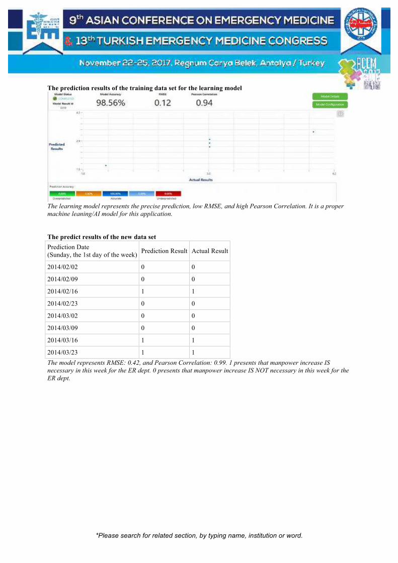
*Please search for related section, by typing name, institution or word.
The prediction results of the training data set for the learning model
The learning model represents the precise prediction, low RMSE, and high Pearson Correlation. It is a proper
machine leaning/AI model for this application. The predict results of the new data set
Prediction Date (Sunday, the 1st day of the week)
Prediction Result Actual Result
2014/02/02 0 0
2014/02/09 0 0
2014/02/16 1 1
2014/02/23 0 0
2014/03/02 0 0
2014/03/09 0 0
2014/03/16 1 1
2014/03/23 1 1
The model represents RMSE: 0.42, and Pearson Correlation: 0.99. 1 presents that manpower increase IS
necessary in this week for the ER dept. 0 presents that manpower increase IS NOT necessary in this week for the
ER dept.

*Please search for related section, by typing name, institution or word.
[S-119]
A Survey of Novel Teaching Unit: A Resident Educational Experience Qais Abuagla1, Najeeb Rahman1, Jaiganesh Thiagarajan1, Mohammed Al Jaberi1, Abdel Noureldin1, Arif Alper Cevik2 1Tawam Hospital, Department of Emergency Medicine, UAE 2UAEU CMHS, Internal Med, Tawam Hosp-Emergency Medicine, UAE Background: Maintaining sufficient supervision and faculty to resident contact time (FRCT) has been a challenge for residency programs. It has been shown that contact time could be increased with a designated teaching unit (TU). We aimed to evaluate the experience of residents in this unique teaching environment. Methods: A survey was created to evaluate trainee perspective on TU. Survey applied to 27 residents at two different periods. The first period was May – August 2015 which had constant core faculty coverage in TU. The second period was May – August 2016 which had a major departmental shortage (None-TU). A survey consisting of 10 questions. The Likert-type "Agree-Disagree" rating scale was used. Results: Twenty-six residents responded to two surveys. During the 2015 period, the majority (46.2%) worked 1 to 5 shifts in a month at TU. 80.7% of residents reported very good - excellent supervision. 88.5% of residents stated agree - strongly agree to have more FRCT. All residents agree on TU providing more opportunity to perform procedures and having a valuable role in patient flow. 88.4% of residents were satisfied with the variety of patients. 73.1% of residents express their experience as very good and excellent. During the 2016 period (none-TU), the experience of residents showed the remarkable change in all categories and low satisfaction rates. 92.2% of residents recommended TU should be continued in the program (p = 0.009). Conclusion: TU provides good clinical and educational experience with high FRCT. Keywords: Education, teaching unit, resident

*Please search for related section, by typing name, institution or word.
[S-121]
An Efficient Medical Training Method in ACLS: Simulation Based Medical Training Erkman Sanrı1, Sinan Karacabey1, Can Özen2, Serkan Emre Eroğlu2, Arzu Denizbaşı1 1Marmara University Faculty of Medicine, Department of Emergency Medicine, Istanbul, Turkey 2Umraniye Training and Research Hospital, Department of Emergency Medicine, Istanbul, Turkey Objective: In this study our objective was to compare the efficacy of traditional and simulation-based medical training (SBMT) methods in advanced cardiac life support (ACLS) training. Material-Methods: This retrospective observational study was conducted in Marmara University. All 6th grade medical faculty students who completed traditional CPR and ACLS training during their EM internship, were participated in SBMT as their routine medical education. This simulation session consisted of the management of a cardiac arrest scenario on a high-fidelity simulation mannequin (SimMan Essential, Laerdal, Norway). First, all subjects formed groups of 4 students, selected a group leader, and manage the scenario with the experience they had from their traditional medical education. Then, a SBMT session was performed. After the SBMT session they managed the same scenario, with the same group, on the mannequin again. The same scenario was used, and all data were recorded before and after the SBMT session. The mortality rate of the mannequin, chest compression quality, respiratory assist quality, drug administration times, doses and defibrillation details were used as outcome measures. Results: Study population included 92 subjects in 23 groups. 45 of the subjects were male (48.9%). All groups were similar with regard to baseline demographic characteristics. The mortality rate of the mannequin before SBMT was significantly higher compared to after the SBMT (82.6% vs 13.0%,p<0.001,respectively) with a decrease of 69.6% in mortality. Conclusion: Simulation-based medical training is an efficient medical training method for ACLS training in combination with traditional medical training method. Keywords: Simulation education, medical training, simulation based medical training

*Please search for related section, by typing name, institution or word.
[S-122]
The Utility of Simulation Training with High-Fidelity Mannequins for Airway Management in Student Paramedics Sinan Karacabey1, Erkman Sanrı1, Serkan Emre Eroğlu2, Arzu Denizbaşı1 1Marmara University Faculty of Medicine, Department of Emergency Medicine, Istanbul, Turkey 2Umraniye Education and Research Hospital, Department of Emergency Medicine, Istanbul, Turkey Objective: In this study we aimed to analyze the utility of simulation training method for airway management in paramedic students. Materials-Method: This retrospective survey analysis took place in the paramedic school of Marmara University. After the approval of institutional ethical board, data of all 2nd grade paramedic students were included in the study who participated in simulation training. They were routinely given simulation education for airway management training during their last term. A high-fidelity mannequin was used for the simulation sessions. All subjects took a pretest composed of 10 questions before the simulation education session and then they took the same test as a posttest in 2 months time period after the simulation education. Subjects with missing test data were excluded. Results: Study population included 39 subjects. Twenty-three of the subjects were male (59%). All groups were similar regarding to baseline demographic characteristics. The median (IQR) posttest score (80 (80.0-90.0)) was significantly higher then the median (IQR) pretest score (30 (20.0,40.0); p<0.001). Conclusion: Simulation training is recommended for learning and enhancing the participants' learning environment from a passive and structured approach, allowing participants to learn through practice, critical thinking and reflection and avoiding the risk of the patient to turn into a learning experience. In a study on paramedic students, success and complication rates were equal between paramedic students trained on the simulator and trained students in the operating room. In our study we showed that simulation training enhance the learning of participants. Further study is also needed to document the utility of simulation for teaching other procedures of airway management. Keywords: Simulation education, manniquin, airway

*Please search for related section, by typing name, institution or word.
[S-123]
The Satisfaction Survey About Emergency Medicine Internship in Phase V of Medicine School Şervan Gökhan1, Mehmet Ergın1, Ayhan Özhasenekler1, Fatih Tanrıverdi1, Gülhan Kurtoğlu Çelık1, Alp Şener2 1Ankara Yıldırım Beyazıt University, Faculty of Medicine, Emergency Medicine Department, Ankara, Turkey 2Ankara Atatürk Training and Research Hospital, Emergency Service, Ankara, Turkey Background: To evaluate emergency medicine internship program in pre-graduation period of medicine school in terms of efficiency and competence of theoretical and practical course. Method: The questionnaire was conducted at Ankara Yıldırım Beyazıt University Medicine School to intern doctors who had completed emergency medicine internship program in Phase V. The questionnaire was in print form and was given to participants who agreed to participate in the survey and requested to respond within 10 minutes. Results: In the evaluation of the theoretical course modules, the highest qualification ratios were given to 'Advanced Life Support Module, Communication Module and Toxicology Modules' (96%, 94%, 93% respectively). In the evaluation of the practical course modules, the highest qualification ratios were given to ‘Defibrillator Use, Basic Life Support and Airway Management, Rhythm Identification Practices' (95%, 91%, 86% respectively). 91% and 86% of the participants were satisfied with the choice of subject and course content for theoretical training. 82% and 70% of the participants were satisfied with the choice of subjects and course content for practical training. It was stated that the ratio of the duration of theoretical to practical training was found to be sufficient by 70% of the participants. Conclusion: We know that criticism is a necessity for progress. The thoughts of students should be considered for trainers to reorganize education program in future. We will continue to get feedback from students and change and improve our program and teaching style in emergency medicine education. Keywords: Medicine education, emergency medicine, pregraduate

*Please search for related section, by typing name, institution or word.
Table 1. The internship program for emergency medicine during 5th grade of faculty of Medicine

*Please search for related section, by typing name, institution or word.
[S-125]
An Anthropological Approach of Pain Management in Emergency Departments: Example of Vaso-occlusives Pain Crisis in Guadeloupe (French Caribbean): Nycrees Moueza1, Marc Eric Gruénais2, Philippe Carrere1, Maryse Etienne Julan3 1University of the French West Indies and Guiana 2University of Bordeaux / Faculty of Social Anthropology - Ethnology 3Transversal Unit - Carribean Sickle Cell Unit, Guadeloupe Objective: Hemoglobinopathies are the most common genetic diseases in Turkey, and the incidence of sickle cell trait is 10.0% in the Cukurova region. Vaso-Occlusives Crisis (VOC) are crisis of intense pain characterising sickle cell disease. Pain is a subjective symptom, hardly palpable for an observer or a care-giver. Numerous surveys highlight problems between patients and care-givers regarding VOC pain management. This study, via a qualitative approach, aims to explain the attitudes of care-givers in Emergency departments regarding the treatment of the pain, taking for example the pain induced by VOC. Methods: The data derive from a study led in 2 phases (observation and interviews of 31 patients and caregivers) during a period of just over 2 months in 3 services of Pointe-Pitre teaching Hospital: The Transversal sickle cell Unit, the pediatrics and adults emergency departments. Results: The study highlights that although the intensity of the pain makes it a priority, in precise cases, practices are not always consistent with a pain-relief logic. In question, working structures and common misconceptions about morphine or the patient’s intention. The results also show a VOC-pain management training mostly based on peer-exchange rather than formal education. This tends to maintain an already on-going pattern. Conclusions: Specific training for the care-givers about their own misconceptions and implementation of protocols which take into account those representations would likely improve VOC-pain management. Keywords: Pain, management, sickle-cell disease

*Please search for related section, by typing name, institution or word.
[S-126]
Are We Poisoning Our Older Patients? The Unrecognized Risk of Polypharmacy and Drug-Drug Interactions in Older Emergency Department Attenders in Trinidad And Tobago Utku Ekin1, Judy Wong1, Satesh Bidaisee2, Darren Dookeeram3 1St. George's University, School of Medicine, University Centre, Grenada, West Indies 2St. George's University, Department of Public Health and Preventive Medicine, University Centre, Grenada, West Indies 3Ministry of Health, Trinidad and Tobago Objective: The prevalence of polypharmacy (patients taking multiple drugs/medications) and drug-drug interactions (DDI) in patients who visit emergency departments is unknown in the Caribbean. Polypharmacy and DDI not only impact the health outcomes of patients but may also make the physician’s job more difficult. The aim of the study is to investigate medication use, polypharmacy and DDI in adults attending the Emergency Department of a large tertiary hospital in Trinidad and Tobago, comparing older and younger patients. The outcome of this study can identify weaknesses in emergency department physician training and education, as well as patient education regarding DDI’s. Material: Data were collected using a data collection sheet designed for the study. The presence of DDI was defined using Micromedix 2.0. Data were analyzed using SPSS version 21. Method: This a cross-sectional study for which data were collected over 4 months. A convenience sampling method was used and patients were recruited during all shifts and all days of the week. Results: There were 649 patients in the study, of which 275 were aged >= 65 years and 374 were 18 – 64 years old. Polypharmacy was more common in older patients, with 154 (56%) taking 5 or more medications compared to 90 (24.1%) younger adults. Over the counter medication was used by 238 (36.7%) patients, while 52 (8%) admitted to using herbal remedies, and there was no significant difference in the use of either with age. Combination drugs were used in 144 (22.2%) patients. Conclusions: The study reveals a high percentage of patients with DDI, and many with multiple potential interactions. Both polypharmacy and DDI were more common in older patients (aged >65 years), and there was an association between polypharmacy and the use of herbal and over-the-counter medications. Keywords: Polypharmacy, older, patients

*Please search for related section, by typing name, institution or word.
Table 1
>=65 Years
18 - 64 Years
All patients
p value
TOTAL
275 (42.4%)
374 (57.6%)
649 (100%)
GENDER Male 119 (43.3%)
149 (39.8%)
268 (41.3%)
0.42
Female
156 (56.7%)
225 (60.2%)
381 (58.7%)
MEDICAL CONDITIONS
Diabetes Mellitus
121 (44%) 46 (12.3%) 167 (25.7%)
< 0.001
Hypertension
153 (55.6%)
74 (19.8%) 227 (35%) < 0.001
Ischaemic Heart Disease
54 (19.6%)
14 (3.7%) 68 (10.5%)
< 0.001
MEDICATION USAGE PATTERNS
Over the Counter Medication
100 (36.4%)
138 (36.9%)
238 (36.7%)
0.934
Herbal Medicines
18 (6.5%) 34 (9.1%) 52 (8%) 0.306
Polypharmacy
123 (44.7%)
52 (13.9%) 175 (27%) <0.001
NUMBER OF POTENTIAL INTERACTIONS SEEN IN EACH PATIENT
0
92 (33.5%)
191 (51.1%)
283 (43.6%)
<0.001
1 - 5
53 (19.3%)
57 (15.2%) 110 (16.9%)
6 - 10
73 (26.5%)
37 (9.9%) 110 (16.9%)
>10
35 (12.7%)
6 (1.6%) 41 (6.3%)
MOST SEVERE INTERACTION SEEN IN EACH PATIENT
Contraindication
6 (3.7%) 2 (2%) 8 (3.1%) 0.007
Potential Major Interaction
62 (38.5%)
22 (22%) 84 (32.2%)
Potential Moderate Interaction
88 (54.7%)
66 (66%) 154 (59%)
Potential Minor Interaction
5 (3.1%) 10 (10%) 15 (5.7%)
Total
161 (100%)
100 (100%)
261 (100%)
Demographic and clinical characteristics of study group, comparing younger and older patients.
(Polypharmacy: >= 5 medications).

*Please search for related section, by typing name, institution or word.
[S-127]
Comparison of Serum levels of Glial Fibrillary Acidic Protein (GFAP) and S100B in Severe Head Trauma or Spontaneous Brain Bleeding with Healthy Individuals İrfan Aydın1, Abdullah Algın1, Mehmet Kaan Poyraz1, Önder Yumrutaş2 1Adıyaman University Faculty of Medicine, Department of Emergency Medicine, Adıyaman, Turkey 2Adıyaman University Faculty of Medicine, Department of Medical Biology, Adıyaman, Turkey Aim: Glial Fibrillary Acidic Protein (GFAP) is a molecule which is specific to the brain tissue that is involved in the blood very soon after traumatic brain injury by mature astrocytes. S100B is pass to cerebrospinal fluid (CSF) and blood easily after brain damage. In this study, we compare the serum level of these two molecules in patients with severe head trauma (bleeding, fracture) or spontaneous brain hemorrhage with healthy individuals. Materials-Methods: Sixtythree patients with cerebral hemorrhage and / or cranial bone fractures and spontaneously cerebral hemorrhages and 30 completely healthy subjects were included to the study. 33 (52.4%) men and 30 (47.6%) women were included in the study. The average age was 27 years (15 days minimum, 86 years old). When we looked at the causes of emergency department admission, it was determined that 32 patients had fallen from high, 18 from traffic accident, 6 from spontaneous subaraknoiad hemorrhage, 4 from animal hunt, 3 from beating. Results: GFAP was 86.37 ng / ml in the study group and 38.07 ng / ml in the control group. In the patient group, the S100B mean was 428.37 pg / ml, while in the control group was 103.44 (Table). Both GFAP and S100B groups were significantly higher (p <0.05). Conclusion: GFAP and S100B blood levels are significantly higher when compared with healthy individuals. We think serum levels of these molecules may help to for the diagnosis of brain injury as a alternative to Brain CT. Keywords: Brain, GFAP, S100B
Patient and control group GFAP and S100B averages
Cases Number GFAP (ng/ml) S100B (pg/ml)
Intracranial Hemorrhage 20 85,80 517,83
Cranial Fractures 17 64,13 222,41
Intracranial hemorrhage + Intracranial fractures
26 101,35 491,21
Control Group 30 38,07 103,44

*Please search for related section, by typing name, institution or word.
[S-129]
How Can Emergency Physicians Protect Their Work in the Era of Pseudo Publishing? Gül Pamukçu Günaydın1, Nurettin Özgür Doğan2 1Ankara Atatürk Training and Research Hospital, Department of Emergency Medicine, Ankara, Turkey 2Kocaeli University, Faculty of Medicine, Department of Emergency Medicine, Kocaeli, Turkey Introduction: Recently scientists have been targets of pseudo journals (fake, hijacked or predatory journals). In this review we aim to increase awareness among especially young emergency physicians. Potential Red Flags to recognize pseudo journals: 1. Publishing fees are not clearly stated on the website. 2. Promising very rapid peer review and publication times. 3. Publishing processes are not transparent, policies are not stated. 4. Guidelines for authors are not detailed or specific or verbatim from other publishers. 5. Editorial board is filled with academicians without their permission or consent or with people not experts in the field. 6. On the journal site the publisher’s full contact information including the address is lacking 7. The journal sends persistent flattering emails. 8. Journal website is poorly maintained and full of grammatical errors and misspellings. 9. The journal publishes special edition or guest edited issues frequently. 10. The journal uses bogus metrics and non-reputable indexing or falsely claims to be indexed in legitimate services. 11. The number of articles in each issue is massive. 12. Journal’s scope and name is excessively broad including more than one discipline of science. Conclusion: Both open-access and subscription publishing models can be abused by ‘predatory’ authors, editors, and journals. Predatory publishing activities are here to stay as long as there is a pressure to publish more in a shorter time. Cyber criminals are developing evermore-sophisticated techniques to entrap not only young researchers, but also experienced academics unaware of the threats focused on scholarly publishing. Efforts must focus on improving oversight to increase publication literacy to avoid publication scams. Keywords: Predatory publishing, pseudo journals

*Please search for related section, by typing name, institution or word.
[S-130]
Assessment of EFAST Training for Final Year Medical Students in an Emergency Medicine Clerkship Arif Alper Cevik1, Abdel Noureldin2, Margret Elzubeir3, Fikri M Abu Zidan4 1UAEU CMHS, Internal Med, Tawam Hosp-Emergency Medicine, ESOGU Emerg. Med. 2Tawam Hospital-Emergency Medicine 3UAEU CMHS, Medical Education, UAE 4UAEU CMHS, Surgery, UAE Objective: Extended Focused Assessment Sonography for Trauma (EFAST) is an important bedside tool in the management of multiple trauma patients. We aimed to evaluate the assessment of our EFAST education in the Emergency Medicine Clerkship (EMC) for final year medical students and correlation of EFAST scores with final exam and other practical skills scores. Methods: The total (54) final year medical students were trained on performing EFAST on simulated patients during their 4-week clerkship. Students received an hour of didactic lecture, a 4 hours practical sessions on human models, and completed a minimum of three EFAST examinations on trauma patients. Finally, the EFAST performance was evaluated on a simulated trauma patient using a standard evaluation form during an Objective Structured Clinical Examination (OSCE). The marks of 51 students who completed the final exam were analyzed. Results: the overall passing rate of the EFAST station was 88% (n: 45). EFAST station mark was significantly higher in males compared with females (p=0.013). EFAST station mark significantly correlated with other OSCE stations marks (p=0.027, rho=0.31), and with the final EMC mark (p=0.032, rho=0.3), but not with the final written exam. Conclusions: Final year medical students demonstrated effective EFAST learning as measured by examination performance. The EFAST final marks were highly correlated with other OSCE station marks and final clerkship marks, but not the final written exam mark which assesses a different cognitive learning domain. Keywords: EFAST, emergency ultrasound, medical student

*Please search for related section, by typing name, institution or word.
The correlation between EFAST mark and other OSCE stations mark

*Please search for related section, by typing name, institution or word.
[S-131]
Do Senior Medical Students Meet Recommended Emergency Medicine Clerkship Curricula Requirements? Sami Shaban1, Arif Alper Cevik2, Mustafa Emin Canakci3, Caglar Kuas3, Margret Elzubeir1, Fikri M Abu Zidan4 1UAEU CMHS, Medical Education, UAE 2UAEU CMHS, Internal Med, Tawam Hosp-Emergency Medicine, ESOGU Emerg. Med., UAE 3ESOGU, Emergency Medicine, Eskişehir, Turkey 4UAEU CMHS, Surgery, UAE Objective: Emergency departments (EDs) offer a variety of learning opportunities for undergraduate medical students. It is however, difficult to evaluate whether they are receiving recommended training during their emergency medicine (EM) clerkship without identifying their clinical activities. We aimed to evaluate the clinical exposure of the final year medical students at our College during their EM clerkship. Methods: This is a retrospective analysis of prospectively collected student logbooks. 75 students rotated in a four-week EM clerkship during 2015-2016. The students rotated in EDs of two hospitals. Each ED treats more than 120,000 cases annually. The students completed 12 eight-hours shifts. Presentations and procedures seen were compared with EM curriculum recommendations. Results: 5,122 patient presentations and 3,246 procedures were recorded in the logbooks, an average (SD) of 68.3 (17.6) patients and 46.1 (14.0) procedures. None of the students encountered all ten recommended presentations. Two students (2.6%) logged all nine procedure categories of the EM curriculum (9 procedures). Conclusion: Recommended presentations and procedures of the EM clerkship were not fully encountered by all our students. Different settings vary in the availability and type of patients and procedures. Each clinical clerkship should tailor their teaching methods based on the available learning opportunities. Keywords: Emergency medicine, clerkship, logbook

*Please search for related section, by typing name, institution or word.
Presentations and Procedures by students’ level of achievement
ABG: arterial blood gas; ID: incision and drainage, CPR: cardiopulmonary resuscitation; ECG:
electrocardiogram; EFAST: Extended focused assessment with sonography for trauma; IV: intravenous; NG:
nasogastric; RUSH: rapid ultrasound for shock and hypotension. * These topics were added to CDEM
procedure list by local EM core faculty group.

*Please search for related section, by typing name, institution or word.
[S-132]
Clinical Procedure Experience of Medical Students Improves Their OSCE Station Marks Arif Alper Cevik1, Fikri Abu Zidan2 1UAEU CMHS, Internal Med, Tawam Hosp-Emergency Medicine, ESOGU Emerg. Med., UAE 2UAEU CMHS, Surgery, UAE Objective: We aimed to study the correlation between procedure experiences in clinical setting and Objective Structural Clinical Examination (OSCE) marks achieved at the end of an Emergency Medicine clerkship for final year medical students. Methods: This is a retrospective analysis of prospectively collected clinical data of 141 final year medical students and their OSCE marks for two consecutive academic years (2015 - 2017). The experience of practical skills including suturing, Extended Focused Assessment Sonography for Trauma (EFAST), airway management, and cardiopulmonary resuscitation were correlated with the final OSCE marks in the same areas. Results: Weighted experiences of the four procedures were significantly correlated with the total OSCE station marks (p = 0.027, Spearman's rho = 0.19). Suturing OSCE marks were significantly higher than the other stations (p < 0.0001, Wilcoxon signed ranks test). There was significant correlation between suturing experience and its OSCE mark (p = 0.036, Spearman’s rho = 0.18). There was also a strong trend in correlation between EFAST experience and its OSCE mark (p = 0.063, Spearman’s rho = 0.16). There was significant difference in weighted experience between each of the four procedures (p < 0.0001, Wilcoxon signed ranks test). In all cut-off levels (75 – 95) of OSCE marks, students showed higher weighted procedure experience for those who had higher marks. Statistical significance was found only for students who scored more than 90% of the OSCE mark. Conclusion: Clinical experience of procedures improved OSCE marks of the same procedures. The top students showed significant higher weighted procedure experience. Keywords: Medical student, procedure, OSCE

*Please search for related section, by typing name, institution or word.
Table 1

*Please search for related section, by typing name, institution or word.
[S-133]
How to Learn Emergency Medicine Specialist Best? Tuğba Atmaca Temrel1, Şervan Gökhan2, Adnan Karaibrahimoğlu3, Mehmet Ergin2, Fatih Tanrıverdi2, Gülhan Kurtoğlu Çelik2, Ayhan Özhasenekler2 1Ankara Atatürk Training and Research Hospital, Emergency Service, Ankara, Turkey 2Ankara Yıldırım Beyazıt University, Faculty of Medicine, Emergency Medicine Department, Ankara, Turkey 3Expert in Biostatistics, Asist. Prof., Süleyman Demirel University, Faculty of Medicine, Department of Biostatistics, Isparta, Turkey Introduction: Learning is the process of the student's concept of knowledge, imprinted on the mind, and the ability to use that knowledge. This study aimed to determine the learning preferences of emergency physicians. Materials-Methods: A total of 223 emergency physicians working in eleven public hospitals and agreed to participate the study were included in the study. Participants evaluated with a questionnaire consisting of seven questions and Turkish translation of VARK (version 7.8) consisting of sixteen questions, by face-to-face or by mail. We probed the participants’ the position in emergency department, whether they had given training to emergency staff, and whether they had already received training of trainer. Results: 63.2% (n = 141) of the physicians working in the university hospital. It was found that 57.4% (n = 128) of the participants gave training to emergency staff and 67.3% (n = 150) did not receive training of trainers. In the learning preferences of the group, AK was the most favored by 15,7% (n = 35) and ARK was 14,8% (n = 33). (Graphic 1) Participants' learning preferences were 43% (n = 96) bimodal and 31.8% (n = 71) trimodal distribution. Discussion and Conclusion: The best learning style has two components. One of those to provide appropriate training to the preference of the learner, and the other is that the learner is aware of his own learning style. Implementation of training activities by trained trainers, provides an excellent learning environment in which the training materials contain visual, auditory, read-write and kinesthetic modules. The awareness of the emergency physicans own learning preference and the preparation of curriculum containing the items appropriate for all types of medical training preferences will contribute positively to the success and quality of education. Keywords: Medical Education, Emergency Medicine, VARK
Graphic 1:
The learning preferences of the participants

*Please search for related section, by typing name, institution or word.
[S-134]
Simmerk: Our Experience in Simulation Based Training Can Özen1, Erkman Sanrı2, Serkan Emre Eroğlu1, Sezen Avtan3, Fatih Karaaslan3 1University of Health Sciences Umraniye Education and Research Hospital, Department of Emergency Medicine, Istaanbul, Turkey 2Marmara University Pendik Education and Research Hospital Department of Emergency Medicine, Istanbul, Turkey 3Istanbul Health Directorate Medical Simulation Center, Istanbul, Turkey Simulation based training is an expending area within medical training. It is considered as an important tool especially in procedure oriented specialities since simulation provides a risk free environment eliminating the human error. Cardiopulmonary resuscitation (CPR) is one of those areas where providers should be ready to perform a series of complex and basic procedures in harmony with a team. Being the leader of this team requires adequate knowledge and experience as well as a clear mind to make important decisions. Simulation based CPR training provides the trainees an excellent opportunity to experience all the aspects of this operation. In this presentation we aim to report our own experience in simulation based training. A course was established involving classical lessons and a 8 minute simulation session using SimMan 3g ® advanced patient simulator. Introduction to simulation, basic and advanced cardiac life support and post resuscitative care topics were covered in three 40 minute lessons. Participants were surveyed on their skill level of leading a team in CPR, chest compressions, recognising the presenting rythym on monitor and thie competency in CPR using a 10 point visual analogue scale (VAS) at the begining and at the end of each course. 59 participants were included. 18/59 (30.50%) were first year residents and 41/52 (69.50%) were last year medical students. 31/59 (52.55%) were male. Mean VAS of participants for their competancy in CPR was 5.09 ± 1.68 (95 CI = 4,61 – 5,49) in pretest and 7.37 ± 1,99 (95% CI = 6.85- 7.90) in posttest which is significantly higher(p<0.001). Simulation based CPR training lets trainees to realise their shortcomings in procedures and level of knowledge while giving them the opportunity to discover the responsibilities of each team member during CPR. It can be used both in medical education and emergency medicine resident training. Keywords: Simulation, Cpr, emergency medicine training

*Please search for related section, by typing name, institution or word.
[S-135]
A Pilot Study of Traumatic Laceration Wound Cleansing in Emergency Department (ED): Saline versus Reversed Osmosis (RO) Water, in Hospital Universiti Sains Malaysia (HUSM), Kelantan and Hospital Tengku Ampuan Afzan(HTAA), Pahang, Malaysia Anas Amri Hashim1, Mohd Hashairi Fauzi1, Zainalabidin Mohamed2, Mohd Muzammil Ozair1 1Department of Emergency Medicine, School of Medical Sciences, Universiti Sains Malaysia, 16150 Health Campus, Kelantan, Malaysia 2Emergency & Trauma Department, Hospital Tengku Ampuan Afzan, Jalan Tanah Putih, 25100 Kuantan, Pahang, Malaysia Introduction: Traumatic laceration wound is a common reason for people seeking treatment in emergency care setting. As for current practice, sterile saline is still the most common agent used in cleansing or irrigating the wound in the emergency department. In overseas, there have been studies on alternative cleansing agent particularly tap water, which have showed equal or better outcomes in term of wound infection. There is no previous study been conducted on wound cleansing using Reverse Osmosis water. Objectives: To compare the rate of wound infection between sterile saline and reverse osmosis water cleansing for traumatic laceration wound. Method: This study was a multicenter, prospective, randomised controlled trial conducted at two tertiary hospitals. Subjects were a convenience sample of adults presenting with acute simple traumatic laceration requiring wound cleansing. They were randomized using block randomization method to irrigation with ‘sterile saline’ or ‘reverse osmosis’ water. Wounds were closed in the standard fashion. Follow-ups were done on day 5 and 14 after the intervention to assess for outcome or wound infection. Results: A total of 48 patients (n=48) were selected and randomised into RO water group (n=24) and Saline group (n=20) (4 patients withdrew). The infection rate for sterile saline cleansing was 5% (95% CI -4.55%, 14.55%) compared to RO cleansing which was 4.17% (95% CI -3.83%, 12.17%). Relative risk for RO water group was 0.834 (95%CI 0.056, 12.494) compare to saline group. Fisher’s exact test showed no significant effect on type of cleansing agent on wound infection. Conclusion: There was no significant difference between reverse osmosis and saline on wound infection. Reverse osmosis water can be considered as alternative cleansing agent to sterile saline in acute uncomplicated traumatic laceration wounds. Keywords: Saline, reverse osmosis, uncomplicated traumatic wound
*Please search for related section, by typing name, institution or word.
[S-136]
Deep Tissue Injury Diagnosis in Pressure Ulcer with Ultrasonography Özge Can, Hilal Dönmez, Gülbin Konakçı, Funda Karbek Akarca Department of Emergency Medicine, Ege University, Izmir, Turkey Introduction: Pressure ulcer is a common problem. There is some pressure ulcer classification. These classification based on a visual score. Some research showed Ultrasound may determine deep tissue injury early. We present cases with had pressure ulcers detecting by ultrasound. Case 1: A 55-year-old patient was present with a stage 2 compression wound on coccyx of a bed-dependent patient due to extensive metastasis. The patient underwent an ultrasound with a linear probe. Dermis edema was detected. There was no bone formation. Case 2: A 48 years old patient who depended on the bed due to brain cancer, stage 1 pressure ulcer was detected on right SIAP. Ultrasound showed diffuse edema in the dermis. Case 3: A 60-year-old woman was admitted to ED and stage 3 compression wound was detected on the right calcaneus. When ultrasound was applied, edema of the dermis and extension to the bone tissue were observed. No damage was found in the periosteum. Discussion: Deep tissue injury under the skin has helped diagnosis for pressure ulcers but has suggested that present staging systems may not be sufficient. Deep tissue injury was categorized by localized discolored area of intact skin or subcutaneous wound and bone or muscle vision. The importance of diagnosing deep tissue injury is in the early stages. There are some data showing that %10 of stageI ulcers and %12 of stageII ulcers progress to stageIII and IV ulcers (1) And animal studies show that deeper tissue is affected by the skin lesions in pressure-induced ulcers (2). Detecting subcutaneous damage by computed tomography, magnetic resonance imaging and ultrasound have been reported. Ultrasonography is a safe, economical, noninvasive method and can be easily and repeatedly performed. (1). Conclusion: Ultrasound may a good diagnostic tool for visualization of subcutaneous deep tissues and reliable identification of deep tissue injury. Keywords: Pressure ulcer, ultrasound, deep tissue injury

*Please search for related section, by typing name, institution or word.
[S-138]
Traumatic Pneumomediastinum and Pneumorrhagia Mehmet Esen Emergency Department, Medical Faculty, Gaziosmanpaşa University, Tokat, Türkiye Pneumorrhachis is characterized by the air in the spinal epidural space. In this article, we report a 28-year-old male case with traumatic pneumomediastinum(PM) and accompanied by subcutaneous emphysema who was radiographically diagnosed with PM accompanied by pneumorrhagia(PR) during his follow-up emergency services visits. Case: A 28-year-old male patient was admitted to the emergency room due to a high fall. GCS was 15 and widespread body trauma was present. Respiratory voices in the right lung basal were decreased, and sensitivity in the right lower ribs was present. Vital parameters were stable. Dyspnea, tachypnea, tachycardia were present. There was no neurological deficit. X-rays taken in the mediastinal air was observed subcutaneous emphysema and rib fracture. In the vertebra computerized tomography taken, the air was detected in the spinal range (PR). Computed tomography of the thorax showed mediastinal air, pneumothorax(PTX) and subcutaneous emphysema. Discussion: PM is characterized by the presence of air in the mediastinal area while PR is a radiological condition with air in the spinal epidural space. The coexistence of these two conditions along with traumatic has rarely been reported in the literature. Pneumorrhachis, or epidural pneumatosis, is a rare radiological condition characterized by air in the spinal epidural area that is usually detected accidentally. The causes of PR can be classified as traumatic, nontraumatic, or iatrogenic. Pneumorrhachis is usually iatrogenic and occurs following the administration of epidural analgesia. Furthermore, PR may very rarely occur when mediastinal air passes through to the epidural space, which was the case with our patient. This is necessitated by the lack of real fascia barriers between the posterior mediastinum and epidural space. The coexistence of PM and PR has been rarely reported in the literature. PM, PTX and PR is the combination of a very rare case, we wanted to share with you. Keywords: Trauma, pneumorrhagia, pneumomediastinum

*Please search for related section, by typing name, institution or word.
Figure: Pneumorrhagia, pneumothorax, pneumomediastinum

*Please search for related section, by typing name, institution or word.
[S-139]
Comparison of the Sensitivity for Detecting Foreign Bodies Among Conventional Plain Radiography, Computed Tomography and Ultrasonography in a Chicken’s Thigh Model Deniz Algedik Gursoy1, Kurtulus Aciksari2, Dogac Niyazi Ozucelik3, Halil Dogan4 1Faculty of Medicine, Department of Emergency Medicine, Yeditepe University Hospital, Istanbul, Turkey 2Faculty of Medicine, Department of Emergency Medicine, Istanbul Medeniyet University, Istanbul, Turkey 3Faculty of Health Sciences, Istanbul University, Istanbul, Turkey 4Emergency Medicine Department, Bakirkoy Dr. Sadi Konuk Research and Training Hospital, University of Health Sciences, Istanbul, Turkey Introduction: There are very few studies related to patients admitted to the emergency unit with soft tissue foreign body penetration and their diagnostic methods. In this study the sensitivity of imaging methods, generally used in the emergency unit, consisting of X-ray, computed tomography and ultrasonography were investigated in animal model with foreign body placed (foreign bodies into chicken thigh). Material-Methods: In this randomised blind experimental study 36 pieces of chicken thigh and foreign bodies including stone, metal, glass materials were used. Each material was placed in different locations. Foreign bodies were placed to 27 of the chicken. The incision was made but foreign bodies were not placed to 9 chickens resulting in only tissue trauma. Radiologists independently evaluated the bilateral radiographs computed tomography scan and ultrasound imaging of chicken thighs evaluated. SPSS 23.0 for Windows program was used for statistical analyses. Results: In this animal model study, radiography revealed 70.3% of foreign bodies, ultrasonography 83.9% and computed tomography 100% the foreign bodies. Conclusion: In the emergency department, the sensitivity and specificity of ultrasonography in diagnosing foreign bodies is higher than X-Ray. Also ultrasonography is an inexpensive, radiation-free and reproducible method that can be used over patients. Keywords: Emergency department, foreign bodies, ultrasonography
Detecting to foreign body in study and control groups
Foreign Body Study Control Total P
USG Detected 68 (83.9) 8 (29.6) 76 (70.3) 0.000
USG Non detected 13 (16.1) 19 (70.4) 32 (29.7) 0.000
USG Total 81 (100) 27 (100) 108 (100) 0.000
X-RAY Detected 57 (70.3) 0 (0) 57 (52.7) 0.000
X-RAY Non detected 24 (29.7) 27 (100) 51 (47.3) 0.000
X-RAY Total 81 (100) 27 (100) 108 (100) 0.000
CT Detected 81 (100) 0 (0) 81 (75) 0.000
CT Non detected 0 (0) 27 (100) 27 (25) 0.000
CT Total 81 (100) 27 (100) 108 (100) 0.000

*Please search for related section, by typing name, institution or word.

*Please search for related section, by typing name, institution or word.
[S-140]
Deneysel Akut Mezenter Iskemi Modelinde Plasma Vascular Adhezyon Protein -1 (VAP-1) Düzeyinin Tanısal Değerinin Araştırılması Ve Plasma I-FABP Düzeyi ile Karşılaştırılması Aynur Şahin1, Damla Aydın Altay1, Selim Demir3, Esin Yuluğ4, Ahmet Menteşe2, Özgür Tatlı1, Yunus Karaca1, Abdülkadir Gündüz1, Süleyman Türedi1 1Karadeniz Teknik Üniversitesi Tıp Fakültesi Acil Tıp Ana Bilim Dalı, Trabzon 2Karadeniz Teknik Üniversitesi Tıp Fakültesi Tıbbi Biyokimya Ana Bilim Dalı, Trabzon 3Karadeniz Teknik Üniversitesi Sağlık Bilimleri Fakültesi Beslenme ve Diyetetik Bölümü, Trabzon 4Karadeniz Teknik Üniversitesi Tıp Fakültesi Histoloji ve Embriyoloji Ana Bilim Dalı, Trabzon Amaç: Bu çalışmanın amacı akut mezenter iskeminin (AMI) erken tanısında plasma vascular adhesion protein-1 (VAP-1) seviyesinin değerini araştırmak ve plasma intestinal fatty acid binding proteinin (I-FABP) tanısal değeri ile karşılaştırmaktır. Yöntem: Bu çalışma randomize kontrollü deneysel bir çalışmadır. 42 dişi Sprague Dawley rat 3’ü kontrol, 3’ü iskemi grupları olmak üzere toplam 6 gruba ayrıldı. Kontrol gruplarına basit laparotomi uygulandıktan 30 dakika (grup 1), 2 (grup 3) saat ve 6 saat (grup 5) sonra kan ve doku örnekleri alındı. İskemi gruplarında ise laparotomi sonrası superior mesentrik arteri (SMA)bağlanarak 30 dakika (grup 2), 2 saat (grup 4) ve 6 saat (grup 6) sonra kan ve doku örnekleri alındı. Kan örneklerinden plasma VAP-1 ile I-FABP düzeyi ölçüldü ve terminal ileum dokusuna ise histopatolojik hasar skorlaması uygulanarak bu veriler birbirleriyle karşılaştırıldı. Bulgular: İlk 30 dakika kontrol ve iskemi grupları karşılaştırıldığında anlamlı histopatolojik hasar oluşturulmasına rağmen sadece I-FABP düzeyinde istatistiksel anlamlı yükseklik tespit edildi (p=0.017). 2 saatlik iskemi ve kontrol grubu karşılaştırıldığında, 30 dakikalık iskemiden daha anlamlı bir iskemik hasar oluşmamasına rağmen sadece VAP-1 düzeyinde anlamlı artış mevcuttu (p=0.011). 6 saatlik kontrol ve iskemi grupları karşılaştırıldığında ise hem VAP-1 hem de I-FABP düzeyinde anlamlı artış görüldü (respectively, p=0.007, p=0.002). Total histopatolojik skorla yapılan korelasyon analizinde ise her iki marker anlamlı derecede iskemik hasarla korelasyon gösterdi fakat en yüksek korelasyon plasma VAP-1 düzeyinde tespit edildi (r=0.771). Sonuç: VAP-1 hem iskeminin 2. saatinden itibaren anlamlı artması hemde iskemik hasarla yüksek korelasyon göstermesi nedeniyle AMI’nin erken tanısında kullanılmaya aday bir biyomarkerdır. Anahtar Kelimeler: Akut mezenter iskemi, VAP-1, I-FABP

*Please search for related section, by typing name, institution or word.
[S-141]
Acil Serviste Metil Alkol Zehirlenmesi Tanısı Alan Hastaların Geriye Dönük Değerlendirilmesi Nezihat Rana Dişel, Ali Çebişli, Ayça Açıkalın Akpınar, Ahmet Sebe, Yüksel Gökel, Zeynep Kekeç Çukurova University, Faculty of Medicine, Department of Emergency Medicine, Adana, Türkiye Giriş-Amaç: Bu çalışmada 18.09.2016-31.01.2017 tarihleri arasında hastanemiz acil servisine başvurup metil alkol zehirlenmesi (MAZ) tanısı ile tedavi edilen hastaların irdelenmesi amaçlanmaktadır. Tedavide antidot olarak kullanılan fomepizol (F), intravenöz ve oral (nazogastrik ile) etil alkol (EA) ve hemodiyalizin (HD) etkinlik, sağkalıma katkısı ve prognoz üzerine etkilerini tartışmayı amaçlamaktayız. Bulgular: Sözkonusu zaman aralığında MAZ nedeniyle izlenen biri kadın toplam 42 hastanın yaş ortalaması 52,8±9,7 (min 21-max 72) idi. Kan metil alkol seviyesi bakılabilen 25 hastanın ortalama değeri 102,9±111,7 mg/dL (min 9,1-max 387) idi. Hastalardan başvuru anında eksitus olan ve sekelle başvurup klinik zehirlenmesi olmayan iki olgu dışındaki 40 hastadan; F+EA+HD alan 10, EA+HD alan 17, F+HD alan 4, sadece EA alan 3, sadece F alan 2 ve sadece HD alan 4 hasta bulunmaktadır. Hastaların 10'u eksitus olup, ortalama yatış süreleri 6,1±9,4 (min 1-max 44) gün idi. Onyedi hasta sağlıklı taburcu edilirken, 12 hastada kalıcı görme kaybı (üç hastada hipoksik sekel ile birlikte) gelişmiş, 3 hasta tedavileri tamamlanmadan kendi istekleri ile taburcu olmuştur. (Hastaların istatistiksel karşılaştırma içeren diğer bulguları sunum esnasında paylaşılacaktır.) Tartışma ve Sonuç: Alınan tedavilerin sağkalıma katkısı ve prognoz üzerine etkilerine bakıldığında, fomepizol alanların eksitus ve sekelli taburculuk oranı ile yatış sürelerinin daha az olduğu görülmüştür. Ancak antidot bulunamayan, ya da eksik doz sebebiyle her iki antidot da kullanılan hastalar istatistiksel karşılaştırmayı zorlaştırmaktadır. Hangi antidotun seçileceği ve hemodiyaliz uygulama endikasyonları laboratuvarın yanı sıra hekime bağlı klinik değerlendirme ile de belirlendiğinden, standart bir tedavi planlamasının olmadığı görülmektedir. Kliniklerin hasta yönetimi konusunda kendi protokollerini oluşturması hastaların sağkalım ve prognozu için belirleyici olacaktır. Anahtar Kelimeler: Metil alkol zehirlenmesi, acil servis, antidot

*Please search for related section, by typing name, institution or word.
[S-143]
Kimyasal Gaz Inhalasyonu Sonucu ARDS: Vaka Sunumu Ayça Açıkalın1, Naciye Orçan1, Nezihat Rana Dişel1, Ahmet Sebe1, Zeynep Kekeç1, Emre Karakoç2 1Çukurova Üniversitesi Tıp fakültesi, Acil Tıp AD, Adana 2Çukurova Üniversitesi Tıp Fakültesi, Dahiliye Yoğun Bakım, Adana Giriş-Amaç: Bu yazıdaki amacımız, Suriye’de kimyasal gaz inhalasyonu sonrası akut solunum yetmezliği gelişen iki vakayı tartışmaktır. Vaka: Suriye’de kimyasal toksik gaz inhalasyonu (muhtemel klorin gazı) nedeniyle, akut solunum yetmezliği öntanısıyla, dış merkezden sevkedilen 2 hasta, 112 tarafından kurumumuza getirildi. Getirilen 2 vakanın kliniği; Vaka 1: 22 yaşında erkek hasta, entübe bir şekilde kliniğimize kabul edildi. Gelişinde, Tansiyon Arteriyel (TA): 130/78 mmHg, nabız(Nb): 123/dk, solunum sayısı (SS):28/dk, ateş(A) 37.8 ℃, GKS: 13 idi. Fizik muayenesinde (FM), her iki akciğerde dinlemekle yaygın raller mevcuttu. Ventilatörde, %100 oksijenle solutulan hastanın kan gazında pH:7.40, PCO2:35.8, PO2:61.4 idi. Hesaplanan alveolo-arteriyel oksijen gradiyenti [P(A-a) O2]:43.85, PaO2/FiO2’si:100,65 bulundu. ARDS öntanısıyla çekilen Toraks BT’sinde yaygın infiltrasyonlar ve opasiteler mevcuttu. İki gün mekanik ventilatörde izlenen hasta, 3. Günde solunum yetmezliği kliniğinin düzelmesi üzerine, yoğunbakımdan servise devredildi. Vaka 2: 17 yaşında erkek hasta, gazın etkisine bağlı larinks ödemi nedeniyle trakeostomi açılarak, entübasyonu sağlandıktan sonra, 112 ile kliniğimize sevkedilmiş. Gelişinde, TA: 134/58 mmHg, Nb: 87/dk, SS:30/dk, A: 37.4, GKS:13 idi. Fizik muayenesinde her iki akciğerinde yaygın ralleri mevcuttu. Ventilatörde %100 oksijenle solutulurken alınan kan gazında pH: 7.25, PCO2:50.3, PO2:63 idi. Hesaplanan P(A-a) O2: 24.12, PaO2/FiO2’si: 103.27 idi. Toraks BT’sinde (Şekil 1) yaygın infiltrasyonları ve opasiteleri mevcuttu. Üç gün ventilatörde izlenen hasta solunum yetmezliğinin düzelmesi üzerine, yatışının 8. Gününde servise alındı. Her iki hasta şifa ile taburcu edildi. Tartışma: Klorin gazı inhalasyonu, endüstriyel kazalarda, savaşlarda yada terörist saldırılarda karşımıza çıkabilmektedir. Yoğun maruziyet akut akciğer hasarına yol açmaktadır. Olay yerinde etkin dekontaminasyonla, hastaneye ulaşabilen vakalarda, yoğun maruziyete ikincil akut akciğer hasarlanması gelişse de, etkin solunum desteği ile erken dönemde iyileşme sağlanabildiği görülmüştür. Anahtar Kelimeler: ARDS, kimyasal gaz inhalasyonu, solunum yetmezliği

*Please search for related section, by typing name, institution or word.
Şekil 1
*Please search for related section, by typing name, institution or word.
[S-145]
Örümcek Isırığına Bağlı Miyokardit Ve Rabdomiyoliz Murat Das, Okhan Akdur, Canan Akman, Yasemin Çetinkaya Çanakkale Onsekiz Mart Üniversitesi Acil Tıp Ana Bilim Dalı, Çanakkale Giriş: Karadul örümceğinin ısırığına bağlı toksik etkilerden alfa-latrotoksin sorumludur. Bu toksin özellikle asetilkolin ve nörepinefrin gibi nörotransmitterlerin masif bir şekilde salınmasına neden olur. Biz bu vaka ile karadul örümceği tarafından ısırılarak acil servise başvuran ve sonrasında miyokardit ve rabdomiyoliz gelişen bir vakayı sunmayı amaçladık. Olgu: 24 yaşında erkek hastanın öyküsünden; bahçede çalışırken ayakkabısının içinde siyah renkli bir örümceğin ısırdığını, önemsemeyerek işine devam ettiği, ısırılmadan 30 dakika sonra sol bacakta yukarı doğru yayılan bir yanma ve şiddetli ağrı geliştiği ve bu ağrının kasıklarına ve karına doğru yayılınca hastaneye başvurduğu öğrenildi. Acil serviste; Kan basıncı:140/65mmHg, Nabız:95/dakika solunum:17/dakika Sıcaklık:36,7°C, kan glikozu:154mg/dl’ydi. Genel durumu orta, bilinç açık, oryante-koopere, anksiyöz görünümde ve şiddetli terlemesi mevcuttu. Sol ayak dorsal 1-2. metatarsal kemikler arasında 0,5cm çaplı ekimoz görünüyordu (resim 1). Şiddetli terlemeyle birlikte, akciğer alt zonlarda raller, karın muayenesinde yaygın defans mevcuttu. Hemogramda WBC:18850/ml, RBC:5,86mil/ml Hemoglobin:16,25mg/dl, Platelet:293bin; Kan biyokimyasında, CK: 1838U/L, CKMb 71,6 U/L Potasyum: 6,32 mmol/L troponin 0,14; olarak ölçüldü. Venöz kan gazı pH:7,28, PCO2: 49,2 [HCO3-]: 17.5 laktat 4,5 mmol/L olarak saptandı. Takiplerinde troponin değerleri 3.01ng/ml’ye, kan basıncı 200/120mm/Hg’ya kadar yükseldi. Tedavide; Fentanyl®, Diazem® ve Dormicum® ampül uygulandı. Kreatinin Kinaz değerleri yüksek olan hastaya günlük 4000-4500 ml olacak şekilde intravenöz sıvı uygulandı. Acil serviste 3 gün monitörlü olarak takip edilen hastanın troponin ve CK değerlerinde düşme kan gazındaki metabolik asidozun düzelmesi, terlemenin kaybolması ve vücudundaki ağrıların geçmesi üzerine kontrole gelmek üzere taburcu edildi. Sonuç: Karadul örümceği ısırığına bağlı sistemik bulgular lokal bulgulara göre daha az sıklıkla gelişmekle beraber nöbet, miyokardit ve rabdomiyoliz gibi hayatı tehdit eden bulgular gelişebilir. bizim hastamızda nöbet haricindeki bulgular gelişmiştir. Sistemik semptomların benzodiazepinler ve opioitler ile semptomatik tedavisi önerilmektedir. Bizim hastamızda da terleme ve yaygın vücut ağrısı gibi şikayetleri opiyat ve benzodiyazepin tedavisine iyi yanıt vermiştir. Bu olguda gelişen rabdomiyoliz nadirdir ve literatürde sınırlı sayıda vaka ile bildirilmiştir. Anahtar Kelimeler: Karadul örümceği, miyokardit, rabdomiyoliz
Resim 1

*Please search for related section, by typing name, institution or word.
1. ve 2. metatarsal kemik arasında 0,5 cm çaplı ekimoz

*Please search for related section, by typing name, institution or word.
[S-146]
Akciğer Parankimini Katederek Perikarda Kadar Uzanan Yabancı Cisim Murat Das1, Muhammed Fatih Tütüncü1, Mustafa Reşorlu2, Okhan Akdur1, Canan Akman1, Murat Uygar Yapucu3 1Çanakkale Onsekiz Mart Üniversitesi Acil Tıp Ana Bilim Dalı, Çanakkale 2Çanakkale Onsekiz Mart Üniversitesi Radyoloji Ana Bilim Dalı, Çanakkale 3Çanakkale Devlet Hastanesi Göğüs Cerrahi Bölümü, Çanakkale Giriş: Penetran toraks yaralanması künt toraks yaralanmalarına göre daha az sıklıkta görülmektedir. Yaralanma mekanizması çoğunlukla ateşli silahlar ve delici kesici aletleri içermektedir. Künt göğüs travmalarına göre cerrahi tedavi ile müdahale ve mortalite penetran yaralanmalarda daha fazladır. Toraksı penetre eden yaralanmalarda pnömotoraks, hemotoraks, trakeo-bronşial yaralanma, akciğer kontüzyonu, özofagus yaralanması ve mediastinal yapılarda yaralanmalar görülebilmektedir. Biz düşme sonucu göğüs ön duvarına cam girmiş ve cerrahi müdahale ile çıkartılarak tedavi edilen bir vakayı sunmayı amaçladık. Olgu: Cam pencere üzerine düşerek sağ göğüs ön duvarını yaralayan 12 yaşındaki erkek hasta dış merkezden Çanakkale Onsekiz Mart Üniversitesi Hastanesine sevk edildi. Acil servisimizde, kan basıncı 110/70mmHg, nabız 97/dakika, solunum sayısı 18/dakika ve oksijen saturasyonu %97 olarak ölçüldü. EKG normal sinüs ritmindeydi. İnspeksiyonda sağ hemitoraks mid-klavikular hatla 3. interkostal aralığın kesiştiği bölgede iki adet 0,5-1cm uzunluğunda delici kesici alet yarası mevcuttu(Resim1). Toraks içinde yabancı cisim olup olmadığının tespiti için posterior-anterior ve yan akciğer grafileri çekildi (Resim2). Kardiyotorasik yapıların daha net değerlendirilmesi için kontrastsız toraks bilgisayarlı tomografi(BT) çekildi(Resim2). X-ray ve BT görüntülerinde yabancı cisme ait görüntüler tespit edildi. Bu görüntülerde pnömotoraks, hemotoraks izlenmedi. Merkezimizde göğüs cerrahisi bölümü izinli olduğundan hasta Çanakkale Devlet Hastanesi göğüs cerrahi bölümüne sevk edildi. Burada yapılan açık torakotomi operasyonunda, yabancı cismin (ucu sivri bir cam parçası) 3. İnterkostal aralık ile orta klavikuler hattan toraks boşluğuna girdiği, orta lob medial segment superiorundan akciğer parankimi içine girip orta lob lateral segment inferiorundan çıktığı görüldü. Yaklaşık 6 cm uzunluğundaki yabancı cisim çıkartıldı. Akciğer parankim laserasyonları primer olarak tamir edildi. Kanama ve hava kaçağı kontrolünü müteakip toraks boşluğuna toraks tüpü yerleştirilerek açılan insizyon usülüne uygun olarak kapatıldı. Postoperatif 6. günde şifa ile taburcu edildi. Sonuç: Toraksa penetre yabancı cisimler, hayatı tehdit edici yaralanmalara sebep olabilir. Bu tür durumlarda toraksın görüntülenmesi, sadece oluşan yaralanmanın niteliği hakkında bilgi vermez, aynı zamanda sonrasında yapılacak müdahalenin ne olacağı kararına da yardımcı olur. Anahtar Kelimeler: Bilgisayarlı toraks tomografisi, toraksta yabancı cisim

*Please search for related section, by typing name, institution or word.
Resim 1
a ve b: Posterior-anterior akciğer grafisinde ve yan akciğer grafisinde okun ucunda yabancı cisim opasitesi, c ve
d: Bilgisayarlı toraks tomografisinde okun ucunda toraks duvarından perikarda uzanım gösteren hiperdens
yabancı cisim.

*Please search for related section, by typing name, institution or word.
[S-147]
Karbonmonoksit Zehirlenmesinde MR Bulgusu Salih Kocaoğlu, Sevgi Sarzep Çatlak, Abuzer Coşkun, Mustafa Oğuz Cumaoğlu, Şimşek Çelik, Osman Mahir Okur Sivas Numune Hastanesi, Acil Servis, Sivas Giriş: Karbonmonoksit (CO) zehirlenmeleri en sık bildirilen toksikolojik kaynaklı ölüm nedenlerindendir. CO karbon kaynaklı yakıtların iyi yanmaması sonucu ortaya çıkar (1). Ülkemizde özellikle kış aylarında soba mangal şohben kullanımı sonrası co zehirlenmesi sık görülmektedir (2). CO beyin, kalp, iskelet kası, böbrek, deri gibi organları ve oluşumları etkiler. Ciddi co intoksikasyonlarında bilgisayarlı tomografide bazal gangliyonlarda fokal hipodens lezyonlar, beyin ödemi bulgusu ve globus pallidusda simetrik hipodens görüntü görülebilir. Mr görüntülemesinde de benzer bölgelerde difüzyon kısıtlılığı saptanabilir. Olgu: 21 yaşında erkek hasta acil servisimize bilinç bulanıklığı nedeniyle ilçe hastanesinden sevk edildi. Gece sobalı bir oda da yatan hasta sabah yakınları tarafından bilinci kapalı şekilde bulunmuş. Acil serviste yapılan muayenesinde bilinç kapalı, genel durum kötü, GKS:8 Nabız: 96 Tansiyon: 105/60 ateş:38.2 Kangazı incelemesinde pH 7.40, pO2 80 mm/Hg, pCO2 20 mm/Hg, sO2 %94, COHb %19, laktat 3.75 mg/dl, HCO3 20.7 mmol/L, BE -3.4 mmol/L; hemogram incelemesinde hemoglobin 14.8 gr/dl, tombosit 228.000 /mm3, lökosit 14.5/mm3; biyokimya incelemesinde kreatin kinaz(CK) 123 U/L, CK-MB 30 U/L, troponin I 0.681 ug/L (üst sınır 0.300). PA AKC: normal Beyin BT: belirgin patoloji saptanmamıştır. Hastanın difüzyon MR incelemesinde: her iki globus pallidusta difüzyon kısıtlılığı saptanmıştır. Karbonmonoksit zehirlenmesi tanısı konulan hasta entübe edilerek yoğun bakım ünitesine yatırılmıştır. Takiplerinde nörolojik muayenesinde iyileşme olmayan hasta yatışının 14. gününde ex olmuştur. Tartışma ve Sonuç: Karbonmonoksit zehirlenmelerinde beyinde patolojik olarak görülen değişiklikler globus pallidus nekrozu, serebral beyaz cevherde demyelinizasyon, serebral kortekste süngerimsi nekroz ve hipokampusde nekrozdur (3). Tanı ve takipte BT ve MR’ın önemli bir yeri vardır. Biz vakamızda MR ile saptanan bulguların hem tanıyı doğrulayıcı hem de nörolojik komplikasyonları ve prognozu öngörmede faydalı olabileceğini düşündük. Anahtar Kelimeler: Acil, intoksikasyon, MR

*Please search for related section, by typing name, institution or word.
1

*Please search for related section, by typing name, institution or word.
[S-148]
Acil Serviste Travmatik Ölümlerin Retrospektif Değerlendirilmesi Sema Avcı1, Engin Deniz Arslan2, Fatih Büyükcam3 1Kars Harakani Devlet Hastanesi, Acil Servis, Kars, Türkiye 2Dışkapı Yıldırım Beyazıt Eğitim ve Araştırma Hastanesi, Acil Servis, Ankara, Türkiye 3Acıbadem Hastanesi, Acil Servis, Eskişehir, Türkiye Amaç: Çalışmanın amacı acil serviste ilk 24 saatte meydana gelen travmatik ölümlerin özelliklerini retrospektif olarak değerlendirmektir. Gereç ve Yöntemler: Bu çalışma, retrospektif olarak 2009-2014 yılları arasında Dışkapı Yıldırım Beyazıt Eğitim ve Araştırma Hastanesi Acil Servisine travma nedeniyle başvuran, ilk 24 saat içerisinde travmaya bağlı bir nedenle ölen 214 hasta ile yapıldı. Hastaların yaş, cinsiyet, başvuru saati, başvuruda arrest olup-olmadığı ve anatomik yaralanma bölgeleri kaydedildi. Bulgular: Hastaların başvuru sırasında ortalama Glasgow Koma Skoru 5.11±4.10 ve Revize Travma Skoru 5.874±1.849 idi. Hastaların ortalama yaşı 48.9±20.1; %76’sı (n=163) erkek ve %24’ü (n=51) kadın idi. 100 hasta acil servise başvuru sırasında arrestti. %43 (92) hastanın 0-1 saat içerisinde, %17.3 (37) hastanın 1-3 saat içerisinde, %9.3 (20) hastanın 3-6 saat içerisinde, %4.2 (9) hastanın 6-12 saat içerisinde ve %1.4 (3) hastanın 12-24 saat içerisinde öldüğü tespit edildi. Acil servise başvurusunda arrest olan hastaların 71’inde baş-boyun, 52’sinde toraks, 8’inde abdomen, 14’ ünde pelvis, 58’sinde extremite yaralanması tespit edildi. Sonuç: Ülkemizde travmatik ölüm nedenlerini değerlendirmek için daha çok çalışmaya ihtiyaç vardır. Anahtar Kelimeler: Acil servis, travma, ölüm

*Please search for related section, by typing name, institution or word.
[S-149]
Effects of Spinal Immobilization at a 20° Angle on Cerebral Oxygen Saturations Measured by INVOS™ Gökhan Aksel University of Health Sciences, Umraniye Training and Research Hospital, Emergency Medicine Department, Istanbul, Turkey Background: The practice of spinal cord immobilization has become almost a traditional suggestion for patients suffering from trauma. But this routine procedure has some the harmful and painful side effects. In a recent study, it was reported that by immobilizing patients with a backboard at a 20° angle, instead of the traditional 0°, decrease in pulmonary function secondary to spinal immobilization can be reduced significantly. In this study, we aimed to investigate whether performing the immobilization at 20° instead of 0° changes cerebral oxygenation. Methods: 33 volunteers were put in a hard cervical collar and backboard at 0° and immobilized for 30 minutes. The cerebral oxygen saturations of the volunteers were measured at 1, 5, and 30 minutes after the start of the procedure (Group 1). Group 2 contained the same volunteers as Group 1. The volunteers were asked to return the day after the Group 1 procedure but at the same time. Serial cerebral oxygen saturations were obtained at the same time intervals as in Group 1, but for Group 2, the backboard was set to 20° (figure 1). Results: When the cerebral oxygen saturations of the two groups were compared, there was a slight decrease when the backboard position was changed from 0° to 20°, but it was not statistically significant (P = 0.220 and P = 0.768, respectively; Table 1). The results revealed that immobilizing the patients with a spinal backboard at 20° instead of 0° did not alter the cerebral oxygen saturations. Conclusion: Our study results revealed that spinal immobilization at 20°, which was a new suggestion for spinal immobilization following a report that this position reduced the decrease in pulmonary function secondary to spinal immobilization, did not alter the cerebral oxygenation, so this suggestion is safe at least from the standpoint of cerebral oxygenation. Anahtar Kelimeler: Brain hypoxia; spinal immobilization; cervical collar; backboard

*Please search for related section, by typing name, institution or word.
A=Back board at 0° (group 1), B= Back board at 20° (group 2)
Change in cerebral oxygen saturations of the volunteers along 3 time intervals in Groups 1 and 2.
Minute 1 Minute 5 Minute 30 P*
Group 1 77.97 ±7.56 78.74 ±7.47 78.11 ±6.91 0.220
Group 2 76.89 ±6.99 77.05 ±7.52 77.20 ±6.71 0.768
*When investigating the changes in cerebral oxygen saturations by time intervals in each group, a repeated-
measures analysis of variance was used

*Please search for related section, by typing name, institution or word.
[S-150]
Correlation Between Presence of Fragmented QRS and Pathological ECG Changes in 12 Lead ECG and Culprit Coronary Artery Lesion on Non-ST-Elevation Acute Coronary Syndrome Patients Who Applied Percutaneous Coronary Intervention Ertan Ararat, Nalan Kozaci, Mustafa Avci Department of Emergency Medicine, Antalya Education and Research Hospital, Antalya, Turkey Objective: The aim of this study is to evaluate the predictive role of the fragmanted QRS (fQRS) wave and pathological electrocardiography (ECG) findings in the ECGs of the patients with non-ST-elevation acute coronary syndrome (Non-STEACS) in predicting the culprit coronary artery lesion. Materials-Methods: This retrospective study included patients who were diagnosed as non-STEACS and underwent percutaneous coronary intervention. Coronary distributions were evaluated as left anterior descending (LAD) (V1-V5), left circumflex (Cx) (I, aVL and V6) and right coronary artery (RCA) (II, III and aVF). When fQRS, pathological Q wave, ST segment depression or T wave inversion were detected in two of the consecutive ECG leads, pathology was determined as ECG. Two groups were formed in statistical analysis. Patients with fQRS in the first group and patients without fQRS in the second group were separated. Also group comparisons were made to include all ECG pathologies. Results: The study included 191 patients, 35% were female and 65% were male. The mean age was 62±13 (31-93). 152 (80%) patients had coronary artery disease. Lesions were determinated in 83 (43%) patients with RCA, 65 (34%) patients with Cx, 106 (55%) patients with LAD. Sensivity and specifity of fQRS in RCA, Cx and LAD was 76%, 46%; 25%, 87%; 34%, 82%; respectively. Sensivity and specifity of pathological ECG changes in RCA, Cx and LAD was 80%, 40%; 45%,72%; 44%,74%, respectiveliy. There was a statistically significant correlation between the fQRS numbers and the ejection fractions (EF) of the patients (r: -0.156, p:0.031). Blood urea nitrogen (BUN) values were found to be higher in the patient group with fQRS wave (p=0.036). Discussion: In the presence of fQRS the highest sensitivity was seen when the lesion was RCA. In addition, the fQRS can be used for predicting the ejection fraction of Non-STEACS patients. Keywords: Fragmente QRS, electrocardiography, percutaneous coronary intervention

*Please search for related section, by typing name, institution or word.
[S-151]
Sentetik Kanaboid Üretimi Sırasında Meydana Gelen Patlama: 3 Yanik Olgusu Efken Toptop, Halil Doğan, Şeyhmus Işık, Fatma Oruç, Ufuk Duran Bakırköy Dr. Sadi ̇ Konuk Eği ̇ti ̇m Ve Araştırma Hastanesi ̇ Aci ̇l Tıp Kli ̇ni ̇ği ̇, İstanbul Giriş: Sentetik Kannabinoidler (SK), Spice veya K2” gibi ticari isimler ile merdiven altı laboratuarlar üretilmektedirler. SK’ların kullanıma sunuma hazırlanmaları sırasında organik solventlerde çözdürülen bileşik solüsyonları bitkisel materyallerin üzerine püskürtülür. Merdiven altında yüksek miktarlarda üretilen bu maddelerin neden olduğu patlama ve yanıklar ile nadir görülen SK üretimine bağlı 3 yanık olgusunu literatür eşliğinde tartıştık Olgu: Uyuşturucu madde üretimi sırasında patlama sonucu yanık gelişen 3 olgu acil servise özel araç ile getirildi. 30 dakika önce ev ortamında uyuşturucu madde imalatı sırasında meydana gelen patlama sonucu vücutta yanık gelişmiş. Olgu 1: 26 yaşında erkek hasta, fizik muayenesinde: Genel durum kötü, ajite, Kan basıncı: 185/109 mm/Hg, Nabız: 140 atım/dakika, Solunum sayısı: 38 soluk/dakika, Saturasyon: %80 idi. GKS: E4M4V1, Hastanın yanık alanı toplam % 54, 2. – 3. derece Solunum yolu yanığı nedeniyle oratrekeal entübe edildi. Olgu 2: 25 yaşında erkek hasta. Fizik muayenesinde: Genel durum kötü, ajite, Kan basıncı: 89/46 mm/Hg, Nabız: 113 atım/dakika, Solunum sayısı: 10 soluk/dakika, Saturasyon: %90 idi. GKS: E4M4V1, Hastanın yanık alanı toplam % 63, 2. – 3. Derece. Solunum yolu yanığı nedeniyle oratrekeal entübe edildi. Olgu 3: 33 yaşında erkek hasta. Fizik Muayenesinde: Genel durum kötü, ajite, Kan Basıncı 71/28 mm/Hg, Nabız: 100 atım/dakika, Solunum Sayısı: 20, Ateş: 36,50 C, Saturasyon: %50 idi. GKS: E4M4V1, yanık alanı toplam % 54, 2-3 derece. Solunum yolu yanığı nedeniyle oratrekeal entübe edildi. Tartışma ve Sonuç: Bütün yanık ünitelerine olan kabullerin % 10 kadarı kimyasal yanıklardan kaynaklanmaktadır. Total yanıkların küçük bir kısmı olmasına rağmen, mortalitesi yüksektir ve bütün yanık ölümlerinin % 30'u kadardır. Eşlik eden doku zedelenmesi ve kimyasal maruziyetten ötürü kimyasal yanıklarda kişiye özel ilgi gereklidir. Sentetik kannabinoidlerin ülkemizin mega şehirlerinde kullanımı çok sıktır. Merdiven altında yüksek miktarlarda üretilen bu maddelere eklemen katkı maddelerinden dolayı patlama ve yanıkların olabileceği unutulmamalıdır. Anahtar Kelimeler: Sentetik kannabinoidler, yanık, patlama

*Please search for related section, by typing name, institution or word.
[S-152]
Hayvan Saldırısı: Bilateral Mandibula Fraktürü Göksu Afacan Başkent Üniversitesi İstanbul Araştırma ve Uygulama Hastanesi, Acil Servis, İstanbul Son zamanlarda kentsel göçün fazla olmasına rağmen ülkemiz nüfusunun halen daha büyük bir çoğunluğu kırsal alanda ikamet etmektedir. Bu sebeple kırsal alandaki insanlar gerek vahşi hayvan gerekse evcilleştirilmiş hayvan saldırılarına daha açıktır. Biz de hayvan saldırısına maruziyet sonucu acil servisimize başvuran bir olguyu incelemek istedik. Olgu: 20 yaş erkek hasta, kendi evlerinde bulunan at ile evlerinden tarlaya gitmeye çalışırken atın ürkmesi sonucu düşmüş ve çenesini yere çarpmış. Daha sonra her iki yüz yarımında şişme ve çenesini açamama şikayetleri başlayınca da acil servisimize başvurdu. Hastanın geliş GKS'si 15 olup vital bulgularında herhangi bir patoloji saptanmadı. Yapılan Tetkiklerinde hastada bilateral mandibula fraktürü saptandı ve PRC servisine yatışı verildi. Sonuç: Kırsal bölgede yaşayan hastalar; hem hayvansal kaynaklı hastalıklara (kuduz, Şarbon vs.) ve hayvan kaynaklı travmalara (öküz tepmesi, attan düşme vs.) daha açıktırlar; bu sebeple cil servis başvurularında daha dikkatli olunmalı ve bu tip travmaların yüksek enerjili travmalar olduğu akıldan çıkarılmamalıdır. Anahtar Kelimeler: Attan düşme, kırsal bölge, mandibula fraktürü

*Please search for related section, by typing name, institution or word.
[S-153]
Sırtta Yabancı Cisim Göksu Afacan1, Sultan Tuna Akgöl Gür2, Serdar Can3, Ahmed Işıklar1 1Başkent Üniversitesi İstanbul Araştırma ve Uygulama Hastanesi, Acil Servis, İstanbul 2Erzurum Bölge Eğitim ve Araştırma Hastanesi, Acil Servis, Erzurum 3Uşak Üniversitesi Tıp Fakültesi Eğitim ve Araştırma Hastanesi, Acil Servis, Uşak Ev kazaları bir evin içinde ya da ona ait yakın çevrede olan kazalara denir. Ülkeler ve yaş grupları arasında değişmekle birlikte ev kazaları tüm kazaların %25'ini oluşturur. Türkiye'de ev kazalarının tüm kazaların %18-25'ini oluşturduğu gösterilmiştir. Ev kazaları tüm yaş gruplarında görülmekle birlikte çocuklar ve yaşlılar için daha çok risk oluşturmaktadır. Olgu: 9 yaş erkek hasta, kardeşleri ile annesinin yere serdiği yorganın üzerinde oynarken birden bağırmaya başlaması üzerine acil servisimize yakınları tarafınca getirildi. Yapılan fizik muayenede T8 vertebra üzerine denk gelen yerden sızıntı tarzında bir kanaması olduğu ve kanamanın olduğu yerde de sert bir cisim varlığı saptandı. Yapılan görüntüleme sonrasında hastanın sırtında vertebra penetrasyonu göstermeyen yorgan iğnesi saptandı. Acil servis şartlarında yorgan iğnesi çıkarıldı; önerilerle taburcu edildi. Sonuç: Ev kazaları minimal travma ile maksimum katastrofik sonuçlar ortaya çıkabileceği için önemli olduğu akıldan çıkarılmamalıdır. Anahtar Kelimeler: Çocuk hasta, ev kazası, yorgan iğnesi

*Please search for related section, by typing name, institution or word.
[S-154]
Minimal Travmaya Bağlı Patella Çıkığı Göksu Afacan1, Sultan Tuna Akgöl Gür2 1Başkent Üniversitesi İstanbul Araştırma ve Uygulama Hastanesi, Acil Servis, İstanbul 2Erzurum Bölge Eğitim ve Araştırma Hastanesi, Acil Servis, Erzurum Akut patella çıkığı genel olarak ortopedistlerin çok sık karşılaştığı problemlerden biridir. Tüm diz yaralanmalarının %2-3'ünü oluşturur. Travmatik patella çıkıkları genellikle genç sporcularda ortaya çıkar ve yıllık insidansı 100.000'de 5,8'dir. Bu hastaların büyük çoğunluğu konservatif tedaviden sonra %15-44 tekrarlama oranıyla beraber daha sonra instabilite göstermemişlerdir. Patellofemoral eklem; stabilizasyon için statik ve dinamik yumuşak doku komponentlerinin olduğu ve kemik uygunluğunun sağlandığı kompleks bir eklemdir. Normal anatomiyi bozabilecek dizilim bozukluğu, displazi, travma gibi herhangi bir durum bu dengeyi etkiler ve patellar instabiliteye yol açabilir. Patellanın akut lateral çıkıklarında muayene sırasında patella çıkık şekilde kalır, hastada akut bir ağrı ve korku ile beraber fleksiyon pozisyonunda tutulan bir diz bulunur. Geleneksel olarak ilk radyolojik değerlendirilme direk grafi ile yapılır. Dizin 4 yönlü grafisi ve Skyline ya da Merchant grafileri çekilmelidir. Akut travmatik patellar instabilite gelişen hastaların önemli bir bölümü kronik instabiliteye yada kronik patellofemoral ağrıya maruz kalır. İlk tedavi patellanın atravmatik redüksiyondur. Bu, en iyi şekilde sedasyon altında yapılır. patellar çıkıklarda en az tartışmalı cerrahi tedavi endikasyonu kuşkusuz büyük boyutlu ayrılmış osseokondral kırıktır. Biz de minimal travma ile patellar çıkığa maruz kalan bir hastayı sunmak istedik Olgu: 15 yaş erkek hasta, halı saha maçından sonra servisimize sol dizinde ağrı ve şişlik şikayeti ile başvurdu. Hasta dizinin üzerine basamadığını belirtmekteydi. Geliş GKS'si 15 olan hasta yapılan sistemik muayenesinde ve vitallerinde herhangi bir patoloji saptanmadı. Direkt grafide patella çıkığı saptanan hasta, Ortopedi ve Travmatoloji yardımı ile sedasyon altında redükte edildi. Sonuç: Patellar çıkıklar her ne kadar nadir görülse de minimal travma ile dahi oluşabileceği akılda tutulmalıdır. Anahtar Kelimeler: Çıkık, patella, travma

*Please search for related section, by typing name, institution or word.
[S-155]
Acil Servise Başvuran 65 Yaş Üstü Travma Hastaların Klinik Özellikleri Mevlüt Engin1, Cemil Kavalcı2, Kaan Çelik1, Beliz Öztok1 1Abant İzzet Baysal Üniversitesi Tıp Fakültesi, Acil Tıp Anabilim Dalı, Bolu 2Başkent Üniversitesi Tıp Fakültesi, Acil Tıp Anabilim Dalı, Ankara Çalışmamızda 65 yaş ve üzeri travma hastaların klinik özelliklerinin ve maliyetlerinin değerlendirilmesi amaçlanmıştır. Hastaların yaş, cinsiyet, başvuru şekli, başvuru zamanı (saat ve ay olarak), başvuru şikayeti, travma mekanizması, travma şekli, belirlenen travmatik lezyon ve travma bölgesi, travma genişliği (basit travma, multitravma), GKS, RTS, tanı, yatış durumları, sonuçlar ve maliyet analizleri değerlendirildi. Veriler SPSS 22.0 for Windows paket programı ile analiz edildi. Değişkenlerin normal dağılıp/dağılmadığı Kolmogorov Smirnov testi ile test edildi. Nonparametrik verilerin analizinde Kruskal Wallis; niteliksel verilerin analizinde Ki-kare testi kullanıldı. Hastaların yaş ortalaması 75,5±8,1 yıl olup, hastaların %51,5’i kadındı. Yaş ve cinsiyet arasında ilişkiye rastlanmadı (p>0,05). En sık başvuru saati 0800-1600 saatleri (%55) arasında, en sık başvuru Temmuz ve Ağustos aylarındaydı. Olguların %87,1’i künt travma ve %92,1’i izole travmaydı. Travma şekli ve yaş arasında ilişki saptanmazken (p>0,05), izole travma sıklığı 85 yaş üzerinde yüksekti (p<0,05). En sık yaralanma ekstremitelerdeydi. 85 yaş üzerinde toraks travması, 75-84 yaşta pelvis, 65-74 yaş arasında ekstremite yaralanma sıklığı yüksek saptandı (p<0,05). GKS ve RTS skorunun yaş ile ilişkisi saptanmadı (p>0,05).75-84yaş aralığında konsültasyon sıklığı yüksekti (p<0,05). 75-84 yaş aralığında Ortopedi, 85 yaş üzerinde Beyin cerrahi ve Göğüs cerrahi kliniklerine daha fazla oranda danışıldığı belirlendi (p<0,05). Hastaların acil servisten taburculuk oranı %65,3, yatış oranı %30,7 ve mortalite oranı %0,5 olarak saptandı. Hasta sonlanımı ile yaş arasında ilişkiye rastlanmadı (p>0,05). Hastaların en sık Ortopedi ve Beyin cerrahi kliniklerine yatırıldığı saptandı. Hasta yatırılan klinik ile yaş arasında ilişki saptanmadı. Ayaktan hastaların maliyet ortancası 157,4 TL, yatan hastaların maliyet ortancası 4406 TL ve toplam maliyet ortancası 4542 TL olarak hesaplandı. 85 yaş ve üzeri hastaların acil maliyeti en yüksek iken, hastaların toplam maliyeti 65-74 yaş arasında en düşüktü (p<0,05). Sonuç olarak; GTH’lerde basit travmalar bile yüksek maliyetli ciddi patolojilere yol açabilmektedir. Bu nedenle bu hastaların travmalardan korunması, acil servise başvuran hastaların çok yönlü değerlendirilmesi gerekmektedir. Anahtar Kelimeler: Gariatrik hastalar, travma, 65 yaş

*Please search for related section, by typing name, institution or word.
[S-156]
Travmatik Asfiksi: İki Olgu Sunumu Neşe Karslıoğlu, Halil Doğan, Esra Kaynak, Fatih Savran, Müge Arslan Bakırköy Dr. Sadi ̇ Konuk Eği ̇ti ̇m Ve Araştırma Hastanesi ̇ Aci ̇l Tıp Kli ̇ni ̇ği ̇, İstanbul Giriş: Travmatik asfiksi ani, şiddetli, sıkıştırıcı ve künt bir torokoabdominal travma sonrası servikofasiyal siyanoz, ödem ve peteşi, subkonjoktival kanama ve nörolojik semptomlarla ilişkili bir klinik sendromdur. Travmatik asfiksi ciddi torasik kompresyon sonucu oluşur. Morbidite ve mortalite beraberinde görülen kardiyovasküler, pulmoner ve nörolojik yaralanmalar ve bunların şiddeti ile ilkişkilidir. Çenaze namazı sırasında çatı çökmesi sonucu göçük altında kalan iki olguda torokoabdominal travma sonucu asfiksiye sekonder kardiyopulmoner arrest gelişin ve acil servisete başarılı resüstasyon ile yanıt alınan iki olgu ile travmatik asfiksi olgularının resüsrasyonunu literatür eşliğinde tartıştık. Olgu 1: 68 yaşında kadın hasta; cami çatısının yoğun kar yağışı nedeni ile çökmesi sonucu göçük altında kalması nedeni ile kardiyopulmoner arrest olarak acil servise getirildi. Kardiyopulmoner resusitasyon ile 20. Dakikada yanıt alındı. Hastanın yapılan fizik muayenesinde sternum ½ de her iki meme etrafında batın üzerinde yaygın ekimoz boyun ve tüm yüzde ileri derecede siyanotik ve ödemli bir görünüm, yaygın peteşiyal döküntü ve her iki göz özellikle lateral inde subkonjoktival hemoraji vardı (Resim 1). Radyolojik incelemelerinde T12 ve L1 transver proces fraktürü mevcuutu. 71. gününde taburcu edildi (GKS:E1M4V2 ). Olgu2: 70 erkek hasta, cenaze sırasında altında bekledikleri caminin çatısının çökmesi sonucu göçük altında kalması nedeni ile kardiyopulmoner arrest olarak acil servisimize getirildi Kardiyopulmoner resusitasyon ile 30. dakikada yanıt alındı. Hastanın yapılan fizik muayenesinde sol göz kapağı ve çenede ekimoz, solda yaygın subkonjoktival kanama, toraks anterior yüzde proksimalden başlayıp boyun ve yüzü kaplayan siyanoz peteşiyal döküntüler mevcuttu. Torakstomografisinde sağda 4-5-6-7. kosta anteriorlarında ve sol 2-3-4-5-6-7. Kosta anteriorlarında hafif deplase fraktür hatları, bilateral akciğerlerde pnömotoraks ve sol akciğer alt lob da kontüzyon saptandı. Spinal tomografisinde L1 vertebra korpus sağ anterioiferiorunda fraktür izlendi Hastanın yapılan ekokardiyografik incelemesinde en geniş yerinde 0,6 mm ulaşan perikrdiyal effüzyon tespit edildi. Tüp torakostomi uygulanan hasta; yoğun bakım yatışının 18. gününde ex oldu. Anahtar Kelimeler: Travma, asfiksi, resusitasyon
Resim 1

*Please search for related section, by typing name, institution or word.
Her iki meme etrafında batın üzerinde yaygın ekimoz peteşiyal döküntüler.

*Please search for related section, by typing name, institution or word.
[S-157]
2015-2016 Yılları Arasında Araç İçi Trafik Kazası İle Acil Servisimize Başvuran Hastaların Demografik İncelemesi Mustafa Varlık1, Serkan Emre Eroğlu2, Serdar Özdemir2, Hande Akbal Kahraman2, Mehmet Nejdet Yıldız3 1Sağlık Bilimleri Üniversitesi fatih sultan mehmet Eğitim ve Araştırma Hastanesi, Acil Tıp Kliniği, İstanbul 2Sağlık Bilimleri Üniversitesi Ümraniye Eğitim ve Araştırma Hastanesi, Acil Tıp Kliniği, İstanbul 3Sağlık Bilimleri Üniversitesi sultanbeyli Hastanesi, Acil Tıp Kliniği, İstanbul Amaç: Çalışmamızda, araç içi trafik kazası sonucu yaralanıp acil servise başvuran hastaların demografik özellikleri incelenmesi amaçlanmıştır. Materyal-Metod: Çalışmamızda, Sağlık Bilimleri Üniversitesi Ümraniye Eğitim ve Araştırma Hastanesi Acil Tıp kliniğine araç içi trafik kazası şikâyeti ile 01.01.2015-31.12.2015 tarihleri arasında ayaktan veya 112 ambulans servisi ile başvurup kaydı tutulan tüm hastalar incelemeye alınmıştır. Bu şekilde 1338 hasta, retrospektif olarak incelenmiştir. Yaş aralığı olmaksızın tüm yaş grupları çalışmaya dâhil edilmiştir. Hastaların yaş, cinsiyet, Glasgow Koma Skalası, patolojik bulguları, yatırılan servisler, mortalite, alkol oranları, başvuru ayları, yaralanma bölgeleri, acil servisten istenilen konsültasyonlar, istenilen ultrasonografiler ve tomografilerin sonuçlar incelenmiştir. Bulgular: Çalışmaya dahil edilen 1338 hastanın, 932 ‘si (%69,7) erkek, 406‘sı (%30,3) kadındı. Yatış durumu açısından bakıldığında, cinsiyetler arasında istatistiksel anlamlı fark yoktu (p=0.585). Sonuç: Trafik kazaları tüm dünyada olduğu gibi ülkemizde de genç ve orta yaş grubunda ölüm ve kalıcı sakatlığa neden olan en önemli halk sağlık sorunlarından biridir. Görülme sıklığı, tüm ölümler arasındaki yeri, meydana getirdiği sakatlıklar, maddi kayıplar ve iş gücü kayıplarından dolayı trafik kazalarının, en aza indirgenmesi ilk amaç olmalıdır. Kazaların yoğun görüldüğü yaş grubu, saat ve aylarda kural ihlallerini önleyici tedbirlerin arttırılması gerektiği aşikardır. Anahtar Kelimeler: Araç içi trafik kazasi, acil servis, alkol

*Please search for related section, by typing name, institution or word.
[S-158]
Utility of Biochemical Markers and RVD/LVD Ratio in Acute Pulmonary Embolism Risk Classification in Emergency Department Mehmet Oguzhan Ay1, Nalan Kozaci2, Mustafa Avci2, Bulent Cekic3, Neset Cerit4, Olgun Keskin5, Ahmet Celik2 1Hitit University, Erol Olcok Training and Research Hospital, Department of Emergency Medicine, Corum, Turkey 2Antalya Training and Research Hospital, Department of Emergency Medicine, Antalya, Turkey 3Antalya Training and Research Hospital, Department of Radiology, Antalya, Turkey 4Antalya Training and Research Hospital, Department of Anesthesia, Antalya, Turkey 5Antalya Training and Research Hospital, Department of Pulmonary Diseases, Antalya, Turkey Objective: We aimed to determine the efficacy of troponin I, D-dimer and lactate levels and right ventricular diameter (RVD)/left ventricular diameter (LVD) ratio on pulmonary computed tomography angiography (PCTA) in the risk classification of the patients who were diagnosed with acute pulmonary embolism (APE) in emergency department (ED). Patients and Methods: Patients who were diagnosed as having APE by PCTA in ED were included in this retrospective study. Patients were grouped as high-risk (undergoing cardiopulmonary resuscitation or given thrombolytic therapy), moderate-risk (with non-high-risk and positive ECO findings) and low risk (others). Troponin I, D-dimer and lactate levels of the patients were determined. RVD, RVD/LVD ratio and interventricular septum deviation were calculated from PCTA images. Results: Total 121 patients were included (35 high, 36 moderate, 50 low risk). Lactate was different in the high-risk group from the other groups, whereas there was no difference between the moderate and low-risk groups. Troponin I levels were not different between the high-risk and moderate-risk groups. There were statistically significant differences between the high, moderate and low-risk groups in terms of mean RVD/LVD ratios. ROC analyses performed in order to define high-risk group revealed a cut-off value of > 2.3 (AUC= 0.848, sensitivity= 70%, specificity= 90%, + Likelihood ratio (LR)= 7, -LR= 0.33, 95% CI= 0.752-0.943) for lactate and > 1.40 (AUC= 0.695 sensitivity= 71%, specificity= 80%, + LR= 3.6, -LR= 0.36, 95% CI= 0.668-0.822) for RVD/LVD ratio. Conclusions: In conclusion, lactate levels and RVD/LVD ratio were shown to be useful in distinguishing high-risk patients from other patient groups. Troponin I is important in terms of showing cardiac involvement, but it is inadequate in distinguishing between high and moderate-risk patients. Lactate, troponin I, and RVD/LVD ratio may be used together for a more accurate separation of patients with high, intermediate and low-risk. Keywords: Pulmonary embolism, lactate, RVD/LVD ratio
Table I. Statistical comparison of study parameters according to risk groups of acute pulmonary embolism.
p value (mean difference) of risk groups
High - Medium High - Low Medium - Low
RVD 0.008 (5.7) 0.001 (9.5) 0.067 (3.8)
RVD/LVD ratio 0.006 (0.3) 0.001 (0.5) 0.023 (0.2)

*Please search for related section, by typing name, institution or word.
Interventricular septum deviation 0.204 (0.6) 0.004 (1.0) 0.321 (0.4)
D-Dimer 0.820 (536) 0.25 (2093) 0.143 (1556)
Troponin I 0.989 (0.01) 0.001 (0.3) 0.001 (0.3)
Lactate 0.002 (2.2) 0.001 (2.9) 0.422 (0.8)
LVD= Left ventricular diameter; RVD= Right ventricular diameter; SI= Shock index; RSI= Respiratory shock
index

*Please search for related section, by typing name, institution or word.
[S-159]
Measurement of Pain Severity of Adult Patients Admitted to the Emergency Department with Extremity Trauma and Evaluation of Attitudes to Pain Menagement of Emergency Physicians Emrah Akgun1, Nalan Kozaci2, Mustafa Avci2 1Department of Emergency Medicine, Kepez State Hospital, Antalya, Turkey 2Department of Emergency Medicine, Antalya Education and Research Hospital, Antalya, Turkey Aim: We aimed that the evaluation of attitudes to pain menagement of emergency physicians and measurement of pain severity with extremity trauma via three pain scales Methods: Demographic data of the patients, trauma mechanism, examination findings and trauma-related pain of patients at the time of application and 45 minutes after the intervention and treatments applied during this period were recorded by using the scores of 'Wong-Baker Faces Pain Scale', 'Verbal Category Pain Scale' and 'Numerical Pain Scale'. Findings: 236 patients included in the study. It was noticed closed soft tissue injury on 64,4%, open soft tissue trauma on 6,4%, bone fracture on 15,7%, skin laceration on 12,3% and burning on 1,3% of the patients. 157 of the patients (66.5%) were treated. It was determined that 33.5% of the patients were not treated for injuries except vaccination, 39.4% were splinted, 7.6% had incisional suture, 7.2% dressed and 0.8% hematoma drainage. While analgesia was applied to 11% of the patients, it was observed that emergency physicians as analgesics only applied diclofenac sodium 75mg as intramuscular. Patients treated on emergency service had a statistically significant decrease in scores obtained with using Wong-Baker Faces Pain Scale, verbal pain scale, and numerical pain scale according to untreated patients (p<0.001). 51% of the patients was discharged from emergency service with evident and over pain according to Wong-Baker Faces Pain Scale, 48% with severe and over pain according to verbal pain scale, 53% 60 and over pain according to numerical pain scale. Results: In this study, it is found that pain severity of the patient with low-energy simple extremity trauma is high and that pain is reduced with the proper treatment in emergency service. Nevertheless, it is observed that the treatment intended to the patient's relief was not adequately implemented by emergency physicians. Keywords: Extermity trauma, pain menagement, pain scale

*Please search for related section, by typing name, institution or word.
[S-161]
Detecting Papilledema with Ultrasound in ED: Is It Possible? Özge Can, Funda Karbek Akarca, Yusuf Ali Altuncı Department of Emergency Medicine, Ege University, Izmir, Turkey Introduction: Trans Ocular ultrasound determines retinal detachment, foreign bodies, ocular tumors, optic nerve head drusen, globe rupture, vitreous hemorrhage, lens dislocation and ICP with measurement of ONSD. In our case, young women with blurred vision admitted to ED and determined papilledema with the US. Case: A 28 years girl present to ED with blurred vision. On her history, she denies any disease or symptoms. On her physical exam, she had no cranial nerve palsy, no motor or sensory deficit on her extremities. Her cranial CT was performed and no any mass, hemorrhage or infarct area. In ED we performed her ocular US and determined papilledema. She refers to ophthalmic surgeon and papilledema was shown with the fundoscopic examination. Discussion: Because of optic nerve location and easy access, the optic nerve is suitable for imaging (1). In papilledema, transorbital sonography may determine a thickening of the retrobulbar portion of the optic nerve (2). In literature, there are some reports with papilledema in optic neuritis patients (3). In one case report shows papilledema in head trauma and encephalitis in the pediatric patient (4). Papilledema is defined like as ONDS measurement. The linear transducer is used and posterior of the globe entire of the optic nerve papilledema can determine. In our case, we perform both eyes and on the right side papilledema was shown. The patient was consulted for neurology physician and prednisolone treatment was started. Conclusion: It is important to determine papilledema in ED. The fundoscopic examination is difficult and needs a device. The ocular US may better diagnostic tool. Keywords: Emergency US, ocular US, papilledema

*Please search for related section, by typing name, institution or word.
[S-162]
A New Radiologic Method for Evaluating the Cardiac Function and Cardiomegaly: Cardiothoracic Area Ratio (CTAR) Serhat Karaman1, Murat Uysal2, Ahmet Doksöz3, Özlem Bilir4 1Gaziosmanpasa University, Faculty of Medicine, Department of Emergency Medicine, Tokat, Turkey 2Gaziosmanpasa University, Faculty of Medicine, Deparment of Anatomy, Tokat, Turkey 3Tokat State Hospital, Department of Cardiology, Tokat, Turkey 4Recep Tayyip Erdoğan University Faculty of Medicine, Department of Emergency Medicine, İslampaşa, Rize Objective: Chest radiograph is very popular diagnostic modality, and it provides sufficient pathological information about cardiac size, pneumonia-shadow, and mass-lesions, with low cost and high reproducibility. The advent of digital radiology and computerized workstations renders it very easy to measure one-dimensional(1D-CTR) and two-dimensional cardiothoracic ratio (2D-CTR) on digital chest radiographs. We hypothesize that cardiac size can be evaluated through measurement of the cardiac area in comparison to the thoracic area by use of a cardiothoracic area ratio (CTAR). This study aimed to assess whether CTAR correlates better than the 1D-CTR with an objective measureof cardiac function (ejection fraction), to evaluate the cardiac size in healthy individuals using CTAR, and to use this measure for evaluating patients with cardiomegaly. Methods: The study included 49 healthy individuals and 78 heart failure patients undergoing echocardiography and digital telecardiography. The CTAR was extracted by defining a region of interest around the cardiac and thoracic areas and calculating a ratio of the two pixel counts obtained. The 1D-CTR was calculated in the traditional manner. Regression analysis was used to study the association between these ratios and the ejection fraction. Results: The traditional linear cardiothoracic ratio showed an inverse correlation with ejection fraction in both healthy individuals (r = -0.29) and heart failure patients (r = -0.38). The CTAR showed an improved correlation (r = -0.42 for healthy individulas and r = -0.47 for heart failure patients). Assuming that ejection fraction of 55% or more indicates normal cardiac function in healthy individuals, the normal CTAR was determine as 0.45 for men and 0.55 for women. Conclusion: The CTAR correlates better with cardiac function as assessed by ejection fraction than the traditional linear CTR. The ratio of cardiac and thoracic area is easily extracted and improves the data yield from digital telecardiography on PACS systems. Keywords: Cardiothoracic ratio, cardiothoracic area ratio, digital radiography
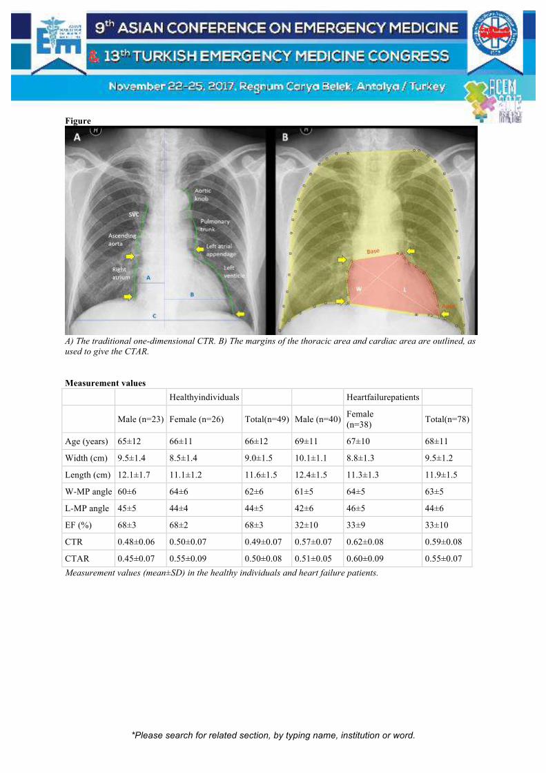
*Please search for related section, by typing name, institution or word.
Figure
A) The traditional one-dimensional CTR. B) The margins of the thoracic area and cardiac area are outlined, as
used to give the CTAR. Measurement values
Healthyindividuals
Heartfailurepatients
Male (n=23) Female (n=26) Total(n=49) Male (n=40)
Female (n=38)
Total(n=78)
Age (years) 65±12 66±11 66±12 69±11 67±10 68±11
Width (cm) 9.5±1.4 8.5±1.4 9.0±1.5 10.1±1.1 8.8±1.3 9.5±1.2
Length (cm) 12.1±1.7 11.1±1.2 11.6±1.5 12.4±1.5 11.3±1.3 11.9±1.5
W-MP angle 60±6 64±6 62±6 61±5 64±5 63±5
L-MP angle 45±5 44±4 44±5 42±6 46±5 44±6
EF (%) 68±3 68±2 68±3 32±10 33±9 33±10
CTR 0.48±0.06 0.50±0.07 0.49±0.07 0.57±0.07 0.62±0.08 0.59±0.08
CTAR 0.45±0.07 0.55±0.09 0.50±0.08 0.51±0.05 0.60±0.09 0.55±0.07
Measurement values (mean±SD) in the healthy individuals and heart failure patients.
*Please search for related section, by typing name, institution or word.
[S-163]
Japanese Emergency Transportation System and Issues on the System in Rural Regions Tetsunori Ikegami1, Toshio Fukuoka1, Yoshihito Ujike2 1Department of Emergency Medicine, Kurashiki Central Hospital, Kurashiki, Japan 2Department of Emergency and General Medicine, Kwasaki Medical University, Kurasiki, Japan Objective: We aim to explain the emergency transportation system in Japan and to reveal the issues on the emergency transportation system in the rural regions. Material: The emergency transportation system in Japan has been operated through the system that patients are transported to the hospitals within each firefighting headquarter’s jurisdiction area as far as possible. However, recently, especially in the rural regions, cases where patients can not be treated simply by transporting them to the hospitals within the jurisdiction area due to the population decline and cutbacks in hospital functions. Okayama Prefecture has 14 firefighting headquarters. Previously, we had no systems that these headquarters cross-jurisdictionally share information in real-time. Method: We introduced a new emergency transportation information system using Information-Communication Technology (ICT) in September 2016. Through this system, we cross-jurisdictionally learn the situation of the medical institutions in real-time using the smartphones and tablets. Three months after the introduction, we conducted a survey using the questionnaire on the usage of the new system. Results: Eight of 14 headquarters answered "The frequency of using the new system to select a destination is less than 20%." Seven of those eight headquarters answered "The reason for this is that there are a few or no choices for the destination". Many headquarters gave the negative answers to the question "Was the transportation time shortened?" or "Was the number of inquiries for acceptance decreased?" Our survey revealed that the primary function of the new system was not effectively utilized at 3 months after introduction. Conclusion: Medium- or small-scale firefighting headquarters which have limited number of hospitals had many negative opinions on the new system. This is because they could not adequately shift to the new system. We report the problems of the emergency transportation system in the rural regions of Japan and its future issues. Keywords: Emergency transportation system, information-communication technology (ICT)

*Please search for related section, by typing name, institution or word.
[S-164]
Establishment of Ambulance Satellite System to Improve Ambulance Response Time in Kota Kinabalu, Sabah Phee Kheng Cheah1, Chen Sek Yim2, Su Na Chin2, Nurul Liyana Idrus1, Muhamad Yaakub Arifin1 1Emergency and Trauma Department, Sabah Women and Children’s Hospital, Kota Kinabalu, Sabah, Malaysia 2Faculty of Science and Natural Resources, Universiti Malaysia Sabah, Kota Kinabalu, Sabah, Malaysia Introduction: Many studies have shown that the ambulance response time (ART) has a strong correlation with mortality such that response time interval is shorter for low mortality rates. Our objective of this study is to determine the ART and identify significant factors for improvement by introducing the ambulance satellite system (SS) into our emergency medical service (EMS). Methods: The SS areas were determined through the analysis of historical emergency call data. The Global Positioning System locations were plotted into our ambulance geolocation system to create visible zones of activity. This was plotted against time. Two geographical areas were deemed to have received a high number of emergency calls during peak hours. Our EMS team was put on standby at these locations during peak hours. Data on ART, distance, clinical information and type of emergencies were collected. We compared data of ambulance runs despatched from Sabah Women and Children’s Hospital (SWACH) during the same period (January to March) of 2016 and 2017 which was the study period. Results: A total of 67 patients were enrolled which consisted of 29 (43%) patients from January to March 2016 and 38 (57%) patients from January to March 2017. Trauma accounted for 51.7% (n=15) of cases in in 2016 and 34.2% (n=13) in 2017. The mean (SD) of ART for dispatch point SWACH was 17.72 ± 4.96 minutes. While, the mean (SD) of ART for dispatch point SS was 13.68 ± 6.99 minutes. Discussion and Conclusion: There was a significant reduction between mean (SD) of ART before and after the establishment of ambulance satellite system. Ambulance travel distance, age of patients and dispatch point was found to be significantly affecting ART in SWACH’s EMS system. This ART improvement is multi-factorial including reduction of distance to caller and also improved team alertness during peak hours. Keywords: Ambulance response time, ambulance satellite system

*Please search for related section, by typing name, institution or word.
[S-165]
The Survey Study About Causes and Preventive Measures for Ambulance Accidents Cemal Albayrak1, Mehmet Ergin2, Ayhan Özhasenekler2, Şervan Gökhan2 1Central Organization, Republic of Turkey Ministry of Health, Ankara, Turkey 2Department of Emergency Medicine, Medicine School, Ankara Yıldırım Beyazıt University, Ankara, Turkey Objective: Ambulances are vitally important in protecting the health of the community and in maintaining survival. A questionnaire study was conducted with 112 emergency medicine service employees in Ankara to understand the causes of ambulance accidents and determining the necessary precautions. Method: There were 558 participants from Ankara 112 EMS employees who agreed to participate in the study were included. The questionnaire consists of five parts. The questions were about the demographic information of participants in the first part, the reasons of ambulance accidents in the second part, the problems related to the use of ambulance in the third part, the situations that increase the risk of accidents during transporting patient by ambulance in the fourth part and the measures to reduce the risk of ambulance accident in the fifth part. Results: 55.2% of the participants were women, 42.47% were 29-34 years of age and 49.64% were Emergency Medicine Technicians. 74.91% of the participants stated that it was an accident because 'other drivers did not obey the rule of passage superiority'. 71% of the participants stated that performance criteria for EMS quality standards include the 'duration of travel to hospital', which forces the driver to speed up. Conclusion: The causes of ambulance accidents should be determined and precautions should be taken for these reasons. This situation is unavoidable in terms of the job safety of EMS workers moving with the 'safety is the first and foremost for you and your patient'. Keywords: Emergency medicine service, ambulance, ambulance accidents

*Please search for related section, by typing name, institution or word.
[S-166]
Analysis of Accidents of 112 Emergency Medicine Service Ambulances During 2016 Cemal Albayrak1, Mehmet Ergin2, Ayhan Özhasenekler2, Şervan Gökhan2 1Central Organization, Republic of Turkey Ministry of Health, Ankara, Turkey 2Department of Emergency Medicine, Medicine School, Ankara Yıldırım Beyazıt University, Ankara, Turkey Aim: To investigate the accidents of ambulances of emergency health services and to reveal the causes and to determine necessary precautions to prevent them. Method: The police records and insurance damage reports for accidents of EMS ambulances during 2016 were examined and the required information was entered into the previously prepared study form. The study form included the driver's gender and license class, the model of the vehicle, the cause of the damage, the location of the accident, the total amount of damage. Results: 2953 ambulance-related traffic accidents were detected. The median value of the expenditure made due to the accident was 1856 TL (min - max: 27 - 96845). 98.31% of the drivers who made an accident were males. 29.63% of the vehicles making the accident modeled in 2014. The most important cause of the accident was the collision with 72.6%. 78.7% of the incidents occurred within the city. Conclusion: It is understood that the majority of the ambulance-related accidents occurred in the city and during the crash. In spite of this, further studies should be carried out, such as examining accident imagesor videos and conducting surveys to understand the exact causes of the accidents. Keywords: Ambulance, ambulance accidents, emergency medicine service

*Please search for related section, by typing name, institution or word.
Table 1. Frequency Distribution Chart According to Data Taken from Police Accident Reports and Insurance Claim Report

*Please search for related section, by typing name, institution or word.
[S-167]
Review of Over 65 Years Patients as Forensisc Cases Admitted Emergency Department in Two Years Gül Bahar Alkaş1, Ahmet Demircan2, Gültekin Kadı2 1Gazi University Health Research and Aplication Center, Ankara, Turkey 2Gazi University Faculty of Medicine, Emergency Department, Ankara, Turkey Introduction: Today; the crowd in the emergency department is the biggest problem. An important part of the admission to the emergency departments is the elderly patients. Because of short-term treatment of traumatic diseases is not possible, these diseases require long-term follow-up and observation, they increase the use of place, time and labour. Results: Between the dates 01.01.2014 and 31.12.2016, 152 people were admitted to the emergency services as forensic cases over 65 years old. 56.6% of patients are male, 43.4% are females. 29,6% of the patients were admitted with beating, followed by 24,3% falling-down, 11,6% suicide and 18,4% in case of carbon monoxide poisoning. 2% of the patients were admitted with dog bite. 6 people (3.9%) were admitted to emergency service due to gunshot injures. Patients were admitted most frequently in April at 10.5%. All of the 8 patients who received sharp object injuries were male. At the same time, all the patients who come for eye and orbital injury are male. The most traumatic region in the patients was 32.2% head region and 14.5% upper extremities. Intracranial pathology was encountered in 9 of 49 patients who presented with head trauma. 53.3% of the patients were admitted between 08.00 and 17.00 hours and 13.8% between 00.00 and 08.00 hours of emergency services. Conclusion: Along with aging the number of chronic diseases increases, together with the trauma of the group of 65 years, increases the number of admission to emergency departments. Because of this admissions complications, mortality, morbidity and hospitalization rates increased. Careful consideration of additional diseases and risk factors for this group of patients in the emergency department's triage to the evaluation of the patient will also reduce the cost of this age group each year that will not reduce mortality and morbidity. Keywords: Geriatrics, emergency, forensic
Başvuru Saatleri / Travma Bölgeleri / Başvuru Hafta İçi/Dışı

*Please search for related section, by typing name, institution or word.
[S-168]
Bedside Ultrasonography of the Optic Nerve Sheath Diamater to Assess Intracranial Pressure and Evaluation of The Response to Treatment in Patients Diagnosed with Hyponatremia in Emergency Department Tayfun Anıl Demir1, Fevzi Yılmaz1, Mehmet Akif Karadaş1, Resmiye Nur Okudan1, Olgun Keskin2 1Sağlık Bilimleri University, Antalya Education and Research Hospital, Emergency Department, Antalya, Turkey 2Sağlık Bilimleri University, Antalya Education and Research Hospital, Department of Pulmonary Medicine, Antalya, Turkey Background-Aim: The aim of this study is to determine whether the thickness of ONSD as a noninvasive method in the diagnosis and treatment of intracranial pressure increase in patients with hyponatremia is a successful method to be used. Methods and Findings: Our study was performed prospectively with 79 patients with hyponatremia and 72 control group patients. Serum sodium value, optic nerve sheath diameter and symptoms, given treatments and ONS diameters after treatment were evaluated in addition to demographic information such as age and sex of the patients. The average age of the patients we work with was 62.3 ± 17.6 years, and 48.1% were male. ONSD at the time of admission and after treatment of patients were higher than the control group (p <0.05). ONSD lower and sodium elevation were detected after treatment with the disease (p <0.05). This change was greater when 3% saline was used. Symptoms were present in 97.5% of the patients at the time of admission and this rate decreased to 96.2% after the treatment. The most common symptoms were dizziness, nausea and headache. Symptoms of dizziness, palpitations and headache were significantly reduced (p <0.05). In patients with pre-treatment symptoms, ONSD were high (p <0,05), no relation was found between ONSD and symptoms before treatment type and after treatment. (p> 0.05). Negative correlations were found between patients' sodium level and ONSD during admission and sodium value before/after treatment (p <0,05),it was determined that this relationship deteriorated after treatment (p> 0,05). For our work cut-off 0.49; sensitivity was 81% and specificity was 81.9%. Fourteen of our patients (17,7%) were hospitalized and treated. It was determined that the patients had a sodium median of 115.5 mg / dl and that the median of the number of hospitalized days was 6. Keywords: Hyponatremia; optic nerve sheath diameter; intracranial pressure

*Please search for related section, by typing name, institution or word.
Table 1
Comparison of ONSD before and after treatment in groups

*Please search for related section, by typing name, institution or word.
[S-169]
Retrospective Analysis of Patients Under 18 Years Old and Admitteded Emergency Department due to Forensic Situations Atiye Kubra Okcesiz, Nalan Kozaci, Mustafa Avci Emergency Department, Antalya Education and Research Hospital, Antalya, Turkey Introduction: The aim of this study was to investigate forensic cases of the childhood defined to be between 0 and 18 years of age. Materials-Method: This study included 1624 patients between 0-18 years of age. Age, gender, nationality, time on admission, season of admission, cause of admission, imaging techniques, consultation, interventions, hospitalized department, termination and need for cardiopulmoner resuscitation were evaluated. Findings: Mean age of the patients was 9,2±6,2 years, most frequent injury was in the adolescent age group (49,4%). Among the patients, 61% were male, no relation was detected between age and gender (p>0,05). Most frequent causes of admission were mint (28,7%) and drug poisoning (22,4%). Mint, drug poisoning, traffic accident, substance abuse, cutter penetrating tool injuries, fall off from high and animal bite were common among adolescents, whereas simple fall, corosoive substance intake and burn were common among infants (p<0,05). Mint, cutter penetrating tool injuries and gunshot wound were common among males, whereas drug and substance intake were common among females (p<0,05). 1.3% of the admittants were foreigners. Patients were mostly admitted to the emergency units between 1600 and 2400 (57%). Imaging was performed in 46,4% of the patients and it was performed commonly in patients with trauma. Consultation was requested in 42,4% of the patients. Discharge rate from emergency unit was 66,9%. Direct graphy and computed tomography request rate was observed to be significantly high among the discharged (p<0,05). Conclusion: Forensic cases are commonly observed during adolescents and school ages of childhood. Most common forensic cases are mint and poisoning. Forensic cases are more common among males. Most frequently observed cases are mint for males and poisoning for females. Most of the forensic cases are discharged from the emergency units. Imaging is more common for those discharged from the emergency units compared to the others. Keywords: Trauma, child, forensic cases

*Please search for related section, by typing name, institution or word.
[S-170]
Trauma Analysis of Patients Over Age Of 65 Who Apply to Emergency Service Nazlı Görmeli Kurt1, Songül Araç2, Evren Ekingen1, Murat Orak2, Mehmet Üstündağ2 1State Hospital, Batman, Department of Emergency, Batman, Turkey 2Department of Emergency, Faculty of Medicine, Univesity of Dicle, Diyarbakır, Turkey Background: The purpose of this study is to make demographic analysis of geriatrics patients who apply to emergency service of Batman Regional State Hospital. Method: Our study involves patients over the age of 65 who applied to Batman Regional State Hospital in 1st February 2016-31st January 2017. 615 cases which were considered convenient with study criteria, had been examined retrospectively. The reason of application of cases, social demographic characteristics, specifications, additional diseases, the month of trauma, trauma area, trauma hour, mortality rates, GKS (Glasgow Coma Scale) and RTS (Revised Trauma Score) were obtained from automation system and case files. Results: 615 patients were included into study 252 of which were male and 363 were female. The average age of patients was 74,36. %70 of patients (n=433) were illiterate. The most common trauma was falling. Women patients were applied to service due to reason of falling than men. We used an anatomic scoring system called “AbbreviatedInjuryScale (AIS)” and we divided patients into two groups as local and multiple according to the number of trauma areas. %58 (n=354) of patients were local cases while %42 (n=259) of them were multiple trauma cases and in both of the groups the most of affected body parts were extremities. In total, %6,17 (n=38) of patients were exitus. 11 of them died in emergency services. Most of patients died (%78,94) due to multiple trauma and they were mostly subject to traffic accidents. The most accompanying comorbid disease to deaths is hypertension. Conclusion: Both in our country and globally, the elderly population is increasing rapidly The deaths due to trauma has a significant share in geriatric age group. Thus, it is significantly important to eliminate geriatric traumas and to intervene to the cases with multidisciplinary approach beginning from emergency services. Keywords: Trauma, geriatrics, emergency service, Keywords: Trauma, geriatrics, emergency service

*Please search for related section, by typing name, institution or word.
distrubution of local trauma areasbased on bady parts

*Please search for related section, by typing name, institution or word.
[S-171]
Comparison of the Rems and Rems-L in Prediction Of 28-Days Mortality of Elderly Patients Who Admitted Ed Serkan Demircan, Mehmet Ergin, Fatih Tanrıverdi, Çağrı Serdar Elgörmüş, Gülhan Kurtoğlu Çelik, Ayhan Özhasenekler, Şervan Gökhan Emergency Department, Ankara Yıldırım Beyazıt University Medicine School, Ankara, Turkey Objective: Our purpose was to compare the safety and efficacy of Rapid Emergency Medicine Score (REMS) and Rapid Emergency Medicine Score-Lactate (REMS-L) scores in predicting 28-day mortality after emergency department (ED) visit and hospitalization from ED for elderly patients. Material-Methods: The study was carried out between February 29 – April 30, 2016 at the Atatürk Educational & Research Hospital Emergency Service. The prospective observational study included elderly patients who were referred to ED, had red and yellow triage coding and did not have any trauma story. An emergency medicine resident or specialist in charge of the relevant area assessed patients. Vital signs, medical and past history were recorded. The sample for blood gas analysis was taken when indicated. The data written on patient chart was used to calculate REMS and REMS-L scores (REMS + lactate (mmol / L)). The primary end point of our study was 28 day mortality while secondary end point was hospitalization from ED. Results: The mean age of 1106 patients was calculated as 77.23 ± 7.41 years and the median value was 77 years. 52,3% (n = 578) of the patients was female. In the prediction of hospital admission, the Area Under Curve (AUC) on ROC for REMS and REMS-L were 0,837; 0,918 respectively (p = 0,001; p < 0,001 respectively). In the prediction of 28-day mortality AUC for REMS and REMS-L were 0,659; 0,695 respectively (p < 0,001; p < 0,001 respectively). Conclusion: According to our results, REMS scoring system were powerful in predicting hospital admission from ED and had moderate force in predicting 28-day mortality. REMS-L scoring system was more powerful than isolated REMS in predicting both for hospitalization and 28-day mortality. Keywords: Geriatry, lactate, rapid emergency medicine score

*Please search for related section, by typing name, institution or word.
[S-172]
Comparison of the MEWS and MEWS-L in Prediction of 28-Days Mortality of Elderly Patients Who Admitted ED Serkan Demircan, Mehmet Ergin, Fatih Tanrıverdi, Çağrı Serdar Elgörmüş, Gülhan Kurtoğlu Çelik, Ayhan Özhasenekler, Şervan Gökhan Emergency Department, Ankara Yıldırım Beyazıt University Medicine School, Ankara, Turkey Objective: Our purpose was to compare the safety and efficacy of Modified Early Warning Score (MEWS) and Modified Early Warning Score-Lactate (MEWS-L) scores in predicting 28-day mortality after emergency department (ED) visit and hospitalization from ED for elderly patients. Material-Methods: The study was carried out between February 29 – April 30, 2016 at the Atatürk Educational & Research Hospital Emergency Service. The prospective observational study included elderly patients who were referred to ED, had red and yellow triage coding and did not have any trauma story. An emergency medicine resident or specialist in charge of the relevant area assessed patients. Vital signs, medical and past history were recorded. The sample for blood gas analysis was taken when indicated. The data written on patient chart was used to calculate MEWS and MEWS-L (MEWS + lactate (mmol / L)) scores. The primary end point was 28 day mortality while secondary end point was hospitalization from ED. Results: The mean age of 1106 patients was 77.23 ± 7.41 years and the median value was 77 years. 52,3% (n = 578) of the patients was female. In the prediction of hospital admission, the Area Under Curve (AUC) on ROC for MEWS and MEWS-L were 0,817; 0,927 respectively (p = 0;002; p < 0,001 respectively). In the prediction of 28-day mortality AUC for MEWS and MEWS-L were 0,647; 0,681 respectively (p < 0,001; p < 0,001 respectively). Conclusion: According to our results, MEWS scoring system were powerful in predicting hospital admission from ED and had moderate force in predicting 28-day mortality. MEWS-L scoring system was more powerful than isolated MEWS in predicting both for hospitalization and 28-day mortality. Keywords: Geriatry, lactate, modified early warning score

*Please search for related section, by typing name, institution or word.
[S-173]
Epidemiological Characteristics of Elderly Patients Admitted to ED Serkan Demircan, Mehmet Ergin, Fatih Tanrıverdi, Çağrı Serdar Elgörmüş, Gülhan Kurtoğlu Çelik, Ayhan Özhasenekler, Şervan Gökhan Emergency Department, Ankara Yıldırım Beyazıt University Medicine School, Ankara, Turkey Objective: Patients aged 65 years and older constitute a special group of patients who are referred to emergency services. Elderly populations are increasing day by day in society of both developing and developed countries. We aimed to determine patient characteristics, reasons for referral, and outcomes of ED visits and hospitalization in elderly patients. Material-Methods: The study was carried out between February 29 – April 30, 2016 at the Atatürk Educational & Research Hospital ED. The prospective observational study included elderly patients who were referred to ED, had red and yellow triage coding and did not have any trauma story. Vital signs, medical and past history and 28 day mortality were recorded. Results: The mean age of 1106 patients was 77.23 ± 7.41 years and the median value was 77 years. 52,3% (n = 578) of the patients was female. The most frequent complaints were dyspnea (18.4%, n=204), abdominal pain (7.2%, n=80), and fever (6.2%, n=66). The most frequently diagnosed disorders in the ED were cardiovascular, pulmonary and neurology diseases. 56.2% (n=622) were discharged from the hospital. A significant difference in 28 day mortality results was observed between patients who were admitted to the intensive care unit (ICU) and discharged (p=0,001). Conclusion: With an increased proportion of elderly individuals in the general population, the number of elderly patients referred to emergency departments continues to increase. This study presents the demographic features and clinical course of elderly patients referred to emergency department. Keywords: Demographics, emergency, geriatry

*Please search for related section, by typing name, institution or word.
[S-174]
Diabetic Ketoacidosis Presenting with a Normal Anion Gap Sinan Görgülü, Ilter Ağaçkıran, Yasemin Özdamar, Muhammed Sarıer, Zehra Akçora, Meltem Akkaş, Bülent Erbil Emergency Service, Hacettepe University, Ankara, Turkey Although historically, diabetic ketoacidosis (DKA) is associated with an increased anion gap, normal anion gap (NAG) doesn’t exclude the diagnosis of DKA. We report a case of DKA with NAG secondary to hyperchloremia. Case: A-48 years old woman was admitted to emergency department with chest pain and palpitation. Her pulse rate was 122/min and the other vital signs were normal. On past medical history, the patient had no known diseases and had no history of use of medications. There were no ischemic findings in her ECG. When she complaint dryness of mouth, the bedside capillary blood glucose level was measured as 320mg/dl. Physical examination findings were unremarkable. The patognomonic laboratory test results were as follows; pH:7.03, HCO3:3,8 mMol/L, Na:132 mEq/L, Cl:121mEq/L, albümin:2.88 g/dl. Urinalysis showed ++++ ketones, glucose ++++. With these findings the anyon gap was normal. She treated with normal saline and insulin infusion. She admitted the intensive care unit by internal medicine. Acidosis and ketonuria were treated in ten days. She discharged with basal and bolus insulin treatment. In some rare cases, DKA may have NAG. A reduction in serum bicarbonate is matched by an approximately equivalent increase in serum chloride concentration. Some authors claim that some patients tend to be well hydrated with renal function preserved following excretion of ketone anions and the kidney reabsorbs chloride to compensate for the loss of these bicarbonate equivalents, generating a hyperchloremic metabolic acidosis. In addition, they assert that normal saline can induce hyperchloremic acidosis. Conclusion: Acute non anion gap metabolic acidosis is now recognized to be as a cause of metabolic acidosis, particularly in the emergency department. Controlled studies to determine which fluid should be given, at what level of serum bicarbonate should base therapy be initiated are needed. Keywords: Diabetic ketoacidosis normal anion gap

*Please search for related section, by typing name, institution or word.
[S-176]
Evaluation of Geriatric Patients Admitted with Falls to Emergency Department Betül Akbuğa Özel, Göksu Bozdereli Berikol, Elmas Burcu Mamak Ekinci Baskent University Faculty of Medicine, Istanbul, Turkey Background: Mechanical injuries of geriatric patients are common problems of emergency medicine (ED) crowd. Falls and accidents are the main causes of these injuries. The incidence of fall is 30 percent of community-dwelling people over the age of 65 years. Fracture, head trauma or major lacerations, as defined serious wounding, occur 10-25% of elderly cases. Aim: We aimed to evaluation the geriatric patients admitted to emergency medicine with the diagnose of falls. Method: The study is a retrospective, descriptive study. Between July 2016 and July 2017, 700 geriatric patients admitted with falls to the ED were included to the study. Results: The geriatric patients admitted to ED with falls are found 8,7% of all geriatric patients and 2,1% of total patients. Four hundred eighty of them are women (68,6%). One hundred and ninty five of the patients (27,9%) are over 85 years old. Most of the patients have the diagnosing code of unspecified fall (57,4%) and fall on same level from slipping, tripping and stumbling (36,7%). Patients with the need of computerized tomography, x-ray, blood test and consultation are 57,4%, 75,9%, 35,4% and 48,4% respectively. Neurosurgery and orthopedics are the most consultated departments. Average duration of stay in ED is found 262 min. and follow-up in ED is 364 min. The need for hospitalization in 88 patients (12,6%) was deemed necessary. Average cost per patient is found 134,0TRY. Conclusion: Women and 75-84-year-olds are more likely to fall. Head trauma and orthopedic injuries are more common. Most of the patients are discharged from ED. The percentage of patients requiring hospitalization is 12.6%. Cost per patient is the least in the 65-74 age group. Epidemiological and risk and resolution-focused analytical studies of geriatric mechanical falls should be increased to reduce the incidence of a preventable morbidity and mortality cause. Keywords: Emergency, fall, geriatric

*Please search for related section, by typing name, institution or word.
[S-177]
Mortality Factors in Geriatrics with Non-Traumatic Abdominal Pain at the Emergency Department Süha Serin1, Bahadır Çağlar2, Gökhan Yılmaz3, Alper Torun3, Başak Göl Serin4, Ismet Parlak3 1Emergency Medicine Clinic, Urla City Hospital, Urla, Izmir, Turkey 2Emergency Medicine Clinic, Elazig Training and Research Hospital, Elazig, Turkey 3Emergency Medicine Clinic, Bozyaka Training and Research Hospital, Izmir, Turkey 4Infectious Disease and Clinical Microbiology Clinic, Tepecik Training and Research Hospital, Izmir, Turkey Introduction-Purpose: The purpose of this study was to determine the factors affecting mortality in geriatrics presenting at ED with non-traumatic abdominal pain. Method: This cross-sectional, retrospective study included patients aged >=65 years who presented at the ED with non-traumatic abdominal pain. The demographic characteristics, laboratory test results and in-hospital courses of the patients were examined. The relationship between mortality and the data obtained was analyzed. The data were analyzed at 95% confidence level and a value of p<0.05 was considered statistically significant. The study was conducted following the approval of the Ethics Committee. Results: Total of 1110 patients were included in the study, comprising 619 (55.8%) females.719 (64.8%) patients admitted to the general surgery clinic, 211 (19%) were operated on. Of those admitted to the general surgery clinic, 106 (9.5%) cases resulted in mortality. The cut-off value of age for mortality was found to be 73 years (73.6% sensitivity, 40.4% specificity, ROC-AUC 0.581) (figure 1). A high lactate value (cut-off value 2.4) was found to be associated with mortality (78.2% sensitivity, 68.8% specificity, ROC-AUC 0.786) (figure 1). The most common predictors of mortality were determined to be perforation (odds ratio [OR] 20.7), ileus (OR 17.9), and the laboratory values of high lactate (OR 7.6), hypocalcemia (OR 3.9) (table 1). Conclusion: Electrolyte and lactate values were determined to be the main determinants of mortality in geriatric patients who presented at ED with abdominal pain. It is important to note that mortality increases over the age of 73 years. Keywords: Geriatrics, non-traumatic abdominal pain, mortality

*Please search for related section, by typing name, institution or word.
The mortality risk for the cut-off value for age is > 73 years & the cut-off value for lactate is > 2.4
Factors affecting mortality when confounding effects are excluded
Independent Variables B±Sd P-value Odds-Ratio (95% CI)
Undergoing surgery -0.929±0.459 0.043 2.53 [1.03-6.22]
Lactate (>2.4) -2.037±0.472 <0.001 7.67 [3.04-19.32]
Ileus and Subileus 2.888±1.144 0.012 17.96 [1.91-169.09]
Perforation 3.030±1.270 0.017 20.70 [1.72-249.51]
Hypocalcemia -1.367±0.494 0.006 3.92 [1.49-10.33]
Dependent Variable: Mortality Predicted Survivals = 95% PredictedEx=%63 Predicted: %88.1 P Model<0.001
MultipleLogisticRegression (Method = BackwardStepwise (Wald)) C.I: Confidence Interval B: regression
coefficients Sd: Standard Deviation

*Please search for related section, by typing name, institution or word.
[S-178]
A 66-Years-Old Man with Tetanus: A Case Report Emrah Yılmaz, Mehmet Eman, Mustafa Silcan, Alper Torun, Ismet Parlak Emergency Medicine, Bozyaka Training and Research Hospital, Izmir, Turkey Tetanus is one of the many diseases that can be protected through vaccination. The tetanospasmin named toxin causing the disease affects the central nervous system and the neuromuscular junction. The prevalence of this disease decreased with repeated doses at birth and after. In our case study, a 66-year-old male patient came to our emergency department with a complaint of difficulty in speaking, locking in jaw and pain in the neck. It was learned that he had a story of working in the garden 6 days ago. After 1 day, a soft tissue infection was suspected in the patient who complained of an standing rash. Only the tetanus vaccination was done because the patient had no trauma of injury and the lesion was clean. Diabetes mellitus, chronic heart disease and hypertension were present. The patient did not know how many doses of tetanus vaccine had been made so far. Overall condition was good, consciousness clear, oriented, tansıon arterıal 200/100 pulse:97 O2 saturatıon:97%. There was no high body temperature. Trismus and right foot dorsal had 5*4cm of redness and irritation, there was no flow. The patient's laboratory and cranial imaging were usual. The patient was diagnosed with tetanus in the clinic and the infectious diseases were transferred to the service After 1 day, opsoclonus and dysphagia were formed, and intubated for respiratory safety. Then, the patient was transferred to anesthesia and reamination department. He died after 6 days. In conclusion, tetanus is a rare infectious disease with high mortality. The diagnosis is mainly clinical. When patients are exposed to tetanus risk, it is vitally important that the condition of immunization, age and especially the additional diseases are questioned in detail and immunoprophylaxis is performed in full. Keywords: Tetanus, tetanus prophylaxis, tetanus in developing countries

*Please search for related section, by typing name, institution or word.
[S-179]
The Viral Prevalence in Adult Patients Who Meet the Case Definition of Severe Acute Respiratory Infection in ED and to be able to predict the mortality by using CURB-65, PSI, Q-SOFA and SOFA Hakan Aydın, Halil Doğan, Doğaç Niyazi Özüçelik, Mustafa Saad Salem Altay Bakırköy Dr. Sadi ̇ Konuk Eği ̇ti ̇m Ve Araştırma Hastanesi ̇ Aci ̇l Tıp Kli ̇ni ̇ği ̇ Background: Acute respiratory infections lead to the death in about 3,1 million people worldwide each year. An influenza pandemic may increase Emergency Depertment (ED) attendance 7-fold. Our aim in this study is to determine the viral prevalence in adult patients who meet the case definition of severe acute respiratory infection (SARI) in ED and to be able to predict the mortality by using CURB-65, PSI, Q-SOFA and SOFA criteria in these patients. Methods: This prospective, observational, single-center, and cross-sectional study was conducted at ED between january 5 and march 31, 2016. Staff completed a standard form and collected a nasopharyngeal swab which was tested for selected respiratory viruses by rt-pcr. The end point was defined as all-cause mortality. Results: A total of 159 patients (median age 66years; 45,9% male) with SARI were enrolled in this study. Patients who had at least one comorbidity disease 78%. Of 159 cases tested, 28,3% were positive for influenza viruses. Among the 45influenza-positive specimens, 54 % H1N1pdm09 (Table 1). During hospitalization 57 patients were admitted to intensive care unit, and 28 died. Innon-servived patients; Respiratory distress, confusion, increased heart rate and respiratory rate, low saturation, smoking, diabetes mellitus, oncologic diseases; Increase in urea, LDH and lactate values, decrease in hematocrit, albumin and calcium levels were statistically significant (Table 2). Disease severity scores, described as a low score on the low mortality rate, while those with high scores have increased mortality rates. Conclusion: A significant portion of SARI cases were found to be ınfluenza viruses positive, but there was no difference in mortality between influenzavirus-infected patients and those not identified, CURB-65, PSI, Q-SOFA and SOFA severty criteria can be used to predict the need of intensive care unit and mortality. Keywords: Influenza, SARI, Q-SOFA
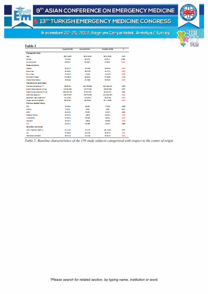
*Please search for related section, by typing name, institution or word.
Tablo 1
Tablo 2: Baseline characteristics of the 159 study subjects categorized with respect to the center of origin

*Please search for related section, by typing name, institution or word.
[S-180]
Retrospective Evaluation of Patients with Acute Renal Infarction in Emergency Department; A Series of 15 Cases Ilhan Uz, Enver Özçete, Murat Ersel, Güçlü Selahattin Kiyan Ege University, Faculty of Medicine, Department of Emergency Medicine, Izmir, Turkey. The prevalence of acute renal infarction (ARI) in Turkey remains unclear. ARI is unexpected diagnosis and coincidentally dedected in emergency department if the patient has still renal colic pain and planned radiological methods. This single-center, retrospective, observational study included 15 patients with newly diagnosed ARI (2015–2017). Their clinical features (age, gender, complaint, type of pain, right/left kidney, laboratory results, radiological methods) and during six months mortality rates were recorded. Results: The prevalence of ARI among emergency room patients was 0.000035 % (mean age 62.53 ± 18.07 years; men 53 %). Computed tomography anjiography or renal ultrasonography were obtained to diagnose ARI. ARI involved the left kidney in 53.3 %, right kidney in 40 %, and both kidneys in 6.7 % of patients. 4 (26%) cases had splenic infarction, 2 (13%) cases had renal artery thrombosis, 1 (0.06%) case had renal artery dissection and 8 (53.3%) cases had atrial fibrillation. We noted abdominal or flank pain in 60% and nausea/vomiting in 20% of patients. The mortality rates were 40% at 0-6 month, respectively. Various causes result in renal infarction, but atrial fibrillation (AF) is the key risk factor of ARI. ARI must be considered especially in patients over 65 years of age, patients with valvular heart disease, atrial fibrillation or embolic episodes, acute flank pain after abdominal trauma in all age groups. Conclusions: We determined the prevalence of ARI among emergency room patients and mortality rate. The majority of ARI cases were of cardiac origin and the others were due to trauma or systemic thrombotic disease. Clinicians should be aware of ARI as a fatal arterial thrombotic disease. Keywords: acute renal infarction, emergency department

*Please search for related section, by typing name, institution or word.
Figure 1.
CT Angiography of Renal infarction and Renal Artery Occlusion

*Please search for related section, by typing name, institution or word.
[S-182]
Pneumoperitoneum with Hydrogen-Containing Carbon Dioxide Ameliorates Bowel Inflammation Atsunori Nakao, Tetsuya Yumoto, Taihei Yamada, Hiromichi Naito Department of Emergency and Critical Care Medicine, Okayama University Hospital, Japan Objective: Laparoscopic procedures, which are less invasive than conventional open surgeries, have become common for acute care abdominal surgery. Bowel function recovers more quickly with laparoscopic surgery than with open surgery. However, in critically ill patients, gastrointestinal motility impairment caused by intestinal manipulation-associated inflammation occurs, even after laparoscopic procedures. Therefore, an effective method to enhance bowel recovery from bowel dysfunction is required to shorten hospitalization after acute care abdominal surgery. Our laboratory has shown that inhalation of hydrogen gas can reduce bowel inflammation and help recovery from postoperative ileus. In this study, we hypothesized that pneumoperitoneum with carbon dioxide containing hydrogen gas can be effectively used to ameliorate bowel inflammation by surgical manipulation. Material-Methods: Ileus was induced in a rat model by applying two cotton-tipped applicators to the small intestine and using a rolling motion (surgical manipulation). After surgical manipulation, the peritoneal cavity was filled with 10 mL carbon dioxide plus nitrogen (1.3%) or carbon dioxide plus hydrogen (1.3%). Gastrointestinal transit was measured and cytokine levels and neutrophil extravasation in the intestinal muscularis propria was assessed. Results: In the nitrogen group, surgical manipulation was associated with a notable delay in intestinal transit, proinflammatory cytokine upregulation, and greater neutrophil extravasation. On the other hand, pneumoperitoneum containing hydrogen significantly mitigated bowel dysmotility caused by surgical manipulation and inflammatory events. Production of nitric oxide in the bowel muscle layers was inhibited by pneumoperitoneum with gas containing 1.3% hydrogen. Conclusion: This study demonstrated the protective effects of pneumoperitoneum with hydrogen-containing carbon dioxide on a rat surgical ileus model by hindering inflammation in the muscularis propria. These effects on bowel dysmobility associated with proinflammatory response may be partially derived from inhibition of nitric oxide production. Keywords: Hydrogen, pneumoperitoneum, bowel movement

*Please search for related section, by typing name, institution or word.
[S-183]
Cerebral Blood Flow Changes in Synthetic Cannabinoid Intoxication Cases Uygar Utku1, Onur Karakayalı2, Serkan Yılmaz2, Nevzat Uzuner3 1Kocaeli Derince Training and Research Hospital, Clinic of Neurology, Kocaeli, Turkey 2Kocaeli Derince Training and Research Hospital, Clinic of Emergency Medicine, Kocaeli, Turkey 3Department of Neurology, Medical School, Eskişehir Osmangazi University, Eskişehir, Turkey Objective: Synthetic cannabinoids (SC) are compounds with psychoactive properties similar to Δ9-tetrahydrocannabinol found in marijuana. Patients with SC intoxication have recently generated the majority of emergency service intoxication cases. We aimed to evaluate the changes in cerebral blood flow in patients with SC intoxication. Material-Methods: All patients with SC intoxication enrolled to the emergency service were taken to the study between January 2016 and May 2017. Changes in cerebral blood flow were measured with transcranial Doppler (TCD) ultrasonography by a neurosonologist. General physical and neurological examinations of the patient including blood pressure and meningeal signs were performed. Hemogram and routine biochemistry tests were checked. Brain MRI or CT were performed for differential diagnosis. TCD ultrasonography examination was performed with a Multidop X DWL TCD device after the patient had rested for 10 minutes in the supine position by 2 MHz pulsed Doppler probes. Bilateral MCA, ACA and PCA were studied at three different depths. Blood flow velocities, Gosling's pulsatility index and Pourcelot's resistance index were recorded. The control group consisted of 40 ages and sex-matched randomly selected patients. Diseases that can disrupt cerebral hemodynamics, closed temporal window or alcohol usage were excluded from the study. Ethics committee approval for the study was taken from the Ethics Committee of the Faculty of Medicine. Results: The mean ages were 34.7 ± 5.9 years in SC intoxication group (all male: 38) and 32.3 ± 4.7 years in the control group (n: 40). Bilateral blood flow velocities were significantly lower in SC intoxication group than in the control group. The values of the pulsatility index and the resistance index were significantly higher in the SC intoxication group than in the control group. Conclusion: Brain blood flow changes consistent with encephalopathy were detected in SC intoxication cases. Keywords: Synthetic cannabinoid, intoxication, cerebral blood flow

*Please search for related section, by typing name, institution or word.
[S-184]
The Releationship Between Serum Levels of Gamma Glutamyl Transferase, Mean Thrombocyte Volume, Red Blood Cell Distribution Width, Neutrophil to Lymphocyte Ratio with Prognose And Mortality in Patients Admitted Emergency Department with Acute Ischemic Cahfer Güloğlu1, Nazlı Görmeli Kurt2, Yusuf Tamam3, Mehmet Üstündağ1, Murat Orak1 1Department of Emergency, Faculty of Medicine, Univesity of Dicle, Diyarbakır, Turkey 2State Hospital, Batman, Department of Emergency, Batman, Turkey 3Department of Neurology, Faculty of Medicine, Univesity of Dicle, Diyarbakır, Turkey Purpose: Stroke is the third most common cause of death after cardiac diseases and cancers and is also one of the most common causes of morbidity and mortality both in Turkey and around the world. In the present study, we aimed to evaluate serum levels of gamma glutamyl transferase (GGT), mean thrombocyte volume (MPV), red blood cell distribution width (RDW), neutrophil to lymphocyte ratio (NLR) as well as lateralization, localization, and size of ischemic stroke, subtypes of stroke, and clinical progress and mortality and ultimately to identify guiding parameters for the diagnosis and treatment of stroke. Material-Method: 473 patients who presented to Emergency department of Dicle Univesity between January 1, 2013 and April 30, 2015 and had a diagnosis of stroke were enrolled in the study. In addition, a control group was formed with 40 ages and gender-matched healthy individuals. Findings: The 473 patients included 236 (49.9%) men and 237 (50.1%) women. Total mortality occurred in 58 (12.3%) patients, including 31 (53.4%) women and 27 (46.6%) men. Mean age was 74.47+11.21 years in the mortality group and 61.00+15.70 in the surviving group. In the patient group, mean GGT, MPV, RDW, and NLR values were higher compared to the control group. According to the TOAST and OCSP classifications, GGT, MPV, RDW, and NLR values were higher in the patient group compared to the control group. However, increased NIHSS and decreased GCS were found to be associated with increased GGT, MPV, RDW, and NLR. Moreover, GGT, MPV, RDW, and NLR were significantly higher in the mortality group than in the surviving group. Result: We concluded that serum parameters including GGT, MPV, RDW, and NLR are simple, inexpensive and easily reachable tools that can be used in the prediction of prognosis and mortality in patients with ischemic stroke. Keywords: Stroke, GGT MPV RDW NLR, mortality

*Please search for related section, by typing name, institution or word.
[S-187]
Posterior Reversible Encephalopathy Syndrome; Five Cases reports İrfan Aydın1, Abdullah Algın1, Yaşar Altun2, Mehmet Kaan Poyraz1 1Adiyaman University Research and Educational Hospital, Deparment of Emergency Medicine, Adıyaman, Turkey 2Adiyaman University Research and Educational Hospital, Department of Neurology, Adıyaman, Turkey Introduction: Posterior reversible encephalopathy syndrome (PRES) is a reversible syndrome accompanied by neurological and radiological findings by Hynchy J et al. in 1996 (1). This syndrome is often referred with hypertension, headache, confusion, convulsions, vomiting, and visual impairment (2). Two hypotheses have been proposed for pathophysiology. The first being cerebral hyperperfusion due to suddenly elevated blood pressure and the associated vasogenic edema and the second endothelial dysfunction caused by endogenous and exogenous toxins (1). It is clinically characterized by various neurological findings which may develop acutely or subacutely. It can last for several hours or days and weeks (3). Cases: All our cases were female, Brain Computerized tomography of all of them was normal. However, there was hyperintense lesion in brain lobe in magnetic resonance imaging (MRI) so we diagnosed as PRES (Table). Conclusion: As a result, when the patient comes with these kind of complaints to the emergency service, the emergency physician rightly thinks more about preeclampsia / eclampsia and hypertensive encephalopathy. However Emergency physicians should be aware of the PRES syndrome, which reports with similar complanints. Reference: 1) Fischer M, Schmutzhard E (2017), Posterior reversible encephalopathy syndrome, J Neurol DOI 10.1007/s00415-016-8377-8 2) Hinchey, Judy, et al. "A reversible posterior leukoencephalopathy syndrome." New England Journal of Medicine 334.8 (1996): 494-500. 3) Fugate JE, Rabinstein AA (2015) Posterior reversible encephalopathy syndrome: clinical and radiological manifestations, pathophysiology, and outstanding questions. Lancet Neurol 14(9):914–925. Keywords: Pres, convulsions, headache

*Please search for related section, by typing name, institution or word.
Demographic and MRI Findings of Patients
Cases Age (Year)
Sex Complaint Gravidity Blood Pressure (mmHg)
MRI (Magnetic Resonans Imaging) Hiperintens
Case 1
23 female Convulsions Postpartum (1 week after birth)
110/70 pons and cerebellum
Case 2
18 female Loss of vission, convulsions, headache, vomiting
Postpartum (2 week after birth)
165/105 parietal, occipital, temporal
Case 3
27 female Convulsions 34-week primigravida
180/110 parietal, occipital
Case 4
26 female Headache 32-week multigravida
165/105 occipital
Case 5
28 female Headache, unconsciousness
Postpartum (2 week after birth)
150/100 occipital
*Please search for related section, by typing name, institution or word.
[S-189]
Relationship Between Ischemia Side and Optic Nerve Sheath Diameter in Acute Ischemic Stroke Patient on Bedside Ocular Ultrasonography Hande Asan1, Ozlem Dikme2, Sehmus Zengin3, Ozgur Dikme3, Hakan Topacoglu3 1Emergency Department, University of Health and Scienses Sultan Abdülhamid Han Training and Research Hospital, Istanbul, Turkey 2Emergency Department, Koc University Hospital, Istanbul, Turkey 3Emergency Department, University of Health and Scienses Istanbul Training and Research Hospital, Istanbul, Turkey Introduction: The increase of optic nerve sheath diameter is a reliable, noninvasive marker of intracranial hypertension. In this trial, we aimed to reach meaningful relationship between acute ischemic stroke side and widening optic nerve sheath diameter side. In this reason, we evaluated acute ischemic stroke patients at emergency department with bedside ocular ultrasonography for bilateral optic nerve sheath diameter measurements. Methods: This prospectively cross-sectional study covered 18+ years-old patients who admitted to the emergency between July 15,2016 and September 15,2016 with acute ischemic stroke. The patients had diagnosis with Diffusion Weighted Imaging Magnetic Resonance and also hadn’t exclusion criteria. Collected data were included age, gender, admission symptoms and its duration, both eyes optic nerve sheath diameters results, National Institutes of Health Stroke Scale score and infarction side. Results: 67 patients were enrolled this study. 37 (55,2%) of them were male and average age was 68,4±13,1. Average of right eyes optic nerve sheath diameter was found 0,485±0,090 cm (Interval 0,29-0,73, median 0,490). Average of left eyes optic nerve sheath diameter was found 0,486±0,101 cm (Interval 0,28-0,71, median 0,480) and both eyes optic nerve sheath diameter's average was found 0,486±0,092 cm (Interval 0,29-0,72, median 0,478). The left optic nerve sheath diameter value of right infarcted patients increased positively relation with right optic nerve sheath diameter value. The observation was same for left infracted patients. There was no statistical significant difference between age, symptoms duration and National Institutes of Health Stroke Scale score. Conclusion: Both of right or left optic nerve sheath diameter values can be considered while detecting to intracranial pressure increase with bedside ocular ultrasonography for patients with acute ischemic stroke. Increasing of optic nerve sheath diameter for both eyes, correlates positively. There is no side-to-side correspondence between the side finding and the optic nerve sheath diameter side. Keywords: Stroke, bedside ultrasonography, emergency department
Figure

*Please search for related section, by typing name, institution or word.
Optic nerve sheath diameter measurement tech and normal and widening diameter samples

*Please search for related section, by typing name, institution or word.
[S-190]
Nonaneurysmal Subarachnoid Hemorrhage Secondary to Pneumococcal Meningitis Hiromu Naraba1, Tomohiro Sonoo1, Yuji Takahashi1, Hideki Hashimoto1, Kensuke Nakamura1, Naoto Morimura2 1Hitachi General Hospital, Japan 2Tokyo University Hospital, Japan Introduction: Nontraumatic subarachnoid hemorrhage, one of the life-threatening diseases seen in Emergency Departments, is rarely caused by conditions other than rupture of saccular aneurysms. We report a case of pneumococcal meningitis complicated with nonaneurysmal subarachnoid hemorrhage, with some literature reviews. Case : A 63 year-old man was admitted to our department with impaired consciousness. About two days before admission, the patient presented with a high fever and headache. Initial cranial computed tomography (CT) scan did not show any abnormalities. Lumbal puncture was performed and cerebrospinal fluid (CSF) analysis revealed pleocytosis and hypoglycemia with microbiological evidence for pneumococcal meningitis. After initiation of appropriate antibiotic and dexamethasone therapy, the patient developed heavy convulsive episode with conjugate deviation. Contrast-enhanced CT scan performed after seizure showed nonaneurysmal subarachnoid hemorrhage in the left postcentral gyrus and thrombosis of the left sigmoid sinus. Meanwhile the patient deteriorated despite treatment for pneumococcal bacteremia, and then required mechanical ventilation. A magnetic resonance imaging (MRI) coupled with magnetic resonance angiogramphy (MRA) performed ten days after admission revealed no signs of infarction, ischemia, thrombosis or arteriovenous malformation. After systemic complications such as septic shock and disseminated intravascular coagulation were ameliorated gradually, the patient withdrew the respirator. However, neurologic complications such as a major motor deficit and impaired cognition or speech were persistent. Discussion: The course of this patient provides two important clinical suggestions. First, pneumococcal meningitis can present with nonaneurysmal subarachnoid hemorrhage. These findings suggest that subarachnoid hemorrhage occurred due to oozing rupture of infected cerebrovascular wall affected from bacterial meningitis. Second, during treatment for bacterial meningitis, daily sedation interruption and intracranial pressure monitoring might have potential usefulness in early detection of neurologic complications. Keywords: Bacterial meningitis, subarachnoid hemorrhage, cerebral venous thrombosis

*Please search for related section, by typing name, institution or word.
[S-191]
A Rare Cause of Encephalopathy: Reversible Splenial Lesion Syndrome Due to Infection and Acute Renal Failure Meltem Akkaş, Sinan Görgülü, Hatice Gökçe Saraç, Yunus Emre Dündar, Zehra Akçora, Nalan Metin Aksu Hacettepe University, School of Medicine, Emergency Department, Ankara, Turkey Introduction: Reversible splenial lesion syndrome (RESLES) which may present with encephalopathy is a rare disorder which is defined as reversible isolated splenial lesions in the corpus callosum identified by magnetic resonance imaging (MRI). Herein we presented a case of RESLES due to infection and acute renal failure. Case: A 62-year-old male was admitted to emergency department (ED) with the complaints of slow and stuttering speech, tremor in his hands, blurring of consciousness for two days and nausea, vomiting, fatigue and coughing for seven days. The day when the patient was admitted to ED, he could not find his home way and started to talk slowly and stuttering. Hypertension and gout were identified in his past medical history. He responded to two stage commands. Slow and stuttering speech, walking with ataxia, dysmetria, dysdiadochokinesia were found in his neurological examination. The patognomonic laboratory test results were as follows; platelet count:134000, BUN: 40,5 mg/dL, uric acide: 8,96 mg/dL, creatinine: 3,14 mg/dL. MRI of head showed a T2 hyperintense corpus splenium lesion showing diffusion restriction. Reversible splenium lesion syndrome due to infection and acute renal failure was considered as a preliminary diagnosis in the patient. Intravenous hydration with isotonic sodium chloride was started. Creatinine level dropped to 1.4mg/dL and his symptoms had resolved. Nine days after his admission, the new MRI showed that the diffusion restriction observed in previous MRI was completely lost Conclusion: RESLES presents in a variety of disorders including infection, seizures, antiepileptic drug withdrawal, chronic alcoholism, tumors, trauma, metabolic and electrolyte disturbance. The complete reversibility of the lesion on follow up imaging and full clinical recovery occuring within days to weeks are hall marks of the syndrome. The clinical course of the syndrome is usually benign with a favorable outcome unless underlying severe disorders. Keywords: Reversible splenial lesion
Figure 1: Brain MRI. Axial T2W image (A) shows a hyperintense well circumscribed, small, oval lesions in the midline within the splenium of the corpus calosum. The lesion shows restricted diffusion (B, C).

*Please search for related section, by typing name, institution or word.
Follow-up brain MRI (D, E) 10 days later sho

*Please search for related section, by typing name, institution or word.
[S-192]
Comparison of Oral Paracetamol and Zolmitriptan Efficacy in The Treatment of Acute Migraine Headache in Emergency Department Cüneyt Arıkan1, İbrahim Türkçüer1, Atakan Yılmaz1, Ezgi Demirözoğul2, Mert Özen1, Aykut Uyanık1, Bülent Erdur1, Gizem Öncel1 1Department of Emergency medicine, Pamukkale University, Denizli, Turkey 225 Aralık State Hospital, Gaziantep, Turkey Study Objective: Migraine is a chronic and sometimes progressive disorder, characterized by headache, recurrent episodes, and other associated symptoms. Migraine is the most common cause of headache among patients who applied to emergency services. Our purpose is to compare the efficacy of oral paracetamol and zolmitriptan in the treatment of acute migraine headache in an emergency department. Methods: A prospective, randomized, controlled researchwe have designed, after approval by the ethics committee; was performed between January and December 2016 in the Pamukkale University Faculty of Medicine, Department of Emergency Medicine. In our study, which included a total of 200 patients presenting with a migraine headache complaint, the patients were divided into 2 groups. 1st group oral paracetamol, 2nd group oral zolmitriptan applied. A 100 mm visual analogue scale (VAS) and a numeric rating scale (NRS) were used as the evaluation scale. Results: The mean decrease in the sixtieth minute VAS scores was 61.2 ± 17.5 mm in paracetamol and 59.2 ± 19.3 mm in zolmitriptan (p = 276).The mean decrease in the 60th minute NRS scores was 5.9 ± 1.7 points in paracetamol and 5.7 ± 2.0 points in zolmitriptan (p = 0,127). Conclusion: As a result of our study, it was determined that oral paracetamol and zolmitriptan were similarly effective in the treatment of acute migrainewithout aura headache. Oral medications can be used to control attacks of acutemigraine without aura, in this way the number of patients presenting emergency services with an acute migraine attack and the rate of iv intervention in patients presenting emergency services may be significantly reduced. Keywords: Migraine, paracetamol, zolmitriptan

*Please search for related section, by typing name, institution or word.
[S-193]
Association of Transmyocardial Repolarisation Parameters with the Size of the Infarct Area Huseyin Uzunosmanoğlu1, Osman Korucu2, Emine Emektar1, Şeref Kerem Çorbacıoğlu1, Çiğdem Hacıfazlıoğlu3, Yunsur Çevik1 1Keçiören Training and Research Hospital, Department of Emergency Medicine, Ankara, TURKEY 2Keçiören Training and Research Hospital, Department of Neurology, Ankara, TURKEY 3Keçiören Training and Research Hospital, Department of Radiology, Ankara, TURKEY Aim: In this study, we have aimed to evaluate to the relationship between transmyocardial repolarization parameters and the size of the infarct area measured with diffusion weighted magnetic resonance imaging (DWMRI) in patients diagnosed with ischemic stroke without known cardiac diseases. Methods: The study is a prospective, observational clinical study. The patients without cardiac disease with acute ischemic stroke were included the study. Electrocardiography (ECG) was received from the patients. The P, QT, Tp-e, Tp-e dispersions were calculated. All the patients had computerized brain tomography (CT) and then DWMRI and were calculated infarct area’s. Results: Seventy ischemic stroke patients and 30 control patients were included in the study. All parameters except for QTc dispersion (p = 0.88) were higher in the stroke group than in the control group (p <0.05 for all values). The infarct area calculated with DWMRI was divided into 4 groups according to Quartiles and QT, QTc, P, Tp-e dispersions of patients were evaluated. Patients were found to have a prolonged dispersion as the infarct area expanded and this difference was statistically significant (p <0.05 for all values). Conclusions: When we compared the patients with ischemic stroke patients who had no known cardiac disease with those in the control group, we found an increase in these parameters. As infarct areas grew larger, QT, QTc, P, Tp-e dispersions are increasing. We think that physicians should be aware of dysrhythmias and sudden cardiac death in acute stroke and observe these patients, especially with those with larger stroke lesions. Keywords: Stroke, infarct area, ECG

*Please search for related section, by typing name, institution or word.
[S-194]
Spontaneous Spinal Epidural Hemorrhage While Push-Ups Doing: A Case Report Tzu Chi Wu, Ping Wun Huang Show-Chwan Memorial Hospital Spontaneous spinal epidural hematoma (SSEH) is a relatively rare entity. It progress quickly and significantly. Prompt diagnosis is required to to prevent serious permanent neurological sequelae. We have a case of spontaneous spinal epidural hematomas associated with bilateral lower paraplegia after push-ups exercise about two hours. We have suspected with early diagnosis by CT and MRI. The case is treated successfully by surgical intervention with laminectomy. Keywords: Spine, epidural hemorrhage, idiopathic
Figure(3)
T2-weighted sagittal magnetic resonance images showed hyperintense hematoma compressing the spinal cord at
the C4-C7 level
*Please search for related section, by typing name, institution or word.
[S-196]
A Rare Case of Low Osmolality Iodinated Contrast Induced Myasthenic Crisis Mimicking Anaphylaxis Reshma Krishna, Fabith Moideen Department of Emergency Medicine, Baby Memorial Hospital, Calicut, Kerala, India Objective: Myasthenic Crisis is an uncommon life threatening neurological emergency. Although low osmolality iodinated contrast induced crisis is uncommon, the need for timely recognition and appropriate management has to be emphasized. We present a unique case of low osmolality iodinated contrast induced Myasthenic crisis, which closely mimicked Anaphylaxis. Materials-Methods: The patient’s medical charts and imaging studies were reviewed thoroughly. Results: We present to you a 44year old gentleman who was found to be extremely agitated in the Computed Tomography(CT) room, unable to complete sentences, with dyspnea and decreased air entry. His vitals included HR: 110bpm, BP: 180/90mmHg, SpO2: 87% R.A and RR: 35. He had developed these symptoms following Contrast Enhanced CT thorax (CECT) and hence the probability of contrast-induced anaphylaxis was considered. He was managed with adrenaline, steroids, oxygen and bronchodilators for which he showed minimal improvement even after repeated adrenaline doses; hence an alternate diagnosis was considered. He had been to the hospital for an elective CECT thorax, which showed a Thymic mass. After reviewing patient’s medications the diagnosis of Myasthenia Gravis(MG) was considered and the possibility of Myasthenic Crisis was thought of. He was given a trial of Non Invasive Ventilation (NIV) and other supportive measures. He started to improve and his vitals began to stabilize. He was saved from the burden of mechanical ventilation and no offending medications were used which exacerbates the crisis. Conclusion: • All reactions due to contrast need not be anaphylaxis; an alternate diagnosis should always strike the mind when patients show minimal improvement. • A trial of NIV should always be given for such patients before opting for intubation. • Include MG as a relative contraindication for Contrast CT in the hospital checklist and closely monitor them for at least 24 hours after contrast. Keywords: Myasthenic Crisis, low osmolality iodinated contrast, NIV

*Please search for related section, by typing name, institution or word.
[S-197]
Pregnancy and Status Epilepticus Ertan Comertpay, Yeliz Simsek Numune Education and Research Hospital, Adana Objectives: Status epilepticus is a life-threatening condition. In pregnancy period, patients management become more complex. Our case is a 30-week-old pregnancy with a known epilepsy history and admidded to the emergency department with status epilepticus. Methods: A 24-year-old female patient with a 30-week pregnancy was admitted to the emergency department with a complaint of seizures. There was epilepsy in patient history and she was using carbamazepine for epilepsy. She had no problem in the pregnancy period and she hadn’t got preeclampsia/eclampsia history. She was postictal at the time of admission and after one hour she had a tonic-clonic seizure in the emergency department. We started levetiracetam infusion at a dose of 15 mg/kg. The patient had recurrent seizures for 12 hours and total of 40 mg of dormicum was administered intermittently for seizure. Since the patient's seizures could not be controlled, MgSO4 infusion started at 2 g/h after 6 gr loading, considering that it might be atypical onset eclampsia. There were no abnormalities in biochemical parameters, hemogram and urine specimen. In the vital findings, the lowest blood pressure (BP) 80/60 and the highest (BP) 120/70 were measured. There was no fever. The patient's consciousness opened after 16 hours. In fetal monitoring, fetal non-stress test was normal. Conclusions: If epilepsy and pregnancy coexist, eclampsia should be kept in mind especially in the last trimester. MgSO4 should be considered in treatment due to anticonvulsant properties and eclampsia inhibitory effect. Keywords: Pregnancy, status epilepticus

*Please search for related section, by typing name, institution or word.
[S-198]
Relationship Between the Serum Levels of Procalcitonin, Lactate, Serum Hga1c And Functional Outcome in Acute Ischemic Stroke Patients Zuhal Kalyon, Çiğdem Özpolat, Erkman Sanrı, Haldun Akoğlu, Özge Onur, Arzu Denizbaşı Marmara University Faculty of Medicine, Department of Emergency Medicine, Istanbul, Turkey Introduction: The ability of biomarkers to improve the prognostic accuracy after AIS (acute ischemic stroke) is attractive. Inflammatory and atherosclerotic processes have fundamental roles in AIS. Serum procalcitonin(PCT) has been proposed as an indicator of systemic inflammatory response. Lactate levels in clinical practice are often used as a surrogate for illness severity. HgA1c is important marker indicating the most common underlying disease for atherosclerosis; diabetes mellitus. In this study we aimed to determine whether serum lactate, PCT, HbA1c levels at admission and lactate and PCT changes on the 2nd hour were associated with short-term functional outcome after AIS. Method: We prospectively studied patients with AIS who were admitted to the emergency room within 24 hour after the onset of symptoms. Serum level of lactate, PCT and HgA1c were measured at the time of admission and 2nd hour. Short-term functional outcome was measured by modified Rankin scale (mRS) 3 days after admission. mRS score of 0, 1 and 2 points defined as favorable functional outcome. Results: 75 patients (with a median age 72 (IQR: 63-79)) who completed the 3-day follow-up were included in the analysis. Blood levels of lactate, PCT, HgA1c levels at admission and lactate, PCT changes on the 2rd hour are compared between the mRS groups according to functional outcomes. The results indicated that there were no statistically significant differences in the levels of lactate, and PCT levels on admission, and lactate and PCT changes in the 2nd hour between favorable and unfavorable outcome groups. HbA1c levels were significantly higher in the unfavorable outcome group (p:0,034). Conclusion: There were no relationship between the levels of lactate or PCT levels with the functional outcome in stroke patients. Monitoring glycemic control is useful not only in primary prevention of stroke, but also in prognosis and functional outcome of stroke. Keywords: Ischemic stroke, procalcitonin, lactate

*Please search for related section, by typing name, institution or word.
[S-199]
Comparison of the utility of Syncope Clinical Decision Rules for the prediction of short and long-term mortality Bülent Taşkın, Sinan Karacabey, Serhad Ömercikoğlu, Haldun Akoğlu, Özge Onur, Arzu Denizbaşı Marmara University Faculty of Medicine, Department of Emergency Medicine, Istanbul, Turkey Introduction: Syncope is defined as a transient, self-limited loss of consciousness with an inability to maintain postural tone that is followed by spontaneous recovery. The evaluation and diagnosis of stable ED patients with syncope is challenging. History and physical examination are the most specific and sensitive ways of evaluating syncope. No specific laboratory testing has sufficient power to be absolutely indicated for evaluation of syncope. Clinical decision rules (CDR) are tools designed to assist clinicians in making decisions at the bedside. In this study we aimed to compare the utility of clinical decision rules [SFSR, OESIL, and Anatolian Syncope Rule (ASR)] for the estimation of short and long-term mortality in syncope. Materials-Methods: This prospective, diagnostic accuracy study was performed in a tertiary care hospital between June 2016 to March 2017. Patients demographics, vital signs, physical examination, ECG and cranial CT findings, lab results are recorded. SFSR, OESIL, EGSYS and ASR scores were all calculated and component of scores were recorded. ROC analysis, accuracy, sensitivity and specificity of each score was calculated and compared for both short and long-term mortality. Results: A total of 156 patients were enrolled to this study. Accuracy of SFSR, ASR, OESIL and EGSYS CDRs for admission to a hospital bed were as follows: 0.61, 0.52, 0.72, and 0.57 respectively. The only significant CDR for the prediction of admission to a hospital bed was OESIL. The value of CDRs for the prediction of admission to an ICU bed was higher. Again, with the same order as above, accuracies were as follows: 0.71, 0.58, 0.91, 0.84. Conclusion: In our study we have shown that ASR is not valuable compared to other Clinical Decision Rules. OESIL was found to be most valuable risk scoring system in terms of hospital admission and prediction of I ̇CU admission. Keywords: Syncope, clinical decision rules

*Please search for related section, by typing name, institution or word.
[S-200]
Hyperammonemic Encephalopathy in A Patient Receiving Fluorouracil/Oxaliplatin Chemotherapy Kanae Inoue, Hiromi Ihoriya, Hirotsugu Yamamoto, Tetsuya Yumoto, Kohei Tsukahara, Taihei Yamada, Hiromichi Naito, Atsunori Nakao Department of Emergency and Critical Care Medicine, Okayama University Graduate School of Medicine Dentistry and Pharmaceutical Sciences, Japan Objective: We report a hyperammonemia case with normal liver function receiving chemotherapy and exhibiting an altered mental status in the emergency department. Although the most common causes of hyperammonemia are severely abnormal liver function, liver disease is absent in some cases. Case: A 64-year-old male was transferred to our emergency department due to incontinence and impaired consciousness. He had no history of diabetes or hypertension, did not drink regularly, and had ceased smoking several years prior. He underwent left hemicolectomy for pT3N1M0 Stage IIIa colon cancer three years earlier. The patient was subsequently started on 5-FU/folinic acid and oxaliplatin. An emergent serum biochemistry evaluation indicated normal liver and kidney function: total bilirubin, 0.53 mg/dL; aspartate aminotransferase, 19 IU/L; alanine aminotransferase, 6 IU/L; blood urea nitrogen, 10.0 mg/dL; and creatinine, 0.76 mg/dL. His lactic acid and serum ammonia levels were 5.1 mg/dL and 338 µg /dL, respectively. Computed tomography and magnetic resonance imaging did not reveal any intracranial pathology such as hematoma, metastasis, or stroke. We diagnosed the patient as having a disturbance of consciousness induced by hyperammonemia. Fluid replacement, diuretics and an intravenous glycerol infusion were started. The next day, the patient became fully conscious with a serum ammonia level of 31 µg /dL. Discussion: The actual mechanisms of hyperammonemia by 5-FU administration are unknown. Possibly, accumulation of fluorocitrate, a 5-FU metabolism byproduct, restricts the Krebs cycle, causing the adenosine triphosphate-dependent urea cycle to become impaired. Accordingly, ammonia is not converted to urea, which consequently leads to ammonia accumulation. Conclusion: Hyperammonemia is a rare adverse effect of 5-FU therapy, but can be very serious, even fatal. If it is recognized early and treated correctly, the patient can return to normal very quickly. Keywords: Hyperammonemic encephalopathy, 5-FU therapy

*Please search for related section, by typing name, institution or word.
[S-202]
Can Perfusion Index Values Predict Infarct Volume in Ischemic Stroke Patients? Ozgur Dikme1, Ozlem Dikme2, Tanju Tasyurek3, Hakan Topacoglu1 1Emergency Department, Istanbul Training and Research Hospital, Istanbul, Turkey 2Emergency Department, Koc University Hospital, Istanbul, Turkey 3Radiology Department, Istanbul Training and Research Hospital, Istanbul, Turkey Introduction: Ischemic stroke is characterized by the sudden loss of brain blood circulation, resulting in a suitable loss of neurologic function. In this study, we aimed to reach meaningful relationship between infarct volume and peripherally measured perfusion index (PI). We evaluated the patients who were diagnosed as acute ischemic stroke by Diffusion Weighted Imaging Magnetic Resonance Imaging (DWI-MRI) and measured PI on bedside with using RAD57. Methods: This prospectively cross sectional observational study was performed on acute ischemic stroke diagnosed emergency department (ED) patients with DWI-MRI. PI was measured on both hands forth finger peripherally. We evaluated the age, gender, infarct volume on DWI-MRI and both hand PI values. Results: Consecutive 182 patients were enrolled in this study. 84 (46,2%) of them were male and median age of all patients was 74 (IQR 61-81). 99 (54,4%) of them was right sided acute ischemic stroke on DWI-MRI. Median PI values were measured as 3,35 (IQR 1,40-6,03) on left hand and. 3,10 (IQR 1,38-6,20) on right hand. Spearman correlation of two hands PI values were found statistically significant (p <0,001) but when considered in paired samples it was not (p= 0,675). Median infarct volumes were found as 3,02 cm3 (IQR 0,69-17,18 cm3) of all patients, 3,15 cm3 (IQR 0,61-18,32 cm3) of right sided patients and 2,30 cm3 (IQR 0,74-15,04 cm3) of left sided patients. There was no statistically significant difference between the infarct volumes and side (p= 0,729). When the relation between PI values and infarct volume was evaluated, there wasn’t found any statistically significant difference (p= 0,683 for right hand, p= 0,210 for left hand). Conclusion: In this study, we didn't find any relationship between PI values and infarct volume on both hands measurements. Further work is needed. Different results can be reached in researches involving more samples. Keywords: Ischemic stroke, perfusion index, emergency department

*Please search for related section, by typing name, institution or word.
[S-203]
Diagnosis and Neuroimaging of Acute Stroke Producing Distal Foot Monoparesis Fatih Ahmet Kahraman1, Serkan Şahin1, Erkut Erol2, İshak Şan3 1Ankara Ataturk Training and Research Hospital Emergency Service, Ankara, Turkey 2Health Sciences University Elazığ Training and Research Hospital Emergency Department, Elazığ, Turkey 3Gölbaşı Hasvak State Hospital Emergency Department, Ankara, Turkey Introduction: Pure motor monoparenia(PMM) is a neurological disorder due to isolated corticospinal pathway deficiency without accompanying other neurological findings. Cerebral cortex, corona radiata, internal capsule and pons may be origin. The etiologies include intracerebral hemorrhage, brain abscess and ischemic stroke. Events with lower limb involvement are much less common. Here we report a case of pure motor monoparezi, a cortical diffusion limitation in diffusion-weighted magnetic resonance imaging, with a lower extremity distal involvement. case report A 48-year-old female patient was admitted to our emergency room with complaints of weakness in the left foot, difficulty in walking shoes. Said his complaints started 3 hours ago. In his neurological examination; Left leg anterior tibial muscle weakness was present and motor force was 2/5 on the left foot, bilateral babinski negative. There was a foot stepping on the left due to low foot. Computed tomography imaging was normal. The patient's lumbar MRI was normal, but the diffuse-weighted images of the acquired brain MRI detected acute infarct area in the cortico-subcortical area in the median right prescentral gyrus. This small infarct area was thought to be responsible for the current neurological pathology. The patient was diagnosed with acute stroke and started acetyl salicylic acid 300 mg 1x1. The patient who was shared with neurology clinic was hospitalized. Discussion: Single-limb-limited monopareth without sensory loss is a rare event. Isolated leg monoparesis due to cerebral infarction is rare. In a large study involving 1575 acute stroke patients, 63 (4.8%) patients had weaknesses predominant, only two of them showing PMM distal to the leg. As a result, PMM is rarely seen in lacunar infarct due to vascular pathologies, and more frequently in the hemispherical area due to superficial cortical locating lesions. Patients with single motor monoplexia weakness due to stroke may be misdiagnosed. Emergency services need to pay more attention to this disease. Keywords: Pure motor monoparesis; motor cortex; infarction magnetic resonance imaging

*Please search for related section, by typing name, institution or word.
[S-204]
Symptomatic Relief and Antiemetic Effects of Metoclopramide Over Dimenhydrinate for the Treatment of Peripheral Vertigo in The Emergency Department Taylan Kilic1, Faruk Gungor2, Deniz Kilic3, Erkan Goksu4, Havva Neslihan Korkmaz Sayrac1, Dilek Soydam Güven1, Cihan Bedel1, Eylem Ozaydin Goksu5 1SBU Antalya Training and Research Hospital, Department of Emergency Medicine, Antalya, Turkey 2Mediterranean Health Foundation Life Hospital, Department of Emergency Medicine, Antalya, Turkey 3Kepez State Hospital, Department of Emergency Medicine, Antalya, Turkey 4Akdeniz University Medical Faculty, Department of Emergency Medicine, Antalya, Turkey 5SBU Antalya Training and Research Hospital, Department of Neurology, Antalya, Turkey Objective: Acute peripheral vertigo (APV) is an unpleasant and anxiety provoking disease. There are limited drug options for the acute parenteral treatment of APV in the emergency department (ED). Therefore, the aim of this study is to determine the antiemetic effect and additive relieving benefit of metoclopramide over the dimenhydrinate for the acute treatment of APV in the ED. Material-Method: The study sample consisted of patients over the age of 18 years that presented to the ED with vertigo. After the examinations, patients who were diagnosed as APV and have the ongoing complaints of nausea and vertigo were asked to determine severity of their symptoms (nausea and vertigo) with a visual analogue scale (VAS) (0-100%). Patients who had at least 40% of severity scores for both nausea and vertigo were given the parenteral treatment of either dimenhydrinate (first group) or dimenhydrinate + metoclopramide (second group) in 100 cc of serum physiologic in the ED. After 45 minutes, patients were asked to determine their severity scores again for both nausea and vertigo. Results: A total of 174 patients (87 patients for each group) were included into the final analysis. Initial VAS score for nausea were 68.53% and 71.86% for the first and second groups, respectively. After the treatment, these scores were declined to 23.78% and 21.60%, respectively. Initial VAS score for vertigo were 76.15% and 77.62% for the first and second groups, respectively. After the treatment, these scores were declined to 40.21% and 37.14%, respectively. There wasn’t any significant difference between each group in terms of symptomatic relief for nausea and vertigo. Conclusion: Although, each treatment option has a positive impact on the symptomatic relief for nausea and vertigo in the acute treatment of APV in the ED, there isn’t any significant additive benefit of metoclopramide over dimenhydrinate. Keywords: Peripheral vertigo, dimenhydrinate, metoclopramide

*Please search for related section, by typing name, institution or word.
[S-205]
Analysis of Ectopic Pregnancies Admitted to Emergency Department Yeliz Simsek, Mehmet Oguzhan Ay Department of Emergency Medicine, Numune Training and Research Hospital, Adana, Turkey Objectives: Ectopic pregnancy (EP) may cause significant morbidity and mortality. In this study, we aimed to evaluate the demographic characteristics, presence of risk factors and diagnostic parameters of the patient with EP and predicting parameters for ruptured EP. Methods: Patients who presented to emergency department (ED) and diagnosed as EP within one year were included to the study. The demographic characteristics, β-human chorionic gonadotropin (β-HCG) levels, transvaginal ultrasonography (TVUSG) findings, treatment protocols, pathology reports and hemoglobin levels at the time of admission to ED were obtained from patient files and hospital automation system and statistical analysis was performed. Results: Total 35 patients were included to the study. The mean age of the patients was 30 ± 5.6 years. Among the patients, 46% had a history of caesarean section (C-section). The complaints of the patients at presentation, their age, gestational week and the β-HCG levels were found to be inefficient in predicting ruptured EPs. TVUSG was found statistically significant in terms of demonstrating ruptures in EP. The ratio of salpingectomies was observed to be higher in the surgical treatment of ruptured EPs. Conclusions: C-section was most frequently seen with EP. There is no absolute diagnostic parameter for predicting ruptured EPs and TVUSG may be a clue for diagnosis. Keywords: Emergency, ectopic pregnancy, ultrasonography

*Please search for related section, by typing name, institution or word.
[S-207]
The Cost Analysis of Patients Admitted to Emergency Medicine Following Isolated Foot and Ankle Trauma Onur Polat, Sinan Genç, Müge Günalp Eneyli, Ahmet Burak Oğuz, Yaşar Çatal, Muhammedcan Şen Department of Emergency Medicine, School of Medicine, Ankara University, Ankara, Turkey Objective: Foot and ankle injuries are frequent reasons for Emergency Department admissions. The aim of this study was to assess the epidemiologic features of isolated foot and ankle injuries admitted to emergency department of an academic teaching and research hospital and to provide an analysis of health care costs in these patients. Materials-Methods: The study was planned retrospectively and data were collected from patients with isolated foot and ankle injuries admitted to the emergency department of an academic teaching and research hospital in the period of January 01, 2016 between December 31, 2016. Data regarding gender, age, trauma mechanism, presence of osseous pathology and direct cost of healthcare per case were collected. Differences between groups were evaluated by Kruskall-Wallis and Mann-Whitney tests. P <0.05 was considered as statistically significant. Results: We included 529 patients with isolated foot and ankle injuries. The mean age was 33.7±15.3 and 55.8 % of patients were male. The most common trauma mechanism was falls (69.4%) followed by accidents (traffic, work related and others) (21.3%) and sports injuries (9.3%) respectively. Osseous pathology was found in 17.5 % of patients. Average cost per patient was ₺ 95.7±413.9. There was significant difference between costs according to the presence of osseous pathology (P<0.001). Significant difference was not found between costs according to trauma mechanisms (P: 0.059). Conclusion: In this study, the mean cost per capita was found to be ₺ 95.7±413.9. If the loss of labor and functional losses are considered, this amount could be much higher. The main cost determinant was presence of osseous pathology. Falls were the most common trauma mechanism for foot and ankle injuries. Prevention priorities should include counteractions against falling. Keywords: Ankle, cost analysis, trauma

*Please search for related section, by typing name, institution or word.
[S-208]
5 Years Analysis of Rabies Suspected Animal Contact Cases Which is a Significant Public Health Problem in The Southeast Anatolia Region Nazlı Görmeli Kurt1, Ali Demir1, Songül Araç2, Eşref Araç3, Recep Dursun2 1State Hospital, Batman, Department of Emergency, Batman, Turkey 2Department of Emergency, Faculty of Medicine, Univesity of Dicle, Diyarbakır, Turkey 3Department of İnternal Medicine, Diyarbakır Training and Research Hospital, Diyarbakır, Turkey Purpose: Rabies is among the infections which has the highest fatality rate among all other inflectional diseases. In our study we aimed to predict the precautions to be taken and review demographic characteristics of the cases that apply to Batman Regional State Hospital Emergency Service Rabies Unit. Material-Method: In our study between January 2012 – January 2017, 1591 patients have been examined retrospectively. Ages, genders, residential locations (urban/rural centres), whether the animal is a waif or reverse, species, wound location, type of contact and application period after contact have been taken under registration. Findings: In our study average ages of our 1591 cases was 22,73 ± 17,20. 77,9% (n=1239) of cases were male and 22,1% (n=352) of cases were female and 70,3% (1119) of the cases have applied from the city centres, and the remaining 29,7% (472) of the cases have applied from the rural areas. 58% (n=922) of the animals were waif and the remaining 42% (n=669) of the animals had their owners and 62,7% (n=997) of these animals were dogs and the remaining 32,1% (511) of the animals were cats. 83,4% (n=1327) of the cases have applied to hospital immediately after the first day suspicious contact to emergency service. Totally to 40,7% (n=648) of these 1591 cases 3 doses, to 59,3% (n=943) 5 doses human diploid cell culture vaccine (HDCV) have been applied. Additionally to 616 (38,7%) patients in addition to vaccine Human rabies immune globulin (HRIG) have been applied. Result: As a result rabies infection is a serious public health problem in developing countries like in our country. It is rather significant from both the perspective of public health and from economic perspective to take under control the rabies of pets, raise awareness of public and healthcare staff and control of waif animals. Keywords: Rabies suspected animal contact, public health problem

*Please search for related section, by typing name, institution or word.
[S-209]
Analysis of the Process of Consultations in The Emergency Department Vahide Aslıhan Durak Uludag University Medical Faculty, Emergency Medicine Department, Bursa, Turkey Objective: In this study, we aimed to evaluate the epidemiological characteristics of the patients who are admitted to Emergency Department(ED)and the process of consultations. Materials-Methods: 2013 patients were included in the study. The epidemiological characteristics of patients such as age and gender, prediagnosis/diagnosis, hospitalization or referring to other hospital and mortality rates and the consulted departments, stay length in ED and process of consultation were investigated. Results: 521(25.9%) of patients applied to hospital between 16:00-19:59 hours and these were the most common appeal hours. The most frequent reasons of consultation requests were, not-complicated traumas (33.5%), cardio-vascular diseases (9.5%) in ED.58.6% of patients were discharged from ED, 27.9% of the patients were hospitalized to different clinics, %9.1 of the patients were referred to another health center.407(20.21%) patients stayed in the ED longer than 8 hours. Additional therapies or diagnostic interventions (2.8%)which were performed by any department increased the lenght of consultation. There was a significant relationship between the number of consultation request and the patients age and time of the admission to the ED(p<0.05). Conclusion: Consultations in the emergency department is very important. The emergency physician and the consultant physician cooperation is necessary for a fast and effective consultation system, and all factors involved in process should work for the benefit of the patient. Keywords: Consultations, emergency department

*Please search for related section, by typing name, institution or word.
[S-212]
Trousseau’s Syndrome Associated with Ovarian Cancer Ziba Songur Yücel, Meltem Akkaş, Emre Kudu, Halil Ölmez, Ayşe Fulya Onat, Nalan Metin Aksu University of Hacettepe, School of Medicine, Department of Emergency, Ankara, Turkey Introduction: Trousseau Syndrome is defined to as migratory thrombophlebitis or cancer-associated thromboembolism. Blood coagulation abnormalities can be occured in patients having ovarian cancer but Trousseau's Syndrome has been rarely reported. Case: 59 years old female patient was admitted to emergency department emergeny department with speech and gait disorder. She had ovarian cancer, peritoneal carcinomatosis, hypertension and hypothyroiditis on her medical history. She was administered chemotherapy before 20 days. She had also complain of swelling of her right leg for 20 days. Her vital signs were normal. Hypoesthesia on her left side, dysarthria and diameter difference on her right leg had been detected on her physical examination. Cranial tomography showed no pathognomonic findings. Multiple embolic infarct had been detected on her magnetic resonance screening. Because of hypoxia, high level of troponın-I and swelling of right leg, pulmonary anjiography had been applied and deep venous thrombosis and pulmonary embolism had been detected. Anticoagulant therapy was begun. Because of multiple embolic infarct showed on magnetic resonance, infective endocarditis and Trousseau’s Syndrome springed to mind as prediagnosis. Vegetation didn’t seen on transthoracic and transesophageal echocardiography. She was transferred to oncology unit with the diagnose of Trousseau’s syndrome. She was discharged in her 8th day of her admission with the recommendation to continue anticoagulation theraphy. Conclusion: Patients with Trousseau’s Syndrome have a poor prognosis. As an emergency physician, it is important to be aware of signs and and symptoms associated with this syndrom. Heparin should be continued indefinitely, because stopping treatment may cause recurrence of thromboses and death. Keywords: Trousseau’s syndrome, ovarian cancer

*Please search for related section, by typing name, institution or word.
[S-214]
The Correlation of Vitamin B12 and Folic Acid Deficiency in Patients with Chronic Obstructive Pulmonary Disease (COPD) Bulut Demirel1, Nalan Ogan3, Togay Evrin2, Kemal Zamir Ertürk2 1Ankara Atatürk Education and Research Hospital, Department of Emergency Medicine, Ankara, Turkey 2Ufuk University, Department of Emergency Medicine, Ankara, Turkey 3Ufuk University Medical Faculty, Department of Pulmonology, Ankara, Turkey Aim: Our aim was to measure the levels of vitamin B12 and folate in cases of chronic obstructive pulmonary disease (COPD) and to study the relation between smoking, respiratory functions, and vitamin levels. Material-Methods: Patients, who presented to the pulmonology service and were diagnosed with COPD, as well as a control group of patients not diagnosed with COPD, were taken into the study. The vitamin B12 and folate levels of both groups and the respiratory functions of the COPD group were compared. Results: The vitamin B12 and folate levels of the patients with COPD and control group were measured. This group included 106 patients, 69 (66%) of whom were male and 36 (34%) of whom were female. The control group consisted of 96 patients, 43 (45%) of whom were male and 53 (55%) of whom were female. Vitamin B12 levels in both the patient group and the control group were similar, and folate levels were lower in the patient group. Of the patients with COPD, 57 (54.3%) had levels of mild to moderate severity (FEV1 >=50), and 48 (45.7%) had levels of serious to very serious severity (FEV1< 50). There was no correlation between FEV1, vitamin B12, and folate levels. In smoking and nonsmoking patients, levels of vitamin B12 were similar, and the folate levels of patients with COPD were lower. Conclusions: In this retrospective study, folic acid deficiencies were detected more frequently in patients with COPD than in the control group, and the severity of COPD rose with the use of tobacco. Keywords: Chronic obstructive pulmonary disease, vitamin B12, folate

*Please search for related section, by typing name, institution or word.
[S-215]
Health Impacts of the Southeast Asian Haze Problem – A Time-Stratified Case Crossover Study of the Relationship Between Ambient Air Pollution and Sudden Cardiac Deaths in Singapore Andrew Fu Wah Ho1, Win Wah2, Arul Earnest3, Yih Yng Ng4, Zhenjia Xie5, Shahidah Nur6, Susan Yap6, Pin Pin Pek6, Nan Liu7, Sean Shao Wei Lam8, Marcus Eng Hock Ong6 1Emergency Medicine Residency Programme, Singapore Health Services, Singapore 2Unit for Prehospital Emergency Care, Singapore General Hospital, Singapore 3Department of Epidemiology and Preventive Medicine, School of Public Health and Preventive Medicine, Monash University, Victoria, Australia 4Medical Department, Singapore Civil Defence Force, Singapore 5School of Computing, National University of Singapore, Singapore 6Department of Emergency Medicine, Singapore General Hospital, Singapore 7Health Services Research Centre, Singapore Health Services, Singapore 8Health Services Research and Biostatistics Unit, Division of Research, Singapore General Hospital, Singapore Aims: Studies are divided on the association of air pollution with out-of-hospital cardiac arrest (OHCA). Haze occurs annually in Singapore arising from forest fires in the region. We aimed to investigate the association between air pollution and OHCA incidence in Singapore. Methods: We performed a time-stratified case-crossover design on all reported OHCA cases from 2010-2015 in Singapore. Control days were chosen on the same day of the week earlier and later in the same month in the same year. We fitted a conditional Poisson regression model to daily OHCA incidence that included potential confounders such as daily temperature, rainfall, wind speed, Pollutant Standards Index (PSI) age, gender and cause of arrest. PSI was categorized based on National Environment Agency’s classification (0-50=Good, 51-100=Moderate, 101>=Unhealthy). All models were adjusted for over-dispersion, autocorrelation and population at risk. We assessed the relationship with OHCA incidence and PSI in the entire cohort and in subgroups of demographic and clinical characteristics. Results: There were 8589 OHCA cases during the study period. Only 334 cases (3.89%) survived. Moderate (Risk ratio/RR=1.1, 95%CI=1.07-1.15) and unhealthy (RR =1.37, 95%CI=1.2-1.56) levels of PSI showed significant association with increased OHCA occurrence. Sub-group analysis showed generally significant association between OHCA incidence and moderate and unhealthy range of PSI. The association was most pronounced among cases who were over age 65, male, Indian ethnicity, of non-traumatic etiology and occurring in inter-monsoon periods. Each increment of 30 unit in PSI values on the same day and previous 1 day up to 5 days was significantly associated with 5.8-8.1% increased risk of OHCA (p<0.001). Conclusion: We found a transient effect of short-term air pollution on OHCA incidence after adjusting for other meteorological indicators and individual characteristics. These finding have public health implications for prevention of OHCA and emergency health services during haze. Keywords: Air pollution, out of hospital cardiac arrest, public health

*Please search for related section, by typing name, institution or word.
[S-216]
How Much Do We Estimate the Lengths? Tuğba Atmaca Temrel1, Mehmet Ergin2, Adnan Karaibrahimoğlu3, Eyüp Karaoğlu1, Fatih Tanrıverdi2, Ayhan Özhasenekler2, Şervan Gökhan2 1Ankara Atatürk Training and Research Hospital, Emergency Service, Ankara, Turkey 2Ankara Yıldırım Beyazıt University, Faculty of Medicine, Emergency Medicine Department, Ankara, Turkey 3Süleyman Demirel University, Faculty of Medicine, Department of Biostatistics, Isparta, Turkey Introduction: The central venous pressure (CVP) is an important parameter for the patient follow up. In emergency departments where the technical background is not sufficient and crowded problem is encountered, the U-tube method obtained from the serum set is still used in CVP measurement. In this study, we aimed to determine how accurate measurements can be made with non-numerical systems by simulating the U-tube method obtained from a serum set. Materials-Methods: A total of 619 people working in the emergency departments of public hospitals in Ankara, 202 persons who agreed to participate the study were included in the study. Participants were interviewed face-to-face to ask questions about age, gender, monthly CVP measurement counts, CVP measurement methods and whether they had received training about CVP measurement or not. In addition, four piece of serum set was given to each participant and wanted to estimate the length. (Sample 1: 3 cm, Sample 2: 7 cm, Sample 3: 10 cm, Sample 4: 15 cm) Results: In the study group, 40.6% (n = 82) of the participants, used the system of U-tube method. When the length estimates for examples 1, 2, 3, and 4 were evaluated according to gender, jobs in emergency service, and receiving training about CVP measurement or not; there was no statistical difference between the groups. No zero error was detected in any of the groups. It was also found that as the length of the sample material increased, more absolute error in predictions were made. Discussion and Conclusion: It is important to use systems that give digital results and do not rely on predictions to provide more accurate measurement of CVP. The clinical significance of errors in the measurement estimates made where the digital systems are not available should be investigated by future studies. Keywords: Central venous pressure, measurement methods, qualitative- quantitative measurement

*Please search for related section, by typing name, institution or word.
[S-217]
Acute Colonic Pseudo-Obstruction (Ogilvie’s Syndrome): A case series Meltem Songür Kodik, Murat Ersel, Güçlü Selahattin Kıyan Ege University, Faculty of Medicine, Department Of Emergency Medicine, Izmir, Turkey Introduction: Acute colonic pseudo-obstruction (ACPO) is originated from partial or full dilatation of the colon and rectum in the absence of intrinsic or extrinsic mechanical obstruction. Still the etiologic pathogenesis of ACPO have not been fully clarified. Material- Method: Between June 9, 2015 and June 14, 2017, 19 cases tracked with ACPO were detected in the Emergency Department of Ege University. Findings: In a small retrospective series of 19 patients, the most predisposing conditions were neurologic (no. of patients:14), and among the patients, 4 of them had infections (pneumonia, urinary infections), 2 of them had miscellaneous medical conditions (metabolic, renal failure), and 1 of them had past surgical operations accompanying. Pulmonary embolism was detected in 2 of these neurological patients in addition to ogilvie's syndrome. Other predisposing reasons are the past operations (no. of patient:3), cancer (no of patient:1), and cardiac diseases (no. of patient:1). Only 3 of the patients were operated on general surgery, 3 of them were not considered for the surgical procedure and referred to other hospitals due to their accompanying diseases, and 13 of them have been discharged after performing conservative medical management. Discussion and Conclusion: ACPO in hospitalized or institutionalized patients is often associated with a severe illness or seen in post-surgical situation along with a metabolic imbalance or administration of culprit medication. Abdominal imaging (e.g. computed tomography scan or contrast enema) is a useful method to diagnose acute intestinal pseudo-obstruction which also facilitate the elimination of mechanical obstruction. For the patients not having a significant abdominal pain or signs of peritonitis, conservative therapy can be applied in the early management of acute-colonic pseudo-obstruction. Hence, in order to minimize the morbidity and mortality, it is crucial to recognize and manage the disease in the initial phases. Keywords: Acute colonic pseudo-obstruction, ogilvie's syndrome, conservative medical management

*Please search for related section, by typing name, institution or word.
[S-218]
Assessment of Health Literacy (HL) Levels of Those Who Applied to the Emergency Department of a University Hospital Gülsüm Öztürk Emiral1, Pakize Gözde Gök2, Alaettin Ünsal1, Didem Arslantaş1, Engin Özakın2, Nurdan Acar2 1Department of Public Health, School of Medicine, Eskişehir Osmangazi University, Eskişehir, Turkey 2Department of Emergency Medicine, School of Medicine, Eskişehir Osmangazi University, Eskişehir, Turkey Objective: The examination of the HL levels of the applicants and factors that are thought to be related. Method: Cross-sectional study conducted on of triage category 3 patients in Eskisehir Osmangazi University Medical Faculty Hospital Adult Emergency Department(AED) between February 01-May 30 2017. The minimum number of people to be reached was 384(95% confidence interval, 5% error margin, 50% adequate HL prevelance). The study group includes 431 people. The questionnaire prepared to collect data includes sociodemographic characteristics of patients, characteristics related to AED applications and Chew HL screening scale. Chi-square-logistic regression analysis were used for statistical analysis. Results: The ages of the study group ranged from 18 to 64 years, mean of 29.6 ± 10.8 years. 169 (39.2%) of thepatients had more than one AED application with the same complaint in the last 1 month. The 3 most frequent application reasons were: recurrence of the same complaint (50.0%), no relief for the current complaints (29.3%) and inability-being late to reach outpatient services (10.9%). In the last 6 months, the number of applicants to more than one AED with any-complaint was 243(56.4%). In study, being woman(OR=1.79), the presence of physician diagnosed chronic illness(OR=1.81) were found to be important risk factors for applying to more than one AED with the same complaint in the last 1 month. The prevelance of inadequate HL was 45.7% (n = 197). For insufficient HL, risk factors were found to be over 40(OR=1.75) and poor family income(OR=1.65). No difference could be found between one/more AED applications with the same complaint in the last 1 month in terms of HL adequacy. Conclusion: Lower income levels and older age were important risk factors for inadequate HL. In addition, there is a need for more extensive researchs to show the relationship between HL level and emergency service applications. Keywords: Health literacy, emergency service, recurrent application

*Please search for related section, by typing name, institution or word.
[S-219]
Musculoskeletal Disorders in Healthcare Personnel: An Interdepartmental Comparison Study Nazmiye Koyuncu1, Gönül Koyuncu2, Özgür Karcıoğlu3, Mine Uyanık4 1Dept. of Emergency Medicine, Haydarpaşa Numune Education and Research Hospital, Istanbul, Turkey 2Dept.of Physical Theraphy and Rehabilitation, Sultan Abdulhamit Research and Education Hospital, Istanbul, Turkey 3Dept. of Emergency Medicine, Istanbul Education and Research Hospital, Istanbul, Turkey 4Dep.of Occupational Therapy, Health Sciences Faculty, Hacettepe University, Ankara, Turkey Objective: To investigate musculoskeletal complaints (MSC) in healthcare workers (HCW) in three community hospital-based departments (emergency department-ED, general surgery-GS, and internal medicine-IM) its effects on the quality of work life (QWL). Methods: This prospective cross-sectional study was performed in the 700-bed community training hospital with an annual census of 105.000 patients. All HCW staffed in three departments (ED, GS, IM) of the hospital were asked to respond to items in the study data sheet. Enrolled personnel were inquired about their demographic data, work history and schedule, and medical history. The 16-item Cornell Musculoskeletal Discomfort Questionnaire (CMDQ) Turkish version was applied to evaluate MSC. Demographic characteristics, history, and clinical findings were analyzed. Results: Among all, 103 personnel (47.7%) were women (n=42, 41.1% in doctors, n=57, 87,6% in nurses and n=4, 8% in other HCW) (p=0.000). A total of 173 personnel (79.7%) reported MSC in some part of their bodies. Female personnel had MSC significantly more commonly than male counterparts (Chi-square=40.7, p=0.000). Numbers and percentages of the personnel who reported MSC in three departments (IM, GS, ED) were 51/61, 52/65, 70/90, respectively (Chi-square=0,77, p=0.67). A total of 149 (90.3%) university graduates and 24 (46.1%) other personnel had MSC (Chi-square=47.6, p=0.000). Among all, 57 (65.5%) low-income and 116 (89.2%) high-income personnel had MSC (Chi-square=18.1, p=0.000). Total QWL score of those without MSC was significantly higher than others (74,7+-12 vs 63.2 +-15, respectively; t-test, p=0.000). Total frequency score of MSC as elicited via CMDQ was significantly higher in those without MSC compared to the others (8,1+-7,6 vs. 0,1+-0,6, respectively, t-test, p=0,000). Conclusions: Female sex, high-income, university graduation, being a nurse or a physician, and older age impose risk for having MSC in the healthcare. Presence of MSC affect QWL negatively. Keywords: Musculoskeletal disorders; healthcare workers; occupational disease; quality of work life; musculoskeletal complaint; lumbar pain; neck pain

*Please search for related section, by typing name, institution or word.
[S-220]
White Code Analysis Given Due to Violence in Fatih Sultan Mehmet Education and Research Hospital Ali Şahiner1, Rohat Ak2, Kemal Aygün3, Fatih Doğanay2, Özge Ecmel Onur2, Tuba Cimilli Öztürk2, Muharrem Doğan2 1Sağlık Bilimleri University, Malatya Education and Research Hospital, Department of Emergency Medicine, Malatya, Turkey 2Sağlık Bilimleri University, Fatih Sultan Mehmet Education and Research Hospital, Department of Emergency Medicine, Istanbul, Turkey 3Pendik State Hospital, İstanbul, Turkey Introduction: Healthcare professionals have a different and special place in terms of risk for being exposed to violence. The commission has been formed under the umbrella of the Grand National Assembly of Turkey in order to determine the aspects of the violence against healthcare professional and set forth the precautions to be taken. Besides, new implementations have been developed by the Prime Ministry and the Ministry of Health. “Code White” is one these implementations. Materials-Methods: The number of the studies carried out on “Code White” is limited in Turkey. 162 cases on Code White were retrospectively studied in this paper which is one of the most extensive studies on violence cases officially reported in Turkey. Results: It is determined that 236 healthcare professionals were exposed to violence by 248 in total. According to the Code White reports, the healthcare professionals were subjected to verbal abuse/violence in 98,1% out of 162 cases, to physical violence in 35,2% and to sexual violence in 0,62%. It is determined that the doctors are the professionals who are exposed to the violence most at the emergency department and the nurses are the professionals who are exposed to the violence most at the units other than emergency departments compared to the other professions; the difference is statistically significant. It is found that the it was initiated legal action against all the cases reported with Code White; however only 23 of them (14,2%) have been concluded. Conclusion: This paper aims to analyze the reasons for the violence, how the violence occurred through people-place-time factors and the precautions taken after the violence and the results of legal processes specific to this hospital; thus, to reach the data useful for the development of Code White implementation both in this hospital and across the country. Keywords: Code white, physical violence, verbal/abuse violence

*Please search for related section, by typing name, institution or word.
[S-222]
Acute Unplanned Childhood Hospitalization in the Emergency Department: Anticipatory Factors Influencing Anxiety Level Among Accompanying Parents of Sick Children Seen at the Emergency Department, Universiti Kebangsaan Malaysia Medical Centre Hashim Embong, Chiew Yuen Ting, Husyairi Husyairi National University of Malaysia Introduction: The Acute Unplanned Childhood Hospitalization in the Emergency Department (AUCHED) research programme was established to investigate context and management of anxiety in relation to the creation of a child-friendly emergency department (ED). The part of this programmatic scientific inquiry seeks to explore the anticipatory factors associated with anxiety amongst parents accompanying sick children presenting acutely to the ED. Method: A 12-month cross sectional study, approved by the Institutional Review Board, was conducted at the ED, Universiti Kebangsaan Malaysia Medical Centre (UKMMC). All parents accompanying a child presenting to the study location, fitting the inclusion and exclusion criteria, were invited to participate. Parents were required to fill a self-administered questionnaire on anxiety, State-Trait Anxiety Inventory (STAI). The factors investigated were age of children, presence of co-morbidities, duration of illness, time of presentation, prior medical contact and primary care referral. Results: A total of 233 subjects were recruited. The mean STAI-state anxiety score was 53.48 ± 11.36, compared to a mean score for STAI-trait anxiety of 39.85 ± 7.66, suggesting a heightened state of anxiety. The bottom 25% of the sample population reported a state anxiety score of 45.00 as compared to the 75th percentile with 62.00 or more. Majority of subjects (85.8%) had reported clinically detected anxiety as defined by STAI-state score above 40. There was no significant association between all pre-visit anticipatory factors with parental anxiety level. The child’s state of illness was the dominant psychosocial factor associated with parental anxiety reported by the subjects. Conclusion: Parental anxiety upon arrival appeared to be significantly higher than expected, suggesting for further intervention. Keywords: Emergency department, parental anxiety, state-trait anxiety inventory
Association between anticipatory factors with parental anxiety
High anxiety score STAI-S > 40 (N=200)
Subclinical Anxiety STAI-S <= 40 (N=33)
Median children’s age, IQR (years) 2(4) 3(6) P = 0.197
Median duration of illness, IQR (day) 3(4) 2(3) P = 0.448
Presence of co-morbidities 39 (19.5%) 7 (21.2%) P = 0.819
Prior medical contact 126 (63.0%) 21 (63.6%) P = 0.944
Presence of referral letter 72 (36.0%) 14 (42.4%) P = 0.481
Time of presentation -Night shift
21(10.5%) 179 (89.5%)
2 (6.1%) 31(93.9%)
P = 0.430

*Please search for related section, by typing name, institution or word.
-Day time

*Please search for related section, by typing name, institution or word.
[S-223]
Acute Unplanned Childhood Hospitalization in Emergency Department: Comparison Between Pre And Post Treatment Parental Anxiety Levels of Sick Children Presenting to the Emergency Department, University Kebangsaan Malaysia Medical Centre Hashim Embong, Mohd Supi Ramli, Chiew Yuen Ting, Emilia Mokhtar, Husyairi Harunarashid National University of Malaysia Introduction: High anxiety states are assumed amongst parents of children being treated at the emergency department (ED) but never objectively measured. Furthermore, there is no substantial quantitative evidence that this heightened anxiety is resolved upon discharge from care in this acute unit. The purpose of the study was to determine trend of overall anxiety in parents of children admitted to ED for acute medical care. Methods: Parents of 131 children admitted to ED were recruited and completed the Spielberger self-evaluation State-Trait Anxiety Inventory questionnaire. The primary outcome measured was a comparison between pre and post-treatment anxiety levels. Additionally, the correlation between post-treatment anxiety trend with actual and perceived length of stay in ED was also analysed. Results: The level of anxiety on arrival was high with the mean pre-treatment state anxiety score was 54.25, a 25% increment from their trait, 41.10. There was complete reduction of anxiety score to the baseline trait level at the end of ED stay. There was no significant correlation between post-treatment anxiety trend with duration of ED stay and the mismatch of expectation with the true duration. Conclusion: Parental anxiety is expected to decrease significantly at the end of ED stay despite the heightened level reported at arrival. Emphasis should be on the provision of high quality care supplemented by the promotion of a positive ED experience. Keywords: Emergency department, parental anxiety, state-trait anxiety inventory
State and trait anxiety score of parents
State- trait Anxiety Inventory (STAI) Score (Mean, SD)
STAI- trait 41.10, 7.28
STAI- state -Pre treatment -Post treatment
54.27, 10.48 40.70, 10.00

[S-224]
Pediatric Emergency Department (PED) Admission Instead of Family Medicine
for Non-Urgent Reasons
Ozlem Eskil Cicek1, Irfan Akyuz2, Emrah Cengiz2 1Istanbul Provincial Directorate of Health, Peykhane St. No: 10 İstanbul/Turkey [email protected] 2Istanbul University, Faculty of Political Sciences, Department of Business Istanbul/Turkey
Objective: The purpose of this study is to assess the reasons for non-urgent PED use in the Family
Medicine admission hours but after pediatric polyclinic admission hours
Method: The study is a cross-sectional research conducted through a questionnaire which has
been prepared through literature review and face-to-face interview method at the Education and
Research Hospital after pediatric polyclinic admission hours. The questionnaire was filled out by
caregivers of patients waiting in green triage area of PED. The reasons for admission to the
emergency room are evaluated on a 5-point likert scale. The required ethics committee approval
for the research has been obtained. The questionnaire’s content validity has been analysed by 2 emergency physicians and 1 philologist.
Findings: From the 179 families who were interviewed a total of 164 families took part in the
questionnaire. The top 5 reasons for coming to the PED 1) Laboratory facilities 2) lack of caregivers
knowledge about of emergency situation 3) Trust in the emergency physician 4) Facilities of the
Radiology department 5) Treatment facilities within the hospital 6) Opportunity of a quick medical
examination in PED. We found a strong inverse relationship between physician-based trust and the
patient's age (Pearson correlation coefficient = -0.893 p <0.01). We found an inverse relationship
between the caregiver’s perception of the emergency and the age of the patient (Pearson
correlation coefficient = -0.761 p <0.01).
Conclusion: Caregivers with younger children trust emergency physicians more than those with
older children. Also, misunderstanding related to the emergency situation exist more among in the
same group. Telephone triage by nurses could be discussed for this age groups. Further research is
needed to provide a solution for reducing unnecessary emergency room admissions.
Keywords: Family medicine, pediatric emergency department, non-urgent
*Please search for related section, by typing name, institution or word.

*Please search for related section, by typing name, institution or word.
[S-225]
Investigation of Parenteral Drug Applications of Nurses Working in Pediatric Units Serap Çelik1, Serap Balcı2, Doğaç Niyazi Özüçelik3 1Health Sciences University Bakırköy Dr. Sadi Konuk Education and Research Hospital, Istanbul, Turkey 2Istanbul University Institute of Health Sciences Florence Nightingale Faculty of Nursing, Istanbul, Turkey 3Istanbul University Faculty of Health Sciences, Istanbul, Turkey Aim: The study was carried out to evaluate the parenteral drug administration errors and solutions proposed by the nurses working in Pediatric Units on drug applications Methot: This study was carried out in three phases as scale application and observation application in Hospital Pediatrics Clinic Units. In the first phase, a face-to-face survey consisting of 61 questions was conducted. In the second phase, 61 nurse Malpratice Trend Scaling (MTS) was administered. In the third phase of studying 50 nurses who received the least points from these nurses, the investigator was unaware of the observation. Results: The average age was 31,31 ± 5,90 years. It is stated that nurses' drug application errors are mainly due to reasons such as the number of patients (77%) and extreme working hours (75.4%), fatigue (70.5%) and lack of knowledge about drugs (63.9%). Nurses' suggestion to prevent drug misapplication mistakes was to improve the system. Nurses had a total average of 229,14 ± 14,64 points (nurses average 4,66 ± 0,31). Despite the high scores they had from the MTS, it was determined that the nurse practiced in the evaluation of the observer could cause medical errors or medical errors in the treatment process. There was no difference with the average MTS point according to the age of the nurses, the years of work, the services they were working with, the study style, the education status, the working status, the pediatric nursing request status, the time and place of safe drug application education (p> 0,05). Conclusion: It was seen that nurses had knowledge about drug administration errors but could not reflect this knowledge on the skills and behaviors of their practice. There is a need for institutional incentives, new policing and procedural work to bring down the most to the medical malpractice. Keywords: Medication error, nurse
İlaç uygulama hatalarının önlenmesine ilişkin hemşirelerin önerileri
HEMŞİRELERİN ÖNERİLERİ (N=61) n %
Çalışma şartlarının iyileştirilmesi 18 29,5
İlaç uygulamalarına yönelik eğitim verilmesi 10 16,4
İstemlerin açık anlaşılır yapılması / sözel istem alınmaması 8 13,1
İlaçların verilmeden önce çift kontrolün, etkili denetimin yapılması 7 11,5
Sağlık çalışanının bilgili, nitelikli olması /ekip üyeleri arasında iletişimin etkili olması 4 6,6
İlaçların etken maddeleri ile istem edilmesi ve sekiz doğru ilkeye göre uygulanması 3 4,9
İlaç hazırlığına yönelik protokollerin oluşturulması 2 3,3

*Please search for related section, by typing name, institution or word.
E-order sisteminde doktorların daha iyi eğitim almaları 1 1,6
Hastaya ait öykünün doğru ve eksiksiz alınması 1 1,6
Cevapsız 7 11,5
Toplam 61 100
[S-226]
A Rare Case of Metabolic Acidosis Associated with Paint Thinner Abuse Asım Kalkan1, Selman Yeniocak1, Adnan Yamanoğlu1, Mustafa Öztürk1, Adnan Hut2, Özlem Uzun3 1MD, Haseki Training and Research Hospital, Department of Emergency Medicine, Istanbul, Turkey 2MD, Haseki Training and Research Hospital, Department of Surgery, Istanbul, Turkey 3MD, Bagcılar Training and Research Hospital, Department of Emergency Medicine, Istanbul, Turkey Abstract: Paint thinner is a volatile solvent containing various aromatic hydrocarbons, such as toluene, benzene, acetone and hexane. (1) The level of at least one instance of misuse among adolescents in various parts of the world is reported to be as high as 20%. (2,3) The most common presentation symptoms in the emergency department are vomiting and respiratory distress. This report describes a case of metabolic acidosis in a patient presenting to our emergency department due to vomiting and consciousness after paint thinner abuse. Case: A 30-year-old male patient presented to our emergency department due to nausea-vomiting, clouded consciousness and agitation. At physical examination the patient was confused and agitated, and the pupils were isocoric and mydriatic. At biochemical tests, lactate dehydrogenase (LDH) was 306 U/L (normal <247 U/L), CK-MB 11.1 ng/L (normal 0.6-6.3) and CK 314 U/L (normal <171). Saturation (sO2) in arterial blood gasses was 97.9%, pH 7.189, pCO2 6 mmHg (normal 32-48), pO2 135 (normal 83-108), metHb 1.1% (0-1.5), HCO3 7.3 mmol/L and base deficit -26. Decompensated metabolic deficit was suspected due to the low base deficit and bicarbonate levels and to the anticipated pCO2 not being observed. HCO3 loading at 140 meq/h was performed in order to raise the patient’s bicarbonate levels to above 12 meq/L. Rapid sequence intubation was performed, and the patient was admitted to intensive care. The patient was extubated after 6 h in intensive care. Bicarbonate was administered in line with blood gas status, and the patient was discharged in a healthy condition on the second day after intravenous fluid therapy. Conclusion: Emergency physicians should be aware that paint thinner abuse by inhalation may give rise to isolated metabolic acidosis. Blood gasses should be analyzed in detail in these patients, even if presentation symptoms at the emergency department are mild. Keywords: Thinner abuse, metabolic acidosis

*Please search for related section, by typing name, institution or word.
[S-228]
Evaluation of Obstetric and Gynaecology Retrieval System in Kota Kinabalu, Sabah Nurul Liyana Idrus1, Phee Kheng Cheah1, Muhamad Yaakub Arifin1, Ruey Soon2 1Emergency and Trauma Department, Sabah Women and Children's Hospital, Kota Kinabalu, Sabah, Malaysia 2Obstetric and Gynaecology Department, Sabah Women and Children's Hospital, Kota Kinabalu, Sabah, Malaysia Introduction: While most pregnancies and births are uneventful, all pregnancies are at risk. In settings where maternal mortality is highest, three crucial delays are directly associated with an increase in maternal mortality rate. These are delays in seeking health care, delay in reaching a health facility and delay in obtaining appropriate care upon reaching a health facility. Our objective of this study is to evaluate the Obstetric and Gynaecology (O&G) retrieval system in Kota Kinabalu. Methods: Activation of O&G retrieval was integrated into the PHC system during the 18 months study period. Ambulance calls involving pregnant women would activate both the ambulance service as well as the O&G retrieval team. Response times, diagnosis, on-scene interventions and immediate outcomes for all cases were recorded. Results: A total of 83 cases were enrolled in the study. The O&G retrieval team responded within 15 minutes in 71% of cases. 18 patients (14.8%) were classified as Priority 1, which included cases like postpartum haemorrhage, eclampsia, uterine inversion and cardiac arrest. Most interventions were general care procedures. Only 4 cases received urgent obstetric interventions on-scene before transfer to the hospital. Discussion and Conclusion: On-scene obstetric interventions were rare. However, this small cohort of patients received the much needed specialised obstetric care significantly earlier with the involvement of the O&G retrieval team at the pre-hospital level. This also translated to faster decision making for further management once the patient arrived at the hospital. Early intervention by the O&G team for obstetric cases in the pre-hospital environment may lead to a better outcomes for certain patients. Keywords: Obstetric and gynaecology retrieval system, pre-hospital care

*Please search for related section, by typing name, institution or word.
[S-229]
In-Flight-Intubation Shortens the Transportation Time in Helicopter Emergency Medical Service System Hiroki Maeyama1, Hiromichi Naito1, Francis X Guyette3, Yuki Bansyotani2, Daisaku Matsui2, Tetsuya Yumoto1, Atsunori Nakao1, Makoto Kobayashi2 1Okayama University Graduate School of Medicine Dentistry and Pharmaceutical Sciences, Department of Emergency and Critical Care Medicine, Okayama, Japan 2Toyooka Public Hospital, Department of Emergency and Critical Care Medicine, Toyooka, Japan 3University of Pittsburgh School of Medicine, Department of Emergency Medicine, PA, USA Objective: Endotracheal intubation is an essential skill in emergency medicine. In the Helicopter Emergency Medical Service, it is common to intubate the patient who needs invasive respiratory care prior to take-off because in-flight-intubation (IFI) is considered difficult due to environmental limitations of space, communication, and vibration. In contrast, IFI may shorten the transportation time since the procedure is conducted during the flight. Material-Methods: A retrospective cohort study was conducted with patients transported from Apr 2010 to Mar 2016 in a single center. Patients >= 18 years who received prehospital intubation were included. Endotracheal intubation was conducted by experienced emergency physicians. Patients were divided into two groups. Flight group (FG): included patients intubated during the flight. Ground group (GG): included patients intubated on the ground (before take-off). The primary outcome was the success rate/safety of intubation and transportation time. The secondary outcome was patient mortality. Result: 310 patients during the period were analyzed. There were 147 cases in FG and 163 cases in GG. Intubation success rate was not different between two groups (FG vs GG: 98.6% vs 98.1%, p = 0.74). There were no differences in hypotension (FG vs GG: 2.5% vs 2.8%, p = 1.00) or hypoxia (FG vs GG: 3.1% vs 4.1%, p = 0.76) between two groups. The time from flight physician initial contact to take-off was shorter in FG (FG vs GG: 5.0±0.2 min vs 12.3±0.4 min, p < 0.001). Overall time for transportation was shorter for the FG (FG vs GG: 34.6±0.8 min vs 41.5±0.9 min, p < 0.001). Mortality between two groups were not different (FG vs GG: 59.8% vs 64.2%, p = 0.51). Conclusion: In-flight-intubation on prehospital transport can be safely performed by experienced emergency physician which may decrease transportation time by an average of 7 minutes. Keywords: Helicopter emergency medical service, intubation

*Please search for related section, by typing name, institution or word.
[S-230]
Determining the Knowledge Levels of Paramedics Who Work on Emergency Health Care About Drugs and Invasive Procedures Erdem Haytaç1, Fatıh Tanrıverdı1, Şervan Gökhan1, Ayhan Özhasenekler1, Mehmet Ergın1, Gülhan Kurtoğlu Çelık1, Alp Şener2 1Ankara Yıldırım Beyazıt University, Faculty of Medicine, Emergency Medicine Department, Ankara, Turkey 2Ankara Atatürk Training and Research Hospital, Emergency Service, Ankara, Turkey Aim: Determining the knowledge levels of paramedics who work on emergency health care (emergency room and ambulance) about drugs and invasive procedures. Survey results are important for reviewing and reconfiguring the education, before and after the graduation. Materials-Methods: 324 paramedics working in emergency room or ambulance in the province of Ankara and its towns participated in the study between 2016-2017. While preparing the questionnaire form, demographical data in the first section and indications, contraindications, adverse effects, application doses of intravenous drugs frequently used in emergency rooms and ambulances, and statements prepared about specific circumstances which require attention have been contained. Results: Paramedics between the ages of 18 to 26, graduated from pre-license with 2-6 years of professional experience constitute most of the participants. Most of the participants thinks that their knowledge level about drugs and invasive procedures is inadequate (%48,7) while most of the participants had in-service training (%81,3). The scores they achieved on their knowledge levels of drugs vary from 32,2 to 91,53. Average score of the all participants is 70,57. Those that have had in-service training determined to be more successful on answering the questions. Conclusion: Paramedics working in emergency health services were found to need further training in some subjects especially in cardiac drugs, laryngeal mask airway and intraosseous. Keywords: Paramedics, knowledge levels, drugs and invasive procedures

*Please search for related section, by typing name, institution or word.
[S-231]
State and Trait Anxiety in Emergency Department Patients’ Relatives Sema Avcı1, Engin Deniz Arslan2, Fatih Büyükcam3 1Emergency Department, Kars Harakani State Hospital, Kars, Turkey 2Emergency Department, Dışkapı Research and Training Hospital, Ankara, Turkey 3Emergency Department, Acıbadem Hospital, Eskişehir, Turkey Objective: Anxiety is the state of feeling uneasiness, fear, dread, and distress. In busy emergency departments patient relatives can mediate the communication between the doctor and the patient and may reflect the frustration of the patient to the medical staff while they may also be effective in. The aims of the present study were to determine the components of anxiety and the facts that lower the anxiety levels in the relatives of the patients (cases) in emergency department by using the Spielberger State-Trait Anxiety Inventory (STAI-Y-1 and STAI-Y-2, respectively). Material Method: The study is conducted on cases whose patients were being monitored in the emergency observation room and resuscitation room, 15 minutes after their admission to the emergency department. Patients and cases’ demographic charecteristics, the severity of the patients assesed by physicians by using REM score and by cases by using VAS were recorded. STAI was filled by cases. Results: The mean state and trait anxiety levels were lower in male cases than female cases. There was a medium strength correlation between VAS value with the state anxiety. while there was no correlation between the VAS value and cases’ trait anxiety (p<0.001, r=0.309; p=0.055 r=0.126 respectively). While trait anxiety levels of the cases who were informed by the doctors (43.9±7.9) and the ones who were not informed (45.6±8.5) were similar (p=0.205, t=1.272), state anxiety levels of the cases who were informed by the doctor (42.4±9.3) were significiantly less than of the cases who were not informed (45.9±10.1) (p=0.026, t=2.240). Conclusion: The only commutative way of decreasing anxiety in patients relatives is informing them. More studies are needed in order to reduce the anxiety level of patients relatives in emergency department. Keywords: Anxiety, emergency department

*Please search for related section, by typing name, institution or word.
[S-232]
The Patient with Hyponatremia Caused by Polydypsia Ziba Songur Yücel, Rukiye Demir Kaymakcı, Ahmet Aksan, Faruk Danış, Nalan Metin Aksu, Meltem Akkaş University of Hacettepe, School of Medicine, Department of Emergency, Ankara, Turkey Introduction: Psychogenic polydipsia (PPD) is a multifactorial malfunction of the hypothalamic-pituitary tract and result of chronic intake of excess fluid. It can cause severe hyponatremia and confusion, lethargy, seizures or death. may occur. Case: 42 years old female patient was admitted to emergency department by ambulance with the complaining of seizure. Because of mental alteration and foreign nationality we couldn’t learn her medical history or substance abuse. She had normal vital signs. Her Glascow Coma Score was 10. She had a seizure again at emergency depatment and 5mg diazepam intravenously was administered. Cranial tomography showed no pathognomonic findings. She had normal blood sugar, low blood sodium (Na:119 mEq/L), decreased serum osmolality (254) and increased urine sodium (99 mEq/L) level. She was administered 150cc 3% NaCl twice at the emergency department. Acyclovir had been given due to risk of viral encephaltis. Cerebrospinal fluid’s smear was clear. She was consultated with the departments of infection diseases, internal medicine and neuroloji then, she was hospitalized. Na returned to normal range. She regained concsciousness and expressed drinking of 7 liter water per a day. She was discharged in her 8th day of her admission. Conclusion: In psychiatric patients, psychogenic polydipsia and the syndrome of inappropriate antidiuretic hormone secretion may cause hyponatremia. Hyponatremia. is one of the reasons of seizure. Appropriate, timely clinical assessment with special attention to thirst, fluid intake prevent hyponatremia complications which may be fatal. Keywords: Psychogenic polydipsia, hyponatremia, seizure

*Please search for related section, by typing name, institution or word.
[S-233]
Evaluation of Suicide Attempts and Cost Analysis of the Patients Who Applied to Istanbul Health Sciences University Kanuni Sultan Suleyman Education and Research Hospital Emergency Service Between 01.10.2014 and 31.09.2015 Alp Yilmaz, Dogac Niyazi Özücelik, Serkan Dogan, Utku Murat Kalafat, Rabia Birsen Tapkan, Ali Saglik Emergency Medicine Department of MoH Health Sciences University Kanuni Sultan Suleyman Education and Research Hospital, Istanbul, Turkey Objective: In this study; In order to contribute to the establishment of a more effective emergency service, we evaluated the cost of the emergency department and the hospital as well as assessing the problems which caused suicide and the methods of the patients who applied to emergency service with the suicide attempt. Materials-Method: The study included 120 patients. The study form containing patient information prepared for each patient was recorded by the relevant physician. In order to evaluate the cost analyses for a total of 120 patients who applied for emergency services, the cost of the examination, laboratory tests, treatment and consultation fees recorded in the hospital automation system of the patient were evaluated. Results: 73.3 % of the patients were female, 26.7 % were male. Of the patients, 56.7 % were married. 10.8% of the patients had suicide stories previously. The most frequent suicide attempt was drug or toxic substance intake (94.2 %). 33.3 % of the patients were asked for a psychiatric consultation. 53.3 % of the patients were discharged with the cure from emergency services, 18.4 % of the patients had intensive care unit indication, 5 % were given indications for hospitalization. The average emergency care costs of the patients were 172.25 ± 76,65 TL and the total cost was 176.50 ± 78,81 TL. Patients requiring psychiatry and anesthesia consultation were found to have a higher duration of stay and total costs (p <0.01). Conclusion: The prolongation of the duration of the emergency care visit and the request for consultation have been found to increase the total cost statistically. For this purpose, the sociodemographic characteristics of suicide patients should be known and protocols should be developed for the monitoring and treatment of suicide attempts in the emergency departments. Keywords: Cost, emergency, suicide

*Please search for related section, by typing name, institution or word.
[S-234]
The Importance of Capnography in Pulmonary Embolism; Ziba Songur Yücel, Nalan Metin Aksu, Özgür Bayar, Aslı Bilge Ipek, Meltem Akkaş Hacettepe University, Faculty of Medicine, Emergency Department, Ankara, Turkey Introduction: Pulmonary embolism (PE) is part of a group of tromboembolic disorders which has a high mortality and morbidity. Clinical desicion rules and d-dimer assays can only safely rule out pulmonary embolism in about 30% of suspected cases. The remaining patients frequently undergo costly and time-consuming imaging, principally CT, which take a risk of contrast-induced allergic reactions, nephropathy and radiation. Our hypothesis is the additional use of non-invasive, fast and simple capnography may reduce imaging and may confirm the presence of PE. Materyal and metod: This study included 100 suspected PE patients. Patients’ ages were changed between 19 and 89. The 60% of patients are female. The patients’ 36% were diagnosed as PE. We used the clinical decision rules (wells score and modify geneva score) and included the patients which have only a positive D-dimer assay or high clinical decision rules’ score. We evaluated the cutt-of value of end-tidal carbondioxide (ETCO₂ ) and alveolar dead space fraction (ADSF) respectively 28.5 (sensitivity:75%, spesificity:64.1%) and 0.128 (sensitivity: 80.6%, spesificity:62.5). In the patients which had normal ETCO₂ with low Wells and Modify Geneva score excluded PE respectively 95% and 100%. In the patients which had normal ADSF with low probability test excluded PE 100%. In the patients which had normal ETCO₂ and ADSF with low probability test excluded PE 100%. In the patients which had high ADSF with low probabilty test, PE was confirmed 100%. In the patients which had low ETCO₂ and high ADSF with low probability test, PE was confirmed 100%. Conclusion: Using of the clinical desicion rules with ETCO₂ and ADSF values evaluating with capnography after pozitif D-dimer assays or high clinic probability test can help us to exclude or confirm the PE. Keywords: Pulmonary embolism, capnography, end-tidal carbondioxide

*Please search for related section, by typing name, institution or word.
[S-235]
The Difference Between Computerized Axial Tomography Images and Coronal and Sagittal Reformatted Images in Detecting Rib Fracture in Thoracic Trauma Cases Ersen Ertekin1, Figen Tunalı Türkdoğan1, Özüm Tunçyürek1, Ali Duman2, Selçuk Eren Çanakçı2, Kenan Ahmet Türkdoğan2 1Adnan Menderes University, Department of Radiology, Aydın, Turkey 2Adnan Menderes University, Department of Emergency Medicine, Aydın, Turkey Objective: To examine the contribution of multiplanar reformatted sections to the detection of fractures by reviewing the multislice computed tomography of patients who were admitted to our emergency service with thoracic trauma. Method: 148 cases who were admitted to emergency service with thoracic trauma and who had thoracic multislice computed tomography between the dates 01.06.2016 and 01.06.2017 were reviewed. The differences between computerized axial tomography and coronal and sagittal reformatted images in detecting rib fracture were compared. Result: The average age of 148 cases who were admitted to the emergency service for trauma and who had thoracic tomography was found as 54,8±23,1. 46 (31,1%) of the trauma cases were women. Of the 148 cases, 64(43,2%) were found to have rib fracture as a result of the assessment of both methods, while 84 (56,8%) were not found to have rib fracture and they were considered as normal. When the genders of the cases was analyzed in terms of having rib fracture, it was found that 16 (25%) of those with rib fracture were women, while 30(35,7%) of those who did not have rib fracture were women and the difference between was statistically insignificant (p=0,160). While axial imaging could detect 47(73,43%) of 64 fracture cases, coronal and sagittal reformatted imaging could detect 61 (95,31%) cases and the difference between was statistically insignificant (p<0,001). Conclusion: In thoracic traumas, detection of rib fractures is significant for being a stimulant in terms of the organ damage that can accompany. Sagittal and coronal reformatted images are superior to axial sections in finding out fractures. When assessed with axial and reformatted images, rib fractures can be detected with high sensitivity and specificity. Keywords: Axial tomography images, coronal and sagittal reformatted images, thoracic trauma
*Please search for related section, by typing name, institution or word.
[S-236]
The Difference Between Computerized Axial Tomography Images and Coronal And Sagittal Reformatted Images in Detecting Vertebra Fracture in Trauma Cases Figen Tunalı Türkdoğan1, Özüm Tunçyürek1, Ersen Ertekin1, Ali Duman2, Selçuk Eren Çanakçı2, Kenan Ahmet Türkdoğan2 1Adnan Menderes University, Department of Radiology, Aydın, Turkey 2Adnan Menderes University, Department of Emergency Medicine, Aydın, Turkey Objective: To examine the contribution of multiplanar reformatted sections to the detection of vertebra fractures by reviewing the multislice computed tomography of patients who were admitted to our emergency service with trauma. Method: 896 cases who were admitted to emergency service for trauma and who had multislice computed tomography with the vertebra fracture pre-diagnosis of the emergency physician between the dates 01.06.2016 and 01.06.2017 were reviewed. The differences between computerized axial tomography images and coronal and sagittal reformatted images in detecting vertebra fracture were compared. Result: 78 cases who were admitted for trauma and whose tomographies showed fracture were included in the study. When the cases’ genders were analyzed in terms of the area with the fracture, it was found that 2 (16,7%) of the cases with fracture in the cervical area, 16 (44,4%) of the cases with fracture in the thoracic area and 18 (60,0%) of the cases with fracture in the lumbar area were women and there was a significant difference only between the fractures in the cervical and lumbar area (p=0,006).When the fracture detecting rates of axial and coronal and sagittal reformatted imaging techniques were assessed in terms of the area of fractures;Significant difference was found between axial and coronal and sagittal reformatted imaging techniques in terms of the analysis of toracal fractures (p<0.001). With coronal and sagittal reformatted imaging, 98,5% of the thoracic fractures can be seen. Significant difference was found between axial and coronal and sagittal reformatted imaging techniques in terms of the analysis of lumbar fractures (p=0.001). Conclusion: Coronal sagittal reformate imaging is more sensitive than axial imaging in patients who has a thoracic and lumbar tenderness in physical examination after trauma. We think that coronal sagittal reformate imaging, especially in corpus fraktures can be distinguished and benefit the phsycian. Keywords: Coronal sagittal reformate imaging, axiel imaging, vertebra fracture

*Please search for related section, by typing name, institution or word.
[S-237]
The Importance and the Clinical Use of Ultasonography in the Diagnose and Reduction of Upper Extremity Fractures Presenting to the Emergency Department Eda Çakır, Bülent Erbil, Elif Öztürk, Mehmet Ali Karaca Emergency Department, Hacettepe University, Ankara, Turkey Among the presenting complaints to the emergency department with ortohopedic innjuries, fractures are the most common cause. We aimed to evaluate the importance and the clinical use of ultasonography in the diagnose and reduction of upper extremity fractures caused by trauma admitted to the Emergency Department of Hacettepe University between the dates of November 2015 and May 2015 were included in the study. Integrity of bone cortex, angulation, stepping, multiple fragment, depletion and the type of fracture were evalutaed with both X-ray and ultrasonography to determine diagnose and the need for reduction. To asses accomplishment of the reduction process, the same parameters were checked by X-ray and ultrasonography and were separetly compared. Thus, 27 patients who meets the criteria for the study were analysed. The median age was found 49.6 17.24 years (minimum 29, maximum 87). Assesment of the integrity of cortex in dorsal view X-ray showed impairment in 100 % of patients where ultrasonography showed of 88.9%. Assesment of impairment in the bone cortex integrity with USG, senstivity of %88.9, spesifity of %100 and positive predictive value of %100 were found. In our study angulation rates noted before and after the reduction were 81.5 % and 7.4 % respectively. USG revalead stepping with a senstivity 100%, spesifity 92.3% and a negative predictive value of 100%. To evaluate the presence of multiple fragment physicians commented positive in only 3 patients (11.1%), with X-ray images, where as 5 patients (18.5) were shown by ultasound. USG pointing the depletion had a senstivity 80.7%, spesifity 100 % and a positive predictive value of 100%. Determining the diagnose and reduction of the fracture with the assessment of integrity of bone cortex, angulation, stepping, multiple fragment, depletion and the type of fracture evalutaed with both X-ray and ultrasonography were found to be similar. Keywords: Ultrasonography, extremitiy fracture, reduction

*Please search for related section, by typing name, institution or word.
[S-238]
Acute Effects of Electromagnetic Field Emitted by Mobile Phones on Attention of Emergency Physicions Gürkan Altuntaş1, Davut Sadoğlu2, Şenol Ardıç2, Hakan Yılmaz3, Melih İmamoğlu4, Süleyman Türedi5 1Recep Tayip Erdoğan Univercity Training and Research Hospital, Depermant of Emergency Medicine, Rize, Turkey 2Kanuni Training and Research Hospital, Depermant of Emergency Medicine, Trabzon, Turkey 3Kanuni Training and Research Hospital, Psychology, Trabzon, Turkey 4Rize State Hospital, Emergency Medicine, Rize, Turkey 5Karadeniz Technical University Faculty of Medicine, Depermant of Emergency Medicine, Trabzon, Turkey Aim: This study was intended to investigate the acute effects on the attention levels of emergency department physicians of the electromagnetic waves (EMW) emitted by mobile phones. Materials and methods: This prospective, randomized controlled, double-blinded study was performed at the Ministry of Health University Trabzon Kanuni Training and Research Hospital Emergency Department. Two groups were established in order to evaluate the acute effects of EMW associated with mobile phone use on the attention levels of emergency department physicians between 15.11.2016 and 15.02.2017. Control group; Participants were requested to hold mobile phones in ‘off’ mode to their left ears in their left hands for 15 min. Experimental group; Participants in this group were asked to hold mobile phones in ‘on’ mode to their left ears in their left hands for 15 min, and were thus exposed to a 900-1800 Megahertz (MHz) EMW for 15 min. The d2 test of attention was applied to both groups after the procedure, and differences in attention and concentration levels were compared. Results: 30 emergency department physicians were included in the study. Differences between initial and final d2 tests in terms of total number of symbols marked (TN, p=0.177), test performance (TN-E, p=0.319), test performance percentage rank (PR, p=0.619) and distractibility (FR, p=0.083) were similar in the two groups. In contrast, significant differences were observed in the control and experimental groups between number of symbols skipped without being marked (E1 selective attention, p=0.025), numbers of symbols marked incorrectly (E2, p=0.018) and focusing levels (E, p=0.016). Conclusion: Our study results show that the EMW emitted by mobile phones has no adverse effect on the attention levels of emergency department physicians, and even has a positive impact on selective attention. Keywords: d2 attention test, electromagnetic field, emergency physicion

*Please search for related section, by typing name, institution or word.
[S-240]
Evaluation of Thoracic Wall Thickness with Computed Thorax Tomography and Its Relation with Demographic Data Serkan Şahin1, Fatih Tanrıverdi2, Ayhan Özhasenekler2, Şervan Gökhan2, Alp Şener1, Gülhan Kurtoğlu Çelik2, Mehmet Ergin2 1Ankara Ataturk Training and Research Hospital, Department of Emergency Medicine, Ankara, Turkey 2Ankara Yildirim Beyazit University, Department of Emergency Medicine, Ankara, Turkey Objective: Tension pneumothorax is one of life-threatening injuries. Needle decompression is recommended for its treatment. Different anatomical regions is suggested in different studies. There are some situations where this vital process has failed due to changing chest wall thickness among patients. The aim of this study is to evaluate thoracic wall thickness with computerized tomography (CT) and to determine which anatomical region should be selected for a successful procedure. Matherial and Methods: The 147 patients who applied to Ankara Ataturk Research and Training Hospital Emergency Department were recruited to study. Sociodemographic data, body mass index (BMI) of the patients who underwent thorax CT for any reason were recorded. Patients' right/left second intercostal midclavicular (MC) and fifth intercostal midaxillary (MA) and anterior axillary (AA) thoracic wall thickness measurements were calculated separately. Results: Of the 147 patients recruited to this study, 61.9% were male, 38.1% were female, mean age was 57, BMI median was 25.47. Thoracic wall thicknesses were calculated as 27.2 mm on right side and 27.3 mm on left side of second intercostal MC space.The fifth intercostal space was 43.5 mm on right side and 43.8 mm on left MA line.The fifth intercostal space was measured as 32.2 mm right AA line and 31.2 mm on the left.It is shown that there is a stepwise increase in thoracic wall thickness related to increasing BMI.It is also founded that needle thoracostomy with standard 5-cm catheters would be expected to fail in 12.9% of patients in second MC intercostal space compared with 67.4% at fifth MA intercostal space and 27.2% at fifth AA intercostal space. Conclusion: According to results of our study,5-cm catheter which is used classically in tension pneumothorax cases has higher success rates than fifth intercostal space when applied from second intercostal space. This study suggests classical second MC intercostal space for needle decompression procedure. Keywords: Needle thoracostomy, tension pneumothorax, thoracic wall thickness

*Please search for related section, by typing name, institution or word.
[S-241]
Determination of the Factors Predicting Tube Thoracostomy in Patients with Pneumothorax Associated with Transthoracic Needle Biopsy Ibrahim Ulas Ozturan1, Nurettin Ozgur Dogan1, Cansu Alyesil1, Murat Pekdemir1, Serkan Yilmaz1, Huseyin Fatih Sezer2 1Kocaeli University Medical Faculty, Emergency Medicine Department, Kocaeli, Turkey 2Kocaeli University Medical Faculty, Thoracic Surgery Department, Kocaeli, Turkey Background: Traumatic iatrogenic pneumothorax occurs most often after transthoracic needle biopsy. Since this procedure was became a common outpatient intervention, emergency department (ED) admissions of postbiopsy pneumothorax patients has been increased. The aim of the present study was to determine the factors predicting tube thoracostomy in patients with postbiopsy pneumothorax in the ED. Methods: A retrospective cross-sectional study was conducted of 191 patients with pneumothorax after transthoracic needle biopsy, who admitted to ED between 2010 and 2016. Patient characteristics, clinical findings at the ED presentation, procedural and radiological features were reviewed. A multivariate logistic regression analysis was performed using the variables, which were statistically significant at univariate analysis. Results: Tube thoracostomy was performed 69 (36.1%) out of 191 patients. A total of 122 (63.9%) patients were treated routine oxygen therapy without any other intervention and 126 (66.0%) patients were hospitalized. In multivariate model, variables determining tube thoracostomy need are decreased breath sounds (OR: 5.6, 95%CI: 2.4-12.8), dyspnea (OR: 2.7, 95%CI: 1.2-6.2), decreased systolic blood pressure (SBP) (OR: 0.9, 95%CI: 0.9-0.9), decreased oxygen saturation (OR:0.9, 95%CI:0.7-0.9, and increased pleura-lesion distance (OR: 1.1, 95%CI: 1.1-1.1). A ROC curve was generated to determine at which pleura-lesion distance can predict chest tube need. A pleura-lesion distance of 19.7 mm can predict tube thoracostomy need with a sensitivity of 69.6% and a specificity of 62.3% (area under the curve: 0.715, 95% CI: 0.641-0.790). Conclusion: Decreased breath sounds, dyspnea, decreased SBP, decreased oxygen saturation and increased pleura-lesion distance may predict tube thoracostomy need in patients with pneumothorax after transthoracic needle biopsy. Key words: Pneumothorax, iatrogenic disease, needle biopsy, emergency department
*Please search for related section, by typing name, institution or word.
[S-242]
Impact of the Troponin Levels on Prognosis of the Acute Pulmonary Embolism Diagnosed Patients Gizem Özer Mangal1, Serkan Emre Eroğlu2, Gökhan Aksel2, Öner Bozan3 1Department of Emergency Medicine, Yüksekova State Hospital, Hakkari, Turkey 2Department of Emergency Medicine, University of Health Sciences Umraniye Training and Research Hospital, Istanbul, Turkey 3Department of Emergency Medicine, University of Health Sciences Okmeydanı Training and Research Hospital, Istanbul, Turkey Aim: This study is done to determine whether there is a significant difference regarding their serum troponin levels between the patients who are diagnosed with pulmonary embolism in emergency service and passed away in a month’s long and the patients who do not pass away, or not. Material-Method: This research is made retrospectively on the patients who are presented to the Emergency department of Health Sciences University Ümraniye Training and Research Hospital from 08.31.2013 to 09.01.2016. It involves the patients who are over the age of 18 and whose pulmonary embolism diagnosis is proven by computerized tomography and pulmonary angiography. The patients who are under the age of 18, whose datas are incomplete in the registry and who have acute coronary syndrome story, chronic kidney failure and acute cerebrovascular situations are not involved in this study. The patients are divided into two groups as who passed away in a month’s long after being presented to the emergency service and who did not pass away. Findings: 65,1% of 129 patients examined in this study are women. The average age of the patients is determined as 68,54±15,70 (%95 CI: 65,81-71,28). It is observed that the systolic blood pressure, diastolic blood pressure and oxygen saturation of the patients are connected with the mortality. (p-values respectively; p=0,006, p=0,005, p=0,019). Mid range troponin values of the patients are observed as 0,021 (0-0,130). Troponin levels are identified higher in the patients who passed away in a month compared to the other group and this finding is statistically significant (p=0,031).In a 1-month period, ROC analysis showed a weak-to-moderate significance with AUC 0,636 in terms of mortality prediction. Conclusion: The patients who have high troponin levels should be surveiled by envisaging mortality. Determining the treatment strategies of this type of the patients may contribute to the quality of the prognosis. Keywords: Pulmonary thromboembolism, troponin, mortality

*Please search for related section, by typing name, institution or word.
[S-244]
The Evaluation of Relationship Between Pupil Diameter on Admission and Presence Light Reflex and Return of Spontaneous Circulation in Patients Underwent Cardiopulmonary Resuscitation Mine Kayacı Yıldız1, Yasin Yıldız2, Sonay Ezgi Yıldırım3, Serkan Emre Eroğlu1 1University of Health Sciences, Umraniye Training and Research Hospital, Emergency Medicine Clinic, Istanbul, Turkey 2University of Health Sciences, Sultan Abdulhamid Han Training and Research Hospital, Emergency Medicine Clinic, Istanbul, Turkey 3University of Health Sciences, Kartal Dr. Lütfi Kırdar Training and Research Hospital, Emergency Medicine Clinic, Istanbul, Turkey Aim: Our aim of the present study is to determine the relationship of ROSC with baseline pupil diameter and the presence of pupillary light reflex in patients receiving CPR in ER and to evaluate the effect of baseline pupil diameter and the presence of pupillary light reflex on resuscitation prognosis. Methods: All adult patients and trauma patients younger than 18 years old who were brought with cardiopulmonary arrest(CPA) to the ER or patients who developed CPA in the hospital were included in this prospective descriptive study. Pupillary diameter was measured with a pupillometry and penlight was used for pupillary light reflex(PLR) evaluation. Results: The mean age of patients was 59.95±20.48 years (95% CI:55.86-64.03). 63.6% of the patients was male and 36.4% was female.30 of 99 patients were in-hospital cardiac arrest and 69 of them were out-of-hospital cardiac arrest. ROSC was achieved in 51.5% (n=51) of 99 patients.23 of (45.09%) these patients were in-hospital cardiac arrest and 28 of them (54.91%) were out-of-hospital cardiac arrest. The number of the patients who had the diameter of pupil was >4mm for right eye was 39(39.40%), and 41(41.42%). ROSC ratios for these groups were 82.05% and 80.48%. Direct and indirect PLR’s were significantly conserved in patients with in-hospital CPR and were significantly related with ROSC. Death was significantly associated with baseline pupil diameter >4 mm and lack of direct PLR. A significant positive weak correlation between pupil diameter and duration of ROSC was found. Patients with in-hospital CPR along with the presence of direct and indirect PLR’s achieved ROSC significantly earlier. Conclusion: In this study; it was determined that there was a relationship between baseline pupil diameter and the precence of PLR and ROSC in cases underwent CPR.In the light of these findings, an evaluation can be made about the resuscitation prognosis according to the pupil diameter and the presence of PLR presence. Keywords: Cardiopulmonary resuscitation, prognosis, pupil

*Please search for related section, by typing name, institution or word.
[S-246]
P-selectin and MCP1 in the Differentiation, Determination of 30-Day Survival and Length of Hospitalisation of Sepsis Patients Ali Duman1, Kenan Ahmet Turkdogan1, Mucahit Avcil1, Cigdem Yenisey2, Mevlud Ture3, Ayhan Akoz1, Bekir Dagli1, Serhat Orun4 1Adnan Menderes University, Department of EmergencyMedicine, Aydın, Turkey 2Adnan Menderes University, Department of Biochemistry, Aydın, Turkey 3Adnan Menderes University, Department of Bioistatiscy, Aydın, Turkey 4Bandırma StateHospital, Emergecy Service, Balıkesir, Turkey Aim: Sepsis is considered the leading late diagnosed disease which still has a mortal course. Early diagnosis and a prompt approach to treatment are the most significant factors which can reduce both morbidity and mortality. Thus, in this study of intensive care unit patients, the aim was to examine P-selectin and MCP1 levels which are known to be markers of inflammation. Method: The study included patients with an initial diagnosis of sepsis in the Emergency Intensive Care Unit in a 1-year period. The patients were separated according to infection and sepsis according to the Q-SOFA criteria. The length of stay in hospital and 30-day mortality rates were recorded. Result: In the ROC analysis of the differentiation between patients with and without sepsis, the sensitivity and specificity of the P selectin and MCP1 cutoff values were found to be 95.7%, 73.8% and 97.8%, 73.8% respectively. The sensitivity and specificity of the P-selectin and MCP1 cutoff values in the prediction of mortality were found to be 71.4%, 65.6% and 78.6%, 65.6% respectively. In the effect on length of stay in hospital according to the cutoff values of P-selectin and MCP1, only MCP1 was found to be statistically significant (p=0.039). Conclusion: P-selectin and MCP1 levels can differentiate sepsis patients according to the Q-SOFA criteria and showed 30-day mortality at a significant level. MCP1 can be considered useful for the Emergency Department physician on the subject of length of stay and transfer to Intensive Care Unit for patients thought to have sepsis. Keywords: MCP1, P-selectin, sepsis
ROC analysis of the MCP1, P selectinandlactatevaluesfortheevaluation of mortality in the sepsis patients
AUC 95% CI P Value Cut-off Sensitivity (95%CI) Specificity +LR -LR
P selectin 0.717 0.564-0.839 0.0210 847.8 71.43% (41.9-91.6) 65.62%(46.8-81.4) 2.08 0.44
MCP1 0.746 0.596-0.862 0.0018 363.0 78.57% (49.2-95.3) 65.62%(46.8-81.4) 2.29 0.33
Lactate 0.775 0.647-0.873 0.0002 4.12 57.14% (28.9-82.3) 91.11%(78.8-97.5) 4.02 0.71

*Please search for related section, by typing name, institution or word.
[S-247]
Mean Platelet Volume and Ratio of Mean Platelet Volume to Platelet Count in Acute Appendicitis Serdar Biricik1, Hüseyin Narcı1, Ibrahim Toker1, Cüneyt Ayrık1, Mehmet Özgür Türkmenoğlu2 1Department of Emergency Medicine, Mersin University, Faculty of Medicine, Mersin, Turkey 2Department of General Surgery, Mersin University, Faculty of Medicine, Mersin, Turkey Introduction: Mean platelet volume (MPV) has been reported to be a diagnostic test for acute appendicitis. There is an inverse correlation between platelet count and MPV. It is also suggested that the proportional interpretation of these two values may be more meaningful. The aim of this study was to investigate the diagnostic value of MPV and MPV / PC ratio in acute appendicitis. Method: Our study was conducted retrospectively between 01.01.2013 and 01.05.2016. Results: A total of 424 patients, 193 female and 231 male, were included in the study. The mean age of all patients was 34.9 ± 13.2. The incidence of negative appendectomy was higher in female patients, whereas the incidence of complicated appendicitis was higher in male patients (p< 0.05). There was no age-related difference between appendicitis and normal appendicitis(p> 0.05). However, complicated appendicitis was more common in patients over 50 years of age. There was a statistically significant difference between the appendicitis and control groups in terms of leukocyte count(p<0.001). There was no statistically significant difference between the appendicitis and control groups in terms MPV, platelet count (PC) and MPV/PC. The leukocyte count had strong discriminatory power (AUROC 0.73, p = <0.001). MPV, PC and MPV / PC variables had weak discriminative power. The AUROC values were below 0.65. The leukocyte count (cut-off >11.9) had sensitivity 66.5%, specificity 71.4%, PPV 93.8%, NPV 24.5%, +LR 2.3, and -LR 0.47, to detect appendicitis cases. In our study, there was an inverse correlation between MPV and PC in patients with acute appendicitis (pearson correlation coefficient = - 0.334, p <0.001). Conclusion: In our study, increased leukocyte count was found to be significantly associated with acute appendicitis, whereas MPV, PC and MPV / PC ratios were not useful as diagnostic markers. Keywords: Appendicitis, mean platelet volume, platelet count

*Please search for related section, by typing name, institution or word.
[S-249]
An Unusual Cause of Back Pain: Rupture of a Spinal Arteriovenous Malformation After Playing a Computer Game For 2 Days Straight Non-Stop Mehmet Kocak1, Sefer Burak Aydın2, Ebru Unal Akoglu1, Burcu Azapoglu Kaymak1, Tuba Cimilli Ozturk1, Halil Dogan3, Ozge Ecmel Onur1, Dogac Niyazi Ozucelik4 1University of Health Science, Fatih Sultan Mehmet Training and Research Hospital Department of Emergency Medicine, Istanbul, Turkey 2Istanbul Aydın University WM Medicalpark Hospital, Emergency Service, Istanbul, Turkey 3University of Health Science, Bakırköy Dr. Sadi Konuk Training and Research Hospital Department of Emergency Medicine, Istanbul, Turkey 4Istanbul University, Faculty of Health Since, Istanbul, Turkey Background: Back pain is a common complaint due to benign pathologies in more than 90% of patients. Physicians must be aware of the signs and symptoms for life-threatening and neurologically debilitating causes of back pain. Spontaneous rupture of a spinal arteriovenous malformation is an extremely rare and dangerous neurosurgical emergency which results in the compression of the spinal cord. Study Design: Case report Patient Sample: One patient Outcome Measures: Radiological and functional Case: An 18-year-old healthy male was admitted to our ED with the complaint of an intractable back pain which radiated to his legs. He also expressed progressive weakness of his legs and numbness of his soles. He indicated to play computer games 16 hours a day for two days in a row. Initial physical examination of the legs was consistent with paraplegia and loss of muscle strength (grade 2/5). A repeat exam in 10 minutes revealed further loss of muscle strength (grade 4/5). Magnetic resonance imaging showed the rupture of an AVM at the level of T11-T12 into the intradural-intramedullary area. After an emergency neurosurgical decompression, the patient has recovered without any sequela and returned to his initial level of activity in 6 months after the event. Discussion: In patients with sudden-onset severe back pain with rapidly evolving symptoms and signs of spinal cord compression, rupture of a spinal AVM should be suspected. MRI of the spinal cord and neurosurgical consultation should be ordered early if symptoms suggest this rare entity. Keywords: Spinal arteriovenous malformations, neurosurgical emergency, back pain
MRI of the neuraxis revealed an arteriovenous malformation of the medullary cone dorsal from vertebral corpus T11-12

*Please search for related section, by typing name, institution or word.
T2-weighted MRI (A) and T1-weighted images (B) showing the intramedullary hematoma at T-11 and T-12.
Hyperintense lesions were apparent along the spinal cord on the T2-weighted image.

*Please search for related section, by typing name, institution or word.
[S-255]
Three Patients with Beta-Blockers Overdose in the Emergency Department Selahattin Gürü1, Aysel Begüm Akın2, Şeyda Tuba Savrun1, Şervan Gökhan2, Mehmet Ergin2 1Emergency Department, Ankara Atatürk Training and Research Hospital, Ankara, Turkey 2Emergency Department, Yıldırım Beyazıt University, Ankara, Turkey Introduction: Beta-blockers (BB) are commonly used in hypertension and congestive heart failure. Toxicologic intakes increase with this prevalence, which may lead to bradycardia and hypotension, cardiogenic shock, and death. In this article, we present three cases admitted to ED in a short period of time. Case-1: 37-year-old woman admitted with metoprolol intake of 1000 mg at once for suicide attempt. Vital signs and electrocardiography (ECG) on arrival were in normal range. Gastric lavage with activated charcoal (AC) was performed. By developing hypotension and bradycardia, high dose insulin (HDI) and high dose glucagon (HDG) were administered. Then, Intravenous lipid therapy (ILT) was also performed. Vital signs had been improved and she was admitted to the coronary intensive care unit. Case-2: 19-year-old woman was admitted with propranolol intake of 400 mg at once for suicide. Vital signs and ECG on arrival were in normal range. Gastric lavage with AC was performed. She didn’t have any signs for cardiovascular depression and was discharged from ED with full recovery after follow up. Case-3: 20-year-old woman was admitted with propranolol intake of 400 mg at once for suicide. Vital signs and ECG on arrival were in normal range. Gastric lavage with AC was performed. She didn’t have any signs for cardiovascular instability. She was discharged from ED with full recovery after follow up. Discussion: Management of BB intoxication is intended to reverse myocardial depression. HDI treatment is used to support myocardial contractility and metabolic process in cardiogenic shock. ILT acts as a lipid sink for lipophilic drugs and also regulates contractility by increasing levels of fatty acid and calcium in myocardial cells. Emergency physicians should be alert for cardiovascular depression in BB overdose intakes. In patients with cardiovascular depression, HDI/HDG and ILT treatments can be life saving. Keywords: Beta-blockers, overdose, emergency department

*Please search for related section, by typing name, institution or word.
[S-257]
Intoxications of Methyl Alcohol in Emergency Department Seyran Bozkurt Babuş, Güten İraöz, Ataman Köse, İbrahim Toker Mersin University Medical Faculty, Emergency Medicine Department, Mersin, Turkey Objective: Methyl alcohol is a toxic raw material used in production of miscellaneous chemical substances. Even though methanol isn’t toxic, its metabolytes are toxic. Methanol intoxication can cause death by metabolic acidosis, brain damage, blindness and cardiovascular injury. In this study, we aimed to evaluate the demographic and clinical charactheristics of patients who referred to emergency department with methyl alcohol intake. Methods: Between April 1, 2016 and March 31, 2017, age, gender, complaints at arrival, clinical practices and hospital outcome of patients diagnosed with methyl alcohol intoxication at the emergency department were evaluated. Results: 31 patients were included in the study. All of the patients were male (%100). Mean age was 55.8±11.6. When we evaluate the distribution of reasons for referral of patients, most frequent reason was blurred vision in 71% (n=22) of patients followed by nausea and vomiting in 12,9% (n=4), dyspnea and seizures in %6,5 (n=2) and cardiac arrest in 3,2% (n=1) of patients respectively. 83.9% (n=26) of patients had chronic alcohol intake. We found that 58.1% (n = 18) of the patients diagnosed with methanol intoxication had chronic diseases such as hypertension and coronary artery disease (19.4%), and diabetes mellitus (16.1%). Bicarbonate treatment was applied in 87.1% (n=27), dialysis was performed in 83.9%, ethyl alcohol was applied in %58.1 (n=18) and folic acid treatment was applied in 48.4% (n=15) of the patients. All patients were followed up in the intensive care unit and 25.8% (n=8) of patients have died. It was observed that the pH and Glasgow Coma Score (GCS) of patients who have died were lower (p values were p=0.009 and p= 0.003, respectively) (Table 1). Conclusion: It has been found that methanol intoxication is more frequent in males and those with chronic alcohol intake and may be mortal in patients with low pH and GCS values. Keywords: Emergency department, intoxication, methyl alcohol
Value of the age, pH and GCS according to hospital outcome
Discharged (n=23) Death (n=8)
Mean±s.deviation Mean±s.deviation p
Age 55.70±12.96 56.25±7.32 0.910
pH 7.15±0.17 6.80±0.28 0.009
GCS 14.39±1.59 7.75±4.23 0.003

*Please search for related section, by typing name, institution or word.
[S-258]
Air Pollutants and Its Effects on Emergency Department Care and Public Health in China Erkan Gunay Case Western Reserve University, Department of Epidemiology and Statistics World Health Organization (WHO) describes air pollution as a major environmental risk to health and suggests that reducing air pollution can reduce emergency department burden of chronic illnesses such as stroke, heart diseases and multiple respiratory illnesses including asthma. Outdoor air pollution was estimated to cause 3.7 million premature deaths in 2012 worldwide. The greatest number in the Western Pacific and South East Asia regions with 88% of the premature deaths occurring in low and middle income countries. A 2013 report describes outdoor air pollution as a leading environmental cause of cancer deaths. Sources of air pollution are considered beyond the control of individuals and air quality guidelines were prepared by WHO and Environmental Protection Agency (EPA) for global standardization. People are encouraged to plan their daily outdoor activities based on these guidelines. A group of common air pollutants are regulated by EPA. These are ozone, nitrogen oxides, carbon monoxide, sulfur dioxide, particulate matter and lead. Other than serious carcinogenic effects outdoor air pollution can present with the symptoms like burning eyes, aching lungs, difficulty breathing, wheezing, coughing, headache, irritated throat and nose. These symptoms have been experienced by most of the urban population and described as an important factor for emergency department care. There are two important group of population who are most vulnerable to the effects of the air pollution. Geriatric patients are at higher risk due to heart disease, congestive heart failure, asthma, emphysema and bronchitis. Children are the other important population because their lungs are still at developmental phases and they can be more vulnerable to the environmental effects. This paper will examine air pollutants and its effects, including emergency department care and cancer development in China from the following perspectives; urban development in China, toxicology and health effects of air pollution. Keywords: Toxicology, public health, global health

*Please search for related section, by typing name, institution or word.
AIR POLLUTANTS

*Please search for related section, by typing name, institution or word.
[S-260]
Osborn Waves and Ethanol Serkan Dogan, Utku Murat Kalafat, Rabia Birsen Yolun, Ramiz Yazici, Busra Bildik, Dogac Niyazi Ozucelik Deparment of Emergency Medicine, Kanuni Sultan Suleyman Education and Research Hospital, Istanbul, Turkey Introduction: The Osborn wave is also known as the J wave. it is determined in the most common on the ECG of hypothermia cases.It can develop secondary in many situations such as hypercalcemia, hypoglycemia, brain damage, ischemic heart disease.When the underlying cause is corrected, the osborn wave disappears.We wanted to investigate the relationship between alcohol and Osborn waves in the case that visits emergency department two times with high blood alcohol levels and Osborn waves in the ECG. Case: A 51-year-old male patient was brought to the emergency service due to excessive alcohol intake by 112 teams. At the initial evaluation of the patient, his general condition was good, GCS: 15, he was conscious, but disoriented due to alcohol. Cardiopulmonary arrest developed during follow-up of the patient. A cardiac pulse was detected after a 5 minutes CPR. There was osborn waves in the patient’s ECG.In his laboratory tests was totally normal except his alcohol level was 211mg/dL (normal level:<10mg/dL). After 12 hours of follow-up in emergency department, he was referred to intensive care unit. After about 5 days, he was brought to emergency department by 112 crew with wet clothes and drunk. His body temperature was highly low, there wasn’t any other abnormalities in his vitals. On the ECG of the patient, no additional pathology was detected except Osborn waves again. It was observed that the typical Osborn waves decreased in the serial ECG and after awhile they disappeared totally. The patient was discharged when no pathology was detected after 24 hours follow-up. Discussion: Osborn waves are most commonly observed in hypothermia. We present this case to point to relation between serious pathologies which present Osborn waves on the ECG and high blood alcohol level. We think that this may be a warning to emergency physicians on behalf of close follow-up and treatment of patients. Keywords: Ethanol, osborn wave

*Please search for related section, by typing name, institution or word.
[S-263]
Oxidative Stress in Patients with Carbon Monoxide Poisoning Gülhan Kurtoğlu Çelik1, Gül Pamukçu Günaydın2, Bülent Demir2, Mehmet Yılmaz2, Teoman Erşen2, Merve Ergin2, Havva Şahin Kavaklı1 1Yıldırım Beyazıt University Department of Emergency Medicine, Ankara, Turkey 2Ankara Atatürk Training and Research Hospital Department of Emergency Medicine, Ankara, Turkey Introduction: The aim of this study is to determine oxidative stress developed in CO poisoning, to measure oxidant and antioxidant parameters and to study the effects of the NBO and HBO treatments on these parameters. Background: Oxidative stress refers to formation of more reactive oxygen species (ROS) than that is eliminated by cellular defense mechanisms (antioxidants) Materials-Methods: On admission to emergency department and at the end of 24th hour after the oxygen therapy, total oxidant status (TOS) was measured as an oxidative stress parameter, total antioxidant status (TAS), paraoxonase (PON), serum paroxonase (SPON), arylesterase (ARES), and thiol (TTL) levels levels were measured as indicators of antioxidant capacity, in patients with CO poisoning. Results: TAS, TTL and ARES levels were found to be significantly lower in the patient group when compared to control group. There were no differences in both inital and 24th hour levels of oxidative stress parameters between the patients who received hyperbaric therapy and normobaric therapy. Discussion: Continuing decrease of TAS, PON, SPON, ARES, and TTL may be because the antioxidant capacity has not yet been replaced or reperfusion ischemia is caused by treatment. Conclusions: The results of our study support that oxidative balance turns against antioxidants in cases of CO poisoning. Keywords: Carbon monoxide poisoning, oxidative stress, hyperbaric oxygenation

*Please search for related section, by typing name, institution or word.
[S-265]
Is It Useful to Measure the Levels of Synthetic Cannabinoids in Emergency Department: Unicentral Prospective Clinical Study Sefer Burak Aydın1, Mehmet Koçak2, Yusuf Köksal3, Halil Doğan4, Doğaç Niyazi Özüçelik5 1Istanbul Aydın University Wm Medical Park Hospital, Emergency Service, Istanbul, Turkey 2SBU Fatih Sultan Mehmet Training and Research Hospital, Emergency Medicine Department, Istanbul, Turkey 3Siirt State Hospital, Emergency Service, Siirt, Turkey 4Bakırköy Dr. Sadi Konuk Training and Research Hospital, Emergency Medicine Department, İstanbul, Turkey 5İstanbul University, Faculty of Health Science, Istanbul, Turkey Background: The abuse of synthetic cannabinoids (SC) has emerged as a public health concern in these days.Therefore, frequency of admission of cannabis abusers with various clinical symptoms to the emergency departments is increasing.The diagnosis of toxic effects is often obvious, but can sometimes be more challenging.Further, measurement the levels of SCs by urine or blood samples is difficult in the emergency departments.Our aim was to evaluate whether the laboratory tests for SC are convenient in patients using SC. Methods: A total of 101 patients (95% male, 48,5% unemployed, 71,3% single, Mage= 31) were included to our study in Emergency Department at Bakirkoy Dr. Sadi Konuk Training and Research Hospital, Istanbul between 17 June and 17 September 2015.Sociodemographic data were collected by questionnaire form with interviews. Routine laboratory tests and SC levels (JWH-018, JWH-073 ve AM-220) were assessed by using K2 enzymes Immunoassay kits in urine samples. Results: In the urine samples of 82% of the patients who used SC within the same day of admission to emergency service were not determined SC by immunoassay. Oxygen saturation of test positive patients were lower than test negative patients. Patients with higher blood creatine levels had higher creatine phosphokinase levels. Lipase levels of the patients were higher in urine test positive group. Mean hospitalization duration of cannabis abusers was determined as 188 minutes in the emergency service. There was positive low correlation between duration of SC use and duration of hospitalization of SC abusers (r=0,262-p<0,05). Majority of the patients were discharged uncomplicated, merely 3 patients transferred to intensive care units and they had uneventful recovery. Conclusions: It is hard to determine SC levels by laboratory tests in patients applied to emergency services with SC use because of biochemistrical structure of SC continuously changed by producers. According to our study, positive laboratory results for SC were not effect of treatment nor prognosis. Keywords: Synthetic cannabinoids, toxicology

*Please search for related section, by typing name, institution or word.
[S-266]
Comparison of N-Acetylcysteine Treatment and Intravenous Lipid Emulsion Treatment in Paracetamol Intoxication in Rats Kemal Aygün1, Rohat Ak2, Tuba Cimilli Öztürk2, Cansu Turan Arslan2, Engin Sümer3, Tevfik Patan2, Özge Ecmel Onur2, Muharrem Doğan2 1Pendik State Hospital, Istanbul, Turkey 2Sağlık Bilimleri University, Fatih Sultan Mehmet Education and Research Hospital, Department of Emergency Medicine, Istanbul, Turkey 3Yeditepe University, Experimental Research Center, Istanbul, Turkey Introduction: Paracetamol is widely used as a pain reliever and a fever reducing agent in all age groups. Paracetamol intoxication is one of the most common drug intoxications. In the treatment of this intoxication, N-acetylcysteine (NAS) is an antidote which has been in use for a long time. Intravenous Lipid Emulsion (ILE) therapy has been used successfully in many intoxication tables in recent years and has been used for many studies. In this study, we evaluated the effect of ILE treatment on hepatic and renal toxicity in a paracetamol intoxicated rat model, and compared the efficacy of NAS treatment with that of both treatments. Materials-Methods: The study was carried out at Yeditepe University, Faculty of Medicine- Experimental Animals Research Center Labs in December 2015. 35 Sprague- Dawley rats weighting 300-400 g were divided into 5 groups randomly. The first group was given normal saline, the second group paracetamol, the third group paracetamol and NAS, the fourth group paracetamol and ILE, the fifth group paracetamol, NAS and ILE. After the sixth day of decapitated rats who were followed for five days, blood and tissue samples taken were analyzed in hiding. Results: Biochemical parameters of the subjects, enzyme activities at the tissue level, liver and kidney histopathologies were evaluated. Paracetamol was found to be toxic effects on the liver and kidneys. The antioxidant activity of paracetamol intoxication of NAS treatment was found tobe more successful in combination with ILE treatment. Conclusion: We conclude that ILE therapy is not as effective as NAS in paracetamol intoxication, but theconcurrent use of combined treatments increases the effectiveness of the treatment. Our study is the first study on the efficacy of ILE treatment in paracetamol intoxication and we think that it will be a guide for other future studies. Keywords: Intravenous lipid emulsion, paracetamol, rats

*Please search for related section, by typing name, institution or word.
[S-267]
The Effects of Inravenous Lipid Emulsion Treatment in the Prevention of Depressive Effects of Propofol on Cardiovascular and Respiratory System Fatih Doğanay, Rohat Ak, Tuba Cimilli Öztürk, Özge Ecmel Onur Sağlık Bilimleri University Fatih Sultan Mehmet ETH, Emergency Department, Istanbul, Turkey Introduction: Intravenous lipid emulsion therapy (ILE) is a drug which is first started to be used for the toxic effects of local anesthetics and lipophilic drugs. Our aim in this study is to observe whether the depressive effect on the cardiovascular system and the respiratory system that may occur in propofol-treated rats can be prevented by ILE treatment. Materials-Methods: The study was conducted in May 2016 at Yeditepe University Medical Faculty Experimental Animal Research Unit Laboratories. For the study, 28 Sprague-Dawley adult rats, weighing 200-300 g, randomly divided into 4 groups. The first group was identified as the control group and received only normal saline treatment. The second group received propofol, the third group received intralipid, and the fourth received propofol with intralipid therapy. Blood pressure, pulse and respiratory parameters and mortality were recorded during 60 minutes of follow-up. Results: When the results of our study were evaluated, a drop in blood pressure was observed, especially in the propofol receiving group. In the propofol alone group, there was a steady decrease in pulse, SBP, DBP and MAP, and after 60 minutes all rats in this group died. In the intralipid group with propofol, after a while, decreased blood pressure and pulse rate increased, and these values were observed close to the control group. While 100% mortality was seen in isolated propofol-treated rats, mortality in the intralipid infusion group was found to be 0% in the first 60 minutes. In addition, our study showed that only intralipid treatment increased the number of respiratory rates alone. Conclusion: It has been observed that effects such as hypotension and respiratory depression following treatment with propofol may be alleviated by intralipid therapy and decreased mortality. The effects of lipid therapy alone on the respiratory system need to be investigated further. Keywords: Propofol, intravenous lipid emulsion, animal study

*Please search for related section, by typing name, institution or word.
[S-269]
‘FLYING ICU’: Mass Air Medical Evacuation of Ventilated Burn Patients Alzamani Mohammad Idrose Hospital Kuala Lumpur, Kuala Lumpur, Malaysia Introduction: Transporting multiple critically-ill patients who are ventilated by air requires special arrangement, expertise, equipments and communication. We describe our experience in which a C130 Hercules plane was transformed into mobile intensive care unit (ICU) ward for this purpose. Case Description: A coal mine exploded and the ICU of recipient hospital was full of burn patients. In view of the congested ICU, decision was made to air transfer 6 severely injured burn patients to several ICUs in five states in Malaysia with distances ranging from 500 to 1000 kilometres. Strategies were outlined. This included 1. Co-ordination with Air Force 2. Assembling evacuation teams 3. Pooling resources: oxygen tanks, ventilators, infusion pumps, tubings, bag-valve-masks, extension wires, vacuum mattresses, resucitation medications & equipments and portable ABG machine 4. Synchronizing estimated time of arrival and departure with ICU and receiving teams of place of landing. A team consisting of 4 emergency physicians, 2 aviation medicine consultants, 6 medical and 6 Assistant Medical Officers was formed. One consultant EP stayed at base for co-ordination. Each patient had a set of midazolam, morphine infusion and pre-prepared muscle relaxants apart from respective case notes. They were all placed on vacuum stretchers and strapped. Conclusion: All patients safely reached respective destination. Total mission time was 12 hours and 30 minutes. Co-ordination with emergency medicine network and military aircraft as well as critical care services and careful planning and strategy are essential in ensuring a safe mission. Keywords: Flying icu air transport mass burnt patients

*Please search for related section, by typing name, institution or word.
[S-270]
A Delayed Manifestation of the Traumatic Rupture of the Diaphragm Mustafa Silcan, Hayriye Gönüllü, Çağlar Alptekin, İsmet Parlak Izmir Bozyaka Eğitim ve Araştırma Hastanesi, Acil Tıp Kliniği, Izmir, Turkey Introduction: Acute diaphragmatic hernia is a result of diaphragmatic injury caused by a severe blunt or penetrating thoracoabdominal trauma. Traumatic diaphragm rupture (TDR) is a challenging condition for clinicians. This displacement can result in cardiovascular collapse by interfering with the mechanics of breathing, compressing the vena cava and preventing the normal filling of the heart by reducing the venous return. Case: A 61-year-old male patient was referred to our hospital from an outpatient clinic, to which he had been admitted after several tree logs had fallen onto him. Before. No abnormal physical examination findings were found except for sensitivity in the lumbar region. The first thoracoabdominal CT taken in the first few hours of the trauma showed no solid organ injury, free intraabdominal air or liquid. There were signs of a fracture of the right L1, L2, L3, and L4 transverse processes, TDR were no determined. The patient was hospitalized in the neurosurgery service to perform posterior spine fixation. On the second day of hospitalization, a control chest X-ray and a control thoracoabdominal CT were undertaken due to the presence of respiratory distress. The CT scan revealed the total loss of aerated lung parenchyma, atelectasis, and air bronchograms in the left lung, diaphragm contour abnormalities on the left side of the diaphragm, and a diaphragmatic defect of approximately 6cm. As shown, due to this defect, the splenic flexure of the stomach and the colon, and a part of the colon descending with the transverse colon had migrated to the intrathoracic cavity. Conclusion: This case report emphasizes the importance of the re-examination of patients with multiple trauma whose first tests show normal signs for the possibility that a new sign or symptom occurs later. Keywords: Trauma, diaphragm rupture, emergency

*Please search for related section, by typing name, institution or word.
Resim-1

*Please search for related section, by typing name, institution or word.
[S-271]
Colon Perforation After Compressor Joke Mehmet Serkan Yurdakul1, Burak Demirci2, Isa Başpınar2 1Karaman State Hospital, Karaman, Turkey 2Bağcılar Training and Research Hospital, Istanbul, Turkey Generally, colorectal injuries can be categorized as penetrating, blunt, and iatrogenic injuries, and treatment methods may differ depending on the mechanism of colon injuries. Most traumatic colon injuries have been reported to be caused by penetrating trauma, and colon injury is associated in 20% of abdominal penetrating traumas. We wanted to describe the occurrence of perforation following a pressure trauma in this case. A 35-year-old male patient was admitted to our emergency room with severe abdominal pain and shortness of breath. His vital signs revealed a temperature of 36°C, blood pressure of 100/65mmHg, respiratory rate of 18/min, pulse rate of 120/min, and pulse oximetry reading of 94% on room air. He was tachypnoeic. The patient had abdominal distension, diffuse tenderness and guarding. The patient was scheduled for abdominal tomography and perforation was seen (Figure 1). Since the image was not a frequently seen perforation image, the anamnesis was deepened. They did not want to tell the patient and relatives at first. Then it was learned that they held the air compressor device on their clothes to clean the dust, then the other one held air compressor in the rectal zone of his friend for a joke. And he has continued for 4-5 seconds. Then they came to the emergency service and hide this information. General surgery consultation planned. The patient was immediately operated by general surgery. Intestinal perforations are often associated with severe injuries which are probably be the determining factors in survival. If colon injuries caused by trauma are not treated appropriately, severe complications leading to death may be induced. This case is both interesting and emphasizes the importance of anamnesis and physical examination in abdominal pain. Keywords: Colon, perforation, pressure trauma
Figure 1

*Please search for related section, by typing name, institution or word.
Colon Perforation, Abdominal CT Imaging

*Please search for related section, by typing name, institution or word.
[S-272]
Our Approach to Rib Fractures Following Blunt Thoracic Trauma and Associated Pathologies Turkan Dubus1, Emine Gaffari3, Ozgur Segmen2, Gurhan Celik2, Hakan Topacoglu3, Ozgur Karcioglu3 1Health Sciences University, Istanbul Training and Research Hospital, Thoracic Surgery Department, Istanbul, Turkey 2Health Sciences University, Istanbul Training and Research Hospital, General Surgery Clinic, Istanbul, Turkey 3Health Sciences University, Istanbul Training and Research Hospital, Emergency Medicine Clinic, Istanbul, Turkey Introduction: Rib fracture is the most common pathology following blunt thoracic trauma, with fifth to ninth ribs being the most frequently fractured. Rib fractures can be accompanied by complications such as pulmonary contusion, hemothorax and pneumothorax. Methods: Data belonging to 432 patients with rib fracture presenting to the emergency clinic for blunt thoracic trauma between January 2012 and May 2017 was retrospectively analyzed. Age, gender, type of trauma, number of fractured ribs, which ribs were fractured, associated intrathoracic complications and treatment outcomes were recorded. Results: Rib fracture was the result of traffic accidents in 84 (19.4%) of the patients, falls in 138 (31.9%) and battery in 210 (48.6%). 328 (75.9%) of the patients were male and 104 (24.1%) were female. 87 patients (20.1%) had 1 fractured rib, 144 (33.3%) had 2, 97 (22.4%) had 3, 34 (7.8%) had 4, 47 (10.8%) had 5 and 23 (5.1%) had 6 or more fractured ribs. 245 patients (56.7%) had pulmonary contusion, 92 patients (21.2%) had pneumothorax, 54 patients (12.5%) had hemothorax, 286 patients (66.2%) had hemopneumothorax and 7 patients (0.16%) had pneumomediastinum. Tube thoracostomy was performed in 409 patients (96.6%). 8 patients with minimal hemothorax, 5 with minimal pneumothorax and 10 with minimal hemopneumothorax were managed conservatively. 24 patients underwent rib fixation surgery because of multiple displaced rib fractures, 8 underwent exploration under thoracotomy because of massive hemothorax. 127 patients required follow up in the intensive care unit. 205 patients were admitted to the Thoracic Surgery Clinic for follow up. Only 2 patients admitted to the intensive care unit died. Conclusion: Rib fracture might have various complications. Just as they can present as isolated rib fractures, they can have associated intrathoracic pathologies such as pulmonary contusion, pneumothorax or hemothorax. Treatment consists of pain management, surgical intervention and intensive care follow up if needed. Keywords: Blunt thoracic trauma, rib fractures, treatments

*Please search for related section, by typing name, institution or word.
[S-273]
A Case Presentation with Ring Avulsion Injury Due to Wedding Ring Bulut Demirel, Erdem Haytaç, Fatih Tanrıverdi, Gülhan Kurtoğlu Çelik, Ayhan Özhasenekler Ankara Yıldırım Beyazıt University Atatürk Education and Research Hospital Emergency Department, Ankara, Turkey Introduction: Ring avulsion injury is a rare and severe injury. Injury is due to a sudden force pulling a ring from a finger. This can result in injuries ranging from circumferential soft-tissue laceration to complete amputation. It may involve crushing, shearing and avulsion of neurovascular bundles along with additional flexor tendon, and boney injuries. We are presenting a case to remind emergency physicians management of avusion injury. Case report: 18 years old male admitted to ED after occupational accident. The patients’ ring was stuck to the rolling part of the ice cream machine. The patient’s right fourth digit was amputated through proximal interphalangeal joint. Analgesia and an antibiotic with wide spectrum intravenously were given and tetanus prophylaxis was administered. After radiographic evaluation, patient’s ring was removed without cutting and the wound was closed properly. Palmar side of the fingers skin was used as an anatomic guide and the hand splinting was done. Patient was referred to microvascular surgery department. Discussion: As for any hand injury, first aid is essential,which the patient is stabilized and given adequate analgesia.The amputated part,if completely avulsed,should be preserved appropriately and the wounds should be assessed.Radiography should be planned for both of the injured digit and the amputated part.Elevation,analgesia,and intravenous antibiotics should be initiated,and the patient counseled for appropriate urgent surgical management.Ring avulsion may present with soft tissue,neurovascular,tendon, joint, and bone injury.The most severe forms of injury involve vascular compromise or complete amputation,associated with tendon injury and/or fracture.Based on these types of injuries,Urbaniak,Nissenbaum and Kay offered classification systems and all of them separated patients with vascular injury.In this case total amputation was obviously seen and microvascular surgery planned for the patient. Conclusion: Ring avulsion injury is a rare injury. Most of the cases are seen after occupational accident. We should insist on prevention, which rings must be removed before starting to work. Emergency physicians should know how to manage avulsion injuries and amputation. Keywords: Occupational accident, ring avulsion injury, wedding ring

*Please search for related section, by typing name, institution or word.
[S-276]
Pediatric External Genital Injuries: 5 Years of Experience Ahmet Ali Tuncer1, Didem Baskın Embleton1, Neşe Nur User2, Salih Çetinkurşun1 1Pediatric Surgery, Afyon Kocatepe University Faculty of Medicine, Afyonkarahisar, Turkey 2Emergency Medicine, Afyon Kocatepe University Faculty of Medicine, Afyonkarahisar, Turkey Aim: External genital injuries are rare in children but they need special management. Evaluation of patients with external genital organ injury (EGOI) and suitability of Onen’s genital injury scoring (GIS) for the management was aimed. Methods: Patients with EGOI, treated in the hospital between March 2011 and March 2016 were evaluated retrospectively according to age, sex, trauma type, hospitalization time, injury type and classification, presence of additional pathologies, and treatment methods. Genital injuries were classified according to Onen’s GIS, and the appropriateness of the scoring to our treatment modalities was investigated. SPSS software was used for statistical analyses Results: EGOI was detected in a total of 57 patients (F: 19; M: 38). Mean age of the patients were 8.11±4.56 years for males and 7.32±2.84 for females. The difference was statistically important (P= 0.006). Duration of hospitalization was 36.3 hrs for boys and 72.6 hrs for girls. (P=0.001). The most frequent injury in boys was bicycle injuries and saddle injury in girls (saddle injury: 21, bicyclette accident: 19, traffic accident: 8, post circumcision bleeding: 4, Zipper squeeze of foreskin: 3, chemical burn; 1). Patients were classified according to Onen’s GIS (G1:42, G2:7, G3:5, G4: 2, and G5:1 patients). Grade 5 patient had a colostomy according to Onen’s classification. Two patients had repair of external anal sphincter and there was no incontinence on follow-up. One G3 patient with testicular hematoma had testicular atrophy one year later. Rest of the patients received either medical treatment or non-specific surgical treatment for their injuries. There was no mortality in the series. Conclusion: EGOI in childhood ranges from simple derma-abrasion to complete tears of anorectum which necessitates colostomy. Using genital damage scoring will ensure better management of these patients. To understand the exact nature of the injury, physical evaluation with sedation, especially in girls is mandatory. Keywords: Genital trauma, pediatric

*Please search for related section, by typing name, institution or word.
[S-277]
Pediatric Bicycle Traumas: Where We Are Didem Baskın Embleton1, Neşe Nur User2, Ahmet Ali Tuncer1, Evrim Boyacı1, Salih Çetinkurşun1 1Pediatric Surgery, Afyon Kocatepe University Faculty of Medicine, Afyonkarahisar, Turkey 2Emergency Medicine, Afyon Kocatepe University Faculty of Medicine, Afyonkarahisar, Turkey Aim: There are not many studies about the pediatric bicycle rider accidents in our country. Legislations are recently being developed. The epidemiologic data and costs of pediatric bicycle riders that were admitted to a university emergency department because of bicycle trauma within one year were evaluated retrospectively. Method: Data of pediatric bicycle trauma patients in 2015 was retrieved. Age, gender, the mechanism of the injury, the injured body parts, imaging techniques, treatment, result and costs were analyzed. Results: A total of 251 patients were found. There were 197 boys and 54 girls. Mean age was 8.79 years. The mechanism of accident was fall from the bicycle in 210 (84.7%), injury of an organ that was stuck to the bicycle part in 25 (10%) and others in 15. All children were without helmets. The most frequent traumas were to the extremities (n=130, 52%)and to the head (n=72, 28.8%). The most frequently used imaging technique was computed tomography (CT) (n=196) and it was mostly asked for head traumas. The most frequently asked consultation was for orthopedics. Nineteen patients were hospitalized. There was no mortality in the series. The total cost of the bicycle injury patients was 48 999.89 TL, mean cost for patient was 195.99 TL (min. 19.57, max. 3184.5). Conclusion: Although children were not using helmets, there were no serious head traumas. On the contrary, most of the head trauma patients were evaluated by computerized brain tomography. This might increase the cost of such traumas, and also increases the risk of malignancy in children. The accidents of non-helmet wearing child bicycle riders should be compared with helmet-riding group to find out the effect of helmets on head traumas and use of CT. Keywords: Children, bicycle accidents

*Please search for related section, by typing name, institution or word.
[S-278]
Penetrating Screw in The Eye Lobe – From Eye to Heart Possible Rare Condition of Oculo-Cardiac Reflex (Aschner Phenomenon) Baran Palanimuthu, Alzamani Mohammad Idrose Emergency and Trauma Department, Kuala Lumpur General Hospital, Malaysia Introduction: We present a case of 24 years old man referred to our hospital as suffered from penetrating injury through right eye through right temporo parieatal region. Case Description: A 24 years old man, referred to our hospital following fall in metal factory. Cause penetration of rod like metal screw through right orbital into skull. The initial vital signs stable. The primary survey were unremarkable. Noted patient presented with rod kind of metal insitu through right orbital. We encountered few episode of low heartrate between Hr:60-65. Other parameters were normal, patient was not hypotensive, no suspicious bleed, ECG shows SR, no acute ischemic changes. Discussion: Oculocardiac reflex is a rare disorder (Aschner phenomenon) Defined as decreased in pulse rate associated with traction applied to extraocular muscles and or compression of the eyeball. The Pathophysiology well understood and will be explained in detail during presentation. Bradycardia algorithm was not used in this patient as heart rate will be normalised (70-85) as well as patient did not show symptoms of bradycardia. Foreign Body(FB) removed by neurosurgical team and opthal team. Conclusions: Oculo cardiac reflex is a rare condition which can cause life threatening event if not been anticipated. Hence understanding in pathophysiology of this condition are crucial for further management of patient condition. Nevertheless, adequate pain management, stabilisation of FB, continuation of antibiotic are also important aspect of management before patient sent to Operating theatre for removal of FB. Heart rete restored and no bradycardia encountered during recovery phase. We explain challenges including phschological and medical condition encountered by patient and handling method, stabilisation of FB, multideparmental communication and management of patient. Keywords: Trauma, FB (Foreign Body)

*Please search for related section, by typing name, institution or word.
Panetrating injury

*Please search for related section, by typing name, institution or word.
[S-279]
Evaluation of Traumatic Sternal Fractures in Emergency Department Enver Özçete, İlhan Uz Emergency department, Medical School, Ege University, Izmir, Turkey Aim: Sternal fractures occur in about 3% to 8% of all blunt trauma patients. However, they are seen with an increasing frequency in road accidents, especially after the introduction of seatbelt legislation. The purpose of our retrospective study was to determine the incidence, morbidity, and mortality in patients with sternal fractures. Materials and Methods: We retrospectively analyzed the 40 patients who sternal fractures due to blunt trauma. The medical records of these patients were retrospectively reviewed and the mechanism of injury, associated injuries and management were analyzed. Results: There were 29 men and 11 women with a mean age of 45,4±17,4 /years (range 9 to 84 years). The reason for the trauma was a vehicular traffic accident in 19 patients (47.5%) and 10 (52.6%) of these patients wore a seat belt. Sternal fractures were localized at corpus in 30 patients (75%). Eighty associated injuries were observed. The most common among them were rib fractures, which were sustained by 22,5% of our patients. Other frequently associated injuries were spinal fractures (n 14), pneumothorax (n:11) and pulmonary contusion (n:10). Cardiac enzyme levels increased in 5 patients, but didn't get progression. Pathologic findings in echocardiography were observed in 13 patients. The average duration of sternal fracture patients stay in the emergency department was 33,9 ± 26,3 hour (range 4 to 120 hours). According to the patients outcomes, 67,5% (n:27) of them were hospitalized, 32,5% (n:13) were discharged from the emergency department. Conclusion: Sternal fractures are usually self-limiting and do not appear to be a significant association with life threatening intra thoracic injuries. Keywords: Trauma, sternal fractures

[S-280]
Tracheobronchial Disruption After Blunt Chest Trauma
Neşe Nur User1, Evrim Boyacı2, Ahmet Ali Tuncer2, Ersin Günay3, Gürhan Öz4, Ahmet Dumanlı4 Ahmet Celal Doğan1, Didem Baskın Embleton2
1Emergency Medicine, Afyon Kocatepe University Faculty of Medicine, Afyonkarahisar, Türkiye2Pediatric Surgery, Afyon Kocatepe University Faculty of Medicine, Afyonkarahisar, Türkiye3Chest Diseases, Afyon Kocatepe University Faculty of Medicine, Afyonkarahisar, Türkiye4Thoracic Surgery, Afyon Kocatepe University Faculty of Medicine, Afyonkarahisar, Türkiye
Introduction: Tracheobronchial tear is a rare and serious complication of blunt chest trauma. It may range from a mucosal tear to complete transection of the trachea or bronchi. We present two patients with tracheobronchial rupture.
Case 1: A 4-year-old girl was referred our hospital after a traffic accident. She had a chest tube insertion in another center for pneumothorax. Control chest x-ray showed persistent left pneumothorax and collapsed left lung. Computed tomography (CT) of the chest was done and fallen lung was seen. Fiberoptic bronchoscopy performed under general anesthesia revealed totally ruptured main left bronchus. Surgical repair was performed. She was discharged on postoperative 8th day.
Case 2: A 18-year-old man was referred to our hospital after a traffic accident. He complained shortness of breath. He has mild tachycardia and tachypnea, but his arterial blood pressure was normal. A chest tube was inserted after the initial diagnosis of pneumothorax. A serious air leak continued after chest tube insertion. In CT of the chest fallen lung and rupture of right main bronchus were revealed. During fiberoptic bronchoscopy he was deteriorated. It was found that carina was deformed and right main bronchus was ruptured at the level of the carina. He was immediately intubated, but had a cardiac arrest. Open cardiac massage with thoracotomy was performed without success. He was pronounced dead about three hours after the accident.
Discussion: Traumatic disruptions of the trachea and bronchi are rare but life threatening, and 75-80% of the tracheobronchial injuries due to blunt trauma are located within 2.5 cm of the carina. The right main bronchus is more commonly injured than the trachea or left main bronchus. An unresolved pneumothorax after chest tube insertion should alert the physician to suspect bronchial injuries. In intubated patient with tracheobronchial rupture, low pressure mechanical ventilation is recommended.
Keywords: Tracheobronchial rupture, carina tears

*Please search for related section, by typing name, institution or word.
Fallen Lung

*Please search for related section, by typing name, institution or word.
[S-283]
Comparison of Ketofol vs. Midafen in Procedural Sedation and Analgesia in the Emergency Department: A Prospective Randomized Controlled Study Burak Özcan, Erkman Sanrı, Haldun Akoğlu, Özge Onur, Arzu Denizbaşı Marmara University Faculty of Medicine, Department of Emergency Medicine, Istanbul, Turkey Objectives: The primary aim of this study was to compare the efficacy in pain relief, between two drug combinations in procedural sedation and analgesia. The secondary aim of this study is to compare the physician satisfaction scores, adverse events, vital sign changes, sedation times and sedation depths and the need for additional dose between the two drug combinations. Material-Methods: This study is a prospective, randomized-controlled, single-blinded study with survey analysis. All adult patients with upper limb trauma, who have been decided to receive procedural sedation and analgesia (PSA) with either ketofol (combination of ketamine and propofol) or midafen (combination of midazolame and fentanyl) were prospectively randomized. Vital and hemodynamic parameters, vena cava inferior (VCI) diameters and pain scores of the patients were recorded in 0th, 3rd, 10th and 30th minutes by automated equipment and visual analog scale (VAS) charts. Results: This study is an ongoing study. Seventy seven percent (n=62) of the estimated sample size of the study (n=80) was achieved. Study population included 62 patients, 30 received ketofol and 32 received midafen. Both treatment arms were similar with regard to baseline demographic characteristics. The decrease in pain scores in ketofol group was significantly greater then the the decrease in midafen group (5.0; 4.0, p<.001, respectively). The physician satisfaction scores were significantly higher in ketofol group when compared to midafen group (9.0; 7.0, p<.001, respectively). Adverse events, sedation times, sedations depths and need for additional dose for ketofol group (5; 17.1; 5.8; 0%, respectively) was significantly different when compared to midafen group (18;20.5;4.9;96.9%, respectively) (Table1). Conclusion: Ketofol is a promising combination for the PSA in adult patients with higher efficacy in pain relief, higher physician satisfaction and less adverse events. Keywords: Procedural sedation, trauma, inferior vena cava
*Please search for related section, by typing name, institution or word.
[S-284]
Cervical Spinal Cord Injury Utku Murat Kalafat, Serkan Dogan, Doganay Can, Melis Dorter, Gulcan Tanrikulu, Dogac Niyazi Ozucelik Department of Emergency Medicine, Istanbul Kanuni Sultan Süleyman Education and Research Hospital, Istanbul, Turkey Introduction: Spinal cord injury is very important in terms of outcomes and rehabilitation continuity, especially in the 16-30 age group, and this age group covers 53.1% of the general population. Traffic accidents, falls and gunshot wounds are seen as the main reasons. The seriousness of the injury is related to the level of injury. The closer the damage is to the brain, the greater the loss of function. More motor and sensory function can be maintained in the following injury. Complete spinal cord injury is a condition of complete paralysis under the injured part. Approximately half of the spinal cord injuries are in the form of incisions. Both sides of the body are affected equally. Often loss of function occurs due to contusion and ischemic damage. Case: A 54-year-old male patient with no known disease history is brought to our department due to a fall in construction. In the examination of the patient, gcs:15, conscious open coopera, quadriplegic, loss of sensation in 4 extremities, respiratory sounds natural cardiac sounds natural pulse palpable, on abdomen examination, there is no defense, no rebound. All-body CT planned for the patient(Figure1). Pathologic findings were not observed in the images. As a result of the consultation the patient was evaluated by neurology and neurosurgeon. Cervical MRI was planned(Figure2). The patient with cervical cord contusion and edema at C3-C4 level was referred to intensive care unit. Conclusion: Spinal cord injury is an emergency that is rarely seen. The results can be devastating for both the patient and the family. Early and accurate diagnosis of spinal cord injuries helps prevent intervention, mortality and morbidity. The treatment that can be performed with spinal injuries is limited and narrow, but the early treatment is the one that affects the prognosis positively. In patients with no detectable intracranial pathology with trauma, spinal cord injuries should be kept in mind. Keywords: Cervical vertebra, quadriplegia, spinal injury
*Please search for related section, by typing name, institution or word.
[S-285]
A Delayed Referring Case with Pneumothorax Utku Murat Kalafat, Serkan Dogan, Ahmet Erdur, Doganay Can, Busra Bildik, Dogac Niyazi Ozucelik Department of Emergency Medicine, Istanbul Kanuni Sultan Süleyman Education and Research Hospital, Istanbul, Turkey Introduction: Pneumothorax appears when air enters into a potential space between parietal and visceral pleura and causes pulmonary collapse. Smoking is the most common risk factor for spontaneous pneumothorax which develops after rupture of subpleural bulla. Pneumothorax-induced symptoms depend on dimensions and progression. Dyspnoea, tachycardia, hypotension and hypoxia may appear. Respiratory sounds decrease. Subcutaneous emphysema may be detected rarely. No symptoms may be detected. We aimed to present a delayed referring case with pneumothorax. The Case: A patient with Alzheimer's disease under home-care was taken due to falling from same distance for 2 days by an ambulance. Medical history could not be obtained since medical history of Alzheimer's. No tenderness was detected on four limbs during physical examination. A subcutaneous emphysema extending longitudinally along lateral wall of the right rib cage as well as decrease in respiratory sounds which was significant on the right. The oxygen saturation was 92%, blood pressure was 90/65 and the pulse was 122. The PA chest X-ray revealed a pneumothorax site over 2 cm and pneumomediastinum image on the right. Subcutaneous emphysema image, a pneumothorax of 26 mm and hemothorax on the right; fractures at ribs 10 and 11 were detected in the thoracic CT scan. Tube thoracostomy was opened to provide aeration of the right chest. The lung became expanded. A haemorrhagic efflux of 200 cc was observed. Thoracic surgery department was consulted; they suggested to monitor the patient with tube thoracostomy under intensive care unit conditions. We referred the patient to a centre with ICU. Conclusion: Besides expected traumatic events such as femur fracture due to falling at home in elder patients, life threatening cases such as pneumothorax may also appear. A complete physical examination is essential especially for elder patents with unstable vital signs who cannot express themselves clearly. Keywords: Pneumothorax, subcutaneous emphysema, trauma

*Please search for related section, by typing name, institution or word.
[S-286]
Odontoid Frakture Utku Murat Kalafat, Serkan Doğan, Doganay Can, Melis Dorter, Gulcan Tanrikulu, Dogac Niyazi Ozucelik Department of Emergency Medicine, Istanbul Kanuni Sultan Süleyman Education and Research Hospital, Istanbul, Turkey Introduction: Odontoid fractures constitute approximately 18% of all cervical fractures and are frequently encountered in practice. Mechanisms of odontoid fractures are usually responsible for hyperflexion or hyperextension injuries of the cervical spine. Posttraumatic neck pain is often the only complaint. The best imaging method is BT. The classification currently used for odontoid fractures is the classification described by Anderson and D'alonzo (1974). Type I fractures are avulsion fractures that occur on the top of odontoids on transverse ligaments. Type II fractures are vertebral body odontoid junction fractures. Type III fractures describe odontoid fractures involving the anterior proximal portion of the vertebrae corpus. We will describe the c2 vertebral odontoid fracture seen after trauma in this case. Case: A 72-year-old male patient, a known COPD story, was admitted to our hospital with neck pain, numbness in the hands and feet. The patient was found to have struck the stone floor with his head tilted when he was sitting after drinking alcohol. In the examination of the patient, consciousness open oreente coopere, all four extremities spontaneously moving, no motor defect, numbness in the hands, respiratory sounds were natural, cardiac examination was natural, abdomen examination was natural. On the imaging of the patient, brain CT: natural, cervical CT: C2 vertebra odontoid proximal fracture and cervical lysthesis to posterior about 6 mm was detected(Figure1). As a result of the consultation, the operation planned for the patient by neurosurgeon. Conclusion: The use of cervical collar and immobilization in cervical trauma are very important. Type II odontoid fractures are the most common type and the most controversial type in terms of treatment options. The probability of not recovering is 30%. The first treatment option is early surgical stabilization. In type II fractures, as in our case, the brain surgery consultation should not be delayed and the patient should be immobilized. Keywords: Odontoid fracture, odontoid process, Spinal trauma

*Please search for related section, by typing name, institution or word.
c2 frakture

*Please search for related section, by typing name, institution or word.
[S-287]
Retrospective Analysis of Patients Admitted to Emergency Service with Chest Trauma Eyüp Karaoğlu1, Mehmet Ergin1, Adnan Karaibrahimoğlu2, Fatih Tanrıverdi1, Ayhan Özhasenekler1, Gülhan Kurtoğlu Çelik1, Şervan Gökhan1 1Ankara Yıldırım Beyazıt University, Faculty of Medicine, Emergency Medicine Department, Ankara, Turkey 2Konya Necmettin Erbakan University, Faculty of Medicine, Medical Education and Informatics Department, Konya, Turkey Objective: Among all trauma cases, chest traumas rank as the third entity after head-neck and extremity traumas and they cause 25% of trauma associated deaths. This study aims to determine the demographic and clinical characteristics of the patients admitted to emergency service with chest trauma. Methods-Materials: In our study, the patients who were admitted to our emergency service with thoracic trauma for one year were evaluated retrospectively. These patients were examined in terms of age, gender, type of emergency admission, mechanism of trauma, accompanying injuries, length of hospital stay and mortality. Also, statistical values were calculated for the shock index in terms of 28-day mortality. Results: A total of 449 cases with thoracic trauma were included in the study. Of them, 72.6% were male. While 95% of chest traumas occurred as blunt trauma, penetrating injury mechanisms was observed at the rate of only 5.6%. The most important causes of blunt thoracic traumas included motor vehicle accident (41.9%), falling from high (24.7%) and falling (13.1%). Of the patients, 11.1% required blood transfusion and 8.9% mechanical ventilation. Tube thoracostomy was performed in 13.4% and thoracotomy in 0.024% of the patients in the ED. The most frequent diagnosis was rib fracture (71.5%). The shock index in terms of 28-day mortality was calculated to be AUC = 0.915 (p value). The threshold for the shock index was 0.935 while the accuracy rate was 90.4%, sensitivity 80% and specificity 91.1%. Conclusion: Since thoracic traumas cause accompanying extrathoracic injuries and injuries involving vital organs, it should be assessed quickly and necessary treatment should be administered quickly. Blunt thoracic traumas are more common than penetrating traumas. The awareness of public should be risen on thoracic trauma associated with traffic accidents, an important cause of mortality in the first four decades in our country. Keywords: Trauma, chest traumas, blunt thoracic traumas

*Please search for related section, by typing name, institution or word.
[S-288]
The Influence of Alcohol in Traffic Accidents and Health Care Costs of It in Urban Ankara Mehmet Ergin1, Bulut Demirel2, Ayhan Özhasenekler1, Cahit Teke1, Fatih Tanrıverdi1, Gülhan Kurtoğlu Çelik1, Alp Şener2, Gül Pamukçu Günaydın2, Şervan Gökhan1 1Ankara Yıldırım Beyazıt Üniversitesi Atatürk Eğitim ve Araştırma Hastanesi Acil Tıp Kliniği, Ankara, Turkey 2Ankara Atatürk Education and Research Hospital, Department of Emergency Medicine, Ankara, Turkey Introduction: It is a fact that the likelihood of accident after vehicle use after alcohol consumption is much higher than usual. This condition is important not only for motor vehicle drivers, but also for those who use any vehicle. Accidents caused by alcohol may be considered to be heavier than accidents due to other factors. At the same time, after the accident, in-hospital interventions will cover not only with trauma but also with the effects of alcohol on the body. We investigated the effect on the health cost of accidents after alcohol consumption. Method: Patients who were 18years old, who applied with emergency service after an accident were included to the study. Patients' ethanol levels, demographic data, and health system costs were recorded. Results: 675 patients were included to study.460 of these patients were male (68.10%). The mean age of the patients was 35.57±15.18.61 of the patients (9.04%)were found to be ethanol positive.47 patient (%77.0) of ethanol positive patients and 413 patients (67.3%) of non-ethanol patients were male. Health care cost was 374,75±251,3 in ethanol positive patients and 283,17±222,72 in patients without ethanol. Age was similar between the ethanol positive and non-ethanol patient groups p 0.198). However, the cost was significantly higher in the ethanol positive patient group(p=003). Discussion: Ethanol is the most commonly consumed alcohol because of its pleasurable properties. But like other alcohol types, ethanol has harmful effects on the body. These effects have a wide range from simple dizziness to coma and death. Alcohol consumption is one of the the most common causes of accidents. Both the lack of consciousness to protect from the traumatic mechanism and ethanol’s side effects cause higher health care costs. In a Latin American study, it was stated that the cost of accidents caused by ethanol was higher. On the other hand, it is shown in a study in Japan that using bicycles after alcohol consumption causes similar high health care system costs. Result: Accidents that happen after alcohol consumption are more costly than other accidents. Keywords: Health costs, alcohol consumption, traffic accident

*Please search for related section, by typing name, institution or word.
[S-289] Can Chronic Obstructive Pulmonary Disease Assessment Test Predict Severe Exacerbations? Hanène Ghazali, Ines Chermiti, Mohamed Mguidiche, Ahmed Souayah, Bessem Chatbri, Najla El Hani, Ahlem Azouzi, Sami Souissi Emergency Department, Regional Hospital of Ben Arous, Tunisia Objective: Chronic obstructive pulmonary disease (COPD) exacerbations are managed in all emergency departments (ED). Patients are getting more frequent and exacerbations are more severe. The COPD Assessment Test or CAT is a tool used to quantify the impact of COPD on patients’health. The objective of our study was to evaluate the CAT and determine if this questionnaire can predict severe exacerbations. Material: Prospective observational study over four months (November 2016-February 2017). Inclusion of adult patients admitted to the ED for COPD exacerbations. Evaluation of CAT. Collection of demographic, comorbidities and treatment characteristics. Management was directed by 2016 GOLD guidelines. Methods: Comparison of two groups: group 1 (CAT>=10) and group 2 (CAT<10). Results: Inclusion of 198 patients. Mean age: 67±11 years. Sex ratio = 3.95. Comorbidities n(%): hypertension 39(20), diabetes 33(17)and active smoking 137(69). CAT>=10 n(%): 145(81). Patients in group 1 were older than in group 2 (67 vs 62; p=0.03) Comparison of groups (group 1 vs group 2) n(%): men (112(77) vs 31(58)), hypertension (33(23) vs 3(6);p=0.04), non-invasive ventilation (28(19) vs 4(7);p=0.03), antibiotic prescription (20(14) vs 2(4);p=0.04), (4(3) vs 0) and hospital admission (52(36) vs 16(30)). Conclusions: Patients with CAT superior or equal to 10 were older and had more comorbidities. They were more often managed with non-invasive ventilation and require more use of antibiotics. No patients within group 2 needs mechanical ventilation. CAT can identify severe COPD exacerbations. Keywords: COPD, exacerbation, test

*Please search for related section, by typing name, institution or word.
[S-290] Acute self poisoning in emergency department: epidemiology and prognosis Ahmed Souayah, Hanen Ghazali, Mohamed Mguidich, Aymen Zoubli, Hela Ben Turkia, Najla El Heni, Ahlem Azouzi, Sami Souissi emergency department of regional hospital of ben arous, Tunisia Objective: Acute self-poisoning (ASP) represents a frequent reason of admission in emergency department (ED) and intensive care units (ICU). The exact epidemiology of acute self-poisoning is variable according to cultural and social status. The description of the clinical pathway and the characteristics of patients with ASP could lead to an improvement in emergency care. The objective of the study was to identify epidemiological, clinical and therapeutic features and the prognosis of ASP in ED. Materiel and Method: Prospective and monocentric study from July 2012 to June 2016. All patients admitted to ED with a diagnosis of acute self-poisoning were included. Clinical and toxicological data were analysed. Prognosis and patient charasterictics were evaluated. Results: During study period, 300 patients were included. Mean age was 26±10 years. Sex ratio was 0.82. The median delay of consultation was two hours after ingestion. Patients had psychiatric history in 28.3% of cases. The offending toxic was a drug in 252 of cases and a pesticide in 48 of cases. The initial examination showed: consciousness disorders: n=53 with coma in 25 cases, digestive disorders: n=93, hypotension (<90mmHg): n=22 and fasciculation in 22 cases. The toxidrome was identified in 91 patients: anticholinergic: n=25, muscle relaxant: n=18, cholinergic: n=21, membrane stabilizer: n=3, opioid: n=10, and adrenergic: n=7. The initial management in ED was: digestive decontamination in 44.3%, antidote in 13%, intubation in 13% and vasoactive drugs in 2% of cases. No deaths were noted in ED. Seventy three patients were admitted in ICU and three patient died in ICU. Conclusion: The epidemiologic profile of acute self-poisoning found through the study objectified young woman with psychiatric history having an acute intoxication with pesticide (chloralose). This patient profile shows the importance of psychiatric assessment of ASP patients which may reduce the rate of recurrence. Keywords: Self poisning, epidemiology, emergency department
*Please search for related section, by typing name, institution or word.
[S-291] Loss of conscious and seizure after ingestion of Moxidectin for killing housefly Volkan Ülker1, Abdullah Cüneyt Hocagil1, Hilal Hocagil1, Beliz Öztok2 1Emergency Medicine Department, Faculty of Medicine, Bulent Ecevit University, Zonguldak, Turkey 2Emergency Medicine Department, Faculty of Medicine, Abant İzzet Baysal University, Bolu, Turkey This case represents clinical and toxicity from oral administration of Cydectin ® (Pfizer HealthCare Systems). Cydectin helps worm control throughout the grazing season, based on a set stocking system. In cattle weighing from 100 to 500 kg bodyweight, CYDECTIN 10% LA is indicated for the treatment and prevention of mixed infestations by mature and immature gastro-intestinal nematodes (stomach & gut worms), respiratory nematodes (lung worms) and certain arthropod parasites. In our case, 14 years old male came to our emergency department with his family due to loss of conscious and chilling one hour ago after ingestion of a one cup of animal drug. In physical examination he has tachycardia (130 beats/min) and hypotension (70/45 mmHg) and low Glasgow Coma Scale (GCS:7). After first examination he was intubated and monitorizated. Blood samples were collected and they were normal. During this procedures, one of family member said that he ingested an animal drug one hour ago and after he became sleepy and fasciculation. After we learned the drug name, we called poison call center (114). Call center said that this Cydectin contains moxidectin and small amounts of benzy alcohol for preservation. Call center recommended gastric lavage and supportive therapy. There was no antidote therapy. After intubation and aggressive fluid therapy, his vital signs became normal and admitted to intensive care unit. During his admission his GCS increased and neurological examination became normal. He extubated and discharged after 48 hours of monitorization. There was no any other side effects. We want to mention about childhood toxicity with animal drugs in rural areas. We must teach our farmers about side effects of endotoxicides. Farmers must keep animal drugs far away from their cildren. Keywords: Cydectin, loss of conscious, moxidectin

*Please search for related section, by typing name, institution or word.
[S-292] Neurologic pain relief or Neurologic disorientation; Tramadol induced delirium of non-small cell Lung cancer patient Volkan Ülker, Hilal Hocagil, Abdullah Cüneyt Hocagil, Levent Koca Emergency Medicine Department, Faculty of Medicine, Bulent Ecevit University, Zonguldak, Turkey Delirium is an acute confusional state commonly seen in clinics but rarely precipitated by drugs. Tramadol is an effective treatment for neuropathic pain that is thought to work via a week effect on opioid receptors and by limiting reuptake of serotonin and norepinephrine. We report a case of tramadol induced acute delirium which occurred in emergency services. A 64 years old man was brought to emergency services for pain relief with history of metastatic non-small cel lung cancer. His blood counts, renal and liver profiles, and biochemistries were normal and chest grapht and electrocardiogram revealed no abnormality. For pain relief he took 100 mg intravenous tramadol in one hour according to advise of pulmonologist. After half an hour he became tachycardic (130 beats/min) and restlessness. He began to scream, developed irrelevant talk, inability to recognize family members and made aimless extremity movements. Normally he has no cranial metastasis. According to this situation, we stopped tramadol infusion and began treatment of delirium via diazepam 5 mg parenteral route. Due to adverse features, a psychiatric consultation was asked for patient. After monitorization he became normal. Tramadol has adverse effects between 5 and 15 percent of patients. There is no life-threatening adverse effects were reported, tramadol can lower the seizure threshold and be careful to give patients with a history of seizure and also has potential to lead to delirium. It is important for pulmonologist sand oncologists to have a high index of suspicion in order to be able to recognize promptly adverse effects of the drug. Keywords: Delirium, Metastatic Cancer, Tramadol side effects
Copyright © 2022 FDOKUMEN