Age-related defects in B lymphopoiesis underlie the myeloid dominance of adult leukemia
33
doi:10.1182/blood-2007-01-069401 Prepublished online June 6, 2007; Dorshkind Robert A.J. Signer, Encarnacion Montecino-Rodriguez, Owen N Witte, Jami McLaughlin and Kenneth adult leukemia Age-related defects in B lymphopoiesis underlie the myeloid dominance of (4217 articles) Neoplasia (3131 articles) Hematopoiesis and Stem Cells Articles on similar topics can be found in the following Blood collections http://bloodjournal.hematologylibrary.org/site/misc/rights.xhtml#repub_requests Information about reproducing this article in parts or in its entirety may be found online at: http://bloodjournal.hematologylibrary.org/site/misc/rights.xhtml#reprints Information about ordering reprints may be found online at: http://bloodjournal.hematologylibrary.org/site/subscriptions/index.xhtml Information about subscriptions and ASH membership may be found online at: digital object identifier (DOIs) and date of initial publication. the indexed by PubMed from initial publication. Citations to Advance online articles must include final publication). Advance online articles are citable and establish publication priority; they are appeared in the paper journal (edited, typeset versions may be posted when available prior to Advance online articles have been peer reviewed and accepted for publication but have not yet Copyright 2011 by The American Society of Hematology; all rights reserved. 20036. the American Society of Hematology, 2021 L St, NW, Suite 900, Washington DC Blood (print ISSN 0006-4971, online ISSN 1528-0020), is published weekly by For personal use only. by guest on May 30, 2013. bloodjournal.hematologylibrary.org From
-
Upload
independent -
Category
Documents
-
view
5 -
download
0
Transcript of Age-related defects in B lymphopoiesis underlie the myeloid dominance of adult leukemia
doi:10.1182/blood-2007-01-069401Prepublished online June 6, 2007;
DorshkindRobert A.J. Signer, Encarnacion Montecino-Rodriguez, Owen N Witte, Jami McLaughlin and Kenneth adult leukemiaAge-related defects in B lymphopoiesis underlie the myeloid dominance of
(4217 articles)Neoplasia � (3131 articles)Hematopoiesis and Stem Cells �
Articles on similar topics can be found in the following Blood collections
http://bloodjournal.hematologylibrary.org/site/misc/rights.xhtml#repub_requestsInformation about reproducing this article in parts or in its entirety may be found online at:
http://bloodjournal.hematologylibrary.org/site/misc/rights.xhtml#reprintsInformation about ordering reprints may be found online at:
http://bloodjournal.hematologylibrary.org/site/subscriptions/index.xhtmlInformation about subscriptions and ASH membership may be found online at:
digital object identifier (DOIs) and date of initial publication. theindexed by PubMed from initial publication. Citations to Advance online articles must include
final publication). Advance online articles are citable and establish publication priority; they areappeared in the paper journal (edited, typeset versions may be posted when available prior to Advance online articles have been peer reviewed and accepted for publication but have not yet
Copyright 2011 by The American Society of Hematology; all rights reserved.20036.the American Society of Hematology, 2021 L St, NW, Suite 900, Washington DC Blood (print ISSN 0006-4971, online ISSN 1528-0020), is published weekly by
For personal use only. by guest on May 30, 2013. bloodjournal.hematologylibrary.orgFrom
1
Age-related defects in B lymphopoiesis underlie the myeloid dominance of adult leukemia Robert A.J. Signer1, Encarnacion Montecino-Rodriguez1, Owen N. Witte2,3,4, Jami McLaughlin2, Kenneth Dorshkind1
1 Department of Pathology and Laboratory Medicine and the Hematopoietic Malignancies Program, Jonsson Comprehensive Cancer Center, David Geffen School of Medicine, University of California, Los Angeles, CA 90095, USA. 2 Department of Microbiology, Immunology and Molecular Genetics, David Geffen School of Medicine, University of California, Los Angeles, CA 90095, USA. 3 Department of Molecular and Medical Pharmacology, David Geffen School of Medicine, University of California, Los Angeles, CA 90095, USA. 4 Howard Hughes Medical Institute, University of California, Los Angeles, CA 90095, USA. Corresponding author: Kenneth Dorshkind Department of Pathology and Laboratory Medicine and the Hematopoietic Malignancies Program Jonsson Comprehensive Cancer Center David Geffen School of Medicine University of California, Los Angeles 10833 Le Conte Avenue Los Angeles, CA 90095 USA Tel.: 1-310-206-9535 Fax: 1-310-206-9391 E-mail: [email protected] Scientific Category: Hematopoiesis
Total Word Count: 4879
Abstract Word Count: 147
Running Title: Effects of aging on leukemogenesis
Key Words: aging, BCR-ABL, CML, hematopoiesis, hematopoietic stem cells, leukemia,
leukemia stem cells, lymphopoiesis, myelopoiesis, senescence.
Blood First Edition Paper, prepublished online June 6, 2007; DOI 10.1182/blood-2007-01-069401
Copyright © 2007 American Society of Hematology
For personal use only. by guest on May 30, 2013. bloodjournal.hematologylibrary.orgFrom

2
Abstract
Reduced lymphopoiesis during aging contributes to declines in immunity, but little
consideration has been given to its effect on the development of hematological disease.
This report demonstrates that age-related defects in lymphopoiesis underlie the myeloid
dominance of adult leukemia. Using a murine model of chronic myeloid leukemia
(CML), an adult-onset malignancy arising from transformation of hematopoietic stem
cells (HSC) by the BCR-ABLP210 oncogene, we demonstrate that young bone marrow
(BM) cells transformed with BCR-ABLP210 initiated both a myeloproliferative disorder
(MPD) and B lymphoid leukemia while BCR-ABLP210 transformed old BM cells
recapitulated the human disease by inducing a MPD with rare lymphoid involvement.
Further, the lesser severity of MPDs initiated from old BCR-ABLP210 transduced BM
cells revealed unappreciated defects in aged myeloid progenitors. These data demonstrate
that aging affects patterns of leukemogenesis and indicate that the effects of senescence
on hematopoiesis are more extensive than previously appreciated.
For personal use only. by guest on May 30, 2013. bloodjournal.hematologylibrary.orgFrom

3
Introduction
Hematopoietic stem cells (HSC) are the precursors from which all mature blood cells are
generated1,2. HSC normally maintain the hematopoietic system through balanced
differentiation into common myeloid (CMP) and lymphoid specified progenitors. CMP,
the earliest myeloid specified progenitors to be defined, subsequently differentiate into
granulocyte-macrophage (GMP) and megakaryocyte-erythroid progenitors (MEP) from
which most myeloid and erythroid cells respectively arise3. While there is uncertainty
regarding the characteristics of the most HSC proximal lymphoid specified progenitors4,
there is general agreement that common lymphoid progenitors (CLP)5 are a canonical
intermediate through which B cell development progresses. The progeny of CLP include
pre-pro-B and pro-B cells, and upon successful rearrangement of immunoglobulin (Ig)
heavy chain genes, pre-B cells that express Ig µ heavy chain in their cytoplasm are
produced. Surface IgM+ B cells are generated from pre-B cells following rearrangement
and expression of Ig light chain genes6,7.
Recent analyses have shown that the balance of hematopoietic cell production
becomes severely perturbed during aging. B lymphocyte development begins to decline
early in adult life, and the production of B cells is dramatically diminished in aged
individuals8,9. This decline is manifest at all stages of B cell development, as both the
frequency and number of CLP and their downstream pre-pro-B, pro-B, and pre-B cell
progeny are significantly reduced with age10-14. Since HSC accumulate multiple
functional defects with increasing age15-18, it has been proposed that the decline in B
lymphopoiesis is the result of age-related deficiencies in the potential of HSC to generate
lymphoid progeny19,20 as well as to proliferative and differentiative defects intrinsic to
lymphoid intermediates9,21. Despite increasing evidence that aged HSC do not proliferate
and differentiate as efficiently as their young counterparts22, myelopoiesis has been
reported to be unaffected by aging11,19. This conclusion is based on the finding that the
frequency of CMP and their downstream progeny remains normal or is increased in old
mice.
The consequences of HSC aging and reduced lymphocyte production have, for the
most part, been considered in the context of their impact on the quality of the adaptive
immune response, which is diminished in the elderly23. Less attention has been given to
For personal use only. by guest on May 30, 2013. bloodjournal.hematologylibrary.orgFrom
4
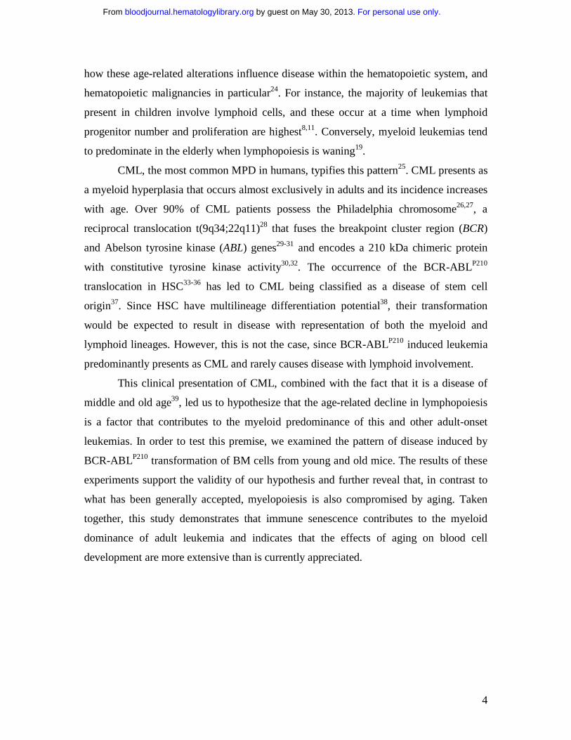
how these age-related alterations influence disease within the hematopoietic system, and
hematopoietic malignancies in particular24. For instance, the majority of leukemias that
present in children involve lymphoid cells, and these occur at a time when lymphoid
progenitor number and proliferation are highest8,11. Conversely, myeloid leukemias tend
to predominate in the elderly when lymphopoiesis is waning19.
CML, the most common MPD in humans, typifies this pattern25. CML presents as
a myeloid hyperplasia that occurs almost exclusively in adults and its incidence increases
with age. Over 90% of CML patients possess the Philadelphia chromosome26,27, a
reciprocal translocation t(9q34;22q11)28 that fuses the breakpoint cluster region (BCR)
and Abelson tyrosine kinase (ABL) genes29-31 and encodes a 210 kDa chimeric protein
with constitutive tyrosine kinase activity30,32. The occurrence of the BCR-ABLP210
translocation in HSC33-36 has led to CML being classified as a disease of stem cell
origin37. Since HSC have multilineage differentiation potential38, their transformation
would be expected to result in disease with representation of both the myeloid and
lymphoid lineages. However, this is not the case, since BCR-ABLP210 induced leukemia
predominantly presents as CML and rarely causes disease with lymphoid involvement.
This clinical presentation of CML, combined with the fact that it is a disease of
middle and old age39, led us to hypothesize that the age-related decline in lymphopoiesis
is a factor that contributes to the myeloid predominance of this and other adult-onset
leukemias. In order to test this premise, we examined the pattern of disease induced by
BCR-ABLP210 transformation of BM cells from young and old mice. The results of these
experiments support the validity of our hypothesis and further reveal that, in contrast to
what has been generally accepted, myelopoiesis is also compromised by aging. Taken
together, this study demonstrates that immune senescence contributes to the myeloid
dominance of adult leukemia and indicates that the effects of aging on blood cell
development are more extensive than is currently appreciated.
For personal use only. by guest on May 30, 2013. bloodjournal.hematologylibrary.orgFrom

5
Materials and Methods
Mice
Four to 7 week old C57BL/6J (B6) and B6.129S7-Rag1tm1Mom/J (Rag1-/-) mice were
purchased from The Jackson Laboratory, and B6.SJL mice were obtained from Taconic
Farms. Ten to 24 month old B6 mice were purchased from the National Institute on
Aging colony. Animals were housed in the vivarium of the University of California at
Los Angeles Division of Laboratory Animal Medicine. Experiments were conducted
according to the guidelines of the UCLA Institutional Animal Care and Use Committee.
B6, B6.SJL, and Rag1-/- recipients were preconditioned with 500 R 12-30 hours
before intravenous injection of transformed cells from a 137Cs irradiator (120 R/min;
Mark I-68A; JL Shepperd and Associates).
Generation of retroviral stocks
The retroviral vector pMSCV40 containing a 5' LTR-driven BCR-ABL internal ribosome
entry site (IRES) enhanced GFP (EGFP) or a 5' LTR-driven IRES EGFP was used to
generate high-titer helper-free retrovirus following transient cotransfection of 293T cells.
293T cells were grown on Poly-L-Lysine (Sigma) coated 10 cm tissue culture treated
plates (Becton Dickinson) in Iscove’s modified Dulbecco’s minimum essential medium
(IMDM, Mediatech) supplemented with 10% fetal calf serum (FCS, Hyclone), 1mM L-
glutamine, 100 U/ml streptomycin, and 100 µg/ml penicillin (complete IMDM; all from
Gibco). Transfections were done by co-precipitating 15 µg of retroviral vector with 15 µg
of an ecotropic packaging vector41 with the CalPhos Mammalian Transfection Kit (BD
Biosciences). Medium was replaced every 12 hours for 3 days with complete IMDM.
Viral stocks were prepared by pooling supernatants collected at 36, 48, and 60 hours
post-transfection. Viral titers were determined following infection of 3T3 cells with serial
dilutions of the pooled virus supernatant, and found to range between 2 x 10⁶ and 7 x 10⁶
virus particles/ml.
Retroviral transduction and bone marrow transplantation
Young mice were administered a single intravenous dose of 5-Fluorouracil (5-FU, 150
mg/kg body weight; Sigma). Due to their higher susceptibility to 5-FU, middle-age and
For personal use only. by guest on May 30, 2013. bloodjournal.hematologylibrary.orgFrom

6
old mice were administered a dose of 115 mg/kg body weight. On the eighth day after 5-
FU administration, BM cell suspensions were prepared as described42. Cells were
distributed in 5 ml polystyrene tubes (Becton Dickinson) and incubated with 1 ml of
retrovirus supplemented with 10% horse serum (Hyclone), 1mM L-glutamine, 100 U/ml
streptomycin, and 100 µg/ml penicillin, 8 µg/ml polybrene (Sigma), 50 µM β-
mercaptoethanol (Sigma), 25 mM HEPES (Gibco), 100 ng/ml SCF (Biosource), 100
ng/ml Flt-3L (R&D), and 10 ng/ml IL-11 (R&D) for 2 hours at 37 ºC in 5% CO2 and air
and constant humidity. Cells were centrifuged for 5 minutes at 400g, and viral
supernatant was replaced with 1 ml of virus stock supplemented as described above. This
procedure was repeated twice. After 6 hours, cells were washed, counted, and
resuspended in PBS. Purified pro/pre-B cells were infected in a modified progenitor cell
culture system42. One million cells were seeded in 6 well plates in RPMI 1640 (Gibco)
supplemented with 10% FCS, 1mM L-glutamine, 100 U/ml streptomycin, 100 µg/ml
penicillin, 8 µg/ml polybrene, 50 µM β-mercaptoethanol, 50 µg/ml gentamycin (Sigma),
20 ng/ml SCF, 20 ng/ml IL-3, 20 ng/ml IL-6 (Biosource), 10 ng/ml Flt-3L, and 10 ng/ml
IL-7. 0.4-µm transwell inserts in which confluent S17 stromal layers had been pre-
established were inserted into each well. Retrovirus was added to the bottom well 4 times
over a 24 hour period, at the end of which cells were washed, counted, and resuspended
in PBS. Five to 7 week old irradiated B6, B6.SJL, or Rag1-/- mice received an intravenous
injection of 2 x 10⁵ transduced BM cells or 1 x 10⁵ pro/pre B cells per mouse. For
secondary transplants 5 x 10⁶ splenocytes from primary diseased mice were injected
intravenously into irradiated 5-7 week old B6 recipients.
Immunophenotypic analysis of leukemic cells and cell sorting
BM and spleen cell suspensions were prepared as previously described42. Cell
suspensions were incubated with anti-CD16/32 (FcγRII-III; clone 2.4G2; eBiosciences)
or total mouse IgG (for CMP and GMP stains only; Axell) to reduce nonspecific labeling.
Cells were incubated with combinations of antibodies to the following cell surface
determinants, conjugated to fluoro-isothiocyanate, phycoerythrin, tricolor,
indodicarbocyanine, biotin, or allophycocyanin: CD3ε (clone KT31.1), CD4 (clone
GK1.5), CD8α (clone 53-6.7), CD11b (clone M1/70), CD16/32 (Fc RII/III; clone 2.4G2),
For personal use only. by guest on May 30, 2013. bloodjournal.hematologylibrary.orgFrom

7
CD19 (clone 1D3), CD45.1 (clone A20), CD45.2 (clone 104), CD45R (B220, clone
RA3-6B2), CD117 (c-Kit, clone 2B8), CD127 (IL-7Rα, clone A7R34), Ter119 (clone
Ter-119), TCRβ (clone H57-597), TCRγδ (clone UC7-13D5), NK1.1 (clone PK136), Ly-
6C (clone AL-21), AA4.1 (clone C1qRp), Sca-1 (clone E13-161.7), IgM (clone II/41),
and Gr-1 (clone RB6-8C5). Biotinylated cells were visualized by incubation with
tricolor-, allophycocyanin-, or allophycocyanin-Alexa Fluor 750- conjugated streptavidin.
All reagents were obtained from Becton Dickinson or eBiosciences except for goat anti–
mouse IgM (Southern Biotech), tricolor-conjugated CD11b and allophycocyanin- Alexa
Fluor 750– conjugated streptavidin (Caltag Laboratories). All incubations were for
approximately 30 minutes at 4°C. After the last wash, live cells were acquired with Cell
Quest Software (Becton Dickinson) on a FACScan, FACSCalibur or Cytek modified
FACSscan (all BD Biosciences) located at the Flow Cytometry Core of Jonsson
Comprehensive Cancer Center at the University of California at Los Angeles.
Disease phenotype was established by determining the frequencies of BM or
spleen cells co-expressing GFP (BCR-ABLP210) and lineage specific cell surface antigens
(Myeloid: CD11b+Gr-1+, B lymphoid: CD45R+CD19+, Erythroid: Ter119+, T-Lymphoid:
CD4+ and/or CD8+). Disease was considered present when at least 7.5% of the total GFP+
cells in either the BM or spleen co-expressed the specific cell surface antigens.
Populations enriched for CMP and GMP3 were defined as Lin– (Lin = CD3ε,
CD8α, CD45R, Gr-1, TER-119, TCRαβ, TCRγδ, and NK1.1) Sca-1–CD127–
CD16/32+/LoCD117Hi and Lin–Sca-1–CD127–CD16/32HiCD117Hi, respectively.
Populations enriched for HSC38 were defined as Lin– (Lin = CD3ε, CD8α, CD11b,
CD45R, Gr-1, TER-119, TCRαβ, TCRγδ, and NK1.1) Sca-1HiCD117Hi. Pre-pro B cells
and pro/pre-B cells43 were defined as Lin– (Lin = CD3ε, CD4, CD8α, CD11b, Gr-1, TER-
119, Ly-6C, IgM, TCRαβ, TCRγδ, and NK1.1) CD45R+CD19-AA4.1+ and Lin–
CD45R+CD19+AA4.1+, respectively.
Cells to be purified were resuspended in minimum essential medium-α (αMEM;
Gibco) supplemented with 2% FCS, 25 mM Hepes, 1mM L-glutamine, 100 U/ml
streptomycin, 100 µg/ml penicillin, and 50 µg/ml gentamycin before being sorted on a
FACSaria (Becton Dickinson). Sorted fractions were examined by reanalysis, and were
routinely 95% pure.
For personal use only. by guest on May 30, 2013. bloodjournal.hematologylibrary.orgFrom

8
B cell progenitor cultures
BM cells from day 8 5-FU treated mice or purified pro/pre-B cells were retrovirally
transduced as described above. 3 x 10⁵ BM cells or 3.8 x 10⁵ GFP+ pro/pre-B cells (which
had been expanded for one week in the modified progenitor assay described above) were
seeded onto S17 stromal cells44 in T12.5 flasks (Becton Dickinson) in RPMI 1640
supplemented with 5% FCS, 50 µM β-mercaptoethanol, 1 mM L-glutamine, 100 U/ml
streptomycin, and 100 µg/ml penicillin45. Cultures were incubated at 37 ºC in 5% CO2
and air and constant humidity, and fed twice weekly for up to three weeks.
Myeloid cultures
Myeloid colony assays were performed by resuspending target cells in 1 ml of
methylcellulose supplemented with 30% FCS, 40% αMEM, 50 µM β-mercaptoethanol, 1
mM L-glutamine, 100 U/ml streptomycin, 100 µg/ml penicillin, 50 µg/ml gentamycin, 20
ng/ml SCF, 10 ng/ml GM-CSF (Biosource), 30 ng/ml IL-3, and 10 ng/ml IL-11 in 3.5 cm
petri dishes (Becton Dickinson) in triplicate3. Colonies were counted on day 8. Individual
colonies were picked under a dissecting microscope, resuspended in 300 µl of PBS, and
analyzed for GFP expression on a FACScan for 30 seconds at a fixed flow rate of 60
µl/min. Live cells were gated, and total cell numbers were calculated as [total live
events/(flow rate x acquisition time)] x sample volume.
Liquid suspension cultures were initiated by seeding 1.5 x 10⁵ whole BM cells or
2 x 10³ sorted CMP in complete IMDM supplemented with 50 µM β-mercaptoethanol, 50
µg/ml gentamycin, 20 ng/ml SCF, 10 ng/ml GM-CSF, 30 ng/ml IL-3, 30 ng/ml IL-6, and
10 ng/ml IL-11 at 37 ºC in 5% CO2 and air and constant humidity. Cultures were fed on
day 4, and on day 6 cells were harvested and assessed by immunostaining with Gr-1 and
CD11b as described above.
Statistical analysis
Unless indicated otherwise, data are expressed as a mean ± SEM. Differences between
groups were tested by a two-tailed, unpaired t-test, with an α of 0.05.
For personal use only. by guest on May 30, 2013. bloodjournal.hematologylibrary.orgFrom

9
Results
Hematopoietic cell age alters BCR-ABLP210 induced leukemia phenotype
To assess the impact of aging on BCR-ABLP210 induced leukemogenesis46, BM cells
harvested from day 8 5-FU treated young (5-7 weeks) or old (90-104 weeks) mice were
infected with a retrovirus carrying a bicistronic IRES expression vector encoding BCR-
ABLP210 and an EGFP reporter gene and subsequently injected into sublethally irradiated
syngeneic young mice (BMBCR-ABL recipients; Figure 1A). Control animals were similarly
transplanted with BM cells infected with a retrovirus containing EGFP alone (BMEGFP
recipients). The use of congenic CD45.1+ recipients in one experiment confirmed that
GFP+ (BCR-ABL+) leukemic cells co-expressed the donor derived CD45.2 cell surface
antigen (data not shown).
Recipients of both young and old BMBCR-ABL cells exhibited weight loss,
cachexia, and poor grooming and were sacrificed between 2 to 7 weeks post-
transplantation. These animals had decreased BM and increased splenic cellularity
compared to BMEGFP recipients (Figure 1B and 1C). Hematopoietic infiltration of the
liver and lungs with occasional lymphadenopathy was evident at necropsy (data not
shown). In contrast, all BMEGFP recipients were observed for up to four months post-
transplantation and remained healthy.
Disease patterns in BMBCR-ABL recipients were assessed by phenotypic and
morphologic characterization of hematopoietic cells in their BM and spleen (Figure 1D
and Table 1). Ninety percent (26/29) of young and 100% (24/24) of old BMBCR-ABL
recipients developed MPDs characterized by the expansion of GFP+Gr-1+CD11b+
granulocytes in the BM and spleen that was frequently accompanied by the presence of
GFP+Ter119+ erythroid cells (Figure 1D and 1E). Similar to the situation in humans, 96%
(23/24) of old BMBCR-ABL recipients lacked significant B lineage involvement in their
disease, as determined by the minimal frequency of GFP+CD45R+CD19+ B lineage cells
and absence of lymphoid blasts in their BM and spleen. In stark contrast, 35% (9/26) of
young BMBCR-ABL recipients that developed MPDs concurrently developed B lymphoid
leukemia. In addition, 10% (3/29) of young BMBCR-ABL recipients presented with B
lymphoid leukemia without significant myeloid involvement, a disease profile that was
never observed in any old BMBCR-ABL recipients.
For personal use only. by guest on May 30, 2013. bloodjournal.hematologylibrary.orgFrom
10
Overall, 41% (12/29) of young BMBCR-ABL recipients developed B lymphoid
leukemia compared with 4% (1/24) of old BMBCR-ABL recipients (Figure 1E). These
differences were not the result of decreased engraftment of old cells18, because
engraftment of GFP+ cells was comparable in the BM of recipients of young and old
BMEGFP (Table 1), all recipients of old BMBCR-ABL developed myeloid disease, and
increasing the number of transplanted old BMBCR-ABL cells did not increase the incidence
of lymphoid disease in the recipients (data not shown). Therefore, these data strongly
indicate that BCR-ABLP210 transduced old hematopoietic cells lack significant potential
to initiate B lymphoid leukemia.
This conclusion was confirmed by transplanting BCR-ABLP210 transduced young
and old BM cells into Rag1-/- recipient mice, whose lack of lymphocytes creates a more
favorable environment for normal and dysplastic lymphoid development47-49. All BMBCR-
ABL Rag1-/- recipients developed MPDs, and recipients of young BMBCR-ABL also
developed B lymphoid leukemia with an incidence markedly higher (63%; 5/8) than
observed in wild type mice. In contrast, none (0/7) of the old BMBCR-ABL Rag1-/-
recipients showed any significant B lineage component to their disease (Figure 1F).
The incidence of B lymphoid leukemia correlates with age-related declines in B
lymphopoiesis
The decline in B lymphopoiesis does not abruptly initiate in old age, but instead begins in
relatively young animals and progresses gradually thereafter8. For example, middle-age
mice (42-44 weeks) have approximately half the number of B lineage cells as young mice
(Figure 2A and 2B). Similarly, the frequency of pre-pro-B, pro-B, and pre-B cells is
progressively reduced in the BM of 5-FU treated mice of increasing age, demonstrating a
reduced capacity to generate B lineage cells de novo in old mice11.
If the age-related decline in B cell production underlies the reduced capacity of
BMBCR-ABL cells to initiate B lymphoid leukemia, then the incidence of BCR-ABLP210
induced lymphoid disease should decline gradually with increasing age. This hypothesis
was tested by transplanting BCR-ABLP210 transduced BM cells from young, middle-age,
and old mice into Rag1-/- recipients. As shown in Figure 2C, the incidence of B lymphoid
leukemia is highest amongst young BMBCR-ABL recipients (63%; 5/8), intermediate in
For personal use only. by guest on May 30, 2013. bloodjournal.hematologylibrary.orgFrom

11
middle-age BMBCR-ABL recipients (25%; 2/8), and nil in recipients of old BMBCR-ABL (0%;
0/7).
Cell intrinsic defects diminish the leukemogenic potential of aged B lineage cells
In addition to its emergence from HSC36, B lymphoid leukemia can also initiate from
direct transformation of committed lymphoid progenitors50-52. This raised the possibility
that the reduced incidence of B lymphoid leukemia from aged BM cells observed in the
above study resulted from the lower number of lymphoid progenitors available for
transformation (Figure 2B). However, this was not the case. When an equivalent number
of BCR-ABLP210 transduced pro/pre-B cells from young and old mice were injected into
sublethally irradiated Rag1-/- recipients, 25% of recipients of young BCR-ABLP210
transduced B lineage cells developed B lymphoid leukemia 8 weeks later, while none of
the recipients of old BCR-ABLP210 transduced pro/pre-B cells developed disease (Figure
3A).
Further in vitro analysis showed that young and old BCR-ABLP210 transduced 5-
FU BM differed significantly in the potential to establish long-term B lineage cultures.
Following 3 weeks in B lymphoid permissive conditions, recovery of GFP+ B linage cells
in cultures initiated with young BMBCR-ABL was 100 times higher than in cultures
established with young BMEGFP cells (Figure 3B). In contrast, BCR-ABLP210 transformed
old BM cells expanded only 6 fold when compared to old BMEGFP cells (Figure 3C).
Morphologic and phenotypic analyses confirmed these observations (Figure 3D). As in
the in vivo experiments, this was not due to differences in numbers of B lineage
progenitors present in young and old BM. When an equivalent number of young and old
BCR-ABLP210 transduced pro/pre-B cells were cultured in B lymphoid permissive
conditions the young BCR-ABLP210 transduced pro/pre-B cells had expanded
approximately 10 fold after 5 days, while no increase in cell number was observed in the
cultures established with old B lineage cells (Figure 3E).
Taken together, these observations indicate that BCR-ABLP210 expression cannot
overcome the proliferative and differentiative defects that have accumuatled in aged B
lymphoid precursors, and these defects underlie their reduced leukemogenic potential.
For personal use only. by guest on May 30, 2013. bloodjournal.hematologylibrary.orgFrom

12
Decreased severity of MPDs derived from old BMBCR-ABL
Compared to recipients of old BMBCR-ABL cells, young BMBCR-ABL recipients consistently
presented with increased wasting and a more pronounced invasion of organs such as liver
and lung with leukemic cells (data not shown). Although young and old BMBCR-ABL
recipients had comparable numbers of cells in their BM and spleen (Figure 1B and 1C),
the proportion of cells that were GFP+ was higher in young BMBCR-ABL recipients. For
example, the proportion of GFP+ cells in the spleen of young and old BMBCR-ABL
recipients was 62% and 52%, respectively and 47% and 29% in the BM (P<0.002),
respectively. Since this increased tumor burden in young BMBCR-ABL recipients could
have reflected the addition of B lymphoid leukemia to their MPDs, we compared young
and old BMBCR-ABL recipients that developed MPDs without B lineage involvement.
Surprisingly, the difference in tumor burden was accentuated in these animals due to a
higher frequency of GFP+Gr-1+CD11b+ cells present in their BM and spleen (Figure 4A
and 4B). This also resulted in tissue disruption as demonstrated by an enhanced
displacement of endogenous B lineage cells in the spleen (Figure 4C).
The increase in leukemic myeloid cells in young BMBCR-ABL recipients was
accompanied by an increased frequency and number of GFP+ HSC, CMP and GMP in the
BM and spleen (Figure 4D and 4E), and splenocytes from young BMBCR-ABL recipients
formed more GFP+ colonies than splenocytes from old BMBCR-ABL recipients when tested
in myeloid colony assays (data not shown).
This discrepancy in tumor burden did not result from an increased transduction
efficiency of young BM cells. Following transduction, young and old BM cells were used
to establish myeloid colonies in two independent experiments. Eight days later, 12
colonies derived from young and old BMEGFP were examined for GFP expression by flow
cytometry. The number of young and old derived colonies that contained GFP expressing
cells (8/12) was the same.
Identification of myelopoietic defects in old mice
The above data were surprising because the frequency and absolute number of HSC and
myeloid progenitors is increased in the BM of old mice (Figure 5A)11,19. Therefore, we
considered the possibility that aging affected the quality of old myeloid progenitors.
For personal use only. by guest on May 30, 2013. bloodjournal.hematologylibrary.orgFrom

13
Further analysis revealed that while old BM cells formed 1.3 fold more colonies than
young BM cells, this value was less than would be predicted from the 2.3 fold increase in
myeloid progenitor frequency observed by flow cytometry. This could be due in part to a
2-3 fold increase in the number of apoptotic Annexin V+ CMP and GMP in old as
compared to young BM (Figure 5B). In addition, individual colonies derived from old
BM cells contained approximately 50% fewer cells than those derived from young BM
cells (Figure 5C), and old CMP similarly generated smaller colonies than their
counterparts from young mice (Figure 5D).
This age-related reduction in myelopoietic potential was also observed when
whole BM cells or sorted CMP isolated from young and old mice were grown in liquid
culture supplemented with myelopoietic cytokines. Consistent with results from the
colony assays, BM cells (Figure 5E) and CMP (Figure 5F) from old mice produced
approximately 45% less Gr-1+CD11b+ myeloid cells than those isolated from young
animals. Taken together, these results demonstrate that aged myeloid progenitors harbor
intrinsic proliferative and/or differentiative defects.
Age-related hematopoietic defects alter the potential of leukemia stem cells (LSC)
Hematopoietic stem and progenitor cells play a critical role in the pathogenesis of CML.
HSC have been implicated as the leukemic cell of origin, while committed myeloid and
lymphoid progenitors have been deemed the LSC in more advanced stages of
disease36,37,53. Consequently, we assessed whether age-related differences in the
pathogenesis of BCR-ABLP210 induced leukemia was a reflection of changes intrinsic to
LSC. Since LSC are defined by their potential to transplant disease54,55, splenocytes
isolated from diseased mice grafted with young or old BMBCR-ABL were transplanted into
sublethally irradiated syngeneic mice. Disease patterns and progression were then
analyzed in these secondary recipients.
Secondary recipients developed disease symptoms within 3 weeks post-
transplantation and were sacrificed. All these mice (27/27) presented with MPDs.
However, 60% (9/15) of mice grafted with splenocytes from young BMBCR-ABL recipients
also presented with B lymphoid leukemia (Figure 6A). In contrast, none (0/12) of the
mice grafted with splenocytes from old BMBCR-ABL recipients developed lymphoid
For personal use only. by guest on May 30, 2013. bloodjournal.hematologylibrary.orgFrom

14
disease (Figure 6B). These data suggest that either no B lymphoid LSC were produced
following BCR-ABLP210 transduction of old hematopoietic cells, or that if they were
produced, they have intrinsic defects that disrupt their leukemia initiating potential.
In addition, we compared secondary recipients that developed MPDs without B
lineage involvement for disease severity. Leukemic burden was 3 fold greater in
secondary recipients transplanted with young BMBCR-ABL derived tumors when compared
with secondary recipients of old BMBCR-ABL derived tumors (Figure 6C and 6D). These
data demonstrate that myeloid LSC derived from young BMBCR-ABL have a greater
expansive potential through either enhanced self-renewal and/or increased production of
mature leukemic myeloid progeny. Taken together, these data indicate that age-related
intrinsic hematopoietic defects ultimately alter the leukemogenic potential of LSC
generated following transformation of hematopoietic cells by BCR-ABLP210.
Discussion
The present report demonstrates that age-related intrinsic defects that accumulate in B
lineage cells limit lymphoid involvement in CML. This finding provides a biological
explanation for the myeloid predominance of adult-onset leukemia. Although this study
focused on B cell development, T cell production also declines with increasing age21,
which may explain the extreme rarity with which T cell leukemia presents in older
humans.
In the murine CML model used in this study, transformation of young BM cells
with BCR-ABLP210 consistently resulted in myeloid and lymphoid disease while
transformation of old BM cells primarily resulted in a MPD with no lymphoid
involvement. Since the 5-FU BM used in these experiments contains HSC as well as B
lineage specified progenitors, B lymphoid leukemia could have developed from
transformation of either population. However, the origin of the leukemia does not
influence the interpretation of our results, which demonstrate that age-related intrinsic
defects that accumulate in B lineage cells limit their involvement in CML. This
conclusion is supported by the observation that BCR-ABLP210 transduced old B lineage
precursors lack lymphoid leukemia initiating potential. Furthermore, expression of a
powerful oncogene such as BCR-ABLP210, which greatly enhanced the growth of young
For personal use only. by guest on May 30, 2013. bloodjournal.hematologylibrary.orgFrom

15
B lineage cells, was unable to significantly augment the growth of old B cell progenitors
in vitro. These data, combined with intrinsic age-related defects in the ability of HSC to
generate lymphoid progeny19, can explain the clinical presentation of CML. Inefficient
production of early lymphoid progenitors from older HSC, in combination with intrinsic
proliferative and developmental defects in the progenitors that are generated, would
contribute to a low incidence of lymphoid leukemia and a predomincance of myeloid
disease in the aged. In contrast, young HSC efficiently generate lymphoid progeny,
which in turn are highly susceptible to the effects of BCR-ABLP210, thereby resulting in
MPD and B lymphoid leukemia56.
Our finding that B lymphoid leukemia initiated more frequently with young than
old BMBCR-ABL is consistent with the clinical observation that the incidence of B lineage
leukemia is highest in children. That children are predisposed to lymphoid leukemia is
consistent with results from murine studies demonstrating that B lineage progenitors from
neonates and young adults cycle at levels above their aged counterparts11. While this
increased proliferation may be necessary to fill peripheral lymphoid compartments, it
could increase the chance that an aberrant genetic event could occur, particularly since
developing B lineage cells possess active gene rearrangement machinery57. This
possibility, combined with the presence of chromosomal translocations in neonatal B
lineage progenitors that cause a predisposition to leukemia development58, may increase
the likelihood of transformation during early B lymphopoiesis.
The observation that MPDs initiated from young BMBCR-ABL cells were more
aggressive than those initiated from old BMBCR-ABL cells was quite unexpected. This
pattern led to the discovery that defects do in fact accumulate in aged myeloid
progenitors. Thus, it is reasonable to propose that the milder MPDs are the result of age-
related defects in growth and survival of myeloid progenitors and/or their progeny. It is
surprising that age-related changes in myeloid progenitors were not previously reported.
However, previous studies primarily examined myeloid progenitors as a population rather
than on a per-cell basis, and as a result, the cell intrinsic defects defined herein had been
overlooked. The age-related defects that accumulate in myeloid progenitors are similar to
the those exhibited by aged HSC, including increased frequency and cycling, decreased
survival, and diminished per-cell repopulating potential22. These observations suggest
For personal use only. by guest on May 30, 2013. bloodjournal.hematologylibrary.orgFrom

16
that age-related defects intrinsic to myeloid progenitors may be the result of intrinsic
changes in old HSC. However, whether old HSC and CMP share common molecular
changes59 remains to be determined.
While the detrimental effects of advancing age and decreased lymphocyte
production on the adaptive immune response have been well documented23, the
consequences of aging on the innate immune system remain largely unexplored60. The
innate immune system is a critical first responder to infection. Despite defects in old
myeloid progenitors, their increased number may compensate for the fact that they do not
produce progeny as efficiently as their young counterparts, which results in a relatively
normal number of mature myeloid cells. However, reports of diminished function of aged
neutrophils61, macrophages62, and dendritic cells63 indicates that the innate immune
response may become significantly compromised with age. Consequently, the unexpected
observation that myeloid progenitors from old mice are intrinsically defective suggests
that age-related defects may contribute the overall reduction in myeloid cell function in
the elderly.
While this study used CML as a model with which to investigate the larger
question of how aging affects leukemia development, the data nevertheless provide new
insights into this disease. First, the demonstration that the degree of lymphoid disease
mediated by BCR-ABLP210 is related to overall levels of B lymphopoiesis provides an
explanation for the paradoxical observation that while CML is considered a stem cell
disease, it presents as a MPD with relatively rare lymphoid involvement. Second, age-
related defects in myeloid progenitors may explain in part why human CML presents
with a chronic rather than an acute course64. Finally, we demonstrate that patterns of
lymphoid and myeloid disease exhibited in primary recipients of young and old BMBCR-
ABL are conserved following transplantation of leukemic cells into secondary recipients.
This finding suggests an intrinsic role for senescence in governing the behavior of LSC
and demonstrates the impact of aging on disease development.
Acknowledgements
This work was supported by grants from the National Institutes of Health (AG-21459)
and the U.S. Department of Defense (W81XWHO410795). O.N.W is an investigator of
For personal use only. by guest on May 30, 2013. bloodjournal.hematologylibrary.orgFrom

17
the Howard Hughes Medical Institute. R.A.J.S. is supported by a fellowship from the
California Institute for Regenerative Medicine (TI-00005). The UCLA Flow Cytometry
Core Facility is supported by grants from the National Institutes of Health (CA-16042,
AI-28697).
Author Contributions
The study was designed by R.A.J.S., E.M.-R., O.N.W. and K.D.; experiments were
performed and data was analyzed by R.A.J.S. with assistance from E.M.-R.; critical
reagents were provided by J.M.; the manuscript was written by R.A.J.S., E.M.-R., and
K.D.
For personal use only. by guest on May 30, 2013. bloodjournal.hematologylibrary.orgFrom

18
References 1. Shizuru JA, Negrin RS, Weissman IL. Hematopoietic stem and progenitor cells: clinical and preclinical regeneration of the hematolymphoid system. Annu Rev Med. 2005;56:509-538. 2. Till JE, McCulloch EA. A direct measurement of the radiation sensitivity of normal mouse bone marrow cells. Radiat Res. 1961;14:213-222. 3. Akashi K, Traver D, Miyamoto T, Weissman IL. A clonogenic common myeloid progenitor that gives rise to all myeloid lineages. Nature. 2000;404:193-197. 4. Igarashi H, Gregory SC, Yokota T, Sakaguchi N, Kincade PW. Transcription from the RAG1 locus marks the earliest lymphocyte progenitors in bone marrow. Immunity. 2002;17:117-130. 5. Kondo M, Weissman IL, Akashi K. Identification of clonogenic common lymphoid progenitors in mouse bone marrow. Cell. 1997;91:661-672. 6. Hardy RR, Carmack CE, Shinton SA, Kemp JD, Hayakawa K. Resolution and characterization of pro-B and pre-pro-B cell stages in normal mouse bone marrow. J Exp Med. 1991;173:1213-1225. 7. Hirose J, Kouro T, Igarashi H, Yokota T, Sakaguchi N, Kincade PW. A developing picture of lymphopoiesis in bone marrow. Immunol Rev. 2002;189:28-40. 8. Montecino-Rodriguez E, Dorshkind K. Evolving patterns of lymphopoiesis from embryogenesis through senescence. Immunity. 2006;24:659-662. 9. Allman D, Miller JP. The aging of early B-cell precursors. Immunol Rev. 2005;205:18-29. 10. Miller JP, Allman D. The decline in B lymphopoiesis in aged mice reflects loss of very early B-lineage precursors. J Immunol. 2003;171:2326-2330. 11. Min H, Montecino-Rodriguez E, Dorshkind K. Effects of aging on the common lymphoid progenitor to pro-B cell transition. J Immunol. 2006;176:1007-1012. 12. Stephan RP, Sanders VM, Witte PL. Stage-specific alterations in murine B lymphopoiesis with age. Int Immunol. 1996;8:509-518. 13. Riley RL, Van der Put E, King AM, Frasca D, Blomberg BB. Deficient B lymphopoiesis in murine senescence: potential roles for dysregulation of E2A, Pax-5, and STAT5. Semin Immunol. 2005;17:330-336. 14. Cambier J. Immunosenescence: a problem of lymphopoiesis, homeostasis, microenvironment, and signaling. Immunol Rev. 2005;205:5-6. 15. Chen J, Astle CM, Harrison DE. Development and aging of primitive hematopoietic stem cells in BALB/cBy mice. Experimental Hematology. 1999;27:928-935. 16. Ogden DA, Mickliem HS. The fate of serially transplanted bone marrow cell populations from young and old donors. Transplantation. 1976;22:287-293. 17. Morrison SJ, Wandycz AM, Akashi K, Globerson A, Weissman IL. The aging of hematopoietic stem cells. Nat Med. 1996;2:1011-1016. 18. Liang Y, Van Zant G, Szilvassy SJ. Effects of aging on the homing and engraftment of murine hematopoietic stem and progenitor cells. Blood. 2005;106:1479-1487. 19. Rossi DJ, Bryder D, Zahn JM, et al. Cell intrinsic alterations underlie hematopoietic stem cell aging. Proc Natl Acad Sci U S A. 2005;102:9194-9199.
For personal use only. by guest on May 30, 2013. bloodjournal.hematologylibrary.orgFrom

19
20. Sudo K, Ema H, Morita Y, Nakauchi H. Age-associated Characteristics of Murine Hematopoietic Stem Cells. J Exp Med. 2000;192:1273-1280. 21. Min H, Montecino-Rodriguez E, Dorshkind K. Effects of aging on early B- and T-cell development. Immunological Reviews. 2005;205:7-17. 22. Geiger H, Van Zant G. The aging of lympho-hematopoietic stem cells. Nat Immunol. 2002;3:329-333. 23. Linton PJ, Dorshkind K. Age-related changes in lymphocyte development and function. Nat Immunol. 2004;5:133-139. 24. Signer RAJ, Montecino-Rodriguez E, Dorshkind K. Aging, B lymphopoiesis, and patterns of leukemogenesis. Experimental Gerontology. 2007;42:391-395. 25. Passegue E. Hematopoietic stem cells, leukemic stem cells and chronic myelogenous leukemia. Cell Cycle. 2005;4:266-268. 26. Shepherd P, Suffolk R, Halsey J, Allan N. Analysis of molecular breakpoint and m-RNA transcripts in a prospective randomized trial of interferon in chronic myeloid leukaemia: no correlation with clinical features, cytogenetic response, duration of chronic phase, or survival. Br J Haematol. 1995;89:546-554. 27. Nowell PC, Hungerford DA. A minute chromosome in human chronic granulocytic leukemia. Science. 1960;132:1497. 28. Rowley JD. Letter: A new consistent chromosomal abnormality in chronic myelogenous leukaemia identified by quinacrine fluorescence and Giemsa staining. Nature. 1973;243:290-293. 29. Shtivelman E, Lifshitz B, Gale RP, Canaani E. Fused transcript of abl and bcr genes in chronic myelogenous leukaemia. Nature. 1985;315:550-554. 30. Ben-Neriah Y, Daley GQ, Mes-Masson AM, Witte ON, Baltimore D. The chronic myelogenous leukemia-specific P210 protein is the product of the bcr/abl hybrid gene. Science. 1986;233:212-214. 31. Stam K, Heisterkamp N, Grosveld G, et al. Evidence of a new chimeric bcr/c-abl mRNA in patients with chronic myelocytic leukemia and the Philadelphia chromosome. N Engl J Med. 1985;313:1429-1433. 32. Wong S, Witte ON. The BCR-ABL story: bench to bedside and back. Annu Rev Immunol. 2004;22:247-306. 33. Fialkow PJ, Gartler SM, Yoshida A. Clonal origin of chronic myelocytic leukemia in man. Proc Natl Acad Sci U S A. 1967;58:1468-1471. 34. Fialkow PJ, Jacobson RJ, Papayannopoulou T. Chronic myelocytic leukemia: clonal origin in a stem cell common to the granulocyte, erythrocyte, platelet and monocyte/macrophage. Am J Med. 1977;63:125-130. 35. Takahashi N, Miura I, Saitoh K, Miura AB. Lineage involvement of stem cells bearing the philadelphia chromosome in chronic myeloid leukemia in the chronic phase as shown by a combination of fluorescence-activated cell sorting and fluorescence in situ hybridization. Blood. 1998;92:4758-4763. 36. Castor A, Nilsson L, Astrand-Grundstrom I, et al. Distinct patterns of hematopoietic stem cell involvement in acute lymphoblastic leukemia. Nat Med. 2005;11:630-637. 37. Kabarowski JH, Witte ON. Consequences of BCR-ABL expression within the hematopoietic stem cell in chronic myeloid leukemia. Stem Cells. 2000;18:399-408.
For personal use only. by guest on May 30, 2013. bloodjournal.hematologylibrary.orgFrom
20
38. Spangrude GJ, Heimfeld S, Weissman IL. Purification and characterization of mouse hematopoietic stem cells. Science. 1988;241:58-62. 39. Michor F, Iwasa Y, Nowak MA. The age incidence of chronic myeloid leukemia can be explained by a one-mutation model. PNAS. 2006;103:14931-14934. 40. Hawley RG, Lieu FH, Fong AZ, Hawley TS. Versatile retroviral vectors for potential use in gene therapy. Gene Ther. 1994;1:136-138. 41. Muller AJ, Young JC, Pendergast AM, et al. BCR first exon sequences specifically activate the BCR/ABL tyrosine kinase oncogene of Philadelphia chromosome-positive human leukemias. Mol Cell Biol. 1991;11:1785-1792. 42. Montecino-Rodriguez E, Leathers H, Dorshkind K. Identification of a B-1 B cell-specified progenitor. Nat Immunol. 2006;7:293-301. 43. Allman D, Li J, Hardy RR. Commitment to the B lymphoid lineage occurs before DH-JH recombination. J Exp Med. 1999;189:735-740. 44. Collins LS, Dorshkind K. A stromal cell line from myeloid long-term bone marrow cultures can support myelopoiesis and B lymphopoiesis. J Immunol. 1987;138:1082-1087. 45. Whitlock CA, Witte ON. Long-term culture of B lymphocytes and their precursors from murine bone marrow. Proc Natl Acad Sci U S A. 1982;79:3608-3612. 46. Wong S, Witte ON. Modeling Philadelphia chromosome positive leukemias. Oncogene. 2001;20:5644-5659. 47. Mombaerts P, Iacomini J, Johnson RS, Herrup K, Tonegawa S, Papaioannou VE. RAG-1-deficient mice have no mature B and T lymphocytes. Cell. 1992;68:869-877. 48. Bhattacharya D, Rossi DJ, Bryder D, Weissman IL. Purified hematopoietic stem cell engraftment of rare niches corrects severe lymphoid deficiencies without host conditioning. J Exp Med. 2006;203:73-85. 49. Lim YM, Wong S, Lau G, Witte ON, Colicelli J. BCR/ABL inhibition by an escort/phosphatase fusion protein. Proc Natl Acad Sci U S A. 2000;97:12233-12238. 50. Hu Y, Swerdlow S, Duffy TM, Weinmann R, Lee FY, Li S. Targeting multiple kinase pathways in leukemic progenitors and stem cells is essential for improved treatment of Ph+ leukemia in mice. PNAS. 2006:0606509103. 51. Krause DS, Lazarides K, von Andrian UH, Van Etten RA. Requirement for CD44 in homing and engraftment of BCR-ABL-expressing leukemic stem cells. Nat Med. 2006;12:1175-1180. 52. Li S, Ilaria RL, Jr., Million RP, Daley GQ, Van Etten RA. The P190, P210, and P230 forms of the BCR/ABL oncogene induce a similar chronic myeloid leukemia-like syndrome in mice but have different lymphoid leukemogenic activity. J Exp Med. 1999;189:1399-1412. 53. Jamieson CH, Ailles LE, Dylla SJ, et al. Granulocyte-macrophage progenitors as candidate leukemic stem cells in blast-crisis CML. N Engl J Med. 2004;351:657-667. 54. Bonnet D, Dick JE. Human acute myeloid leukemia is organized as a hierarchy that originates from a primitive hematopoietic cell. Nat Med. 1997;3:730-737. 55. Lapidot T, Sirard C, Vormoor J, et al. A cell initiating human acute myeloid leukaemia after transplantation into SCID mice. Nature. 1994;367:645-648. 56. Huntly BJ, Shigematsu H, Deguchi K, et al. MOZ-TIF2, but not BCR-ABL, confers properties of leukemic stem cells to committed murine hematopoietic progenitors. Cancer Cell. 2004;6:587-596.
For personal use only. by guest on May 30, 2013. bloodjournal.hematologylibrary.orgFrom

21
57. Jung D, Giallourakis C, Mostoslavsky R, Alt FW. Mechanism and control of V(D)J recombination at the immunoglobulin heavy chain locus. Annu Rev Immunol. 2006;24:541-570. 58. Greaves MF, Wiemels J. Origins of chromosome translocations in childhood leukaemia. Nat Rev Cancer. 2003;3:639-649. 59. Janzen V, Forkert R, Fleming HE, et al. Stem-cell ageing modified by the cyclin-dependent kinase inhibitor p16INK4a. Nature. 2006;443:421-426. 60. Plackett TP, Boehmer ED, Faunce DE, Kovacs EJ. Aging and innate immune cells. J Leukoc Biol. 2004;76:291-299. 61. Fu YK, Arkins S, Li YM, Dantzer R, Kelley KW. Reduction in superoxide anion secretion and bactericidal activity of neutrophils from aged rats: reversal by the combination of gamma interferon and growth hormone. Infect Immun. 1994;62:1-8. 62. Stout RD, Suttles J. Immunosenescence and macrophage functional plasticity: dysregulation of macrophage function by age-associated microenvironmental changes. Immunol Rev. 2005;205:60-71. 63. Komatsubara S, Cinader B, Muramatsu S. Polymorphism of age-related changes in stimulatory capacity of murine dendritic cells. Mech Ageing Dev. 1986;37:163-173. 64. Li S, Gillessen S, Tomasson MH, Dranoff G, Gilliland DG, Van Etten RA. Interleukin 3 and granulocyte-macrophage colony-stimulating factor are not required for induction of chronic myeloid leukemia-like myeloproliferative disease in mice by BCR/ABL. Blood. 2001;97:1442-1450.
For personal use only. by guest on May 30, 2013. bloodjournal.hematologylibrary.orgFrom

22
Figure 1. Age alters the phenotype of BCR-ABLP210 induced leukemia.
(A) BM from young or old B6 mice was harvested on day 8 following 5-FU treatment,
infected with a retrovirus carrying a bicistronic IRES expression vector encoding BCR-
ABLP210 and a reporter EGFP gene, and transplanted into sublethally irradiated, young
syngeneic or congenic recipients. Control recipients received BM transduced with EGFP
alone. Leukemias are accompanied by decreased BM (B) and increased splenic (C)
cellularity. Cell numbers represent mean values ± SEM obtained from 5 independent
experiments with 29 recipients of young BMBCR-ABL, 24 recipients of old BMBCR-ABL, 9
recipients of young BMEGFP, and 5 recipients of old BMEGFP. (D) Leukemic cells in the
BM were characterized by flow cytometry for expression of GFP (BCR-ABLP210) in
combination with lineage specific cell surface antigens. Examples of recipients with a
MPD (upper panels), B lymphoid leukemia (middle panels), and both a MPD and
lymphoid leukemia (lower panels) are shown. Recipients that developed a MPD had
increased granulocytes and decreased lymphocytes in the spleen (upper right panel),
while the ones that developed B lymphoid leukemia had increased lymphoid cells and
blasts in the spleen (middle right panel) compared to controls. Cytospin preparations of
spleen cells were visualized following wright giemsa staining (400x magnification).
Summary of the incidence of leukemia by phenotype in B6 (E) and Rag1-/- (F) recipients
of young and old BMBCR-ABL cells. Data in (E) are based on the same recipients as in (B)
and (C). Data in (F) are based on 8 recipients of young and 7 recipients of old BMBCR-ABL
cells.
Figure 2. B lymphoid leukemogenic potential declines in parallel with age-related
declines in B lymphopoiesis.
(A) Immunostaining used to define pre-pro-B cells (Lin-CD19-CD45R+AA4.1+) and
pro/pre-B cells (Lin-CD19+CD45R+AA4.1+) in murine BM. (B) The frequency of
lymphoid progenitor populations in the BM progressively declines in mice of increasing
age. Groups of young (5-7 weeks; n=4), middle-age (42-44 weeks; n=2) and old (90-104
weeks; n=3) mice were analyzed. Steady state frequencies are presented as the mean ±
SEM, and 2 pooled middle-age mice. 5-FU frequencies are presented from the pooled
BM of 4 young, 7 middle-age, and 4 old mice. Total B lineage cells represents
For personal use only. by guest on May 30, 2013. bloodjournal.hematologylibrary.orgFrom

23
CD19+CD45R+ cells. Pre-pro B cell frequencies are 0.103% in young, 0.073% in middle-
age, and 0.01% in old 5-FU treated mice, respectively. (C) The incidence of B lymphoid
leukemia in Rag1-/- recipients of BMBCR-ABL cells is reduced with increasing BM age.
Recipients of middle-age (n=8) BMBCR-ABL cells develop B lymphoid leukemia less
frequently than recipients of young (n=8) BMBCR-ABL cells, but more frequently than
recipients of old (n=7) BMBCR-ABL cells. The recipients of young and old BMBCR-ABL are
the same as shown in Figure 1F.
Figure 3. Age-related intrinsic defects in B lineage progenitors diminish their
leukemogenic potential.
(A) Pro/pre-B cells (Lin-CD19+CD45R+AA4.1+) purified from the BM of young and old
mice were transduced with BCR-ABLP210 and the same number of young and old cells
was transplanted into Rag1-/- recipients. 8 weeks later, 25% of recipients of BCR-ABL
transduced young pro/pre-B cells (n=8) developed B lymphoid leukemia, while recipients
of old pro/pre-B cells (n=8) did not develop any characteristics of disease. 3 x 10⁵ young
(B) and old (C) BM cells transduced with EGFP or BCR-ABLP210 were used to establish
hematopoietic cultures in B lineage permissive conditions45. Cultures were examined 3
weeks later. Young BMBCR-ABL cells expanded 100 fold while only a 6-fold expansion
was observed with old cells compared to controls. (D) Phenotypic and morphologic
analysis of cultures described in B and C. Cultures derived from young BMBCR-ABL have
increased cellularity and a higher frequency of GFP+ B lineage cells compared to those
initiated from old BMBCR-ABL which produced cells primarily with a myeloid
morphology. (E) 3.8 x 10⁵ BCR-ABLP210 expressing (GFP+) young and old pro/pre-B
cells were seeded on stromal layers in B lineage permissive conditions45. After 5 days,
the number of GFP+ cells increased 10 fold in the cultures seeded with young pro/pre-B
cells, while the old pro/pre-B cells did not show any significant expansion. One of two
representative experiments is shown.
Figure 4. MPDs derived from old BMBCR-ABL are characterized by a reduced tumor
burden.
For personal use only. by guest on May 30, 2013. bloodjournal.hematologylibrary.orgFrom

24
The frequency of total leukemic GFP+ (BCR-ABL+) cells and leukemic myeloid GFP+Gr-
1+CD11b+ cells in the BM (A) and spleen (B) of recipients of young BMBCR-ABL cells is
increased compared to recipients of old BMBCR-ABL cells. (C) Decreased frequency of B
lineage cells in the spleen of young BMBCR-ABL recipients compared to old BMBCR-ABL
recipients. Cell frequency in (A-C) is presented as the mean frequency of cells ± SEM
from 17 recipients of young and 23 recipients of old BMBCR-ABL that developed MPDs
analyzed in 5 independent experiments. (D) Immunostaining used to define populations
enriched for leukemic HSC (top; GFP+Lin-Sca-1HiCD117Hi), CMP (middle; GFP+Lin–
Sca-1–CD127-CD16/32+/LoCD117Hi), and GMP (bottom; GFP+Lin–Sca-1–CD127-
CD16/32+/LoCD117Hi ) in the BM and spleen of BMBCR-ABL recipients. (E) Recipients of
young BMBCR-ABL cells have more leukemic (GFP+) HSC, CMP, and GMP in their BM
(upper panel) and spleen (lower panel) compared to those transplanted with old BMBCR-
ABL cells. The frequency of GFP+ HSC, CMP, and GMP is presented as the mean ± SEM
of 12 recipients of young and 13 recipients of old BMBCR-ABL that developed MPDs with
no B lineage involvement.
Figure 5. Myelopoietic defects are present in old mice.
(A) The frequency of CMP and GMP is increased in the BM of old (78-105 weeks)
compared to young (5 weeks) mice both at steady state and at day 8 post 5-FU treatment.
Steady state frequencies are presented as the mean ± SEM of 16 young and 12 old mice
analyzed in 4 independent experiments. 5-FU frequencies are presented from the pooled
BM of 9 young and 7 old mice analyzed in two independent experiments. (B) The
number of Annexin V+ CMP and GMP is increased in the BM of old compared to young
mice. Cell numbers are presented as the mean ± SEM of 12 young and 8 old mice
analyzed in 3 independent experiments and normalized per 50,000 CMP or GMP
respectively. Myeloid colonies derived from whole BM cells (C) and sorted CMP (D)
isolated from young mice contained more cells than those derived from BM and CMP
isolated from old mice. 5x 10⁴ whole BM cells and 250 CMP were plated per dish. Cell
numbers in (C) are presented as the mean ± SEM of 24 colonies derived from young BM
cells and 24 colonies derived from old BM cells picked in 2 independent experiments.
Cell numbers in (D) are presented as the mean of 12 colonies derived from young CMP
For personal use only. by guest on May 30, 2013. bloodjournal.hematologylibrary.orgFrom

25
and 12 colonies derived from old CMP ± SEM picked in 1 of 2 representative
experiments. (E) 1.5 x 10⁵ BM cells and 2 x 10³ CMP (F) plated in liquid culture
supplemented with myelopoietic cytokines isolated from young mice produce more Gr-
1+CD11b+ myeloid cells when compared to their old counterparts. Numbers are presented
as the mean ± SEM of 3 to 6 wells analyzed in 1 of 2 representative experiments.
Figure 6. Age-related hematopoietic defects alter the leukemogenic potential of LSC.
(A) Secondary recipients of splenocytes from leukemic mice grafted with young BMBCR-
ABL cells developed MPDs and B lymphoid leukemia. 5 x 10⁶ splenocytes from primary
young BMBCR-ABL recipients were transplanted into secondary recipients (n=15). (B)
Secondary recipients of 5 x 10⁶ splenocytes from leukemic mice grafted with old BMBCR-
ABL cells (n=12) develop MPDs with no significant involvement of B lineage cells. (C)
The frequency of total leukemic GFP+ cells and leukemic myeloid GFP+Gr-1+CD11b+
cells in the spleens of secondary recipients of splenocytes from tumors derived from
young BMBCR-ABL cells is increased compared to secondary recipients of tumors derived
old BMBCR-ABL cells. (D) The frequency of B lineage cells in the spleen of secondary
recipients of tumors derived from young BMBCR-ABL cells is decreased when compared to
secondary recipients of tumors derived from old BMBCR-ABL cells. In all transfers, donor
splenocytes were transplanted into at least 3 secondary recipients. At least three primary
recipients were analyzed in each experiment. Cell frequency in (C-D) is presented as the
mean frequency of cells ± SEM. The mice analyzed in (C-D) developed MPDs with no B
lineage involvement.
For personal use only. by guest on May 30, 2013. bloodjournal.hematologylibrary.orgFrom

26
Table 1. Disease Characteristics of Mice Transplanted with Young or Old EGFP or BCR-ABLP210 Transuced BM Cells In One Representative Experiment
BM Spleen Disease
Donor Total Cells x106
%GFP+ %GFP+ Gr-1+ CD11b+
%GFP+ CD45R+ CD19+
Total Cells x106
%GFP+ %GFP+ Gr-1+ CD11b+
%GFP+
CD45R+
CD19+
Myeloid (M) or Lymphoid (B)
Young EGFP1 9.4 1.9 1.1 0.6 15.8 5.9 2.8 0.9 None EGFP2 26.0 1.5 0.8 0.2 52.8 1.8 0.5 1.3 None EGFP3 13.2 2.1 0.9 0.3 48.0 2.5 0.4 1.7 None EGFP4 18.8 1.9 0.9 0.3 61.6 5.3 0.3 1.5 None BCR1 9.2 62.0 39.1 0.2 52.8 75.3 55.1 1.0 M BCR2 N/A 64.1 12.7 2.5 N/A 84.8 16.5 1.3 M BCR3 11.0 76.3 52.3 0.3 90.2 86.7 42.8 0.9 M BCR4 10.2 65.4 25.7 0.1 92.2 76.6 50.0 0.7 M BCR5 12.0 55.7 44.7 5.2 108.5 66.0 38.9 6.4 M + B BCR6 7.2 32.0 10.9 19.5 130.4 74.5 9.9 15.1 M + B BCR7 11.6 60.7 1.3 58.8 56.8 58.6 5.2 48.4 B BCR8 10.2 82.0 56.9 0.5 199.2 56.0 37.6 2.9 M Old EGFP1 17.8 2.4 1.7 0.1 72.0 1.2 0.7 0.4 None EGFP2 23.4 1.4 1.0 0.2 82.6 2.2 1.4 0.3 None BCR1 8.8 68.9 27.5 0.2 148.8 65.6 18.2 2.6 M BCR2 20.0 17.8 12.9 0.1 136.3 43.7 12.8 1.2 M BCR3 15.6 34.4 26.6 0.2 144.0 58.4 32.2 1.7 M BCR4 9.4 56.0 23.1 0.1 168.0 53.8 21.0 2.2 M BCR5 15.2 45.4 24.0 0.1 178.6 62.4 19.5 1.2 M BCR6 14.8 30.7 25.2 0.1 121.0 50.7 26.5 1.8 M BCR7 20.6 22.7 18.4 0.1 136.3 52.6 20.3 1.3 M BCR8 20.8 2.2 1.6 0.1 67.2 21.7 9.0 0.7 M *Numbers of BM cells are from 2 femurs and 2 tibias.
For personal use only. by guest on May 30, 2013. bloodjournal.hematologylibrary.orgFrom

27
For personal use only. by guest on May 30, 2013. bloodjournal.hematologylibrary.orgFrom

28
For personal use only. by guest on May 30, 2013. bloodjournal.hematologylibrary.orgFrom

29
For personal use only. by guest on May 30, 2013. bloodjournal.hematologylibrary.orgFrom

30
For personal use only. by guest on May 30, 2013. bloodjournal.hematologylibrary.orgFrom

31
For personal use only. by guest on May 30, 2013. bloodjournal.hematologylibrary.orgFrom

32
For personal use only. by guest on May 30, 2013. bloodjournal.hematologylibrary.orgFrom