
Treatment of experimental periodontal disease by photodynamic therapy in immunosuppressed rats
Upload
rr-researchCategory
view
1download
0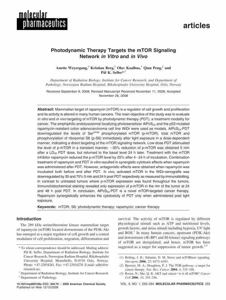
Photodynamic Therapy Targets the mTOR SignalingNetwork in Vitro and in Vivo
Anette Weyergang,† Kristian Berg,† Olav Kaalhus,† Qian Peng,‡ andPål K. Selbo*,†
Department of Radiation Biology, Institute for Cancer Research, and Department ofPathology, Norwegian Radium Hospital, Rikshospitalet UniVersity Hospital, Oslo, Norway
Received September 9, 2008; Revised Manuscript Received November 11, 2008; AcceptedNovember 26, 2008
Abstract: Mammalian target of rapamycin (mTOR) is a regulator of cell growth and proliferationand its activity is altered in many human cancers. The main objective of this study was to evaluatein vitro and in vivo targeting of mTOR by photodynamic therapy (PDT), a treatment modality forcancer. The amphiphilic endolysosomal localizing photosensitizer AlPcS2a and the p53 mutatedrapamycin-resistant colon adenocarcinoma cell line WiDr were used as models. AlPcS2a-PDTdownregulated the levels of Ser2448 phosphorylated mTOR (p-mTOR), total mTOR andphosphorylation of ribosomal S6 (p-S6) immediately after light exposure in a dose-dependentmanner, indicating a direct targeting of the mTOR signaling network. Low-dose PDT attenuatedthe level of p-mTOR in a transient manner; ∼35% reduction of p-mTOR was obtained 5 minafter a LD35 PDT dose, but returned to the basal level 24 h later. Treatment with the mTORinhibitor rapamycin reduced the p-mTOR level by 25% after 4-24 h of incubation. Combinationtreatment of rapamycin and PDT in vitro resulted in synergistic cytotoxic effects when rapamycinwas administered after PDT. However, antagonistic effects were obtained when rapamycin wasincubated both before and after PDT. In vivo, activated mTOR in the WiDr-xenografts wasdownregulated by 35 and 75% 5 min and 24 h post PDT respectively as measured by immunoblotting.In contrast to untreated tumors where p-mTOR expression was found throughout the tumors,immunohistochemical staining revealed only expression of p-mTOR in the rim of the tumor at 24and 48 h post PDT. In conclusion, AlPcS2a-PDT is a novel mTOR-targeted cancer therapy.Rapamycin synergistically enhances the cytotoxicity of PDT only when administered post lightexposure.
Keywords: mTOR; S6; photodynamic therapy; rapamycin; cancer therapy
IntroductionThe 289 kDa serine/threonine kinase mammalian target
of rapamycin (mTOR) located downstream of the PI3K-Akthas emerged as a major regulator of cell growth and a centralmodulator of cell proliferation, migration, differentiation and
survival. The activity of mTOR is regulated by differentphysiological stimuli such as ATP and nutritional levels,growth factors, and stress stimuli including hypoxia, UV lightand ROS.1 In many human cancers, upstream (PI3K-Akt)and downstream (4E-BP1 and S6 kinase) signaling pathwaysof mTOR are deregulated, and hence, mTOR has beensuggested as a target for suppression of tumor growth.2,3
* To whom correspondence should be addressed. Mailing address:Pål K. Selbo, Department of Radiation Biology, Institute forCancer Research, Norwegian Radium Hospital, RikshospitaletUniversity Hospital, Montebello, N-0310 Oslo, Norway.Phone: +47-22934261. Fax: +47-22934270. E-mail: [email protected].
† Department of Radiation Biology, Institute for Cancer Research.‡ Department of Pathology.
(1) Reiling, J. H.; Sabatini, D. M. Stress and mTORture signaling.Oncogene 2006, 25, 6373–6383.
(2) Bjornsti, M. A.; Houghton, P. J. The TOR pathway: a target forcancer therapy. Nat. ReV. Cancer 2004, 4, 335–348.
(3) Rosen, N.; She, Q. B. AKT and cancersis it all mTOR? CancerCell 2006, 10, 254–256.
articles
10.1021/mp800156e CCC: $40.75 2009 American Chemical Society VOL. 6, NO. 1, 255–264 MOLECULAR PHARMACEUTICS 255Published on Web 12/15/2008

Two functionally distinct mTOR complexes (mTORC),mTORC1 and mTORC2, have been identified.4 mTORC1consists of mTOR, mLST8 and raptor (regulatory associatedprotein of mTOR) and mTORC2 consists of mTOR, mLST8and rictor (rapamycin insensitive companion of mTOR).mTORC1 is sensitive to the macrolide antibiotic rapamycinand its synthetic analogues CCI-779, RAD001 and AP23573,all specific inhibitors perturbing mTORC1 activity. mTORC2was thought to be insensitive to rapamycin; however, recentlyit was shown that long-time incubation with rapamycininhibited the assembly of the protein complex.5 The mech-anism of action of rapamycin, and its analogues, has createdsignificant interest in their use as anticancer agents. Rapa-mycin binds to its intracellular receptor FKB12-bindingprotein (FKBP) making a complex that binds to mTOR andthereby inhibits activation of downstream targets. Inhibitionof mTOR by rapamycin induces G1 arrest or, in some cases,apoptosis.2,4 However, intrinsic or acquired resistance torapamycin treatment of cancer cells has been demonstrated.6
Potent rapamycin analogues (rapalogs) or alternative mTORtargeting strategies are therefore highly warranted.7,8 Inaddition, cells with mutated p53 have deregulated mTORexpression.9
Photodynamic therapy (PDT) is an approved treatmentmodality of a range of cancers worldwide and was recentlyrecommended as a first line treatment for non-melanoma skincancers.10 PDT is a noninvasive and cancer-selective modal-ity based on local or systemic administration of a tumor-accumulating photosensitizer that is subsequently activatedby local light exposure resulting in generation of cytotoxicreactive oxygen species (ROS).11-13 PDT inactivates cancercells by oxidation of biomolecules, such as some amino acids,unsaturated fatty acids and cholesterol, which cause stress-
induction resulting in necrosis and/or apoptotic cell death.14,15
Recently, it was shown by independent groups that autophagyis an alternative mode of cell death post PDT.16,17 However,it has been reported that autophagy can protect cells fromphototoxicity post low-dose PDT.18 PDT-generated antitumoractivity can also be achieved by targeting the microvascu-lature and/or activation of the host immune system.19 Cellularsignaling after PDT is dependent on the physiochemicalproperties and subsequent intracellular localization of thephotosensitizer. In the present work an amphiphilic photo-sensitizer, AlPcS2a, known to localize to endosomes andlysosomes was used.
The main aim of this study was to evaluate PDT as analternative mTOR-targeting strategy. We here report on thePDT effects on mTOR signaling both in ViVo and in Vitro inp53-mutated and rapamycin-resistant WiDr colon adenocar-cinoma cells and subcutaneously growing tumors. Thespatiotemporal p-mTOR expression in ViVo was also inves-tigated after PDT. The ability of the mTOR inhibitorrapamycin to enhance the cytotoxic effect of PDT was inaddition evaluated in Vitro.
Experimental SectionCell Line and Culture Conditions. The colon adenocar-
cinoma cell line WiDr (ATCC CCL-218), a derivative ofHT-29, with a p53 mutation (G f A, Arg273 f His), wasused.20 The WiDr cells were subcultured twice a week inRPMI 1640 medium (Sigma, St. Louis, MO), supplementedwith 10% fetal calf serum (GIBCO BRL, Paisley, U.K.), 100U/mL penicillin and 100 µg/mL streptomycin and 2 mMglutamine (Sigma). The cells were grown and incubated in75 cm2 flasks (Nalge Nunc Int., Naperville, IL) at 37 °C ina humidified atmosphere containing 5% CO2.
(4) Wullschleger, S.; Loewith, R.; Hall, M. N. TOR signaling ingrowth and metabolism. Cell 2006, 124, 471–484.
(5) Sarbassov, D. D.; Ali, S. M.; Sengupta, S.; Sheen, J. H.; Hsu,P. P.; Bagley, A. F.; Markhard, A. L.; Sabatini, D. M. Prolongedrapamycin treatment inhibits mTORC2 assembly and Akt/PKB.Mol. Cell 2006, 22, 159–168.
(6) Kurmasheva, R. T.; Huang, S.; Houghton, P. J. Predictedmechanisms of resistance to mTOR inhibitors. Br. J. Cancer 2006,95, 955–960.
(7) Guertin, D. A.; Sabatini, D. M. An expanding role for mTOR incancer. Trends Mol. Med. 2005, 11, 353–361.
(8) Faivre, S.; Kroemer, G.; Raymond, E. Current development ofmTOR inhibitors as anticancer agents. Nat. ReV. Drug DiscoVery2006, 5, 671–688.
(9) Feng, Z.; Zhang, H.; Levine, A. J.; Jin, S. The coordinateregulation of the p53 and mTOR pathways in cells. Proc. Natl.Acad. Sci. U.S.A. 2005, 102, 8204–8209.
(10) Braathen, L. R.; Szeimies, R. M.; Basset-Seguin, N.; Bissonnette,R.; Foley, P.; Pariser, D.; Roelandts, R.; Wennberg, A. M.;Morton, C. A. Guidelines on the use of photodynamic therapyfor nonmelanoma skin cancer: an international consensus. J. Am.Acad. Dermatol. 2007, 56, 125–143.
(11) Dougherty, T. J.; Gomer, C. J.; Henderson, B. W.; Jori, G.; Kessel,D.; Korbelik, M.; Moan, J.; Peng, Q. Photodynamic therapy.J. Natl. Cancer Inst. 1998, 90, 889–905.
(12) Dolmans, D. E.; Fukumura, D.; Jain, R. K. Photodynamic therapyfor cancer. Nat. ReV. Cancer 2003, 3, 380–387.
(13) Brown, S. B.; Brown, E. A.; Walker, I. The present and futurerole of photodynamic therapy in cancer treatment. Lancet Oncol.2004, 5, 497–508.
(14) Piette, J.; Volanti, C.; Vantieghem, A.; Matroule, J. Y.; Habraken,Y.; Agostinis, P. Cell death and growth arrest in response tophotodynamic therapy with membrane-bound photosensitizers.Biochem. Pharmacol. 2003, 66, 1651–1659.
(15) Oleinick, N. L.; Morris, R. L.; Belichenko, I. The role of apoptosisin response to photodynamic therapy: what, where, why, and how.Photochem. Photobiol. Sci. 2002, 1, 1–21.
(16) Buytaert, E.; Callewaert, G.; Hendrickx, N.; Scorrano, L.; Hart-mann, D.; Missiaen, L.; Vandenheede, J. R.; Heirman, I.; Grooten,J.; Agostinis, P. Role of endoplasmic reticulum depletion andmultidomain proapoptotic BAX and BAK proteins in shaping celldeath after hypericin-mediated photodynamic therapy. FASEB J.2006, 20, 756–758.
(17) Kessel, D.; Vicente, M. G.; Reiners, J. J., Jr. Initiation of apoptosisand autophagy by photodynamic therapy. Lasers Surg. Med. 2006,38, 482–488.
(18) Kessel, D.; Arroyo, A. S. Apoptotic and autophagic responses toBcl-2 inhibition and photodamage. Photochem. Photobiol. Sci.2007, 6, 1290–1295.
(19) Castano, A. P.; Mroz, P.; Hamblin, M. R. Photodynamic therapyand anti-tumour immunity. Nat. ReV. Cancer 2006, 6, 535–545.
(20) Rodrigues, N. R.; Rowan, A.; Smith, M. E.; Kerr, I. B.; Bodmer,W. F.; Gannon, J. V.; Lane, D. P. p53 mutations in colorectalcancer. Proc. Natl. Acad. Sci. U.S.A. 1990, 87, 7555–7559.
articles Weyergang et al.
256 MOLECULAR PHARMACEUTICS VOL. 6, NO. 1
Drugs and Chemicals. AlPcS2a was purchased fromPorphyrin Products (Logan, UT). AlPcS2a was first dissolvedin 0.1 M NaOH and thereafter diluted in PBS, pH 7.5 to aconcentration of 1.25 mg/mL and a final concentration of0.02 M NaOH. The photosensitizer was light protected andstored at -20 °C until use. Rapamycin was purchased fromSigma (St. Louis, MO) and dissolved in 99.9% dimethyl-sulfoxide (DMSO) (Sigma) to a final concentration of 4.38mM and stored as aliquots at -20 °C until use.
In Vitro Light Source. The light exposure of cells inculture was performed by using LumiSource (PCI Biotech,Oslo, Norway). The lamp consists of a bank of 4 light tubes(4 × 18 W Philips Fluotone 18W/950), which emits red light(610-720 nm) filtered through a 620 nm long pass filter(Vink Plast, Kolbotn, Norway). The irradiance varies lessthan 10% across the whole illumination area (765 cm2) withan output of 1.5 mW/cm2. The light source is air-cooledduring light exposure, which prevents cells from beingexposed to hyperthermia and keeps the irradiance stable.
Photochemical Treatments in Vitro. Cells were harvestedwith 500 µg/L trypsin and 200 mg/mL ethylenediaminetet-raacetic acid (EDTA) (Sigma) and seeded out at 2000 cellsper well in 96-well plates (Nunclon, Nunc, Roskilde,Denmark) for the MTT-assay, 1000 cells/well in 6-well plates(Nunclon) for colony-forming ability experiments, 50 000cells/well in 6-well plates for the experiments with Coultercounter and 500 000 cells/wells in Falcon 3003 63 cm2 wellsfor sample preparation to SDS-PAGE and Western blotting.The cells were allowed to attach to the bottom of the wellsfor 24 h prior to start of the experiments. Cells were thenincubated with 20 µg/mL AlPcS2a for 18 h followed by 2×wash with culture medium and subsequently a 4 h chaseperiod in drug free medium before the cells were exposedto increasing light doses.
Combination of PDT and Rapamycin. Two different inVitro treatment strategies were examined: (i) Rapamycin, atdifferent concentrations as indicated in the figures, wasincubated in the medium throughout the entire experiment,i.e. coincubated with PS for 18 h, incubated during PS-freechasing period (4 h), incubated during light exposure, andpresent until cytotoxic assessments 96 h after light exposure.Total rapamycin incubation time was therefore 5 days. (ii)Alternatively, rapamycin was incubated only directly afterlight exposure and was present in the medium until cytotoxicevaluation, which gave a total rapamycin incubation timeof 4 days.
Statistic Evaluations of the Combined Effect of PDTand Rapamycin. To assess possible synergistic/antagonisticeffects when rapamycin was used in combination with PDTwe used the parameter DL ) -(ln Scomb - ln Sadd), which isthe difference in log values between the observed survivalfraction of the combined regimen and the expected survivalfraction when rapamycin and PDT are supposed to actindependently. The latter term, ln Sadd ) ln SPDT + ln SRap,is given from the single agent experiments at the same doses.The DL values from repeated experiments are averaged forthe case of rapamycin either being added after PDT or when
it is present both before and after PDT. Significant differencesfrom zero of positive (synergistic) or negative (antagonistic)DL are assessed by two-tailed t tests with a significance levelof p ) 0.05.
Cytotoxic Evaluations. Three independent assays wereemployed to measure the cytotoxic responses of the treat-ments: (i) The MTT-assay was performed 24 or 96 h afterlight exposure as indicated in the figure legends. Then theculture medium was removed and new medium containing0.25 mg/mL MTT reagent was added to the cells. The cellswere incubated for 2-4 h and the formazan crystals dissolvedin DMSO. Absorbance at 570 nm was used as a measure ofcell viability. (ii) Cell counting was performed 96 h afterlight exposure. The cells were then trypsinized, pelleted, andthe pellets dissolved in 20 mL of PBS. Two milliliter portionsof the final suspensions were counted by an in-house madeCounter coulter. (iii) To verify the MTT assessments, colonyforming ability assays were performed 7-10 days post PDTas recently described.21
SDS-PAGE, Western Blotting and Immunostaining.Harvesting of in Vitro growing cells and in ViVo growingtumors was done at different time points after lightexposure, and the relative number of cells in each samplewas measured as recently described.22 Samples were thensubjected to SDS-PAGE with 7.5% gels for mTOR- andAkt-analysis and 12% gels for S6-analysis prior to Westernblotting. The PVDF transfer membranes (GE Healthcare,Amershamplace, U.K.) were then incubated with differentprimary antibodies from Cell Signaling Technology (Dan-vers, MA); mTOR (#2972), p-mTOR (Ser2448) (#2971),Akt (#9272), p-Akt (Ser473) (#4051), p-Akt (Thr308)(#4056) and p-S6 (Ser235/236) (#4856). Horse radishperoxidase (HRP) conjugated donkey-antirabbit antibody(Promega, Madison, WI) and horse-antimouse antibody(Cell Signaling Technology) were used as secondaryantibodies. ECL plus Western blotting detection systems(GE Healthcare) and Image Quant software (Moleculardynamics, GE Healthcare) were used to detect andquantify the protein bands. Membranes were stained withPonceau S to verify even loading and transfer of proteins.
Immunohistochemistry (IHC). Five-micrometer sectionsfrom formalin-fixed and paraffin-embedded WiDr tumortissue blocks were cut and mounted on positively chargedmicroscopy slides. The sections were then dried for 45 minat 60 °C, overnight at 37 °C and stored at 4 °C until use.The deparaffinized tissue sections were pretreated in amicrowave oven at 750 W for 5 min and 500 W for 15 minin buffer (pH 6; Dako, code no. S1699) for target retrievalbefore they were incubated with the IHC specific rabbitprimary antibody against human p-mTOR (Cell SignalingTechnology) (#2976) diluted 1:50 for 30 min at roomtemperature. The sections were then stained with the Dako-Cytomation EnVision+ System-HRP (DAP) Rabbit (Dako-Cytomation, Inc., Carpentaria, CA) (#K4011), counterstainedwith hematoxylin, and finally, dehydrated and mounted inEukitt (Sigma). A negative control was included with thebuffer solution only instead of the primary antibody.
Targeting of mTOR by PDT articles
VOL. 6, NO. 1 MOLECULAR PHARMACEUTICS 257

Animal Studies and in ViWo PDT. BALB/c (nu/nu) nudefemale mice were bred at the Animal department of theNorwegian Radium Hospital. The mice were kept underspecific pathogen-free conditions, and all procedures involv-ing mice were carried out in agreement with the protocolsapproved by the Animal Care Committee at the hospitalunder control by the National Ethical Committee’s guidelineson animal welfare. All mice were acclimated in the Animaldepartment for at least 1 week before tumor implantation.The mice were on average 20-25 g (5-8 weeks) at the startof the experiment. WiDr cells from subcutaneous (sc)growing tumors were mixed to homogeneity by a scalpel,and 20 µL of the tumor-tissue suspension was injected s.c.on the left hip of each mouse. Ten mg/kg AlPcS2a wasinjected intraperitoneally (ip) when the tumors approacheda volume of 100 mm3. The tumor volume was calculatedusing the following formula:
V) (W2 × L) ⁄ 2
according to the US National Cancer Institute protocolswhere W is the width and L the length of the tumorsmeasured 2 or 3 times per week with a caliper. Tumors(75-125 mm3) were illuminated 48 h after AlPcS2a admin-istration by 670 nm light with an irradiance rate of 100 mW/cm2 and a total dose of 30 J/cm2 using a diode laser fromCeramOptec Industries Inc. (Bonn, Germany). The animalswere protected from light with aluminum foil except above
the tumor area including an adjacent rim of approximately2 mm of normal skin during illumination.
ResultsImmediate Response to PDT on mTOR Signaling. The
direct response to PDT (by increasing light-doses givingincreased cytotoxicity) on p-mTOR and the phosphorylation-status of its downstream effector the ribosomal protein S6was evaluated in Vitro 5 min after light exposure (Figure 1).It was demonstrated that after a LD35 dose the phosphory-lation of mTOR and S6 was attenuated by 30% and 60%,respectively. The reduction in phosphorylated mTOR andS6 increased with increasing PDT doses. Total mTOR wasalso reduced with increasing light doses. Surprisingly,treatment with the photosensitizer in the absence of lightcaused a 20% reduction in p-S6, but not in the p-mTORlevel (Figure 1).
The WiDr cells were found to continuously expressThr308phoshorylated Akt when incubated in culture mediumwith 10% FCS. However, no photochemical effects on Thr308
phosphorylated Akt nor total Akt were observed after thephotochemical treatment (Figure 1) compared to cells thatwere incubated with photosensitizer only. A Ser473 phos-phorylation of Akt was detected neither in control nor inPDT-treated cells (data not shown).
Time-Dependent Dephosphorylation of mTOR afterRapamycin or PDT in Vitro. Serum starvation of cellsovernight before incubation with growth factors is a commonmethod to stimulate phosphorylation of mTOR.23 However,in WiDr cells activated mTOR could be detected withoutserum starvation. Four hour incubation of the WiDr cellswith 10 nM rapamycin attenuated Ser2448 phosphorylationof mTOR by 25% as assessed by Western blot analysis(Figure 2A). A prolongation of the 10 nM rapamycinexposure to 24 h did not further decrease the level ofp-mTOR (Figure 2A) nor did increasing rapamycin concen-trations up to 100 nM (data not shown). Total mTORexpression was not influenced by the 24 h rapamycintreatment. Twenty-four hours of 10 or 100 nM rapamycindid not induce decreased viability as measured by the MTTmethod. Sustained incubation of more than 72 h did,however, reduce the viability as presented in Figure 3. Thetime dependent effects on mTOR were also studied after PDT(LD35) in Vitro. As can be seen in Figure 2B, downregulationof p-mTOR and total mTOR was transient and returned to
(21) Yip, W. L.; Weyergang, A.; Berg, K.; Tonnesen, H. H.; Selbo,P. K. Targeted Delivery and Enhanced Cytotoxicity of Cetuximab-Saporin by Photochemical Internalization in EGFR-PositiveCancer Cells. Mol. Pharmaceutics 2007, 4, 241–251.
(22) Weyergang, A.; Selbo, P. K.; Berg, K. Y1068 phosphorylation isthe most sensitive target of disulfonated tetraphenylporphyrin-based photodynamic therapy on epidermal growth factor receptor.Biochem. Pharmacol. 2007, 74, 226–235.
Figure 1. Downregulation of mTOR signaling pathwayafter PDT. Light dose dependent mTOR signaling 5 minpost AlPcS2a-PDT. Immunoblots of p-mTOR(Ser2448),total mTOR, p-S6(Ser235/236), p-Akt(Thr308) and total Aktin cells subjected to increasing light doses withindicated cytotoxicity. The amounts of p-mTOR, totalmTOR and p-S6 were quantified as relative to untreatedcells and are presented. MTT was assessed at 24 hpost PDT. Efficiency of transfer and even loading oflanes were verified by using Ponceau S staining. Thedata are the average of triplicates based on threeindependent experiments. Bars, standard errors.
articles Weyergang et al.
258 MOLECULAR PHARMACEUTICS VOL. 6, NO. 1
control levels 24 h after PDT. Even loading of proteins wasverified by using Ponceau S staining (data not shown).
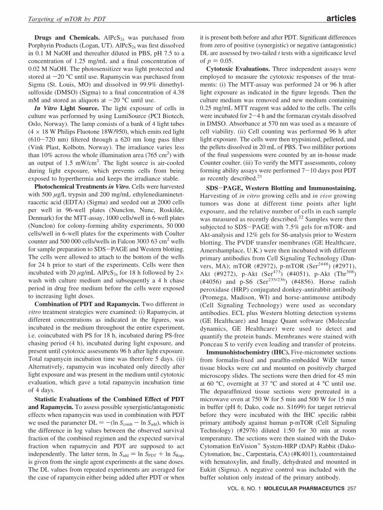
Rapamycin-Induced Synergistic or AntagonisticResponses in Combination with PDT. The WiDr cells werechallenged with a combination of PDT and rapamycin toevaluate possible synergistic effects on cell inactivation.Synergistic effects as measured by the synergy parameterDL (as described in Experimental Section) were observedwhen the WiDr cells were exposed to rapamycin for 96 hafter PDT (Figure 3A and 3B, with corresponding synergyparameters in Figure 3E). The DL values are all positive,which implies synergy with respect to cytotoxicity. Thiseffect is larger for the 4 min light exposure than for the 3min exposure. The variation with rapamycin concentrationis rather small. Except for the DL values at 0.3 nM (p )0.10) and 30 nM (p ) 0.07) of rapamycin after 3 min oflight exposure, all DL values in Figure 3E are significantlypositive. Averaging DL over the rapamycin concentrations,the observed cell viability was ∼1.5 and 2-fold lower thanexpected from the calculated additive effects after PDT with3 and 4 min of light exposure, respectively. In contrast,antagonistic effects were observed in WiDr cells that wereexposed to rapamycin throughout the whole procedure, i.e.for 22 h prior to light exposure and until the time of analyses
(Figure 3C and 3D, with corresponding synergy parametersin Figure 3F). Figure 3F shows the DL values obtained fromexperiments where rapamycin is present both before and afterPDT. In contrast to the regime where rapamycin was addedafter PDT, these DL values are all negative, which impliesantagonism, but only treatment with 4 min of light (and the3 min of light in combination with 1000 nM rapamycin, p) 0.047) is significant. Averaging DL over rapamycinconcentration gave an antagonistic survival enhancementfactor of ∼1.15 for 2 and 3 min of light exposure, and ∼1.7
(23) Sekulic, A.; Hudson, C. C.; Homme, J. L.; Yin, P.; Otterness,D. M.; Karnitz, L. M.; Abraham, R. T. A direct linkage betweenthe phosphoinositide 3-kinase-AKT signaling pathway and themammalian target of rapamycin in mitogen-stimulated andtransformed cells. Cancer Res. 2000, 60, 3504–3513.
Figure 2. Time-dependent inhibition of mTOR aftersubtoxic rapamycin or PDT treatment. Total andp-mTOR 4 and 24 h after treatment of WiDr cells withA, 10 nM rapamycin and B, PDT (LD35) 5 min, 4 and24 h after light exposure. Levels of p-mTOR werequantified as relative to untreated cells. MTT wasassessed at 24 h post PDT. Efficiency of transfer andeven loading of lanes were verified by using Ponceau Sstaining. The data are the average values of threeindependent experiments. Bars, standard errors.
Figure 3. Rapamycin-induced synergistic or antagonisticresponses in combination with PDT are dependent ontime of rapamycin administration. Cytotoxic effects ofPDT alone or in combination with rapamycin after twodifferent experimental treatment regimes. A and B, rapa-mycin was administrated to the cells immediately afterPDT. C and D, rapamycin was present throughout thewhole experiment from the initiation of the AlPcS2a incuba-tion until the MTT measurements (96 h post PDT). A andC, light dose was kept constant (LD40) and the rapamy-cin concentration varied. B and D, the rapamycin concen-tration was kept constant at 10 nM and the light dosevaried. The data are representative results of reproducedexperiments and show the average of triplicates presentedas compared to untreated control cells. E and F, DLvalues after combination treatment with constant PDTdoses and increasing rapamycin doses. E, experimentswhere rapamycin was added immediately after light ex-posure while F, experiments where rapamycin was presentboth before and after the photochemical treatment. Dataare the average of at least three independent experi-ments. Bars, standard errors.
Targeting of mTOR by PDT articles
VOL. 6, NO. 1 MOLECULAR PHARMACEUTICS 259
in the case of the 4 min of light exposure when compared tothe expected additive effect.24 In general, the treatment-dependent observation of synergism or antagonism washighly dependent on the PDT-dose and less dependent onthe rapamycin concentration. Rapamycin-induced antago-nistic and synergistic responses in combination with PDTwere confirmed by counting individual cells using Coultercounter and by colony forming ability measurements asalternative methods for cytotoxicity evaluations.
In ViWo PDT-Effects on Downstream p-mTOR. Micewith subcutaneously growing WiDr tumors were subjectedto PDT causing a temporary reduction in tumor volume anda 2-3 week delay in tumor growth as recently described.22
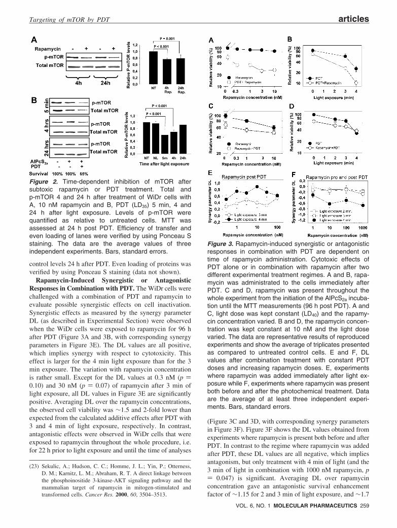
It was demonstrated that the level of activated mTOR wasattenuated by PDT also in ViVo (Figure 4A and 4C). Fiveminutes post PDT the p-mTOR level was reduced by 35%compared to nontreated tumors as evaluated on Western blotsof whole tumor extracts (P ) 0.024). At 24 h post PDT theactivated mTOR in the WiDr-tumors was further reduced
by >75% compared to nontreated control tumors (P )0.002). However, the level of mTOR phosphorylationbetween 5 min and 24 h post PDT varied strongly amongtreated tumors as presented in Figure 4A. Figure 4B is arepresentative blot for total Akt, indicating no change ofexpression over time post PDT, except a slight decrease after5 min. However, this was not statistically significant whenjudging several immunoblots (P > 0.05). Correspondingtumor growth data were recently presented.22
Spatiotemporal Expression of p-mTOR after in ViWoPDT. Analysis of tumor tissue extracts on Western blots asdescribed above provides only information on the presenceof phosphorylated mTOR in the whole tissue extracts. Toobtain information about the spatiotemporal activation ofmTOR at Ser2448, immunohistochemistry (IHC) on sectionsfrom paraffin-embedded WiDr tumor xenografts was per-formed. In control tumors, phosphorylation of mTOR wasfound to be heterogeneously distributed all over the tissue(Figure 5A). At 1 and 4 h post PDT, p-mTOR was stilldetectable throughout the tumor, especially in the viableislands of colon adenocarcinoma cells. Twenty-four and 48 hafter PDT almost no staining of p-mTOR was observed inthe tumor except in the peripheral area still showing a similarlevel of p-mTOR expression to that of control tumors (Figure5A and 5B). The fraction of necrotic tissue, in particular inthe central area, was increased from 24 to 48 h post PDT.Additional IHC sections of p-mTOR expression from thexenograft tissues are shown in Supplementary Figures 1-3in the Supporting Information (controls, 1, 4, 24 and 48 hpost PDT).
Interestingly, a significant expression of p-mTOR wasobserved in the epidermis overlaying the tumors, while thedermis was negative for activated mTOR. In addition, someof the hair follicles were also highly positive for p-mTOR(Figure 5C).
DiscussionThe tumor-suppressor p53 inhibits mTOR activity as a part
of its action mechanism.9 p53 is mutated in about 50% ofall cancers.25 Therefore, we found it interesting to study thePDT effect on mTOR in the p53 mutated colon adenocar-cinoma cell line WiDr compared to the outcome of rapa-mycin treatment. Rapamycin is a specific inhibitor of themTORC1 complex and a cell type dependent inhibitor ofAkt and mTORC2.26
In the present study we show that 4 and 24 h rapamycinexposure reduced the phosphorylation of mTOR in the WiDrcell line by 25%. The rapamycin treatment of WiDr cellsinduced low cytotoxicity, i.e. <30% of the cells wereinactivated after 96 h treatment with 10 nM rapamycin, whichis in accordance with a recent report on rapamycin treatment
(24) If the additive cytotoxic effect of PDT and rapamycin were causedby the same mechanism (named Mode II by Steel and Peckham)instead of by mutually independent mechanisms (Mode I), theexpected survival fraction would be larger and correspond to alarger synergy effect in the case when rapamycin is used afterPDT. Conversely, the additive survival fraction would be smallerin the Mode II calculations and indicate a larger antagonism whenrapamycin is present during the whole experiment compared tothe Mode I calculation. Steel, G. G.; Peckham, M. J. Exploitablemechanisms in combined radiotherapy-chemotherapy: the conceptof additivity. Int. J. Radiat. Oncol. Biol. Phys. 1979, 5, 85–91.
(25) Selivanova, G.; Wiman, K. G. Reactivation of mutant p53:molecular mechanisms and therapeutic potential. Oncogene 2007,26, 2243–2254.
(26) Sabatini, D. M. mTOR and cancer: insights into a complexrelationship. Nat ReV. Cancer 2006, 6, 729–734.
Figure 4. Expression of phosphorylated mTOR in vivoafter PDT. A, Effects of PDT on the p-mTOR level insubcutaneously growing WiDr tumors. B, Representativeblot of total Akt (from experiment 4). Efficiency of transferand even loading of lanes were verified by using PonceauS staining. C, Activated mTOR was quantified relative tountreated tumors. Four independent experiments (Exp)were performed. Each protein band represents one tumor.Bars ) standard errors.
articles Weyergang et al.
260 MOLECULAR PHARMACEUTICS VOL. 6, NO. 1

of colon carcinomas, including the WiDr cell line.27 PDTeffects on mTOR are here demonstrated for the first timeand shown to induce rapid inhibition of Ser2448 phosphory-lated mTOR both in Vitro and in ViVo. In addition, PDTinduced dephosphorylation of S6, an effector protein down-stream of S6 kinase that is downstream of mTOR. Akt is
located both upstream and downstream of mTOR.28,29
Interestingly, Akt was not affected by AlPcS2a-PDT, sug-
(27) Nozawa, H.; Watanabe, T.; Nagawa, H. Phosphorylation ofribosomal p70 S6 kinase and rapamycin sensitivity in humancolorectal cancer. Cancer Lett. 2007, 251, 105–113.
Figure 5. Spatiotemporal expression of mTOR post in vivo PDT. Immunohistochemistry of p-mTOR (Ser2448) in WiDrtumors post PDT. A, Nontreated (NT) and PDT-treated animals 4, and 24 h post light exposure. Arrows indicate therim of p-mTOR post PDT. Bars ) 2 mm. B, Cellular and subcellular localization of p-mTOR in nontreated (NT) andPDT- treated tumors 1, 4 and 24 h post light exposure. Left panel is from the marginal zone and right panel is fromthe central zone of tumor. Small panels (magnifications) are from the corresponding areas. Bar in upper left panel(large window) ) 100 µm. Small panel bar ) 20 µm. C, p-mTOR staining of skin above tumor. Large panel bar )2000 µm and small panel bar ) 10 µm. The upper small panel is a magnification of epidermis, while the other smallpanel shows a hair follicle. Control IHC, see Supplementary Figure 3C in the Supporting Information.
Targeting of mTOR by PDT articles
VOL. 6, NO. 1 MOLECULAR PHARMACEUTICS 261

gesting that AlPcS2a is not colocalized with Akt. The presentresults show that the reduction in p-mTOR after PDT waslarger than that observed after rapamycin treatment in theWiDr cells. The lack of additional attenuation of mTORphosphorylation by increasing the rapamycin treatmentconcentration above 10 nM indicate that mTORC1 activityis completely inhibited by rapamycin and that the remainingp-mTOR is due to phosphorylated mTOR in the mTORC2complexes. The differences in effects on p-mTOR betweenrapamycin and PDT reported here may therefore be causedby PDT-mediated attenuation of both mTORC1 and mTORC2compared to rapamycin which mainly acts on mTORC1.However this has to be further explored and confirmed byimmunoprecipitation of mTORC1 and mTORC2 and furtherevaluation of PDT mediated attenuation of mTOR in bothcomplexes.
The present results show a reduction in total mTOR andmTOR signaling immediately after PDT in Vitro, which may
indicate that the photochemical treatment causes a directoxidation of mTOR. PDT induces ROS of which singletoxygen (1O2) is suggested to dominate and cause oxidationof surrounding biomolecules. Singlet oxygen has an ex-tremely short lifetime (<10-40 ns) and diffusion length(<10-20 nanometers) in cells. Hence, the cytotoxic actionof PDT takes place in the very close vicinity of thesubcellular site of 1O2 formation.30-32 The photosensitizerused in this study, AlPcS2a, is shown both in Vitro and inViVo to localize to endosomes and lysosomes.33-35 Inaddition, TOR2 has been shown to be localized in lysosomes/vacuoles in yeast.36 Based on these observations, we suggestthat mTOR and AlPcS2a may be colocalized in endocyticvesicles of cells not exposed to light. Furthermore, it hasbeen documented, both in Vitro and in ViVo, that AlPcS2a isreleased from the endocytic vesicles during light exposure,and relocalizes to other cellular compartments except thenucleus.34,35 In mammalian cells, mTOR has been suggestedto be associated with intracellular membranes of whichendoplasmatic reticulum (ER) has in particular been docu-mented or to the plasma membrane and a minor fraction inthe cytosol.37,38 The photosensitizer TPPS2a, which has thesame cellular localization pattern as AlPcS2a, relocalizes toER upon light exposure and it has been suggested that PDT-induced targeting of ER is of importance for the cytotoxic
(28) Sarbassov, D. D.; Guertin, D. A.; Ali, S. M.; Sabatini, D. M.Phosphorylation and regulation of Akt/PKB by the rictor-mTORcomplex. Science 2005, 307, 1098–1101.
(29) Phung, T. L.; Ziv, K.; Dabydeen, D.; Eyiah-Mensah, G.; Riveros,M.; Perruzzi, C.; Sun, J.; Monahan-Earley, R. A.; Shiojima, I.;Nagy, J. A.; Lin, M. I.; Walsh, K.; et al. Pathological angiogenesisis induced by sustained Akt signaling and inhibited by rapamycin.Cancer Cell 2006, 10, 159–170.
(30) Moan, J.; Berg, K. The photodegradation of porphyrins in cellscan be used to estimate the lifetime of singlet oxygen. Photochem.Photobiol. 1991, 53, 549–553.
(31) Niedre, M.; Patterson, M. S.; Wilson, B. C. Direct near-infraredluminescence detection of singlet oxygen generated by photody-namic therapy in cells in vitro and tissues in vivo. Photochem.Photobiol. 2002, 75, 382–391.
(32) Redmond, R. W.; Kochevar, I. E. Spatially-Resolved CellularResponses to Singlet Oxygen. Photochem. Photobiol. 2006, 82,1178–1186.
(33) Moan, J.; Berg, K.; Anholt, H.; Madslien, K. Sulfonated alu-minium phthalocyanines as sensitizers for photochemotherapy.Effects of small light doses on localization, dye fluorescence andphotosensitivity in V79 cells. Int. J. Cancer 1994, 58, 865–870.
(34) Selbo, P. K.; Sandvig, K.; Kirveliene, V.; Berg, K. Release ofgelonin from endosomes and lysosomes to cytosol by photo-chemical internalization. Biochim. Biophys. Acta 2000, 1475, 307–313.
(35) Selbo, P. K.; Sivam, G.; Fodstad, O.; Sandvig, K.; Berg, K. InVivo Documentation of Photochemical Internalization, a NovelApproach to Site Specific Cancer Therapy. Int. J. Cancer 2001,92, 761–766.
(36) Cardenas, M. E.; Heitman, J. FKBP12-rapamycin target TOR2is a vacuolar protein with an associated phosphatidylinositol-4kinase activity. EMBO J. 1995, 14, 5892–5907.
(37) Withers, D. J.; Ouwens, D. M.; Nave, B. T.; van der Zon, G. C.;Alarcon, C. M.; Cardenas, M. E.; Heitman, J.; Maassen, J. A.;Shepherd, P. R. Expression, enzyme activity, and subcellularlocalization of mammalian target of rapamycin in insulin-responsive cells. Biochem. Biophys. Res. Commun. 1997, 241,704–709.
(38) Sabatini, D. M.; Barrow, R. K.; Blackshaw, S.; Burnett, P. E.;Lai, M. M.; Field, M. E.; Bahr, B. A.; Kirsch, J.; Betz, H.; Snyder,S. H. Interaction of RAFT1 with gephyrin required for rapamycin-sensitive signaling. Science 1999, 284, 1161–1164.
Figure 6. Schematic model of the mTOR signalingpathway and its inhibition by rapamycin and the suggestedrole of AlPcS2a-PDT. Upon light activation, lysosomal/ER-associated AlPcS2a mediates photochemical genera-tion of ROS, of which singlet oxygen (1O2) is the mostabundant, and thereby cause a direct oxidative damage tomTOR. This attenuates the downstream signaling ofmTOR, including deactivation of S6.
articles Weyergang et al.
262 MOLECULAR PHARMACEUTICS VOL. 6, NO. 1
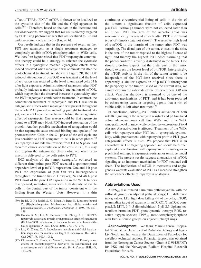
effect of TPPS2a-PDT.39 mTOR is shown to be localized tothe cytosolic side of the ER and the Golgi apparatus incells.40,41 Therefore, based on the data in the literature andour observations, we suggest that mTOR is directly targetedby PDT using photosensitizers that are localized to ER andendolysosomal compartments (Figure 6).
Our results indicate that in the presence of serum neitherPDT nor rapamycin as a single treatment manages tocompletely abolish mTOR phosphorylation in WiDr cells.We therefore hypothesized that PDT-rapamycin combina-tion therapy could be a strategy to enhance the cytotoxiceffects in a synergistic manner. Synergistic effects wereindeed observed when rapamycin was administered after thephotochemical treatment. As shown in Figure 2B, the PDTinduced attenuation of p-mTOR was transient and the levelof activation was restored to the level in untreated cells 24 hafter light exposure. Administration of rapamycin after PDTprobably induces a more sustained attenuation of mTOR,which may explain the observed increase in cytotoxicity afterthe PDT-rapamycin combination treatment. In contrast, thecombination treatment of rapamycin and PDT resulted inantagonistic effects when rapamycin was present throughoutthe whole PDT procedure including prelight treatment. Asyet, we do not know the mechanism behind the antagonisticeffect of rapamycin. One reason could be that rapamycinbound to mTOR may block PDT-induced attenuation of theprotein. Another possible mechanism of antagonism couldbe that rapamycin cause reduced binding and uptake of thephotosensitizer. Cells in the G1 phase of the cell cycle areless sensitive to PDT compared to cells in other phases.42
As rapamycin inhibits the traverse from G1 to S phase andtherefore causes accumulation of the cells in G1, this mayalso explain the antagonistic effects observed when rapa-mycin is delivered to the cells prior to PDT.
IHC analysis of the tumor xenografts collected atdifferent time points post PDT revealed a spatiotemporaldependent level of p-mTOR expression. One and 4 h postPDT the expression of p-mTOR was heterogeneousthroughout the tumor tissue. However, 24 and 48 h postPDT most of the p-mTOR expression in the WiDr tumorsdisappeared, including areas with high density of viablecells in the central part of the tumor, consistent with thefinding from the Western blots. However, in a thin
continuous circumferential lining of cells in the rim ofthe tumors a significant fraction of cells expressedp-mTOR. Although tumors were not collected later than48 h post PDT, the size of the necrotic areas wasmacroscopically increased at 98 h after PDT in differenttypes of tumors (data not shown). The relative high levelof p-mTOR in the margin of the tumor after PDT wassurprising. The distal part of the tumor, closest to the skin,is the area of the tumor exposed to the highest fluence oflight, and thereby the highest PDT doses assuming thatthe photosensitizer is evenly distributed in the tumor. Oneshould therefore expect that the distal part of the tumorshould express the lowest level of p-mTOR. Interestingly,the mTOR activity in the rim of the tumor seems to beindependent of the PDT-dose received since there isapparently a similar expression of p-mTOR throughoutthe periphery of the tumor. Based on the current data, wecannot explain the rationale of the observed p-mTOR rimeffect. Vascular shutdown is assumed to be one of theantitumor mechanisms of PDT, and it has been reportedby others using vascular-targeting agents that a rim ofviable cells is left after treatment.43
In conclusion, AlPcS2a-PDT inhibits activation of bothmTOR signaling in the rapamycin resistant and p53-mutatedcolon adenocarcinoma cell line WiDr and in a WiDrxenograft model in mice, while neither the expression of totalAkt nor Akt-activation is affected. Treatment of the WiDrcells with rapamycin after PDT led to synergistic cytotox-icity, while pretreatment with rapamycin prior to PDT gaveantagonistic effects in Vitro. We propose that PDT is analternative mTOR targeting approach and should be furtherexplored in combination with rapamycin or its analogues inpreclinical settings, in rapamycin resistant or sensitive modelsystems. The present results suggest attenuation of mTORsignaling as an important mechanism for PDT mediated celldeath. The implication of mTOR in metastasis and angio-genesis warrants evaluation of PDT as a means to strengthenthe anticancer effects of rapamycin analogues.
Abbreviations UsedAlPcS2a, disulfonated aluminum phthalocyanine with the
sulfonate groups on adjacent phthalate rings; DL, differencein log values; LDx, light dose killing x% of the cells; mTOR,mammalian target of rapamycin; mTORC1/2, mTOR com-plex1/2; MTT, 3-(4,5-dimethylthiazol-2-yl)-2,5-diphenyltet-razolium bromide; PDT, photodynamic therapy; ROS, re-active oxygen species; TPPS2a, meso-tetraphenylporphinewith two sulfonate groups on adjacent phenyl rings.
Acknowledgment. We thank Marie-Therese Roppes-tad Strand at the Department of Radiation Biology and Inger-Liv Nordli and her team at the Department of Pathology forexcellent technical support. Financial support was obtainedfrom the Norwegian Cancer Society (Grant # C 96138/007)for PKS and the Norwegian Radium Hospital ResearchFoundation for A.W.
(39) Rodal, G. H.; Rodal, S. K.; Moan, J.; Berg, K. Liposome-boundZn (II)-phthalocyanine. Mechanisms for cellular uptake andphotosensitization. J. Photochem. Photobiol. B 1998, 45, 150–159.
(40) Drenan, R. M.; Liu, X.; Bertram, P. G.; Zheng, X. F. FKBP12-rapamycin-associated protein or mammalian target of rapamycin(FRAP/mTOR. localization in the endoplasmic reticulum and theGolgi apparatus. J. Biol. Chem. 2004, 279, 772–778.
(41) Liu, X.; Zheng, X. F. Endoplasmic reticulum and Golgi localiza-tion sequences for mammalian target of rapamycin. Mol. Biol.Cell 2007, 18, 1073–1082.
(42) Christensen, T.; Feren, K.; Moan, J.; Pettersen, E. Photodynamiceffects of haematoporphyrin derivative on synchronized andasynchronous cells of different origin. Br. J. Cancer 1981, 44,717–724.
Targeting of mTOR by PDT articles
VOL. 6, NO. 1 MOLECULAR PHARMACEUTICS 263

Supporting Information Available: Figure SI 1: IHC ofp-mTOR. WiDr tumor tissues from nontreated (NT) and PDT-treated animals 1, 4, 24 and 48 h post light exposure. P-mTORis shown in left panels and corresponding HE staining on theright panels. Figure SI 2: IHC of p-mTOR. Reproduction ofnontreated and treated (24 h post PDT) WiDr tumors. Arrowsindicate the rim of p-mTOR post PDT. Figure SI 3: ControlIHC (24 h post PDT) with an irrelevant primary antibody nottargeting phosphorylated mTOR and the same secondary
antibody as used in Figure 5. This material is available free ofcharge via the Internet at http://pubs.acs.org.
MP800156E
(43) Davis, P. D.; Dougherty, G. J.; Blakey, D. C.; Galbraith, S. M.;Tozer, G. M.; Holder, A. L.; Naylor, M. A.; Nolan, J.; Stratford,M. R.; Chaplin, D. J.; Hill, S. A. ZD6126: a novel vascular-targeting agent that causes selective destruction of tumor vascu-lature. Cancer Res. 2002, 62, 7247–7253.
articles Weyergang et al.
264 MOLECULAR PHARMACEUTICS VOL. 6, NO. 1
Copyright © 2022 FDOKUMEN