A novel cardioprotective p38MAPK/mTOR pathway
21
A novel cardioprotective p38-MAPK/mTOR pathway Gonzalo Hernández 1 , Hind Lal 2 , Miguel Fidalgo 1 , Ana Guerrero 1 , Juan Zalvide 1 , Thomas Force 2 , and Celia M. Pombo 1 Gonzalo Hernández: [email protected]; Hind Lal: [email protected]; Miguel Fidalgo: [email protected]; Ana Guerrero: [email protected]; Juan Zalvide: [email protected] 1 Department of Physiology. School of Medicine. University of Santiago de Compostela. 15705 Santiago de Compostela. Spain 2 Center for Translational Medicine, Thomas Jefferson University, Philadelphia. Pennsylvania 19107 USA Abstract Despite intensive study, the mechanisms regulating activation of mTOR and the consequences of that activation in the ischemic heart remain unclear. This is particularly true for the setting of ischemia/reperfusion (I/R) injury. In a mouse model of I/R injury, we observed robust mTOR activation, and its inhibition by rapamycin increased injury. Consistent with the in-vivo findings, mTOR activation was also protective in isolated cardiomyocytes exposed to two models of I/R. Moreover, we identify a novel oxidant stress-activated pathway regulating mTOR that is critically dependent on p38-MAPK and Akt. This novel p38-regulated pathway signals downstream through REDD1, Tsc2, and 14-3-3 proteins to activate mTOR and is independent of AMPK. The protective role of p38/Akt and mTOR following oxidant stress is a general phenomenon since we observed it in a wide variety of cell types. Thus we have identified a novel protective pathway in the cardiomyocyte involving p38-mediated mTOR activation. Furthermore, the p38-dependent protective pathway might be able to be selectively modulated to enhance cardio-protection while not interfering with the inhibition of the better-known detrimental p38-dependent pathways. Keywords Ischemia/reperfusion; oxidant stress; mTOR; p38 MAPK; cell survival INTRODUCTION Reperfusion is the definitive treatment for acute coronary syndromes including myocardial infarction (MI), but reperfusion injury is, at this point, largely unavoidable [1]. Reactive oxygen species (ROS), which activate a host of signaling pathways including, among others, the stress-activated protein kinases, are key mediators of I/R injury. In an attempt to reduce reperfusion injury, pre-clinical studies have identified a large number of putative targets of ROS, but very few have been validated and much remains to be done to better understand the consequences of modulating their activity in the ischemic heart. The p38 MAPKs are © 2011 Elsevier Inc. All rights reserved. Address correspondence to: Celia Pombo. Phone number: 34 (881) 812290. Fax: 34 (881) 812432 [email protected] or Thomas Force. Phone number: 1 (215) 503-9520 [email protected]. Publisher's Disclaimer: This is a PDF file of an unedited manuscript that has been accepted for publication. As a service to our customers we are providing this early version of the manuscript. The manuscript will undergo copyediting, typesetting, and review of the resulting proof before it is published in its final citable form. Please note that during the production process errors may be discovered which could affect the content, and all legal disclaimers that apply to the journal pertain. NIH Public Access Author Manuscript Exp Cell Res. Author manuscript; available in PMC 2012 December 10. Published in final edited form as: Exp Cell Res. 2011 December 10; 317(20): 2938–2949. doi:10.1016/j.yexcr.2011.09.011. NIH-PA Author Manuscript NIH-PA Author Manuscript NIH-PA Author Manuscript
-
Upload
independent -
Category
Documents
-
view
0 -
download
0
Transcript of A novel cardioprotective p38MAPK/mTOR pathway

A novel cardioprotective p38-MAPK/mTOR pathway
Gonzalo Hernández1, Hind Lal2, Miguel Fidalgo1, Ana Guerrero1, Juan Zalvide1, ThomasForce2, and Celia M. Pombo1
Gonzalo Hernández: [email protected]; Hind Lal: [email protected]; Miguel Fidalgo:[email protected]; Ana Guerrero: [email protected]; Juan Zalvide: [email protected] of Physiology. School of Medicine. University of Santiago de Compostela. 15705Santiago de Compostela. Spain2Center for Translational Medicine, Thomas Jefferson University, Philadelphia. Pennsylvania19107 USA
AbstractDespite intensive study, the mechanisms regulating activation of mTOR and the consequences ofthat activation in the ischemic heart remain unclear. This is particularly true for the setting ofischemia/reperfusion (I/R) injury. In a mouse model of I/R injury, we observed robust mTORactivation, and its inhibition by rapamycin increased injury. Consistent with the in-vivo findings,mTOR activation was also protective in isolated cardiomyocytes exposed to two models of I/R.Moreover, we identify a novel oxidant stress-activated pathway regulating mTOR that is criticallydependent on p38-MAPK and Akt. This novel p38-regulated pathway signals downstream throughREDD1, Tsc2, and 14-3-3 proteins to activate mTOR and is independent of AMPK. Theprotective role of p38/Akt and mTOR following oxidant stress is a general phenomenon since weobserved it in a wide variety of cell types. Thus we have identified a novel protective pathway inthe cardiomyocyte involving p38-mediated mTOR activation. Furthermore, the p38-dependentprotective pathway might be able to be selectively modulated to enhance cardio-protection whilenot interfering with the inhibition of the better-known detrimental p38-dependent pathways.
KeywordsIschemia/reperfusion; oxidant stress; mTOR; p38 MAPK; cell survival
INTRODUCTIONReperfusion is the definitive treatment for acute coronary syndromes including myocardialinfarction (MI), but reperfusion injury is, at this point, largely unavoidable [1]. Reactiveoxygen species (ROS), which activate a host of signaling pathways including, among others,the stress-activated protein kinases, are key mediators of I/R injury. In an attempt to reducereperfusion injury, pre-clinical studies have identified a large number of putative targets ofROS, but very few have been validated and much remains to be done to better understandthe consequences of modulating their activity in the ischemic heart. The p38 MAPKs are
© 2011 Elsevier Inc. All rights reserved.Address correspondence to: Celia Pombo. Phone number: 34 (881) 812290. Fax: 34 (881) 812432 [email protected] orThomas Force. Phone number: 1 (215) 503-9520 [email protected]'s Disclaimer: This is a PDF file of an unedited manuscript that has been accepted for publication. As a service to ourcustomers we are providing this early version of the manuscript. The manuscript will undergo copyediting, typesetting, and review ofthe resulting proof before it is published in its final citable form. Please note that during the production process errors may bediscovered which could affect the content, and all legal disclaimers that apply to the journal pertain.
NIH Public AccessAuthor ManuscriptExp Cell Res. Author manuscript; available in PMC 2012 December 10.
Published in final edited form as:Exp Cell Res. 2011 December 10; 317(20): 2938–2949. doi:10.1016/j.yexcr.2011.09.011.
NIH
-PA Author Manuscript
NIH
-PA Author Manuscript
NIH
-PA Author Manuscript
clear examples of this. p38s are members of the stress-activated protein kinase family [2]and are activated by various stresses including I/R in the heart [3]. Although several studiesreport that p38 activation enhances injury in hearts subjected to I/R, other studies suggestthat p38 activation may confer protection in some circumstances [4] and reviewed in [5].There are many reasons for these disparate results. Most notably, different animal modelsand different protocols have been employed and this likely leads to different magnitudes andtime courses of activity. This culminates in different profiles of activation of downstreamtargets. Importantly, some of the downstream targets of p38 are protective, while others areinducers of cell death, and the overall result of p38 activation may depend on the balancebetween these [5].
Recently, Downward and co-workers reported the surprising finding that p38 activatesmTOR in response to oxidant stress in the A547 cancer cell line [6]. This was surprising ontwo fronts: 1) mTOR is typically inhibited by stressors [7][8], and 2) p38 had never beenimplicated in regulating mTOR. Herein we examine whether this novel pathway is active innon-transformed cells, whether it plays an important biological role, and what are themechanisms regulating activity of the pathway. We chose to focus on cardiomyocytes, sinceoxidant stress injury plays such a major role in the cell death seen in the setting of ischemia/reperfusion (I/R). We now report that activation of mTOR is protective in the setting of I/Rin vivo and H/R in vitro. Furthermore, we delineate an extensive signaling cascade regulatedprimarily by p38 but also by Akt, that recruits multiple factors that converge on mTOR. Webelieve that many of these factors could serve as novel targets to limit I/R injury. Our studiessignificantly advance understanding of I/R injury and the factors regulating it.
MATERIALS AND METHODSIschemia/reperfusion model
C57/Bl6 mice were utilized in accordance with the Guide for the Care and Use ofLaboratory Animals. These studies were approved by the Institutional Animal Care and UseCommittee of Thomas Jefferson University. 11 week old male mice, were injected intra-peritoneally with vehicle (DMSO 2%, EtOH 48%, PBS 50%) or rapamycin (2 mg/kg inDMSO 2%, EtOH 48%, PBS 50%) 24 h and 3 h before surgery. They were thenanesthetized with 2.0% isofluorane and their heart was exposed using a verticalpericardiotomy. Ischemia was induced by occluding the left anterior descending coronaryartery for thirty minutes, followed by release of the occlusion [9]. Twenty-four hours afterreperfusion, the animals were anesthetized with 2.0% isofluorane and area at risk (AAR orischemic region) was determined by injection of Evans Blue solution (SIGMA). Hearts wereexcised, quickly frozen in dry ice, sliced from apex to base in four 1 mm sections, andincubated in triphenyl tetrazolium chloride (TTC) for 30 minutes at RT to determine the sizeof the infarct zone (MI). Sections were photographed through a direct light microscope.AAR and MI were quantified using Image J software. AAR was expressed as a percent oftotal left ventricular area, and the extent of MI was expressed as percent of the AAR. Whenprocessed for protein extraction, the animals were sacrificed, the heart was rapidly excised,and the LV was snap-frozen in liquid nitrogen. LV tissue was homogenized in 10 volumesof lysis buffer (50 mM Tris-HCl (pH7.4), 150 mM NaCl, 1mM EDTA, 0.25% sodiumdeoxycholate, 1% NP-40, with the protease and phosphatase inhibitor cocktail present.
Cells and reagentsPrimary neonatal rat ventricular myocyte (NRVM) cultures were prepared from 1–2-day-oldrats using standard methods as previously described [10]. Cells were cultured in growthmedium consisting of 10 % Horse Serum (HS), 5 % Fetal Bovine Serum (FBS) and 1 %Penicillin/Streptomycin (PS) in F-10 Nutrient Mixture Media. The experiments were carried
Hernández et al. Page 2
Exp Cell Res. Author manuscript; available in PMC 2012 December 10.
NIH
-PA Author Manuscript
NIH
-PA Author Manuscript
NIH
-PA Author Manuscript
36 h after cells were seeded. HCA2 human fibroblasts, immortalized with telomerase(HCA2-hTERT), were a kind gift from Dr. Gavin Wilkinson (Department of Medicine,University of Wales College of Medicine, Cardiff, UK). Human osteosarcoma (SaOS2) andhuman embryonic kidney (HEK293) cell lines were purchased from ATCC. Primary mouseembryonic fibroblasts (MEFs) were a gift from Dr. Anxo Vidal (Department of Physiology,University of Santiago de Compostela, Santiago de Compostela, Spain). p38α−/− MEFswere a gift of Dr. Angel Nebreda (CNIO, Spain). SaOS2 culture medium consisted ofDulbecco’s Modified Eagle’s Medium (DMEM) with 5 % FBS and 1% PS. Both HCA2cells and MEFs were cultured in DMEM supplemented with 5% FBS and 1% Glutamine/Penicillin/Streptomycin (GPS) 1%. Rapamycin was purchased from SIGMA (forexperiments with cell lines), or from LC laboratories (in vivo experiments). Wortmannin,SB202190, SP600125, and PD98059 were obtained from Calbiochem, SB239063 wasobtained from GlaxoSmithKline, VX-702 was obtained from Vertex Pharmaceuticals andDorsomorphin (Compound C), cobaltum chloride (CoCl2), and LY294002 from SIGMA.For the in vitro experiments done with SaOS2, HCA2-htert, and MEFs, all cell culturereagents were acquired from SIGMA; for experiments with NRVMs the reagents used werefrom GIBCO. All other chemicals were purchased from SIGMA.
Hypoxia/reoxygenation protocolsNRVM cultures were subject to the following hypoxia/reoxygenation (H/R) protocol: 36 hafter being seeded, cells were placed in modified KRH media (NaCl 115.0 mM, KH2PO41.3 mM, NaHCO3 25.0 mM, MgCl2·6H2O 0.5 mM, CaCl2·7H2O 0.9 mM, sodium lactate20.0 mM, 2-de-oxi-glucose (2-DG) 2.5 mM, KCl 12.0 mM and sodium dithionite 1.0 mM)adapted from Punn et al. (2000) [11] that had been pre-equilibrated with 5% CO2/95% N2overnight. Cells were placed in an airtight chamber that was purged at 25 L/min with 5%CO2/95% N2 and were kept at 37 °C for 45 min. Cells were removed from the chamber andplaced in KRH media (NaCl 115.0 mM, KCl 3.6 mM, KH2PO4 1.3 mM, NaHCO3 25.0 mM,MgCl2·6H2O 0.5 mM, CaCl2·7H2O 0.9 mM and glucose 10.0 mM) that had been pre-equilibrated in air. Cells were then maintained in normoxic conditions at 37 °C, CO2 5% forthe times indicated in the figure legends.
H2O2 treatment protocolsNRMV cultures were placed in KRH media during 120 min at 37 °C, 5% CO2. After that,H2O2 50 μM was added at t = 0, and the cells were kept at 37 °C, 5% CO2 the time needed.When used, inhibitors were added to the media prior to treatment.
SaOS2, HCA2-htert and MEFs cultures were placed in KRH medium during 60 min at 37°C, 5% CO2. After that, H2O2 100 μM was added at t = 0, and the cells were kept at 37 °C,5% CO2 the time needed. When used, inhibitors were added to the media prior to treatment.
For determination of NRVMs cell death by apoptosis after H/R treatment, cells seeded on 8-well chamber slides (coated with laminin 100 μg/mL) and submitted for 36 hours to the H/Rprotocol in the presence of DMSO 0,1%, or rapamycin 20 nM. After 14 h of reoxygenation,cells were processed for TUNEL staining using the ApopTag® Fluorescein In SituApoptosis Detection Kit (Millipore) following manufacturer’s instructions. Coverslips wereviewed with a Nikon Eclipse E800 microscope and a minimum of five fields were randomlyselected and photographed with a Hammamatsu Orca digital camera. Then TUNEL-positivenuclei and total (DAPI-stained) nuclei were counted. TUNEL-positive nuclei wereexpressed as a percent of total nuclei. For determination of NRVM cell death by necrosis,cells were seeded in 6-well plates and 36 h hypoxia performed in the presence of DMSO0,1% or rapamycin 20 nM as described above. Samples from cell culture media were
Hernández et al. Page 3
Exp Cell Res. Author manuscript; available in PMC 2012 December 10.
NIH
-PA Author Manuscript
NIH
-PA Author Manuscript
NIH
-PA Author Manuscript

obtained 4 and 8 h after reoxygenation and used to estimate cell viability using theTOXYLIGHT assay (Lonza).
Viability assays in SaOS2 and HCA2-htert cell lines were performed both by trypan blueexclusion, as described by Nogueira et al (2008) [12], and by MTT. In the latter assay at theend of the treatment, cells were incubated in 100 μl of a 0.5 mg/ml solution of 3-(4,5-dimethylthiazol-2-yl)-2,5-diphenyltetrazolium bromide (MTT) (Sigma-Aldrich) at 37°C for4h and lysed in 100 μl of the solubilization solution (0.01M HCl, 10% SDS) at 37°C forovernight. The absorbance of each well was measured at 550 nm in a microplate reader.
siRNA-mediated knockdownPre-designed siRNA targeting rat p38 mRNA and an siRNA control were obtained fromInvitrogen (siRNA p38 s135447 and siRNA control AM4611). siRNA transfection wasperformed using Lipofectamine RNAiMAX according to the manufacturer instructions(Invitrogen) with slight modifications. Briefly, 0.5 × 10⁶ NRVMs were transfected in 2 mlof F-10 medium containing 500μl of Opti-MEM (Invitrogen), 8 μl of LipofectamineRNAiMAX and 100 nmol of siRNA.
ImmunoblottingCell lysates were prepared as previously described (Nogueira et al (2008) [12], Shao et al.(2006)) [10], resolved by SDS-PAGE and proteins were analyzed by western blot onnitrocellulose membranes. Membranes were incubated for 1 h at room temperature with oneof the following antibodies: S6, phospho S6 (S235/236), phospho Akt (S473), phospho Akt(T308), phospho mTOR (S2448), AMPK (T172), phospho GSK3β (S9), 4EBP1, phospho4EBP1 (S65), phospho p38 (T180/Y182) that were purchased from Cell SignalingTechnology; Akt, p38 and phospho p38 (T180/Y182) that were purchased from Santa CruzBiotechnology; phospho ACC (Ser79) from Millipore; REDD1 (ProteinTech Group); 14.3.3(Thermo Scientific), α-tubulin (SIGMA); GAPDH (BD biosciences). Antibody binding wasdetected either with a peroxidase-conjugated goat anti-rabbit or anti-mouse IgG (Pierce)followed by a chemiluminescence kit West Dura (Pierce) or either using Alexa Fluor® 700goat anti-mouse, Alexa Fluor® 700 goat anti-rabbit (Invitrogen) followed by OdysseyImager (LI-COR) scanning. All immunoblots shown are representative of at least n = 3experiments. The bands were quantified by Image J software(http://rsbweb.nih.gov/ij/download.html).
ImmunoprecipitationHCA2-htert cell extracts were prepared in lysis buffer (HEPES 20 mM pH 7.4, EGTA 2mM, glycerol 10 %, Triton X-100 1 %, β-glyceraldehyde phosphate, PMSF 2 mM, NaF 1mM, DTT 20 mM, sodium orthovanadate 1 mM, leupeptin 2.0 g/mL and aprotinin 1.4 mg/L). 1 mg of total protein was pre-cleared with Protein G agarose (Roche). Then Lysates wereincubated with 1 μg of 14.3.3 antibody (Thermo Scientific), and collected with Protein Gagarose. Beads were washed three times with lysis buffer and proteins were resolved bySDS-PAGE and analyzed by Western blot.
Statistical analysesResults are presented as mean ± S.E.M. Statistical significance was evaluated using unpairedStudent’s t-test. p < 0.05 was considered significant.
Hernández et al. Page 4
Exp Cell Res. Author manuscript; available in PMC 2012 December 10.
NIH
-PA Author Manuscript
NIH
-PA Author Manuscript
NIH
-PA Author Manuscript

RESULTSmTOR is activated by I/R and is cardioprotective
The mTOR pathway has been reported to be inhibited by hypoxia and anoxia in cancercells[13]. We first asked whether mTOR activity was modulated in the heart in the setting ofI/R based on the mTOR readout, phosphorylation of the ribosomal S6 protein. We foundquite striking activation following reperfusion in vivo that, as expected, was abolished bythe mTOR inhibitor, rapamycin (Figure 1A). To further evaluate mTOR activation at thecellular level, neonatal rat ventricular myocytes (NRVMs) were treated with rapamycin vs.vehicle and then were subjected to a period of 45 min of hypoxia followed by re-oxygenation. We found, as expected, that S6 phosphorylation was very low during hypoxiain NRVMs, but, similar to in vivo, phosphorylation increased significantly afterreoxygenation (Figure 1B). Consistent with the in vivo observation, the increase in phosphoS6 was abolished by rapamycin confirming that S6 phosphorylation during re-oxygenationis dependent on mTOR (Figure 1B).
To determine the consequences of mTOR activation, we subjected C57/BI6 mice to I/R afterpre-treatment with rapamycin (2 mg/kg IP), or vehicle. Area at risk (AAR) as a percent oftotal left ventricular area (LV), was similar in the two groups, but infarct area (IA) as apercent of AAR was significantly increased in the rapamycin-treated mice (Figure 1C). Thusactivation of mTOR is cardioprotective in the setting of I/R. This is, to our knowledge, thefirst demonstration of a cardioprotective role of mTOR following I/R in vivo.
mTOR inhibition leads to hypoxia/reoxygenation-induced cardiomyocyte cell deathOne of the key features of I/R is the burst of ROS following reperfusion [1]. This can lead tocell death by either apoptosis or necrosis [14]. To determine the role of mTOR activation incell death, NRVMs were treated with rapamycin vs. vehicle and then subjected to a periodof 45 min of hypoxia followed by re-oxygenation. At the indicated time points after re-oxygenation (14 hours for apoptosis and 4 or 8 hours for necrosis) the extent of apoptosisand necrosis was determined. H/R did not induce apoptosis to any significant degree (Figure2A) and rapamycin did not modify this response. In contrast, H/R induced necrotic celldeath, and this was significantly increased in cells treated with rapamycin (Figure 2B).Therefore, under these conditions, mTOR inactivation increases cell death, indicating thatmTOR serves a pro-survival function in the setting of H/R.
p38 is required for H/R-induced activation of mTORTo understand the molecular mechanism of mTOR mediated cardioprotection, we nextexamined mechanisms responsible for H/R-induced activation of mTOR. We found that p38MAPK was significantly activated both in vivo (Figure 3A) and in vitro (Figure 3B) inresponse to I/R and H/R respectively. This activation was not inhibited by rapamycintreatment. In fact, there is a small increase in p38 phosphorylation in rapamycin treatedanimals. This, together with the results below suggests the existence of feedback loopsbetween p38 and mTOR. Akt was also significantly activated by H/R (Figure 3B).
Importantly, activation of mTOR, as determined by ribosomal S6 protein phosphorylation,was markedly reduced when p38 activity was inhibited by three unrelated small moleculeinhibitors, SB202190, SB239063 (a third generation p38 inhibitor) (Figure 3C), and VX-702(Figure 3D). This same result was obtained when p38 levels were depleted using siRNAstratagies (Figure 3E). To our knowledge, these data demonstrate for the first time that p38is a central regulator of the mTOR pathway in H/R.
Hernández et al. Page 5
Exp Cell Res. Author manuscript; available in PMC 2012 December 10.
NIH
-PA Author Manuscript
NIH
-PA Author Manuscript
NIH
-PA Author Manuscript
During reperfusion or re-oxygenation, the production of reactive oxygen species (ROS) isgreatly increased as a sudden supply of oxygen becomes available to the reducedcomponents of the respiratory chain [15]. Therefore we asked if mTOR is also activated byoxidative stress, induced by H2O2, and if so, whether p38 modulates this activation. To thatend, we subjected NRVMs to H2O2 for 30 min in KRH media. This significantly increasedS6 phosphorylation and, as with reoxygenation, Akt and p38 MAPK were also activated(Figure 4A). Also shown for purposes of comparison is the maximal stimulation of mTORand maximal phosphorylation of S6 with growth media (figure 4A, left panels).
We then asked whether either p38 or Akt regulate mTOR activity after ROS exposure. Boththe PI3K/Akt inhibitor, wortmannin, and the p38 inhibitor SB239063, abrogated H2O2-induced S6 phosphorylation (Figure 4B). However, p38 inhibition affected neither Aktphosphorylation nor phosphorylation of the Akt target GSK3β following H2O2 (Figure 4C;see right panel for quantification). Of note, inhibition of PI3K/Akt by wortmannin did notaffect p38 activation by ROS (Figure 4B, C). Therefore, activation of the mTOR pathway byROS depends on both PI3K/Akt and p38 MAPK acting in parallel. Taken together, thesedata demonstrate that p38 is an activator of mTOR in the setting of H/R and oxidant stress,and suggest that ROS may be one of the stressors that activate mTOR in the reperfusedheart.
PI3K/Akt- and p38 MAPK-dependent activation of mTOR by ROS is a general phenomenonthat protects cells from dying
We next asked two questions: 1) is the p38-mediated activation of mTOR following oxidantstress a general phenomenon in mammalian cells; and 2) what are the biologicalconsequences of this activation. We exposed a variety of human cell lines as well as mouseembryonic fibroblasts (MEFs) to H2O2. S6 phosphorylation was increased in all cell typestested, specifically the human osteosarcoma cell line SaOS, the human fibroblast HCA2 cellline, and MEFs (Figure 5A). Consistent with the findings in cardiomycytes, S6phosphorylation is mTOR dependent as pretreatment with rapamycin prevented it (figure5B). Furthermore, the time course of activation of the mTOR pathway after oxidative stressin HCA2 cells is concurrent with that of p38, with both being fully activated as early as 30minutes post treatment (Figure 5C). These data indicate that the concurrent activation ofmTOR and p38 by ROS is a general mechanism and is not restricted to cardiomyocytes.Furthermore, mTOR activation after oxidative stress is dependent both on Akt and p38(Figure 5D), as was the case with cardiomyocytes.
We also wanted to see whether mTOR activity protects from oxidative stress in the variouscell lines. In both SaOS and HCA2 cells, pretreatment with rapamycin significantlyincreased cell death in cells treated with H2O2 but not in control cells, a result that has beenobtained using two different techniques to evaluate cell death: trypan blue exclusion assay(figure 5E) and MTT viability assay (Figure 5F). To further confirm the regulation of mTORby p38, we treated wild type or p38α−/− MEFs with two different concentrations of H2O2.Consistent with findings above, deletion of p38 impaired oxidative stress-induced S6phosphorylation (Figure 5F). Furthermore, p38α −/− cells were more prone to die afteroxidative stress than wild-type cells (Figure 5G). Moreover, pretreatment with rapamycinclearly diminished the survival advantage of the wild-type cells (Figure 5G). Thus p38αprotects against oxidative stress-induced cell death and this protective effect depends at leastin part on mTOR activation. These data confirm that activation of mTOR is protectiveagainst oxidative stress and that this is a general phenomenon.
Hernández et al. Page 6
Exp Cell Res. Author manuscript; available in PMC 2012 December 10.
NIH
-PA Author Manuscript
NIH
-PA Author Manuscript
NIH
-PA Author Manuscript

Oxidant stress-induced activation of mTOR is independent of AMPKWe have shown that the activation of mTOR by stress requires the positive influence ofupstream factors (i.e. p38 and PI3K). However, activation of mTOR also requires that inputsfrom negative regulators are inhibited. The AMP-activated protein kinase (AMPK) is onekey negative regulator of mTOR in response to low energy levels [13]. As recent findingssuggest that ROS activate AMPK, which should inhibit mTOR [16], we next examinedAMPK phosphorylation and its impact on mTOR activation in our system. As expected,AMPK was activated in cells deprived of serum and amino acids (Fig 6A, top panel, lane 2vs 3), and this activity correlated with a repressed mTOR pathway (middle panel, lanes 2and 3). However, H2O2-induced AMPK activation (top panel, lane 4) did not impair theH2O2-induced activation of mTOR (Figure 6A, middle panel, compare lanes 2 and 4).Moreover, when we treated HCA2 cells with Compound C (an inhibitor of AMPK; Figure6B) or AICAR (an activator of AMPK; Figure 6C), either in the presence or in the absenceof H2O2, we saw that neither inhibition nor activation of AMPK modified S6phosphorylation. This lack of effect is specific to oxidative stress since stimulation ofAMPK with AICAR inhibited the mTOR activation induced by aminoacids (Figure 6C).These data show that although AMPK is stimulated by ROS, it does not modulate mTORactivity in this setting.
ROS downregulate REDD1 and promote TSC2/14-3-3 associationModulation of mTOR activity by various factors converges at the level of the TSC1/2complex (tuberous sclerosis tumor suppressor proteins). Genetic and biochemical evidencehas established that this complex is a negative regulator of mTOR activity [17, 18]. Underhypoxic conditions, enhanced expression of the hypoxia-inducible gene REDD1/RTP801suppresses mTOR activity by releasing TSC2 from its growth factor-induced associationwith inhibitory 14-3-3 proteins. To ask whether ROS could modulate REDD1 levels,TSC2/14-3-3 association, and, consequently, mTOR activity, SaOS2 and HCA2 cells wereplaced in KRH media containing H2O2 vs. vehicle. REDD1 levels were elevated in KRHmedia, but declined significantly in cells treated with H2O2 (Figure 7A). Phosphorylation ofmTOR and S6 is low when REDD1 levels are high, and, in contrast, phosphorylation is highwhen REDD1 levels are low (Figure 7A). We also found that we could recover TSC2 from14-3-3 immunoprecipitates when cells were treated with H2O2 and REDD1 levels were low(figure 7B, compare lane 5 vs. 4), and inhibition of p38 MAPK but not PI3K/Akt disruptsthis interaction (figure 7C, compare lane 5 vs 7 and 5 vs 6). Moreover, downregulation ofREDD1 by H2O2 was also dependent on p38 (Figure 7D, panel 9).
DISCUSSIONHerein we show that mTOR is markedly activated in the setting of I/R and that activation iscardioprotective in vivo as well as in various in vitro models of I/R. Furthermore wedelineate a novel signaling pathway regulated by the stress activated MAPK, p38, that leadsto activation of mTOR. Key to this is p38-mediated inhibition of the mTOR regulator TSC2,which is due, at least in part, to down-regulation of the REDD1 protein. Akt also contributesto mTOR activation by stresses since we find that inhibiting Akt reduces mTOR activation.Our findings are summarized in Figure 8.
Based on the central role of p38 in regulating levels of inflammatory cytokines, p38inhibitors were developed for use in patients with chronic inflammatory disorders such asrheumatoid arthritis. However liver toxicity was dose-limiting in animal models. Recent pre-clinical studies with SB239063 showed that endothelial dysfunction and atheroscleroticplaque inflammation were reduced. More recently, patients treated with a low dose of p38inhibitor for 3 months had reduced levels of C-reactive protein (indicative of less
Hernández et al. Page 7
Exp Cell Res. Author manuscript; available in PMC 2012 December 10.
NIH
-PA Author Manuscript
NIH
-PA Author Manuscript
NIH
-PA Author Manuscript
inflammation) and improved vascular reactivity. Another p38 inhibitor, GW856553 hasentered into a clinical trial in patients with acute coronary syndromes(http://clinicaltrials.gov/ct2/show/NCT00910962?term=solstice&rank=1). While theevidence is mounting that p38 inhibition might be beneficial in reducing inflammation, it isobviously critical to be certain that p38 inhibition will not exacerbate I/R injury.Unfortunately, there is no clear answer to this question. In a recent review examining ourunderstanding of the role of p38 in ischemic injury, the conclusion was reached that“dissecting the beneficial vs. detrimental aspects of p38 signaling will be a major issue to beaddressed in the future.”[5] [19]
Herein, we believe we have identified one key beneficial role for p38 in the setting of I/R:activation of mTOR. Recently there has been an increase in interest in the role of mTOR inthe heart. Activation of mTOR via over-expression of the wild-type protein was shown toprotect against pressure overload induced by thoracic aortic constriction [20]. Deletion ofRaptor, a key component of the mTORC1 complex, leads to reduced cardiomyocyte growthand heart failure that is particularly pronounced in the setting of pressure overload.Furthermore, rapamycin was recently proposed as a therapeutic strategy to limit thepathologic hypertrophy seen in LEOPARD syndrome, one of the ras-opathies caused by amutation in the tyrosine phosphatase SHP2 [21]. Thus mTOR appears to be protective in thesetting of excess hypertrophic signaling, possibly irrespective of cause.
The role of mTOR in regulating ischemic injury is less clear in that it has been examined ina number of settings with what appear to be disparate conclusions. [22, 23], [24–26]. Severalstudies have examined the role of mTOR in insulin cardioprotection and have shown thatmTOR inhibition with rapamycin or its analogs negate the beneficial effects of insulininfusion. Rapamycin also reduces the protection provided by ischemic pre-conditioning(IPC) [25]. More recently, Buss et al. examined the role of mTOR in a chronic MI modeland reported that inhibition of mTOR with everolimus reduced infarct size. In contrast,Lajoie et al.[27] used a similar model to that of Buss et al. [26]and concluded that rapamycinincreased infarct size in female rats.
The only other report of which we are aware that addressed the role of mTOR in “standard”I/R (i.e. no IPC, no insulin infusion, etc.) stated that rapamycin decreased infarct size in aLangendorf model. Our findings clearly differ from those of Khan et al. [24]since wedemonstrate that rapamycin increased I/R injury. The disparate conclusions underline theconfusion in this area and, we believe, in order to understand the true roles of mTOR in theheart, it is key to identify molecular mechanisms regulating mTOR in the stressed heart.
Therefore, we set out to identify the signaling pathways that regulate mTOR activity in theheart exposed to I/R. Our central finding is that the mTOR pathway is positively regulatedby p38 MAPK and inability to activate mTOR via this mechanism increases cell death inmodels of I/R injury. We have also delineated the pathway upstream of mTOR activation inresponse to H/R and oxidant stress, defining p38 and Akt as two essential activators of themTOR pathway in these situations. Importantly, we were able to reproduce this p38-dependent mTOR activation by ROS in different cell types, confirming that this is not a cell-type-specific phenomenon.
The fact that stress activates mTOR is a relatively new concept, and in fact the vast majorityof work has suggested that stress inactivates mTOR. This inactivation was reported todepend on AMPK-mediated TSC2 phosphorylation in the setting of energy stress andhypoxia, and by HIF-1 inducible REDD1-mediated displacement of inhibitory 14-3-3 fromTSC2 in the setting of hypoxic stress ([28][13][29][30] and reviewed in [31]. In contrast, wefound that with oxidative stress, despite AMPK being activated, AMPK was unable to
Hernández et al. Page 8
Exp Cell Res. Author manuscript; available in PMC 2012 December 10.
NIH
-PA Author Manuscript
NIH
-PA Author Manuscript
NIH
-PA Author Manuscript
inactivate mTOR or to block its activation. Regulation of TSC2 by AMPK is known to be acomplex event that requires phosphorylation of TSC2 at several residues, in which a primingphosphorylation by AMPK is followed by additional phosphorylation events by GSK3β. Wespeculate that inactivation of GSK3β by Akt in cells exposed to ROS (Figure 4C) mayimpair the GSK3β-dependent phosphorylation of TSC2. Experimental confirmation of thishypothesis will depend on the availability of antibodies specific to GSK3β-dependentphosphorylation sites of TSC2.
We also find that REDD-1 is down-regulated by reactive oxygen species and this likelyaccounts for the inactivation of TSC2 and activation of mTOR in this setting. p38 MAPK,but not Akt, is necessary for this downregulation. Overall, this shows that p38 stimulates themTOR pathway in a specific and an unexpected manner.
To our knowledge, there have been only two reports prior to this showing that p38 canactivate mTOR. Li et al [32], have proposed a model whereby the p38 substrate MK2phosphorylates TSC2 at Ser1210, thereby inducing its binding to 14-3-3.We however wereunable to detect phosphoSer1210 TSC2 after H2O2 treatment using commercially availableantibodies (data not shown). However, we do see a clear downregulation of REDD1 afterH2O2 treatment that is dependent on p38. Since TSC2 is inactive in hypoxia mainly due tohigh REDD1 levels, the mechanism we describe here is likely to be generally important inischemic syndromes. The second publication was from Cully and coworkers who recentlyidentified p38 as an upstream regulator of TORC1 activity, at least in Drosophilamelanogaster cells and in a transformed human cell line, but the functional significance ofthis remains unclear. The effects of p38 described by Cully et al. are independent of TSC2,at least in Drosophila, and, as such, the mechanism differs significantly from our findings[6]. The existence of several possible routes by which p38 can modulate the mTOR pathwaysuggests that this may be a central mechanism of mTOR regulation.
In summary, mTOR activation protects cardiomyocytes from I/R injury and cell death.Mechanistically, ROS-induced activation of mTOR is mediated by p38-driven TSC2inactivation which occurs despite activation of AMPK. These data underline the fact thatregulation of downstream targets by p38 is complex as it is regulating both pro-survival andpro-death pathways, sometimes simultaneously. Our findings raises some concerns over theuse of small molecule inhibitors of p38, some of which are advancing through clinical trials.The findings also suggest that the development of effective treatments based on inhibition ofthe p38 pathway might be better-targeted at factors downstream of p38 itself.
AcknowledgmentsThis work was supported by grants from Xunta de Galicia to CMP (INCITE 09 208 110 PR), from FISss to JZ(PI080655 , co-financed with Regional Development European Funds) and from NHLBI (grants HL061688 andHL091799), and donations from the Scarperi family to TF. GH, MF and AG were fellows from the Ministerio deCiencia e Innovación.
NON-STANDARD ABBREVIATIONS AND ACRONYMS
I/R ischemia/ reperfusion injury
MI myocardial infarction
H/R hypoxia/reoxigenation
ROS reactive oxygen species
AAR area at risk
Hernández et al. Page 9
Exp Cell Res. Author manuscript; available in PMC 2012 December 10.
NIH
-PA Author Manuscript
NIH
-PA Author Manuscript
NIH
-PA Author Manuscript
IA infarct area
NRVM neonatal rat ventricular myocytes
References1. Machado NG, Alves MG, Carvalho RA, Oliveira PJ. Mitochondrial involvement in cardiac
apoptosis during ischemia and reperfusion: Can we close the box? Cardiovasc Toxicol. 20092. Coulthard LR, White DE, Jones DL, McDermott MF, Burchill SA. p38(MAPK): Stress responses
from molecular mechanisms to therapeutics. Trends Mol Med. 2009; 15:369–379. [PubMed:19665431]
3. Bogoyevitch MA, Gillespie-Brown J, Ketterman AJ, Fuller SJ, Ben-Levy R, Ashworth A, MarshallCJ, Sugden PH. Stimulation of the stress-activated mitogen-activated protein kinase subfamilies inperfused heart. p38/RK mitogen-activated protein kinases and c-jun N-terminal kinases areactivated by ischemia/reperfusion. Circ Res. 1996; 79:162–173. [PubMed: 8755992]
4. Clark JE, Sarafraz N, Marber MS. Potential of p38-MAPK inhibitors in the treatment of ischaemicheart disease. Pharmacol Ther. 2007; 116:192–206. [PubMed: 17765316]
5. Rose BA, Force T, Wang Y. Mitogen-activated protein kinase signaling in the heart: Angels versusdemons in a heart-breaking tale. Physiol Rev. 2010; 90:1507–1546. [PubMed: 20959622]
6. Cully M, Genevet A, Warne P, Treins C, Liu T, Bastien J, Baum B, Tapon N, Leevers SJ,Downward J. A role for p38 stress-activated protein kinase in regulation of cell growth via TORC1.Mol Cell Biol. 2010; 30:481–495. [PubMed: 19917724]
7. Wullschleger S, Loewith R, Hall MN. TOR signaling in growth and metabolism. Cell. 2006;124:471–484. [PubMed: 16469695]
8. Zheng M, Wang YH, Wu XN, Wu SQ, Lu BJ, Dong MQ, Zhang H, Sun P, Lin SC, Guan KL, HanJ. Inactivation of rheb by PRAK-mediated phosphorylation is essential for energy-depletion-induced suppression of mTORC1. Nat Cell Biol. 2011; 13:263–272. [PubMed: 21336308]
9. Gao E, Lei YH, Shang X, Huang ZM, Zuo L, Boucher M, Fan Q, Chuprun JK, Ma XL, Koch WJ. Anovel and efficient model of coronary artery ligation and myocardial infarction in the mouse. CircRes. 2010; 107:1445–1453. [PubMed: 20966393]
10. Shao Z, Bhattacharya K, Hsich E, Park L, Walters B, Germann U, Wang YM, Kyriakis J,Mohanlal R, Kuida K, Namchuk M, Salituro F, Yao YM, Hou WM, Chen X, Aronovitz M,Tsichlis PN, Bhattacharya S, Force T, Kilter H. c-jun N-terminal kinases mediate reactivation ofakt and cardiomyocyte survival after hypoxic injury in vitro and in vivo. Circ Res. 2006; 98:111–118. [PubMed: 16306447]
11. Punn A, Mockridge JW, Farooqui S, Marber MS, Heads RJ. Sustained activation of p42/p44mitogen-activated protein kinase during recovery from simulated ischaemia mediates adaptivecytoprotection in cardiomyocytes. Biochem J. 2000; 350(Pt 3):891–899. [PubMed: 10970806]
12. Nogueira E, Fidalgo M, Molnar A, Kyriakis J, Force T, Zalvide J, Pombo CM. SOK1 translocatesfrom the golgi to the nucleus upon chemical anoxia and induces apoptotic cell death. J Biol Chem.2008; 283:16248–16258. [PubMed: 18364353]
13. Liu L, Cash TP, Jones RG, Keith B, Thompson CB, Simon MC. Hypoxia-induced energy stressregulates mRNA translation and cell growth. Mol Cell. 2006; 21:521–531. [PubMed: 16483933]
14. Miyamoto S, Murphy AN, Brown JH. Akt mediated mitochondrial protection in the heart:Metabolic and survival pathways to the rescue. J Bioenerg Biomembr. 2009; 41:169–180.[PubMed: 19377835]
15. Ferrari R, Ceconi C, Curello S, Cargnoni A, De Giuli F, Visioli O. Occurrence of oxidative stressduring myocardial reperfusion. Mol Cell Biochem. 1992; 111:61–69. [PubMed: 1588944]
16. Cao C, Lu S, Kivlin R, Wallin B, Card E, Bagdasarian A, Tamakloe T, Chu WM, Guan KL, WanY. AMP-activated protein kinase contributes to UV- and H2O2-induced apoptosis in human skinkeratinocytes. J Biol Chem. 2008; 283:28897–28908. [PubMed: 18715874]
Hernández et al. Page 10
Exp Cell Res. Author manuscript; available in PMC 2012 December 10.
NIH
-PA Author Manuscript
NIH
-PA Author Manuscript
NIH
-PA Author Manuscript
17. Tee AR, Manning BD, Roux PP, Cantley LC, Blenis J. Tuberous sclerosis complex gene products,tuberin and hamartin, control mTOR signaling by acting as a GTPase-activating protein complextoward rheb. Curr Biol. 2003; 13:1259–1268. [PubMed: 12906785]
18. Tee AR, Fingar DC, Manning BD, Kwiatkowski DJ, Cantley LC, Blenis J. Tuberous sclerosiscomplex-1 and -2 gene products function together to inhibit mammalian target of rapamycin(mTOR)-mediated downstream signaling. Proc Natl Acad Sci U S A. 2002; 99:13571–13576.[PubMed: 12271141]
19. Wang Y. Mitogen-activated protein kinases in heart development and diseases. Circulation. 2007;116:1413–1423. [PubMed: 17875982]
20. Song X, Kusakari Y, Xiao CY, Kinsella SD, Rosenberg MA, Scherrer-Crosbie M, Hara K,Rosenzweig A, Matsui T. mTOR attenuates the inflammatory response in cardiomyocytes andprevents cardiac dysfunction in pathological hypertrophy. Am J Physiol Cell Physiol. 2010;299:C1256–66. [PubMed: 20861467]
21. Marin TM, Keith K, Davies B, Conner DA, Guha P, Kalaitzidis D, Wu X, Lauriol J, Wang B,Bauer M, Bronson R, Franchini KG, Neel BG, Kontaridis MI. Rapamycin reverses hypertrophiccardiomyopathy in a mouse model of LEOPARD syndrome-associated PTPN11 mutation. J ClinInvest. 2011; 121:1026–1043. [PubMed: 21339643]
22. Jonassen AK, Sack MN, Mjos OD, Yellon DM. Myocardial protection by insulin at reperfusionrequires early administration and is mediated via akt and p70s6 kinase cell-survival signaling. CircRes. 2001; 89:1191–1198. [PubMed: 11739285]
23. Fuglesteg BN, Tiron C, Jonassen AK, Mjos OD, Ytrehus K. Pretreatment with insulin beforeischaemia reduces infarct size in langendorff-perfused rat hearts. Acta Physiol (Oxf). 2009;195:273–282. [PubMed: 19143095]
24. Khan S, Salloum F, Das A, Xi L, Vetrovec GW, Kukreja RC. Rapamycin confers preconditioning-like protection against ischemia-reperfusion injury in isolated mouse heart and cardiomyocytes. JMol Cell Cardiol. 2006; 41:256–264. [PubMed: 16769083]
25. Kis A, Yellon DM, Baxter GF. Second window of protection following myocardialpreconditioning: An essential role for PI3 kinase and p70S6 kinase. J Mol Cell Cardiol. 2003;35:1063–1071. [PubMed: 12967629]
26. Buss SJ, Muenz S, Riffel JH, Malekar P, Hagenmueller M, Weiss CS, Bea F, Bekeredjian R,Schinke-Braun M, Izumo S, Katus HA, Hardt SE. Beneficial effects of mammalian target ofrapamycin inhibition on left ventricular remodeling after myocardial infarction. J Am Coll Cardiol.2009; 54:2435–2446. [PubMed: 20082935]
27. Lajoie C, El-Helou V, Proulx C, Clement R, Gosselin H, Calderone A. Infarct size is increased infemale post-MI rats treated with rapamycin. Can J Physiol Pharmacol. 2009; 87:460–470.[PubMed: 19526041]
28. Sofer A, Lei K, Johannessen CM, Ellisen LW. Regulation of mTOR and cell growth in response toenergy stress by REDD1. Mol Cell Biol. 2005; 25:5834–5845. [PubMed: 15988001]
29. Brugarolas J, Lei K, Hurley RL, Manning BD, Reiling JH, Hafen E, Witters LA, Ellisen LW,Kaelin WG Jr. Regulation of mTOR function in response to hypoxia by REDD1 and the TSC1/TSC2 tumor suppressor complex. Genes Dev. 2004; 18:2893–2904. [PubMed: 15545625]
30. DeYoung MP, Horak P, Sofer A, Sgroi D, Ellisen LW. Hypoxia regulates TSC1/2-mTORsignaling and tumor suppression through REDD1-mediated 14-3-3 shuttling. Genes Dev. 2008;22:239–251. [PubMed: 18198340]
31. Zoncu R, Efeyan A, Sabatini DM. mTOR: From growth signal integration to cancer, diabetes andageing. Nat Rev Mol Cell Biol. 2011; 12:21–35. [PubMed: 21157483]
32. Li Y, Inoki K, Vacratsis P, Guan KL. The p38 and MK2 kinase cascade phosphorylates tuberin,the tuberous sclerosis 2 gene product, and enhances its interaction with 14-3-3. J Biol Chem. 2003;278:13663–13671. [PubMed: 12582162]
Hernández et al. Page 11
Exp Cell Res. Author manuscript; available in PMC 2012 December 10.
NIH
-PA Author Manuscript
NIH
-PA Author Manuscript
NIH
-PA Author Manuscript

Figure 1. Activation of mTOR in reperfused hearts is cardioprotectiveA. Anesthetized C57/BI6 mice were subject to left coronary artery ischemia (30 min)followed by reperfusion for 24 h. Mice were either previously pretreated with rapamycin (2mg/kg, 24h and again at 3h before ischemia, n= 20), or left untreated (vehicle, DMSO 2%,EtOH 48%, PBS 50%, n= 17). Hearts were processed for immunoblotting as described inMethods. Immunoblot shows S6 phosphorylation (a readout of mTOR activity) in heartssubjected to sham I/R or I/R, either following rapamycin treatment or vehicle treatment (leftpanel with quantification shown in right panel). B. Reoxygenation following hypoxiaactivates mTOR as determined by p-S6. NRVMs in modified KRH medium were subjectedto hypoxia (45 min) followed by reoxygenation for 30 min, in the presence of 0,1% DMSOor 20 nM rapamycin. Immunoblot shows levels of phosphorylated S6 (p-S6). C, mTORinhibition increases infarct size. Anesthetized C57/BI6 mice were treated as in A. Tissuewas processed as described in Methods. Quantification of area at risk of infarction (AAR) asa percentage of total left ventricular area (LV; AAR/LV)), and infarct area (IA) aspercentage of the AAR (IA/AAR) are shown. ** p< 0,01
Hernández et al. Page 12
Exp Cell Res. Author manuscript; available in PMC 2012 December 10.
NIH
-PA Author Manuscript
NIH
-PA Author Manuscript
NIH
-PA Author Manuscript
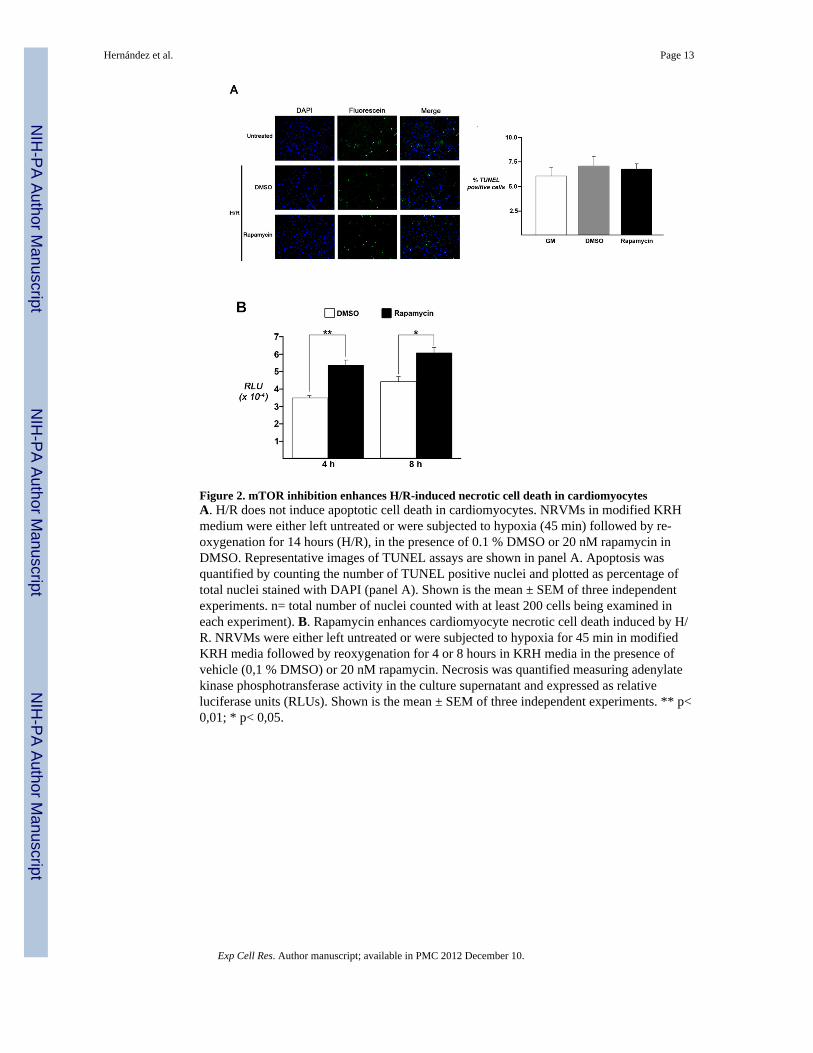
Figure 2. mTOR inhibition enhances H/R-induced necrotic cell death in cardiomyocytesA. H/R does not induce apoptotic cell death in cardiomyocytes. NRVMs in modified KRHmedium were either left untreated or were subjected to hypoxia (45 min) followed by re-oxygenation for 14 hours (H/R), in the presence of 0.1 % DMSO or 20 nM rapamycin inDMSO. Representative images of TUNEL assays are shown in panel A. Apoptosis wasquantified by counting the number of TUNEL positive nuclei and plotted as percentage oftotal nuclei stained with DAPI (panel A). Shown is the mean ± SEM of three independentexperiments. n= total number of nuclei counted with at least 200 cells being examined ineach experiment). B. Rapamycin enhances cardiomyocyte necrotic cell death induced by H/R. NRVMs were either left untreated or were subjected to hypoxia for 45 min in modifiedKRH media followed by reoxygenation for 4 or 8 hours in KRH media in the presence ofvehicle (0,1 % DMSO) or 20 nM rapamycin. Necrosis was quantified measuring adenylatekinase phosphotransferase activity in the culture supernatant and expressed as relativeluciferase units (RLUs). Shown is the mean ± SEM of three independent experiments. ** p<0,01; * p< 0,05.
Hernández et al. Page 13
Exp Cell Res. Author manuscript; available in PMC 2012 December 10.
NIH
-PA Author Manuscript
NIH
-PA Author Manuscript
NIH
-PA Author Manuscript
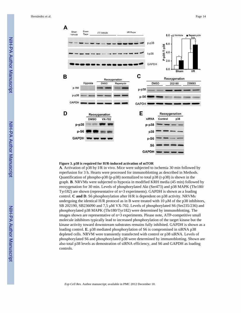
Figure 3. p38 is required for H/R-induced activation of mTORA. Activation of p38 by I/R in vivo. Mice were subjected to ischemia 30 min followed byreperfusion for 3 h. Hearts were processed for immunoblotting as described in Methods.Quantification of phospho-p38 (p-p38) normalized to total p38 (t-p38) is shown in thegraph. B. NRVMs were subjected to hypoxia in modified KRH media (45 min) followed byreoxygenation for 30 min. Levels of phosphorylated Akt (Ser473) and p38 MAPK (Thr180/Tyr182) are shown (representative of n=3 experiments). GAPDH is shown as a loadingcontrol. C and D. S6 phosphorylation after H/R is dependent on p38 activity. NRVMsundergoing the identical H/R protocol as in B were treated with 10 μM of the p38 inhibitors,SB 202190, SB236090 and 7,5 μM VX-702. Levels of phosphorylated S6 (Ser235/236) andphosphorylated p38 MAPK (Thr180/Tyr182) were determined by immunoblotting. Theimages shown are representative of n=3 experiments. Please note, ATP-competitive smallmolecule inhibitors typically lead to increased phosphorylation of the target kinase but thekinase activity toward downstream substrates remains fully inhibited. GAPDH is shown as aloading control. E. p38 mediated phosphorylation of S6 is compromised in siRNA p38depleted cells. NRVM were transiently transfected with control or p38 siRNA. Levels ofphosphorylated S6 and phosphorylated p38 were determined by immunoblotting. Shown arealso total p38 levels as demostration of siRNA efficiency, and S6 and GAPDH as loadingcontrols.
Hernández et al. Page 14
Exp Cell Res. Author manuscript; available in PMC 2012 December 10.
NIH
-PA Author Manuscript
NIH
-PA Author Manuscript
NIH
-PA Author Manuscript
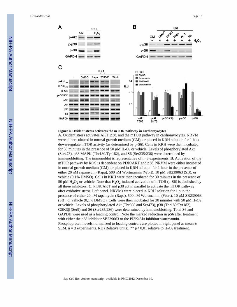
Figure 4. Oxidant stress activates the mTOR pathway in cardiomyocytesA. Oxidant stress activates AKT, p38, and the mTOR pathway in cardiomyocytes. NRVMwere either cultured in normal growth medium (GM), or placed in KRH solution for 1 h todown-regulate mTOR activity (as determined by p-S6). Cells in KRH were then incubatedfor 30 minutes in the presence of 50 μM H2O2 or vehicle. Levels of phosphorylated Akt(Ser473), p38 MAPK (Thr180/Tyr182), and S6 (Ser235/236) were determined byimmunoblotting. The immunoblot is representative of n=3 experiments. B. Activation of themTOR pathway by ROS is dependent on PI3K/AKT and p38. NRVM were either incubatedin normal growth medium (GM), or placed in KRH solution for 1 hour in the presence ofeither 20 nM rapamycin (Rapa), 500 nM Wortmannin (Wort), 10 μM SB239063 (SB), orvehicle (0,1% DMSO). Cells in KRH were then incubated for 30 minutes in the presence of50 μM H2O2 or vehicle. Note that H2O2-induced activation of mTOR (p-S6) is abolished byall three inhibitors. C. PI3K/AKT and p38 act in parallel to activate the mTOR pathwayafter oxidative stress. Left panel. NRVMs were placed in KRH solution for 1 h in thepresence of either 20 nM rapamycin (Rapa), 500 nM Wortmannin (Wort), 10 μM SB239063(SB), or vehicle (0,1% DMSO). Cells were then incubated for 30 minutes with 50 μM H2O2or vehicle. Levels of phosphorylated Akt (Thr308 and Ser473), p38 (Thr180/Tyr182),GSK3β (Ser9) and S6 (Ser235/236) were determined by immunoblotting. Total S6 andGAPDH were used as a loading control. Note the marked reduction in pS6 after treatmentwith either the p38 inhibitor SB239063 or the PI3K/Akt inhibitor wortmannin.Phosphoprotein levels normalized to loading controls are plotted in right panel as mean ±SEM. n = 3 experiments. RU (Relative units). ** p< 0,01 relative to H2O2 treatment.
Hernández et al. Page 15
Exp Cell Res. Author manuscript; available in PMC 2012 December 10.
NIH
-PA Author Manuscript
NIH
-PA Author Manuscript
NIH
-PA Author Manuscript
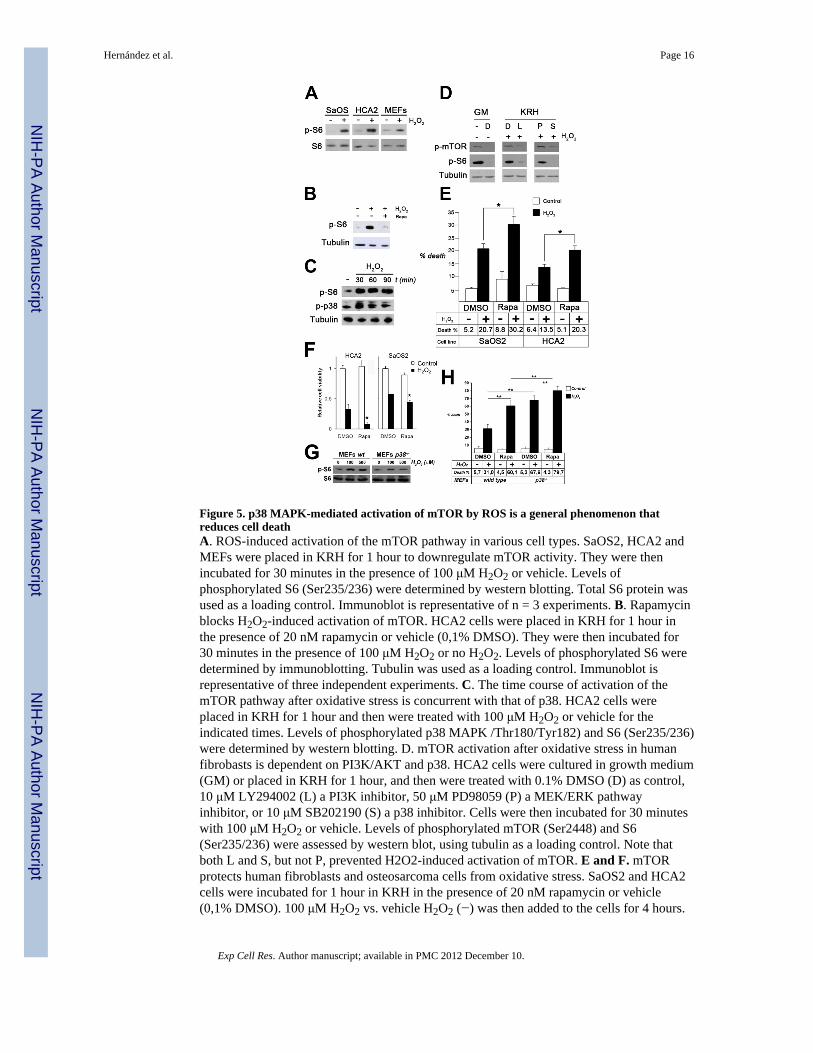
Figure 5. p38 MAPK-mediated activation of mTOR by ROS is a general phenomenon thatreduces cell deathA. ROS-induced activation of the mTOR pathway in various cell types. SaOS2, HCA2 andMEFs were placed in KRH for 1 hour to downregulate mTOR activity. They were thenincubated for 30 minutes in the presence of 100 μM H2O2 or vehicle. Levels ofphosphorylated S6 (Ser235/236) were determined by western blotting. Total S6 protein wasused as a loading control. Immunoblot is representative of n = 3 experiments. B. Rapamycinblocks H2O2-induced activation of mTOR. HCA2 cells were placed in KRH for 1 hour inthe presence of 20 nM rapamycin or vehicle (0,1% DMSO). They were then incubated for30 minutes in the presence of 100 μM H2O2 or no H2O2. Levels of phosphorylated S6 weredetermined by immunoblotting. Tubulin was used as a loading control. Immunoblot isrepresentative of three independent experiments. C. The time course of activation of themTOR pathway after oxidative stress is concurrent with that of p38. HCA2 cells wereplaced in KRH for 1 hour and then were treated with 100 μM H2O2 or vehicle for theindicated times. Levels of phosphorylated p38 MAPK /Thr180/Tyr182) and S6 (Ser235/236)were determined by western blotting. D. mTOR activation after oxidative stress in humanfibrobasts is dependent on PI3K/AKT and p38. HCA2 cells were cultured in growth medium(GM) or placed in KRH for 1 hour, and then were treated with 0.1% DMSO (D) as control,10 μM LY294002 (L) a PI3K inhibitor, 50 μM PD98059 (P) a MEK/ERK pathwayinhibitor, or 10 μM SB202190 (S) a p38 inhibitor. Cells were then incubated for 30 minuteswith 100 μM H2O2 or vehicle. Levels of phosphorylated mTOR (Ser2448) and S6(Ser235/236) were assessed by western blot, using tubulin as a loading control. Note thatboth L and S, but not P, prevented H2O2-induced activation of mTOR. E and F. mTORprotects human fibroblasts and osteosarcoma cells from oxidative stress. SaOS2 and HCA2cells were incubated for 1 hour in KRH in the presence of 20 nM rapamycin or vehicle(0,1% DMSO). 100 μM H2O2 vs. vehicle H2O2 (−) was then added to the cells for 4 hours.
Hernández et al. Page 16
Exp Cell Res. Author manuscript; available in PMC 2012 December 10.
NIH
-PA Author Manuscript
NIH
-PA Author Manuscript
NIH
-PA Author Manuscript
Medium was then changed to media lacking H2O2 with 20 nM rapamycin or vehicle (0,1%DMSO), and 24 hours later the percentage of non viable cells was determined by trypan blueexclusion (E, shown is the mean ± SEM of three independent experiments. *, p< 0,05) or byMTT assay expressed as the percentage of cell survival (F, values are the mean + SEM offour samples. *, p< 0,05). G. Activation of the mTOR pathway afer oxidative stress isabrogated in p38α−/− cells. WT and p38 α −/− MEFs were placed in KRH for 1 hour. Cellswere then treated with vehicle H2O2 (−), 100 μM or 500 μM H2O2 for 60 min.Phosphorylated S6 (Ser235/236) was determined by western blotting. Also shown are totalS6 levels.H. p38α deficiency and mTOR inhibition increase susceptibility of cells tooxidative stress. WT and p38α−/− MEFs were placed in KRH for 1 hour in the presence of20 nM rapamycin or vehicle (0,1% DMSO). 100 μM H2O2 or vehicle was then added to themedia for 4 hours. The medium was then changed to growth medium with 20 nM rapamycinor vehicle (0,1% DMSO), and 24 hours later cells were trypsinized and the percentage ofnon viable cells was determined by trypan blue exclusion. Shown is the mean ± SEM ofthree independent experiments. ** p< 0,01.
Hernández et al. Page 17
Exp Cell Res. Author manuscript; available in PMC 2012 December 10.
NIH
-PA Author Manuscript
NIH
-PA Author Manuscript
NIH
-PA Author Manuscript
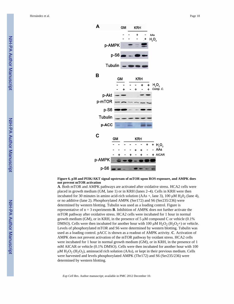
Figure 6. p38 and PI3K/AKT signal upstream of mTOR upon ROS exposure, and AMPK doesnot prevent mTOR activationA. Both mTOR and AMPK pathways are activated after oxidative stress. HCA2 cells wereplaced in growth medium (GM, lane 1) or in KRH (lanes 2–4). Cells in KRH were thenincubated for 30 minutes in amino acid-rich solution (AAs +, lane 3), 100 μM H202 (lane 4),or no additive (lane 2). Phosphorylated AMPK (Ser172) and S6 (Ser235/236) weredetermined by western blotting. Tubulin was used as a loading control. Figure isrepresentative of n = 3 experiments B. Inhibition of AMPK does not further activate themTOR pathway after oxidative stress. HCA2 cells were incubated for 1 hour in normalgrowth medium (GM), or in KRH, in the presence of 5 μM compound C or vehicle (0.1%DMSO). Cells were then incubated for another hour with 100 μM H2O2 (H2O2+) or vehicle.Levels of phosphorylated mTOR and S6 were determined by western blotting. Tubulin wasused as a loading control. pACC is shown as a readout of AMPK activity. C. Activation ofAMPK does not prevent activation of the mTOR pathway by oxidant stress. HCA2 cellswere incubated for 1 hour in normal growth medium (GM), or in KRH, in the presence of 1mM AICAR or vehicle (0.1% DMSO). Cells were then incubated for another hour with 100μM H2O2 (H2O2), aminoacid rich solution (AAs), or kept in their previous medium. Cellswere harvested and levels phosphorylated AMPK (Thr172) and S6 (Ser235/236) weredetermined by western blotting.
Hernández et al. Page 18
Exp Cell Res. Author manuscript; available in PMC 2012 December 10.
NIH
-PA Author Manuscript
NIH
-PA Author Manuscript
NIH
-PA Author Manuscript

Figure 7. Regulation of mTOR by ROS, REDD1 downregulation and TSC2/14-3-3 associationA. Oxidative stress downregulates REDD1 protein. SaOS2 and HCA2 cells were placed inKRH for 1 h. They were then incubated for 30 minutes in the presence 100 μM H2O2 orvehicle. For HCA2 cells, the third lane is from cells incubated in normal growth medium(GM) and is shown for comparison. Levels of phosphorylated mTOR and S6 and totalREDD1 were determined by western blotting. Tubulin was used as a loading control.Immunoblot is representative of n = 3 experiments. B. Oxidative stress induces dissociationof 14-3-3 from REDD1 and association with TSC2. HCA2 cells were either incubated ingrowth medium (GM), or placed in KRH for 1 h. Cells in KRH were then incubated for 30minutes in the presence of 100 μM H2O2 where indicated. Cell extracts were either rundirectly, or immunoprecipitated with anti-14-3-3 or an non-specific antibody (Mock). TSC2,14-3-3 and REDD1 levels were determined by western blot. The arrowhead points to a non-specific band seen only in the extracts immunoprecipitated with the non-specific antibody.C. Association of TSC2 with 14-3-3 after oxidative stress is dependent on p38 but not onPI3K/AKT. HCA2 cells were either incubated in growth medium (GM), or placed in KRHfor 1 h, in the presence or absence of 10 μM LY294002 (L), 10 μM SB202190 (S), orvehicle (DMSO, D). They were then incubated for 30 minutes in the presence 100 μM ofH2O2 where indicated. Cell extracts were either run directly, or immunoprecipitated withanti-14-3-3 or a non-specific antibody (Mock). TSC2 and 14-3-3 levels in theimmunoprecipitate were determined by western blotting. Note the markedly lower amount
Hernández et al. Page 19
Exp Cell Res. Author manuscript; available in PMC 2012 December 10.
NIH
-PA Author Manuscript
NIH
-PA Author Manuscript
NIH
-PA Author Manuscript

of TSC2 associating with 14-3-3 in the setting of p38 inhibition with SB202190 (lane 7). D.REDD1 downregulation after oxidative stress is dependent on p38. HCA2 cells wereincubated for 1 hour in KRH in the presence of 20 μM SB239063 or vehicle. They were thenincubated for 30 minutes in the presence 100 μM of H2O2 or vehicle. Cell extracts wereeither run directly, or immunoprecipitated with anti-14-3-3 antibody. Shown are TSC2,REDD1 and 14-3-3 in the inmunoprecipitate, and phosphorylated Akt (Ser473),phosphorylated S6 (Ser235/236), total Akt, S6, TSC2, REDD1 and 14-3-3 in whole cellextracts. GAPDH was used as a loading control. Note the reduction in the amount of TSC2in the 14-3-3 immunoprecipitate when the p38 inhibitor SB239063 is present (5th panel,compare lanes 2 and 3).
Hernández et al. Page 20
Exp Cell Res. Author manuscript; available in PMC 2012 December 10.
NIH
-PA Author Manuscript
NIH
-PA Author Manuscript
NIH
-PA Author Manuscript
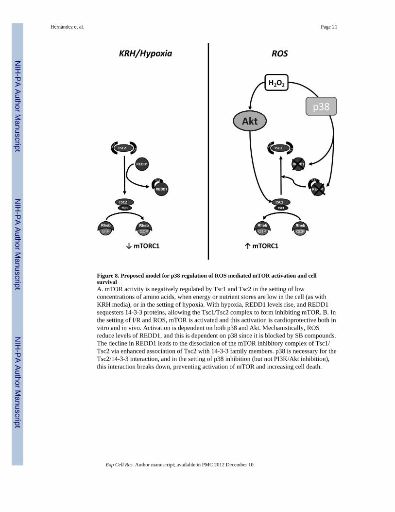
Figure 8. Proposed model for p38 regulation of ROS mediated mTOR activation and cellsurvivalA. mTOR activity is negatively regulated by Tsc1 and Tsc2 in the setting of lowconcentrations of amino acids, when energy or nutrient stores are low in the cell (as withKRH media), or in the setting of hypoxia. With hypoxia, REDD1 levels rise, and REDD1sequesters 14-3-3 proteins, allowing the Tsc1/Tsc2 complex to form inhibiting mTOR. B. Inthe setting of I/R and ROS, mTOR is activated and this activation is cardioprotective both invitro and in vivo. Activation is dependent on both p38 and Akt. Mechanistically, ROSreduce levels of REDD1, and this is dependent on p38 since it is blocked by SB compounds.The decline in REDD1 leads to the dissociation of the mTOR inhibitory complex of Tsc1/Tsc2 via enhanced association of Tsc2 with 14-3-3 family members. p38 is necessary for theTsc2/14-3-3 interaction, and in the setting of p38 inhibition (but not PI3K/Akt inhibition),this interaction breaks down, preventing activation of mTOR and increasing cell death.
Hernández et al. Page 21
Exp Cell Res. Author manuscript; available in PMC 2012 December 10.
NIH
-PA Author Manuscript
NIH
-PA Author Manuscript
NIH
-PA Author Manuscript