Proteomic response of human neuroblastoma cells to azaspiracid-1
Upload
independentCategory
view
2download
0
OPEN
p38MAPK inhibition: a new combined approach toreduce neuroblastoma resistance under etoposidetreatment
B Marengo1, CG De Ciucis1, R Ricciarelli1, AL Furfaro1, R Colla1, E Canepa1, N Traverso1, UM Marinari1, MA Pronzato1
and C Domenicotti*,1
Neuroblastoma (NB) is the second most common solid pediatric tumor and is characterized by clinical and biologicalheterogeneity, and stage-IV of the disease represents 50% of all cases. Considering the limited success of present chemotherapytreatment, it has become necessary to find new and effective therapies. In this context, our approach consists of identifying andtargeting key molecular pathways associated with NB chemoresistance. This study has been carried out on three stage-IV NBcell lines with different status of MYCN amplification. Cells were exposed to a standard chemotherapy agent, namely etoposide,either alone or in combination with particular drugs, which target intracellular signaling pathways. Etoposide alone induced aconcentration-dependent reduction of cell viability and, at very high doses, totally counteracted cell tumorigenicity andneurosphere formation. In addition, etoposide activated p38 mitogen-activated protein kinase (MAPK), AKT and c-Jun N-terminalkinase. Pre-treatment with SB203580, a p38MAPK inhibitor, dramatically sensibilized NB cells to etoposide, strongly reducing thedosage needed to inhibit tumorigenicity and neurosphere formation. Importantly, SB203580–etoposide cotreatment alsoreduced cell migration and invasion by affecting cyclooxygenase-2, intercellular adhesion molecule-1, C–X–C chemokinereceptor-4 and matrix metalloprotease-9. Collectively, our results suggest that p38MAPK inhibition, in combination with standardchemotherapy, could represent an effective strategy to counteract NB resistance in stage-IV patients.Cell Death and Disease (2013) 4, e589; doi:10.1038/cddis.2013.118; published online 11 April 2013Subject Category: Cancer
Neuroblastoma (NB) is the second most common pediatricsolid malignant tumor and is characterized by biological andclinical heterogeneity.1,2 While low-risk NB may sponta-neously regress or differentiate into benign ganglioneuroblas-toma, high-risk NB (stage-IV) results in metastaticdissemination3 and only 20% of patients survive 5 years fromdiagnosis in spite of aggressive chemotherapy.4
Current therapeutic stratification of patients with NB isbased on risk assessment according to combinations of age,tumor stage, MYCN status, DNA ploidy status and histo-pathology. This biological heterogeneity renders it necessaryto identify additional markers for stratification and prognos-tication, as well as molecular pathways that can be targeted incombination with standard chemotherapy. However, theefficacy of the single-agent therapy is limited by mechanismsof resistance that hinder its clinical success. A factor thatcontributes to the malignancy of NB is the presence of a sub-population of chemo- and radio-resistant stem cells in thetumor bulk.5 These cancer stem-like cells (CSCs) contributeto both cancer progression and metastases. In NB, in fact,
neurospheres (NBSs), the CSCs of neuronal origin, havebeen found in primary tumor specimens, as well as inestablished cell lines.6
Moreover, it has been demonstrated that therapy-resistantaggressive NBs frequently overexpress and secrete highlevels of growth factors and chemokines,7 which are able toactivate growth signaling pathways, thereby providing asuitable microenvironment for tumor development.8,9
In this study, we analyzed the main survival and deathpathways triggered by etoposide, a commonly used chemo-therapeutic compound, in two MYCN-amplified and onenon-amplified cell lines. In particular, our study was stronglyfocused on HTLA-230, one of the MYCN-amplified NB celllines, isolated from the bone-marrow aspirate of a patient withthe stage-IV disease.10 These cells are highly tumorigenic11
and phenotypically similar to other metastatic bone marrow-isolated NB cells.12
Our results demonstrate that the etoposide resistance ofNB cells is due to the presence of NBSs and suggest thatSB203580, a specific p38 mitogen-activated protein kinase
1Department of Experimental Medicine, University of Genoa, Via L.B. Alberti 2, Genoa, Italy*Corresponding author: C Domenicotti, Department of Experimental Medicine, University of Genoa, Via L.B. Alberti 2, Genoa 16132, Italy. Tel: +39 010 3538830;Fax: +39 010 3538836; E-mail: [email protected]
Received 24.10.12; revised 07.3.13; accepted 12.3.13; Edited by G Raschella
Keywords: MAPK; neuroblastoma; etoposideAbbreviations: bFGF, basic fibroblast growth factor; COX, cyclooxygenase; CSCs, cancer stem-like cells; CXCR, C–X–C chemokine receptor; EGF, epidermal growthfactor; FBS, fetal bovine serum; FITC, fluorescein isothiocyanate; ICAM, intercellular adhesion molecule; JNK, c-Jun N-terminal kinase; MAPK, mitogen-activatedprotein kinase; MEK, MAP kinase kinase; MKP-1, MAPK phosphatase-1; MMP, matrix metalloprotease; MTT, dimethylthiazolyl-2-5-diphenyltetrazolium bromide; NB,neuroblastoma; NBSs, neurospheres; PI, propidium iodide; PI-3 kinase, phosphatidylinositol 3-kinases; PKC, protein kinase C; ROS, reactive oxygen species; SDF,stromal-derived factor; VEGF, vascular endothelial growth factor
Citation: Cell Death and Disease (2013) 4, e589; doi:10.1038/cddis.2013.118& 2013 Macmillan Publishers Limited All rights reserved 2041-4889/13
www.nature.com/cddis

(MAPK) inhibitor, in combination with etoposide, may beeffective in preventing cell growth, invasion, migration,angiogenesis and NBS generation, which are all factorsresponsible for the relapse and progression of NB.
Results
Etoposide induces a dose-dependent reduction of cellviability and high doses totally counteract the tumori-genicity of NB cells and the formation of NBSs. NB cellswere exposed for 24 h to increasing concentrations ofetoposide (0.07–225 mM). As shown in Figure 1a, etoposideinduced a dose-dependent reduction of cell viability, startingat a 10 mM concentration and reaching a 70% decrease at225mM. As shown in Figure 1b (left panel), untreated cellswere able to form colonies (68 colonies of 450 cells).Similarly, NB cells exposed for 24 h to 1.25 mM etoposide, aconcentration that mimics in vitro the dose used in clinicaltherapy,13 formed colonies (44 colonies of 450 cells). On thecontrary, higher doses of etoposide (from 10 to 225 mM)totally suppressed the clonogenicity of these cells.
Since the anchorage-independent growth is useful indetecting colonies, not appreciated by a clonogenic assay,14
cells treated for 24 h with etoposide were grown in a semisolidagar. Similarly, as shown in Figure 1b (right panel), colonieswere detected only in untreated samples (25 colonies of 425cells) and in 1.25 mM etoposide samples (16 colonies of 425cells).
When cells were plated above the clonal density (10 cellsper ml) and grown under appropriate conditions, many NBSswere observed within 1 week in both untreated and 1.25 mMetoposide-treated cells (Figure 1c). Interestingly, the quantityof NBSs increased with the passage number (Figure 1c), buthigher doses of etoposide (10–100 mM) prevented the forma-tion of NBSs, already during the first week (Figure 1c).
As shown in Figure 1d, untreated and etoposide-treatedmonolayer cells expressed CD133 and Oct4, known stem cellmarkers.15 Moreover, in NBSs, at the eighth passage, eitheroriginating from the control or from the treated cells, CD133increased twofold, whereas Oct4 increased by 40 and 85% inuntreated and treated NBSs, respectively (Figure 1d).
Since the effects on cell viability and clonogenicity, inducedby 100mM etoposide, were comparable to that induced by thehigher doses, the subsequent analyses were performed until100mM.
At 24 h of treatment of cells with etoposide (10–100 mM)induced a dose-dependent increase of apoptotic cells and anecrotic effect was observed at 50 and 100 mM etoposide.
Etoposide produced a dose-dependent increase in dichlor-ofluorescein (DCF)-positive cells that became fivefold higherat 100mM, and g-H2AX, a marker of DNA double-strandbreaks, was induced in etoposide-treated cells (data notshown).
Etoposide activates p38MAPK, AKT and JNK. As shownin Figure 2a, 24-h etoposide increased protein kinase C(PKC)d and reduced PKCa levels. By analyzing the down-stream molecular pathways of PKC, etoposide induced adose-dependent activation of p38MAPK, already at 1.25 mM
(Figure 2b, left panel). In addition, c-Jun N-terminal kinase(JNK) was activated by 60 and 30%, at 1.25 and 10 mM,respectively, but no effect was observed at the otherconcentrations (Figure 2b, right panel). Moreover, etoposideincreased the activity of Akt (phospho-Thr-Akt/Akt) by 70% incells treated with the dose of 1.25mM and by 50 and 35%(phospho-Ser-Akt/Akt), respectively, in cells exposed to 50and 100mM (Figure 2b, lower panel).
SB203580/etoposide cotreatment decreases cell viabi-lity, reduces the clonogenicity and inhibits the formationof NBSs. Since all signaling molecules analyzed werealready activated at 1.25 mM etoposide, the effects of specificenzymatic inhibitors were investigated at this concentrationof etoposide. As shown in Figure 3a (left panel), a reductionof cell viability was observed when etoposide-treated cellswere pre-exposed to LY290042 (phosphatidylinositol3-kinase (PI-3-kinase)/Akt inhibitor; 15% decrease), toSB203580 (p38MAPK inhibitor; 17% decrease) and toSP600125 (JNK inhibitor; 30% decrease). As shown inFigure 3a (right panel), while PD98059 (MEK inhibitor) pre-treatment increased the ability of NB cells to form colonies inthe presence of etoposide, surprisingly, pre-treatment withSB203580 markedly reduced the tumorigenicity of etoposide-treated cells (60% decrease).
Treatment with the inhibitors that affected cell viability andtumorigenicity did not alter per se the number of NBSs (datanot shown). As shown in Figure 3b, etoposide did not modifythe number of NBSs, even in the presence of pre-treatmentwith LY290042 or SP600125 (first passage). However, whencells were pre-treated with SB203580 and then exposed toetoposide, the formation of NBSs was totally absent, evenfrom the first passage (Figure 3b).
In addition, the progressive increase in NBSs observed inuntreated, etoposide- and cotreated cells was dependent onpassages and lasted for a period of 5 weeks (Figure 3b). After6 weeks, the cotreatments did not change the number ofNBSs (Figure 3b).
In the NBSs originating from untreated and etoposide-treated cells, p38MAPK was activated 18-fold compared withmonolayer cells (Figure 3c, left panel), whereas the expres-sion of MAPK phosphatase-1 (MKP-1), p38MAPK inhibitor,did not change (Figure 3c, right panel).
SB203580/etoposide or SP600125/etoposide cotreat-ments inhibit the formation of capillary-like structures.The ability of NB cells to form a network of tubes was notmodified by etoposide or LY290042 after 24 h treatment(Figure 4a). Instead, SB203580 and SP600125 alonedecreased the number of branches in the tube network by55% with regard to untreated cells (Figure 4a, graph).
While the association of LY290042 with etoposide did notalter the formation of tubes, the cotreatment with SB203580 orSP600125 decreased the number of branches by 90% withregard to etoposide-treated cells (Figure 4a, graph). More-over, tubes formed by untreated, etoposide- or LY290042-treated and cotreated cells persisted for up to 3 days. Similarresults were observed in cells incubated in medium withoutbasic fibroblast growth factor (bFGF) and vascular endothelialgrowth factor (VEGF) (data not shown).
p38MAPK role in chemoresistanceB Marengo et al
2
Cell Death and Disease

Furthermore, SB203580, alone or in combination withetoposide, reduced VEGF by 61 and 69%, respectively(Figure 4b). SP600125 alone was able to increase the VEGFamount twofold, but its combination with etoposide did notmodify the VEGF expression (Figure 4b).
SB203580/etoposide cotreatment reduces cell migrationand invasion by affecting COX-2, ICAM-1, CXCR4expression and MMP-9 secretion. Cell migration was notaltered by etoposide (Figure 4c) or by LY290042 orSB203580 or SP600125 administered alone (data not shown).
Figure 1 Etoposide decreases cell viability and, at high drug concentrations, inhibits the tumorigenic potential of HTLA-230 NB cells and prevents NBS formation. (a) Cellviability was determined by MTT assays in cells exposed to increasing concentrations of etoposide (0.07–225mM) for 24 h. Histograms summarize quantitative data ofmeans±S.D. of five independent experiments. *Po0.01 versus untreated cells (Ctr). (b) Left panel, clonogenic assay. HTLA-230 cells were seeded in six-well plates andthen incubated with 1.25, 10, 50, 100, 150 and 225mM etoposide, as indicated, for 24 h. Subsequently, cells were incubated in fresh medium without the drug for an additional20 days before staining and counting the colonies. Right panel, soft-agar colony formation assay. HTLA-230 cells were treated with 1.25, 10, 50, 100, 150 and 225mMetoposide, as indicated, for 24 h, washed and re-plated in agar-containing medium. After 25 days, colonies were stained and counted. (c) Left panel, NBS formation. Aftertreatment with etoposide (1.25–100mM), HTLA-230 cells were cultured in serum-free culture conditions containing bFGF and EGF for 1 week (first passage). Originalmagnification � 10. Right panel, graph. At every passage (one per week), the NBSs in untreated (Ctr) and in 1.25mM etoposide-treated cells were counted by analysis underan inverted microscope. The histogram summarizes quantitative data of means±S.D. of five independent experiments. (d) RT-PCR analyses of CD133 and Oct4 in untreatedand etoposide-treated monolayer cells and in NBSs originating from by the same monolayer cells. The glyceraldehyde 3-phosphate dehydrogenase (GAPDH) signal is theinternal loading control. Results are representative of three independent experiments with essentially similar results. Histogram, reported in the right panel, summarizesquantitative data of means, normalized to GAPDH expression±S.D. of three independent experiments. *Po0.01 versus monolayer cells
p38MAPK role in chemoresistanceB Marengo et al
3
Cell Death and Disease

Similarly, cotreatments of etoposide with LY290042 orSP600125 did not affect the cell migration (data not shown).It is worth noting that pre-treatment with SB203580was able to reduce cell migration by 65% and 50%,evaluated by the scratch and Transwell assays, respec-tively (Figure 4c).
Cell invasion was reduced by 33% after etoposide treat-ment and was further inhibited by 51% and 80% afterLY290042 and SB203580 cotreatments, respectively(Figure 4d). Moreover, SP600125 cotreatment did not changethe number of membrane-invading cells (Figure 4d).LY290042 or SB203580 alone reduced the cell invasion by34% and 60%, respectively, while SP600125 per se wasuneffective (data not shown).
Considering the effects induced by SB203580 cotreatmenton cell migration and invasion, some molecular markers,known to be related to the invasive phenotype, wereinvestigated.
As shown in Figure 5a, etoposide induced a 60% increasein the cyclooxygenase (COX)-2 levels, an effect that wastotally inhibited by the pre-treatment with SB203580. More-over, treatment with SB203580 alone did not modify theCOX-2 levels in untreated cells (Figure 5a).
Intercellular adhesion molecule-1 (ICAM-1) was reduced by25% after etoposide and by 65% after SB203580 alone withregard to untreated cells (Figure 5b). Moreover, SB203580cotreatment reduced the ICAM-1 levels found after etoposideby 40% (Figure 5b).
Figure 2 Etoposide activates p38MAPK, Akt and JNK. (a) Protein levels of PKC d and a in cells treated with etoposide (1.25–100mM). Immunoblots shown arerepresentative of three independent experiments. b-Actin is the internal loading control. (b) p38MAPK, JNK and Akt activation. Histograms summarize quantitative data ofmeans±S.D. of three independent experiments. *Po0.05 and **Po0.01 versus untreated (Ctr) cells. Data are expressed as a ratio of the levels of phosphorylated proteinsto the levels of unphosphorylated ones, whose values have been previously normalized against b-actin
p38MAPK role in chemoresistanceB Marengo et al
4
Cell Death and Disease

Figure 3 Effects of SB203580 (SB) cotreatment on cell viability, clonogenicity and formation of NBSs. (a) Left panel, cell viability. Cells were pre-treated for 1 h with thedifferent inhibitors (0.1mM chelerythrine chloride (Chele), 500 nM LY2940042 (LY), 50 mM PD98059 (PD), 10mM SB203580 or 4 mM SP600125 (SP)) and then exposed to1.25mM etoposide for an additional 24 h. Histogram summarizes quantitative data of means±S.D. of five independent experiments. *Po0.05 versus etoposide-treated cellsand **Po0.01 versus etoposide-treated cells. Right panel, clonogenic assay. Histogram summarizes quantitative data of means±S.D. of five independent experiments.11Po0.01 versus untreated (Ctr) cells and **Po0.01 versus etoposide-treated cells. (b) Growth curve of NBSs. At every passage (one per week), the NBSs originating frometoposide and inhibitor pre-treated cells were counted by analysis under an inverted microscope. Histogram summarizes quantitative data of means±S.D. of threeindependent experiments. *Po0.05 and **Po0.01 versus etoposide-treated cells. (c) Left panel, p38MAPK activation in untreated monolayer cells and in NBSs. Histogramsummarizes quantitative data of means±S.D. of three independent experiments. *Po0.05 versus untreated monolayer cells and **Po0.01 versus monolayer cells. Thedata are expressed as a ratio of the levels of phosphorylated proteins to unphosphorylated proteins, whose values have been previously normalized with the relativeglyceraldehyde 3-phosphate dehydrogenase (GAPDH) levels. Right panel, immunoblot analysis of MKP-1 in untreated monolayer cells and in NBSs. Histogram summarizesquantitative data of means±S.D. of three independent experiments
p38MAPK role in chemoresistanceB Marengo et al
5
Cell Death and Disease

As shown in Figure 5c, etoposide or SB203580 alonedid not alter the C–X–C chemokine receptor-4 (CXCR4)levels, while cotreatment was able to decrease the CXCR4by 60%.
Analyses of matrix metalloprotease (MMP) activity demon-strated that MMP-9 was secreted by untreated cells(Figure 5d). In addition, etoposide or SB203580 alone didnot influence the MMP-9 secretion (Figure 5d). However,
Figure 4 SB203580 (SB) or SP600125 (SP) inhibit the formation of capillary-like structures and SB203580 cotreatment reduces migration and invasion of etoposide-treated cells. (a) Formation of capillary-like structures. Representative micrographs of the complete network of tubes formed by untreated (Ctr), treated (with etoposide,LY2940042, SB203580 or SP600125 alone) and cotreated cells (etoposide plus inhibitors). The negative control is obtained by cell exposure to 10 mM sulforaphane. Originalmagnification � 10. The graph reports the number of branches of the tube network formed by cells under the treatment conditions as described above. Quantitative data arethe means±S.D. of three independent experiments. 11Po0.01 versus untreated (Ctr) cells and **Po0.01 versus etoposide-treated cells. (b) Immunoblot analysis of VEGF.The histogram summarizes quantitative data of means±S.D. of three independent experiments 1Po0.05 versus untreated (Ctr) cells and *Po0.05 versus etoposide-treatedcells. The data are expressed as a ratio of VEGF to glyceraldehyde 3-phosphate dehydrogenase (GAPDH) amounts. (c) Migration assay. Cell migration was evaluated by thescratch and Transwell assays. In the scratch assay, the rate of migration was quantified by measuring the distance between the migrating cell boundaries. In the Transwellassay, migration was quantified by counting the number of cells, which moved to the underside of the membrane after 24 h of treatment. Histogram summarizes quantitativedata of means±S.D. of three independent experiments. *Po0.01 versus etoposide-treated cells. (d) Invasion assay. Cell invasion was quantified by counting the number ofcells, which moved to the underside of the coated membrane after 24 h of treatment. Histogram summarizes quantitative data of means±S.D. of six fields per membrane ofthree independent experiments. 1Po0.01 versus untreated (Ctr) cells and *Po0.01 versus etoposide-treated cells
p38MAPK role in chemoresistanceB Marengo et al
6
Cell Death and Disease
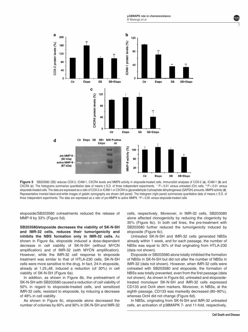
etoposide/SB203580 cotreatments reduced the release ofMMP-9 by 33% (Figure 5d).
SB203580/etoposide decreases the viability of SK-N-SHand IMR-32 cells, reduces their tumorigenicity andinhibits the NBS formation only in IMR-32 cells. Asshown in Figure 6a, etoposide induced a dose-dependentdecrease in cell viability of SK-N-SH (without MYCNamplification) and of IMR-32 (with MYCN amplification).However, while the IMR-32 cell response to etoposidetreatment was similar to that of HTLA-230 cells, SK-N-SHcells were more sensitive to the drug. In fact, 24-h etoposide,already at 1.25 mM, induced a reduction (of 30%) in cellviability of SK-N-SH (Figure 6a).
In addition, as shown in Figure 6b, the pretreatment ofSK-N-SH with SB203580 caused a reduction of cell viability of50% in regard to etoposide-treated cells, and sensitizedIMR-32 cells, resistant to etoposide, by inducing a decreaseof 48% in cell viability.
As shown in Figure 6c, etoposide alone decreased thenumber of colonies by 60% and 90% in SK-N-SH and IMR-32
cells, respectively. Moreover, in IMR-32 cells, SB203580alone affected clonogenicity by reducing the clogenicity by35% (Figure 6c). In both cell lines, the pre-treatment withSB203580 further reduced the tumorigenicity induced byetoposide (Figure 6c).
Untreated SK-N-SH and IMR-32 cells generated NBSsalready within 1 week, and for each passage, the number ofNBSs was equal to 30% of that originating from HTLA-230(data not shown).
Etoposide or SB203580 alone totally inhibited the formationof NBSs in SK-N-SH but did not alter the number of NBSs inIMR-32 (data not shown). However, when IMR-32 cells werecotreated with SB203580 and etoposide, the formation ofNBSs was totally prevented, even from the first passage (datanot shown). As shown in Figure 6d, untreated and etoposide-treated monolayer SK-N-SH and IMR-32 cells expressedCD133 and Oct4 stem markers. Moreover, in NBSs, at theeighth passage, CD133 was markedly decreased (80–90%),whereas Oct4 did not change (Figure 6d).
In NBSs, originating from SK-N-SH and IMR-32 untreatedcells, an activation of p38MAPK 7- and 11-fold, respectively,
Figure 5 SB203580 (SB) reduces COX-2, ICAM-1, CXCR4 levels and MMP9 activity in etoposide-treated cells. Immunoblot analyses of COX-2 (a), ICAM-1 (b) andCXCR4 (c). The histograms summarize quantitative data of means±S.D. of three independent experiments. 11Po0.01 versus untreated (Ctr) cells; **Po0.01 versusetoposide-treated cells. The data are expressed as a ratio of COX-2 or ICAM-1 or CXCR4 to glyceraldehyde 3-phosphate dehydrogenase (GAPDH) amounts. MMP9 activity (d).Representative inverted black-and-white images of gelatin zymography are shown (left panel). The histogram (right panel) summarizes quantitative data of means±S.D. ofthree independent experiments. The data are expressed as a ratio of pro-MMP9 to active MMP9. *Po0.05 versus etoposide-treated cells
p38MAPK role in chemoresistanceB Marengo et al
7
Cell Death and Disease

Figure 6 Effects of SB203580 (SB) cotreatment on viability, clonogenicity, CC133/Oct4 expression and p38MAPK activation in SK-N-SH and IMR-32 cells. (a) Cell viability.SK-N-SH (left panel) and IMR-32 (right panel) cells were exposed to increasing concentrations of etoposide (0.07–225mM) for 24 h. Histograms summarize quantitative data ofmeans±S.D. of five independent experiments. *Po0.05 and **Po0.01 versus untreated cells (Ctr). (b) Cell viability. SK-N-SH (left panel) and IMR-32 (right panel) cells weretreated with 1.25mM etoposide/10mM SB203580 alone or pre-treated for 1 h with 10mM SB203580 and then exposed to 1.25mM etoposide for an additional 24 h. Histogramsummarizes quantitative data of means±S.D. of five independent experiments. 1Po0.05 versus untreated cell (Ctr) and *Po0.05 versus etoposide-treated cells.(c) Clonogenic assay. SK-N-SH (left panel) and IMR-32 (right panel) cells were seeded in six-well plates and then incubated with 1.25mM etoposide/10mM SB203580 alone orcotreated with SB203580þ etoposide for 24 h. Subsequently, cells were incubated in fresh medium without the drug for an additional 20 days before staining and counting thecolonies. Histogram summarizes quantitative data of means±S.D. of five independent experiments. 11Po0.01 versus untreated cell (Ctr) and *Po0.05 versus etoposide-treated cells. (d) RT-PCR analyses of CD133 and Oct4 in untreated and etoposide-treated monolayer cells and in NBSs originating from the same monolayer cells. Thehistogram summarizes quantitative data of means, normalized to glyceraldehyde 3-phosphate dehydrogenase (GAPDH) expression,±S.D. of three independent experiments.**Po0.01 versus monolayer cells. (e) p38MAPK activation in monolayer cells and in NBSs. Histogram summarizes quantitative data of means±S.D. of three independentexperiments. 1Po0.05 and 11Po0.01 versus untreated monolayer cells and **Po0.01 versus etoposide-treated monolayer cells. The data are expressed as a ratio of thelevels of phosphorylated proteins to unphosphorylated proteins, whose values have been previously normalized with the relative GAPDH levels
p38MAPK role in chemoresistanceB Marengo et al
8
Cell Death and Disease

was found in comparison to the monolayer cells (Figure 6e).Furthermore, in the NBSs from etoposide-treated IMR-32cells, p38MAPK was activated eightfold compared withmonolayer etoposide-treated ones (Figure 6e). No changein MKP-1 was observed (data not shown).
SB203580 plus etoposide decreases VEGF levels, mark-edly reduces cell migration/invasion and MMP-9 secre-tion. SK-N-SH and IMR-32 cells were unable to formcapillary-like structures (Figure 7a). However, in these celllines, etoposide alone reduced VEGF by 30% in SK-N-SHand by 15% in IMR-32 cells (Figure 7b). Similarly, SB203580decreased VEGF by 38% in SK-N-SH and by 48% in IMR-32cells (Figure 7b). In addition, SB203580, in combination withetoposide, further reduced VEGF by 20% and 50% in SK-N-SH and IMR-32 cells, respectively (Figure 7b).
As shown in Figure 7c, cotreatment of SB203580 withetoposide was able to reduce the cell migration of SK-N-SH by77% and of IMR-32 cells by 40%, respectively (Figure 7c). Inaddition, etoposide and SB203580 alone were able to reducecell migration of SK-N-SH by 45% and 40%, respectively(Figure 7c, left panel).
SB203580 alone or in combination with etoposidedecreased by 80–83% the invasiveness of both cell lines(Figure 7d).
As shown in Figure 7e (left panel), etoposide or SB203580alone reduced the secretion of MMP-9 from SK-N-SH cells by30% and 75%, respectively. In IMR-32, etoposide did notinfluence the MMP-9 secretion (Figure 7e, right panel), whileSB203580 alone reduced the MMP-9 release by 60%(Figure 7e, right panel). However, etoposide plus SB203580reduced the release of MMP-9 by 22% in SK-N-SH and by42% in IMR-32, with regard to cells treated with etoposidealone (Figure 7e).
Discussion
In this paper, we demonstrate that HTLA-230 and IMR-32,both MYCN-amplified cells, are highly etoposide-resistant, asonly the doses ranging from 10 to 225 mM are able to reducecell survival. In addition, HTLA-230 are more resistant thanIMR-32 because 1.25 mM etoposide, a concentration thatin vitro mimics the dose used in clinical therapy,13 exerts a lessmarked antitumorigenic effect on HTLA-230, whereas thesame dose of etoposide strongly decreases the clonogenicityof IMR-32 cells.
However, all NB cells analyzed are able to generate NBSs,but only in cells with MYCN-amplification, the treatment withetoposide does not interfere with NBS formation. Theseresults are in agreement with a paper demonstrating thatNBS-derived cells, originating from pediatric brain tumors,have an increased resistance to etoposide compared withmonolayer-derived cells.16 In addition, it has been demon-strated that only NB stage-IV-derived cells generate spheres,but that the MYCN expression status is not related to thesphere formation.17
In this paper, we demonstrate that in NBSs, originatingfrom HTLA-230 cells, the levels of stemness markers (CD133and Oct4) are enhanced, while in NBSs, originating fromSK-N-SH and IMR-32 cells, the expression of CD133 is
reduced and Oct4 do not change. These results are in linewith a report demonstrating that CD133 expression isincreased in spheres but not in every analyzed spherederived from NB samples and cell lines.17,18 Probably, theoverexpression of stemness markers contributes to render-ing HTLA-230 more resistant to etoposide, and in this regard,it has been demonstrated that CD133 expression in NB cellsis associated with resistance to doxorubicin, vincristine andcisplatin.19
In accordance with other studies,20 we have recentlyreported that etoposide causes DNA damage and an over-production of reactive oxygen species (ROS),21 which havebeen demonstrated to mediate both cell damage andbiological functions.22 In this regard, herein we show thatetoposide induces a dose-dependent increase in the levels ofthe proapoptotic PKCd23 and a parallel decrease of PKCa, theantiapoptotic isoform.24
However, given that PKCs are upstream molecules in theROS signaling pathway leading to DNA damage andapoptosis,21,25,26 it is important to identify the downstreammediators of the NB response to etoposide and we show thatetoposide induces the activation of Akt and MAPKs (i.e. JNK,and p38). It is worth noting that the activation of MAPKs hasbeen reported in over 50% of acute myelogenous andlymphocytic leukemia and that MAPKs are also stimulated inother tumors,27,28 therefore implying that the inhibition of theMAPK pathways could represent an important strategy tocounteract tumor growth. In this context, our results demon-strate that the viability of HTLA-230 exposed to 1.25 mMetoposide is reduced by the cotreatment with MAPKs and Aktinhibitors.
Moreover, cotreatment with etoposide and SB203580,a specific p38MAPK inhibitor, markedly reduces the tumor-igenicity, while PD98059, an inhibitor of MEK, increases theability to form colonies. These findings are in line with studiesdemonstrating, on the one hand, that p38MAPK activationplays a key role in tumorigenicity29 and, on the other hand, thatPD98059 fails to reduce the toxicity of etoposide.30
For the first time, we have demonstrated that SB203580,synergizing with etoposide, totally inhibits the formation ofNBSs. This is probably related to the evidence that NBSsoriginating from MYCN-amplified cells have higher levels ofp38MAPK activity in comparison to the same cells grown inmonolayer and to NBSs originating from MYCN non-amplifiedcells. To confirm the fundamental role played by p38MAPKactivation in CSC generation and propagation, the role ofMKP-1, an endogenous inhibitor of MAPKs,31 was investi-gated with the result that, in NBS cells, no changes wereobserved in MKP-1. Recently, it has also been demonstratedthat p38MAPK activity enhances the expression of a specificsubset of Oct4 target genes.32 In this regard, SB203580 plusetoposide does not allow the formation of NBSs, probably dueto its acting on CD133 and Oct4. Moreover, it has been foundthat CD133-positive cells maintain self-renewal and CSC-likeproperties by involving Oct4,15 whose transcript is detected inmany human carcinomas,33 including NB.11
Noteworthy is that our data confirm previous evidenceindicating that the p38 kinase is involved in the production ofVEGF34 and in VEGF-induced endothelial migration.35 Anadditional mechanism of tumor angiogenesis is represented
p38MAPK role in chemoresistanceB Marengo et al
9
Cell Death and Disease

by the vascular mimicry whereby cancer cells may acquirefeatures that are typical of endothelial cells.36 Recently,Pezzolo et al.11 have suggested that targeting the ability of
HTLA-230 cells to transform into endothelial-like cells maycounteract the contribution of NB-derived endothelial cells totumor relapse and chemoresistance.11 To our knowledge, our
p38MAPK role in chemoresistanceB Marengo et al
10
Cell Death and Disease

work is the first that demonstrates that the ability of untreatedand treated HTLA-230 cells to acquire the typical features ofendothelial cells is strongly reduced by p38MAPK and JNKinhibition. Moreover, our results demonstrate that p38MAPKinhibition decreases VEGF expression in all NB cellsanalyzed, suggesting that p38MAPK regulated VEGF via anMYCN-independent mechanism. However, considering thatonly SB203580 reduces VEGF in etoposide-treated cells,while both SB203580 and SP600125 inhibit vascular mimicry,it is possible that p38MAPK and JNK inhibitors may act bymodulating other growth factors and matrix-relatedcomponents.37
The p38MAPK pathway is known to regulate cancerdevelopment by modulating not only angiogenesis but alsocell motility and invasion. In this context, our resultsdemonstrate that migration and invasiveness of etoposide-treated NB cells is dependent on p38MAPK and also suggestthat the inhibition of this pathway could be a new strategy inlimiting the invasiveness of stage-IV NB. Accordingly, it hasbeen demonstrated that SB203580 negatively affects, in vivo,breast cancer cell invasiveness,38 whereas in vitro studiesshow that migration and invasion of bladder and hepatocarci-noma cells are linked to p38MAPK activity.39,40
Growing evidence also demonstrates that CXCR4, thechemokine stromal-derived factor 1 (SDF1/CXCL12) recep-tor, plays a key role in NB biology41 and exerts a promigratoryeffect by activating p38MAPK.42 Another modulator of thecancer invasiveness, whose expression is associated with theactivation of MAPKs, is COX-2, (ref. 43–45) which increasesmigration and modulates the expression of the ICAM-1, aninducible surface glycoprotein that mediates adhesion-dependent cell-to-cell interactions.46 In this regard, we havedemonstrated that in HTLA-230 cells, etoposide markedlyincreases COX-2 expression according to the evidence thatchemo/radiotherapies induce COX-2 in cancer.47 On theother hand, etoposide reduces ICAM-1 and, although it doesnot affect cell survival, markedly reduces the cellinvasiveness.
Probably, the effect of etoposide is the result of a balancebetween the increased expression of COX-2, linked to a majorsurvival and migratory ability, and the reduced ICAM-1expression, leading to a reduction of the metastatic potentialof tumor cells. The cotreatment of etoposide with SB203580,by downregulating the expression of both proteins, on the onehand, determines a cytotoxic and antiangiogenetic effect and,on the other hand, reduces the invasive and metastaticproperties. The demonstration that p38MAPK may regulateNB cell migration and invasiveness is confirmed by theinhibitory effect of SB203580 on the MMP-9 activity.
Therefore, these results strongly suggest that the combina-tion of etoposide with SB203580 might be effective in blockingtumor growth and metastases.
Some preclinical studies have demonstrated thatSB203580 is pharmacologically active in vivo in severalanimal models and is a potent inhibitor of cytokine productionwith only minor effects on the immune system.48 Several p38inhibitors tested for the treatment of inflammatory diseaseshave been well tolerated with minimal side effects.49 Ofparticular note, there is increasing evidence that the differentproinflammatory chemokines are implicated in the growth andinvasivity of NB.50
In conclusion, we believe that, due to the dual activity oncancer cells and tumor microenvironment, standardchemotherapy combined with p38 inhibitors could representa successful therapeutic strategy for the treatment ofstage-IV NB.
Materials and MethodsMaterials. Etoposide, chelerythrine chloride, LY2940042 and PD98059 wereobtained from Calbiochem (Merck KGaA, Darmstadt, Germany). SB203580 andSP600125 were from Sigma Chemicals Co. (St. Louis, MO, USA). Matrigel(basement membrane matrix) was from Becton, Dickinson and Company (FranklinLakes, NJ, USA).
Cell cultures and treatments. The MYCN-amplified human stage-IV NBcell lines, HTLA-230 and IMR-32, and the MYCN-non-amplified stage-IV SK-N-SHcells were obtained from Dr. V Pistoia (G Gaslini Institute, Genoa, Italy). The cellline was tested for mycoplasma contamination (Mycoplasma Reagent Set;Euroclone s.p.a, Pavia, Italy). Cell morphology and proliferation were analyzedafter thawing and within eight passages in culture. Cells were cultured inRPMI1640 (Euroclone) supplemented with 10% fetal bovine serum (FBS;Euroclone), 2 mM glutamine (Euroclone), 1% penicillin/streptomycin (Euroclone),1% sodium pyruvate (Sigma) and 1% of amino-acid solution (Sigma). Cells weretreated for 24 h with etoposide doses ranging from 0.07 to 225mM, and then, in aseries of experiments, were pre-treated for 1 h with various enzymatic inhibitors ofdifferent signaling pathways: 0.1mM chelerythrine chloride (PKC, pan inhibitor),500 nM LY290042 (PI-3-kinase/Akt inhibitor), 50 mM PD98059 (MAP kinasekinase, MEK, inhibitor), 10mM SB203580 (p38MAPK inhibitor) or 4mM SP600125(JNK inhibitor).
The stock solutions of etoposide and chemical inhibitors were prepared in DMSOand pilot experiments have demonstrated that the final DMSO concentrations didnot change the cell responses analyzed.
MTT assay. Cell viability was determined using the dimethylthiazolyl-2-5-diphenyltetrazolium bromide (MTT; Sigma) staining. Briefly, cells were seededinto 96-well plates (Corning Incorporated, Corning, NY, USA) and then treated withetoposide and/or inhibitors for 24 h. Next, the cells were incubated with 0.5 mg/mlMTT for 3 h at 37 1C. After incubation, the supernatant was discarded, insolubleformazan precipitates were dissolved in HCl (0.1 N in isopropanol) and theabsorbance at 570 nm was recorded using a microplate reader (EL-808; BioTekInstruments Inc., Winooski, VT, USA).
Figure 7 SB203580 cotreatment markedly reduces migration, invasion and MMP9 activity of etoposide-treated cells. (a) Formation of capillary-like structures.Representative micrographs of the complete network of tubes in untreated (Ctr) and treated cells. Original magnification � 10. (b) Immunoblot analysis of VEGF. Thehistogram summarizes quantitative data of means±S.D. of three independent experiments 11Po0.01 versus untreated (Ctr) cells and **Po0.01 versus etoposide-treatedcells. The data are expressed as a ratio of VEGF to glyceraldehyde 3-phosphate dehydrogenase (GAPDH) amounts. (c) Migration assay. Cell migration was evaluated inSK-N-SH (left panel) and IMR-32 (right panel) cells by the Transwell assay. Histogram summarizes quantitative data of means±S.D. of three independent experiments.11Po0.01 versus untreated (Ctr) cells and **Po0.01 versus etoposide-treated cells. (d) Invasion assay. Cell invasion was evaluated in SK-N-SH (left panel) and IMR-32(right panel) cells and was quantified by counting the number of cells, which moved to the underside of the coated membrane after 24 h of treatment. Histogram summarizesquantitative data of means±S.D. of six fields per membrane of three independent experiments. 11Po0.01 versus untreated (Ctr) cells and **Po0.01 versus etoposide-treated cells. (e) MMP9 activity. The histograms (SK-N-SH, left panel, and IMR-32, right panel) summarize quantitative data of means±S.D. of three independentexperiments. 11Po0.01 versus untreated (Ctr) cells and **Po0.01 versus etoposide-treated cells
p38MAPK role in chemoresistanceB Marengo et al
11
Cell Death and Disease

Clonogenic assay. NB cells (150 per well) were seeded in six-well plates(Corning) and treated with etoposide and/or inhibitors for 24 h. Subsequently, themedium was changed and the cells were maintained in drug-free medium for 20days. Cells were then fixed with methanol and stained with crystal violet (0.5% inwater with 50% methanol). Colonies containing more than 50 cells were countedand the images were acquired with a Nikon Coolpix L22 camera (NikonCorporation, Tokyo, Japan).
Soft-agar colony formation assay. NB cells were plated into six-wellplates in the presence or absence of etoposide and/or inhibitors for 24 h.Anchorage-independent growth was carried out as follows: base agar (0.5% agar,RPMI1640 and 10% FBS) was added to each well and allowed to solidify, andthen an equal volume of top agar (0.35% agar, RPMI1640 and 10% FBS),containing untreated or treated cells (103 cells per cm2), was added to each well.Plates were incubated in a 5% CO2 humidified incubator at 37 1C for 25 days.Colonies were stained with 0.005% crystal violet. Colonies containing more than25 cells were counted by a Leica DMIRB microscope (Leica, Wetzlar, Germany)and the images were acquired with a Nikon Coolpix L22 camera (Nikon).
Sphere culture of NB cells. After treatment, NB cells were cultured inDMEM-F12 knockout medium (Invitrogen, Paisley, UK) containing 1% penicillin/streptomycin (Euroclone), 2% B27 supplement (Invitrogen), 40 ng/ml bFGF (R&DSystems Inc., Minneapolis, MN, USA) and 20 ng/ml epidermal growth factor (EGF;Invitrogen).51 Half of the medium was then replaced with fresh culture mediumevery 7 days.
The self-renewal capacity of NBSs was determined by counting the spheres inthe liquid culture. Growth curves were established by mechanically dissociating thepassaged tumor spheres, plating 16� 104 single cells in 25 cm2 flasks andassessing the NBSs number at every passage.
RT-PCR analysis. Total RNA was extracted using TRIZOL reagent (Invitrogen)according to the manufacturer’s instructions. Total RNA (1 mg) was reverse-transcribed into cDNA by random hexamer primer and SuperScript II ReverseTranscriptase (Invitrogen).
Amplification of cDNA by a polymerase chain reaction was performed usingAmpliTaq Polymerase (Invitrogen) and specific primers for CD133, Oct4 andGAPDH. Primer sequences were: CD133 Fw – 50-ACATCTCAACATTAATGAGC-30;CD133 Rv – 50-TTTGCTTCTAGATCATATGC-30(222 bp); Oct4 Fw – 50-CAGTGCCCGAAACCCACAC-30; Oct4 Rv – 50-GGAGACCCAGCAGCCTCAAA-30
(160 bp); GAPDH Fw–50-AGCCACATCGCTCAGACACC-30; and GAPDH Rv – 50-TGAGGCTGTTGTCATACTTCTC-30 (426 bp).
Fluorescence microscopy analysis of apoptotic and necroticcells. For the assessment of apoptosis and necrosis, cells were analyzed asdescribed previously.25 Following treatment, cells were incubated with 0.5mg/mlfluorescein isothiocyanate (FITC)-labeled recombinant Annexin-V and 0.5mg/mlpropidium iodide (PI; BioVision, Mountain View, CA, USA). Cells werethen visualized and counted (four fields of 200–400 cells) by fluorescencemicroscopy using a Leica DMIRB microscope (Leica) with a dual filter set for FITCand rhodamine. Images were acquired with a Leica DCF320 camera. Cell deathwas evaluated as a percentage of Annexin-V (apoptotic) or PI-positive (necrotic)cells.
Detection of ROS production. Detection of ROS was performed asreported previously.25 After treatment, cells were incubated with 20 mM20,70-dichlorofluorescein-diacetate (DCFH-DA; Sigma) and the accumulation ofDCF was analyzed at 530/485 nm. The cells were then observed and counted(four fields of 200–400 cells) by fluorescence microscopy using a Leica DMIRBmicroscope with a standard set of filters for fluorescein. The images were acquiredwith a Leica DCF320 camera.
DNA damage. Cells were seeded on chamber slides (Iwaky Seiyaku Co.,Tokyo, Japan) treated with etoposide, fixed with paraformaldehyde (4% in PBSpH 7.5) and permeabilized with Triton 0.1%.
Nonspecific antibody binding was blocked by a 30 min incubation with 1% BSAand 1% gelatine. Cells were treated with anti-g-H2AX antibody (1 : 500; Abcam,Cambridgeshire, UK) and then incubated with an FITC-conjugated anti-mouseantibody (Upstate, Lake Placid, NY, USA). Nuclei were identified by PI staining.Images were collected by fluorescence microscopy (Leica DMIRB microscope) with
a dual filter set for FITC and rhodamine. The images were acquired with a LeicaDCF320 camera.
Immunoblot analysis. Immunoblots were carried out according to standardmethods25 using monoclonal mouse antibodies, anti-PKCa (Upstate, Lake Placid,NY, USA), anti-b-actin (Sigma), anti-g-H2AX, anti-ICAM-1, anti-JNK (Abcam), anti-VEGF (Abnova, Taipei, Taiwan), and polyclonal rabbit antibodies, anti-humanPKCd, anti-Akt, anti-phospho-Akt (Thr308), anti-phospho-Akt (Ser473), anti-p38MAPK, anti-phospho-p38MAPK (Cell Signalling Technology Inc., Danvers, MA,USA), anti-phospho-JNK (Thr183) and anti-phospho-JNK (Tyr185), anti-CXCR4,anti-COX2 (Abcam), anti- MKP-1 and anti-GAPDH (Sigma).
Anti-mouse and anti-rabbit secondary antibodies were coupled with horseradishperoxidase (Amersham International, Buckinghamshire, UK). Proteins werevisualized with an enzyme-linked chemiluminescence detection kit according tothe manufacturer’s (Amersham) instructions. Chemiluminescence was monitoredby exposure to film and the signals were analyzed under non-saturating conditionswith an image densitometer connected to Quantity One software (Bio-RadLaboratories, Hercules, CA, USA).
Formation of capillary-like structures. In vitro formation of capillary-likestructures was carried out on untreated and treated HTLA-230 cells (2� 104 cellsper well), seeded into a 96-Matrigel-coated well plate, adding endothelial basalmedium in the presence or absence of VEGF (15 ng/ml) and bFGF (50 ng/ml).11 Inparallel, 10mM sulforaphane was added as a control inhibitor of the capillary-likestructure formation. Samples were analyzed over a 4–48 h period with amicroscope (Leica DMIRB) using � 10 and � 20 lenses.
‘Scratch’ assay. HTLA-230 cells were plated into 24-well plates and cultureduntil confluent. A 200ml pipette tip was used to ‘scratch’ the cell monolayers andthen etoposide and/or inhibitors were added for 24 h. Photomicrographs weretaken using an inverted microscope (Leica DMIRB) equipped with a � 10 lens. Toevaluate the cell migration rate, images were recorded at time 0 and 24 h after thetreatments. The distance between the two margins of the wound was analyzed byAdobe Photoshop 7.0.1.
Migration assay. Cell migration assay was carried out using the Transwellsystem (Corning) equipped with 8-mm pore size polycarbonate filters. Cells(5� 104), suspended in serum-free medium, were plated into the upper chambersin the presence or absence of etoposide and/or inhibitors and allowed to migratetowards the lower chamber containing medium with 5% FBS, as achemoattractant, for 24 h. Subsequently, the unmigrating cells in the uppercompartment were removed using cotton swabs and the cells that had migrated tothe lower surface of the filters were fixed with glutaraldehyde 2.5% (Sigma) andstained with Gill’s hematoxylin no.1 solution (Sigma), following the manufacturer’sinstructions. The quantity of cells that had migrated through the filter wasevaluated by microscopy analysis (Leica DMIRB microscope) using a � 10 lens.
Invasion assay. In vitro invasion assay was carried out in BD BioCoatMatrigel Invasion Chambers (BD) with 8 mm pores into 24-well plates, following themanufacturer’s instructions. Cells (5� 104), suspended in serum-free medium,were plated into the upper coated chambers in the presence or absence ofetoposide and/or inhibitors and allowed to migrate towards the lower chambercontaining medium with 5% FBS, as a chemoattractant, for 24 h. Subsequently,the unmigrated cells in the upper chamber were gently scraped off the filter. Thequantity of cells that had migrated through the filter was evaluated by crystal violetstaining and microscopy analysis (Leica DMIRB microscope) using a � 10 lens.
MMP activity. MMP activity in the conditioned media was determined byzymography, according to the methods described by Bernhard and Muschel.52
Cells were cultured and treated in serum-free medium. Subsequently, theconditioned medium was harvested and centrifuged at 14 000 r.p.m.� 10 min atroom temperature and the resulting supernatant was concentrated by usingAmicon Ultra Centrifugal Filters (Millipore Ireland Ltd, Country Cork, UK). The totalprotein amount was determined by the BCA method (Pierce, Thermo Scientific,Rockford, IL, USA).
Substrate polyacrylamide gel electrophoresis was carried out by a modifiedprotocol of Heussen and Dowdle,53 using the gelatine Ready Gel Zymogram 10%(Bio-Rad Laboratories). Briefly, samples were mixed with Laemmli buffer (in a ratioof 3 : 1), warmed at 37 1C for 30 min and subjected to gel electrophoresis.
p38MAPK role in chemoresistanceB Marengo et al
12
Cell Death and Disease

Subsequently, the gel was incubated for 48 h in developing buffer (50 mM Tris(pH 7.4), 0.2 M NaCl, 1% Triton X-100, 0.02 sodium azide and 5 mM CaCl2).
After this step, the gel was stained for 1 h (0.2% Coomassie Blue, 30% ethanoland 10% acetic acid) and then de-stained in a solution containing 10% acetic acid.The gelatine digestion was analyzed with an image densitometer connected toQuantity One software (Bio-Rad Laboratories).
Data analysis. Results were expressed as mean±S.D. from at least threeindependent experiments. The statistical significance of parametric differencesamong sets of experimental data was evaluated by one-way ANOVA andDunnett’s test for multiple comparisons.
Conflict of InterestThe authors declare no conflict of interest.
Acknowledgements. We thank Mr Giuseppe Catalano (DIMES-University ofGenoa) for his technical assistance and Ms Suzanne Patten for language editing.
1. Brodeur GM. Neuroblastoma: biological insights into a clinical enigma. Nat Rev Cancer2003; 3: 203–216.
2. Maris JM, Hogarty MD, Bagatell R, Cohn SL. Neuroblastoma. Lancet 2007; 369:2106–2120.
3. Hansford LM, McKee AE, Zhang L, George RE, Gerstle JT, Thorner PS et al.Neuroblastoma cells isolated from bone marrow metastases contain a naturally enrichedtumor-initiating cell. Cancer Res 2007; 67: 11234–11243.
4. Tonini GP, Pistoia V. Molecularly guided therapy of neuroblastoma: a review of differentapproaches. Curr Pharm Des 2006; 12: 2303–2317.
5. Jordan CT, Guzman ML, Noble M. Cancer stem cells. N Engl J Med 2006; 355:1253–1261.
6. Hirschmann-Jax C, Foster AE, Wulf GG, Nuchtern JG, Jax TW, Gobel U et al. A distinct‘side population’ of cells with high drug efflux capacity in human tumor cells. Proc Natl AcadSci USA 2004; 101: 14228–14233.
7. Geminder H, Sagi-Assif O, Goldberg L, Meshel T, Rechavi G, Witz IP et al. A possible rolefor CXCR4 and its ligand, the CXC chemokine stromal cell-derived factor-1, in thedevelopment of bone marrow metastases in neuroblastoma. J Immunol 2001; 167:4747–4757.
8. Gerard C, Rollins BJ. Chemokines and disease. Nat Immunol 2001; 2: 108–115.9. Burger JA, Kipps TJ. CXCR4: a key receptor in the crosstalk between tumor cells and their
microenvironment. Blood 2006; 107: 1761–1767.10. Matsushima H, Bogenmann E. Modulation of neuroblastoma cell differentiation by the
extracellular matrix. Int J Cancer 1992; 51: 727–732.11. Pezzolo A, Parodi F, Marimpietri D, Raffaghello L, Cocco C, Pistorio A et al. Oct- 4þ /
Tenascin Cþ neuroblastoma cells serve as progenitors of tumor-derived endothelial cells.Cell Res 2011; 21: 1470–1486.
12. Castriconi R, Dondero A, Cilli M, Ognio E, Pezzolo A, De Giovanni B et al. Human NK cellinfusions prolong survival of metastatic human neuroblastoma-bearing NOD/scid mice.Cancer Immunol Immunother 2007; 56: 1733–1742.
13. Karlsson J, Ora I, Porn-Ares I, Pahlman S. Arsenic trioxide-induced death ofneuroblastoma cells involves activation of Bax and does not require p53. Clin CancerRes 2004; 10: 3179–3188.
14. Franken NA, Rodermond HM, Stap J, Haveman J, van Bree C. Clonogenic assay of cellsin vitro. Nat Protoc 2006; 1: 2315–2319.
15. Chen YC, Hsu HS, Chen YW, Tsai TH, How CK, Wang CY et al. Oct-4 expressionmaintained cancer stem-like properties in lung cancer-derived CD133-positive cells. PLoSOne 2008; 3: e2637.
16. Hussein D, Punjaruk W, Storer LC, Shaw L, Othman R, Peet A et al. Pediatric brain tumorcancer stem cells: cell cycle dynamics, DNA repair, and etoposide extrusion. Neuro Oncol2011; 13: 70–83.
17. Khalil MA, Hrabeta J, Cipro S, Stiborova M, Vicha A, Eckschlager T. Neuroblastoma stemcells – mechanisms of chemoresistance and histone deacetylase inhibitors. Neoplasma2012; 59: 37–46.
18. Coulon A, Flahaut M, Muhlethaler-Mottet A, Meier R, Liberman J, Balmas-Bourloud K et al.Functional sphere profiling reveals the complexity of neuroblastoma tumor-initiating cellmodel. Neoplasia 2011; 13: 991–1004.
19. Sartelet H, Imbriglio T, Nyalendo C, Haddad E, Annabi B, Duval M et al. CD133 expressionis associated with poor outcome in neuroblastoma via chemoresistance mediated by theAKT pathway. Histopathology 2012; 60: 1144–1155.
20. Oh SY, Sohn YW, Park JW, Park HJ, Jeon HM, Kim TK et al. Selective cell death ofoncogenic Akt-transduced brain cancer cells by etoposide through reactive oxygen speciesmediated damage. Mol Cancer Ther 2007; 6: 2178–2187.
21. Marengo B, De Ciucis C, Ricciarelli R, Passalacqua M, Nitti M, Zingg JM et al.PKCdelta sensitizes neuroblastoma cells to L-buthionine-sulfoximine and etoposide
inducing reactive oxygen species overproduction and DNA damage. PLoS One 2011; 6:e14661.
22. Gopalakrishna R, Jaken S. Protein kinase C signaling and oxidative stress. Free Radic BiolMed 2000; 28: 1349–1361.
23. Basu A, Woolard MD, Johnson CL. Involvement of protein kinase C-delta in DNA damage-induced apoptosis. Cell Death Differ 2001; 8: 899–908.
24. Jiang XH, Tu SP, Cui JT, Lin MC, Xia HH, Wong WM et al. Antisense targeting proteinkinase C alpha and beta1 inhibits gastric carcinogenesis. Cancer Res 2004; 64:5787–5794.
25. Domenicotti C, Marengo B, Verzola D, Garibotto G, Traverso N, Patriarca S et al. Role ofPKC-delta activity in glutathione-depleted neuroblastoma cells. Free Radic Biol Med 2003;35: 504–516.
26. Marengo B, De Ciucis C, Verzola D, Pistoia V, Raffaghello L, Patriarca S et al. Mechanismsof BSO (L-buthionine-S,R-sulfoximine)-induced cytotoxic effects in neuroblastoma. FreeRadic Biol Med 2008; 44: 474–482.
27. Mebratu Y, Tesfaigzi Y. How ERK1/2 activation controls cell proliferation and cell death: Issubcellular localization the answer? Cell Cycle 2009; 8: 1168–1175.
28. McCubrey JA, Steelman LS, Abrams SL, Bertrand FE, Ludwig DE, Basecke J et al.Targeting survival cascades induced by activation of Ras/Raf/MEK/ERK, PI3K/PTEN/Akt/mTOR and Jak/STAT pathways for effective leukemia therapy. Leukemia 2008; 22:708–722.
29. Sato T, Yamochi T, Yamochi T, Aytac U, Ohnuma K, McKee KS et al. CD26 regulates p38mitogen-activated protein kinase-dependent phosphorylation of integrin beta1, adhesion toextracellular matrix, and tumorigenicity of T-anaplastic large cell lymphoma Karpas 299.Cancer Res 2005; 65: 6950–6956.
30. Amran D, Sancho P, Fernandez C, Esteban D, Ramos AM, de Blas E et al.Pharmacological inhibitors of extracellular signal-regulated protein kinases attenuatethe apoptotic action of cisplatin in human myeloid leukemia cells via glutathione-independent reduction in intracellular drug accumulation. Biochim Biophys Acta 2005;1743: 269–279.
31. Owens DM, Keyse SM. Differential regulation of MAP kinase signalling by dual-specificityprotein phosphatases. Oncogene 2007; 26: 3203–3213.
32. Saxe JP, Tomilin A, Scholer HR, Plath K, Huang J. Post-translational regulation of Oct4transcriptional activity. PLoS One 2009; 4: e4467.
33. Gidekel S, Pizov G, Bergman Y, Pikarsky E. Oct-3/4 is a dose-dependent oncogenic fatedeterminant. Cancer Cell 2003; 4: 361–370.
34. Pages G, Berra E, Milanini J, Levy AP, Pouyssegur J. Stress-activated protein kinases(JNK and p38/HOG) are essential for vascular endothelial growth factor mRNA stability.J Biol Chem 2000; 275: 26484–26491.
35. McMullen ME, Bryant PW, Glembotski CC, Vincent PA, Pumiglia KM. Activation of p38 hasopposing effects on the proliferation and migration of endothelial cells. J Biol Chem 2005;280: 20995–21003.
36. Pezzolo A, Parodi F, Corrias MV, Cinti R, Gambini C, Pistoia V. Tumor origin of endothelialcells in human neuroblastoma. J Clin Oncol 2007; 25: 376–383.
37. Paulis YW, Soetekouw PM, Verheul HM, Tjan-Heijnen VC, Griffioen AW. Signallingpathways in vasculogenic mimicry. Biochim Biophys Acta 2010; 1806: 18–28.
38. Jang BC, Sanchez T, Schaefers HJ, Trifan OC, Liu CH, Creminon C et al. Serumwithdrawal-induced post-transcriptional stabilization of cyclooxygenase-2 mRNA in MDA-MB-231 mammary carcinoma cells requires the activity of the p38 stress-activated proteinkinase. J Biol Chem 2000; 275: 39507–39515.
39. Kumar B, Koul S, Petersen J, Khandrika L, Hwa JS, Meacham RB et al.p38 mitogen-activated protein kinase-driven MAPKAPK2 regulates invasionof bladder cancer by modulation of MMP-2 and MMP-9 activity. Cancer Res 2010; 70:832–841.
40. Hsieh YH, Wu TT, Huang CY, Hsieh YS, Hwang JM, Liu JY. p38 mitogen-activated proteinkinase pathway is involved in protein kinase Calpha-regulated invasion in humanhepatocellular carcinoma cells. Cancer Res 2007; 67: 4320–4327.
41. Raffaghello L, Cocco C, Corrias MV, Airoldi I, Pistoia V. Chemokines in neuroectodermaltumour progression and metastasis. Semin Cancer Biol 2009; 19: 97–102.
42. Hiratsuka S, Duda DG, Huang Y, Goel S, Sugiyama T, Nagasawa T et al. C-X-Creceptor type 4 promotes metastasis by activating p38 mitogen-activated protein kinase inmyeloid differentiation antigen (Gr-1)-positive cells. Proc Natl Acad Sci USA 2011; 108:302–307.
43. Hwang D, Jang BC, Yu G, Boudreau M. Expression of mitogen-inducible cyclooxygenaseinduced by lipopolysaccharide: mediation through both mitogen-activated protein kinaseand NF -kappaB signaling pathways in macrophages. Biochem Pharmacol 1997; 54:87–96.
44. Tsujii M, Kawano S, DuBois RN. Cyclooxygenase-2 expression in human colon cancercells increases metastatic potential. Proc Natl Acad Sci USA 1997; 94: 3336–3340.
45. Fulton AM, Ma X, Kundu N. Targeting prostaglandin E EP receptors to inhibit metastasis.Cancer Res 2006; 66: 9794–9797.
46. Zimmerman T, Blanco FJ. Inhibitors targeting the LFA-1/ICAM-1 cell-adhesion interaction:design and mechanism of action. Curr Pharm Des 2008; 14: 2128–2139.
47. Khan Z, Khan N, Tiwari RP, Sah NK, Prasad GB, Bisen PS. Biology of Cox-2: anapplication in cancer therapeutics. Curr Drug Targets 2011; 12: 1082–1093.
48. Badger AM, Bradbeer JN, Votta B, Lee JC, Adams JL, Griswold DE. Pharmacologicalprofile of SB 203580, a selective inhibitor of cytokine suppressive binding protein/p38
p38MAPK role in chemoresistanceB Marengo et al
13
Cell Death and Disease

kinase, in animal models of arthritis, bone resorption, endotoxin shock and immunefunction. J Pharmacol Exp Ther 1996; 279: 1453–1461.
49. Lee MR, Dominguez C. MAP kinase p38 inhibitors: clinical results and an intimatelook at their interactions with p38alpha protein. Curr Med Chem 2005; 12:2979–2994.
50. Gross N, Meier R. Chemokines in neuroectodermal cancers: the crucial growth signal fromthe soil. Semin Cancer Biol 2009; 19: 103–110.
51. Toma JG, McKenzie IA, Bagli D, Miller FD. Isolation and characterization of multipotentskin-derived precursors from human skin. Stem Cells 2005; 23: 727–737.
52. Bernhard EJ, RJ Muschel. Ras metastasis, and matrix metalloproteinase 9. MethodsEnzymol 2001; 333: 96–104.
53. Heussen C, Dowdle EB. Electrophoretic analysis of plasminogen activators inpolyacrylamide gels containing sodium dodecyl sulfate and copolymerized substrates.Anal Biochem 1980; 102: 196–202.
Cell Death and Disease is an open-access journalpublished by Nature Publishing Group. This work is
licensed under a Creative Commons Attribution-NonCommercial-NoDerivs 3.0 Unported License. To view a copy of this license, visithttp://creativecommons.org/licenses/by-nc-nd/3.0/
p38MAPK role in chemoresistanceB Marengo et al
14
Cell Death and Disease
Copyright © 2022 FDOKUMEN