Angiotensin II-induced pro-fibrotic effects require p38MAPK activity and transforming growth factor...
11
(This is a sample cover image for this issue. The actual cover is not yet available at this time.) This article appeared in a journal published by Elsevier. The attached copy is furnished to the author for internal non-commercial research and education use, including for instruction at the authors institution and sharing with colleagues. Other uses, including reproduction and distribution, or selling or licensing copies, or posting to personal, institutional or third party websites are prohibited. In most cases authors are permitted to post their version of the article (e.g. in Word or Tex form) to their personal website or institutional repository. Authors requiring further information regarding Elsevier’s archiving and manuscript policies are encouraged to visit: http://www.elsevier.com/copyright
Transcript of Angiotensin II-induced pro-fibrotic effects require p38MAPK activity and transforming growth factor...

(This is a sample cover image for this issue. The actual cover is not yet available at this time.)
This article appeared in a journal published by Elsevier. The attachedcopy is furnished to the author for internal non-commercial researchand education use, including for instruction at the authors institution
and sharing with colleagues.
Other uses, including reproduction and distribution, or selling orlicensing copies, or posting to personal, institutional or third party
websites are prohibited.
In most cases authors are permitted to post their version of thearticle (e.g. in Word or Tex form) to their personal website orinstitutional repository. Authors requiring further information
regarding Elsevier’s archiving and manuscript policies areencouraged to visit:
http://www.elsevier.com/copyright

Author's personal copy
The International Journal of Biochemistry & Cell Biology 44 (2012) 1993– 2002
Contents lists available at SciVerse ScienceDirect
The International Journal of Biochemistry& Cell Biology
journa l h o me page: www.elsev ier .com/ locate /b ioce l
Angiotensin II-induced pro-fibrotic effects require p38MAPK activity andtransforming growth factor beta 1 expression in skeletal muscle cells
María Gabriela Moralesb, Yaneisi Vazqueza, María José Acunab, Juan Carlos Riverac, Felipe Simond,José Diego Salasa, Joel Álvarez Rufe, Enrique Brandanb, Claudio Cabello-Verrugioa,c,∗
a Laboratorio de Biología y Fisiopatología Molecular, Departamento de Ciencias Biológicas, Facultad de Ciencias Biológicas & Facultad de Medicina, Universidad Andres Bello, Santiago,Chileb Centro de Regulación Celular y Patología (CRCP), Centro de Regeneración y Envejecimiento (CARE), Departamento de Biología Celular y Molecular, Facultad de Ciencias Biológicas,Pontificia Universidad Católica de Chile, Santiago, Chilec Centro de Genética Humana (CGH), Facultad de Medicina, Clínica Alemana, Universidad del Desarrollo, Santiago, Chiled Laboratorio de Fisiología Integrativa, Departamento de Ciencias Biológicas, Facultad de Ciencias Biológicas & Facultad de Medicina, Universidad Andres Bello, Santiago, Chilee Laboratorio de Biomecánica, Escuela de Kinesiología, Facultad de Medicina, Clínica Alemana, Universidad del Desarrollo, Santiago, Chile
a r t i c l e i n f o
Article history:Received 3 April 2012Received in revised form 26 July 2012Accepted 30 July 2012Available online xxx
Keywords:Angiotensin IICTGFFibrosisNAD(P)H oxidasep38MAPKReactive oxygen species (ROS)Skeletal muscleTGF-�
a b s t r a c t
Fibrotic disorders are typically characterised by excessive connective tissue and extracellular matrix(ECM) deposition that preclude the normal healing of different tissues. Several skeletal muscle dys-trophies are characterised by extensive fibrosis. Among the factors involved in skeletal muscle fibrosisis angiotensin II (Ang-II), a key protein of the renin-angiotensin system (RAS). We previously demon-strated that myoblasts responded to Ang-II by increasing the ECM protein levels mediated by AT-1receptors, implicating an Ang-II-induced reactive oxygen species (ROS) by a NAD(P)H oxidase-dependentmechanism.
In this paper, we show that in myoblasts, Ang-II induced the increase of transforming growth factor beta1 (TGF-�1) and connective tissue growth factor (CTGF) expression through its AT-1 receptor. This effectis dependent of the NAD(P)H oxidase (NOX)-induced ROS, as indicated by a decrease of the expressionof both pro-fibrotic factors when the ROS production was inhibited via the NOX inhibitor apocynin. Theincrease in pro-fibrotic factors levels was paralleled by enhanced p38MAPK and ERK1/2 phosphorylationin response to Ang-II. However, only the p38MAPK activity was critical for the Ang-II-induced fibroticeffects, as indicated by the decrease in the Ang-II-induced TGF-�1 and CTGF expression and fibronectinlevels by SB-203580, an inhibitor of the p38MAPK, but not by U0126, an inhibitor of ERK1/2 phospho-rylation. Furthermore, we showed that the Ang-II-dependent p38MAPK activation, but not the ERK1/2phosphorylation, was necessary for the NOX-derived ROS. In addition, we demonstrated that TGF-�1expression was required for the Ang-II-induced pro-fibrotic effects evaluated by using SB-431542, aninhibitor of TGF-�RI kinase activity, and by knocking down TGF-�1 levels by shRNA technique.
These results strongly suggest that the fibrotic response to Ang-II is mediated by the AT-1 receptor andrequires the p38MAPK phosphorylation, NOX-induced ROS, and TGF-�1 expression increase mediatedby Ang-II in skeletal muscle cells.
© 2012 Elsevier Ltd. All rights reserved.
Abbreviations: Ang-II, angiotensin-II; AT-1, angiotensin-II receptor type 1; ARB,angiotensin II receptor type I blocker; CTGF, connective tissue growth factor;DMD, Duchenne muscular dystrophy; ECM, extracellular matrix; MAPK, mitogen-activated protein kinase; NAC, N-acetyl cystein; NOX, NAD(P)H oxidase; ROS,reactive oxygen species; TGF-�, transforming growth factor type beta.
∗ Corresponding author at: Laboratorio de Biología y Fisiopatología Molecular,Departamento de Ciencias Biológicas, Facultad de Ciencias Biológicas & Facultad deMedicina, Universidad Andres Bello, Avenida Republica 239, Postal Code 8370146,Santiago, Chile. Tel.: +56 27703665.
E-mail address: [email protected] (C. Cabello-Verrugio).
1. Introduction
Angiotensin II (Ang-II), the main effector of the renin angiotensinsystem (RAS), is involved primarily in the regulation of blood pres-sure. However, Ang-II has been associated with the genesis andprogression of a fibrotic disorder in several tissues (Bataller et al.,2005; Brecher, 1996; Guo et al., 2001). Thus, an increase in Ang-IIlevels has been linked to the pathogenesis of fibrotic disorders intissues such as the liver, cardiac muscle, and kidney (Bataller andBrenner, 2005; Brecher, 1996; Guo et al., 2001). Ang-II is a potentinducer of extracellular matrix (ECM) proteins such as type III colla-gen and fibronectin through the angiotensin II receptor type 1 (AT-1
1357-2725/$ – see front matter © 2012 Elsevier Ltd. All rights reserved.http://dx.doi.org/10.1016/j.biocel.2012.07.028
Author's personal copy
1994 M.G. Morales et al. / The International Journal of Biochemistry & Cell Biology 44 (2012) 1993– 2002
receptor) in several tissues and cell types, among them skeletalmuscle cells (Seccia et al., 2003; Tarif and Bakris, 1997; Lakshmananet al., 2011). One of the mechanisms by which Ang-II induces andmaintains fibrosis is an increase of pro-fibrotic factor expression,as in the transforming growth factor type beta 1 (TGF-�1) and theconnective tissue growth factor (CTGF) (Che et al., 2008; Iwanciwet al., 2003; Morales et al., 2011; Kupfahl et al., 2000; Cohn et al.,2007).
There is evidence that Ang-II activates the MAPK members,among them p38MAPK (de Boer et al., 2004; Kiribayashi et al.,2005), which is essential for the regulation of many cellular pro-cesses, including inflammation, cell differentiation, cell growth,and cell death. p38MAPK mediates the signals that are relevantto the development of fibrosis associated with a dysfunction in theheart, smooth muscle, retina, and kidney (Ma et al., 2009; Parsonset al., 2007; Clark et al., 2007).
Skeletal muscle fibrosis is developed in several skeletal musclediseases such as Duchenne muscular dystrophy (DMD) and in itsmurine model, mdx mice (Mezzano et al., 2007; Fadic et al., 2006).Interestingly, pro-fibrotic factors CTGF and TGF-�1 have beeninvolved in the fibrotic phenotype of the murine model of DMD(Zhou et al., 2006; Vial et al., 2008). Components of the RAS axissuch as the angiotensin I-converting enzyme (ACE) and AT-1 recep-tor increase in the dystrophic skeletal muscle (Sun et al., 2009). Thisfact, in addition to evidence that shows the regulatory effect of theAT-1 receptor blocker (ARB) on TGF-�1 and CTGF activity, suggeststhat the local classical RAS axis is activated in the dystrophic skele-tal muscle and contributes to the fibrotic phenotype of this disease(Cabello-Verrugio et al., 2012; Morales et al., 2011). Therefore, it isimportant to evaluate the mechanism through which Ang-II mod-ulates skeletal muscle fibrosis. Previously, we demonstrated thatAng-II induced skeletal muscle fibrosis via the AT-1 receptor bya NAD(P)H oxidase (NOX)-derived reactive oxygen species (ROS)-dependent mechanism (Cabello-Verrugio et al., 2011a). In addition,it has recently been reported that NOX protein levels and activityare increased in the mdx muscle, which are a major source of ROSproduction in dystrophic muscles (Whitehead et al., 2010).
In the present study, we delved deeper into the mechanisminvolved in Ang-II-induced skeletal muscle fibrosis. We demon-strated in myoblasts that Ang-II induced an increase in theexpression of pro-fibrotic factors TGF-�1 and CTGF through its AT-1 receptor by a mechanism dependent on the NOX-derived ROSgeneration. Ang-II also activated p38 and ERK1/2 MAPK throughthe AT-1 receptor, but only the p38MAPK activity was critical forthe Ang-II-dependent pro-fibrotic effects that require NOX-derivedROS production. Interestingly, the Ang-II-dependent fibronectinand CTGF increase is dependent on the TGF-�1 induction mediatedby Ang-II. Thus, our results expand and complement the insightinto the mechanisms involved in Ang-II-induced fibrosis in skeletalmuscle cells.
2. Materials and methods
2.1. Cell cultures
The skeletal muscle cell line C2C12, obtained from an adultmouse leg (American Type Culture Collection) was grown asdescribed previously (Cabello-Verrugio and Brandan, 2007). Thecells were serum-starved for 18 h and then subjected to differenttreatments. For the Ang-II treatment, the myoblasts were incubatedfor 48 h with Ang-II (500 nM) (Sigma, USA). For treatment with theAT-1 and AT-2 receptor blockers, the myoblasts were pre-incubatedfor 1 h with losartan (10 �M) and PD-123319 (10 �M) (both fromTocris Bioscience, USA), respectively, and subsequently incubatedwith Ang-II, as indicated in the figures. For treatment with reducing
agents, the cells were pre-incubated for 1 h with N-acetyl cysteine(NAC) (10 mM, Sigma, USA) in the presence or absence of Ang-II(500 nM) for 48 h. For the NAD(P)H oxidase inhibition, the cellswere pre-incubated for 1 h with the inhibitor apocynin (1 mM,Sigma, USA) and subsequently incubated with Ang-II (500 nM)for 48 h. For the inhibition of p38MAPK, the activity cells wereincubated with SB-203580 (5 �M) (Alomone, Israel); for the inhibi-tion of ERK1/2, phosphorylation cells were incubated with U0126(5 �M, an inhibitor of MEK) (Alomone, Israel); and for the inhibitionof TGF-�, signalling cells were incubated with SB-431542 (10 �M,an inhibitor of TGF-� receptor I kinase activity) (Tocris Bioscience,USA).
2.2. Short hairpin RNA transfection
The short hairpin RNA (shRNA) specific for the TGF-�1 mouse(Addgene plasmid 14973) was obtained from Addgene (Thomasand Massague, 2005). The TGF-�1 or control shRNA were trans-fected using the LipofectAMINE 2000 method (Invitrogen, USA).Following transfection, the FBS was added to the medium, andcells were cultured for 24 h. After that, treatment with Ang-IIand/or inhibitors was performed for the lengths of time describedabove, and the extracts were obtained for the RT-qPCR, ELISA, orimmunoblot analysis.
2.3. Measurement of intracellular ROS production by flowcytometry
The treated C2C12 cells were harvested with trypsin/EDTA,washed twice in ice-cold PBS, resuspended, and loaded with oneof two cell permeate dyes—dichlorodihydrofluorescein (DCF, 5 �M)or dihydroethidium (DHE, 10 �M) (both from Invitrogen, USA)—forROS determination for 15–30 min in the dark at room temperature.They were then analysed immediately using a flow cytometry sys-tem (FACSCanto, BD Biosciences, USA). A minimum of 10,000 cellswere analyzed per sample. The cellular intensity of the dyes wasanalyzed using FACSDiva software v4.1.1 (BD Biosciences, USA).
2.4. RNA isolation, reverse transcription, and quantitativereal-time PCR
The myoblasts were serum-starved for 18 h and then incubatedfor different times with Ang-II (500 nM). The total RNA was isolatedfrom the cell cultures at the times indicated in the figures usingTrizol (Invitrogen, USA), according to the manufacturer’s instruc-tions. The total RNA (1 �g) was reverse-transcribed to cDNA usingrandom hexamers and superscript reverse transcriptase (Invit-rogen, USA). Taqman quantitative real-time PCR reactions wereperformed twice on a Stratagene MX 3005 Termocyler (AgilentTechnology, USA), using predesigned primer sets for the TGF-�1and CTGF mice as well as the GAPDH housekeeping gene (TaqmanAssays-on-Demand, Applied Biosystems, USA). The mRNA expres-sion was quantified using the comparative �Ct method (2−��Ct),using GAPDH as the reference gene. The mRNA levels are expressedrelative to the mean expression in the control group. The values cor-respond to the mean of the �Ct value ± standard deviation (SD) ofthe three independent experiments.
2.5. Immunoblot analysis
For the cell immunoblot analyses, the protein extracts fromthe myoblasts were prepared in 50 mM Tris–HCl, pH 7.4, 0.1 MNaCl, and 0.5% Triton X-100 with a cocktail of protease inhibitors,1 mM PMSF, and a cocktail of phosphatase inhibitors. The aliquotswere subjected to the SDS-PAGE in 10% polyacrylamide gels,electrophoretically transferred onto PDVF membranes (Millipore,

Author's personal copy
M.G. Morales et al. / The International Journal of Biochemistry & Cell Biology 44 (2012) 1993– 2002 1995
USA), and probed with rabbit anti-fibronectin, mouse anti-tubulin(Sigma–Aldrich, USA), goat anti-CTGF (Santa Cruz, USA), rabbitanti-total ERK1/2, rabbit anti-phospho ERK1/2, rabbit anti-totalp38MAPK, and rabbit anti-phospho p38MAPK (Cell signaling, USA).All immunoreactions were visualised by enhanced chemilumines-cence (Thermo Scientific, USA).
2.6. Enzyme-linked immunosorbent assay
A TGF-�1 ELISA assay was performed to determine the TGF-�1levels secreted to the medium in C2C12 that was exposed to Ang-IIunder different conditions, following the manufacturer’s protocol(TGF-�1 EIA kit, Enzo Life Science, USA). The results were nor-malised by protein amount and were expressed as percentage ofAng-II-induced TGF-�1 levels.
2.7. Protein determination
The proteins were determined in aliquots of cell extracts usingthe bicinchoninic acid protein assay kit (Thermo Scientific, USA),with BSA as the standard.
2.8. Statistics
Statistical significance was evaluated using one-way analysis ofvariance (ANOVA) with a post hoc Bonferroni multiple compar-ison test (Sigma Stat 3.5 Software). A difference was consideredstatistically significant at a P-value <0.05.
3. Results
3.1. The angiotensin II-induced expression of pro-fibrotic factorsis AT-1 receptor dependent in skeletal muscle cells
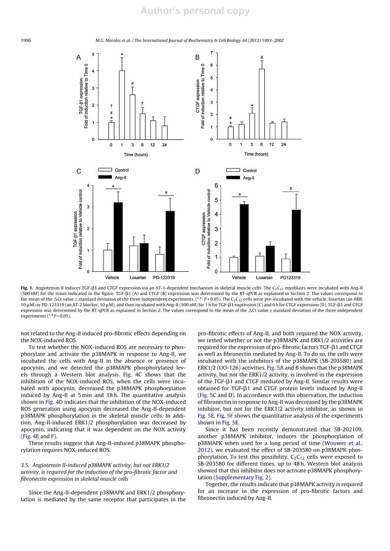
Since angiotensin II (Ang-II) contributes to a fibrotic response inskeletal muscle cells (Cabello-Verrugio et al., 2011a), we decidedto study the effect of Ang-II on the expression of pro-fibroticfactors such as TGF-�1 and CTGF. To evaluate the kinetic ofthe Ang-II-dependent expression of these factors, the C2C12 cellswere incubated with Ang-II, and their expression was evalu-ated by RT-qPCR assays. Fig. 1A shows an early increase in theAng-II-dependent TGF-�1 expression, reaching a peak after 1 hof Ang-II treatment and decreasing to basal levels 12 h after theAng-II treatment. Fig. 1B shows that the induction of the Ang-II-dependent CTGF expression started in 3 h, showing a peak 6 hafter the Ang-II treatment. Since the Ang-II-induced fibrosis ismediated by its transmembrane receptor AT-1 in skeletal musclecells (Cabello-Verrugio et al., 2011a), we evaluated its participa-tion in the Ang-II-dependent expression of pro-fibrotic factors inskeletal muscle cells. Fig. 1C and D, respectively, show that theincrease in the TGF-�1 and CTGF expression induced by Ang-IIin the C2C12 myoblasts was prevented when the cells were pre-incubated with the Ang-II receptor type 1 blocker (ARB) losartan,but no effect was observed when the cells were pre-incubated withthe blocker receptor of the Ang-II receptor type 2 PD-123319. Simi-lar results were obtained when the TGF-�1 and CTGF protein levelswere determined by ELISA and immunoblot assays, respectively(Supplementary Fig. 1).
3.2. These results indicate that Ang-II increases TGF-ˇ1 and CTGFexpression through an AT-1 receptor-dependent mechanism
Pro-fibrotic factor expression induced by angiotensin II isdependent on NAD(P)H oxidase-induced reactive oxygen species(ROS) in skeletal muscle cells.
Since we previously demonstrated that the Ang-II-dependentNOX-induced ROS participates in the generation and develop-ment of fibrosis in skeletal muscle cells, we evaluated whether theincrease of the TGF-�1 and CTGF expression induced by Ang-II isalso mediated by this mechanism. The TGF-�1 and CTGF mRNA lev-els were determined in cells treated with Ang-II and pre-incubatedin the presence or absence of apocynin, an inhibitor of the NOXactivity (Becerra et al., 2011; Nunez-Villena et al., 2011; Simon andFernandez, 2009). Fig. 2 shows that the inhibition of the NOX activ-ity by apocynin completely prevented TGF-�1 (Fig. 2A) and CTGF(Fig. 2B) expression in response to Ang-II. In addition, apocynindecreased the Ang-II dependent TGF-�1 (Fig. 2C) and CTGF (Fig. 2D)protein levels.
We previously demonstrated that NOX produced an increase ofintracellular ROS induced by Ang-II (Cabello-Verrugio et al., 2011a).To assay the role of the NOX-induced ROS under treatment withAng-II on the pro-fibrotic factors’ expression, we incubated the cellswith Ang-II in the absence or presence of the antioxidant N-acetylcysteine (NAC). Fig. 2A and B shows that the NAC decreased theTGF-�1 and CTGF expression induced by Ang-II, respectively. Sim-ilar results were obtained for the Ang-II-induced TGF-�1 and CTGFprotein levels (Fig. 2C and D).
Together, these results suggest that Ang-II induces TGF-�1 andCTGF expression via a mechanism involving the NOX-induced ROS.
3.3. Angiotensin II induces p38 and ERK1/2 MAPKphosphorylation through the AT-1 receptor in skeletal muscle cells
Evidence indicates that Ang-II induces MAPK phosphorylation(Kiribayashi et al., 2005; Li et al., 2011; Lakshmanan et al., 2012).We studied the effect of Ang-II on the MAPKs’ phosphorylationin skeletal muscle cells. Fig. 3A shows that the kinetic of thep38MAPK phosphorylation increased, with a first peak between 2.5and 7.5 min, reaching basal levels in 10 min. A second peak of thep38MAPK phosphorylation was observed at 12 and 18 h after theAng-II incubation. When the ERK1/2 phosphorylation was evalu-ated, an early activation was observed with a peak between 2.5and 7.5 min and a decrease to basal levels at later times. Interest-ingly, both the Ang-II-induced p38MAPK phosphorylation (5 minand 18 h) and ERK1/2 phosphorylation (5 min) were preventedwhen the cells were pre-incubated with ARB losartan (Fig. 3C andD, respectively), indicating that the p38MAPK and ERK1/2 phos-phorylation induced by Ang-II is mediated by the AT-1 receptor.
3.4. Angiotensin II-induced p38MAPK phosphorylation isdependent on the NAD(P)H oxidase-induced ROS in skeletalmuscle cells
Since Ang-II activates the production of the NOX-induced ROSand p38MAPK, we evaluated the relationship between these twosignalling pathways.
First, we treated the C2C12 myoblasts with Ang-II in the absenceor presence of SB-203580, an inhibitor of p38MAPK activity; subse-quently, the intracellular ROS production in response to Ang-II wasdetermined by flow cytometry using two different ROS-sensitivefluorescent probes, the DCF and DHE. Fig. 4A and B shows that theincrease in the NOX-induced ROS production mediated by Ang-IIdoes not change when the cells are incubated with the p38MAPKactivity inhibitor. In the same figures, we observed that the ROSdecreased to basal levels, as shown by the control cells when wetreated the cells with Ang-II in the presence of an inhibitor ofNOX activity (apocynin), such as we have previously demonstrated(Cabello-Verrugio et al., 2011a). Interestingly, we observed that thecells treated with Ang-II, together with the inhibitor of the MEKactivity UO-126, which avoid the ERK1/2 phosphorylation, did notchange the ROS production, confirming that the ERK1/2 activity is
Author's personal copy
1996 M.G. Morales et al. / The International Journal of Biochemistry & Cell Biology 44 (2012) 1993– 2002
Fig. 1. Angiotensin II induces TGF-�1 and CTGF expression via an AT-1-dependent mechanism in skeletal muscle cells. The C2C12 myoblasts were incubated with Ang-II(500 nM) for the times indicated in the figure. TGF-�1 (A) and CTGF (B) expression was determined by the RT-qPCR as explained in Section 2. The values correspond tothe mean of the �Ct value ± standard deviation of the three independent experiments. (*,#,†P < 0.05). The C2C12 cells were pre-incubated with the vehicle, losartan (an ARB,10 �M) or PD-123319 (an AT-2 blocker, 10 �M), and then incubated with Ang-II (500 nM) for 1 h for TGF-�1 expression (C) and 6 h for CTGF expression (D). TGF-�1 and CTGFexpression was determined by the RT-qPCR as explained in Section 2. The values correspond to the mean of the �Ct value ± standard deviation of the three independentexperiments (*,#P < 0.05).
not related to the Ang-II induced pro-fibrotic effects depending onthe NOX-induced ROS.
To test whether the NOX-induced ROS are necessary to phos-phorylate and activate the p38MAPK in response to Ang-II, weincubated the cells with Ang-II in the absence or presence ofapocynin, and we detected the p38MAPK phosphorylated lev-els through a Western blot analysis. Fig. 4C shows that theinhibition of the NOX-induced ROS, when the cells were incu-bated with apocynin, decreased the p38MAPK phosphorylationinduced by Ang-II at 5 min and 18 h. The quantitative analysisshown in Fig. 4D indicates that the inhibition of the NOX-inducedROS generation using apocynin decreased the Ang-II-dependentp38MAPK phosphorylation in the skeletal muscle cells. In addi-tion, Ang-II-induced ERK1/2 phosphorylation was decreased byapocynin, indicating that it was dependent on the NOX activity(Fig. 4E and F).
These results suggest that Ang-II-induced p38MAPK phospho-rylation requires NOX-induced ROS.
3.5. Angiotensin II-induced p38MAPK activity, but not ERK1/2activity, is required for the induction of the pro-fibrotic factor andfibronectin expression in skeletal muscle cells
Since the Ang-II-dependent p38MAPK and ERK1/2 phosphory-lation is mediated by the same receptor that participates in the
pro-fibrotic effects of Ang-II, and both required the NOX activity,we tested whether or not the p38MAPK and ERK1/2 activities arerequired for the expression of pro-fibrotic factors TGF-�1 and CTGFas well as fibronectin mediated by Ang-II. To do so, the cells wereincubated with the inhibitors of the p38MAPK (SB-203580) andERK1/2 (UO-126) activities. Fig. 5A and B shows that the p38MAPKactivity, but not the ERK1/2 activity, is involved in the expressionof the TGF-�1 and CTGF mediated by Ang-II. Similar results wereobtained for TGF-�1 and CTGF protein levels induced by Ang-II(Fig. 5C and D). In accordance with this observation, the inductionof fibronectin in response to Ang-II was decreased by the p38MAPKinhibitor, but not for the ERK1/2 activity inhibitor, as shown inFig. 5E. Fig. 5F shows the quantitative analysis of the experimentsshown in Fig. 5E.
Since it has been recently demonstrated that SB-202109,another p38MAPK inhibitor, induces the phosphorylation ofp38MAPK when used for a long period of time (Wouwer et al.,2012), we evaluated the effect of SB-203580 on p38MAPK phos-phorylation. To test this possibility, C2C12 cells were exposed toSB-203580 for different times, up to 48 h. Western blot analysisshowed that this inhibitor does not activate p38MAPK phosphory-lation (Supplementary Fig. 2).
Together, the results indicate that p38MAPK activity is requiredfor an increase in the expression of pro-fibrotic factors andfibronectin induced by Ang-II.

Author's personal copy
M.G. Morales et al. / The International Journal of Biochemistry & Cell Biology 44 (2012) 1993– 2002 1997
Fig. 2. Angiotensin II-induced TGF-�1 and CTGF expression requires NOX-induced ROS in skeletal muscle cells. The C2C12 cells were pre-incubated with the vehicle, N-acetylcysteine (NAC, 10 mM) or apocynin (a NOX inhibitor, 1 mM), and then incubated with Ang-II (500 nM) for 1 h for TGF-�1 expression (A) and 6 h for CTGF expression (B).TGF-�1 and CTGF expression was determined by the RT-qPCR as explained in Section 2. The values correspond to the mean of the �Ct value ± standard deviation of the threeindependent experiments (*P < 0.05). (C) Detection by ELISA of TGF-�1 protein levels into the medium of C2C12 exposed to Ang-II (500 nM) for 48 h in the absence (vehicle)or presence of apocynin (1 mM) or NAC (10 mM). The values are expressed as a percentage of the TGF-�1 levels secreted by C2C12 cells incubated with Ang-II and the vehicleand are representative of three independent experiments (*,#P < 0.05). (D) CTGF protein levels evaluated by Western blot analysis in an extract of a C2C12 cell incubatedwith Ang-II (500 nM) for 48 h in the absence (vehicle) or presence of apocynin (1 mM) or NAC (10 mM). The levels of tubulin are shown as loading control. The images arerepresentative of the three independent experiments. The molecular weight standards are indicated in kilo Daltons (kDa).
3.6. Angiotensin II-induced TGF-ˇ1 expression is required for latep38MAPK phosphorylation and pro-fibrotic effects mediated byangiotensin II in skeletal muscle cells
Since Ang-II induces TGF-�1 expression, we evaluated if thepro-fibrotic effects mediated by Ang-II are dependent on the TGF-�1 induction. For that, the C2C12 cells were incubated with Ang-IIin the absence or presence of an inhibitor of the TGF-� receptor I(TGF-�RI) kinase activity SB-431542 to inhibit the TGF-� signalling(Cabello-Verrugio et al., 2011b), and the p38MAPK phosphoryla-tion was evaluated by Western blot analysis. Fig. 6A shows that theinhibition of the TGF-�1 signalling by SB-431542 did not have anyeffect on the early p38MAPK phosphorylation induced by Ang-II at5 min, but completely inhibited its phosphorylation at 18 h.
To corroborate these results, we decreased the TGF-�1 lev-els using a specific shRNA for TGF-�1. The Ang-II-dependentTGF-�1 expression (Supplementary Fig. 3A) and protein levels(Supplementary Fig. 3B) were significantly decreased by transfec-tion of the specific shRNA for TGF-�1 compared to cells transfectedwith the control shRNA. Under these conditions, we determined thep38MAPK phosphorylation mediated by Ang-II. Fig. 6B shows thatthe Ang-II-induced p38MAPK phosphorylation is inhibited in cellstransfected with the shRNA for TGF-�1 at 18 h after the Ang-II incu-bation. However, it did not have any effect on its phosphorylationat 5 min.
Further, we evaluated the participation of the TGF-�1 sig-nalling on the Ang-II-induced fibronectin and CTGF levels. Fig. 6C
shows that the Ang-II-induced fibronectin and CTGF protein levelsdecreased in cells treated with the inhibitor of TGF-�RI kinase activ-ity (SB-431542). Fig. 6D shows the observation that the decrease ofthe TGF-�1 levels by a specific shRNA for TGF-�1 diminished theAng-II-induced fibronectin and the CTGF protein levels comparedwith cells transfected with a control shRNA.
Together, these results suggest that the pro-fibrotic effectsmediated by Ang-II require the increase of TGF-�1 expression andits signalling.
4. Discussion
In this report, we show that the expression of CTGF and TGF-�1, two potent pro-fibrotic factors involved in skeletal musclefibrosis, is induced by Ang-II in skeletal muscle cells by an AT-1receptor-dependent mechanism involving p38MAPK NOX-derivedROS and TGF-�1 expression and signalling (Fig. 7). Moreover,we demonstrated that p38MAPK is critical not only to CTGF andTGF-�1 expression increases, but also to ECM protein inductionmediated by Ang-II (Fig. 7). Interestingly, we have demonstratedthat the pro-fibrotic effects induced by Ang-II are dependenton TGF-�1 expression and signalling, supported by the decreaseof CTGF and fibronectin expression when an inhibitor of TGF-�RI kinase activity and a specific shRNA for TGF-�1 was used(Fig. 7).
The participation of Ang-II in the fibrotic phenotype inskeletal muscle was previously demonstrated by our group

Author's personal copy
1998 M.G. Morales et al. / The International Journal of Biochemistry & Cell Biology 44 (2012) 1993– 2002
Fig. 3. Angiotensin II induces the phosphorylation of p38 and ERK-1/2 MAPK via the AT-1 receptor in skeletal muscle cells. (A) The C2C12 myoblasts were incubated withAng-II (500 nM). After the times indicated in the figure, the obtained extracts were separated by SDS-PAGE, and levels of the p38MAPK phosphorylated and p38MAPK totalwere evaluated by Western blot analysis. (B) The C2C12 cells were treated as described in (A). ERK1/2-phosphorylated and ERK1/2 total protein levels were evaluated byWestern blot analysis. To evaluate the participation of the AT-1 receptor in the activation of the MAPK mediated by Ang-II, the cells were pre-incubated with losartan (an AT-1blocker, 10 �M) and then incubated with Ang-II (500 nM). The protein levels of the p38MAPK phosphorylated, p38MAPK total (C), ERK1/2 phosphorylated, and ERK1/2 total(D) were evaluated by Western blot. In A–D, the images are representative of the two independent experiments. The molecular weight standards are indicated in kilodaltons(kDa).
(Cabello-Verrugio et al., 2011a). However, the relationship betweenAng-II and the expression of TGF-�1 and CTGF with the mech-anism involved has not been studied in skeletal muscle fibrosis(Cabello-Verrugio et al., 2012; Morales et al., 2011; Cabello-Verrugio and Brandan, 2007).
Ang-II increases CTGF and TGF-�1 expression by several differ-ent mechanisms depending on cell types. Ang-II was found to stim-ulate TGF-�1 expression in rat heart endothelial cells blocked bylosartan in a dose- and time-dependent manner (Chua et al., 1994).In human mesangial cells (HMCs), the induction of ECM proteins
(fibronectin) and TGF-� production by Ang-II is mediated by thep38MAPK activation (Bolick et al., 2003). In cardiac myofibroblasts,the TGF-�1 production and secretion can be modulated by thespecific Ang-II receptor blockers, suggesting the participation ofAng-II as the inducer of this fibrotic growth factor (Campbell andKatwa, 1997). On the other hand, CTGF has been demonstrated tobe induced by Ang-II in an AT-1-dependent manner (Ahmed et al.,2004; Cabello-Verrugio et al., 2011a; Iwamoto et al., 2010). More-over, pathways dependent on calcineurin-, PPAR�-, PKC-, or serum-and glucocorticoid-inducible kinase SGK1 have been described to

Author's personal copy
M.G. Morales et al. / The International Journal of Biochemistry & Cell Biology 44 (2012) 1993– 2002 1999
Fig. 4. p38MAPK phosphorylation induced by angiotensin II is dependent on NOX activity in skeletal muscle cells. C2C12 cells were exposed to the vehicle alone—apocynin (aninhibitor of NOX, 1 mM), SB-203580 (an inhibitor of p38MAPK activity, 5 �M), or UO126 (an inhibitor of MEK, 5 �M)—in the absence (control) or presence of Ang-II (500 nM);DCF (A) or DHF (B) fluorescence from the three independent experiments was measured. The bars represent the percentage (mean ± standard deviation) (*,#P < 0.05). (C) Themyoblasts were pre-incubated with the vehicle or apocynin (1 mM) and then were incubated with the Ang-II (500 nM) for 5 min or 18 h. The protein levels of the p38MAPKphosphorylated and p38MAPK total were evaluated by Western blot. The molecular weights are indicated in kilodaltons (kDa). (D) Quantification of three independentexperiments representative of C (*,#P < 0.05). (E) The myoblasts were treated as described in C, and the incubation with Ang-II was for 5 min. The protein levels of the ERK1/2phosphorylated and ERK1/2 total were evaluated by Western blot. The molecular weight standards are indicated in kilodaltons (kDa). (F) Quantification of three independentexperiments representative of E (*,#P < 0.05).
participate in Ang-II-dependent CTGF induction (Finckenberg et al.,2003; Gao et al., 2007; He et al., 2005; Hussain et al., 2008). In car-diac fibroblasts, the Ang-II-induced TGF-�1 expression is mediatedby the ERK-1/2 MAPK-dependent mechanism, and the CTGF expres-sion is mediated by the activation of p38MAPK (Li et al., 2011). In ourmodel, we previously demonstrated that CTGF expression is mod-ulated by TGF-�1, lysophosphatidic acid (LPA) through the TGF-�receptor transactivation, and JNK activity (Cabello-Verrugio et al.,2011b). In this paper, our results suggest that the p38MAPK acti-vation induced by Ang-II in skeletal muscle cells is required for theinduction of TGF-�1 and CTGF expression. However, the participa-tion of the ERK-1/2 activity induced by Ang-II in other processesoccurring in skeletal muscle cells requires further experimentalevidence.
Interestingly, there is evidence that Ang-II can cause a rapidactivation of Smad signalling independent of the TGF-� and late
effects which are Smad-dependent and TGF-�-mediated (Carvajalet al., 2008). In our model, we have observed that Ang-II does notphosphorylate directly to the Smad 2/3 (data not shown). How-ever, the participation of TGF-�1 on the pro-fibrotic effects inducedby Ang-II were determined. TGF-�1 expression was required forthe Ang-II-induced p38MAPK phosphorylation observed in a sec-ond peak after incubation of Ang-II (12–18 h). Ang II-inducedfibronectin and CTGF increase was dependent on the stimulationof TGF-�1 expression, as demonstrated using either an inhibitor ofTGF-�RI kinase activity or short hairpin RNA (shRNA) for TGF-�1. Asimilar requirement of TGF-�1 was observed in the effect of Ang-IIon PPAR� activity in aortic smooth muscle cells (Subramanian et al.,2012).
Ang-II is not only a fibrotic factor but also a pro-oxidantcytokine in several tissues, including the skeletal muscle (Cabello-Verrugio et al., 2011a; Cozzoli et al., 2011). Interestingly, the

Author's personal copy
2000 M.G. Morales et al. / The International Journal of Biochemistry & Cell Biology 44 (2012) 1993– 2002
Fig. 5. p38MAPK, but not ERK1/2 activity, is required for angiotensin II-induced pro-fibrotic factor expression and fibrosis in skeletal muscle cells. The C2C12 cells werepre-incubated with the vehicle, SB-203580 (an inhibitor of p38MAPK activity, 5 �M) or UO-126 (an inhibitor of MEK, 5 �M), and then incubated with Ang-II (500 nM) for 1 hfor the TGF-�1 expression (A) and 6 h for CTGF expression (B). TGF-�1 and CTGF expression was determined by the RT-qPCR as explained in Section 2. The values correspondto the mean of the �Ct value ± standard deviation of the three independent experiments (*P < 0.05). (C) Detection by ELISA of TGF-�1 protein levels into the medium of C2C12
pre-incubated with the vehicle, SB-203580 (an inhibitor of p38MAPK activity, 5 �M) or UO-126 (an inhibitor of MEK, 5 �M), and then incubated with Ang-II (500 nM) for48 h. The values are expressed as a percentage of the TGF-�1 levels secreted by C2C12 cells incubated with Ang-II and the vehicle and are representative of three independentexperiments (*P < 0.05). (D) The myoblasts were pre-incubated with the vehicle, SB-203580 (an inhibitor of p38MAPK activity, 5 �M) or UO-126 (an inhibitor of MEK, 5 �M),and then incubated with Ang-II (500 nM) for 48 h. The levels of CTGF were determined by Western blot. The levels of tubulin are shown as a loading control. The molecularweight standards are indicated in kilodaltons (kDa). The images are representative of the three independent experiments. (E) The myoblasts were pre-incubated with thevehicle, SB-203580 (an inhibitor of p38MAPK activity, 5 �M) or UO-126 (an inhibitor of MEK, 5 �M), and then incubated with Ang-II (500 nM) for 48 h. The levels of fibronectin(FN) were determined by Western blot. The levels of tubulin are shown as a loading control. The molecular weight standards are indicated in kilodaltons (kDa). The imagesare representative of the three independent experiments that are quantified and shown in graph (F) (*P < 0.05).
pro-fibrotic effects of Ang-II in the skeletal muscle are depen-dent on ROS production. The NOX, one of the sources of ROS,mediates the actions of Ang-II and plays a critical role in fibro-genesis (Bataller et al., 2003). In our previous report and inthis paper, the experiments with an antioxidant such as NAC,or the use of NOX inhibitors such as apocynin, strongly sug-gest the participation of NOX-induced ROS as critical fibroticmediators (Cabello-Verrugio et al., 2011a). This is in agreementwith the decrease of the Ang-II-dependent TGF-�1 expression,ECM proteins, and p38MAPK signalling observed by treatmentwith anti-oxidant molecules (Bolick et al., 2003). In the con-text of skeletal muscle fibrosis, the identification of novel genes
that participate in the fibrosis induced by Ang-II in a redox-sensitive manner could be further studied through microarrayanalysis.
To summarise, in this report, we show that Ang-II induces theexpression of TGF-�1 and CTGF in the skeletal muscle throughits AT-1 receptor by a mechanism involving p38MAPK and NOX-derived ROS. Moreover, we observed that p38MAPK and TGF-�1induction are also critical to increase ECM protein levels mediatedby Ang-II. These results strongly reinforce the importance of Ang-II in the fibrotic phenotype in the skeletal muscle and contributeto understanding the mechanism involved in its effects on skeletalmuscle fibrosis.

Author's personal copy
M.G. Morales et al. / The International Journal of Biochemistry & Cell Biology 44 (2012) 1993– 2002 2001
Fig. 6. Angiotensin II-induced TGF-�1 expression and signalling is required for the pro-fibrotic effects mediated by angiotensin II in skeletal muscle cells. (A) C2C12 cellswere exposed to the vehicle or to SB-431542 (an inhibitor of TGF-� receptor I kinase activity, 10 �M) in the absence (control) or presence of Ang-II (500 nM) for the timesindicated in the figure. The protein levels of the p38MAPK phosphorylated and p38MAPK total were evaluated by Western blot. (B) C2C12 myoblasts were transfected witha shRNA control or specific shRNA for TGF-�1. The transfected myoblasts were incubated in the absence (control) or presence of Ang-II (500 nM) for the times indicated inthe figure. The protein levels of the p38MAPK phosphorylated and p38MAPK total were evaluated by Western blot. (C) C2C12 cells were pre-incubated with the vehicle orSB-431542 (10 �M) in the absence (control) or presence of Ang-II (500 nM). The fibronectin (FN), CTGF, and tubulin protein levels were evaluated by Western blot. (D) Thetransfected myoblasts, as described in B, were incubated in the absence (control) or presence of Ang-II (500 nM). The fibronectin (FN), CTGF, and tubulin protein levels wereevaluated by Western blot. For A–D, the molecular weight standards are indicated in kilodaltons (kDa).
Fig. 7. Scheme for the signalling pathways involved in the angiotensin II-inducedpro-fibrotic effects in skeletal muscle cells. When C2C12 myoblasts are incubatedwith Ang-II, the AT-1 receptor is activated. Downstream, the AT-1-dependentNAD(P)H oxidase (NOX) activation (Cabello-Verrugio et al., 2011a) is required forthe Ang-II-induced reactive oxygen species (ROS) production and p38MAPK phos-phorylation. These events are required for the TGF-�1 expression, which is essentialfor the CTGF and fibronectin increase mediated by Ang-II.
Conflict of interest statement
The authors confirm that there are no conflicts of interest.
Acknowledgments
This study was supported by research grants from FONDECYT# 1120380, 1121078, 1110426, CARE PFB12/2007, Conicyt AT-24100061 and AT-24100047, MDA 89419, and UNAB DI 33-11/R.
Appendix A. Supplementary data
Supplementary data associated with this article can befound, in the online version, at http://dx.doi.org/10.1016/j.biocel.2012.07.028.
References
Ahmed MS, Oie E, Vinge LE, Yndestad A, Oystein Andersen G, Andersson Y, et al.Connective tissue growth factor—a novel mediator of angiotensin II-stimulatedcardiac fibroblast activation in heart failure in rats. Journal of Molecular andCellular Cardiology 2004;36:393–404.
Bataller R, Brenner DA. Liver fibrosis. Journal of Clinical Investigation2005;115:209–18.
Bataller R, Gabele E, Parsons CJ, Morris T, Yang L, Schoonhoven R, et al. Systemicinfusion of angiotensin II exacerbates liver fibrosis in bile duct-ligated rats.Hepatology 2005;41:1046–55.
Bataller R, Schwabe RF, Choi YH, Yang L, Paik YH, Lindquist J, et al. NADPH oxidasesignal transduces angiotensin II in hepatic stellate cells and is critical in hepaticfibrosis. Journal of Clinical Investigation 2003;112:1383–94.
Becerra A, Echeverria C, Varela D, Sarmiento D, Armisen R, Nunez-VillenaF, et al. Transient receptor potential melastatin 4 inhibition preventslipopolysaccharide-induced endothelial cell death. Cardiovascular Research2011;91:677–84.
Bolick DT, Hatley ME, Srinivasan S, Hedrick CC, Nadler JL. Lisofylline, a novelantiinflammatory compound, protects mesangial cells from hyperglycemia-and angiotensin II-mediated extracellular matrix deposition. Endocrinology2003;144:5227–31.
Brecher P. Angiotensin II and cardiac fibrosis. Trends in Cardiovascular Medicine1996;6:193–8.

Author's personal copy
2002 M.G. Morales et al. / The International Journal of Biochemistry & Cell Biology 44 (2012) 1993– 2002
Cabello-Verrugio C, Acuna MJ, Morales MG, Becerra A, Simon F, Brandan E. Fibroticresponse induced by angiotensin-II requires NAD(P)H oxidase-induced reac-tive oxygen species (ROS) in skeletal muscle cells. Biochemical and BiophysicalResearch Communications 2011a;410:665–70.
Cabello-Verrugio C, Brandan E. A novel modulatory mechanism of transforminggrowth factor-beta signaling through decorin and LRP-1. Journal of BiologicalChemistry 2007;282:18842–50.
Cabello-Verrugio C, Cordova G, Vial C, Zuniga LM, Brandan E. Connective tissuegrowth factor induction by lysophosphatidic acid requires transactivation oftransforming growth factor type beta receptors and the JNK pathway. CellularSignalling 2011b;23:449–57.
Cabello-Verrugio C, Morales MG, Cabrera D, Vio CP, Brandan E. Angiotensin IIreceptor type 1 blockade decreases CTGF/CCN2-mediated damage and fibro-sis in normal and dystrophic skeletal muscles. Journal of Cellular and MolecularMedicine 2012;16:752–64.
Campbell SE, Katwa LC. Angiotensin II stimulated expression of transforming growthfactor-beta1 in cardiac fibroblasts and myofibroblasts. Journal of Molecular andCellular Cardiology 1997;29:1947–58.
Carvajal G, Rodriguez-Vita J, Rodrigues-Diez R, Sanchez-Lopez E, Ruperez M, CartierC, et al. Angiotensin II activates the Smad pathway during epithelial mesenchy-mal transdifferentiation. Kidney International 2008;74:585–95.
Clark JE, Sarafraz N, Marber MS. Potential of p38-MAPK inhibitors in the treat-ment of ischaemic heart disease. Pharmacology & Therapeutics 2007;116:192–206.
Cohn RD, van Erp C, Habashi JP, Soleimani AA, Klein EC, Lisi MT, et al. AngiotensinII type 1 receptor blockade attenuates TGF-beta-induced failure of mus-cle regeneration in multiple myopathic states. Nature Medicine 2007;13:204–10.
Cozzoli A, Nico B, Sblendorio VT, Capogrosso RF, Dinardo MM, Longo V, et al. Enalapriltreatment discloses an early role of angiotensin II in inflammation- and oxida-tive stress-related muscle damage in dystrophic mdx mice. PharmacologicalResearch 2011;64:482–92.
Che ZQ, Gao PJ, Shen WL, Fan CL, Liu JJ, Zhu DL. Angiotensin II-stimulated colla-gen synthesis in aortic adventitial fibroblasts is mediated by connective tissuegrowth factor. Hypertension Research 2008;31:1233–40.
Chua CC, Diglio CA, Siu BB, Chua BH. Angiotensin II induces TGF-beta 1 productionin rat heart endothelial cells. Biochimica et Biophysica Acta 1994;1223: 141–7.
de Boer RA, Pokharel S, Flesch M, van Kampen DA, Suurmeijer AJ, Boomsma F,et al. Extracellular signal regulated kinase and SMAD signaling both mediate theangiotensin II driven progression towards overt heart failure in homozygousTGR(mRen2)27. Journal of Molecular Medicine (Berlin) 2004;82:678–87.
Fadic R, Mezzano V, Alvarez K, Cabrera D, Holmgren J, Brandan E. Increase in decorinand biglycan in Duchenne muscular dystrophy: role of fibroblasts as cell sourceof these proteoglycans in the disease. Journal of Cellular and Molecular Medicine2006;10:758–69.
Finckenberg P, Inkinen K, Ahonen J, Merasto S, Louhelainen M, Vapaatalo H,et al. Angiotensin II induces connective tissue growth factor gene expres-sion via calcineurin-dependent pathways. American Journal of Pathology2003;163:355–66.
Gao DF, Niu XL, Hao GH, Peng N, Wei J, Ning N, et al. Rosiglitazone inhibits angiotensinII-induced CTGF expression in vascular smooth muscle cells—role of PPAR-gamma in vascular fibrosis. Biochemical Pharmacology 2007;73:185–97.
Guo G, Morrissey J, McCracken R, Tolley T, Liapis H, Klahr S. Contributions ofangiotensin II and tumor necrosis factor-alpha to the development of renalfibrosis. American Journal of Physiology Renal Physiology 2001;280:F777–85.
He Z, Way KJ, Arikawa E, Chou E, Opland DM, Clermont A, et al. Differential regula-tion of angiotensin II-induced expression of connective tissue growth factor byprotein kinase C isoforms in the myocardium. Journal of Biological Chemistry2005;280:15719–26.
Hussain A, Wyatt AW, Wang K, Bhandaru M, Biswas R, Avram D, et al. SGK1-dependent upregulation of connective tissue growth factor by angiotensin II.Kidney and Blood Pressure Research 2008;31:80–6.
Iwamoto M, Hirohata S, Ogawa H, Ohtsuki T, Shinohata R, Miyoshi T, et al. Connectivetissue growth factor induction in a pressure-overloaded heart ameliorated bythe angiotensin II type 1 receptor blocker olmesartan. Hypertension Research2010;33:1305–11.
Iwanciw D, Rehm M, Porst M, Goppelt-Struebe M. Induction of connective tissuegrowth factor by angiotensin II: integration of signaling pathways. Arterioscle-rosis, Thrombosis, and Vascular Biology 2003;23:1782–7.
Kiribayashi K, Masaki T, Naito T, Ogawa T, Ito T, Yorioka N, et al. Angiotensin II inducesfibronectin expression in human peritoneal mesothelial cells via ERK1/2 and p38MAPK. Kidney International 2005;67:1126–35.
Kupfahl C, Pink D, Friedrich K, Zurbrugg HR, Neuss M, Warnecke C, et al. AngiotensinII directly increases transforming growth factor beta1 and osteopontin and indi-rectly affects collagen mRNA expression in the human heart. CardiovascularResearch 2000;46:463–75.
Lakshmanan AP, Thandavarayan RA, Watanabe K, Sari FR, Meilei H, Giridharan VV,et al. Modulation of AT-1R/MAPK cascade by an olmesartan treatment attenuatesdiabetic nephropathy in streptozotocin-induced diabetic mice. Molecular andCellular Endocrinology 2012;348:104–11.
Lakshmanan AP, Watanabe K, Thandavarayan RA, Sari FR, Harima M, GiridharanVV, et al. Telmisartan attenuates oxidative stress and renal fibrosis in strep-tozotocin induced diabetic mice with the alteration of angiotensin-(1–7) masreceptor expression associated with its PPAR-gamma agonist action. Free Radi-cal Research 2011;45:575–84.
Li L, Fan D, Wang C, Wang JY, Cui XB, Wu D, et al. Angiotensin II increases periostinexpression via Ras/p38 MAPK/CREB and ERK1/2/TGF-beta1 pathways in cardiacfibroblasts. Cardiovascular Research 2011;91:80–9.
Ma FY, Sachchithananthan M, Flanc RS, Nikolic-Paterson DJ. Mitogen activatedprotein kinases in renal fibrosis. Frontiers in Bioscience (Scholar Edition)2009;1:171–87.
Mezzano V, Cabrera D, Vial C, Brandan E. Constitutively activated dystrophic musclefibroblasts show a paradoxical response to TGF-beta and CTGF/CCN2. Journal ofCell Communication and Signaling 2007;1:205–17.
Morales MG, Cabello-Verrugio C, Santander C, Cabrera D, Goldschmeding R, BrandanE. CTGF/CCN-2 over-expression can directly induce features of skeletal muscledystrophy. Journal of Pathology 2011;225:490–501.
Nunez-Villena F, Becerra A, Echeverria C, Briceno N, Porras O, Armisen R,et al. Increased expression of the transient receptor potential melastatin 7channel is critically involved in lipopolysaccharide-induced reactive oxygenspecies-mediated neuronal death. Antioxidants and Redox Signalling 2011;15:2425–38.
Parsons CJ, Takashima M, Rippe RA. Molecular mechanisms of hepatic fibrogenesis.Journal of Gastroenterology and Hepatology 2007;22(Suppl. 1):S79–84.
Seccia TM, Belloni AS, Kreutz R, Paul M, Nussdorfer GG, Pessina AC, et al. Cardiacfibrosis occurs early and involves endothelin and AT-1 receptors in hypertensiondue to endogenous angiotensin II. Journal of the American College of Cardiology2003;41:666–73.
Simon F, Fernandez R. Early lipopolysaccharide-induced reactive oxygen speciesproduction evokes necrotic cell death in human umbilical vein endothelial cells.Journal of Hypertension 2009;27:1202–16.
Subramanian V, Golledge J, Heywood EB, Bruemmer D, Daugherty A. Regulation ofperoxisome proliferator-activated receptor-gamma by angiotensin II via trans-forming growth factor-beta1-activated p38 mitogen-activated protein kinase inaortic smooth muscle cells. Arteriosclerosis, Thrombosis, and Vascular Biology2012;32:397–405.
Sun G, Haginoya K, Dai H, Chiba Y, Uematsu M, Hino-Fukuyo N, et al. Intramuscularrenin-angiotensin system is activated in human muscular dystrophy. Journal ofthe Neurological Sciences 2009;280:40–8.
Tarif N, Bakris GL. Angiotensin II receptor blockade and progression of nondiabetic-mediated renal disease. Kidney International Supplement 1997;63:S67–70.
Thomas DA, Massague J. TGF-beta directly targets cytotoxic T cell func-tions during tumor evasion of immune surveillance. Cancer Cell 2005;8:369–80.
Vial C, Zuniga LM, Cabello-Verrugio C, Canon P, Fadic R, Brandan E. Skeletalmuscle cells express the profibrotic cytokine connective tissue growth factor(CTGF/CCN2), which induces their dedifferentiation. Journal of Cellular Physiol-ogy 2008;215:410–21.
Whitehead NP, Yeung EW, Froehner SC, Allen DG. Skeletal muscle NADPH oxidaseis increased and triggers stretch-induced damage in the mdx mouse. PLoS One2010;5:e15354.
Wouwer MV, Couzinie C, Serrano-Palero M, Gonzalez-Fernandez O, Galmes-VarelaC, Menendez-Antoli P, et al. Activation of the BRCA1/Chk1/p53/p21(Cip1/Waf1)pathway by nitric oxide and cell cycle arrest in human neuroblastoma NB69cells. Nitric Oxide 2012;26:182–91.
Zhou L, Porter JD, Cheng G, Gong B, Hatala DA, Merriam AP, et al. Temporal andspatial mRNA expression patterns of TGF-beta1, 2, 3 and TbetaRI, II, III in skeletalmuscles of mdx mice. Neuromuscular Disorders 2006;16:32–8.