Derivation and characterization of human fetal MSCs: An alternative cell source for large-scale...
10
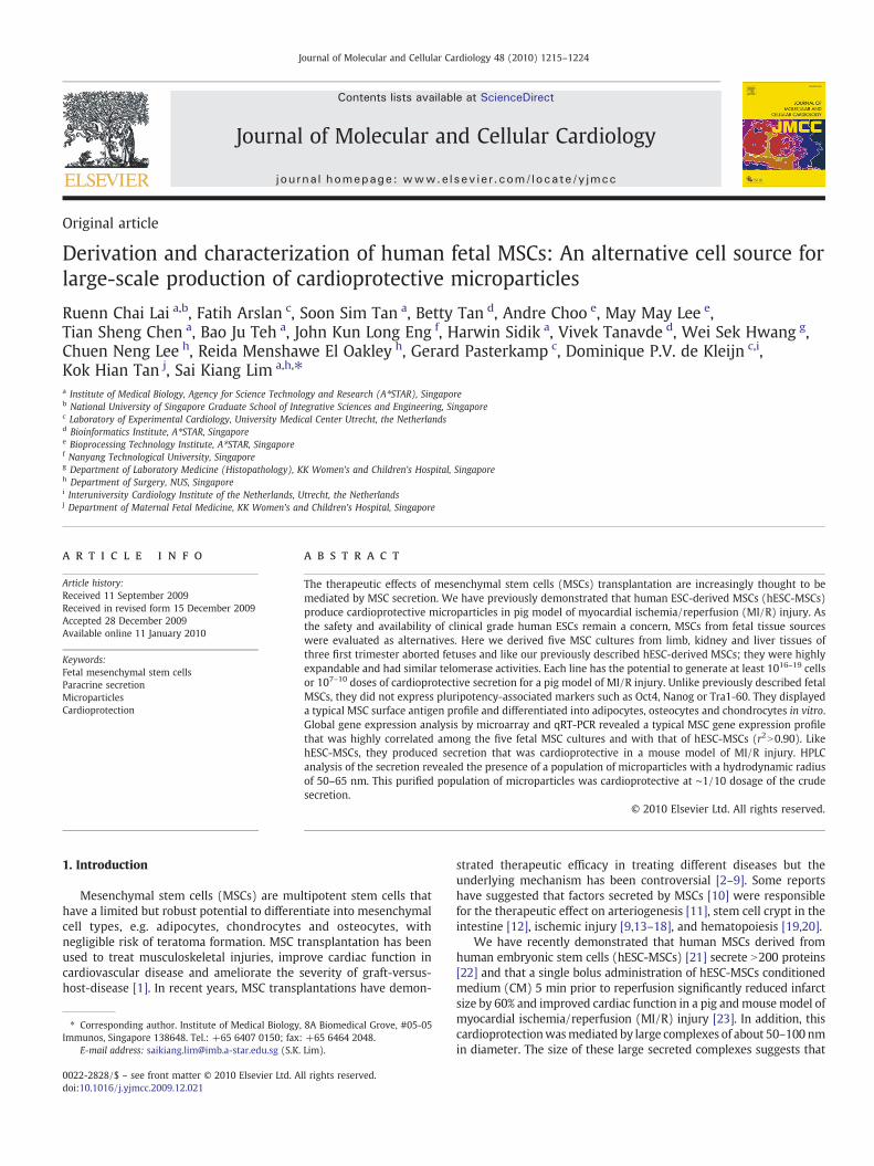
Original article Derivation and characterization of human fetal MSCs: An alternative cell source for large-scale production of cardioprotective microparticles Ruenn Chai Lai a,b , Fatih Arslan c , Soon Sim Tan a , Betty Tan d , Andre Choo e , May May Lee e , Tian Sheng Chen a , Bao Ju Teh a , John Kun Long Eng f , Harwin Sidik a , Vivek Tanavde d , Wei Sek Hwang g , Chuen Neng Lee h , Reida Menshawe El Oakley h , Gerard Pasterkamp c , Dominique P.V. de Kleijn c,i , Kok Hian Tan j , Sai Kiang Lim a,h, ⁎ a Institute of Medical Biology, Agency for Science Technology and Research (A⁎STAR), Singapore b National University of Singapore Graduate School of Integrative Sciences and Engineering, Singapore c Laboratory of Experimental Cardiology, University Medical Center Utrecht, the Netherlands d Bioinformatics Institute, A⁎STAR, Singapore e Bioprocessing Technology Institute, A⁎STAR, Singapore f Nanyang Technological University, Singapore g Department of Laboratory Medicine (Histopathology), KK Women's and Children's Hospital, Singapore h Department of Surgery, NUS, Singapore i Interuniversity Cardiology Institute of the Netherlands, Utrecht, the Netherlands j Department of Maternal Fetal Medicine, KK Women's and Children's Hospital, Singapore abstract article info Article history: Received 11 September 2009 Received in revised form 15 December 2009 Accepted 28 December 2009 Available online 11 January 2010 Keywords: Fetal mesenchymal stem cells Paracrine secretion Microparticles Cardioprotection The therapeutic effects of mesenchymal stem cells (MSCs) transplantation are increasingly thought to be mediated by MSC secretion. We have previously demonstrated that human ESC-derived MSCs (hESC-MSCs) produce cardioprotective microparticles in pig model of myocardial ischemia/reperfusion (MI/R) injury. As the safety and availability of clinical grade human ESCs remain a concern, MSCs from fetal tissue sources were evaluated as alternatives. Here we derived five MSC cultures from limb, kidney and liver tissues of three first trimester aborted fetuses and like our previously described hESC-derived MSCs; they were highly expandable and had similar telomerase activities. Each line has the potential to generate at least 10 16–19 cells or 10 7–10 doses of cardioprotective secretion for a pig model of MI/R injury. Unlike previously described fetal MSCs, they did not express pluripotency-associated markers such as Oct4, Nanog or Tra1-60. They displayed a typical MSC surface antigen profile and differentiated into adipocytes, osteocytes and chondrocytes in vitro. Global gene expression analysis by microarray and qRT-PCR revealed a typical MSC gene expression profile that was highly correlated among the five fetal MSC cultures and with that of hESC-MSCs (r 2 N 0.90). Like hESC-MSCs, they produced secretion that was cardioprotective in a mouse model of MI/R injury. HPLC analysis of the secretion revealed the presence of a population of microparticles with a hydrodynamic radius of 50–65 nm. This purified population of microparticles was cardioprotective at ∼1/10 dosage of the crude secretion. © 2010 Elsevier Ltd. All rights reserved. 1. Introduction Mesenchymal stem cells (MSCs) are multipotent stem cells that have a limited but robust potential to differentiate into mesenchymal cell types, e.g. adipocytes, chondrocytes and osteocytes, with negligible risk of teratoma formation. MSC transplantation has been used to treat musculoskeletal injuries, improve cardiac function in cardiovascular disease and ameliorate the severity of graft-versus- host-disease [1]. In recent years, MSC transplantations have demon- strated therapeutic efficacy in treating different diseases but the underlying mechanism has been controversial [2–9]. Some reports have suggested that factors secreted by MSCs [10] were responsible for the therapeutic effect on arteriogenesis [11], stem cell crypt in the intestine [12], ischemic injury [9,13–18], and hematopoiesis [19,20]. We have recently demonstrated that human MSCs derived from human embryonic stem cells (hESC-MSCs) [21] secrete N 200 proteins [22] and that a single bolus administration of hESC-MSCs conditioned medium (CM) 5 min prior to reperfusion significantly reduced infarct size by 60% and improved cardiac function in a pig and mouse model of myocardial ischemia/reperfusion (MI/R) injury [23]. In addition, this cardioprotection was mediated by large complexes of about 50–100 nm in diameter. The size of these large secreted complexes suggests that Journal of Molecular and Cellular Cardiology 48 (2010) 1215–1224 ⁎ Corresponding author. Institute of Medical Biology, 8A Biomedical Grove, #05-05 Immunos, Singapore 138648. Tel.: +65 6407 0150; fax: +65 6464 2048. E-mail address: [email protected] (S.K. Lim). 0022-2828/$ – see front matter © 2010 Elsevier Ltd. All rights reserved. doi:10.1016/j.yjmcc.2009.12.021 Contents lists available at ScienceDirect Journal of Molecular and Cellular Cardiology journal homepage: www.elsevier.com/locate/yjmcc
-
Upload
sabanciuniv -
Category
Documents
-
view
1 -
download
0
Transcript of Derivation and characterization of human fetal MSCs: An alternative cell source for large-scale...
Journal of Molecular and Cellular Cardiology 48 (2010) 1215–1224
Contents lists available at ScienceDirect
Journal of Molecular and Cellular Cardiology
j ourna l homepage: www.e lsev ie r.com/ locate /y jmcc
Original article
Derivation and characterization of human fetal MSCs: An alternative cell source forlarge-scale production of cardioprotective microparticles
Ruenn Chai Lai a,b, Fatih Arslan c, Soon Sim Tan a, Betty Tan d, Andre Choo e, May May Lee e,Tian Sheng Chen a, Bao Ju Teh a, John Kun Long Eng f, Harwin Sidik a, Vivek Tanavde d, Wei Sek Hwang g,Chuen Neng Lee h, Reida Menshawe El Oakley h, Gerard Pasterkamp c, Dominique P.V. de Kleijn c,i,Kok Hian Tan j, Sai Kiang Lim a,h,⁎a Institute of Medical Biology, Agency for Science Technology and Research (A⁎STAR), Singaporeb National University of Singapore Graduate School of Integrative Sciences and Engineering, Singaporec Laboratory of Experimental Cardiology, University Medical Center Utrecht, the Netherlandsd Bioinformatics Institute, A⁎STAR, Singaporee Bioprocessing Technology Institute, A⁎STAR, Singaporef Nanyang Technological University, Singaporeg Department of Laboratory Medicine (Histopathology), KK Women's and Children's Hospital, Singaporeh Department of Surgery, NUS, Singaporei Interuniversity Cardiology Institute of the Netherlands, Utrecht, the Netherlandsj Department of Maternal Fetal Medicine, KK Women's and Children's Hospital, Singapore
⁎ Corresponding author. Institute of Medical Biology,Immunos, Singapore 138648. Tel.: +65 6407 0150; fax:
E-mail address: [email protected] (S.K.
0022-2828/$ – see front matter © 2010 Elsevier Ltd. Adoi:10.1016/j.yjmcc.2009.12.021
a b s t r a c t
a r t i c l e i n f oArticle history:Received 11 September 2009Received in revised form 15 December 2009Accepted 28 December 2009Available online 11 January 2010
Keywords:Fetal mesenchymal stem cellsParacrine secretionMicroparticlesCardioprotection
The therapeutic effects of mesenchymal stem cells (MSCs) transplantation are increasingly thought to bemediated by MSC secretion. We have previously demonstrated that human ESC-derived MSCs (hESC-MSCs)produce cardioprotective microparticles in pig model of myocardial ischemia/reperfusion (MI/R) injury. Asthe safety and availability of clinical grade human ESCs remain a concern, MSCs from fetal tissue sourceswere evaluated as alternatives. Here we derived five MSC cultures from limb, kidney and liver tissues ofthree first trimester aborted fetuses and like our previously described hESC-derived MSCs; they were highlyexpandable and had similar telomerase activities. Each line has the potential to generate at least 1016–19 cellsor 107–10 doses of cardioprotective secretion for a pig model of MI/R injury. Unlike previously described fetalMSCs, they did not express pluripotency-associated markers such as Oct4, Nanog or Tra1-60. They displayeda typical MSC surface antigen profile and differentiated into adipocytes, osteocytes and chondrocytes in vitro.Global gene expression analysis by microarray and qRT-PCR revealed a typical MSC gene expression profilethat was highly correlated among the five fetal MSC cultures and with that of hESC-MSCs (r2N0.90). LikehESC-MSCs, they produced secretion that was cardioprotective in a mouse model of MI/R injury. HPLCanalysis of the secretion revealed the presence of a population of microparticles with a hydrodynamic radiusof 50–65 nm. This purified population of microparticles was cardioprotective at ∼1/10 dosage of the crudesecretion.
8A Biomedical Grove, #05-05+65 6464 2048.Lim).
ll rights reserved.
© 2010 Elsevier Ltd. All rights reserved.
1. Introduction
Mesenchymal stem cells (MSCs) are multipotent stem cells thathave a limited but robust potential to differentiate into mesenchymalcell types, e.g. adipocytes, chondrocytes and osteocytes, withnegligible risk of teratoma formation. MSC transplantation has beenused to treat musculoskeletal injuries, improve cardiac function incardiovascular disease and ameliorate the severity of graft-versus-host-disease [1]. In recent years, MSC transplantations have demon-
strated therapeutic efficacy in treating different diseases but theunderlying mechanism has been controversial [2–9]. Some reportshave suggested that factors secreted by MSCs [10] were responsiblefor the therapeutic effect on arteriogenesis [11], stem cell crypt in theintestine [12], ischemic injury [9,13–18], and hematopoiesis [19,20].
We have recently demonstrated that human MSCs derived fromhuman embryonic stem cells (hESC-MSCs) [21] secrete N200 proteins[22] and that a single bolus administration of hESC-MSCs conditionedmedium (CM) 5 min prior to reperfusion significantly reduced infarctsize by 60% and improved cardiac function in a pig andmousemodel ofmyocardial ischemia/reperfusion (MI/R) injury [23]. In addition, thiscardioprotectionwasmediated by large complexes of about 50–100 nmin diameter. The size of these large secreted complexes suggests that
1216 R.C. Lai et al. / Journal of Molecular and Cellular Cardiology 48 (2010) 1215–1224
they are microparticles which are broadly defined as secretedmembrane particles in the size range of 0.05–1 μm [24].
A requisite for translating cardioprotective MSC secretion intoclinical applications is a clinical grade MSC source but this is currentlylimited by restricted access to clinical grade hESCs. Therefore,alternative tissue or cell sources that are amenable to the generationof highly expandable, clinical grade MSCs have to be developed. Herewe examined fetal tissues as a candidate tissue source.
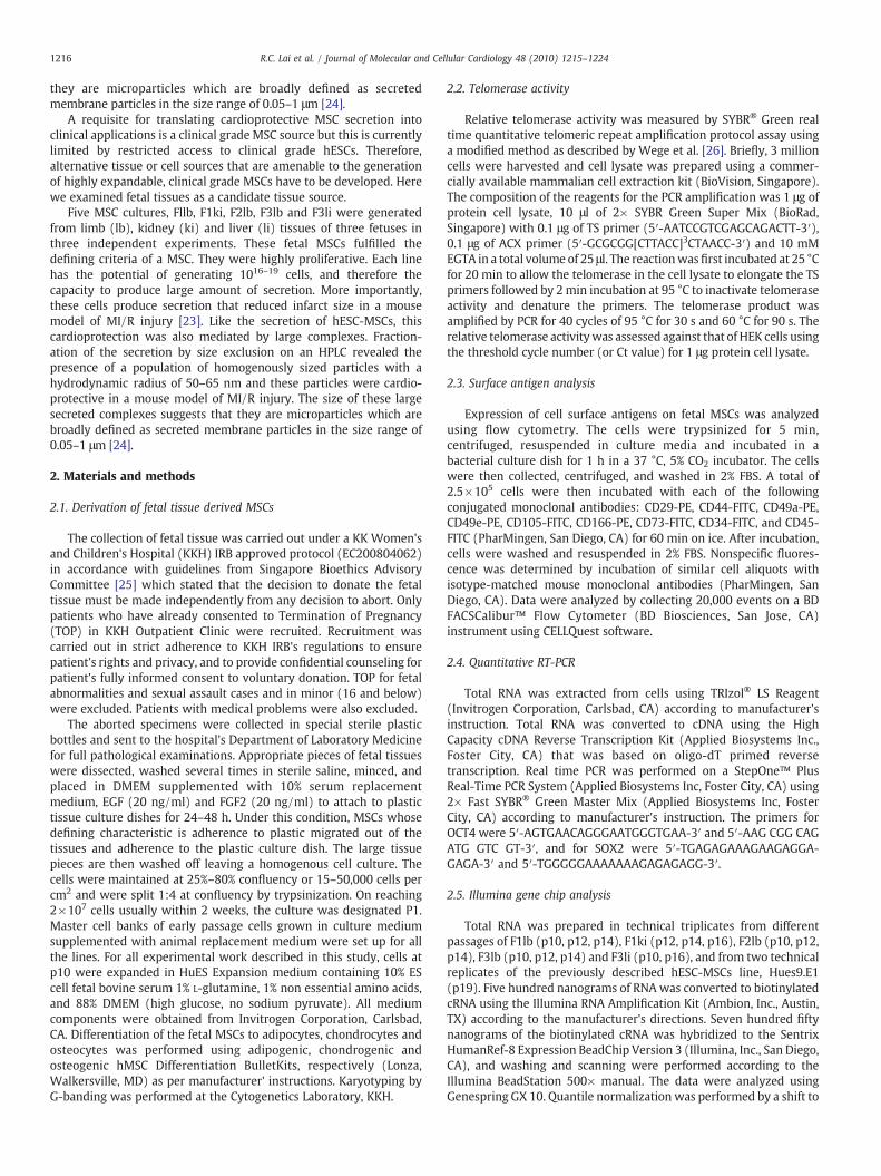
Five MSC cultures, Fllb, F1ki, F2lb, F3lb and F3li were generatedfrom limb (lb), kidney (ki) and liver (li) tissues of three fetuses inthree independent experiments. These fetal MSCs fulfilled thedefining criteria of a MSC. They were highly proliferative. Each linehas the potential of generating 1016–19 cells, and therefore thecapacity to produce large amount of secretion. More importantly,these cells produce secretion that reduced infarct size in a mousemodel of MI/R injury [23]. Like the secretion of hESC-MSCs, thiscardioprotection was also mediated by large complexes. Fraction-ation of the secretion by size exclusion on an HPLC revealed thepresence of a population of homogenously sized particles with ahydrodynamic radius of 50–65 nm and these particles were cardio-protective in a mouse model of MI/R injury. The size of these largesecreted complexes suggests that they are microparticles which arebroadly defined as secreted membrane particles in the size range of0.05–1 μm [24].
2. Materials and methods
2.1. Derivation of fetal tissue derived MSCs
The collection of fetal tissue was carried out under a KK Women'sand Children's Hospital (KKH) IRB approved protocol (EC200804062)in accordance with guidelines from Singapore Bioethics AdvisoryCommittee [25] which stated that the decision to donate the fetaltissue must be made independently from any decision to abort. Onlypatients who have already consented to Termination of Pregnancy(TOP) in KKH Outpatient Clinic were recruited. Recruitment wascarried out in strict adherence to KKH IRB's regulations to ensurepatient's rights and privacy, and to provide confidential counseling forpatient's fully informed consent to voluntary donation. TOP for fetalabnormalities and sexual assault cases and in minor (16 and below)were excluded. Patients with medical problems were also excluded.
The aborted specimens were collected in special sterile plasticbottles and sent to the hospital's Department of Laboratory Medicinefor full pathological examinations. Appropriate pieces of fetal tissueswere dissected, washed several times in sterile saline, minced, andplaced in DMEM supplemented with 10% serum replacementmedium, EGF (20 ng/ml) and FGF2 (20 ng/ml) to attach to plastictissue culture dishes for 24–48 h. Under this condition, MSCs whosedefining characteristic is adherence to plastic migrated out of thetissues and adherence to the plastic culture dish. The large tissuepieces are then washed off leaving a homogenous cell culture. Thecells were maintained at 25%–80% confluency or 15–50,000 cells percm2 and were split 1:4 at confluency by trypsinization. On reaching2×107 cells usually within 2 weeks, the culture was designated P1.Master cell banks of early passage cells grown in culture mediumsupplemented with animal replacement medium were set up for allthe lines. For all experimental work described in this study, cells atp10 were expanded in HuES Expansion medium containing 10% EScell fetal bovine serum 1% L-glutamine, 1% non essential amino acids,and 88% DMEM (high glucose, no sodium pyruvate). All mediumcomponents were obtained from Invitrogen Corporation, Carlsbad,CA. Differentiation of the fetal MSCs to adipocytes, chondrocytes andosteocytes was performed using adipogenic, chondrogenic andosteogenic hMSC Differentiation BulletKits, respectively (Lonza,Walkersville, MD) as per manufacturer' instructions. Karyotyping byG-banding was performed at the Cytogenetics Laboratory, KKH.
2.2. Telomerase activity
Relative telomerase activity was measured by SYBR® Green realtime quantitative telomeric repeat amplification protocol assay usinga modified method as described by Wege et al. [26]. Briefly, 3 millioncells were harvested and cell lysate was prepared using a commer-cially available mammalian cell extraction kit (BioVision, Singapore).The composition of the reagents for the PCR amplification was 1 μg ofprotein cell lysate, 10 μl of 2× SYBR Green Super Mix (BioRad,Singapore) with 0.1 μg of TS primer (5′-AATCCGTCGAGCAGACTT-3′),0.1 μg of ACX primer (5′-GCGCGG[CTTACC]3CTAACC-3′) and 10 mMEGTA in a total volume of 25 μl. The reactionwasfirst incubated at 25 °Cfor 20 min to allow the telomerase in the cell lysate to elongate the TSprimers followed by 2min incubation at 95 °C to inactivate telomeraseactivity and denature the primers. The telomerase product wasamplified by PCR for 40 cycles of 95 °C for 30 s and 60 °C for 90 s. Therelative telomerase activitywas assessed against that of HEK cells usingthe threshold cycle number (or Ct value) for 1 μg protein cell lysate.
2.3. Surface antigen analysis
Expression of cell surface antigens on fetal MSCs was analyzedusing flow cytometry. The cells were trypsinized for 5 min,centrifuged, resuspended in culture media and incubated in abacterial culture dish for 1 h in a 37 °C, 5% CO2 incubator. The cellswere then collected, centrifuged, and washed in 2% FBS. A total of2.5×105 cells were then incubated with each of the followingconjugated monoclonal antibodies: CD29-PE, CD44-FITC, CD49a-PE,CD49e-PE, CD105-FITC, CD166-PE, CD73-FITC, CD34-FITC, and CD45-FITC (PharMingen, San Diego, CA) for 60 min on ice. After incubation,cells were washed and resuspended in 2% FBS. Nonspecific fluores-cence was determined by incubation of similar cell aliquots withisotype-matched mouse monoclonal antibodies (PharMingen, SanDiego, CA). Data were analyzed by collecting 20,000 events on a BDFACSCalibur™ Flow Cytometer (BD Biosciences, San Jose, CA)instrument using CELLQuest software.
2.4. Quantitative RT-PCR
Total RNA was extracted from cells using TRIzol® LS Reagent(Invitrogen Corporation, Carlsbad, CA) according to manufacturer'sinstruction. Total RNA was converted to cDNA using the HighCapacity cDNA Reverse Transcription Kit (Applied Biosystems Inc.,Foster City, CA) that was based on oligo-dT primed reversetranscription. Real time PCR was performed on a StepOne™ PlusReal-Time PCR System (Applied Biosystems Inc, Foster City, CA) using2× Fast SYBR® Green Master Mix (Applied Biosystems Inc, FosterCity, CA) according to manufacturer's instruction. The primers forOCT4 were 5′-AGTGAACAGGGAATGGGTGAA-3′ and 5′-AAG CGG CAGATG GTC GT-3′, and for SOX2 were 5′-TGAGAGAAAGAAGAGGA-GAGA-3′ and 5′-TGGGGGAAAAAAAGAGAGAGG-3′.
2.5. Illumina gene chip analysis
Total RNA was prepared in technical triplicates from differentpassages of F1lb (p10, p12, p14), F1ki (p12, p14, p16), F2lb (p10, p12,p14), F3lb (p10, p12, p14) and F3li (p10, p16), and from two technicalreplicates of the previously described hESC-MSCs line, Hues9.E1(p19). Five hundred nanograms of RNA was converted to biotinylatedcRNA using the Illumina RNA Amplification Kit (Ambion, Inc., Austin,TX) according to the manufacturer's directions. Seven hundred fiftynanograms of the biotinylated cRNA was hybridized to the SentrixHumanRef-8 Expression BeadChip Version 3 (Illumina, Inc., San Diego,CA), and washing and scanning were performed according to theIllumina BeadStation 500× manual. The data were analyzed usingGenespring GX 10. Quantile normalization was performed by a shift to
1217R.C. Lai et al. / Journal of Molecular and Cellular Cardiology 48 (2010) 1215–1224
75th percentile, and the normalized data were baseline transformedto the median of all samples.
2.6. SDS–PAGE analysis and western blot hybridization
For SDS–PAGE analysis, total proteins in CM were separated on4–12% SDS–polyacrylamide gels and stained with silver. For westernblot hybridization, the proteins were electroblotted onto a nitro-cellulose membrane after first separating on an SDS–PAGE. Themembrane was blocked and incubated with the primary anti-humanantibodies that included 1:60 dilution of mouse anti-CD9, 1:60
Fig. 1. Characterization of fetal MSC cultures. (A) Cellular morphology under phase contrast.the limb and kidney tissues of the same fetus; F2lb (p8) derived from the limb of a second fetfetus. (B) Karyotype analysis by G-banding was performed each of the fetal MSC cultures, F
dilution of mouse anti-CD81, 1:56 dilution of mouse anti-Alix, 1:200dilution of mouse anti-pyruvate kinase (PK), 1:60 dilution of mouseanti-SOD-1 or 1:60 dilution of goat anti-TSP-1. The blot was thenincubated with a horseradish peroxidase-coupled secondary anti-body. The secondary antibodies used were 1:1250 or 1:1364 dilutionof goat anti-mouse IgG or 1:1364 dilution of donkey anti-goat IgG.All antibodies were obtained from Santa Cruz Biotechnology, SantaCruz, CA except mouse anti-PK which is from Abcam Inc., Cambridge,MA. The blot was then incubated with HRP-enhanced chemilumi-nescent substrate (Thermo Fisher Scientific Inc., Waltham, MA) andthen exposed to an X-ray film.
Representative images of the five different MSCs, F1lb (p8) and F1ki (p8) derived fromus; and F3lb (p8) and F3li (p8) derived from the limb and liver tissues of the same third1lb (p10) F2lb (p10), F3lb (p10), F1ki (p12), and F1li (p12).
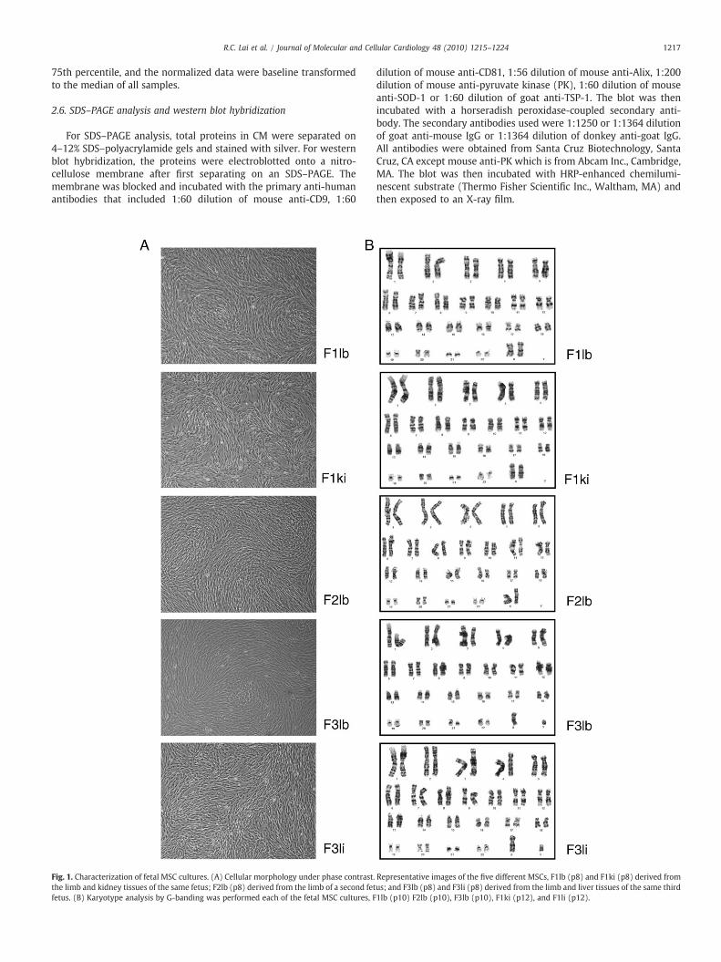
Fig. 2. Telomerase activity in hESC-MSCs and fetalMSCs. Relative telomerase activitywasmeasured by real time quantitative telomeric repeat amplification protocol. This qPCR-based assay quantifies product generated in vitro by telomerase activity present in thesamples. The relative telomerase activity which is directly proportional to the amount oftelomerase products was assessed by the threshold cycle number (or Ct value) for 1 μgprotein cell lysate. Hues9.E1 referred to a previously described hESC-MSCs line and HEKis a human embryonic kidney cell line. The Ct value for each fetal MSCswas themean forthree passages, P16, P18, and P20, and that for Hues9.E1 was themean for two passages,P20 and P22. The assay was performed in triplicate for each passage.
1218 R.C. Lai et al. / Journal of Molecular and Cellular Cardiology 48 (2010) 1215–1224
2.7. HPLC purification of microparticles
The instrument setup consisted of a liquid chromatography systemwith a binary pump, an auto injector, a thermostated column ovenand a UV–visible detector operated by the Class VP software fromShimadzu Corporation (Kyoto, Japan). The Chromatography columnsused were TSK Guard column SWXL, 6×40 mm and TSK gel G4000SWXL, 7.8×300 mm from Tosoh Corporation (Tokyo, Japan). Thedetectors Dawn 8 (light scattering), Optilab (refractive index) andQELS (dynamic light scattering) were connected in a series followingthe UV–visible detector. The last three detectors were from WyattTechnology Corporation (California, USA) and were operated by theASTRA software. The components of the sample were separated bysize exclusion, that is, the larger molecules will elute before thesmaller molecules. The eluent buffer used was 20 mM phosphatebuffer with 150mM of NaCl at pH 7.2. This buffer was filtered througha pore size of 0.1 μm and degassed for 15 min before use. Thechromatography system was equilibrated at a flow rate of 0.5 ml/minuntil the signal in Dawn 8 stabilized at around 0.3 detector voltageunits. The UV–visible detector was set at 220 nm and the column wasoven equilibrated to 25°C. The elution mode was isocratic and the runtime was 40 min. The volume of sample injected ranged from 50 to100 μl. The hydrodynamic radius, Rh was computed by the QELS andDawn 8 detectors. The highest count rate (Hz) at the peak apex wastaken as the Rh. Peaks of the separated components visualized at 220nm were collected as fractions for further characterization studies.
2.8. Testing secretion for cardioprotection
The secretion was prepared by growing the fetal MSCs in achemically defined serum-free culture medium for 3 days aspreviously described [22]. Briefly, cells at p12 were first expandedin serum-containing culture medium as described above. At p15, 80%confluent cell cultures were washed three times with PBS and thenincubated in a chemically defined medium consisting of DMEMwithout phenol red (Invitrogen Corporation, Carlsbad, CA) andsupplemented with insulin, transferrin, and selenoprotein (ITS)(Invitrogen Corporation, Carlsbad, CA), 5 ng/ml FGF2 (InvitrogenCorporation, Carlsbad, CA), 5 ng/ml PDGF AB (Peprotech, Rocky Hill,NJ), glutamine–penicillin–streptomycin, and β-mercaptoethanolovernight. The cell culture was then washed with PBS and replacedwith fresh chemically defined medium for another 3 days to producethe conditioned medium. This CM was collected and clarified bycentrifugation at 500×g. The clarified CM concentrated 50 times byreducing its volume by a factor of 50 using a tangential flow filtrationsystem with membrane MW cutoff of 100 kDa (Satorius, Goettingen,Germany). The use of a membrane MW cutoff of 100 kDa allowsmolecules with MW of less than 100 kDa to pass through the filterresulting a preferential loss of molecules less than 100 kDa. Theconcentrated CM was then sterilized by filtration through a 220 nmfilter.
The CM was tested in a mouse model of MI/R injury. MI wasinduced by 30 min left coronary artery (LCA) occlusion andsubsequent reperfusion. Five minutes before reperfusion, mice wereintravenously infused with 200 μl saline diluted CM containing 3 μgprotein for Hues9.E1 (hESC-MSCs) CM or 150 μg protein for fetal MSCCM or 10 μg protein for HPLC F1 via the tail vein. Control animals wereinfused with 200 μl saline. After 24 h of reperfusion, infarct size (IS) asa percentage of the area at risk (AAR) was assessed using Evans' bluedye injection and TTC staining as described previously [23].
2.9. Statistical analysis
Two-way ANOVA with post-hoc Dunnett was used to test thedifference in infarct size between groups. Correlation coefficient ofeach pairs of array was assessed using Pearson correlation test.
3. Results
3.1. Generating MSC cultures from human fetal tissues
We generated five MSC cultures from fetal limb (F1lb, F2lb, F3lb),kidney (F1ki) and liver (F3li) tissues of three fetuses in threeindependent experiments using feeder- and serum-free culturecondition as previously described [21]. A homogenous culture ofputative fibroblast-like MSCs migrated out of the tissues and adheredto the plastic culture dish, 2 days after fetal tissues were plated ongelatinized tissue culture plates. This observation was consistent withthe defining characteristic of MSCs i.e. adherence to plastic. The largetissue pieces were then washed off leaving a homogenous cell culture.This procedure was performed on five different fetal tissuesoriginating from 3 fetuses in 3 independent experiments. Each time,a homogenous culture of putative MSCs was obtained that form atypical fingerprint whorl at confluency (Fig. 1A). The cultures weredesignated P1 when 2×10⁷ cells were generated. The averagepopulation doubling time of all five cultures was between 48 and72 h and was most optimal at between 25% and 80% confluency or15–50,000 cells per cm2. The cells could be maintained in continuousculture for at least 20 passages at a 1:4 split with minimal changes inpopulation doubling time. The karyotype of all five cultures at p10–12was normal i.e. 46 XX or 46XY as determined byG-banding (Fig. 1B). Atpassages 14, 16 and 18, telomerase activity in all fiveMSC cultures wasdetermined and the average cellular telomerase activity over threepassages in each of the five cultures was equivalent to that of thepreviously describedHues9.E1 hESC-MSCs [21] (Fig. 2). Allfive culturescould be expanded to at least passage 20 without obvious changes inthe population doubling time. At a 1:4 split ratio and starting with 107
cells, each line would generate 1016 cells at passage 15 or 1019 cells atpassage 20.
3.2. Assessment of fetal cultures as MSCs
The five putative MSC cultures derived from fetal tissues wereassessed according to the ISCT minimal criteria for the definition ofhuman MSCs [27]. All five presumptive MSC cultures derived fromthree different fetuses were grown on plastic culture dishes as amonolayer of adherent spindle-shaped cells (Fig. 1A). They were allCD29+, CD44+, CD49a+ CD49e+, CD105+, CD166+, MHC I+, CD34−

Fig. 3. Marker profiling. (A, B) F1lb MSCs at p11 or p12 were stained with a specific antibody conjugated to a fluorescent dye and analyzed by FACS. Nonspecific fluorescence wasdetermined by incubation of similar cell aliquots with isotype-matched mouse monoclonal antibodies. (C) Relative transcription level of OCT4 and SOX2 was measured usingquantitative RT-PCR. hES3, a human embryonic stem cells line was set as the baseline for comparison.
1219R.C. Lai et al. / Journal of Molecular and Cellular Cardiology 48 (2010) 1215–1224
and CD45− as represented by F1lb MSCs (Fig. 3A). We observedthat F1lb MSCs were HLA-DRlo but the remaining four cultures wereHLA-DR−. In contrast to previous reports [28–37], these fetal MSCs-like hESC-MSCs did not express pluripotency-associated proteinsOct4, SSEA-4, and Tra1-60 as exemplified by F1lb MSCs (Fig. 3B).However, transcripts of OCT4 and SOX2 were readily detected byreal time PCR but their levels were at least ten times lower thanthose in hES3 human ESCs (Fig. 3C). All five presumptive fetal MSCcultures could be induced to differentiate to osteoblasts, adipocytesand chondroblasts in vitro (Fig. 4).
Fig. 4. Differentiation of fetal MSCs. Fetal MSCs were induced to undergo osteogenesichondrogenesis, the differentiated cells were stained with von Kossa stain, Oil Red and AlcianF3lb MSCs at 100×magnification.
3.3. Gene expression profile
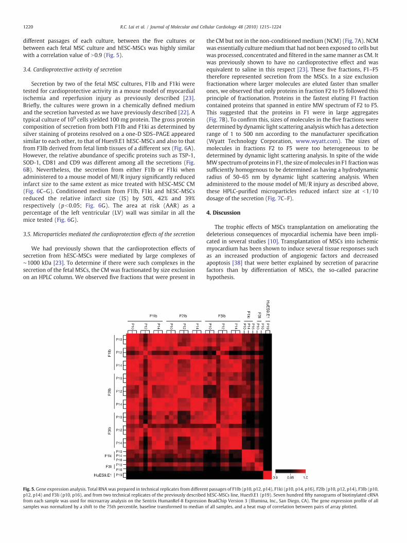
Genome-wide gene expression profiling of the fetal MSCs andhESC-MSCs was performed using microarray hybridization to assess1) the relatedness among the five fetal MSC cultures derived fromthree different tissues and 2) the relatedness between the fetal MSCcultures and hESC-MSCs. Microarray hybridization was performed induplicate on Sentrix Human Ref-8 Expression BeadChip version 3(Illumina, Inc., San Diego, CA) using RNA from two or three differentpassages of the MSC cultures. The gene expression profile between
s, adipogenesis and chondrogenesis. After a) osteogenesis, b) adipogenesis and c)blue, respectively. Images of differentiated fetal MSCs as represented by differentiated
1220 R.C. Lai et al. / Journal of Molecular and Cellular Cardiology 48 (2010) 1215–1224
different passages of each culture, between the five cultures orbetween each fetal MSC culture and hESC-MSCs was highly similarwith a correlation value of N0.9 (Fig. 5).
3.4. Cardioprotective activity of secretion
Secretion by two of the fetal MSC cultures, F1lb and F1ki weretested for cardioprotective activity in a mouse model of myocardialischemia and reperfusion injury as previously described [23].Briefly, the cultures were grown in a chemically defined mediumand the secretion harvested as we have previously described [22]. Atypical culture of 109 cells yielded 100 mg protein. The gross proteincomposition of secretion from both F1lb and F1ki as determined bysilver staining of proteins resolved on a one-D SDS–PAGE appearedsimilar to each other, to that of Hues9.E1 hESC-MSCs and also to thatfrom F3lb derived from fetal limb tissues of a different sex (Fig. 6A).However, the relative abundance of specific proteins such as TSP-1,SOD-1, CD81 and CD9 was different among all the secretions (Fig.6B). Nevertheless, the secretion from either F1lb or F1ki whenadministered to a mouse model of MI/R injury significantly reducedinfarct size to the same extent as mice treated with hESC-MSC CM(Fig. 6C–G). Conditioned medium from F1lb, F1ki and hESC-MSCsreduced the relative infarct size (IS) by 50%, 42% and 39%respectively (pb0.05; Fig. 6G). The area at risk (AAR) as apercentage of the left ventricular (LV) wall was similar in all themice tested (Fig. 6G).
3.5. Microparticles mediated the cardioprotection effects of the secretion
We had previously shown that the cardioprotection effects ofsecretion from hESC-MSCs were mediated by large complexes of∼1000 kDa [23]. To determine if there were such complexes in thesecretion of the fetal MSCs, the CM was fractionated by size exclusionon an HPLC column. We observed five fractions that were present in
Fig. 5. Gene expression analysis. Total RNA was prepared in technical replicates from differenp12, p14) and F3li (p10, p16), and from two technical replicates of the previously describedfrom each sample was used for microarray analysis on the Sentrix HumanRef-8 Expressionsamples was normalized by a shift to the 75th percentile, baseline transformed to median
the CM but not in the non-conditionedmedium (NCM) (Fig. 7A). NCMwas essentially culturemedium that had not been exposed to cells butwas processed, concentrated and filtered in the samemanner as CM. Itwas previously shown to have no cardioprotective effect and wasequivalent to saline in this respect [23]. These five fractions, F1–F5therefore represented secretion from the MSCs. In a size exclusionfractionation where larger molecules are eluted faster than smallerones, we observed that only proteins in fraction F2 to F5 followed thisprinciple of fractionation. Proteins in the fastest eluting F1 fractioncontained proteins that spanned in entire MW spectrum of F2 to F5.This suggested that the proteins in F1 were in large aggregates(Fig. 7B). To confirm this, sizes of molecules in the five fractions weredetermined by dynamic light scattering analysis which has a detectionrange of 1 to 500 nm according to the manufacturer specification(Wyatt Technology Corporation, www.wyatt.com). The sizes ofmolecules in fractions F2 to F5 were too heterogeneous to bedetermined by dynamic light scattering analysis. In spite of the wideMWspectrumof proteins in F1, the size ofmolecules in F1 fractionwassufficiently homogenous to be determined as having a hydrodynamicradius of 50–65 nm by dynamic light scattering analysis. Whenadministered to the mouse model of MI/R injury as described above,these HPLC-purified microparticles reduced infarct size at b1/10dosage of the secretion (Fig. 7C–F).
4. Discussion
The trophic effects of MSCs transplantation on ameliorating thedeleterious consequences of myocardial ischemia have been impli-cated in several studies [10]. Transplantation of MSCs into ischemicmyocardium has been shown to induce several tissue responses suchas an increased production of angiogenic factors and decreasedapoptosis [38] that were better explained by secretion of paracrinefactors than by differentiation of MSCs, the so-called paracrinehypothesis.
t passages of F1lb (p10, p12, p14), F1ki (p10, p14, p16), F2lb (p10, p12, p14), F3lb (p10,hESC-MSCs line, Hues9.E1 (p19). Seven hundred fifty nanograms of biotinylated cRNABeadChip Version 3 (Illumina, Inc., San Diego, CA). The gene expression profile of all
of all samples, and a heat map of correlation between pairs of array plotted.
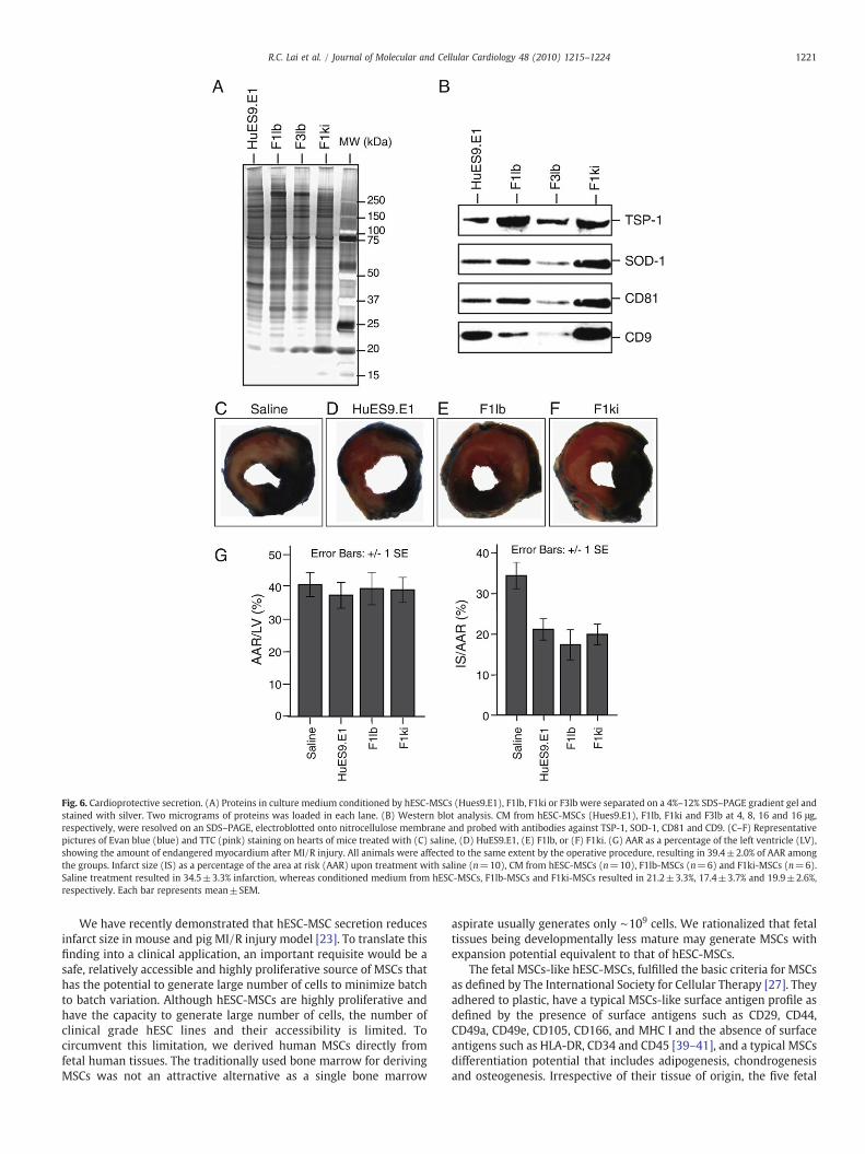
Fig. 6. Cardioprotective secretion. (A) Proteins in culture medium conditioned by hESC-MSCs (Hues9.E1), F1lb, F1ki or F3lb were separated on a 4%–12% SDS–PAGE gradient gel andstained with silver. Two micrograms of proteins was loaded in each lane. (B) Western blot analysis. CM from hESC-MSCs (Hues9.E1), F1lb, F1ki and F3lb at 4, 8, 16 and 16 μg,respectively, were resolved on an SDS–PAGE, electroblotted onto nitrocellulose membrane and probed with antibodies against TSP-1, SOD-1, CD81 and CD9. (C–F) Representativepictures of Evan blue (blue) and TTC (pink) staining on hearts of mice treated with (C) saline, (D) HuES9.E1, (E) F1lb, or (F) F1ki. (G) AAR as a percentage of the left ventricle (LV),showing the amount of endangered myocardium after MI/R injury. All animals were affected to the same extent by the operative procedure, resulting in 39.4±2.0% of AAR amongthe groups. Infarct size (IS) as a percentage of the area at risk (AAR) upon treatment with saline (n=10), CM from hESC-MSCs (n=10), F1lb-MSCs (n=6) and F1ki-MSCs (n=6).Saline treatment resulted in 34.5±3.3% infarction, whereas conditioned medium from hESC-MSCs, F1lb-MSCs and F1ki-MSCs resulted in 21.2±3.3%, 17.4±3.7% and 19.9±2.6%,respectively. Each bar represents mean±SEM.
1221R.C. Lai et al. / Journal of Molecular and Cellular Cardiology 48 (2010) 1215–1224
We have recently demonstrated that hESC-MSC secretion reducesinfarct size in mouse and pig MI/R injury model [23]. To translate thisfinding into a clinical application, an important requisite would be asafe, relatively accessible and highly proliferative source of MSCs thathas the potential to generate large number of cells to minimize batchto batch variation. Although hESC-MSCs are highly proliferative andhave the capacity to generate large number of cells, the number ofclinical grade hESC lines and their accessibility is limited. Tocircumvent this limitation, we derived human MSCs directly fromfetal human tissues. The traditionally used bone marrow for derivingMSCs was not an attractive alternative as a single bone marrow
aspirate usually generates only ∼109 cells. We rationalized that fetaltissues being developmentally less mature may generate MSCs withexpansion potential equivalent to that of hESC-MSCs.
The fetal MSCs-like hESC-MSCs, fulfilled the basic criteria for MSCsas defined by The International Society for Cellular Therapy [27]. Theyadhered to plastic, have a typical MSCs-like surface antigen profile asdefined by the presence of surface antigens such as CD29, CD44,CD49a, CD49e, CD105, CD166, and MHC I and the absence of surfaceantigens such as HLA-DR, CD34 and CD45 [39–41], and a typical MSCsdifferentiation potential that includes adipogenesis, chondrogenesisand osteogenesis. Irrespective of their tissue of origin, the five fetal
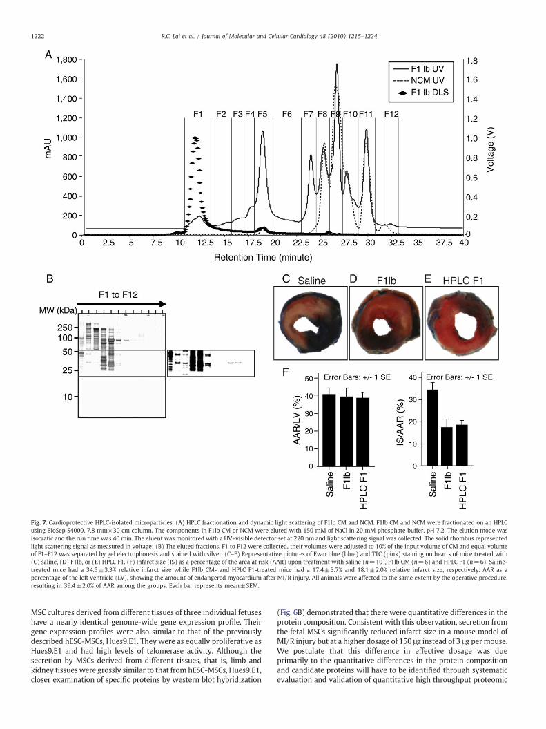
Fig. 7. Cardioprotective HPLC-isolated microparticles. (A) HPLC fractionation and dynamic light scattering of F1lb CM and NCM. F1lb CM and NCM were fractionated on an HPLCusing BioSep S4000, 7.8 mm×30 cm column. The components in F1lb CM or NCM were eluted with 150 mM of NaCl in 20 mM phosphate buffer, pH 7.2. The elution mode wasisocratic and the run time was 40 min. The eluent was monitored with a UV–visible detector set at 220 nm and light scattering signal was collected. The solid rhombus representedlight scattering signal as measured in voltage; (B) The eluted fractions, F1 to F12 were collected, their volumes were adjusted to 10% of the input volume of CM and equal volumeof F1–F12 was separated by gel electrophoresis and stained with silver. (C–E) Representative pictures of Evan blue (blue) and TTC (pink) staining on hearts of mice treated with(C) saline, (D) F1lb, or (E) HPLC F1. (F) Infarct size (IS) as a percentage of the area at risk (AAR) upon treatment with saline (n=10), F1lb CM (n=6) and HPLC F1 (n=6). Saline-treated mice had a 34.5±3.3% relative infarct size while F1lb CM- and HPLC F1-treated mice had a 17.4±3.7% and 18.1±2.0% relative infarct size, respectively. AAR as apercentage of the left ventricle (LV), showing the amount of endangered myocardium after MI/R injury. All animals were affected to the same extent by the operative procedure,resulting in 39.4±2.0% of AAR among the groups. Each bar represents mean±SEM.
1222 R.C. Lai et al. / Journal of Molecular and Cellular Cardiology 48 (2010) 1215–1224
MSC cultures derived from different tissues of three individual fetuseshave a nearly identical genome-wide gene expression profile. Theirgene expression profiles were also similar to that of the previouslydescribed hESC-MSCs, Hues9.E1. They were as equally proliferative asHues9.E1 and had high levels of telomerase activity. Although thesecretion by MSCs derived from different tissues, that is, limb andkidney tissues were grossly similar to that from hESC-MSCs, Hues9.E1,closer examination of specific proteins by western blot hybridization
(Fig. 6B) demonstrated that there were quantitative differences in theprotein composition. Consistent with this observation, secretion fromthe fetal MSCs significantly reduced infarct size in a mouse model ofMI/R injury but at a higher dosage of 150 μg instead of 3 μg per mouse.We postulate that this difference in effective dosage was dueprimarily to the quantitative differences in the protein compositionand candidate proteins will have to be identified through systematicevaluation and validation of quantitative high throughput proteomic
1223R.C. Lai et al. / Journal of Molecular and Cellular Cardiology 48 (2010) 1215–1224
analysis. We further demonstrate that consistent with our previousobservations [23], the cardioprotective effect of secretion from fetalMSCs was mediated by large complexes in the range of 100 to 200 nmor possibly, microparticles.
Microparticle is a minimally defined term that encompasses abroad spectrum of secreted particles in the size range of 0.05–1 μm.Microparticles are known to be produced by numerous cell types[42–48]. Although the function of microparticles remains poorlyunderstood, there is irrefutable evidence that microparticles haveimportant functions. As summarized by many recent reviews [46–57],microparticles are definitively associated with many diseases such ascancer, atherosclerosis and cerebral ischemia. However, the specificrole ofmicroparticles in disease process and/or progress remains to bedetermined. As some microparticles carried biologically activematerials includingproteins andRNAs that can be transferredbetweencells, it is likely that microparticles would have an impact eitherpositively or negatively on a disease process and/or progress. This hasled to suggestions that microparticles could be exploited as potentialtherapeutic vectors [58]. However, the broad definition of micro-particles which encompasses all secreted membrane vesicles toinclude the more defined exosomes (50–100 nm), microvesicles(100–1000 nm), ectosomes (50–200 nm), membrane particles(50–80 nm), exosome-like vesicles (20–50 nm) and apoptotic vesicles(50–500 nm) [24] has impeded our understanding of microparticles.Other than exosomes which are the most stringently definedmicroparticles to date, the major distinguishing parameter for thesedifferent classes of microparticles is their size.
In this report where we not only demonstrated directly for the firsttime that purified microparticles in MSC secretion were cardiopro-tective, we also purified these microparticles by size exclusion on anHPLC which is currently one of the most precise methods for sizefractionation of molecules or particles. The purified microparticleswere determined by dynamic light scattering analysis to be apopulation of homogenously sized particles with a hydrodynamicradius of 50–65 nm. This particle size which translated into anapproximate diameter of 100–130 nm was consistent with ourprevious observation that filtration through a membrane with anMW cutoff of 1000 kDa caused the filtrate to lose its cardioprotectivefunction while that part of secretion retained by the same filter or theretentate was cardioprotective [23]. A membrane with an MW cutoffof 1000 kDa has a 100-nm nominal pore size according to themanufacturer (Pall Corp. http://www.pall.com/). This essentiallymeant that the filtrate contained particles with diameter of less than50–100 nm and these b50–100 nm particles were not cardioprotec-tive. On the other hand, the retentate containing particles N50–100 nmwas cardioprotective. Together, this study and our previous study [23]demonstrated that cardioprotection by the secretion of hESC-MSCsand fetal MSCs was similarly mediated by microparticles withdiameters of N50–100 nm. Our previous study also suggested thatsecretion devoid of themicroparticles but containing all other secretedproteins as represented by the CM filtrate through a membrane withan MW cutoff of 1000 kDa was not cardioprotective [23]. Thisobservation of no cardioprotective activity could therefore beextrapolated to HPLC fraction 2 to 4 where there were no micro-particles and the secreted proteins were eluted according to the sizeexclusion principle, and were therefore soluble.
The involvement of microparticles in mediating the paracrineeffect of MSC transplantation on tissue repair represents a radical shiftin our current understanding of the paracrine effect of MSC whichhitherto has been limited to cytokine, chemokine or growth factor-mediated extracellular signaling [10].
Together our report demonstrated that MSCs derived from fetaltissues are a viable alternative to human ESCs as a tissue source ofhighly proliferative MSCs that produces cardioprotective secretion. Asingle fetal tissue could potentially generate 1016 to 1019 MSCs. A 109
cell culture secretes 100 mg protein. We have previously shown that
the effective cardioprotective dose for a pig model of MI/R injury was10 mg protein per 60–70 kg pig [23]. Therefore, a single fetal tissuecould potentially generate sufficient MSCs to produce 108–11 doses.Together with the relatively simple purification of these micro-particles to a population of homogenously sized microparticles, theuse of secretion from fetal MSCs represents a viable strategy toaddress an urgent unmet therapeutic need for treating MI/R injury[59].
5. Disclosure statement
No potential conflict of interest.
Acknowledgments
We thank J.J. Chee (KKH) for her help in patient recruitment andtissue harvest, and Jayanthi Padmanabhan (BTI) for technicalassistance in preparing the secretion.
References
[1] Le Blanc K, Pittenger M. Mesenchymal stem cells: progress toward promise.Cytotherapy 2005;7:36–45.
[2] Minguell JJ, Erices A. Mesenchymal stem cells and the treatment of cardiac disease.Exp Biol Med (Maywood) 2006;231:39–49.
[3] Schuleri KH, Boyle AJ, Hare JM. Mesenchymal stem cells for cardiac regenerativetherapy. Handb Exp Pharmacol 2007:195–218.
[4] Abdel-Latif A, Bolli R, Tleyjeh IM, Montori VM, Perin EC, Hornung CA, et al. Adultbone marrow-derived cells for cardiac repair: a systematic review and meta-analysis. Arch Intern Med 2007;167:989–97.
[5] Mazhari R, Hare JM. Advances in cell-based therapy for structural heart disease.Prog Cardiovasc Dis 2007;49:387–95.
[6] Ohnishi S, Nagaya N. Prepare cells to repair the heart: mesenchymal stem cells forthe treatment of heart failure. Am J Nephrol 2007;27:301–7.
[7] Behfar A, Terzic A. Optimizing adult mesenchymal stem cells for heart repair. J MolCell Cardiol 2007;42:283–4.
[8] Atsma DE, Fibbe WE, Rabelink TJ. Opportunities and challenges for mesenchymalstem cell-mediated heart repair. Curr Opin Lipidol 2007;18:645–9.
[9] Gnecchi M, He H, Liang OD, Melo LG, Morello F, Mu H, et al. Paracrine actionaccounts for marked protection of ischemic heart by Akt-modified mesenchymalstem cells. Nat Med 2005;11:367–8.
[10] Caplan AI, Dennis JE. Mesenchymal stem cells as trophic mediators. J Cell Biochem2006;98:1076–84.
[11] Kinnaird T, Stabile E, Burnett MS, Shou M, Lee CW, Barr S, et al. Local delivery ofmarrow-derived stromal cells augments collateral perfusion through paracrinemechanisms. Circulation 2004;109:1543–9.
[12] Leedham SJ, Brittan M, McDonald SA, Wright NA. Intestinal stem cells. J Cell MolMed 2005;9:11–24.
[13] Togel F, Hu Z,Weiss K, Isaac J, Lange C,Westenfelder C. Administeredmesenchymalstem cells protect against ischemic acute renal failure through differentiation-independent mechanisms. Am J Physiol Renal Physiol 2005;289:F31–42.
[14] Patschan D, Plotkin M, Goligorsky MS. Therapeutic use of stem and endothelialprogenitor cells in acute renal injury: ca ira. Curr Opin Pharmacol 2006;6:176–83.
[15] Miyahara Y, Nagaya N, Kataoka M, Yanagawa B, Tanaka K, Hao H, et al. Mono-layered mesenchymal stem cells repair scarred myocardium after myocardialinfarction. Nat Med 2006;12:459–65.
[16] Gnecchi M, He H, Noiseux N, Liang OD, Zhang L, Morello F, et al. Evidencesupporting paracrine hypothesis for Akt-modified mesenchymal stem cell-mediated cardiac protection and functional improvement. FASEB J 2006;20:661–9.
[17] Mayer H, Bertram H, Lindenmaier W, Korff T, Weber H, Weich H. Vascularendothelial growth factor (VEGF-A) expression in human mesenchymal stemcells: autocrine and paracrine role on osteoblastic and endothelial differentiation.J Cell Biochem 2005;95:827–39.
[18] Nakagami H, Maeda K, Morishita R, Iguchi S, Nishikawa T, Takami Y, et al. Novelautologous cell therapy in ischemic limb disease through growth factor secretionby cultured adipose tissue-derived stromal cells. Arterioscler Thromb Vasc Biol2005;25:2542–7.
[19] Van Overstraeten-Schlogel N, Beguin Y, Gothot A. Role of stromal-derived factor-1in the hematopoietic-supporting activity of human mesenchymal stem cells. Eur JHaematol 2006;76:488–93.
[20] Cheng L, Qasba P, Vanguri P, Thiede MA. Human mesenchymal stem cells supportmegakaryocyte and pro-platelet formation from CD34(+) hematopoietic pro-genitor cells. J Cell Physiol 2000;184:58–69.
[21] Lian Q, Lye E, Suan Yeo K, Khia Way Tan E, Salto-Tellez M, Liu TM, et al. Derivationof clinically compliant MSCs from CD105+, CD24− differentiated human ESCs.Stem Cells 2007;25:425–36.
[22] Sze SK, de Kleijn DP, Lai RC, Khia Way Tan E, Zhao H, Yeo KS, et al. Elucidating thesecretion proteome of human embryonic stem cell-derived mesenchymal stemcells. Mol Cell Proteomics 2007;6:1680–9.
1224 R.C. Lai et al. / Journal of Molecular and Cellular Cardiology 48 (2010) 1215–1224
[23] Arslan F, Smeets MB, O'Neill LA, Keogh B, McGuirk P, Timmers L, et al. Myocardialischemia/reperfusion injury is mediated by leukocytic toll-like receptor-2 andreduced by systemic administration of a novel anti-toll-like receptor-2 antibody.Circulation 2010;121:80–90.
[24] Thery C, Ostrowski M, Segura E. Membrane vesicles as conveyors of immuneresponses. Nat Rev Immunol 2009;9:581–93.
[25] Ethical, legal and social issues in human stem cell research, reproductive andtherapeutic cloning. Singapore Bioethics Advisory Committee Report 2002.
[26] Wege H, Chui MS, Le HT, Tran JM, Zern MA. SYBR Green real-time telomeric repeatamplification protocol for the rapid quantification of telomerase activity. NucleicAcids Res 2003;31:E3.
[27] Dominici M, Le Blanc K, Mueller I, Slaper-Cortenbach I, Marini F, Krause D, et al.Minimal criteria for defining multipotent mesenchymal stromal cells. The Interna-tional Society for Cellular Therapy position statement. Cytotherapy 2006;8:315–7.
[28] Tsai MS, Lee JL, Chang YJ, Hwang SM. Isolation of human multipotentmesenchymal stem cells from second-trimester amniotic fluid using a noveltwo-stage culture protocol. Hum Reprod 2004;19:1450–6.
[29] Guillot PV, Gotherstrom C, Chan J, Kurata H, Fisk NM. Human first-trimester fetalMSC express pluripotency markers and grow faster and have longer telomeresthan adult MSC. Stem Cells 2007;25:646–54.
[30] Peng HH, Wang TH, Chao AS, Chang SD. Isolation and differentiation of humanmesenchymal stem cells obtained from second trimester amniotic fluid; experi-ments at Chang Gung Memorial Hospital. Chang Gung Med J 2007;30:402–7.
[31] Roubelakis MG, Pappa KI, Bitsika V, Zagoura D, Vlahou A, Papadaki HA, et al.Molecular and proteomic characterization of human mesenchymal stem cellsderived from amniotic fluid: comparison to bone marrow mesenchymal stemcells. Stem Cells Dev 2007;16:931–52.
[32] YenML, ChienCC, Chiu IM,HuangHI,ChenYC,HuHI, et al.Multilineagedifferentiationand characterization of the human fetal osteoblastic 1.19 cell line: a possible invitro model of human mesenchymal progenitors. Stem Cells 2007;25:125–31.
[33] Jo CH, Kim OS, Park EY, Kim BJ, Lee JH, Kang SB, et al. Fetal mesenchymal stem cellsderived from human umbilical cord sustain primitive characteristics duringextensive expansion. Cell Tissue Res 2008;334:423–33.
[34] Kermani AJ, Fathi F, Mowla SJ. Characterization and genetic manipulation ofhuman umbilical cord vein mesenchymal stem cells: potential application in cell-based gene therapy. Rejuvenation Res 2008;11:379–86.
[35] Poloni A, Rosini V, Mondini E, Maurizi G, Mancini S, Discepoli G, et al.Characterization and expansion of mesenchymal progenitor cells from first-trimester chorionic villi of human placenta. Cytotherapy 2008;10:690–7.
[36] Wang XY, Lan Y, He WY, Zhang L, Yao HY, Hou CM, et al. Identification ofmesenchymal stem cells in aorta–gonad–mesonephros and yolk sac of humanembryos. Blood 2008;111:2436–43.
[37] Zhang ZY, Teoh SH, Chong MS, Schantz JT, Fisk NM, Choolani MA, et al. Superiorosteogenic capacity for bone tissue engineering of fetal compared to perinatal andadult mesenchymal stem cells. Stem Cells 2009;27:126–37.
[38] Tang YL, Zhao Q, Qin X, Shen L, Cheng L, Ge J, et al. Paracrine action enhances the effectsof autologous mesenchymal stem cell transplantation on vascular regeneration in ratmodel of myocardial infarction. Ann Thorac Surg 2005;80:229–36 discussion 36-7.
[39] Javazon EH, Beggs KJ, Flake AW. Mesenchymal stem cells: paradoxes of passaging.Exp Hematol 2004;32:414–25.
[40] Barry FP, Murphy JM. Mesenchymal stem cells: clinical applications and biologicalcharacterization. Int J Biochem Cell Biol 2004;36:568–84.
[41] Majumdar MK, Keane-Moore M, Buyaner D, Hardy WB, Moorman MA, McIntoshKR, et al. Characterization and functionality of cell surface molecules on humanmesenchymal stem cells. J Biomed Sci 2003;10:228–41.
[42] Meziani F, Tesse A, Andriantsitohaina R. Microparticles are vectors of paradoxicalinformation in vascular cells including the endothelium: role in health anddiseases. Pharmacol Rep 2008;60:75–84.
[43] Redman CW, Sargent IL. Circulating microparticles in normal pregnancy and pre-eclampsia. Placenta 2008;29(Suppl A):S73–7.
[44] Zwicker JI. Tissue factor-bearing microparticles and cancer. Semin ThrombHemost 2008;34:195–8.
[45] Aharon A, Brenner B. Microparticles, thrombosis and cancer. Best Pract Res ClinHaematol 2009;22:61–9.
[46] Burnier L, Fontana P, Kwak BR, Angelillo-Scherrer A. Cell-derivedmicroparticles inhaemostasis and vascular medicine. Thromb Haemost 2009;101:439–51.
[47] Castellana D, Kunzelmann C, Freyssinet JM. Pathophysiologic significance ofprocoagulant microvesicles in cancer disease and progression. Hamostaseologie2009;29:51–7.
[48] Chironi GN, Boulanger CM, Simon A, Dignat-George F, Freyssinet JM, Tedgui A.Endothelial microparticles in diseases. Cell Tissue Res 2009;335:143–51.
[49] Horstman LL, Jy W, Bidot CJ, Nordberg ML, Minagar A, Alexander JS, et al. Potentialroles of cell-derived microparticles in ischemic brain disease. Neurol Res 2009;31:799–806.
[50] Esmon CT. Basic mechanisms and pathogenesis of venous thrombosis. Blood Rev2009;23:225–9.
[51] Davizon P, Lopez JA. Microparticles and thrombotic disease. Curr Opin Hematol2009;16:334–41.
[52] Al-Nedawi K, Meehan B, Rak J. Microvesicles: messengers and mediators of tumorprogression. Cell Cycle 2009;8:2014–8.
[53] Doeuvre L, Plawinski L, Toti F, Angles-Cano E. Cell-derived microparticles: a newchallenge in neuroscience. J Neurochem 2009;110:457–68.
[54] Sabatier F, Camoin-Jau L, Anfosso F, Sampol J, Dignat-George F. Circulatingendothelial cells, microparticles and progenitors: key players towards thedefinition of vascular competence. J Cell Mol Med 2009;13:454–71.
[55] Stein E,McMahonB,KwaanH,Altman JK, FrankfurtO, TallmanMS.The coagulopathyof acute promyelocytic leukaemia revisited. Best Pract Res Clin Haematol 2009;22:153–63.
[56] Milsom C, Rak J. Tissue factor and cancer. Pathophysiol Haemost Thromb 2008;36:160–76.
[57] Hartvigsen K, ChouMY, Hansen LF, Shaw PX, Tsimikas S, Binder CJ, et al. The role ofinnate immunity in atherogenesis. J Lipid Res 2009;50 Suppl:S388–93.
[58] Benameur T, Andriantsitohaina R, Martinez MC. Therapeutic potential of plasmamembrane-derived microparticles. Pharmacol Rep 2009;61:49–57.
[59] Knight DR. Editorial overview: cardioprotective drugs for myocardial ischemicinjury—a therapeutic area at risk. Curr Opin Investig Drugs 2007;8:190–2.