Evaluation of solid lipid microparticles produced by spray congealing for topical application of...
9
JPP 2009, 61: 559–567 ß 2009 The Authors Received September 26, 2008 Accepted February 04, 2009 DOI 10.1211/jpp/61.05.0003 ISSN 0022-3573 Correspondence: Nadia Passerini, Dipartimento di Scienze Farmaceutiche, Universita ` di Bologna, Via S. Donato 19/2, 40127 Bologna, Italy. E-mail: [email protected] Research Paper Evaluation of solid lipid microparticles produced by spray congealing for topical application of econazole nitrate Nadia Passerini a , Elisabetta Gavini b , Beatrice Albertini a , Giovanna Rassu b , Marcello Di Sabatino a , Vanna Sanna b , Paolo Giunchedi b and Lorenzo Rodriguez a a Dipartimento di Scienze Farmaceutiche, Universita ` di Bologna, Bologna and b Dipartimento di Scienza del Farmaco, Universita ` di Sassari, Sassari, Italy Abstract Objectives The aims of this study were to evaluate the suitability of the spray congealing technique to produce solid lipid microparticles (SLMs) for topical administration and to study the skin permeation of a drug from SLMs compared with solid lipid nanoparticles (SLNs). Methods Econazole nitrate was used as model drug and Precirol ATO 5 as the lipidic carrier. SLMs and SLNs were both prepared at 5 : 1, 10 : 1 and 12.5 : 1 lipid : drug weight ratios and characterised in terms of particle size, morphology, encapsulation efficiency and chemical analysis of the particle surface. SLMs and SLNs were also incorporated into HPMC K 100M hydrogels for ex-vivo drug permeation tests using porcine epidermis. Key findings SLMs had particle sizes of 18–45 mm, while SLNs showed a mean diameter of 130–270 nm. The encapsulation efficiency was 80–100%. Permeation profiles of econazole nitrate were influenced by both particle size (significant difference until 9 h) and the amount of lipid. Conclusions The results confirm the usefulness of SLNs as carriers for topical administration and suggest the potential of SLMs for the delivery of drugs to the skin. Keywords econazole nitrate; skin delivery; solid lipid microparticles; solid lipid nanoparticles; spray congealing Introduction In recent years, solid lipid micro- and nanoparticles (SLMs and SLNs) have emerged as attractive carriers for topical application in the pharmaceutical and in cosmetic fields because of their advantages over polymeric systems. [1–3] SLNs have been proposed to enhance the percutaneous absorption of drugs [4] and allow drug targeting to the skin. [5–8] Improved dermal absorption of active pharmaceutical ingredients loaded into lipid carriers may result from increases in the surface contact of drug and corneocytes, skin occlusion, rapid and steady release, and surfactant effects. [9] It is well demonstrated that, after application to the skin, the small particles can make close contact with superficial junctions of corneocytes on the outermost skin layers because of a pronounced adhesive effect. [10] Adherence of lipid nanoparticles to the skin leads to the formation of a film and subsequently to an occlusion effect. [11] The first model for film formation by SLNs on the skin was developed by Mu ¨ller and Dingler [12] and the occlusion effect has been widely demonstrated in in-vitro models [13] and after application in vivo. [14] The main approaches used for the production of finely dispersed lipid nanoparticles include high-pressure homogenisation, [15] microemulsions, [16] solvent emulsification– evaporation or –diffusion, [17,18] water/oil/water double-emulsion method [19] and high-speed stirring and/or ultrasonication. [20] SLMs have been less extensively investigated for skin application compared with SLNs but have recently been shown to be potentially useful for both topical and transdermal drug delivery. [21–25] SLMs can be prepared by different processes such as solvent evaporation, melt dispersion, hot and cold homogenisation, spray drying and spray congealing. [26] Spray congealing has 559
-
Upload
independent -
Category
Documents
-
view
4 -
download
0
Transcript of Evaluation of solid lipid microparticles produced by spray congealing for topical application of...
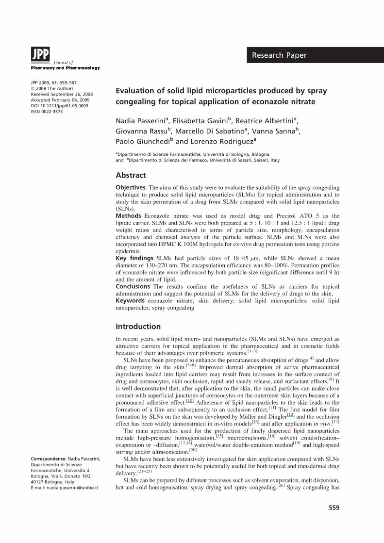
JPP 2009, 61: 559–567
! 2009 The Authors
Received September 26, 2008
Accepted February 04, 2009
DOI 10.1211/jpp/61.05.0003
ISSN 0022-3573
Correspondence: Nadia Passerini,
Dipartimento di Scienze
Farmaceutiche, Universita di
Bologna, Via S. Donato 19/2,
40127 Bologna, Italy.
E-mail: [email protected]
Research Paper
Evaluation of solid lipid microparticles produced by spray
congealing for topical application of econazole nitrate
Nadia Passerinia, Elisabetta Gavinib, Beatrice Albertinia,
Giovanna Rassub, Marcello Di Sabatinoa, Vanna Sannab,
Paolo Giunchedib and Lorenzo Rodrigueza
aDipartimento di Scienze Farmaceutiche, Universita di Bologna, Bologna
and bDipartimento di Scienza del Farmaco, Universita di Sassari, Sassari, Italy
Abstract
Objectives The aims of this study were to evaluate the suitability of the spray congealing
technique to produce solid lipid microparticles (SLMs) for topical administration and to
study the skin permeation of a drug from SLMs compared with solid lipid nanoparticles
(SLNs).
Methods Econazole nitrate was used as model drug and Precirol ATO 5 as the
lipidic carrier. SLMs and SLNs were both prepared at 5 : 1, 10 : 1 and 12.5 : 1 lipid : drug
weight ratios and characterised in terms of particle size, morphology, encapsulation
efficiency and chemical analysis of the particle surface. SLMs and SLNs were also
incorporated into HPMC K 100M hydrogels for ex-vivo drug permeation tests using porcine
epidermis.
Key findings SLMs had particle sizes of 18–45 mm, while SLNs showed a mean
diameter of 130–270 nm. The encapsulation efficiency was 80–100%. Permeation profiles
of econazole nitrate were influenced by both particle size (significant difference until 9 h)
and the amount of lipid.
Conclusions The results confirm the usefulness of SLNs as carriers for topical
administration and suggest the potential of SLMs for the delivery of drugs to the skin.
Keywords econazole nitrate; skin delivery; solid lipid microparticles; solid lipid
nanoparticles; spray congealing
Introduction
In recent years, solid lipid micro- and nanoparticles (SLMs and SLNs) have emerged as
attractive carriers for topical application in the pharmaceutical and in cosmetic fields
because of their advantages over polymeric systems.[1–3]
SLNs have been proposed to enhance the percutaneous absorption of drugs[4] and allow
drug targeting to the skin.[5–8] Improved dermal absorption of active pharmaceutical
ingredients loaded into lipid carriers may result from increases in the surface contact of
drug and corneocytes, skin occlusion, rapid and steady release, and surfactant effects.[9] It
is well demonstrated that, after application to the skin, the small particles can make close
contact with superficial junctions of corneocytes on the outermost skin layers because of a
pronounced adhesive effect.[10] Adherence of lipid nanoparticles to the skin leads to the
formation of a film and subsequently to an occlusion effect.[11] The first model for film
formation by SLNs on the skin was developed by Muller and Dingler[12] and the occlusion
effect has been widely demonstrated in in-vitro models[13] and after application in vivo.[14]
The main approaches used for the production of finely dispersed lipid nanoparticles
include high-pressure homogenisation,[15] microemulsions,[16] solvent emulsification–
evaporation or –diffusion,[17,18] water/oil/water double-emulsion method[19] and high-speed
stirring and/or ultrasonication.[20]
SLMs have been less extensively investigated for skin application compared with SLNs
but have recently been shown to be potentially useful for both topical and transdermal drug
delivery.[21–25]
SLMs can be prepared by different processes such as solvent evaporation, melt dispersion,
hot and cold homogenisation, spray drying and spray congealing.[26] Spray congealing has
559

attracted increasing attention in recent years because this
technique does not require the use of organic or aqueous
solvents and hence it is environmentally friendly and less time
consuming than other methods; moreover the process can be
easily employed on an industrial scale. Spray congealing, also
called spray chilling, involves the atomisation of a fluid
(solution or suspension of the active pharmaceutical ingredient
in a melted carrier) into an environment maintained at a
temperature below the carrier melting point. The atomisation
leads to the formation of melted droplets, which then solidify
upon cooling, producing the final microparticles.[27] Ultra-
sonic atomisers[28–30] and, more recently,[31] a wide pneumatic
nozzle have been proposed as new atomisers, allowing the
preparation of non-aggregated and spherical microparticles
with high encapsulation efficiency suitable for oral adminis-
tration. Until now, spray congealing has not been considered
as a production technique for SLMs for skin application.
The aims of this study were to evaluate the feasibility of
producing SLMs suitable for topical administration using the
spray congealing technique and to study the skin permeation
of a drug from the SLMs compared with SLNs. Econazole
nitrate (ECN) was used as a model drug and Precirol ATO 5 as
the lipidic carrier; microparticles and nanoparticles were both
prepared at three different lipid : drug weight ratios (5 : 1,
10 : 1 and 12.5 : 1). The particles obtained were characterised
in terms of particle size, morphology, encapsulation efficiency
and chemical analyses of their surface.
After incorporation of micro- and nanoparticles into
hydrogels, the ex-vivo permeation studies of the ECN from
formulations through porcine skin were then carried out and
compared with the non-encapsulated ECN.
Materials and Methods
Materials
ECN (molecular weight 444.7, log P 5.61) was kindly
provided by Erregierre SpA (Bergamo, Italy) and glyceryl
palmitostearate (Precirol ATO 5) from Gattefosse (Cedex,
France). PEG sorbitan monooleate (Tween 80) was purchased
from Sigma-Aldrich Chemie GmbH (Steinheim, Germany)
and hydroxypropylmethylcellulose (HPMC K100M) from
Dow Chemicals (Midland, MI, USA). Methanol (Chromasolv
for HPLC, gradient grade) and NH4H2PO4 were obtained from
Rielden-de Haen AG (Seelze, Germany). Other chemicals
were of HPLC or analytical grade.
Preparation of solid lipid microparticles (SLM)
SLMs were produced by the spray congealing process using
three different ratios of Precirol ATO 5 : ECN (5 : 1, 10 : 1
and 12.5 : 1% w/w) and are designated SLM 1, SLM 2 and
SLM 3, respectively. Precirol ATO 5 was heated at 10!C
above the melting point. The drug was then added to the
molten carrier and magnetically stirred to obtain a suspen-
sion, which was then loaded into a thermostatted feeding
chamber placed above the wide pneumatic nozzle, which is
an innovative external-mix two-fluid atomiser described in
detail in a previous paper.[31] The main differences between
the wide pneumatic nozzle and commercial two-fluid
atomisers are the following: the internal diameter of the
orifice is bigger than usual, being 4.5 mm, and it works in an
unusual configuration (Figure 1). The molten fluid is
delivered to the orifice from A (molten fluid inlet) by the
Venturi effect, while the atomisation air is delivered from
B (air inlet), in a radial direction with respect to the molten
fluid. Atomisation occurs in C, where the air input converges
with the molten fluid. Another important aspect of the wide
pneumatic nozzle is that the whole device is heated by two
resistors connected to an inverter (not shown in the figure); in
this way, the atomisation air is heated inside the nozzle and
inlet air at ambient temperature can be used. Two operating
parameters can be set: pressure of the air and temperature
of the device. In preliminary studies atomisation was
carried out varying the air pressure from 2 to 3 bar and the
nozzle temperature from 85 to 120!C. Thereafter, micro-
particles were obtained with the air pressure set at 2.5 bar
and the nozzle temperature at 100!C. Atomisation leads to
the formation of melted droplets which then solidify during
the fall in the chamber at room temperature, producing the
final microparticles, which were collected and stored in a
vacuum desiccator at room temperature. Each SLM formula-
tion was prepared in triplicate.
Preparation of solid lipid nanoparticles
Preparation of SLN 1 and SLN 2 dispersions (lipid : drug
ratio of 5 : 1, 10 : 1% w/w respectively) by modification of
the high-shear homogenisation method has been reported
previously by Sanna and colleagues.[8] In the current work
the SLN 3 dispersion was produced using the same ratio of
lipid : drug as for SLM 3 (12.5 : 1% w/w). Briefly, the drug
was added to the lipid material (Precirol ATO 5) previously
melted at 80!C. The hot lipid phase was then slowly
dispersed under stirring in a surfactant (Tween 80, 2.5%
w/w) solution at the same temperature, and the mixture was
homogenised using a Silverson L4R mixer (Crami, Italy), at
6200 rpm for 5 min. The nanoemulsion obtained was
solidified by rapid cooling at -5!C with magnetic stirring
to give the SLN dispersions. Each SLN formulation was
prepared in triplicate.
A
C
B B
O
Figure 1 Schematic representation of the wide pneumatic nozzle:
A, fluid feed (molten fluid inlet); B, air inlet; C, atomisation of fluid;
O, O-ring
560 Journal of Pharmacy and Pharmacology 2009; 61: 559–567

Lyophilisation of solid lipid nanoparticles
A weighed amount of SLN aqueous dispersion was frozen
overnight at -80!C and then lyophilised using a 5Pascal LIO
5P apparatus (Cinquepascal srl, Milano, Italy). The freeze-
drying process was carried out at -54.5!C under vacuum
(0.909 mbar) for 8 h and the SLN powders were collected for
successive experiments.
Characterisation of particles
Particle size analysisThe particle size of the SLMs was measured by laser
diffraction (Coulter LS 100 Q Laser Sizer, Beckman Coulter,
Miami, FL, USA) after dispersion of microparticles in a
surfactant aqueous solution. The average particle size was
expressed as the volume–surface diameter, dvs (mm). The
particle size distribution was expressed in terms of the SPAN
index, calculated from the following equation: SPAN = (d90 -
d10) / d50, where d10, d50 and d90 are the diameter sizes and the
value is the percentage of particles smaller than that size, a
high SPAN value indicating a wide size distribution.[32]
Particle size analysis of the original SLN 3 dispersion was
performed by photon correlation spectroscopy (PCS) (Coulter
N5 submicron particle sizer, Beckman Coulter). PCS gives
the mean particle size and the polydispersity index (PI) as a
measure of the width of the distribution. Prior to analysis,
each sample was diluted with distilled water to achieve an
appropriate concentration of particles. All measurements were
done in triplicate and data were expressed as means ± SD.
Drug contentSamples (50 mg) of SLMs or lyophilised SLNs were dissolved
in methanol (10 ml) under stirring at 80!C and then slowly
cooled to room temperature to precipitate the lipid. After
centrifugation (3000 rpm for 5 min), an aliquot of supernatant
was diluted 100 times with methanol. The drug content in the
solution was determined by HPLC after filtration, using a
Varian Prosta 210 liquid chromatography system equippedwith
a Varian 330 diode array detector (Varian Deutschland GmbH,
Dramstad, Germany) using an Apherisorb RP-C8 column
(5 mm, 250 ¥ 4.6 mm; Supelco, Milano, Italy). The mobile
phase was methanol and 0.05 M NH4H2PO4 (85 : 15 v/v),
delivered at a flow rate of 1.0 ml/min. Detection was at
200 nm. The elution period was 8 min and the retention time of
ECNwas about 5.8 min.[8]Calibration curves were linear in the
range 0.5–20 mg/ml. The results are expressed as the mean of
three replicates for each batch of SLMs or SLNs.
The encapsulation efficiency was calculated as a percen-
tage with respect to the theoretical amount of ECN used for
preparation of the particles.
Scanning electron microscopyThe shape and surface characteristics of SLMs and lyophilised
SLNs were determined by scanning electron microscopy
(SEM). Samples were sputter-coated with Au/Pd using a
vacuum evaporator (Edwards, Milano, Italy) and examined
using an ESEM-FEI Quanta 200 SEM (FEI Company,
Hillsboro, OR, USA) at an accelerating voltage of 25 kV
using the secondary electron technique.
X-ray photoelectron spectroscopyX-ray photoelectron spectroscopy (XPS) analyses were
performed in an ultra-high-vacuum chamber working at a
base pressure of about 7 ¥ 10-10 Torr, equipped with a
conventional Mg-anode X-ray source (Leybold-Heraeus
EA11 SCD, Cologne, Germany) (hv = 1253.6 eV) and a
double-pass cylindrical mirror analyser. The samples were
prepared by pressing a suitable amount of SLNs and SLMs
onto pure tantalum foil (99.999% purity, Goodfellow,
Huntingdon, UK). XPS measurements were carried out on
ECN (raw material) and on ECN-loaded SLNs and SLMs.
Preparation of hydrogels
Two per cent gelling agent (HPMC K100M) was added to the
original SLN dispersion of SLN 3 and to SLM batches 1, 2
and 3 dispersed in a surfactant aqueous solution (Tween 80,
2.5% w/w as used for the preparation of the original
dispersions of SLN 1–3) in a beaker and gently stirred at
room temperature for 15 min to yield gels. A gel containing
non-encapsulated ECN was prepared as the reference
formulation. The final preparations contained 1% ECN (w/w).
Ex-vivo permeation studies
Pig skin is frequently used as a model membrane for
permeation studies because the stratum corneum is similar
in thickness to the human membrane and it has similar
permeability properties to human skin, showing an analogous
penetration for topically applied compounds.[33–35]
Ears from adult domestic pigs were obtained from a local
slaughterhouse. The ears were removed from the carcass
before the steam cleaning process. Any ears that were
obviously damaged were discarded. The ears were washed
with water and dried using soft tissue. Hairs were removed
and the skin was heated in distilled water at 60!C for 2 min,
and the epidermis gently peeled off and used for the
permeation studies.[36] The epidermal membrane was chosen
for these studies because of the lipophilic nature of the
permeant, ECN and because it most accurately represents the
in-vivo situation and hence is the tissue of choice for most
ex-vivo permeation studies.[36]
Permeation studies were carried out on gels containing
SLMs or SLNs and on non-encapsulated ECN gel.
The epidermis was cut and clamped by means of a plastic
ring to the bottom of a support consisting of a plastic tube
(height 1.91 cm, diameter 2.28 cm).[37,38] A weighed amount
of each gel (about 200 mg) was spread uniformly on the
surface of the skin (area = 4.08 cm2). The cylindrical support
was connected to the drive shaft of the dissolution apparatus
(Erweka DT 70, Erweka GmbH, Heusenstamm, Germany).
The system was then inserted into the vessel containing the
receptor medium, so that the dermis side touched the surface
of the fluid, taking care not to trap air under the membrane.
The working conditions were 200 ml methanol/water
(50 : 50 v/v) as receptor medium, 32!C and 25 rpm. The
composition of the receptor medium was chosen because
ECN dissolved readily in it whereas the solid lipid particles
did not. Alcohols are commonly used as a co-solvent with
water for receptor solutions for poorly soluble permeants.
Solid lipid microparticles Nadia Passerini et al. 561

The amount of ECN permeated through the epidermis at
different times (1, 2, 3, 4, 5, 6, 7, 8 and 24 h) was determined
by HPLC analysis using the method described above. Linear
regression analysis of the permeation data was performed by
plotting the cumulative amount of ECN determined in the
receptor solution against the square root of time (in h). From
these plots, the drug permeation rate (corresponding to the
slope) and the lag time (obtained by extrapolating the linear
portion of the curve to the abscissa) were determined for each
formulation.
Statistical analysis
The statistical significance of ex-vivo permeation data was
tested using the Kruskal–Wallis test. Individual differences
between formulations were evaluated using Dunn’s test as a
non-parametric post-hoc test, using GraphPad Prism, version
2.01 (GraphPad Software Inc. La Jolla, CA, USA). The
differences were considered to be statistically significant
when P was less than 0.05.
Results
Preparation and characterisation of micro- andnanoparticles
Preliminary experiments in which the operating parameters
of the atomiser were varied (air pressure from 2 to 3 bar
and nozzle temperature from 85 to 120!C) were performed
to determine the parameters required to produce micro-
particles with a particle size in the range 10–50 mm, which is
considered optimal for topical applications.[22] Satisfactory
particle size was obtained using an air pressure of 2.5 bar and
the nozzle temperature at 100!C. In fact, as shown in Table 1,
the dvs varied between 18 and 45 mm for all the SLM
samples. In addition, yields were in the range 80–92% and
the encapsulation efficiencies were 103.2, 83.4 and 104.9%
for SLM 1, SLM 2 and SLM 3, respectively.
For comparison, SLN 3, which had the same composition
as SLM 3, were produced by the high-shear homogenisation
method used by Sanna and colleagues[8] for the preparation
of SLN 1 and SLN 2 ECN-loaded nanoparticles, which had
the same lipid : drug ratio as SLM 1 and SLM 2. The yields
of SLN were in the range 75–88%, and the encapsulation
efficiency varied from 97 to 102%. PCS data (Table 1)
showed that SLN formulations were characterised by a mean
diameter varying from 135 to 270 nm; the PI data indicated a
narrow and unimodal distribution[39] for SLN 1 and SLN 2,
while with increased amount of lipid, as in SLN 3, the PI
value increased to 0.8.
The shape and morphology of both SLNs and SLMs were
studied by SEM. Figure 2 shows photomicrographs of the
lyophilised SLN 1, SLN 2 and SLN 3 formulations, revealing
that the nanoparticles were aggregated and fused. SEM
analysis of SLM 1, 2 and 3 at low magnification (Figure 3a, c
and e) showed non-aggregated microparticles with a regular
and spherical shape. Higher magnification of the biggest
microspheres (Figure 3b, d and f) revealed that the surfaces
of the SLMs were not completely smooth and had some
surface irregularities.
Figure 4 shows the XPS results for ECN (raw material)
and drug-loaded SLNs and SLMs. The spectrum of the ECN
molecule (curve 1) exhibited the Cl and N peaks at about 210
and 410 eV, respectively, in addition to the O and C peaks.
By contrast, the curves for SLM 1, SLM 2 (curves 2 and 3),
SLN 1 and SLN 2 (curves 4 and 5) are characterised by the
disappearance of Cl and N peaks, suggesting the absence of
drug molecules on the surface of the particles. It can
therefore be reasonably hypothesised that SLN 3 and SLM 3,
having lower drug content, would also be characterised by
the complete encapsulation of ECN.
Ex-vivo permeation studies
Figure 5 shows the amount of ECN that permeated through
the porcine skin from SLM and SLN gels against the square
root of time; Table 2 gives the Higuchi’s rate constant lag
time and the total amount of ECN permeated from different
gels after 24 h.
The non-encapsulated ECN is able to permeate the
epidermis. The cumulative amount of drug released from
ECN gel within the first hour is negligible (lag time 60 min);
the total amount permeated through the epidermis ranged
from about 32 mg/cm2 after 2 h to 124 mg/cm2 at the end of
the test (24 h), with a release rate of 25.34 mg/cm per h½
(Figure 5).
Permeation profiles and permeation parameters show that
all SLM gels are characterised by a similar release rate of
ECN (24–28 mg/cm per h½) and the same total amount of
drug permeated after 24 h. However, SLM 1 and SLM 2 gels
have a very short lag time (about 12 min) while SLM 3,
containing the highest amount of lipid, is characterised by a
longer lag time (Table 2).
The release rate of ECN from gels containing SLN 1, 2 or
3 showed that the cumulative amount permeated after 24 h
Table 1 Particle size characterisation of the solid lipid microparticles (SLMs) and nanoparticles (SLNs)
Formulation dvs (mm) SPAN index Mean diameter (nm) Polydispersity index
SLM 1 18.0 ± 3.17 1.27 ± 0.02
SLM 2 44.7 ± 5.16 1.13 ± 0.04
SLM 3 31.6 ± 3.17 0.6 ± 0.07
SLN 1 135.8 ± 15.37a 0.268 ± 0.08a
SLN 2 156.3 ± 8.43a 0.286 ± 0.07a
SLN 3 271.9 ± 14.64 0.818 ± 0.09
SLN and SLM formulations 1, 2 and 3 have lipid : drug ratios of 5 : 1, 10 : 1 and 12.5 : 1 (% w/w), respectively. dvs, volume–surface diameter.
Values are means ± SD (n = 3). aData from Sanna et al.[8]
562 Journal of Pharmacy and Pharmacology 2009; 61: 559–567

was not significantly different regardless of the amount of
lipid used. The lag times were between 60 and 97 min for all
SLN gels.
Discussion
Preparation and characterisation of micro- andnanoparticles
The spray congealing process for the production of SLMs
suitable for topical administration was evaluated; in particular
the wide pneumatic nozzle, recently successfully employed
to produce both propafenone-hydrochloride- and vitamin-E-
loaded lipid microspheres,[31] was used as the atomiser.
The results demonstrated that the spray congealing
technique using the wide pneumatic nozzle is a suitable
technology for the production of ECN-loaded lipid micro-
particles potentially useful for skin delivery. Selection of
appropriate manufacturing parameters made it possible to
obtain particles with a diameter suitable for topical admin-
istration. All SLM formulations had low SPAN indexes,
demonstrating that the spray congealing technique is able
to produce microparticles characterised by a narrow size
distribution independently of the lipid : drug ratio.
Furthermore, the SLMs were obtained with good yields and
satisfactory encapsulation efficiencies.
With regard to SLNs, the results confirmed that the high-
shear homogenisation method is also suitable for the
preparation of SLN 3, as nanoparticles were obtained with
good yields and encapsulation efficiency. The high encapsula-
tion efficiency values reflect the affinity of the lipophilic drug
for the lipidic material,[40] as well as the lipid compounds
chosen. It is widely reported in the literature that higher
encapsulation efficiencies can be obtained using mixtures of
acylglycerols because of the formation of voids and vacancies
within the lipid matrix of the particles.[3] The particle size of
SLN can be influenced by the lipid : drug weight ratios used
(P < 0.05): increasing the amount of lipid, as in SLN 3,
increased the mean diameter and the PI value. The high lipid
content presents an obstacle to the dispersion of nanoparticles
and makes their aggregation easy, giving a dispersion
characterised by big particle size and no unimodal distribution.
SEM results revealed that the lipid : drug ratio used did
not affect the morphology of either SLNs or SLMs. The surface
of SLMs had some irregularities, which could be attributed to
ECN crystals on the microparticle surface or be caused by the
rapid solidification of Precirol during the spray congealing
process. To clarify this aspect, XPS analysis was performed on
samples at higher drug content. XPS is a spectroscopic surface
a b
c
Figure 2 Scanning electron micrographs of the solid lipid nanoparticle formulations SLN 1 (a), SLN 2 (b) and SLN 3 (c)
Solid lipid microparticles Nadia Passerini et al. 563

chemical analysis used to estimate the elemental composition,
chemical state and electronic state of the elements on the surface
of a material (up to 10 nm).[30] XPS uses a beam of X-rays to
irradiate the material while simultaneously measuring the
kinetic energy and the number of electrons that escape from
the surface of the material being analysed. Each element
produces a characteristic set of XPS peaks at characteristic
binding energy values that directly identify each element. Thus,
information on drug distribution in the particles, present on the
surface or encapsulated within the SLMs and SLNs can be
obtianed. Since ECN is the unique component having Cl and N
atoms in the structure, its exact location in SLMs and SLNs
can be detected from XPS analysis. The results show that
the irregularities on the microparticle surface are not due to the
presence of drug crystals on or close to the external layer of the
microparticles. Thus, the spray congealing technique and high-
shear homogenisation process promote efficient incorporation
of the drug into the micro- and nanoparticles. This can be
a
c d
b
e f
Figure 3 Scanning electron micrographs of the solid lipid microparticle formulations SLM 1 (a and b), SLM 2 (c and d) and SLM 3 (e and f) at low
(a, c, e) and high (b, d, f) magnification
564 Journal of Pharmacy and Pharmacology 2009; 61: 559–567

attributed to the high affinity of ECN for the lipid matrix as well
as the use of a suitable amount of lipid substance sufficient to
entrap the drug into micro- and nanoparticles.
Ex-vivo permeation studies
In order to evaluate the potential of SLM for topical
administration and to compare the permeation ability of
ECN from SLMs and SLNs, ex-vivo permeation studies were
performed after the incorporation of SLNs (in original
dispersions) and SLMs into a gel base which does not induce
dissolution of lipid particles. For comparison, a gel contain-
ing the non-encapsulated drug was also tested.
Considering the permeation data obtained from SLMs,
drug permeation appears to be influenced only by high
lipid : drug ratios. In fact, increasing the lipid content delays
and decreases the drug release from the SLM 3 formulation
compared with both SLM 1 in the range 3–9 h (P < 0.01) and
SLM 2 after 7 and 9 h (P < 0.05). Furthermore, the permeation
profile of SLM 3 is significantly different (P < 0.01) from the
permeation behaviour of non-encapsulated ECN gel at 2–9 h.
This profile may be due to the higher amount of lipid, which
increases the diffusional layer that delays the drug release
from formulation and decreases the total amount of ECN
recovered compared with SLM 1 and SLM 2 gels.
Comparison of permeation results for SLN 1–3 and non-
encapsulated drug shows that SLN 2 and SLN 3 delayed
ECN permeation through the skin (P < 0.05 and P < 0.01
after 9 h; P < 0.001 and P < 0.05 after 24 h, for SLN 2 and
SLN 3, respectively). These results also demonstrate that
permeation of ECN from SLN gels depends on the
formulation: a low lipid content results in permeation of
more ECN. This finding agrees with a proposed structural
model for the incorporation of active compound into SLNs
consisting of a drug-enriched core enclosed in an outer
shell enriched with lipid.[41] Thus, the slower drug
permeation from SLN 2 and SLN 3 gels may result from
the increased diffusional distance due to the higher lipid
content,[19] as well as the higher affinity of the drug for the
lipidic matrix.
Finally, comparison of the gels containing the particles
with the same composition (SLM 1 vs SLN 1; SLM 2 vs
SLN 2; SLM 3 vs SLN 3) shows that both the drug release
O1s N1s
600 500 400 300
Binding energy
Inte
nsi
ty (
a.u
.)
200 100 0
C1sCl2p
Figure 4 X-ray photoelectron spectroscopy photoemission spectra of the
econazole nitrate raw material (a), SLM 1 (b), SLM 2 (c), SLN 1 (d) and
SLN 2 (e). Solid lipid nanoparticle (SLN) and microparticle (SLM)
formulations 1 and 2 have lipid : drug ratios of 5 : 1 and 10 : 1 (% w/w),
respectively.
140
120
100
EC
N p
erm
eati
on
(m
g/c
m2)
80
60
40
20
00 1 2
Time (h1/2)
3 4 5
Non-encapsulated econazole nitrate SLN1
SLN2
SLN3SLM3
SLM1
SLM2
Figure 5 Cumulative amount of econazole nitrate (ECN) permeating
through porcine epidermis. Gels contained non-encapsulated econazole
nitrate, SLN/SLM 1, SLN/SLM 2 and SLN/SLM 3. SLN, solid lipid
nanoparticle; SLM, solid lipid microparticle.
Table 2 Release rate, lag time and cumulative amount of econazole nitrate (ECN) permeated after 24 h through epidermis from gels
Formulation Release rate
(mg/cm2 per h½)
R2
Higuchi model
Lag time (min) Cumulative amount of ECN
permeated after 24 h
(mg/cm2) % of applied dose
ECN 25.35 ± 0.12 0.935 60.0 ± 1.10 124.2 ± 0.12 25.34
SLM 1 27.62 ± 1.37 0.996 11.6 ± 3.25 121.43 ± 7.13 25.09
SLM 2 25.02 ± 2.37 0.988 12.7 ± 1.54 108.53 ± 11.64 22.44
SLM 3 23.74 ± 1.90 0.967 138 ± 7.29 77.62 ± 2.99 15.83
SLN 1 20.42 ± 1.23 0.995 71.4 ± 1.18a 80.72 ± 8.68a 16.63
SLN 2 14.67 ± 1.43 0.994 96.6 ± 2.28a 48.46 ± 0.80a 12.04
SLN 3 16.57 ± 1.02 0.952 60.0 ± 3.83 69.49 ± 0.52 14.18
SLN and SLM formulations 1, 2 and 3 have lipid : drug ratios of 5 : 1, 10 : 1 and 12.5 : 1 (% w/w), respectively. Data are means ± SD (n = 5).aData from Sanna et al.
[8].
Solid lipid microparticles Nadia Passerini et al. 565

rate and cumulative amount of ECN permeated after 24 h
were not significantly different. This result was unexpected,
as data from the literature demonstrated that the methods
and the production conditions of micro- and nanoparticles
(spray congealing process and high-shear homogenisation)
can lead to different incorporation of drug inside the
particles,[3,41] which influences the release of ECN from
the systems. However, the SLMs and SLNs show different
adhesive forces between the surface of the stratum corneum
and particles as a function of the particle size. It is known
that nanoparticles create a monolayered lipid film of smaller
interparticle pores on the skin, with a consequent occlusion
effect that is higher than with lipid microparticles; the film
formation increases skin hydration.[3] This effect depends on
the particle size and influences the ability of the formulation
to control drug release. Thus, the strict adhesion between
nanoparticles could fix the drug into a formed film and thus
decrease the diffusion through the stratum corneum, thus
leading to localisation of drug on the skin surface.[41] On the
other hand, the lower adhesion to the skin and the larger
pores between the microparticles can improve the accumula-
tion of drug released from microparticles on the skin surface,
allowing more rapid permeation.
Conclusions
The results show that the spray congealing technique using
the wide pneumatic nozzle enables the production of ECN-
loaded SLMs with a diameter suitable for topical adminis-
tration. Moreover, the results confirm that high-shear
homogenisation is a good method for the preparation of
SLNs containing ECN. Both techniques were characterised
by good production yields and high encapsulation efficien-
cies. The ex-vivo permeation studies show that the permea-
tion profiles of ECN through epidermis are influenced both
by the particle size (significant difference until 9 h) and the
amount of lipid. Thus, the results confirm the usefulness of
SLN as carriers for topical administration and suggest the
potential of SLM for the controlled delivery of drugs to the
skin.
Declarations
Conflict of interest
The Author(s) declare(s) that they have no conflicts of
interest to discolse.
Funding
This research received no specific grant from any funding
agency in the public, commercial or not-for-profit sectors.
References
1. Lippacher A et al. Liquid and semisolid SLNTM dispersions for
topical application: rheological characterization. Eur J Pharm
Biopharm 2004; 58: 561–567.
2. Muller RH et al. Nanostructured lipid carriers (NLC) in cosmetic
dermal products. Adv Drug Deliv Rev 2007; 59: 522–530.
3. Souto EB et al. Topical delivery of oily actives using solid lipid
particles. Pharmaceut Technol Europe 2007; 19(12).
4. Jenning V et al. Vitamin A loaded solid lipid nanoparticles for
topical use: occlusive properties and drug targeting to the upper
skin. Eur J Pharm Biopharm 2000; 49: 211–218.
5. Jenning V et al. Vitamin A-loaded solid lipid nanoparticles for
topical use: drug release properties. J Control Release 2000; 66:
115–126.
6. Santos Maia C et al. Drug targeting by solid lipid nanoparticles
for dermal use. J Drug Target 2002; 10: 489–495.
7. Lombardi Borgia S et al. Lipid nanoparticles for skin penetration
enhancement – correlation to drug localization within the particle
matrix as determined by fluorescence and parelectric spectro-
scopy. J Control Release 2005; 110: 151–163.
8. Sanna V et al. Solid lipid nanoparticles (SLN) as carriers for the
topical delivery of econazole nitrate: in-vitro characterization, ex-vivo
and in-vivo studies. J Pharm Pharmacol 2007; 59: 1057–1064.
9. Schafer-Korting M et al. Lipid nanoparticles for improved
topical application of drugs for skin diseases. Adv Drug Deliv
Rev 2007; 59: 427–443.
10. Cevc G. Lipid vesicles and other colloids as drug carriers on the
skin. Adv Drug Deliv Rev 2004; 56: 675–711.
11. Alvarez-Roman R et al. Enhancement of topical delivery
from biodegradable nanoparticles. Pharm Res 2004; 21: 1818–
1825.
12. Muller RH, Dingler A. The next generation after the liposomes:
solid lipid nanoparticles (SLN, Lipopearls) as dermal carrier in
cosmetics. Eurocosmetics 1998; 7–8: 19–26.
13. de Vringer T, de Ronde HA. Preparation and structure of a
water-in-oil cream containing lipid nanoparticles. J Pharm Sci
1995; 84: 466–472.
14. Sivaramakrishnan R et al. Glucocorticoid entrapment into lipid
carriers – characterisation by parelectric spectroscopy and
influence on dermal uptake. J Control Release 2004; 97: 493–502.
15. Siekmann B, Westesen K. Melt-homogenized solid lipid
nanoparticles stabilized by the nontonic surfactant tyloxapol 1.
Preparation and particle size determination. Pharm Pharmacol
Lett 1994; 3: 194–197.
16. Gasco MR. Solid lipid nanospheres from warm microemulsions.
Pharm Technol Eur 1997; 9: 52–58.
17. Hu FQ et al. Preparation of solid lipid nanoparticles with
clobetasol propionate by a novel solvent diffusion method in
aqueous system and physicochemical characterization. Int J
Pharm 2002; 239: 121–128.
18. Trotta M et al. Preparation of solid lipid nanoparticles by a
solvent emulsification-diffusion technique. Int J Pharm 2003;
257: 153–160.
19. Wissing SA et al. Solid lipid nanoparticles for parenteral drug
delivery. Adv Drug Deliv Rev 2004; 56: 1257–1272.
20. Hou D et al. The production and characteristics of solid lipid
nanoparticles (SLNs). Biomaterials 2003; 24: 1781–1785.
21. Gavini E et al. Solid lipid microparticles (SLM) containing
juniper oil as anti-acne topical carriers: preliminary studies.
Pharm Dev Technol 2005; 10: 479–487.
22. Iannuccelli V et al. Influence of liposphere preparation on
butyl-methoxydibenzoylmethane photostability. Eur J Pharm
Biopharm 2006; 63: 140–145.
23. El-Kamel AH et al. Testosterone solid lipid microparticles for
transdermal drug delivery. Formulation and physicochemical
characterization. J Microencapsul 2007; 24: 457–475.
24. Scalia S et al. Influence of solid lipid microparticle carriers on
skin penetration of the sunscreen agent, 4-methylbenzylidene
camphor. J Pharm Pharmacol 2007; 59: 1621–1627.
25. Tursilli R et al. Solid lipid microparticles containing the
sunscreen agent, octyl-dimethylaminobenzoate: effect of the
vehicle. Eur J Pharm Biopharm 2007; 66: 483–487.
566 Journal of Pharmacy and Pharmacology 2009; 61: 559–567

26. Jaspart S et al. Solid lipid microparticles: formulation,
preparation, characterization, drug release and applications.
Expert Opin Drug Deliv 2005; 2: 75–87.
27. Killeen MJ. Spray drying and spray congealing of pharmaceutics.
In: Swarbrick J, Boylan J, eds. Encyclopedia of Pharmaceutical
Technology. New York: Marcel Dekker, 1996: 207–221.
28. Rodriguez L et al. Description and preliminary evaluation of a
new ultrasonic atomizer for spray-congealing processes. Int J
Pharm 1999; 183: 133–143.
29. Passerini N et al. Characterization of carbamazepine-Gelucire
50/13 microparticles prepared by a spray-congealing process
using ultrasounds. J Pharm Sci 2002; 91: 699–707.
30. Passerini N et al. Controlled release of verapamil hydrochloride
from waxy microparticles prepared by spray congealing.
J Control Release 2003; 88: 263–275.
31. Albertini B et al. New spray congealing atomizer for the
microencapsulation of highly concentrated solid and liquid
substances. Eur J Pharm Biopharm 2008; 69: 348–357.
32. Dubey RR, Parikh RH. Studies of PLGA microspheres. Pharm
Tech Eur 2004; 16: 23–34.
33. Simon GA, Maibach HI. The pig as an experimental animal
model of percutaneous permeation in man: qualitative and
quantitative observations – an overview. Skin Pharmacol Appl
Skin Physiol 2000; 13: 229–234.
34. Schmook FP et al. Comparison of human skin or epidermis
models with human and animal skin in in-vitro percutaneous
absorption. Int J Pharm 2001; 215: 51–56.
35. Williams A. Experimental design. In: Transdermal and Topical
Drug Delivery – From Theory to Clinical Practice. London:
Pharmaceutical Press, 2003: 51–82.
36. Davies DJ et al. Multi-species assessment of electrical
resistance as a skin integrity marker for in vitro percutaneous
absorption studies. Toxicol in Vitro 2004; 18: 351–358.
37. Gavini E et al. Mucoadhesive microspheres for nasal admin-
istration of an antiemetic drug, metoclopramide: in-vitro/
ex-vivo studies. J Pharm Pharmacol 2005; 57: 287–294.
38. Gavini E et al. Spray-dried microspheres based on methylpyr-
rolidinone chitosan as new carrier for nasal administration of
metoclopramide. Eur J Pharm Biopharm 2008; 68: 245–252.
39. Donini C et al. Preparation of poly(methacrylic acid-g-poly
(ethylene glycol)) nanospheres from methacrylic monomers for
pharmaceutical applications. Int J Pharm 2002; 245: 83–91.
40. Souto EB et al. Development of a controlled release formulation
based on SLN and NLC for topical clotrimazole delivery. Int J
Pharm 2004; 278: 71–77.
41. Muller RH et al. Solid lipid nanoparticles (SLN) and
nanostructured lipid carriers (NLC) in cosmetic and dermato-
logical preparations. Adv Drug Deliv Rev 2002; 54: S131–155.
Solid lipid microparticles Nadia Passerini et al. 567