In vitro ex vivo assessment of Morinda citrifolia
10
This article was downloaded by: [University of Malaya] On: 24 July 2015, At: 19:47 Publisher: Taylor & Francis Informa Ltd Registered in England and Wales Registered Number: 1072954 Registered office: 5 Howick Place, London, SW1P 1WG Pharmaceutical Biology Publication details, including instructions for authors and subscription information: http://www.tandfonline.com/loi/iphb20 In vitro ex vivo assessment of Morinda citrifolia on drug metabolizing enzymes in spontaneously hypertensive rats A. M. Mahfoudh a , Norhayati Ismail b , Sabariah Ismail c & A. H. Hussin a a Department of Pharmacology, School of Pharmaceutical Sciences, Universiti Sains Malaysia, Minden, Penang, Malaysia b Department of Pharmaceutical Chemistry, School of Pharmaceutical Sciences, Universiti Sains Malaysia, Minden, Penang, Malaysia c Center for Drug Research, Universiti Sains Malaysia, Minden, Penang, Malaysia Published online: 23 May 2015. To cite this article: A. M. Mahfoudh, Norhayati Ismail, Sabariah Ismail & A. H. Hussin (2009) In vitro ex vivo assessment of Morinda citrifolia on drug metabolizing enzymes in spontaneously hypertensive rats, Pharmaceutical Biology, 47:12, 1108-1116 To link to this article: http://dx.doi.org/10.3109/13880200903008658 PLEASE SCROLL DOWN FOR ARTICLE Taylor & Francis makes every effort to ensure the accuracy of all the information (the “Content”) contained in the publications on our platform. However, Taylor & Francis, our agents, and our licensors make no representations or warranties whatsoever as to the accuracy, completeness, or suitability for any purpose of the Content. Any opinions and views expressed in this publication are the opinions and views of the authors, and are not the views of or endorsed by Taylor & Francis. The accuracy of the Content should not be relied upon and should be independently verified with primary sources of information. Taylor and Francis shall not be liable for any losses, actions, claims, proceedings, demands, costs, expenses, damages, and other liabilities whatsoever or howsoever caused arising directly or indirectly in connection with, in relation to or arising out of the use of the Content. This article may be used for research, teaching, and private study purposes. Any substantial or systematic reproduction, redistribution, reselling, loan, sub-licensing, systematic supply, or distribution in any form to anyone is expressly forbidden. Terms & Conditions of access and use can be found at http:// www.tandfonline.com/page/terms-and-conditions
-
Upload
independent -
Category
Documents
-
view
1 -
download
0
Transcript of In vitro ex vivo assessment of Morinda citrifolia

This article was downloaded by: [University of Malaya]On: 24 July 2015, At: 19:47Publisher: Taylor & FrancisInforma Ltd Registered in England and Wales Registered Number: 1072954 Registered office: 5 Howick Place,London, SW1P 1WG
Pharmaceutical BiologyPublication details, including instructions for authors and subscription information:http://www.tandfonline.com/loi/iphb20
In vitro ex vivo assessment of Morinda citrifoliaon drug metabolizing enzymes in spontaneouslyhypertensive ratsA. M. Mahfoudha, Norhayati Ismailb, Sabariah Ismailc & A. H. Hussina
a Department of Pharmacology, School of Pharmaceutical Sciences, Universiti SainsMalaysia, Minden, Penang, Malaysiab Department of Pharmaceutical Chemistry, School of Pharmaceutical Sciences, UniversitiSains Malaysia, Minden, Penang, Malaysiac Center for Drug Research, Universiti Sains Malaysia, Minden, Penang, MalaysiaPublished online: 23 May 2015.
To cite this article: A. M. Mahfoudh, Norhayati Ismail, Sabariah Ismail & A. H. Hussin (2009) In vitro ex vivo assessmentof Morinda citrifolia on drug metabolizing enzymes in spontaneously hypertensive rats, Pharmaceutical Biology, 47:12,1108-1116
To link to this article: http://dx.doi.org/10.3109/13880200903008658
PLEASE SCROLL DOWN FOR ARTICLE
Taylor & Francis makes every effort to ensure the accuracy of all the information (the “Content”) containedin the publications on our platform. However, Taylor & Francis, our agents, and our licensors make norepresentations or warranties whatsoever as to the accuracy, completeness, or suitability for any purpose of theContent. Any opinions and views expressed in this publication are the opinions and views of the authors, andare not the views of or endorsed by Taylor & Francis. The accuracy of the Content should not be relied upon andshould be independently verified with primary sources of information. Taylor and Francis shall not be liable forany losses, actions, claims, proceedings, demands, costs, expenses, damages, and other liabilities whatsoeveror howsoever caused arising directly or indirectly in connection with, in relation to or arising out of the use ofthe Content.
This article may be used for research, teaching, and private study purposes. Any substantial or systematicreproduction, redistribution, reselling, loan, sub-licensing, systematic supply, or distribution in anyform to anyone is expressly forbidden. Terms & Conditions of access and use can be found at http://www.tandfonline.com/page/terms-and-conditions

Pharmaceutical Biology, 2009; 47(12): 1108–1116
R E S E A R C H A R T I C L E
In vitro ex vivo assessment of Morinda citrifolia on drug metabolizing enzymes in spontaneously hypertensive rats
A. M. Mahfoudh1, Norhayati Ismail2, Sabariah Ismail3, and A. H. Hussin1
1Department of Pharmacology, School of Pharmaceutical Sciences, Universiti Sains Malaysia, Minden, Penang, Malaysia, 2Department of Pharmaceutical Chemistry, School of Pharmaceutical Sciences, Universiti Sains Malaysia, Minden, Penang, Malaysia, and 3Center for Drug Research, Universiti Sains Malaysia, Minden, Penang, Malaysia
Address for Correspondence: A. H. Hussin, Assoc. Prof., Department of Pharmacology, School of Pharmaceutical Sciences, Universiti Sains Malaysia, 11800 Minden, Penang, Malaysia. Mob: 0060194419234. Tel: 604-6533888, ext 2232. E-mail: [email protected], [email protected]
(Received 18 April 2008; revised 27 November 2008; accepted 18 January 2009)
Introduction
The interest in and widespread consumption of herbal medicine stress the importance and urgency of the examination of drug interactions of herbal products. Many adverse drug responses are caused by such inter-actions, compounded by the massive consumption of over-the-counter medications, including botanicals/herbal products (Chavez et al., 2006).
Drug metabolism reactions associated with phase I liver metabolism are hydrolysis, reduction, hydration, and oxidation. During the phase I metabolism, new functional groups are introduced into the lipophilic
drug structures. In phase I metabolism, oxidation can be further sub-classified into oxidation performed by microsomal mixed-function oxidase systems (cytochrome P450 dependent) and non-cytochrome-dependent oxidation, which includes a number of enzymes in the body that are not related to the mixed-function oxidase systems. Most of these enzymes are primarily involved in endogenous compounds alcohol dehydrogenase, aldehyde dehydrogenase, xanthine oxidases, amine oxidases, aromatases, and alkylhydra-zine. Complete mixed-function oxidase cytochrome P450 (CYP), NADPH (reduced nicotinamide adenine dinucleotide phosphate)-cytochrome P450 reductase,
ISSN 1388-0209 print/ISSN 1744-5116 online © 2009 Informa UK LtdDOI: 10.3109/13880200903008658
AbstractMorinda citrifolia Linn. (Rubiaceae), common name noni, has been used as a herbal medicine for over 2000 years. The consumption of noni, and especially the fruit, stresses the importance, urgency, and possibility of the examination of drug interaction when concomitantly administered with a drug. The objectives of this study were to determine the effects of noni juice (NJ) on aminopyrine N-demethylase (APND), uridine diphosphoglucuronosyl-transferase (UGT), and cytosolic glutathione S-transferase (GST) drug metabo-lizing enzymes and the molecular mechanism elucidation of NJ on APND using different inhibitors and stimulators. The in vitro results for APND showed that different concentrations of NJ significantly increased the activity in isolated hepatocytes at 1.0 ng/mL, 10 ng/mL, 10 µg/mL, 20 µg/mL, 50 µg/mL, and 100 µg/mL. The ex vivo results demonstrated that NJ (210 mg/kg) produced a statistically significant increase in APND activity following 1 day of NJ treatment. The results for UGT and GST showed a decrease in the activity of UGT at a dose of 21 mg/kg following 1 day of treatment, and at 2.1 and 21 mg/kg following 14 days of treat-ment. GST enzyme demonstrated an increase in activity by 100% for all doses following 1 day of treatment. Molecular mechanism elucidation of the ex vivo effect of NJ on phase I APND showed that KT5720 signifi-cantly reduced the activity as compared to control. A change in activity of APND, UGT, and GST following 1 day and 14 days of treatment suggests that all three metabolic pathways may play a role in herb–drug interaction by modulation of metabolic enzymes.
Keywords: Aminopyrine; UGT; GST; PKA
http://www.informahealthcare.com/phb
Dow
nloa
ded
by [
Uni
vers
ity o
f M
alay
a] a
t 19:
47 2
4 Ju
ly 2
015
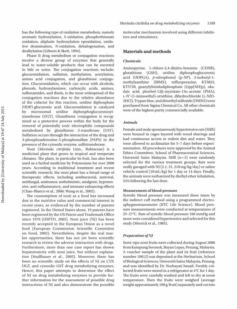
Morinda citrifolia on drug metabolizing enzymes 1109
has the following type of oxidation metabolism, namely aromatic hydroxylation, S-oxidation, phosphothionate oxidation, aliphatic hydroxylation epoxidation, oxida-tive deamination, N-oxidation, dehalogenation, and dealkylation (Gibson & Skett, 1994).
Phase II drug metabolism or conjugation reactions involve a diverse group of enzymes that generally lead to water-soluble products that can be excreted in bile or urine. The conjugation reactions include: glucuronidation, sulfation, methylation, acetylation, amino acid conjugation, and glutathione conjuga-tion. Glucuronidation, which can occur with alcohols, phenols, hydroxylamines, carboxylic acids, amines, sulfonamides, and thiols, is the most widespread of the conjugation reactions due to the relative abundance of the cofactor for this reaction, uridine diphosphate (UDP)-glucuronic acid. Glucuronidation is catalyzed by microsomal uridine diphosphoglucuronosyl- transferase (UGT). Glutathione conjugation is recog-nized as a protective process within the body for the removal of potentially toxic electrophilic compounds metabolized by glutathione S-transferase (GST). Sulfation occurs through the interaction of the drug and 3-phosphoadenosine-5-phosphosulfate (PAPS) in the presence of the cytosolic enzyme, sulfotransferase.
Noni (Morinda citrifolia Linn., Rubiaceae) is a medicinal plant that grows in tropical and temperate climates. The plant, in particular its fruit, has also been used as a herbal medicine by Polynesians for over 2000 years. According to traditional treatment and recent scientific research, the noni plant has a broad range of therapeutic effects, including antibacterial, antiviral, antifungal, antitumor, anthelmintic, analgesic, hypoten-sive, anti-inflammatory, and immune enhancing effects (Chan-Blanco et al., 2006; Wang et al., 2002).
The consumption of noni as a food has increased due to the nutritive value and commercial interest in recent years, as evidenced by the number of patents registered. In the United States alone, 19 patents have been registered by the US Patent and Trademark Office since 1976 (USPTO, 2005). Noni juice (NJ) has been recently accepted in the European Union as a novel food (European Commission Scientific Committee on Food, 2002). Nevertheless, despite the real mar-ket opportunities, there has not yet been scientific research to review the adverse interaction with drugs. Furthermore, more than one case report has shown hepatotoxicity with noni juice, but without explana-tion (Stadlbauer et al., 2005). Moreover, there has been no scientific study on the effects of NJ on CYP, UGT, and cytosolic GST drug metabolizing enzymes. Hence, this paper attempts to determine the effect of NJ on drug metabolizing enzymes to provide fur-ther information for the assessment of possible drug interactions of NJ and also demonstrate the possible
molecular mechanism involved using different inhibi-tors and stimulators.
Materials and methods
Chemicals
Aminopyrine, 1-chloro-2,4-dinitro-benzene (CDNB), glutathione (GSH), uridine diphosphoglucuronic acid (UDPGA), p-nitrophenol (p-NP), 3-isobutyl-1- methylxanthine (IBMX), trifluoperazine, KT5823, KT5720, guanylylimidodiphosphate (Gpp(NH)p), oka-daic acid, phorbol-12-myristate-13-acetate (PMA), l-N5-(1-iminoethyl)-ornithine, dihydrochloride (l-NIO-2HCl), Trypan blue, and dimethyl sulfoxide (DMSO) were purchased from Sigma Chemical Co. All other chemicals were of the highest purity commercially available.
Animals
Female and male spontaneously hypertensive rats (SHR) were housed in cages layered with wood shavings and had continuous access to rodent diet and water. They were allowed to acclimatize for 5–7 days before experi-mentation. All procedures were approved by the Animal Ethics Committee, School of Pharmaceutical Sciences, Universiti Sains Malaysia. SHR (n = 5) were randomly selected for the various treatment groups. Rats were orally gavaged with NJ (2.1, 21, 210 mg/kg/day) or saline vehicle control (10 mL/kg) for 1 day or 14 days. Finally, the animals were euthanized by diethyl ether inhalation, 24 h following the last dose.
Measurement of blood pressureSystolic blood pressure was measured three times by the indirect cuff method using a programmed electro-sphygmomanometer (IITC Life Science). Blood pres-sure measurements were conducted at temperatures of 25–27°C. Rats of systolic blood pressure 160 mmHg and more were considered hypertensive and selected for this study (Merrick et al., 1985).
Preparation of NJ
Semi-ripe noni fruits were collected during August 2006 from Kampung Seronok, Bayan Lepas, Penang, Malaysia. A voucher sample of the plant and its fruit (reference number 10612) was deposited at the Herbarium, School of Biological Sciences, Universiti Sains Malaysia, Penang, and was identified by Dr. Norhayati Ismail. Freshly col-lected fruits were stored in a refrigerator at 4°C for 1 day. The fruits were carefully washed and left to dry at room temperature. Then the fruits were weighed (average weight approximately 100 g/fruit) separately and cut into
Dow
nloa
ded
by [
Uni
vers
ity o
f M
alay
a] a
t 19:
47 2
4 Ju
ly 2
015
1110 A. M. Mahfoudh et al.
small pieces. The noni fruit juice was extracted using a juice extractor (Braun MP80; Germany). Finally the juice was filtered and stored in a dark amber-colored bottle at 4°C throughout the experimental period. The yield of juice per gram of fruit was found to be 0.4 mL, and the yield of dry powder (after freeze drying) per milliliter of juice was found to be 10 mg.
IR spectroscopy and HPTLC of NJ samples
The potassium bromide (KBr) disk technique was used for sample preparation. The infrared (IR) spectrum was recorded in the mid-IR region, 4000– 400 cm−1, using a Thermo Nicolet Fourier transform infrared (FTIR) Nexus spectrophotometer (USA).
NJ samples were applied as bands by means of a high-performance thin layer chromatography (HPTLC) applicator (Camag, Switzerland) on 10 × 20 cm glass-backed silica gel 60 F
254 TLC plates (Merck,
Darmstadt), and the mobile phase used was a mixture of n-butanol:acetic acid:water in a ratio of 4:1:5, respec-tively. The sample was applied as a band with approxi-mate volume of 10 µL. Ascending developments were performed at room temperature in a chamber previ-ously saturated with the mobile phase. The TLC plate was placed in a near vertical position. Finally, after dry-ing, the TLC plate was scanned with a HPTLC scanner (Camag TLC Scanner 3; Switzerland) and images of the TLC plate were taken with a HPTLC image documenta-tion machine (Camag Digistore 2; Switzerland).
In vitro effect of NJ on phase I aminopyrine N-demethylase (APND) on isolated rat hepatocytes
Rat hepatocytes were obtained using a simple non-enzymatic perfusion technique (Petrenko et al., 2002) with slight modifications. Experiments were performed between 8:00 and 11:00 a.m. as this was found to be the most suitable timeframe, as shown in previous studies (Ogata et al., 1990). The cell suspension was filtered through gauze and was centrifuged at 300 rpm for 5 min. The cell pellet was then collected and resuspended in 10 mL of incubation medium. This cell suspension was used to determine both cell viability and cell number. The viability criterion most commonly used is the trypan blue exclusion test, and is based on the premise that viable cells do not allow dye permeation whereas non-viable cells allow the dye to permeate (Fry, 1981; Puviani et al., 1998). Cell preparation that showed an initial via-bility greater than 90% was used in the experiments.
The above procedure was also used to determine the ex vivo effect of NJ on APND after treatment for 1 and 14 days, respectively, of female SHR at dose levels of 2.1, 21, and 210 mg/kg body weight.
Ex vivo effect of NJ on phase II drug metabolizing enzymes in liver subcellular fractions
After the last day of treatment with NJ, rats were sacri-ficed and livers removed, weighed, finely minced, and homogenized with ice-cold 0.25 M sucrose, resulting in 20%, w/v, liver homogenate, using a Potter–Elvehjem glass tube and a Teflon pestle.
Post-mitochondrial supernatantThe liver homogenate was centrifuged at 10,500 rpm for 10 min to pelletize intact cells, cell debris, nuclei, and mitochondria at 4°C. The resultant supernatant con-tained the microsomal plus soluble fractions of the cell (Habig et al., 1974). The supernatant was divided into two portions, one for studying cytosolic enzyme, which was stored at −80°C until used, and the other for the preparation of microsomal liver fractions. This fraction was used for the estimation of GST.
Microsomal liver fractionsA microsomal liver fraction was prepared by the cal-cium precipitation method (Kamath & Rubin, 1972). The post-10,500 rpm supernatant was diluted 1:5 (v/v) with 0.0125 M sucrose, which contained 8 mM CaCl
2.
The solution was stirred for a few seconds and then centrifuged at 550 rpm for 10 min. The microsomal sediments were collected and resuspended with 0.25 M sucrose to remove either a dention protein or excess CaCl
2. Then, they were centrifuged at 2000 rpm
for 10 min. The microsomal pellet was resuspended in 0.25 M sucrose containing 20% glycerin (v/v) and stored at −80°C until use. The microsomal fraction was used to analyze UGT.
The protein contents of the post-mitochondrial super-natant and microsomal fractions were analyzed by the Lowry method cited by Gibson and Skett (1994), using a microplate reader (BioTek Instruments, Inc., USA).
Enzymatic assays
APND enzymatic assay was conducted according to the method described by Nash (1953) using hepatocytes. GST assay was performed according to the method described by Habig et al. (1974). UGT enzyme activity toward p-NP was carried out with slight modification to the method as described (Bock et al., 1983; Isselbacher et al., 1962; Weisburger et al., 2002).
Molecular mechanism elucidation of ex vivo effect of NJ on APND in isolated hepatocytes
Noni juice at a dose of 210 mg/kg was used to treat rats for 1 day. After 24 h, rats were sacrificed and liv-ers removed. Hepatocytes were first preincubated for
Dow
nloa
ded
by [
Uni
vers
ity o
f M
alay
a] a
t 19:
47 2
4 Ju
ly 2
015

Morinda citrifolia on drug metabolizing enzymes 1111
15 min with a particular cellular inducer or inhibitor. Two controls were used, one containing incubation medium, hepatocytes, DMSO (concentration less than 0.1%, v/v), and aminopyrine. The other control contained incubation medium, hepatocytes, distilled water, and aminopyrine. The sample was processed similarly to the in vitro aminopyrine N-demethylase assay.
In this study, particular cellular inducers or inhibi-tors were used at concentrations equivalent to their EC
50 (effective concentration) or IC
50 (inhibitory con-
centration), respectively. The list of cellular inhibitors and inducers used are outlined as follows: 3- isobutyl-1-methylxanthine (IBMX) (IC
50 = 2.0−50.0 × 10⁻⁶ M)
(Beavo & Reifsnyder, 1990; Turner et al., 1993); tri-fluoperazine (IC
50 = 5.8 M) (Zhang et al., 2003);
KT5823 (IC50
= 234.0 nM) (Kase et al., 1987); KT5720 (IC
50 = 56.0 nM) (Kase et al., 1987); guanylylimi-
dodiphosphate (Gpp(NH)p) (IC50
= 1.0 × 10⁻⁹ M) (Natsvlishvili et al., 2001); okadaic acid (IC
50 = 1.0 nM
and 15.0 nM) (Honnor et al., 1989; Liedtke et al., 2005; Mumby et al., 1992); phorbol-12-myristate-13-acetate (PMA) (EC
50 = 50 × 10−8 M) (Hockberger et al.,
1989) and l-N5-(1-iminoethyl)-ornithine, dihydrochlo-ride (l-NIO-2HCl) (IC
50 = 500 nM) (Rees et al., 1990).
Statistical analysis
The computer program GraphPad InStat™ was used to statistically analyze all data obtained from the liver drug metabolism study. The Dunnett test and unpaired t-test were used to analyze results. Data are expressed as mean ± SD. Differences were regarded as significant if p < 0.05.
Results
IR spectroscopy and HPTLC of NJ samples
The IR spectrum (KBr) of the dried powder of NJ displayed a broad band at 3600– 3000 cm−1, a band at 3000– 2800 cm−1, a weak, sharp band at 2400– 2300 cm−1, bands at 1740– 1720 cm−1, 1640– 1610 cm−1, and 1414 cm−1, bands at 1300– 1240 cm−1 and 1100– 1000 cm−1, bands at 981 cm−1, 890– 880 cm−1, 870– 810 cm−1, and 800– 770 cm−1, and at 640– 630 cm−1 and 590– 520 cm−1 (Figure 1). The HPTLC chromatogram of the NJ showed peaks of constituents with R
f (rate of flow) values 0.2,
0.3, 0.38, and 0.41 under ultraviolet (UV) light at 365 nm (Figure 2a); at 254 nm of UV light it displayed peaks with R
f values 0.2, 0.25, and 0.4 of compounds (Figure 2b).
633
777
818
868
897
918
1076
1252
1414
1637
17
18
1741
2337
2929
3385
2
4
6
8
10
12
14
16
18
20
22
24
26
% T
rans
mitt
ance
500 1000 1500 2000 2500 3000 3500 4000
Wavenumbers (cm−1)
Figure 1. IR spectrum of noni juice (NJ). Sample was prepared using the potassium bromide (KBr) disk technique and spectrum recorded using a Thermo Nicolet Fourier transform infrared Nexus spectrophotometer in the mid-IR region of 4000– 400 cm−1.
Dow
nloa
ded
by [
Uni
vers
ity o
f M
alay
a] a
t 19:
47 2
4 Ju
ly 2
015

1112 A. M. Mahfoudh et al.
In vitro and ex vivo effect of NJ on phase I APND in isolated hepatocytes
The in vitro result showed that different concentra-tions of NJ significantly increased APND activity in hepatocytes of different SHR groups: 100 µg/mL of NJ significantly increased (p < 0.01) the activity of APND in hepatocytes of all SHR groups (adult and young male and female); 50 µg/mL of NJ (p < 0.01) increased APND activity in hepatocytes of young male and female SHR; while 1.0 ng/mL, 10 ng/mL, 10 µg/mL, and 20 µg/mL of NJ significantly (p < 0.05) increased aminopyrine phase I metabolism in hepatocytes of young female SHR (Table 1).
The ex vivo results demonstrated that NJ produced a statistically significant increase (p < 0.05) in APND activity at the dose of 210 mg/kg following 1 day of NJ
treatment. However, other doses increased the activ-ity of the enzyme but results were statistically insig-nificant following 1 day of NJ treatment (Table 2). Also, the decrease in N-demethylase activity was statisti-cally insignificant following 14 days of NJ treatment (Table 2).
Ex vivo effect of NJ on phase II drug metabolizing enzymes in liver subcellular fractions
Noni juice decreased the catalytic activity of UGT at 21 mg/kg significantly (p < 0.05) following 1 day of NJ treatment, and at 2.1 and 21 mg/kg (p < 0.01) following 14 days of NJ treatment (Table 2).
Our results for GST enzyme demonstrated an increase in GST activity by 100% for all doses following 1 day of NJ treatment (Table 2). The protein concen-tration of post-mitochondrial supernatant following 1 day of treatment was found to be not significant at all doses. However, the microsomal fraction of the 2.1 mg/kg group showed a significant increase (p < 0.01) (Table 3). The protein concentration of post- mitochondrial supernatant following 14 days of treat-ment showed a decrease that was statistically significant (p < 0.05) for the 2.1 mg/kg group, but differences in all other groups were insignificant (Table 3). The micro-somal fraction demonstrated a significant decrease for the 210 (p < 0.05) and 21 mg/kg (p < 0.01) groups.
Molecular mechanism elucidation of ex vivo effect of NJ on phase I APND in isolated hepatocytes
The results showed the influence of certain cellular inhibitors/stimulants [IBMX, trifluoperazine, KT5720, KT5823, Gpp(NH)p, genistein, PMA, okadaic acid, and l-NIO-2HCl] on the effect of 1 day of treatment of 210 mg/kg NJ in a single dose on aminopyrine metabo-lism in hepatocytes obtained from young female SHR. Our studies showed that all inhibitors/inducers did not
0.00
(a)
(b)
0.05 0.10 0.15 0.20 0.25 0.30 0.35 0.40 [Rf] 0.50
0.00 0.05 0.10 0.15 0.20 0.25 0.30 0.35 0.40 [Rf] 0.50
Figure 2. Chromatogram of the TLC plate of NJ samples shows peaks of constituents with R
f values 0.2, 0.3, 0.38, and 0.41 under UV light
at 365 nm (a); at 254 nm UV light it shows peaks of compounds with R
f values 0.2, 0.25, and 0.4 (b). Using computer winCATS software V.
1.3.3.
Table 1. In vitro effect of noni juice (NJ) on APND activity in hepatocytes of spontaneously hypertensive rats (SHR).
NJ concentration
Group
Adult male Adult female Young male Young female
Control 17.79 ± 0.28 17.02 ± 0.47 17.85 ± 0.25 17.48 ± 0.36
0.1 ng/mL 17.82 ± 0.34 16.90 ± 0.37 17.91 ± 0.25 17.76 ± 0.59
1.0 ng/mL 18.11 ± 0.76 16.93 ± 0.56 17.76 ± 0.21 18.31 ± 0.36*
10 ng/mL 17.76 ± 0.28 16.82 ± 0.25 17.88 ± 0.29 18.31 ± 0.52*
100 ng/mL 17.85 ± 0.51 16.90 ± 0.14 17.74 ± 0.33 18.17 ± 0.40
1.0 µg/mL 17.36 ± 0.31 16.82 ± 0.25 17.91 ± 0.35 18.17 ± 0.60
10 µg/mL 17.85 ± 0.40 16.96 ± 0.14 18.17 ± 0.44 18.31 ± 0.30*
20 µg/mL 17.36 ± 0.54 17.31 ± 0.90 17.99 ± 0.18 18.28 ± 0.40*
50 µg/mL 18.45 ± 0.32 17.56 ± 0.78 18.71 ± 0.76** 18.68 ± 0.62**
100 µg/mL 18.74 ± 0.33** 18.22 ± 1.05** 19.03 ± 0.47** 19.03 ± 0.34**
Each value expressed as mean ± SD (n = 6); significant difference with respect to control: *p < 0.05 and **p < 0.01.APND activities are expressed in produced formaldehyde µmol/min mL−1/106 cells.
Dow
nloa
ded
by [
Uni
vers
ity o
f M
alay
a] a
t 19:
47 2
4 Ju
ly 2
015

Morinda citrifolia on drug metabolizing enzymes 1113
significantly affect the hepatocytes of young female SHR except KT5720, which significantly reduced (p < 0.01) the activity of APND compared to control. All points were compared with respective controls (Table 4).
Discussion
The present study objectives were to investigate the effects of NJ on phase I metabolizing enzymes involv-ing dealkylation, in particular, the N-demethylation reaction responsible for the metabolism of aminopy-rine, and phase II metabolizing enzymes involving O-glucuronidation and GSH conjugation responsible for metabolism of p-NP and CDNB, respectively.
In determination of the N-demethylation reac-tion in phase I metabolism, aminopyrine undergoes N-demethylation to form monomethyl-4-antipyrine and formaldehyde (Gibson & Skett, 1994). The formaldehyde produced can be measured by the colorimetric procedure of Nash (1953), based on the Hantzsch reaction. The enzyme assay employed
Table 3. Effects of NJ on liver weights and protein content of fractions in female SHR.
Parameter Control
NJ (mg/kg)
2.1 21 210
1 day
Liver weight (mg/g body weight) 28.15 ± 2.54 28.61 ± 2.54 28.20 ± 2.51 28.71 ± 1.64
Post-mitochondrial protein (mg/g liver) 112.05 ± 9.60 108.85 ± 18.90 125.10 ± 8.15 121.30 ± 12.35
Microsome protein (mg/g liver) 37.05 ± 0.94 40.61 ± 1.34** 35.91 ± 1.71 37.05 ± 0.85
14 days
Liver weight (mg/g body weight) 33.54 ± 1.50 36.46 ± 2.65 32.38 ± 6.11 31.10 ± 7.53
Post-mitochondrial protein (mg/g liver) 113.65 ± 9.11 99.60 ± 8.62* 98.05 ± 12.31 107.35 ± 8.83
Microsome protein (mg/g liver) 24.32 ± 3.04 24.78 ± 1.79 18.26 ± 3.79** 20.15 ± 1.57*
Female SHR were treated orally with doses of NJ dried juice dissolved in water. Data of body and tissue weights represent mean ± SD for five rats.Values significantly different from control values: *p < 0.05 and **p < 0.01.
Table 4. Effects of inducers/inhibitors on APND in hepatocytes of female SHR.
Inducer/inhibitor Activity
Control (C1) 19.03 ± 2.35
Control (C2) 20.97 ± 1.66
Okadaic acid (1 nM) 18.14 ± 0.36
Okadaic acid (15 nM) 18.02 ± 0.20
l-N5-(1-iminoethyl)-ornithine (l-NIO) 17.88 ± 0.22
Guanylylimidodiphosphate (Gpp(NH)p) 17.99 ± 0.28
Phorbol-12-myristate-13-acetate (PMA) 20.09 ± 3.79
KT5720 18.17 ± 0.34**
KT5823 20.60 ± 2.37
3-Isobutyl-1-methylxanthine (IBMX) 21.49 ± 2.83
Trifluoperazine 21.98 ± 3.11
Values are expressed as mean ± SD (n = 6); significant difference with respect to control: **p < 0.01. Each value expressed as mean ± SD.APND activities are expressed in produced formaldehyde µmol/min mL−1/106 cells; hepatocytes from NJ (210 mg/kg, daily for 1 day) treated SHR. Experimental procedures foresee a preincubation of inducers/inhibitors with hepatocytes followed by incubation in the enzymatic assay.C
1 represents distilled water as solvent for okadaic acid (1 nM and
15 nM), l-NIO, Gpp(NH)p, and PMA; C2 represents DMSO as solvent
for KT5720, KT5823, IBMX, and trifluoperazine.
Table 2. One-day and 14-day effects of NJ on drug metabolizing enzymes in liver of SHR.
Parameter Control
NJ (mg/kg)
2.1 21 210
1 day
APND 20.20 ± 2.40# 20.97 ± 3.19 21.83 ± 3.30 21.17 ± 2.38*
18.54 ± 0.49†
UGT 1.33 ± 0.39 1.17 ± 0.25 0.81 ± 0.24* 0.99 ± 0.25
GST 0.180 ± 0.04 0.318 ± 0.05** 0.302 ± 0.05** 0.336 ± 0.06**
14 days
APND 19.66 ± 1.45# 19.88 ± 2.11 20.31 ± 1.61 19.94 ± 1.33
22.61 ± 3.16†
UGT 1.97 ± 0.35 1.29 ± 0.29** 1.00 ± 0.19** 1.61 ± 0.47
GST 0.130 ± 0.05 0.128 ± 0.06 0.133 ± 0.05 0.122 ± 0.04
Each value expressed as mean ± SD (n = 6); significant difference with respect to control: *p < 0.05 and **p < 0.01. #Represents control for 2.1 and 21 mg/kg groups of rats; †represents control for 210 mg/kg rat group.Aminopyrine N-demethylase (APND) activities are expressed in produced formaldehyde µmol/min mL−1/106 cells; UDP-glucuronosyl transferase (UGT) activities are expressed in p-NP consumed nmol/min/mg microsomal proteins; glutathione S-transferase (GST) activities are express as mmol product formed/min/mg of cytosolic proteins.
Dow
nloa
ded
by [
Uni
vers
ity o
f M
alay
a] a
t 19:
47 2
4 Ju
ly 2
015

1114 A. M. Mahfoudh et al.
allows the determination of formaldehyde as one of the liver phase I metabolites of aminopyrine. This study was undertaken to evaluate the effects of NJ on aminopyrine phase I oxidation metabolism through N-demethylation by CYP450 in the mixed-function oxidase system in rat liver hepatocytes. The N-demethylation reaction is also behind the principle of the liver function test in the aminopyrine breath test, which is clinically used for the diagnosis of liver function (Chiaramonte et al., 2003). Additionally, ami-nopyrine is known to be a substrate for several forms of CYP, and appears to be a good probe substrate for investigating the effects of herbal preparations on drug oxidation (Hasegawa et al., 2002).
The in vitro effect determines the activity of drug metabolizing enzymes following direct addition of NJ to incubation medium containing hepatocytes of rat liver and substrate drug aminopyrine. The in vitro results demonstrated that NJ produced a statistically significant effect on N-demethylase catalytic activity in young female SHR, at age 7 weeks and average weight 140–160 g, but other rat (normal and diabetic) hepato-cytes did not show statistically significant effects (data not shown).
Phase II metabolizing enzymes were studied using p-NP and CDNB as substrates for glucuronidation and conjugation reactions, respectively. UGT enzymes are responsible for glucuronidation, which represents a major means of generating water-soluble substances from xenobiotics or endobiotics, which in turn leads to the ultimate excretion of these substances into urine or bile. The UGT enzyme is an important factor for determining the extent to which xenobiotics or endo-biotics are detoxified (Weisburger et al., 2002). These microsomal enzymes are located in the endoplasmic reticulum of cells from a number of tissues. In deter-mination of the glucouronidation reaction in phase II metabolism, p-NP undergoes O-glucuronidation and the excess amount of p-NP can be measured by the colorimetric procedure of Bock et al. (1983).
GSTs are members of the family of GSH linked enzymes that offer cellular protection against both endogenous and xenobiotic substances. GSTs catalyze the conjugation of GSH to form various electrophilic compounds. These enzymes are located in the cytosol of cells from a number of tissues. Determination of the GSH conjugation reaction in phase II metabolism can be done using the colorimetric procedure of Habig et al. (1974).
The study of the effects of NJ on phase II enzymes was carried out on homogenized fractions (cystolic and microsomal) of SHR. The SHR group was selected for this study because 1 day of treatment of NJ was shown to have increased the aminopyrine phase I metabolism significantly in hepatocytes of SHR at the highest dose
levels (Table 2). In contrast, in the 14-day study, NJ decreased aminopyrine phase I metabolism in the hepa-tocytes of SHR but not at significant levels. The results showed that 14-day NJ treatment significantly inhibited UGT activity in young female SHR at 2.1 and 21 mg/kg NJ, but UGT activity was not significantly different from control at 210 mg/kg NJ. However, 1-day treatment with NJ significantly inhibited UGT activity at 21 mg/kg, but at 2.1 and 210 mg/kg NJ insignificantly inhibited UGT activity (Table 2).
In contrast, GST activity was significantly increased in the 1-day study (Table 2), while in the 14-day study there was no significant effect on GST activity when compared to control (Table 2). GST induction may be mediated via the antioxidant response elements and its transcription factor Nrf2, which is involved in the induction of many phase II enzymes (Thimmulappa et al., 2002). UGT and GST activity have no association in the induction of many phase II enzymes. However, this result disagrees with studies performed by Van der Logt et al. (2003). The authors postulated that both genes are regulated by Nrf2, in concord with the study of Thimmulappa et al. (2002).
To demonstrate the molecular mechanism of the effect of NJ on the activity of phase I APND, several enzyme inducers and inhibitors that covered cAMP, cGMP, and phosphatidyl inositol pathways of drug metabolism were used. These chemicals were added to Petri dishes at concentrations based on their particular IC
50 or EC
50. In the liver, batteries of extracellular signals
regulate various intracellular processes via a complex cascade of receptors, transducers, and second mes-sengers (Bygrave & Benedetti, 1993). The most prolific of these second messengers, cyclic nucleotides (adeno-sine 29,39-cyclic monophosphate (cAMP) and guanos-ine 39,59-cyclic monophosphate (cGMP)), remain the best characterized (Bentley & Beavo, 1992; Iber et al., 2001). The resulting activation of cyclic nucleotide- dependent protein kinases (PK) functions as the basis for many effects of cyclic nucleotides on cellular func-tion (Edelman et al., 1987).
Theoretically, KT5720 inhibits protein kinase A (PKA). Therefore the use of KT5720 is expected to reduce phosphorylation of CYP450 and may lead to an increase in activity of APND. In contrast, the results obtained here showed that the activity of APND was significantly decreased when compared to that of control. Although the result is not as expected, the cAMP/PKA pathway may account for the molecular mechanism of the 1-day effect of NJ on APND in young female SHR hepatocytes. However, more detailed investigation is needed to explain the actual mecha-nism of action.
NJ was compared with two other commercially available products, Hawaiian (HNJ) and Tahitian
Dow
nloa
ded
by [
Uni
vers
ity o
f M
alay
a] a
t 19:
47 2
4 Ju
ly 2
015

Morinda citrifolia on drug metabolizing enzymes 1115
(TNJ), using FTIR spectra and HPTLC chromatograms. There were no differences in the characteristic absorp-tion bands in the spectra of NJ and the commercial products HNJ and TNJ (data not shown), especially in the fingerprint region (1500– 800 cm−1). Images of the TLC plates at UV light = 365 nm of the NJ (Figure 3a) showed an intense blue fluorescent zone against a black background for all sample bands as well as HNJ and TNJ (data not shown). From the FTIR spectra, HPTLC chromatograms, and HPTLC plate images at different UV wavelengths, it was concluded that the tested samples of NJ showed qualitative similarities in their major constituents.
In conclusion, oral NJ treatment modestly increased enzymes involved in APND and GST following 1 day of treatment. NJ on continuous administration for 14 days may decrease the activity of metabolizing enzymes, and therefore changes in APND, UGT, and GST following 1
day and 14 days of NJ treatment suggest that all three metabolic pathways may play a role in herb–drug interaction by modulation of metabolic enzymes. PKA may play a role in the induction of aminopyrine phase I metabolizing enzymes in hepatocytes of rats following 1 day of NJ treatment; however, more experi-ments are needed to confirm the above PKA role. Changes in the phases I and II enzyme activities sug-gest that, at least in rats, NJ may affect the metabolism of drugs that undergo the same liver metabolism path-way as APND, UGT, and GST.
Declaration of interest: The authors wish to acknowl-edge the IRPA grant, number 305/PFARMASI/612205, which supports this research project.
References
Beavo JA, Reifsnyder DH (1990): Primary sequence of cyclic nucle-otide phosphodiesterase isozymes and the design of selective inhibitors. Trends Pharmacol Sci 11: 150–155.
Bentley JK, Beavo JA (1992): Regulation and function of cyclic nucle-otides. Curr Opin Cell Biol 4: 233–240.
Bock KW, Burchell B, Dutton GJ, Hanninen O, Mulder GJ, Owens IS, Siest G, Tephly TR (1983): UDP-glucuronosyltransferase activi-ties guidelines for consistent interim terminology and assay conditions. Biochem Pharmacol 32: 953–955.
Bygrave FL, Benedetti A (1993): Calcium: Its modulation in liver by cross-talk between the actions of glucagon and calcium- mobilizing agonists. Biochem J 296: 1–14.
Chan-Blanco Y, Vaillant F, Perez AM, Reynes M, Brillouet J-M, Brat P (2006): The noni fruit (Morinda citrifolia L.): A review of agricul-tural research, nutritional and therapeutic properties. J Food Compost Anal 19: 645–654.
Chavez ML, Jordan MA, Chavez PI (2006): Evidence-based drug–herbal interactions. Life Sci 78: 2146–2157.
Chiaramonte D, Steiner JM, Broussard JD, Baer K, Gumminger S, Moeller EM, Williams DA, Shumway R (2003): Use of a 13C- aminopyrine blood test: First clinical impressions. Can J Vet Res 67: 183–188.
Edelman AM, Blumenthal DK, Krebs EG (1987): Protein serine/ threonine kinases. Annu Rev Biochem 56: 567–613.
European Commission Scientific Committee on Food (2002): Opinion of the Scientific Committee on Food of Tahitian Noni Juice. SCF/CS/DOS/18 ADD 2. Brussels, ECSCF.
Fry JR (1981): Preparation of mammalian hepatocytes. Methods Enzymol 77: 130–137.
Gibson GG, Skett P (1994): Introduction to Drug Metabolism, 2nd ed. London, Chapman & Hall, pp. 1–33, 217–248.
Habig WH, Pabst MJ, Lakoby WB (1974): Glutathione S-transferases the first enzymatic step in mercapturic acid formation. J Biol Chem 249: 7130–7139.
Hasegawa A, Yoshino M, Nakamura H, Ishii I, Watanabe T, Kiuchi M, Ishikawa T, Ohmori S, Kitada M (2002): Identification of inhibi-tory component in cinnamon-O-methoxycinnamaldehyde inhibits CYP1A2 and CYP2E1. Drug Metab Pharmacokinet 17: 229–236.
Hockberger P, Toselli M, Swandulla D, Lux HD (1989): A diacylglyc-erol analogue reduces neuronal calcium currents independently of protein kinase C activation. Nature 338: 340–342.
Honnor RC, Haystead TA, Sim AT, Carling D, Tsukitani Y, Cohen P, Hardie DG (1989): Effects of the tumour promoter okadaic acid
a b
Figure 3. TLC plates of NJ at UV light = 365 nm (a) and 254 nm (b). NJ samples were applied as bands by means of a HPTLC applicator (Camag, Switzerland) on 10 × 20 cm glass-backed silica gel 60 F
254 TLC
plates (Merck, Darmstadt) and mobile phase used was a mixture of n-butanol:acetic acid:water in a ratio of 4:1:5, respectively. Images of the TLC plate were taken with a HPTLC image documentation machine (Camag Digistore 2; Switzerland).
Dow
nloa
ded
by [
Uni
vers
ity o
f M
alay
a] a
t 19:
47 2
4 Ju
ly 2
015

1116 A. M. Mahfoudh et al.
on intracellular protein phosphorylation and metabolism. Nature 337: 78–81.
Iber H, Li-Masters T, Chen Q, Yu S, Morgan ET (2001): Regulation of hepatic cytochrome P450 2C11 via cAMP: Implications for down-regulation in diabetes, fasting, and inflammation. J Pharmacol Exp Ther 297: 174–180.
Isselbacher KJ, Chrabas MF, Quinn RC (1962): The solubilization and partial purification of a glucuronyl transferase from rabbit liver microsomes. J Biol Chem 237: 3033–3036.
Kamath SA, Rubin E (1972): Interaction of calcium with microsomes: a modified method for the rapid isolation of rat liver micro-somes. Biochem Biophys Res Commun 49: 52–59.
Kase H, Iwahashi K, Nakanishi S, Matsuda Y, Yamada K, Takahashi M, Murakata C, Sato A, Kaneko M (1987): K-252 compounds, novel and potent inhibitors of protein kinase C and cyclic nucleotide-dependent protein kinases. Biochem Biophys Res Commun 142: 436–440.
Liedtke CM, Wang X, Smallwood ND (2005): Role for protein phos-phatase 2A in the regulation of Calu-3 epithelial Na+-K+-2Cl-, type 1 co-transport function. J Biol Chem 280: 25491–25498.
Merrick BA, Davies MH, Cook DE, Holcslaw TL, Schnell RC (1985): Alterations in hepatic microsomal drug metabolism and cyto-chrome P-450 proteins in spontaneously hypertensive rats. Pharmacology 30: 129–135.
Mumby MC, Kamm KE, Stull JT, Nomura M (1992): Site-specific dephosphorylation of smooth muscle myosin light chain kinase by protein phosphatases 1 and 2A. Biochemistry 31: 11915–11920.
Nash T (1953): The colorimetric estimation of formation of formal-dehyde means of Hantzsch reactions. J Biol Chem 55: 416–422.
Natsvlishvili N, Juravliova E, Dzneladze D, Mikeladze D (2001): Haloperidol regulates the binding of guanine nucleotides to synaptic membranes through the NMDA receptor. J Biol Phys Chem 1: 24–28.
Ogata E, Mine T, Kojima I (1990): Difference in sensitivity to gluca-gon action in three different rat liver systems. Metabolism 39: 321–326.
Petrenko A, Kravchenko L, Shanina I, Fuller B (2002): A simple non-enzymatic method for the isolation of high yields of functional rat hepatocytes. Cell Biol Int 26: 1003–1006.
Puviani AC, Ottolenghi C, Tassinari B, Pazzi P, Morsiani E (1998): An update on high-yield hepatocyte isolation methods and on the potential clinical use of isolated liver cells. Comp Biochem Physiol A Mol Integr Physiol 121: 99–109.
Rees DD, Palmer RM, Schulz R, Hodson HF, Moncada S (1990): Characterization of three inhibitors of endothelial nitric oxide synthase in vitro and in vivo. Br J Pharmacol 101: 746–752.
Stadlbauer V, Fickert P, Lackner C, Schmerlaib J, Krisper P, Trauner M, Stauber RE (2005): Hepatotoxicity of noni juice: report of two cases. World J Gastroenterol 11: 4758–4760.
Thimmulappa RK, Mai KH, Srisuma S, Kensler TW, Yamamoto M, Biswal S (2002): Identification of Nrf2-regulated genes induced by the chemopreventive agent sulforaphane by oligonucleotide microarray. Cancer Res 62: 5196–5203.
Turner NC, Burns FM, Wood LJ, Gueremy T, Souness JE (1993): The effect of cyclic AMP and cyclic GMP phosphodiesterase inhibi-tors on the superoxide burst of guinea-pig peritoneal macro-phages. Br J Pharmacol 108: 876–883.
USPTO (2005): Patents (Morinda citrifolia). Patent Full-Text and Image Database. Retrieved January 17, 2005 from the World Wide Web: http://patft.uspto.gov/netacgi/nph.
Van der Logt EM, Roelofs HM, Nagengast FM, Peters WH (2003): Induction of rat hepatic and intestinal UDP-glucuronosyltransferases by naturally occurring dietary anticar-cinogens. Carcinogenesis 24: 1651–1656.
Wang MY, West BJ, Jensen CJ, Nowicki D, Su C, Palu AK, Anderson G (2002): Morinda citrifolia (Noni): A literature review and recent advances in Noni research. Acta Pharmacol Sin 23: 1127–1141.
Weisburger JH, Embola CW, Sohn OS, Fiala ES (2002): Induction of UDP-glucuronosyltransferase 1 (UDP-GT1) gene complex by green tea in male F344 rats. Food Chem Toxicol 40: 841–844.
Zhang L, Feng X, McDonald JM (2003): The role of calmodulin in the regulation of osteoclastogenesis. Endocrinology 144: 4536–4543.
Dow
nloa
ded
by [
Uni
vers
ity o
f M
alay
a] a
t 19:
47 2
4 Ju
ly 2
015


![21242593[VARIMOT EX.].pdf](https://static.fdokumen.com/doc/165x107/6338b562e57b005bf2016bb4/21242593varimot-expdf.jpg)