Significant plant virus diseases in India and a glimpse of modern disease management technology
18
REVIEW Significant plant virus diseases in India and a glimpse of modern disease management technology Narayan Rishi Received: 10 April 2008 / Accepted: 5 November 2008 / Published online: 6 January 2009 Ó The Phytopathological Society of Japan and Springer 2008 Abstract India has a diverse agroclimate representing tropical, subtropical and temperate climates with zones ranging from average to high in temperature, humidity and rainfall; from low to scarce rainfall in deserts to cold to very cold plains and upland areas. Thus it is the home of one of the richest flora and fauna in the world. For pests and pathogens, too, tropical and subtropical climates are ideal for growth and development. A majority of the world’s fruits, vegetables, cereals, pulses, oil seed crops, fiber crops, sugarcane, spices, and ornamentals are cultivated in India. Virus diseases and their vectors are also in abundance. Since ancient times, virus-like diseases and management practices have been known in India. This knowledge has now been revived and adopted in organic farming, espe- cially for medicinal and aromatic plants. Current trends for extensive and intensive agriculture, open international agricultural trade, and thus food security and sustained economy have brought new challenges in the fight against virus diseases. In this changing scenario, current diseases of significance are caused by begomoviruses, badnaviruses, cucomoviruses, potyviruses, ilarviruses in crops such as vegetables, fruits, ornamentals, fiber crops, and sugarcane. Variability in the viruses is also common. Briefly reviewed here is the positive effect of an isolate of Rice necrosis mosaic virus; when artificially inoculated on jute and mesta fiber crops enhanced their fiber contents. Diseases of significance, e.g., leaf fleck disease of sugarcane, citrus yellow mosaic disease, banana bunchy top, banana bract mosaic, mungbean yellow mosaic, mosaic in chrysanthe- mum, gladiolus and orchids are also discussed. Efficient, reliable diagnostic tools have been developed and used extensively. Some of the advanced laboratories have been accredited for virus indexing under the National Certifica- tion System for tissue-cultured plants. International standards for phytosanitary measures have been promul- gated, and a mandatory nodal agency is in place for the conservation and exchange of germplasm; a Containment Level-4 facility to examine incoming transgenics and a fully equipped laboratory to intercept virus-infected plant material are functional. A National Agricultural Biosecurity System will soon be in place. All these measures are essential to protect agricultural systems and to compete in the international agriculture market. Continued vigilance, disease mapping and adopting the latest technology are required to practice sustainable agriculture. Keywords Vrikshayurveda PCR Multiplex PCR ELISA Jute Mesta Badnavirus Introduction The Indian civilization is one of the oldest in world, and so is the practice of agriculture, which are several millennia old. A Sanskrit term, vyadhi, which is equivalent to the word disease in English, was first used in India in Krishi- Parashara compiled by sage Parashara (c. 400 BC) (Nene 2006a). Descriptions of certain specific symptoms like yellowing of leaves, closely resembling what are now known as viral diseases, have been found in Vrikshayurv- eda (The Science of Plant Life) written by Surapala in c. 1000 AD (Sadhale 1996). This treatise is based on the principles of Ayurveda, a millennia-old Indian system of N. Rishi (&) Indian Virological Society, CCS Haryana Agricultural University, 9/48, New Campus, Hisar 125 004, India e-mail: [email protected] 123 J Gen Plant Pathol (2009) 75:1–18 DOI 10.1007/s10327-008-0139-8
-
Upload
independent -
Category
Documents
-
view
0 -
download
0
Transcript of Significant plant virus diseases in India and a glimpse of modern disease management technology

REVIEW
Significant plant virus diseases in India and a glimpse of moderndisease management technology
Narayan Rishi
Received: 10 April 2008 / Accepted: 5 November 2008 / Published online: 6 January 2009
� The Phytopathological Society of Japan and Springer 2008
Abstract India has a diverse agroclimate representing
tropical, subtropical and temperate climates with zones
ranging from average to high in temperature, humidity and
rainfall; from low to scarce rainfall in deserts to cold to very
cold plains and upland areas. Thus it is the home of one of
the richest flora and fauna in the world. For pests and
pathogens, too, tropical and subtropical climates are ideal
for growth and development. A majority of the world’s
fruits, vegetables, cereals, pulses, oil seed crops, fiber crops,
sugarcane, spices, and ornamentals are cultivated in India.
Virus diseases and their vectors are also in abundance.
Since ancient times, virus-like diseases and management
practices have been known in India. This knowledge has
now been revived and adopted in organic farming, espe-
cially for medicinal and aromatic plants. Current trends for
extensive and intensive agriculture, open international
agricultural trade, and thus food security and sustained
economy have brought new challenges in the fight against
virus diseases. In this changing scenario, current diseases of
significance are caused by begomoviruses, badnaviruses,
cucomoviruses, potyviruses, ilarviruses in crops such as
vegetables, fruits, ornamentals, fiber crops, and sugarcane.
Variability in the viruses is also common. Briefly reviewed
here is the positive effect of an isolate of Rice necrosis
mosaic virus; when artificially inoculated on jute and mesta
fiber crops enhanced their fiber contents. Diseases of
significance, e.g., leaf fleck disease of sugarcane, citrus
yellow mosaic disease, banana bunchy top, banana bract
mosaic, mungbean yellow mosaic, mosaic in chrysanthe-
mum, gladiolus and orchids are also discussed. Efficient,
reliable diagnostic tools have been developed and used
extensively. Some of the advanced laboratories have been
accredited for virus indexing under the National Certifica-
tion System for tissue-cultured plants. International
standards for phytosanitary measures have been promul-
gated, and a mandatory nodal agency is in place for the
conservation and exchange of germplasm; a Containment
Level-4 facility to examine incoming transgenics and a
fully equipped laboratory to intercept virus-infected plant
material are functional. A National Agricultural Biosecurity
System will soon be in place. All these measures are
essential to protect agricultural systems and to compete in
the international agriculture market. Continued vigilance,
disease mapping and adopting the latest technology are
required to practice sustainable agriculture.
Keywords Vrikshayurveda � PCR � Multiplex PCR �ELISA � Jute � Mesta � Badnavirus
Introduction
The Indian civilization is one of the oldest in world, and so
is the practice of agriculture, which are several millennia
old. A Sanskrit term, vyadhi, which is equivalent to the
word disease in English, was first used in India in Krishi-
Parashara compiled by sage Parashara (c. 400 BC) (Nene
2006a). Descriptions of certain specific symptoms like
yellowing of leaves, closely resembling what are now
known as viral diseases, have been found in Vrikshayurv-
eda (The Science of Plant Life) written by Surapala in
c. 1000 AD (Sadhale 1996). This treatise is based on the
principles of Ayurveda, a millennia-old Indian system of
N. Rishi (&)
Indian Virological Society,
CCS Haryana Agricultural University,
9/48, New Campus, Hisar 125 004, India
e-mail: [email protected]
123
J Gen Plant Pathol (2009) 75:1–18
DOI 10.1007/s10327-008-0139-8
human health management. Ancient, medieval, and pre-
modern practices used to manage these plant viral diseases
have been described by Nene (2003, 2006b) and are now
being practiced in several laboratories and in organic
farming, especially for growing medicinal and aromatic
plants.
In the modern era, the earliest recorded viral and
phytoplasma diseases in India are root wilt of coconut
(Cocos nucifera L.) present in south Kerala since 1882
(Butler 1908), spike disease of sandal (Santalum album
L.), observed in Karnataka in 1899 (McCarthy 1903),
small cardamom (Elettaria cardamomum Maton) mosaic
(commonly known as ‘Marble’ or ‘Katte’) disease (Mol-
lison 1900), yellow leaf disease of areca nut palm (Areca
catechu L.) observed in 1914 (Varghese 1934), cotton
stenosis (Kottur and Patel 1920), tristeza disease of citrus
(Brown 1920), mosaic disease of sugarcane (Dastur
1923), and yellow vein mosaic disease of bhendi (okra;
Abelmoschus esculentus) (Kulkarni 1924). Root wilt of
coconut, spike disease of sandal, and yellow leaf of are-
canut were later identified as phytoplasma diseases. In
earlier review papers, I chronicled the development of
modern virology in India and its important viral diseases
(Rishi 2004, 2006); here I will elaborate on certain sig-
nificant viral diseases of current importance and the
modern technology developed for investigating viral
diseases.
Bhendi yellow vein mosaic disease
Okra, popularly known as bhendi (Abelmoschus esculentus
L.) in India is a common vegetable grown in several
countries. Bhendi fruits are a rich source of proteins (20–
24% edible) and minerals, and seeds contain 13–22%
edible oil. Ten species of Abelmoschus occur in India; they
are believed to be of Asiatic origin. A. esculentus, the
cultivated species is probably of Indian origin (Dhankar
et al. 2005). Bhendi, exported from India as a fresh vege-
table, comprises 70% of the total fresh vegetable earnings,
excluding onion (APEDA 2000). The annual yield in India
is around 3.8 million tons, the highest in the world
(http://www.postharvestindia.com).
Bhendi yellow vein mosaic disease (BYVMD) was first
reported in 1924 (Kulkarni 1924) during the erstwhile
Bombay Presidency. Different degrees of chlorosis and
yellowing of veins and veinlets, smaller leaves, fewer and
smaller fruits, and stunting are characteristic of BYVMD
(Fig. 1a, b). Fruit yield is also greatly reduced, by as much
as 96% if the crop is infected at early stage (Pun and
Doraiswamy 1999). Several BYVMD-resistant bhendi
varieties have been released, but none have retained
resistance for long (Usha 2008).
Bhendi yellow vein mosaic disease is caused by Bhendi
yellow vein mosaic virus (BYVMV), which is a species of
genus Begomovirus, family Geminiviridae (Fauquet and
Stanley 2005). BYVMV is believed to have originated in
India (Usha 2008). The only known method of transmis-
sion is through whitefly (WF) (Bemisia tabaci Gennadius).
Begomoviruses are very diverse and parallel whitefly in
their geographical distribution throughout the tropics and
parts of the subtropics. Begomoviruses have mono- or
bipartite genomes consisting of circular single-stranded
DNA of 2.6–3.0 kbp.
Bhendi is also rich in polysaccharides and polyphenols.
Polysaccharides are viscous and gluelike; they inhibit Taq
polymerase activity, thus interfering with PCR. Their
texture also creates problems for DNA during pipetting.
For cloning the Begomovirus genome either by directly
restricting the replicative form from total nucleic acid or by
amplifying the full or partial genome with Begomovirus-
specific primers, high quality DNA is a prerequisite. Ike-
gami et al. (1981) handled this problem by extracting DNA
from a nonmucilaginous host. But to characterize all the
genomic components that may be involved in inciting the
disease, the viral DNA should be extracted from natural
host. Jose and Usha (2000) developed a protocol that is
simple, cheaper and enables the isolation of pure viral
DNA without ultrapurification of the virus. For extraction,
they used sodium citrate buffer (pH 6.0), and for virus
precipitation, PEG 6000 was added after alkali lysis. The
citrate buffer eliminated the mucilage and polyphenols, and
the alkali lysis enriched the replicative form of DNA. The
extracted DNA could be digested with restriction enzyme
and cloned successfully with no problems. PCR amplifi-
cation (using begomovirus-specific primers) and cloning
yielded a 2.7-kbp DNA-A of BYVMV. Despite full efforts,
no second genomic component DNA-B was found, leading
to the conclusion that BYVMV is monopartite. Agroinoc-
ulation with infective full-length DNA-A of BYVMV,
however, only induced mild curling in bhendi.
Using non-overlapping abutting primers (beta 1.F and
beta 1.R), a *1.35-kbp DNA b fragment was amplified
from BYVMV-infected bhendi plant. These abutting
primers are located in the highly conserved region of DNA
b sequences. When DNA b was used with DNA-A to
coinoculate bhendi, typical symptoms of BYVMD resulted.
This proved that BYVMD is caused by a complex of
DNA-A and DNA b.
BYVMD-associated DNA b has some commonality
with DNA b associated with Ageratum yellow vein virus
and Cotton leaf curl virus (CLCuV) (Jose and Usha 2003).
Zhou et al. (1998) concluded, on the basis of sequences of
Okra yellow vein virus (OYVMV) and CLCuV in Pakistan,
that recombination of OYVMV with another unidentified
begomovirus in Okra produced a virus that caused an
2 J Gen Plant Pathol (2009) 75:1–18
123
epidemic in cotton. In bipartite begomoviruses, movement
protein (MP) BC1 and nuclear shuttling protein (NSP) BV1
encoded by the DNA-B component induce systemic
symptoms. In monopartite begomoviruses, this role is
carried on either by the coat protein (CP) when it becomes
highly karyophilic and active in the nuclear import/export
of viral nucleoproteins across the nuclear pore complex,
or a functional bC1 transcript that modulates virus-like
symptoms or in some cases suppresses RNA silencing.
Kumar et al. (2006) studied protein–protein interaction
and nuclear trafficking of the CP and bC1 protein associ-
ated with BYVMV. In fusion studies with green fluorescent
protein (GFP), they found that GFP fused with CP
(GFPCP) in the nucleus, whereas GFP fused with bC1
(GFP bC1) near the periphery of epidermal cells. Expres-
sion of bC1 in transgenic Nicotiana benthamiana resulted
in severe abnormalities such as stunting and distorted stem
and leaves. Gopal et al. (2007) showed a differential role of
C4 and bC1 of BYVMV in the suppression of posttran-
scriptional gene silencing (PTGS) and evidence for
transactivation by C2.
There are seven open reading frames (ORFs) in DNA-A
and one in DNA b, that is half the size of DNA-A and
functions as a symptom modulator. The highly conserved
intergenic region (IR) transcribes genes in a bidirectional
manner in monopartite begomoviruses. Many motifs in the
IR control gene expression and replication. In a study of
the functions of C2, C4 and bC1, Gopal et al. (2007)
revealed that C2 is involved in transactivation and mildly
suppresses gene silencing, but C4 and bC1 strongly sup-
press PTGS.
When inheritance of resistance to BYVMV was studied in
interspecific crosses between A. manihot and A. tetraphyllus,
a single dominant gene controlled resistance (Jambhale and
Nerker 1981). Susceptibility to BYVMV in A. manihot ssp.
manihot was controlled by two dominant genes (Sharma and
Dhillon 1983). Several species (A. manihot [some forms],
A. pungens, A. crinitus, A. panduraeformis, and A. vitifolius)
were resistant to BYVMV. A. manihot ssp. manihot, A. spp
‘Ghana’, A. tuberculatus and A. spp (West African okra)
were symptomless carriers of BYVMV and may be useful in
developing BYVMV-resistant hybrids (Dhankar and Mishra
2004).
Mungbean yellow mosaic disease
Of all the yellow mosaic diseases of grain legumes (pul-
ses), mungbean yellow mosaic disease (MYMD) caused by
mungbean yellow mosaic virus (MYMV) is most impor-
tant. MYMV induces yellow mosaic diseases in several
legumes viz. Vigna radiata (mungbean/greengram),
V. mungo (urdbean/blackgram), V. aconitifolia (mothbean),
V. unguiculata (cowpea), Cajanus cajan (arhar/pigeonpea),
Glycine max (soybean), Phaseolus vulgaris (French bean),
and P. lunatus (lima bean) in India, Bangladesh, Pakistan,
Sri Lanka, Thailand, Philippines, and Indonesia. Grain
legumes in India are cultivated year round for both Rabi
and Kharif crops because they help restore soil fertility by
fixing atmospheric nitrogen. In addition, pulses are the
major source of protein for the largely vegetarian popula-
tion in India. The essential amino acid lysine is present in
pulses in high quantities compared to the low quantities in
cereals. Furthermore, the pulse protein and cereal protein
are complementary, together providing a complete source
of amino acids for humans.
Mungbean is native to India, where it has been culti-
vated and domesticated since 2200 BC. India leads
Pakistan, Sri Lanka, Thailand, Laos, Kumpuchia, Vietnam,
Malaysia, and China in mungbean acreage (55%) and
production (45%). Due to its rapid maturity and photo-
insensitivity, mungbean is adaptable to multiple cropping
systems in the lowlands of the tropics and subtropics. It is
easily digestible and free from flatulence-inducing factors.
MYMD is the major constraint in raising healthy,
profitable crops of mungbean and other hosts of MYMV.
It was first seen in New Delhi in 1955 (Nariani 1960)
on mungbean, and the putative causal agent was named
MYMV. It is transmitted by WF in a circulative, non-
propagative manner. In mungbean, yellow mosaic disease
incidence in farmers’ fields may be as high as 100%. The
symptoms appear in the form of small irregular yellow
specs and spots, which enlarge until leaves are completely
yellowed. Diseased plants are stunted, with fewer flowers
and pods that bear smaller, occasionally shriveled seeds
(Fig. 1c). In severe cases, the disease may cause total loss
in yield (Nariani 1960). Varma et al. (1992) reported that
yellow mosaic of mungbean, urdbean, cowpea, and soy-
bean induce an annual yield loss of US$300 million in
India.
Genetics of resistance to mungbean yellow mosaic virus
in mungbean was studied using four resistant cultivars
(LM696, L-24-2, ML513 and ML505) and four susceptible
(P9333, K891, K851 and T44). These cultivars were used
as resistant and susceptible parents for a crossing program
with F1, F2, back cross and reciprocal cross generations.
The cross between resistant parents suggested that resistant
parents had different resistant genes and that an inhibitory
gene interaction was operating in controlling disease
resistance. Crosses of susceptible parents also suggested
similar genetic constitutions (Rishi 2004, 2006). Resistance
was governed by two major genes with complimentary and
inhibitory gene interactions. A similar study of the genetics
of resistance of MYMV-cowpea isolate was done on
cowpea using resistant cvs. CS39, CS55, RC80 and KBB
and susceptible cvs. GC2, HFC42-1 and FS68 as parents.
J Gen Plant Pathol (2009) 75:1–18 3
123

In this case, resistance to the causal virus was governed by
dominant gene and symbols Cym1 and Cym2 were assigned
to the resistant genes (Rishi et al. 1996; Sangwan and Rishi
2004).
Mandal et al. (1997) cloned the DNAs of a blackgram
isolate of MYMV and produced systemic infection in
V. mungo after agroinoculation, thereby proving Koch’s
postulate. The progeny virus particles from the agroin-
oculated plants produced typical yellow mosaic symptoms
when transmitted by WF on healthy blackgram seedlings.
Nucleotide sequence analyses of isolates of yellow mosaic
collected from different legume hosts of MYMV estab-
lished the presence of Mungbean yellow mosaic India
virus (MYMIV) on urdbean, mungbean, soybean,
pigeonpea, French bean, mothbean and cowpea in north-
ern-, central- and eastern-India, Pakistan and Bangladesh
and of Mungbean yellow mosaic virus (MYMV) on
mungbean and urdbean in western and southern India and
Thailand (Girish and Usha 2005; Malathi et al. 2005;
Usharani et al. 2004).
Bipartite begomoviruses have an intergenic region (IR)
in the genomic component of DNA-A and DNA-B. Within
the IR, a short stretch of 118–200 nucleotides, called the
common region (CR), is almost identical (99–100%).
Contrary to this observation in MYMV, there is no CR in
the isolates of MYMIV; rather the IR of DNA-A and DNA-
B is variable. MYMIV Lud-Sb isolate is the only exception
with 93% identity for the CR (Periasamy 2006). After
agroinoculations with the cowpea isolate (MYMIV-Cp)
and blackgram isolate (MYMIV-Bg) of blackgram, cow-
pea, mungbean and French bean, French bean had the
higher disease incidence with both the isolates, but symp-
toms caused by MYMIV-Bg isolate were more severe than
by MYMIV-Cp. On cowpea cv. Pusa Komal, MYMIV-Bg
produced mild typical leaf curl, but MYMIV-Cp caused
severe leaf curl. Cowpea cv. Lal Lobia was not susceptible.
MYMIV-Cp in the field caused yellow mosaic (Fig. 1d),
and at advanced stages, there was complete yellowing of
leaves, stunting, and fewer flowers and fruits were set.
DNA b is commonly found in monopartite begomoviruses,
but recently in a few cases DNA b has been reported in
bipartite begomoviruses including MYMIV (Rouhibakhsh
and Malathi 2005). Co-agroinoculations with MYMIV-Cp
DNA-A and DNA-B components with DNA b on urdbean,
cowpea, French bean and mungbean only marginally
enhanced infectivity and severity of symptoms. Replicative
viral forms of DNA-A and DNA-B were higher, and DNA
b was extremely low (Periasamy 2006). In another study,
the soybean isolate (MYMIV-Sb) was highly infectious
after agroinoculations of French bean, cowpea and mung-
bean but had lower infectivity on urdbean and its natural
host soybean. MYMIV-Sb infected cowpea after agroin-
oculation, but MYMIV-Bg and mungbean isolate
(MYMIV-Mg) did not, indicating strain variation. MY-
MIV-Sb genome had 15% divergence in the CR. WF
transmitted progeny virus particles of MYMIV-Sb from
agroinoculated soybean plants to healthy soybean seed-
lings, thus confirming Koch’s postulates (Usharani et al.
2004). On the basis of the complete nucleotide sequence
analysis of DNA-A and DNA-B from the yellow mosaic
isolates on soybean in central and southern India, the iso-
lates were identified as Mungbean yellow mosaic India
virus-Soybean (MYMIV-Sb) in central India and Mung-
bean yellow mosaic virus-Soybean (MYMV-Sb) in
southern India. Nucleotide sequence similarity in DNA-A
of these isolates was 82% and in DNA-B 71%. The anal-
yses of the CR of these isolates indicated considerable
divergence in the origin of replication (ori), but this did not
impair their infectivity as demonstrated by agroinfection
with partial tandem repeats (PTRs) of MYMIV-Sb (Girish
and Usha 2005).
Movement protein (MP) and nuclear shuttling protein
(NSP) are the two movement proteins encoded by the
bipartite geminiviruses that facilitate the cell-to-cell
movement needed for infectivity and systemic infection by
plant viruses. Replication of geminiviruses takes place in
the nuclei, requiring transport of the viral genome through
the nuclear envelope and plasmembrane. These movement
proteins interact with the viral nucleic acids in a sequence-
nonspecific manner during the transport process. A recog-
nition property of the MP (comprising 298 amino acid
residues) of MYMIV-Sb was studied (Radhakrishnan et al.
2008). In N. benthamiana it was localized at the periphery
of epidermal cells when expressed ectopically. For study-
ing the recognition properties, the MP was expressed in
Escherichia coli as maltose-binding fusion protein (MP-
MBP), which was purified in native condition. The results
of gel mobility shift assays to analyse the DNA recognition
properties of purified MP-MBP indicated sequence non-
specific and concentration-dependent binding of MP of
MYMIV-Sb to both dsDNA and ssDNA with a high
affinity with ssDNA. It did not specifically recognize
plasmid DNA, but showed size selection towards linear
dsDNA.
In a phylogenic study of the viral genome components
involved in yellow mosaic diseases in Southeast Asia, the
DNA-A phylogenic tree of MYMV formed two distinct
branches; one with MYMV strains from India, Thailand
and Pakistan and the other with the MYMIV strains from
India, Pakistan, Bangladesh and Nepal. The phylogenic
tree of DNA-B of MYMV formed three clusters, whereas
that of MYMIV formed one cluster indicating homogeneity
of the DNA-B population in MYMIV isolates. The phy-
logenic tree based on the ori region yielded separate
clusters for the majority of DNA-A and DNA-B compo-
nents because of variability in the ori region (Girish and
4 J Gen Plant Pathol (2009) 75:1–18
123
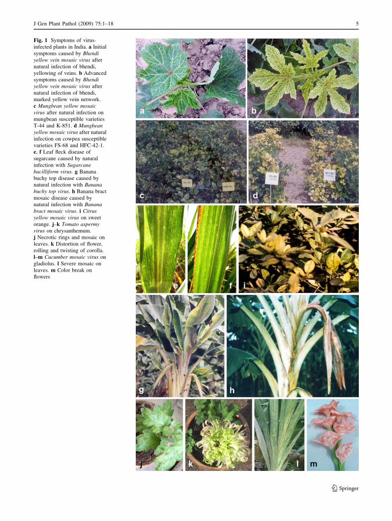
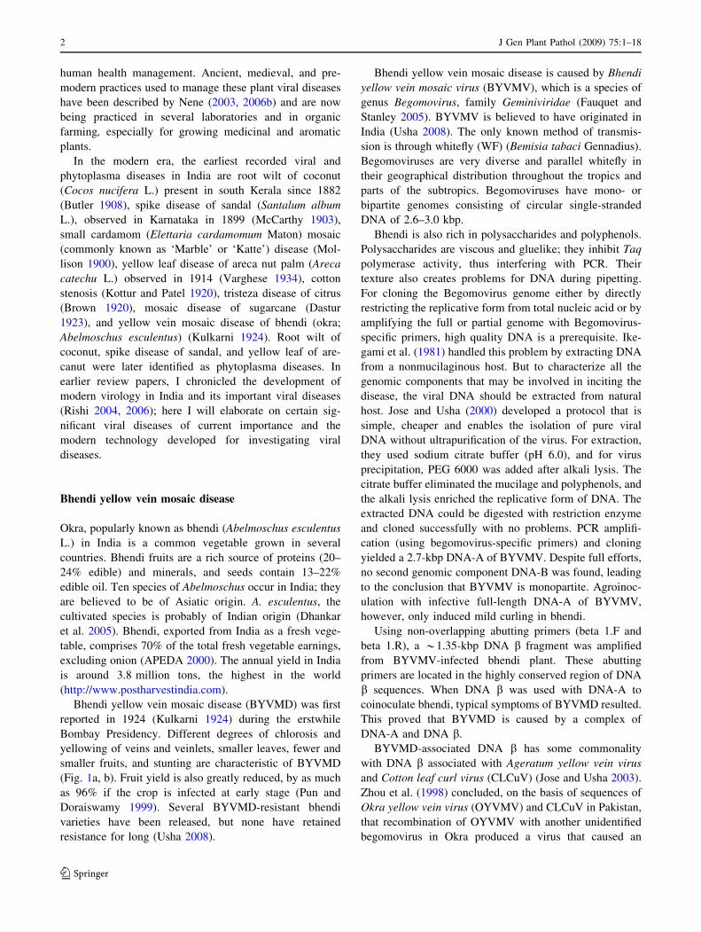
Fig. 1 Symptoms of virus-
infected plants in India. a Initial
symptoms caused by Bhendiyellow vein mosaic virus after
natural infection of bhendi,
yellowing of veins. b Advanced
symptoms caused by Bhendiyellow vein mosaic virus after
natural infection of bhendi,
marked yellow vein network.
c Mungbean yellow mosaicvirus after natural infection on
mungbean susceptible varieties
T-44 and K-851. d Mungbeanyellow mosaic virus after natural
infection on cowpea susceptible
varieties FS-68 and HFC-42-1.
e, f Leaf fleck disease of
sugarcane caused by natural
infection with Sugarcanebacilliform virus. g Banana
buchy top disease caused by
natural infection with Bananabuchy top virus. h Banana bract
mosaic disease caused by
natural infection with Bananabract mosaic virus. i Citrusyellow mosaic virus on sweet
orange. j–k Tomato aspermyvirus on chrysanthemum.
j Necrotic rings and mosaic on
leaves. k Distortion of flower,
rolling and twisting of corolla.
l–m Cucumber mosaic virus on
gladiolus. l Severe mosaic on
leaves. m Color break on
flowers
J Gen Plant Pathol (2009) 75:1–18 5
123
Usha 2005). The only exception was the clustering of the
DNA-B components from two isolates of MYMV with the
DNA-A from MYMIV.
The CP is instrumental in transmitting the virus and in
determining the viral DNA yield in the infected tissue. The
replication initiator protein (Rep) regulates the rolling
replication of viral DNA. Malik et al. (2005) studied the
interaction of the CP and Rep of MYMIV and reported that
CP downregulated the nicking and closing function of Rep.
This result indicates the importance of CP in controlling
viral DNA replication, which might be useful in disease
management.
Tracking the DNA replication mechanism of plant DNA
viruses in a suitable eukaryotic model system may open a
new avenue for the management of DNA plant viruses. At
the present time, it is difficult to manage the serious, ever-
increasing problems of begomoviruses because the only
known vector, WF, is developing resistance to chemical
control, variability is very common in begomoviruses,
large scale movement of plant materials to different areas
and introduction of newer crops. Such technology is new
for understanding replication of plant DNA, which has
been little studied in host plants.
Raghavan et al. (2004) has reported developing a yeast
model system (Saccharomyces cerevisiae W303a strain),
which during budding supports the DNA replication of an
Indian isolate of mungbean yellow mosaic begomovirus.
AC5 viral factor was identified as an important contributor
to viral DNA replication. This yeast model system can also
be exploited for screening for inhibitors of virus replica-
tion. Rep is the most important viral protein for DNA
replication. In further study with MYMIV, Chilakamarthi
et al. (2007) designed an anti-Rep ribozyme to target viral
DNA replication. When evaluated in S. cerevisiae W303a,
which lacks RNAi machinery and is thus a suitable sur-
rogate host (Raghavan et al. 2004) for replication of the
virus DNA, in the presence of the anti-Rep ribozyme the
yeast cell, grew slower because its growth was dependent
on the begomovirus replication. Viral DNA replication also
decreased by *65%. These results are encouraging for
developing a virus-resistant genetically modified plant.
Rice necrosis mosaic virus induced productivity
in jute and Mesta fiber crops
Rice necrosis mosaic virus (RNMV), taxonomically placed
under genus Bymovirus, family Potyviridae (Shukla et al.
1994), was first seen in Japan in 1959 (Fujii 1967). In India
it was first reported by Ghosh (1979). RNMV infection of
rice starts as a mosaic on the emerging leaves, then the
chlorotic spots enlarge into yellow chlorotic streaks and
necrotic spots. Eventually, fewer plant tillers and shorter
plants are produced; yields can be reduced by 12.7–100%
depending upon the variety (Fujii 1967; Ghosh 1980;
Inouye and Fujii 1977). On the other hand, when dicot jute
crops (Corchorus capsularis L. [white jute] and C. olitorius
L. [tossa jute]) and Mesta crops (Hibiscus cannabinus
[kenaf] and H. sabdariffa [roselle]) were sap-inoculated,
they were found to be symptomless carriers of the virus,
and amazingly, the virus infection enhanced productivity in
all these crops without any detrimental effect (Ghosh
1982).
RNMV is bipartite, having flexous particles of
275 9 13–14 nm and 550 9 13–14 nm (Inouye 1977;
Ghosh 1982). The Indian isolate of RNMV is serologically
related to the Japanese isolate, and they are also similar in
morphology and size. The virus is transmitted to rice by a
monocot-specific, soil-inhabiting fungus, Polymixa gra-
minis, and to dicot hosts by sap. There is no known insect
vector of RNMV (Ghosh 1980; Inouye and Fujii 1977).
Initially, rice was the only known host, but later Triticum
aestivum, Avena sativa, Zea mays, Chenopodium amaran-
ticolor, C. quinoa, Nicotiana tabacum, Brachiaria ramose,
Ludwigia perennis, and the aforementioned jute and mesta
crops were also infected by RNMV (Ghosh 1981, 1982;
Inouye and Fujii 1977).
Important fiber crops, jute and mesta are natural
inhabitants of the tropics and subtropics. In India these
crops are cultivated in several states such as West Bengal,
Assam, Meghalaya, Tripura, Orissa, Bihar, Uttar Pradesh,
Madhya Pradesh, Andhra Pradesh. Tossa jute originated in
Africa and white jute in Indo-Burma region. In India tossa
jute is called the ‘‘poor farmers’ golden fiber’’ and is pre-
ferred over white jute for its higher productivity. For some
time, polypropylene fibers captured the market over these
centuries-old natural fibers because of their lower cost and
better fiber strength. Recently, however, as we recognize
that nonbiodegradable polypropylene fibers are a potential
threat to our environment, the market for natural fibers has
been revived.
Productivity promotion
As discussed before, RNMV-sap-inoculated jute and mesta
seedlings were symptomless hosts of the virus and had
enhanced productivity at full maturity (Ghosh 1982). In
relation to the noninoculated control plants, virus infection
improved the vascular system and increased plant biomass
from 8 to 220 g with greater vigor and enhanced juvenility.
Stem diameter, leaf and root size increased, and the crop
required only half of the normally recommended dose of
fertilizer as compared to noninoculated control crop
(Ghosh and Mitra 1987). In a study of the RNMV–host
interactions in jute and mesta, the inherent cytokinin-like
activity in the plants was greatly enhanced while auxin-like
6 J Gen Plant Pathol (2009) 75:1–18
123
activity was retarded (Ghosh 1985). This RNMV-induced
growth promotion in jute and mesta is maintained through
harvested seeds for three generations (Ghosh 2001). Such
seeds have been called energized (E) seeds.
Tetraploidy, is commonly used for crop improvement.
In an investigation (Ghosh and Mitra 2008) of tetraploid
tossa jute varieties JRO-1265 (4n) a progenitor of JRO-
632, JRO-1055 (4n) a progenitor of JRO-878 and JRO
2020 (4n) a progenitor of JRO-3690 and diploid tossa JRO
3690 (2n), JRO 878, JRO-524 (2n), JRO-TJ-40 (2n) and
JRO-36E (2n) (100 plants of each variety), 20 days after
germination of seeds, the apical leaves of each plant were
mechanically inoculated with sap from RNMV-infected
rice leaves. Seeds were harvested at maturity (120 days),
and productivity was analyzed; irrespective of ploidy, all
the varieties except for JRD-878 (2n) increased in plant
height, stem diameter, total fresh mass (with and without
leaves), fiber mass and stick mass. However, the degree of
the increase varied with variety. The fiber mass increased
from 22.43 to 36.00% in diploids and in tetraploids from
2.80 to 19.00%. Similar interesting results were earlier seen
with other varieties (Ghosh 1985). In these fiber plants,
RNMV infection stimulates cell division and increases the
cell size, contributing to the increases in leaf size, stem
diameter and fiber bundle number (Ghosh 1982). RNMV
infection at the seedling growth and a positive serological
test at crop maturity, coupled with enhanced endogenous
growth induced by cytokinin and IAA suggests that a
unique pseudosymbiotic relationship between the host and
virus induced beneficial biochemical changes (Ghosh
1985). Because tetraploidy is a better avenue for higher
production in many crops, using RNMV to further improve
phenotypic characters that increase yield potential in such
tetraploids has opened a new vista that is less time con-
suming and cheaper to develop for commercial use (Ghosh
and Mitra 2008). RNMV is not transmitted from jute to the
succeeding rice crop in a rotation because the fungus vector
P. graminis in soil is specific for monocots and thus makes
the technology even more viable (Ghosh 1985).
Mesta yellow vein mosaic disease of mesta crops
(jute and kneaf)
Mesta crops are an important source of bast fibers and a
substitute for jute. The fibers obtained after retting are used
as the raw material for a number of products such as rope,
fishing net, and canvas. Seeds are a good source of oil
(20%) and used in cooking, soap, paints and lubricants.
Seed cakes left after oil extraction are used as cattle feed.
The plant also has medicinal value against several human
disorders such as coughs and rheumatism. The U.S.
Deparment of Agriculture has identified kneaf as the best
plant for making nonwoody paper.
Mests crops are grown in India in several states, but
overall productivity of the crops is low. Viral diseases,
most importantly Mesta yellow vein mosaic disease
(MYVMD), are an important factor in reducing the fiber
yield. MYVMD was first observed in Bahraich district in
Uttar Pradesh and has spread fast in several other areas of
the country, incurring heavy losses. The causal agent of the
disease has been identified as monopartite begomovirus
associated with satellite DNA b (Chatterjee et al. 2005;
Paul et al. 2006). Southern blot and nucleic acid hybrid-
ization test using Cotton leafcurl Rajasthan virus
(CLCuRV) specific DNA-A and DNA b probes confirmed
that it is caused by a begomovirus related to CLCuRV. The
disease is transmitted by cleft grafting and whitefly
(Bemisia tabaci). Symptoms in emerging leaves start as
abundant pin head spots on the leaf lamina, including
veins. These spots gradually enlarge and coalesce to form
chlorotic to yellow flecks. In severe cases, these yellow
flecks enlarge until the leaves and stems are completely
chlorotic or yellow and plant defoliates. Flower and pod set
is reduced, those that develop are malformed. Life span of
infected plants is also reduced. The host range of this virus
differs from other begomovirus diseases on Hibiscus plants
(Paul et al. 2006; Rajeshwari et al. 2005). The full-length
sequence of DNA-A and DNA b of the virus infecting
mesta was cloned and sequenced. DNA-A had 2728
nucleotides in length having 83.5% identity with Cotton
leafcurl Bangalore virus (CLCuBV) and 83.3% identity
with CLCuRV. There were six conserved ORFs identified
in both orientations of the sequence. Sequence analyses
proved that this new DNA-A was typical of Old World
begomoviruses. It was therefore identified as a separate
species of Begomovirus and was named as Mesta yellow
vein mosaic virus (Chatterjee and Ghosh 2007).
Leaf fleck disease of sugarcane
Leaf fleck disease of sugarcane caused by Sugarcane
bacilliform virus (SCBV) was first reported from Cuba
(Rodrıguez Lema et al. 1985). Later, Lockhart and Autrey
(1988) reported that the sugarcane clone Mex. 57-473
growing in Morocco and Hawaii and CP44-101 in Morocco
contained bacilliform viral particles, closely related to
another identified Badnavirus, Banana streak virus (BSV)
(Lockhart 1986; Lockhart and Olszewski 1993). The virus
is transmitted by sugarcane mealy bug (Saccharicoccus
sacchari) and the grey sugarcane mealy bug (Dysmicoccus
boninsis) in nature, and citrus mealy bug (Planococcus
citri), which does not colonize sugarcane naturally,
J Gen Plant Pathol (2009) 75:1–18 7
123
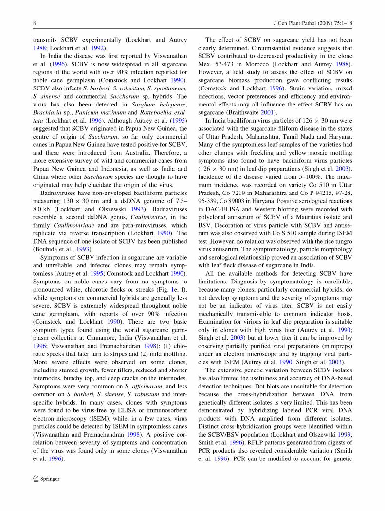
transmits SCBV experimentally (Lockhart and Autrey
1988; Lockhart et al. 1992).
In India the disease was first reported by Viswanathan
et al. (1996). SCBV is now widespread in all sugarcane
regions of the world with over 90% infection reported for
noble cane germplasm (Comstock and Lockhart 1990).
SCBV also infects S. barberi, S. robustum, S. spontaneum,
S. sinense and commercial Saccharum sp. hybrids. The
virus has also been detected in Sorghum halepense,
Brachiaria sp., Panicum maximum and Rotteboellia exal-
tata (Lockhart et al. 1996). Although Autrey et al. (1995)
suggested that SCBV originated in Papua New Guinea, the
centre of origin of Saccharum, so far only commercial
canes in Papua New Guinea have tested positive for SCBV,
and these were introduced from Australia. Therefore, a
more extensive survey of wild and commercial canes from
Papua New Guinea and Indonesia, as well as India and
China where other Saccharum species are thought to have
originated may help elucidate the origin of the virus.
Badnaviruses have non-enveloped bacilliform particles
measuring 130 9 30 nm and a dsDNA genome of 7.5–
8.0 kb (Lockhart and Olszewski 1993). Badnaviruses
resemble a second dsDNA genus, Caulimovirus, in the
family Caulimoviridae and are para-retroviruses, which
replicate via reverse transcription (Lockhart 1990). The
DNA sequence of one isolate of SCBV has been published
(Bouhida et al., 1993).
Symptoms of SCBV infection in sugarcane are variable
and unreliable, and infected clones may remain symp-
tomless (Autrey et al. 1995; Comstock and Lockhart 1990).
Symptoms on noble canes vary from no symptoms to
pronounced white, chlorotic flecks or streaks (Fig. 1e, f),
while symptoms on commercial hybrids are generally less
severe. SCBV is extremely widespread throughout noble
cane germplasm, with reports of over 90% infection
(Comstock and Lockhart 1990). There are two basic
symptom types found using the world sugarcane germ-
plasm collection at Cannanore, India (Viswanathan et al.
1996; Viswanathan and Premachandran 1998): (1) chlo-
rotic specks that later turn to stripes and (2) mild mottling.
More severe effects were observed on some clones,
including stunted growth, fewer tillers, reduced and shorter
internodes, bunchy top, and deep cracks on the internodes.
Symptoms were very common on S. officinarum, and less
common on S. barberi, S. sinense, S. robustum and inter-
specific hybrids. In many cases, clones with symptoms
were found to be virus-free by ELISA or immunosorbent
electron microscopy (ISEM), while, in a few cases, virus
particles could be detected by ISEM in symptomless canes
(Viswanathan and Premachandran 1998). A positive cor-
relation between severity of symptoms and concentration
of the virus was found only in some clones (Viswanathan
et al. 1996).
The effect of SCBV on sugarcane yield has not been
clearly determined. Circumstantial evidence suggests that
SCBV contributed to decreased productivity in the clone
Mex. 57-473 in Morocco (Lockhart and Autrey 1988).
However, a field study to assess the effect of SCBV on
sugarcane biomass production gave conflicting results
(Comstock and Lockhart 1996). Strain variation, mixed
infections, vector preferences and efficiency and environ-
mental effects may all influence the effect SCBV has on
sugarcane (Braithwaite 2001).
In India bacilliform virus particles of 126 9 30 nm were
associated with the sugarcane filiform disease in the states
of Uttar Pradesh, Maharashtra, Tamil Nadu and Haryana.
Many of the symptomless leaf samples of the varieties had
other clumps with freckling and yellow mosaic mottling
symptoms also found to have bacilliform virus particles
(126 9 30 nm) in leaf dip preparations (Singh et al. 2003).
Incidence of the disease varied from 5–100%. The maxi-
mum incidence was recorded on variety Co 510 in Uttar
Pradesh, Co 7219 in Maharashtra and Co P 94215, 97-28,
96-339, Co 89003 in Haryana. Positive serological reactions
in DAC-ELISA and Western blotting were recorded with
polyclonal antiserum of SCBV of a Mauritius isolate and
BSV. Decoration of virus particle with SCBV and antise-
rum was also observed with Co S 510 sample during ISEM
test. However, no relation was observed with the rice tungro
virus antiserum. The symptomatology, particle morphology
and serological relationship proved an association of SCBV
with leaf fleck disease of sugarcane in India.
All the available methods for detecting SCBV have
limitations. Diagnosis by symptomatology is unreliable,
because many clones, particularly commercial hybrids, do
not develop symptoms and the severity of symptoms may
not be an indicator of virus titer. SCBV is not easily
mechanically transmissible to common indicator hosts.
Examination for virions in leaf dip preparation is suitable
only in clones with high virus titer (Autrey et al. 1990;
Singh et al. 2003) but at lower titer it can be improved by
observing partially purified viral preparations (minipreps)
under an electron microscope and by trapping viral parti-
cles with ISEM (Autrey et al. 1990; Singh et al. 2003).
The extensive genetic variation between SCBV isolates
has also limited the usefulness and accuracy of DNA-based
detection techniques. Dot-blots are unsuitable for detection
because the cross-hybridization between DNA from
genetically different isolates is very limited. This has been
demonstrated by hybridizing labeled PCR viral DNA
products with DNA amplified from different isolates.
Distinct cross-hybridization groups were identified within
the SCBV/BSV population (Lockhart and Olszewski 1993;
Smith et al. 1996). RFLP patterns generated from digests of
PCR products also revealed considerable variation (Smith
et al. 1996). PCR can be modified to account for genetic
8 J Gen Plant Pathol (2009) 75:1–18
123

variation through the design of primers based on conserved
sequences. Published primers for SCBV are available that
target consensus sequences within the aspartic protease,
ribonuclease H and reverse transcriptase coding regions
and tRNAMet binding site of several badnaviruses (Brai-
thwaite et al. 1995; Lockhart and Olszewski 1993). DNA
sequencing can reveal the extent of genetic variation
between isolates. When reverse transcriptase and ribonu-
clease H amplicons from SCBV isolated from three noble
canes were sequenced (Braithwaite et al. 2004; Geijskes
et al. 2002), variability was revealed not just among the
SCBV isolates from different canes, but also among iso-
lates within individual cane plants. Thus, conserved or
degenerate primers may not reliably prime the amplifica-
tion of all the isolates.
Sugarcane, particularly the noble canes, appear to con-
tain a large pool of SCBV genomic sequences, perhaps
because of the vegetative nature of sugarcane propagation
and the long history of sugarcane movement and cultiva-
tion. Thus, SCBV variants have had considerable time to
accumulate within the canes. The extensive isolate vari-
ability even within one sugarcane clone and the possibility
of virus integration into the host genome have limited the
acceptance of PCR-based methods for detection of SCBV.
Although ISEM is time consuming and requires sophisti-
cated equipment, it is still considered the most reliable
method of detecting SCBV.
Control of the mealy bug vectors and disease-free
planting material should be helpful in managing SCBV.
Apical meristem culture and heat treatment could not
eliminate SCBV from infected sugarcane (Autrey et al.
1990; Egeskov et al. 1994). Because badnaviruses replicate
via reverse transcription, the use of chemotherapeutic
agents that inhibit the reverse transcriptase offers a novel
approach for control. Antiviral agents have been used
successfully to eliminate BSV from banana (Helliot et al.
2003).
Banana bunchy top disease
Banana is one of the important fruit crops grown in
tropical and subtropical areas of India, the world’s largest
banana producer. Although India contributes 19.71% of
total world production (19.19 million tons) grown on
0.565 million hectares (Singh 2007), many pests of
bananas and plantains diminish the productivity signifi-
cantly, and viruses are considered a serious threat. So far,
four viral diseases viz., bunchy top, streak, bract mosaic
and infectious chlorosis have been reported in India. Of
these virus diseases, banana bunchy top disease (BBTD)
(Fig. 1g), caused by Banana bunchy top virus (BBTV) is
the most serious and devastating disease, which limits or
threatens production in one quarter of the world’s banana-
growing areas. Capoor and Varma (1968) reported mosaic
disease of banana from Deccan area. Infectious chlorosis
caused by Cucumber mosaic virus (CMV) is another
important viral disease, more prevalent in Cavendish and
Mysore group of bananas. Recently, banana bract mosaic
and streak diseases reported at the farm of National
Research Centre for Banana (NRCB), Tiruchirapalli
(India) are assuming more significance owing to their fast
spread and economic loss they cause (R. Selvarajan,
personal communication).
BBTV is a single-stranded DNA virus in the family
Nanoviridae. It is the type species of the genus Babuvirus.
BBTV is isometric, and the particles measure 20 nm. The
molecular weight of the coat protein is 19.3 kDa, and the
virus has a multicomponent genome. Six circular single-
stranded components are known to be associated with the
genome (Selvarajan and Balasubramanian 2008). BBTV,
introduced in India from Sri Lanka in 1943, has been listed
under diseases of national importance. The disease has now
spread to the states of Andhra Pradesh, Tamil Nadu, Orissa,
Maharastra, Bihar, Karnataka, West Bengal, Assam and
Uttar Pradesh and recently to the forests of North Eastern
Hill states of India.
Banana bunchy top affects many commercial cultivars
such as Virupakshi, Nendran, Robusta, Ney Poovan and
Monthan, causing considerable economic loss, estimated at
Rs. 400 million annually in Kerala alone. The highly fla-
voured hill banana, Virupakshi (AAB) has been wiped out
by BBTV in lower Pulany and Sirumalai hills of Tamil
Nadu (Selvarajan and Jeyabaskaran 2006). One of the
reasons for the severity in hill regions is the year-round
presence of inoculum and the insect vector that transmits
the virus. The crop is grown perennially for 10–15 years as
large clumps. The virus is transmitted primarily by infected
suckers and through tissue-propagated material. Banana
aphid Pentalonia nigronervosa transmits the virus in a
semipersistent manner (Selvarajan et al. 2006).
Serological or immunological assays have been devel-
oped and used successfully for a number of years to detect
plant viruses. Using polyclonal antiserum, DAC-ELISA
was found better than DIBA for detection of BBTV.
Simultaneous detection of Indian isolates of BBTV and
BSV by duplex PCR and of BBTV, BBrMV and BSV by
multiplex-PCR technique has been reported (Selvarajan
et al. 2004). Selvarajan et al. (2004) developed IC-PCR
technique for BBTV. Selvarajan et al. (2007) cloned and
sequenced all six DNA components of a hill banana isolate
of BBTV. The nucleotide sequences of BBTV-DNA 1
shared 99, 97 and 93% homology with the South Pacific
group, the Asian group and the Indian group. BBTV-DNA
3 component had 96–97% homologies with the South
Pacific and Asian groups, and BBTV-DNA 4 had 94–96%,
J Gen Plant Pathol (2009) 75:1–18 9
123
BBTV-DNA 5 had 94–99% and BBTV-DNA 6 had 97%
homologies with the South Pacific group.
Selvarajan et al. (2007) reported cloning and sequencing
of the coat protein gene of five BBTV isolates from the
Arunachal Pradesh, Meghalaya, Nagaland, Assam and
Kodaikanal hills. A comparative analysis of the Indian
isolates with Asian isolates indicated that the Indian iso-
lates do not belong to Asian group. Cluster analysis of the
Indian isolate with South Pacific placed the Meghalaya
isolate in a different cluster. A comparative analysis of all
three groups (Asia, South Pacific and Indian isolates) fur-
ther confirmed that the Indian isolates clusterd with the
South Pacific isolates.
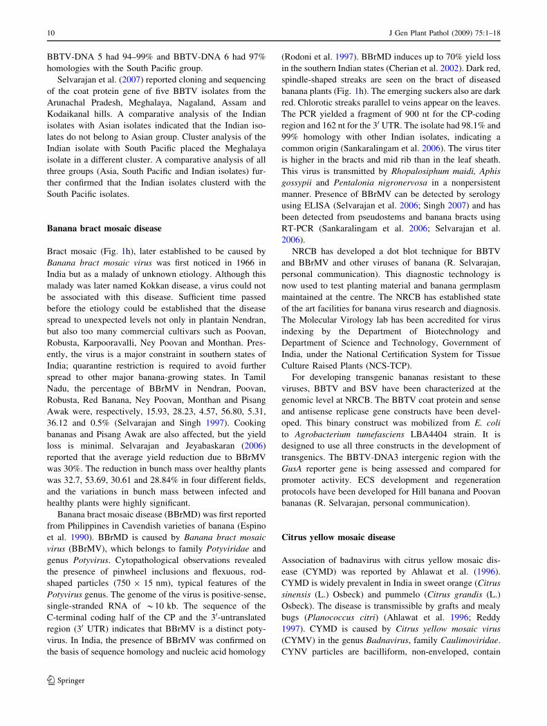
Banana bract mosaic disease
Bract mosaic (Fig. 1h), later established to be caused by
Banana bract mosaic virus was first noticed in 1966 in
India but as a malady of unknown etiology. Although this
malady was later named Kokkan disease, a virus could not
be associated with this disease. Sufficient time passed
before the etiology could be established that the disease
spread to unexpected levels not only in plantain Nendran,
but also too many commercial cultivars such as Poovan,
Robusta, Karpooravalli, Ney Poovan and Monthan. Pres-
ently, the virus is a major constraint in southern states of
India; quarantine restriction is required to avoid further
spread to other major banana-growing states. In Tamil
Nadu, the percentage of BBrMV in Nendran, Poovan,
Robusta, Red Banana, Ney Poovan, Monthan and Pisang
Awak were, respectively, 15.93, 28.23, 4.57, 56.80, 5.31,
36.12 and 0.5% (Selvarajan and Singh 1997). Cooking
bananas and Pisang Awak are also affected, but the yield
loss is minimal. Selvarajan and Jeyabaskaran (2006)
reported that the average yield reduction due to BBrMV
was 30%. The reduction in bunch mass over healthy plants
was 32.7, 53.69, 30.61 and 28.84% in four different fields,
and the variations in bunch mass between infected and
healthy plants were highly significant.
Banana bract mosaic disease (BBrMD) was first reported
from Philippines in Cavendish varieties of banana (Espino
et al. 1990). BBrMD is caused by Banana bract mosaic
virus (BBrMV), which belongs to family Potyviridae and
genus Potyvirus. Cytopathological observations revealed
the presence of pinwheel inclusions and flexuous, rod-
shaped particles (750 9 15 nm), typical features of the
Potyvirus genus. The genome of the virus is positive-sense,
single-stranded RNA of *10 kb. The sequence of the
C-terminal coding half of the CP and the 30-untranslated
region (30 UTR) indicates that BBrMV is a distinct poty-
virus. In India, the presence of BBrMV was confirmed on
the basis of sequence homology and nucleic acid homology
(Rodoni et al. 1997). BBrMD induces up to 70% yield loss
in the southern Indian states (Cherian et al. 2002). Dark red,
spindle-shaped streaks are seen on the bract of diseased
banana plants (Fig. 1h). The emerging suckers also are dark
red. Chlorotic streaks parallel to veins appear on the leaves.
The PCR yielded a fragment of 900 nt for the CP-coding
region and 162 nt for the 30 UTR. The isolate had 98.1% and
99% homology with other Indian isolates, indicating a
common origin (Sankaralingam et al. 2006). The virus titer
is higher in the bracts and mid rib than in the leaf sheath.
This virus is transmitted by Rhopalosiphum maidi, Aphis
gossypii and Pentalonia nigronervosa in a nonpersistent
manner. Presence of BBrMV can be detected by serology
using ELISA (Selvarajan et al. 2006; Singh 2007) and has
been detected from pseudostems and banana bracts using
RT-PCR (Sankaralingam et al. 2006; Selvarajan et al.
2006).
NRCB has developed a dot blot technique for BBTV
and BBrMV and other viruses of banana (R. Selvarajan,
personal communication). This diagnostic technology is
now used to test planting material and banana germplasm
maintained at the centre. The NRCB has established state
of the art facilities for banana virus research and diagnosis.
The Molecular Virology lab has been accredited for virus
indexing by the Department of Biotechnology and
Department of Science and Technology, Government of
India, under the National Certification System for Tissue
Culture Raised Plants (NCS-TCP).
For developing transgenic bananas resistant to these
viruses, BBTV and BSV have been characterized at the
genomic level at NRCB. The BBTV coat protein and sense
and antisense replicase gene constructs have been devel-
oped. This binary construct was mobilized from E. coli
to Agrobacterium tumefasciens LBA4404 strain. It is
designed to use all three constructs in the development of
transgenics. The BBTV-DNA3 intergenic region with the
GusA reporter gene is being assessed and compared for
promoter activity. ECS development and regeneration
protocols have been developed for Hill banana and Poovan
bananas (R. Selvarajan, personal communication).
Citrus yellow mosaic disease
Association of badnavirus with citrus yellow mosaic dis-
ease (CYMD) was reported by Ahlawat et al. (1996).
CYMD is widely prevalent in India in sweet orange (Citrus
sinensis (L.) Osbeck) and pummelo (Citrus grandis (L.)
Osbeck). The disease is transmissible by grafts and mealy
bugs (Planococcus citri) (Ahlawat et al. 1996; Reddy
1997). CYMD is caused by Citrus yellow mosaic virus
(CYMV) in the genus Badnavirus, family Caulimoviridae.
CYNV particles are bacilliform, non-enveloped, contain
10 J Gen Plant Pathol (2009) 75:1–18
123

dsDNA (7559 bp) and measure 130 9 30 nm (Ahlawat
et al. 1996; Huang and Hartung 2001). Infected citrus
plants develop yellow mosaic (Fig. 1i) and fewer flowers
and fruits. Juice and ascorbic acid levels are also reduced.
Serological detection of badnaviruses is not reliable
because the virus particles are weak immunogens. Ahlawat
et al. (1996) obtained a PCR product using degenerate
primers from partially purified virus preparations. How-
ever, the high level of polyphenolics and tannins in citrus
leaves interfered in extracting good quality DNA. Baran-
wal et al. (2003) succeeded in overcoming this problem
using sodium sulphite during the extraction. DNA thus
obtained was higher in yield, better in quality and was
cleaner and more stable. The sodium sulphite may have
prevented oxidation of DNA upon release from host cells.
This protocol has been standardized and is in regular use.
Piper yellow mottle disease
Betel vine (Piper betle), Indian long pepper (Piper longum)
and black pepper (P. nigrum) are economically important
species of family Piperaceae. After black pepper, one of the
most ancient crops cultivated in India, in importance are
betel vine and Indian long pepper. Black pepper may have
originated in the southwestern hills of India, and betel vine
is supposed to be a native of Malaysia. The medicinal
properties of these plants are helpful against diseases of
respiratory tract and are also important ingredients in
the Ayurveda, Siddha and Unani systems of medicines
(Ravindran 2000). Dried berries of P. nigrum (black pepper)
are an important condiment of international commercial
value and India earns Rs. 88 crore annually through export.
Leaves of betel vine are chewed.
Piper yellow mottle disease caused by a badnavirus,
piper yellow mottle virus (PYMoV), and transmitted by
mealy bug (Ferrisia virgata) was reported in Southeast
Asia including India (Bhat et al. 2003, 2005; Lockhart et al.
1997; Sarma et al. 2001). The symptoms on P. betel are
mottling, mosaic and reduced leaf size; on P. longum,
mosaic with dark green patches, blisters and leaf distortion;
and on P. nigrum, vein clearing, mottling, chlorosis, and
distortion of leaves, reduced plant vigor, and fewer and
smaller fruit set. The virus on these hosts spread through
the use of virus-contaminated plant material for vegetative
propagation and via the mealy bug F. virgata and the citrus
mealy bug Planococcus citri (Bhat et al. 2003, 2005).
Badnaviruses are non-enveloped bacilliform particles
(30 9 130–150 nm), having a double-stranded, circular
DNA genome (7.1–7.6 kb). The type member is Comme-
lina yellow mottle virus. With a few exceptions, the
genome has three open reading frames. ORF I and ORF II
encode for putative proteins of unknown functions. ORF III
encodes a polyprotein that is cleaved to produce functional
products such as the movement protein, coat protein,
aspartic protease and a replicase.
Siju et al. (2008) characterized badnavirus infecting
betel vine and Indian long pepper. Degenerate primer pairs
(sense and antisence) of ORF III were designed on the
basis of known badnaviruses and used for PCR. The
products were sequenced and were 597 nt long potentially
coding for 199 amino acid sequence both in the case of
betel vine and Indian long pepper. The sequence identities
were 89.1% for nucleotides and 93.4% for amino acids
when compared to those of known badnaviruses. The
phylogenic tree clearly indicated that badnavirus isolates
infecting betel vine and Indian long pepper and black
pepper were strains of PYMoV.
Chrysanthemum diseases caused by Chrysanthemum
virus B (carlavirus) and Cucumber mosaic virus
(cucumovirus)
Chrysanthemum (Dendranthema 9 grandiflorum Kitam.)
of family Asteraceae is a very popular cut flower crop and
pot plant cultivated in several parts of India (4000 ha) and
world. It is commonly known as garden chrysanthemum
and ‘‘queen of the East.’’ The word chrysanthemum comes
from chrysos = golden and anthos = flower. It ranks only
next to rose and third amongst the cut flowers in production
in India (Singh 2000). The flower is most in demand in
Japan and the Netherlands.
A large number of viruses and viroids viz. Chrysan-
themum virus B, Tomato aspermy virus, Cucumber mosaic
virus, Tomato spotted wilt virus (TSWV), Chrysanthemum
stem necrosis virus, Chrysanthemum spot potyvirus,
Chrysanthemum chlorotic stunt viroid, and Chrysanthe-
mum stunt viroid are known to infect chrysanthemum in
nature (Kumar et al. 2005, Singh et al. 2007a). In India,
Chrysanthemum virus B (CVB), Tomato aspermy virus
(TAV) and Cucumber mosaic virus (CMV) are most
important (Verma et al. 2007). CVB is the most prevalent
and does not cause any symptoms in most varieties of
chrysanthemum. TAV and different strains of CMV induce
significant losses. On some varieties, CVB causes vein
clearing and mottling. It has narrow host range, confined to
*10 species of dicotyledonous plants. Chrysanthemum is
vegetatively propagated by sucker, which aids disease
perpetuation if the mother plant is virus contaminated.
CVB is also transmitted mechanically and by Myzus per-
sicae and M. solani (Hollings 1957). It is distantly related
to carlaviruses viz. Carnation latent virus, Potato virus S
and Potato virus M (van Slogteren et al. 1962). The host
range of CVB does not overlap the host range of these
carlaviruses.
J Gen Plant Pathol (2009) 75:1–18 11
123
Efficient diagnostic tools are important for screening the
plant material and are essential for epidemiological studies,
quarantine and effective marketing of virus-free plant
material (Lawson 1981). Such tools are even more
important for symptomless hosts. In India, protocols of
diagnostic tools like ELISA, PCR, RT-PCR and IC-RT-
PCR have been standardized for CVB (Singh et al. 2007a;
Verma et al. 2003; Zaidi et al. 1993) and are now the
routine for virus detection. In a further step, the coat pro-
tein (CP) gene, the triple gene block (TGB), the nucleic
acid-binding protein (NABP), and Rep have been ampli-
fied, cloned and sequenced. The Rep gene was completely
sequenced. The CP gene sequences of 29 isolates collected
from different places showed variation in size (9921–
969 bp); the middle portion of the CP was conserved but
the N and C terminals varied markedly (Singh et al. 2007a).
In phylogenetic analyses, the relationship of CVB to Lily
symptomless virus was established. All 29 isolates were
classified into three major groups in the phylogenic tree.
CMV induced symptom variability in chrysanthemum is
common in nature. It may be yellow mosaic with green
veins, severe mosaic, yellowing of veins and yellow to
necrotic spots. This variability in symptoms is a strong
indication of the existence of CMV strains in the natural
infection of chrysanthemum (Srivastava et al. 1992). The
virus particles in dip preparation of these samples of varied
symptoms were isometric measuring 29 nm. Association of
CMV with these varied symptoms was confirmed by RT-
PCR. CP gene-specific primers of CMV gave a band of
*650 bp, which hybridized with the cloned probe of CMV
(Kumar et al. 2005).
Chrysanthemum diseases caused by Tomato aspermy
virus (cucumovirus)
Chlorotic ring mosaic of chrysanthemum caused by Tomato
aspermy virus (TAV) in India is characterized by yellow
mosaic, green vein banding, stunting and flower deforma-
tion (Fig. 1j, k). TAV has been identified on the basis of
symptoms, host range and aphid transmission (Aphis
gossypii and Myzus persicae) (Gupta and Singh 1981;
Sastry 1964). Later, TAV was identified by other features:
local lesion hosts (Chenopodium spp. and Cucumis sativus),
electron microscopy (ca. 29 nm isometric particles), crys-
talline inclusions and virions in the central vacuole in cells
of N. clevelandii after sap-inoculation, serological rela-
tionships, molecular weight of the CP, the presence of five
RNAs (Raj et al. 1992, 2007; Verma et al. 2007). Verma
et al. (2007) for the first time obtained complete amplifi-
cation of the TAV genome (RNA 1, RNA 2, RNA 3) using
multiplex PCR. Fifteen isolates of TAV on different culti-
vars of chrysanthemum were collected from different states
of India, from the plains and from hilly tracts at higher
elevations. Both in RT-PCR and IC-RT-PCR, an amplicon
of the expected size (657 bp) was observed on an agarose
gel. The virus could be detected by RT-PCR in plants
without symptoms, but no virus was detected by ELISA in
these samples. Amplified CP gene sequences of four iso-
lates had 97% identity with those of other isolates available
in the database, and the deduced amino acid sequence also
had 99% similarity, probably indicating a common ances-
try. The lower similarity between the CP genes of TAV and
CMV indicated a distant relationship within the group.
Some of the samples with a faint amplification of the 657-bp
fragment in the RT-PCR resulted in a strong amplification
in the IC-RT-PCR.
Gladiolus diseases caused by Cucumber mosaic virus
(cucumovirus)
Among the top six flowers in the growing floral export
industry in India, Gladiolus psittacinus (Iridaceae) is a very
important cut flower widely used in bouquets and floral
baskets because of its very attractive inflorescences in a
wide range of colors. Numerous viruses infect gladioli in
nature: CMV, Bean yellow mosaic virus (BYMV), Broad
bean wilt virus (BBWV), TSWV, Tobacco mosaic virus
(TMV), Tobacco necrosis virus (TobNV), Tobacco black
ring virus (TobBRV), Tobacco ring spot virus (TobRSV),
Tomato black ring spot virus, Arabis mosaic virus,
Tobacco streak virus, Soybean Mosaic virus (SMV) and
Strawberry latent ring spot virus (StrLRSV) (Katoch et al.
2003; Raj et al. 1998, 2002). Of these, CMV and BYMV
are the most important viral pathogens of gladiolus. They
produce mosaic, stripe, floral color breaking and reduced
vigour (Fig. 1l, m). The virus is transmitted mechanically
and by M. persicae in a nonpersistent manner. These
viruses were identified on gladiolus on the basis of symp-
toms, the host range (which is wide), aphid transmission,
electron microscopy, serological tests viz. DAS-ELISA,
TAS-ELISA, direct tissue blotting immunoassay (DTBIA),
ISEM, Western blotting assay (WBA), and RT-PCR and
Southern hybridization. Of all these, RT-PCR followed by
Southern hybridization test was found most effective.
Katoch et al. (2003) reported that DAS-ELISA, DTBIA,
ISEM and RT-PCR were best suited to detect these viruses.
ELISA and DTBIA are most widely used, but the RT-PCR
is more sensitive and can detect virus even at very low titer.
Important virus diseases of orchids in India
About 1300 species of orchids occur in different parts of
India especially the northeastern states, northern hilly
12 J Gen Plant Pathol (2009) 75:1–18
123
areas, Kerala, and the Darjeeling hills. More than 30
viruses have been reported to infect different orchids in
various geographical locations. Among the orchid viruses,
Cymbidium mosaic potexvirus (CymMV) and Odonto-
glossum ringspot tobamovirus (ORSV) are considered most
important due to their worldwide occurrence and severity
of the symptoms in several orchid genera. They reduce the
general vigor of the plant and affect the flower quality,
thereby reducing the marketability and incurring severe
economic losses. These two viruses also occur naturally as
mixed infections in several orchid species. In India, almost
no attention has been given to viral diseases of orchids
because orchid growers have been ignorant about viral
diseases and their economic implications on orchid plants.
Orchid viruses generally produce mosaic, ringspots,
chlorosis, chlorotic and necrotic sunken patches on the
leaves, deformation and a drastic reduction in the size of
flowers and overall stunting of the plant. The most
important symptoms of CymMV and ORSV on orchids are
mosaic and mottling on the leaves, followed by necrosis of
the leaves and flowering stalk. CymMV induces charac-
teristic colour breaking in Cattleya. Calanthe mild mosaic
potyvirus also induces flower color break in Calanthe (Gara
et al. 1998).
Sherpa et al. (2003) first reported the occurrence of
CymMV in India and recorded the virus in Cymbidium
aloifolium, C. iridioides, Epidendron sp., Liparis botan-
ensis, Phaius tankervilleae and Pholidota imbricata and
cymbidium hybrids. The virus was successfully transmitted
to Datura stramonium and Chenopodium murale, which
produced blotchy local lesions. The presence of the virus in
local lesions was confirmed by ELISA and electron
microscopy. Later, Sherpa et al. (2006a) reported ORSV in
42 different species of orchids from Sikkim by slot-blot
hybridization method. They then characterized the coat
protein (CP) gene of CymMV from India and amplified the
gene encoding the CP gene of nine isolates using the
RT-PCR (Sherpa et al. 2006b). They also cloned and
sequenced the amplified product, and multiple gene
sequence alignments of the deduced amino acid sequence
revealed considerable homology to CymMV from other
countries. They concluded that CymMV CP gene is highly
conserved and can be used for diagnosis. Bhat et al. (2006)
reported CymMV in vanilla from the states of Kerala and
Karnataka. Singh et al. (2007b) reported the association of
a potyvirus with C. pendulum and C. tigrinum from
Sikkim. The infected samples when tested by ELISA,
RT-PCR and Northern blot analysis confirmed the presence
of potyvirus. The sequencing of a RT-PCR amplified
amplicon using potyvirus-specific primers revealed that the
virus is closely related to Calanthe mild mosaic virus.
Recently, a large number of orchid germplasms was
scanned by EM and ELISA at the National Research
Centre for Orchids, Pakyong, Sikkim. Electron microscopy
of a negatively stained preparation from the sap of mosaic-
affected cymbidium and other orchid species showed
flexuous particles measuring about 450–550 nm, while sap
from ORSV-affected plants had rigid rods measuring about
300–310 nm. Electron microscopy and ELISA results
revealed that mixed infections of CymMV and ORSV are
common in nature; CymMV and ORSV the most common
viruses in northeastern Himalayan region of India. Besides
these two viruses, potyvirus-, rhabdovirus-, badnavirus-
and tombusvirus-like particles have also been recorded in
different orchid genera and species. Attempts are being
made to characterize these viruses biologically, serologi-
cally and molecularly in order to develop effective
strategies for producing healthy plant materials and thereby
compete with other countries involved in the orchid trade
(Pant et al. 2007).
In addition to the diseases of different crops discussed, a
few more that are caused by begomoviruses are currently
of significance in other crops; also Sugarcane streak
mosaic virus, which is different from Sugarcane mosaic
virus and has been recommended to be renamed as a new
genus of Potyviridae; Tobacco streak virus of genus Ilar-
virus, family Bromoviridae; Mandarivirus, a new genus of
the newly created family Flexiviridae; and Pigeonpea
sterility mosaic virus (unassigned). The diseases they cause
viz. cotton leaf curl (Sharma and Rishi 2007, 2008); sug-
arcane streak (Hema et al. 2008); necrosis diseases of
sunflower, legumes, cucurbits, chili, okra, cotton and sun
hemp (Jain et al. 2008); Indian citrus ring spot (Ahlawat
and Pant 2008); and pigeonpea sterility mosaic (Kumar
et al. 2004) have been adequately dealt with in recent
reviews (Rishi 2004, 2006).
Conclusion
India has a diverse topography, varying from fertile fields
to the deserts of Rajasthan in the plains, a vast peninsular
area, and the uplands of the Himalayas, Aravali and Nilgiri,
that represent equally varied agroclimatic conditions from
tropical, subtropical to temperate. These conditions have
led to the evolution of one of the richest biodiversity of
species in the world.
Indian agriculture is also one of the oldest in world.
There is documented evidence of the export of spices,
cotton and silk for centuries to different parts of the world.
Tropical and subtropical agroclimates are conducive to the
growth of a large number of pests and pathogens, which
have coevolved with the rich vegetation. Symptoms of
some of these biotic stresses such as leaf fleck disease of
sugarcane are so obscure that they can long miss the
attention of even highly experienced scientists and farmers.
J Gen Plant Pathol (2009) 75:1–18 13
123

Literature is replete with examples of the virus contami-
nation of entire lots of germplasm and their movement and
inadvertent spread to different parts of the world (Com-
stock and Lockhart 1990; Hampton et al. 1976; Lockhart
et al. 1996; Viswanathan et al. 1996). These germplasms
were long used for breeding purposes, which disseminated
these viruses. Furthermore, during a long coexistence, part
of the genome of some viral pathogens became integrated
within the host as in sugarcane where propagation is veg-
etative (Braithwaite et al. 2004). In such cases, the results
of PCR are deceptive, and ELISA/ISEM or Western blot-
ting tests are more dependable.
In spite of the fact that India today is one of the five
most vibrant economies in the world, Indian agriculture
still forms the backbone of a rural livelihood and national
economy. The rapid growth of agriculture to ensure food
security and the liberalization of world trade in agriculture
with the advent of the WTO have opened new vistas of
growth while imposing new challenges with the risks of
introducing new pathogens, existing pathogens adapting to
new hosts, and discovering undeclared transgenicity in a
sample. To meet these challenges, the National Bureau of
Plant Genetic Resources, New Delhi is the mandatory
nodal agency for the management of plant genetic resour-
ces including germplasm exchange. It has a Containment
Level-4 facility for processing transgenics and a state of the
art facility and the expertise to detect and intercept seed-
transmitted viruses and virus-contaminated plant material.
An international standard (http://www.ippc.org) for phy-
tosanitary measures and other national regulatory measures
to protect agriculture have been (Khetarpal and Gupta
2002, 2006) promulgated. A National Agricultural Biose-
curity System should soon come to force and will integrate
with the existing regulatory mechanisms under the auspices
of various agencies (Khetarpal, personal communication).
During the last three decades, plant virology has rapidly
developed with the advent of advanced molecular tech-
niques that have led to the identification of a large number
of viruses, thus strengthening the classification and taxon-
omy of plant viruses. Accurate and quick diagnosis of viral
pathogens is imperative for the success of mapping of
epidemics, breeding for resistance, and developing
quarantine and other control measures. Problems with
compounds such as polyphenolics, tannins, and polysac-
charides that interfere with extracting good quality DNA
have been overcome in several cases, and protocols for
highly efficient, reliable diagnostics have been developed
(Jose and Usha 2000). To tackle the multitude of problems
in a vast country like India with diverse crops and vege-
tation, a few well-equipped laboratories of plant virology
with the necessary expertise are functioning in some parts
of India. The National Research Centre for Banana at
Thiruchirapalli, Tamil Nadu and the Institute of Himalayan
Bioresource and Technology at Palampur, Himachal
Pradesh, have been accredited for virus indexing by the
nodal departments of the Government of India, under the
National Certification System for Tissue Culture Raised
Plants (NCS-TCP). They also have developed state of the
art plant virology laboratories for detecting, diagnosing and
managing virus diseases and problems. But there is still
need for several more such laboratories and additional
centres for human resource development in the country.
Ecofriendly measures such as marker-aided selection of
disease resistance as is being developed for transgenic pi-
geonpeas resistant to pigeonpea sterility mosaic virus at
ICRISAT, Hyderabad, pathogen-derived resistance, anti-
viral agents (Helliot et al. 2003), and biocontrol of insect
vectors should be tried vigorously at much larger scales for
long-term disease management.
Acknowledgments Grateful thanks to Dr. Y.L. Nene for some very
useful suggestions in the manuscript. Thanks for the photographs of
virus infected plants provided by Dr. M.K. Kalita (bhendi), the
late Dr. S.S. Bishnoi (mungbean), Dr. K. Kumar (cowpea), Dr. R.
Selvarajan (banana), Dr. G.P. Rao (sugarcane), Dr. V.K. Baranwal
(citrus) and Dr. S.K. Raj (chrysanthemum and gladiolus). Help
extended by Dr. R. Selvarajan and Dr. R.P. Pant in compiling the
sections on banana and orchids are also acknowledged.
References
Ahlawat YS, Pant RP (2008) Indian citrus ringspot virus. In: Rao GP,
Myrta A, Ling K-S (eds) Characterization, diagnosis and
management of plant viruses, vol 2. Studium Press, Houston,
pp 317–361
Ahlawat YS, Pant RP, Lockhart BEL, Srivastava M, Chakraborty NK,
Varma A (1996) Association of a badnavirus with citrus mosaic
disease in India. Plant Dis 80:590–592
APEDA (2000) Agro-export statistics. Agriculture and Processed
Food Export Development Agency, New Delhi
Autrey LJC, Saumtally S, Dookun A, Boolell S (1990) Occurrence of
Sugarcane bacilliform virus in Mauritius. Proc South Afric
Sugar Technol Assoc 64:34–39
Autrey LJC, Boolell S, Lockhart BEL, Jones P, Nadif A (1995) The
distribution of Sugarcane bacilliform virus in various geograph-
ical regions. Proc Int Soc Sugar Cane Technol 21:527–541
Baranwal VK, Majumder S, Ahlawat YS, Singh RP (2003) Sodium
sulphite yields improved DNA of higher stability for PCR
detection of Citrus yellow mosaic virus from citrus leaves. J
Virol Methods 112:153–156
Bhat AI, Devasahayam S, Sarma YR, Pant RP (2003) Association of a
badnavirus in black pepper (Piper nigrum L.) transmitted by
mealybug (Ferrisia virgata) in India. Curr Sci 84:1547–1550
Bhat AI, Devasahayam S, Hareesh PS, Preethi N, Tresa T (2005)
Planococcus citri (Risso) —an additional mealybug vector of
Badnavirus infecting black pepper (Piper nigrum L.) in India.
Entomology 30:85–90
Bhat AI, Bhadramurthy V, Siju S, Hareesh PS (2006) Detection and
identification of Cymbidium mosaic virus infecting vanilla
(Vanilla planifolia Andrews) in India based on coat protein gene
sequence relationships. J Plant Biochem Biotechnol 15:33–37
Bouhida M, Lockhart BEL, Olszewski NE (1993) An analysis of the
complete sequence of a Sugarcane bacilliform virus genome
infectious to banana and rice. J Gen Virol 74:15–22
14 J Gen Plant Pathol (2009) 75:1–18
123
Braithwaite K (2001) New viruses and virus-like diseases of
sugarcane—an overview. In: Rao GP, Ford RE, Tosic M, Teakle
DS (eds) Sugarcane pathology, vol II, virus and phytoplasma
diseases. Science Publishers, Enfield, pp 3–24
Braithwaite KS, Egeskov NM, Smith GR (1995) Detection of
Sugarcane bacilliform virus using the polymerase chain reaction.
Plant Dis 79:792–796
Braithwaite KS, Geijskes RJ, Smith GR (2004) A variable region of
the Sugarcane bacilliform virus (SCBV) genome can be used to
generate promoters for transgene expression in sugarcane. Plant
Cell Rep 23:319–326
Brown WR (1920) The orange: a trial of stock at Peshawar. Bulletin
93, Indian (erstwhile Imperial) Agricultural Research Institute,
Pusa, Bihar, India
Butler EJ (1908) Report on coconut palm diseases in Travancore.
Agricultural Research Institute, Pusa, India, Bull no. 9:1–23
Capoor SP, Varma PM (1968) Investigation on mosaic disease of
banana in the Deccan. Indian Phytopathol 23:62–68
Chatterjee A, Ghosh SK (2007) A new monopartite begomovirus
isolated from Hibiscus cannabinus L. in India. Arch Virol
152:2113–2118
Chatterjee A, Roy A, Padmalatha KV, Malathi VG, Ghosh SK (2005)
Occurrence of a Begomovirus with yellow vein mosaic disease of
mesta (Hibiscus cannabinus and Hibiscus sabdariffa). Australas
Plant Pathol 34:609–610
Cherian KA, Menon R, Suma A, Nair S, Sudhesh MV (2002) Effect
of banana bract mosaic disease on yield of commercial banana
varieties in Kerala. In: Proceedings of global conference on
banana and plantain, Bangalore, India, 28–31 October 2002,
p 155
Chilakamarthi U, Mukherjee SK, Deb JK (2007) Intervention of
geminiviral replication in yeast by ribozyme mediated down-
regulation of its Rep protein. FEBS Lett 581:2675–2683
Comstock JC, Lockhart BEL (1990) Widespread occurrence of
Sugarcane bacilliform virus in U.S. sugarcane germplasm
collections. Plant Dis 74:530
Comstock JC, Lockhart BEL (1996) Effect of Sugarcane bacilliformvirus on biomass production of three sugarcane cultivars. Sugar
Cane 4:12–15
Dastur JF (1923) The mosaic disease of sugarcane in India. Agric
J India 18:505–509
Dhankar BS, Mishra JP (2004) Objectives of okra breeding. In: Singh
PK et al (eds) Hybrid vegetable development. Haworth Press,
Binghamton, pp 195–209
Dhankar BS, Mishra JP, Bisht IS (2005) Okra. In: Dhillon BS, Tyagi
RK, Saxena S, Randhawa GJ (eds) Plant genetic resources:
horticultural crops. Narosa Publishing House, New Delhi, pp 59–
74
Egeskov N, Braithwaite K, Smith G (1994) Development of
techniques for the eradication of Sugarcane bacilliform badna-virus from infected sugarcane. In: Proceedings of the 4th
international society of sugar cane technologists pathology
workshop, p 39 (abstract)
Espino TM, Exconde SB, Zipagan FB, Espino RRC (1990) Banana
bract mosaic, a new disease of banana II: isolation and
purification for monoclonal antibody production. Philipp Agric
73:61–68
Fauquet CM, Stanley J (2005) Revising the way we conceive and
name viruses below the species level: a review of geminivirus
taxonomy calls for new standardized isolate descriptors. Arch
Virol 150:2151–2179
Fujii S (1967) New rice plant disease necrosis mosaic (tentative
name). Shokubutsu Boeki 2:291–299
Gara IW, Kondo H, Maeda T, Inouye N, Tamada T (1998) Calanthe
mild mosaic virus, a new potyvirus causing a mild mosaic
disease of Calanthe orchid in Japan. J Phytopathol 146:357–363
Geijskes RJ, Braithwaite KS, Dale JL, Harding RM, Smith GR (2002)
Sequence analysis of an Australian isolate of Sugarcanebacilliform badnavirus. Arch Virol 147:2393–2404
Ghosh SK (1979) A new virus disease of rice in India. Curr Sci
48:1045
Ghosh SK (1980) Rice necrosis mosaic virus. Indian Acad Sci (Plant
Sci) 2:291–299
Ghosh SK (1981) Weed hosts of rice necrosis mosaic virus. Plant Dis
65:602–603
Ghosh SK (1982) Growth promotion in plants by rice necrosis mosaic
virus. Planta 155:193–198
Ghosh SK (1985) Virus infection: a host dependent reversible
reaction phenomenon. J Agric Sci 10:141–146
Ghosh SK (2001) Atypical yield response of fiber crops to fertilizer
use under Rice necrosis mosaic virus inoculation. Proc Natl
Acad Sci 71B:173–177
Ghosh SK, Mitra GC (1987) Growth and development in diploid and
tetraploid plants of jute (Corchorus olitorius L.) under rice
necrosis mosaic virus inoculation. Proc Ind Nat Sci Acad 53:77–80Ghosh SK, Mitra GC (2008) Effect of Rice necrosis mosaic virus
inoculation in different euploids of jute. Indian J Virol 19:39–41
Girish KR, Usha R (2005) Molecular characterization of two
soybean-infecting begomoviruses from India and evidence for
recombination among legume-infecting begomoviruses from
South-East Asia. Virus Res 108:167–176
Gopal P, Kumar PP, Sinilal B, Jose J, Yadunandam Ak, Usha R
(2007) Differential roles of C4 and bC1 in mediating suppression
of post-transcriptional gene silencing: evidence for transactiva-
tion by the C2 of Bhendi yellow vein mosaic virus, a monopartite
begomovirus. Virus Res 123:9–18
Gupta RP, Singh BP (1981) Studies on Chrysanthemum aspermy
virus on Chrysanthemum. Indian J Mycol Pathol 11:158–160
Hampton RO, Mink GI, Hamilton RI, Kraft JM, Muelhbauer FJ
(1976) Occurrence of pea seedborne mosaic virus in North
American pea breeding lines, and procedures for its elimination.
Plant Dis Rep 60:455–459
Helliot B, Panis B, Frison E, De Clercq E, Swennen R, Lepoivre P,
Neyts J (2003) The acyclic nucleoside phosphonate analogues,
adefovir, tenofovir and PMEDAP, efficiently eliminate Bananastreak virus from banana (Musa spp.). Antiviral Res 59:121–126
Hema M, Subba Reddy Ch V, Savithri HS, Sreenivasulu P (2008)
Sugarcane streak mosaic virus. In: Rao GP, Khurana SMP,
Lenardon SL (eds) Characterization, diagnosis and management
of plant viruses, vol 1. Studium Press, Houston, pp 145–168
Hollings M (1957) Investigation on chrysanthemum virus II. Virus B
(mild mosaic) and chrysanthemum latent virus. Ann Appl Biol
45:589–602
Huang Q, Hartung JS (2001) Cloning and sequence analysis of an
infectious clone of Citrus yellow mosaic virus that can infect
sweet orange via Agrobacterium-mediated inoculation. J Gen
Virol 82:2549–2558
Ikegami M, Haber S, Goodman RM (1981) Isolation and character-
ization of virus-specific double-stranded DNA from tissues
infected by bean golden mosaic virus. Proc Natl Acad Sci USA
78:4102–4106
Inouye T (1977) Rice necrosis mosaic, a soil borne virus disease.
Trop Agric Res Ser 10:185–188
Inouye T, Fujii S (1977) Rice necrosis mosaic virus. Descriptions of
plant viruses, no 172. CAB Ser, Wallingford, p 4
Jain RK, Bhat AI, Varma A (2008) Tobacco streak virus—an
emerging virus in vegetables. In: Rao GP, Kumar PL, Holguın-
Pena RJ (eds) Characterization, diagnosis and management of
plant viruses, vol 3. Studium Press, Houston, pp 203–212
Jambhale ND, Nerker YS (1981) Inheritance of resistance to okra
yellow vein mosaic disease in interspecific crosses of Abelmos-chus. Theor Appl Genet 60:313–316
J Gen Plant Pathol (2009) 75:1–18 15
123
Jose J, Usha R (2000) Extraction of geminiviral DNA from a highly
mucilaginous plant (Abelmoschus esculentus). Plant Mol Biol
Rep 18:349–355
Jose J, Usha R (2003) Bhendi yellow vein mosaic disease in India is
caused by association of a DNA b satellite with a begomovirus.
Virology 305:310–317
Katoch M, Abdin MZ, Ram R, Zaidi AA (2003) An overview of
diagnostics for viruses infecting gladiolus. Crop Prot 22:153–156
Khetarpal RK, Gupta K (2002) Implications of sanitary and phyto-
sanitary agreement of WTO in Plant Protection in India. Annu
Rev Plant Pathol 1:1–26
Khetarpal RK, Gupta K (2006) Policy issues in plant protection. In:
Chahal SS, Khetarpal RK, Thind TS (eds) One hundred years of
plant pathology in India: an overview. ISMPP, Scientific
Publishers, Jodhpur, pp 401–422
Kottur GL, Patel ML (1920) Malformation of the cotton plant leading
to sterility. Agric J India 15:640–643
Kulkarni CS (1924) Mosaic and other related diseases of crops in the
Bombay Presidency. Poona Agriculture College Magazine 16
Kumar PL, Jones PL, Waliyar F (2004) Biology, etiology and
management of pigeonpea sterility mosaic disease. Annu Rev
Plant Pathol 3:77–100
Kumar S, Srivastava A, Raj SK (2005) Molecular detection of
Cucumber mosaic virus in chrysanthemum cultivars. Acta
Phytopathol Entomol Hung 40:205–211
Kumar PP, Usha R, Zrachya A, Levy Y, Spanov H, Gafni Y (2006)
Protein–protein interactions and nuclear trafficking of coat
protein and bC1 protein associated with bhendi yellow vein
mosaic disease. Virus Res 122:127–136
Lawson RH (1981) Controlling virus diseases in major international
flower and bulb crops. Plant Dis 65:780–786
Lockhart BEL (1986) Purification and serology of a bacilliform virus
associated with banana streak disease. Phytopathology 76:995–
999
Lockhart BEL (1990) Evidence for a double-stranded circular DNA
genome in a second group of plant viruses. Phytopathology
80:127–131
Lockhart BEL, Autrey LJC (1988) Occurrence in sugarcane of a
bacilliform virus related serologically to banana streak virus.
Plant Dis 72:230–233
Lockhart BEL, Olszewski NE (1993) Serological and genomic
heterogeneity of Banana streak badnavirus: implications for
virus detection in Musa germplasm. In: Garny J (ed) Breeding
banana and plantain for resistance to diseases and pests. CIRAD/
INIBAP, Montpellier, pp 105–113
Lockhart BEL, Autrey LJC, Comstock JC (1992) Partial purification
and serology of Sugarcane mild mosaic virus, a mealybug-
transmitted closterolike virus. Phytopathology 82:691–695
Lockhart BEL, Irey MJ, Comstock JC (1996) Sugarcane bacilliformvirus, Sugarcane mild mosaic virus and Sugarcane yellow leaf
syndrome. In: Croft BJ et al (eds) Sugarcane germplasm
conservation and exchange. ACIAR Proceedings no 67, Can-
berra, Australia, pp 108–112
Lockhart BEL, Kiratiya-Angul K, Jones P, Eng L, De Silva P,
Olszewski NE, Lockhart N, Deema N, Sangalang J (1997)
Identification of Piper yellow mottle virus, a mealybug-trans-
mitted badnavirus infecting Piper spp. in Southeast Asia. Eur J
Plant Pathol 103:303–311
Malathi VG, Surendranath B, Naghma A, Roy A (2005) Adaptation to
new hosts shown by the cloned components of Mungbean yellowmosaic India virus causing golden mosaic in northern India. Can
J Plant Pathol 27:439–447
Malik PS, Kumar V, Bagewadi B, Mukherjee SK (2005) Interaction
between coat protein and replication initiation protein of Mungbean yellow mosaic India virus might lead to control of viral
DNA replication. Virology 337:273–283
Mandal B, Varma A, Malathi VG (1997) Systemic infection of Vignamungo using the cloned DNAs of the blackgram isolate
Mungbean yellow mosaic geminivirus through agroinoculation
and transmission of the progeny virus by whiteflies. J Phytopa-
thol 145:505–510
McCarthy C (1903) Indian Forester 22:1–29
Mollison JW (1900) Bull Dep Ld Rec Agric, Bombay, 20 pp
Nariani TK (1960) Yellow mosaic of mung (Phaseolus aureus L.).
Indian Phytopathol 13:24–29
Nene YL (2003) Crop disease management practices in ancient,
medieval, and pre-modern India. Asian Agri-Hist 7:185–201
Nene YL (2006a) Prologue: status of plant pathology in India before
1905. In: Chahal SS, Khetarpal RK, Thind TS (eds) One hundred
years of plant pathology in India: an overview. ISMPP, Scientific
Publishers, Jodhpur, pp 1–18
Nene YL (2006b) A glimpse at viral diseases in the ancient period.
Indian J Virol 17:59–66
Pant RP, Pun KB, Medhi RP (2007) Status of orchid viruses occurring
in the north eastern Himalayan region. In: International sympo-sium viruses of ornamental and temperate fruit crops, Indian
Virological Society, 17–18 Dec 2007, IHBT, Palampur (HP),
India, p 34 (Abst)
Paul S, Ghosh R, Roy A, Mir JI, Ghosh SK (2006) Occurrence of a
DNA b-containing begomovirus associated with leaf curl disease
of kenaf (Hibiscus cannabinus L.) in India. Aust Plant Dis Notes
1:29–30
Periasamy M (2006) Studies on Mungbean yellow India mosaic virus(MYMIV) transcripts and satellite DNA b role in pathogenesis.
PhD dissertation, IARI, New Delhi, 115 pp
Pun KB, Doraiswamy S (1999) Effect of age of okra plants on
susceptibility to Okra yellow vein mosaic virus. Indian J Virol
15:57–58
Radhakrishnan GK, Splitter GA, Usha R (2008) DNA recognition
properties of the cell-to-cell movement protein (MP) of soybean
isolate of Mungbean yellow mosaic India virus (MYMIV-Sb).
Virus Res 131:152–159
Raghavan V, Malik PS, Choudhury NR, Mukherjee SK (2004) The
DNA-A component of a plant geminivirus (Indian mung bean
yellow mosaic virus) replicates in budding yeast cells. J Virol
78:2405–2413
Raj SK, Srivastava KM, Raizada RK, Singh BP (1992) Chlorotic ring
mosaic of chrysanthemum caused by a strain of Chrysanthemum
aspermy virus in India. J Plant Dis Prot 98:640–646
Raj SK, Saxena S, Hallan V, Singh BP (1998) Reverse transcription
polymerase chain reaction (RT-PCR) for direct detection of
cucumber mosaic virus (CMV) in gladiolus. Biochem Mol Biol
Int 44:89–95
Raj SK, Srivastava A, Chandra G, Singh BP (2002) Characterization
of cucumber mosaic virus isolate infecting Gladiolus cultivars
and comparative evaluation of serological and molecular meth-
ods for sensitive diagnosis. Curr Sci 83:1132–1137
Raj SK, Kumar S, Choudhari S (2007) Identification of Tomatoaspermy virus as the cause of yellow mosaic and flower
deformation of chrysanthemum in India. Aust Plant Dis Notes
2:1–2
Rajeshwari R, Reddy RVC, Maruthi MN, Colvin J, Seal SE,
Muniyappa V (2005) Host range, vector relationships and
sequence comparison of a begomovirus infecting hibiscus in
India. Ann Appl Biol 147:15–25
Ravindran PN (2000) Black pepper, Piper nigrum. Harwood
Academic Publishers, Amsterdam, p 497
Reddy BVB (1997) Characterization of citrus mosaic virus and to
develop methods for its quick detection. PhD dissertation, Indian
Agricultural Research Institute, New Delhi, India
Rishi N (2004) Current status of begomoviruses in the Indian
subcontinent. Indian Phytopath 57:396–407
16 J Gen Plant Pathol (2009) 75:1–18
123
Rishi N (2006) Significant achievements and current status: virology.
In: Chahal SS, Khetarpal RK, Thind TS (eds) One hundred years
of plant pathology in India: an overview. ISMPP, Scientific
Publishers, Jodhpur, pp 143–206
Rishi N, Lodhi GP, Bishnoi SS, Sangwan RS (1996) Genetics of
resistance to geminiviruses in Vigna crops. In: Xth International
congress virology, Jerusalem, Israel, 11–16 Aug, p 187 (Abst)
Rodoni BC, Ahlawat YS, Varma A, Dale JL, Harding RM (1997)
Identification and characterization of banana bract mosaic virus
in India. Plant Dis 81:669–672
Rodrıguez Lema E, Rodrıguez D, Fernandez E, Acevedo R, Lopez D
(1985) Reporte de un nuevo virus de la cana de azucar. Ciencias
de la Agricultura 23:130
Rouhibakhsh A, Malathi VG (2005) Severe leaf crinkle disease of
cowpea – a new disease of cowpea in northern India caused by
Mungbean yellow mosaic India virus and satellite DNAb. Plant
Pathol 54:259
Sadhale N (trans) (1996) Surpala’s Vrikshayurveda (The science of
plant life by Surpala). Agri-History Bulletin no. 1: Asian Agri-
History Foundation, Secunderabad, India, 104 pp
Sangwan RS, Rishi N (2004) Genetics of resistance to cowpea yellow
mosaic virus in cowpea (Vigna unguiculata (L.) Walp.). Ind J
Mycol Pl Path 34:620–622
Sankaralingam A, Baranwal VK, Ahlawat YS, Devi R, Ramiah M
(2006) RT-PCR detection and molecular characterization of
Banana bract mosaic virus from the pseudostem and bract of
banana. Arch Phytopathol Plant Prot 39:273–281
Sarma YR, Kiranmai G, Sreenivasalu P, Anandaraj M, Hema M,
Venkataramana M, Murthy AK, Reddy DVR (2001) Partial
characterization and identification of a virus associated with
stunt disease of black pepper (Piper nigrum) in India. Curr Sci
80:459–462
Sastry KSM (1964) A new strain of CAV from India. Indian J
Microbiol 4:37–40
Selvarajan R, Balasubramanian V (2008) Banana viruses. In: Rao GP
et al (eds) Characterization, diagnosis and management of plant
viruses, vol 2, horticultural crops. Studium Press, Houston,
pp 109–124
Selvarajan R, Jeyabaskaran KJ (2006) Effect of Banana bract mosaic
virus (BBrMV) on growth and yield of cultivar Nendran
(plantain, AAB). Indian Phytopath 59:496–500
Selvarajan R, Singh HP (1997) Occurrence, geographical distribution
and electron microscopy of BBMV in India. In: Proceedings
international conference on integrated disease management for
sustainable agriculture, New Delhi, 11–15 Nov, 1997
Selvarajan R, Balasubramanian V, Dayakar S, Sathaimoorthy S,
Ahlawat YS (2004) Simultaneous detection of Indian isolates of
Banana bunchy top virus, Banana streak virus and Banana bract
mosaic virus by multiplex-PCR technique. In: Proceedings of
national seminar on banana industry-present scenario and future
strategies at the Bidhan Chandra Krishi Viswavidyala, FTC,
Kalyani, West Bengal, India, 11–13 June 2004
Selvarajan R, Balasubramanian V, Sathiamoorthy S (2006) Vector
transmission of banana bract mosaic and banana streak viruses in
India. In: XVI Annual convention and international symposium
on management of vector-borne viruses, Indian Virological
Society, ICRISAT, Hyderabad, 7–10 Feb 2006, 110 pp (abst)
Selvarajan R, Balasubramanian V, Rajesh T, Lakshmi Dhevi N,
Rajmohan R, Mustaffa MM (2007) Cloning and sequence
analysis of complete genome of BBTV infecting Hill banana in
India. In: International conference on emerging and re-emerging
viral diseases of the tropics and sub-tropics, 11–14 Dec 2007,
New Delhi
Sharma BR, Dhillon TS (1983) Genetics of resistance to yellow vein
mosaic virus in inter-specific crosses of okra. Genet Agrar
37:267–275
Sharma P, Rishi N (2007) Cotton leaf curl disease, an emerging
whitefly transmissible begomovirus complex. Plant Viruses
1:127–134
Sharma P, Rishi N (2008) Cotton leaf curl begomovirus. In: Rao GP,
Khurana SMP, Lenardon SL (eds) Characterization, diagnosis
and management of plant viruses, vol 1. Studium Press, Houston,
pp 361–378
Sherpa AR, Hallan V, Ram R, Vij SP, Pathak P, Garg ID, Zaidi AA
(2003) First report of Cymbidium mosaic virus (CymMV) in
orchids from India. Plant Pathol 52:788
Sherpa AR, Bag TK, Hallan V, Zaidi AA (2006a) Detection of
Odontoglossum ringspot virus in orchids from Sikkim, India.
Australas Plant Pathol 35:69–71
Sherpa AR, Hallan V, Pathak P, Zaidi AA (2006b) Characterization
of the coat protein gene of Cymbidium mosaic virus isolates from
India. J Phytopathol 154:275–280
Shukla DD, Ward CW, Brunt AA (1994) The Potyviridae. CAB Int,
Wallingford, 516 pp
Siju S, Bhat AI, Hareesh PS (2008) Identification and characterization
of a Badnavirus infecting betel vine and Indian long pepper.
J Plant Biochem Biotech 17:73–76
Singh HP (2000) Current status of floriculture—national and inter-
national scenario. In: Singh HP, Dadlani NK (eds) Commercial
floriculture. FAO and DAC, Ministry of Agriculture, Govern-
ment of India, New Delhi, pp 1–26
Singh HP (2007) Research and development of banana and plantain in
India. Adopted from Souvenir for National Conference on
Banana Production and Utilization of Banana for Economic
Livelihood and Nutritional Security, 25–28 Oct 2007
Singh M, Gaur RK, Rao GP (2003) Distribution of sugarcane
bacilliform badnavirus in India. J Mycol Plant Pathol 33:406–410
Singh L, Hallan V, Zabeen N, Singh AK, Ram R, Martin DP, Zaidi
AA (2007a) Coat protein gene diversity among Chrysanthemumvirus B isolates from India. Arch Virol 152:405–413
Singh MK, Sherpa AR, Hallan V, Zaidi AA (2007b) A potyvirus in
Cymbidium spp. in northern India. Australas Plant Pathol Notes
2:11–13
Smith GR, Handley JA, Harding RM, Dale JL, Gambley CF,
Braithwaite KS (1996) Variability in sugarcane bacilliform and
mosaic viruses and consequences for diagnosis. Proc Int Soc
Sugar Cane Technol 23:390–396
Srivastava KM, Raj SK, Singh BP (1992) Properties of a Cucumbermosaic virus strain spontaneously infecting chrysanthemum in
India. Plant Dis 76:474–477
Usha R (2008) Bhendi yellow vein mosaic virus. In: Rao GP, Kumar
PL, Holguın-Pena RJ (eds) Characterization, diagnosis &
management of plant viruses, vol 3. Studium Press, Houston,
pp 387–392
Usharani KS, Surendranath B, Haq QMR, Malathi VG (2004) Yellow
mosaic virus infecting soybean in northern India is distinct from
the species infecting soybean in southern and western India. Curr
Sci 86:845–850
van Slogteren DHM, De Vos NP, Hakkaart FA (1962) Serological
relationship of chrysanthemum virus B to carnation latent virus
and potato viruses M and S. Nature 195:835–836
Varghese MK (1934) Diseases of coconut palm. Department of
Agriculture Fisheries, Travancore, 105 pp
Varma A, Dhar AK, Mandal B (1992) MYMV transmission and its
control in India. In: Green SK, Kin D (eds) Mungbean yellow
mosaic diseases. Asian Vegetable Research and Development
Centre, Taipei, pp 8–27
Verma N, Sharma A, Ram R, Hallan V, Zaidi AA, Garg ID (2003)
Detection, identification and incidence of Chrysanthemum Bcarlavirus in chrysanthemum in India. Crop Prot 22:425–429
Verma N, Kumar K, Kulshrestha Raikhy G, Hallan V, Ram R, Zaidi
AA, Garg ID (2007) Molecular studies on Tomato aspermy virus
J Gen Plant Pathol (2009) 75:1–18 17
123
isolates infecting chrysanthemum. Arch Phytopathol Plant Prot.
doi:10.1080/03235400600951779
Viswanathan R, Premachandran MN (1998) Occurrence and distri-
bution of Sugarcane bacilliform virus in the sugarcane
germplasm collection in India. Sugar Cane 6:9–18
Viswanathan R, Alexander KC, Garg ID (1996) Detection of
Sugarcane bacilliform virus in sugarcane germplasm. Acta Virol
40:5–8
Zaidi AA, Ram R, Zaidi SNH, Mukherjee D (1993) Diagnosis of
viruses in some ornamental plants with special reference to
serological methods: new developments. Indian Rev Life Sci
13:157–174
Zhou X, Liu Y, Robinson DJ, Harrison BD (1998) Four DNA-A
variants among Pakistani isolates of cotton leaf curl virus and
their affinities to DNA-A of geminivirus isolates from okra.
J Gen Virol 79:915–923
18 J Gen Plant Pathol (2009) 75:1–18
123