Maternal Methadone Dose, Placental Methadone Concentrations, and Neonatal Outcomes
Upload
independentCategory
view
0download
0
Molecular & Biochemical Parasitology 127 (2003) 179–191
The 3D7var5.2 (varCOMMON) typevar gene family is commonlyexpressed in non-placentalPlasmodium falciparummalaria�
Gerhard Wintera,b, Qijun Chena,b, Kirsten Flicka,b,1, Peter Kremsnerc,d,Victor Fernandeza,b, Mats Wahlgrena,b,∗
a Microbiology and Tumor Biology Center, Karolinska Institutet, P.O. Box 280, SE-171 77 Stockholm, Swedenb Swedish Institute for Infectious Disease Control, SE-171 82 Stockholm, Sweden
c Department of Parasitology, Institute of Tropical Medicine, University of Tubingen, Wilhelmstrasse 27, 72074 Tubingen, Germanyd Medical Research Unit, Albert Schweitzer Hospital, Lambarene, Gabon
Received 16 September 2002; received in revised form 5 November 2002; accepted 18 November 2002
Abstract
Relapse variants in chronicPlasmodium falciparuminfections are antigenically distinct from the parental parasites. The variableantigen PfEMP1 expressed at the surface of the infected erythrocyte (IE) is encoded by thevar gene family with≈60 copies per haploidgenome. Placental isolates commonly express DBL� containing subtypes ofvar genes with homology to either 3D7var5.2 (varCOMMON)or FCR3varCSA. Here we report thatvarCOMMON related genes are constitutively transcribed in≈60% of malaria infected children inGabon.varCOMMON is conserved in field isolates over at least 2.1 kb. In 3D7 parasitesvarCOMMON is present on chromosome 5 (var5.2)and constitutively transcribed in the opposite direction to most othervar genes. It lacks a regulatory intron, an acidic terminal segment andends in telomeric repeat sequences.varCOMMON encodes a large, hypothetical PfEMP1 of a structure similar to previous placenta-bindingPfEMP1s but it is not present at the IE-surface. IE of a 3D7 clone (3D7S8) transcribevarCOMMON but express a PfEMP1 distinct fromvarCOMMON at the surface and adhere to placental tissues throughvarCOMMON independent novel mechanisms. Our report suggests thatexpression ofvarCOMMON type genes is not restricted to placental malaria.© 2003 Published by Elsevier Science B.V.
Keywords: Plasmodium falciparum; Surface antigens;var gene transcription; Placental malaria; Field isolates
1. Introduction
The display of variable adhesion antigens at the surfaceof the Plasmodium falciparuminfected erythrocyte (IE) isa characteristic feature of erythrocytes infected by matureblood stages and it is critical for the survival and trans-mission of the parasite. In persistent infections the parasiteevades the mounting immune response by antigenic vari-ation leading to characteristic waves of parasitemia[1,2].
Abbreviations:DBL, duffy-binding like; PfEMP1,Plasmodium falci-parum erythrocyte membrane protein 1; IE, infected erythrocyte;RT-PCR, reverse transcriptase polymerase chain reaction
� Note: Nucleotide sequence data are available in the GenBankTM,EMBL and DDBJ databases under accession numbers AF528110-AF528155 and AF528100-AF528109.
∗ Corresponding author. Tel.:+46-8-457-10; fax:+46-8-310-525.E-mail address:[email protected] (M. Wahlgren).1 Present address: Canadian Science Center for Human and Animal
Health, Special Pathogens Program, 1015 Arlington Street, Winnipeg,Manitoba, Canada R3E3R2.
P. falciparumerythrocyte membrane protein 1 (PfEMP1) isthe most prominent of the variable antigens exported to theerythrocyte surface. It mediates the sequestration of IE inthe micro-vasculature of the brain, the placenta and otherorgans by adhering to host receptors on the endothelium (cy-toadhesion) and on uninfected erythrocytes (rosetting). Sex-ual forms (gametocytes) also express PfEMP1 in the early,sequestering stages but not in the later circulating forms[3].
PfEMP1 proteins are encoded by a large and diversefamily of var genes, which typically consist of a highlyvariable 5′ exon encoding the extracellular domain, a rel-atively conserved intron and a conserved 3′ exon encodingan internally located acidic terminal segment[4]. It haspreviously been suggested that each parasite contains ap-proximately 50var genes per haploid genome[5–8,18],an estimate recently confirmed with the elucidation of thegenome sequence of 3D7 parasites revealing 59var geneseach encoding a unique PfEMP1. Additionally, 3D7 para-sites contain several pseudo- and truncatedvar genes[9].
0166-6851/03/$ – see front matter © 2003 Published by Elsevier Science B.V.doi:10.1016/S0166-6851(03)00004-5

180 G. Winter et al. / Molecular & Biochemical Parasitology 127 (2003) 179–191
Most var genes are located in the subtelomeric region ofP. falciparumchromosomes[10–12]. Frequent ectopic re-combination events in this chromosomal area may generatea high degree of diversity in the totalvar gene pool[13–16].In the course of a persistent infection the parasite evadesthe host immune attack by switching the expressedvargene which is paralleled by a change of the antigenic andadhesive phenotype of the IE[17]. While a number ofvargenes are transcribed in ring stage parasites, only one genewas found expressed in the trophozoite suggesting a formof allelic exclusion[8,18,19]. The mechanism by whichthe parasite selectively activates a particularvar gene whilesilencing others is not yet understood. Recently, Deitschet al. [20] identified upstream and in the intron sequencesof var genes that cooperatively act as regulatory sequenceelements involved in the silencing ofvar gene transcription[20]. However, not allvar genes appear to contain theseelements, which may indicate that transcription of a subsetof var genes is regulated by alternative means.
The expression ofvar genes has been extensively studiedin laboratory strains. By contrast, little is known aboutvargene expression in malaria patients. In this study, we there-fore investigatedvar gene expression in clinical isolatesfrom children in Gabon with mild malaria. Unexpectedly, theisolates commonly expressed a highly conservedvar genesequence, here referred to asvarDBL1-COMMON. We foundthis sequence also transcribed in a novel placenta-bindinglaboratory clone 3D7S8, here as part of a large, 9 domainvargene in 3D7 parasites,var5.2. Recent studies onvar geneexpression in placental isolates identified two commonlyexpressed families of DBL� containing genes[22,23].These two families were defined by Rowe et al.[23] on thebasis of their homology to the 5′-end of either thevar5.2 of3D7 parasites or to the previously characterised chondroitinsulfate A (CSA)-bindingvar gene FCR3varCSA [21]. Con-sequently, the data suggested that an effective placentalmalaria vaccine based on the DBL� domain would have totarget only a limited number ofvar genes. By contrast, ourdata presented here show that the expression of avar genefamily highly homologous to the 5′-end of 3D7var5.2 is notrestricted to placental malaria. As thisvar type is commonlytranscribed in the field we propose that it be referred to asvarCOMMON.
varCOMMON is structurally related to FCR3varCSA inthat both gene types contain a similar array of seven DBLdomains. Intriguingly, while being clearly distinct in the 5′region, varCOMMON and FCR3varCSA are highly homolo-gous in the 3′-end of exon1 comprising the DBL5 to DBL7encoding region[23]. It is important to note, however, thatthe varCOMMON gene, in contrast to FCR3varCSA, lacksa common intron and an exon2 domain. Our data suggestthat varCOMMON related genes are commonly transcribedand highly conserved genes, which are not exposed on theIE surface. Furthermore, our results indicate that 3D7S8mature stage trophozoites transcribe at least twovar genes,varCOMMON and a second yet to be elucidated PfEMP1
type, which is expressed at the IE surface and may facilitateplacenta binding of 3D7S8 parasites by novel mechanisms.
2. Materials and methods
2.1. Parasites
3D7S8 was sub-cloned by micromanipulation from3D7AH1 parasites, a clone previously obtained by limit-ing dilution from a patient isolate NF54. FCR3S1.2 is asub-clone obtained previously by micromanipulation fromthe clone FCR3S1[24]. Peripheral blood samples fromrandomly selected children with malaria were obtained in1995 and 1996 at the Albert Schweitzer Hospital in Lam-barene, Gabon[25,26], an area hyperendemic for malaria[27]. The genetic analysis of parasites was approved bythe ethics committee of the international foundation of theAlbert Schweitzer Hospital in Lambarene. After collectionpatient samples were immediately frozen according to stan-dard procedures. Parasites were maintained in culture asdescribed[28,29].
2.2. DNA and total RNA preparation
For preparation of genomic DNA blood samples werethawed on ice and treated with 0.05% saponin in PBS atroom temperature for several minutes until lysis of red bloodcells was observed. Freed parasites were then centrifugedand washed once in phosphate-buffered saline (PBS), pH7.5. DNA was isolated using the Easy-DNATM Kit fromInvitrogen (Invitrogen, Carlsbad, CA). For RNA extractionblood samples were thawed and maintained in culture forup to 72 h until the majority of cells were matured to thetrophozoite stage. To enrich for infected erythrocytes cul-tured cells in RPMI 1640–5% Sorbitol were layered on topof a three-step Percoll gradient (40, 60 and 70%) in RPMI1640–5% Sorbitol and centrifuged at 2000×g for 10 min atroom temperature. Trophozoites were collected from the in-terfaces of the 40–60% and 60–70% layers, washed once inice-cold PBS and then extracted for total RNA as described[30]. RNA obtained from each isolate was dissolved in 10�lwater and frozen in 1�l aliquots at−70◦C.
2.3. RT-PCR and PCR of varCOMMON sequences
The GeneAMP RNA PCR kit (Applied Biosystems, CA,USA) was used to reverse transcribe total RNA. RNAaliquots were first treated with DNase I (Stratagene) for37◦C and then divided in half. One half was primed withrandom hexamer oligonucleotides and reverse transcribed(RT) into single stranded cDNA as suggested by the man-ufacturer. The other half was equally treated except thatreverse transcriptase was omitted in the RT-step; this sam-ple served as a control template for the absence of genomicDNA. The DBL1� domain ofvar genes was then amplified

G. Winter et al. / Molecular & Biochemical Parasitology 127 (2003) 179–191 181
from 10�l cDNA template by polymerase chain reaction(PCR) using the universal primers�-AF and�-BR recentlydescribed by Taylor et al.[6]. The PCR reactions wereperformed in 50�l of 50 mM KCl, 2 mM MgCl2, 200�Mof each NTP, 1�M each primer and 1U of Amplitaq DNApolymerase (Perkin-Elmer). Cycling conditions were asdescribed[6].
For specific amplification of DBL1� of varCOMMON fromgenomic or cDNA, primers vdc-1 (AAGAT CTTTT CCTTGGTGCT CC) and vdc-2 (CACCG TTATA ATTAT GTCCACACCT) were used to give a 264 bp product. The PCRs wereconducted in 1.5 mM MgCl2, 200�M each NTP, 200 nMeach primer. The cycling conditions were 40 cycles of 94◦Cfor 15 s, 50◦C for 30 s, 60◦C for 30 s.
Segments at the 3′-end of thevarCOMMON gene were am-plified from genomic DNA (1�l) and cDNA (1�l) with spe-cific upstream primer vc/DBL7 (TGTAT GGCAC GCTATGTTAT GTGG) and downstream primers vc-rev1 (CCTGAACCC TAAAC CTGAA GATTG) or vc-rev2 (TGAACCCTGA ACCCT AAACC G). A putative intron spanningregion ofvarCOMMON was targeted using upstream primervc/DBL7 and downstream primer L5[8]. All reactions wereperformed in 1.5 mM MgCl2, 200�M each NTP, 1�M eachprimer. Cycling conditions were 35 cycles (genomic DNA)or 40 cycles (RNA) of 94◦C for 15 s, 50◦C for 30 s and60◦C for 2.5 min. To amplifyvarCOMMON gene segmentsspanning the DBL1 domain ofvarCOMMON and a DBL�domain we designed a specific upstream primer vdc-3(TTCTA GTGAT AGGTG TGGAC) targeting a variabledomain in thevarCOMMON DBL1 domain, and a degeneratedownstream primer DBL�-2 (WARWA TRTCW CCNAKATCDG CRAA) targeting a semi-conserved sequence in theDBL� domain of var genes[31]. The PCRs were conductedwith 1�l genomic DNA in 2.5 mM MgCl2, 200 nM eachprimer and 200�M each NTP. Cycling conditions were 40cycles of 94◦C for 15 s, 48◦C for 30 s, and 60◦C for 4 min.Products were analysed for specific product using Southernblot as described below. A 1.9 kbvar amplification productwas gel-purified, cloned and sequenced.
2.4. Cloning and sequence analysis
PCR products were cloned using a TOPO TA cloningkit (Invitrogen, Carlsbad, CA) and cycle sequenced usingBigDye Terminator reactions (Applied Biosystems).Sequence analysis, GeneBank BLAST analysis andCLUSTALW alignment was performed using the McVectorProgram (Genetics Computer Group, Oxford, UK). BLASTanalysis of the 3D7 genome database was performed athttp://www.plasmoDB.org.
2.5. Southern blot analysis
PCR products separated in 1% agarose gels were trans-ferred to positively charged nylon membranes according tostandard procedures. The�-BR primer was 3′-end labelled
with Dig-11-ddUTP employing the DIG Oligo-nucleotide3′-End Labelling Kit (Roche) and used as a probe to detectvar gene products. Probe hybridisation and detection waswith the DIG Luminescent Detection Kit (Roche) accordingto manufacturer instructions. Probe hybridisation was per-formed in standard buffer at 42◦C for 16 h followed by twowashes at 42◦C 2× SSC (150 mM NaCl, 15 mM Na-citrate,pH 7.0), 0.1% SDS, pH 7.0 and two washes 0.5× SSC/0.1%SDS, pH 7.0.
2.6. Placenta-binding assays
All binding assays of 3D7S8 IE to placenta tissue werecarried out as described previously[32]. Inhibition assayswere performed in which IE were incubated with increasingconcentrations (50, 100, 500�g ml−1) of CSA or hyaluronicacid (HA) prior to the binding to placenta. The involvementof immunoglobulins was studied by incubating IE withprotein A (5–50�g ml−1) or placenta sections with 10 or20 mg ml−1 non-immune IgG prior to binding. Numbers ofbound IE were compared to untreated samples.
2.7. Surface analysis of IE
Synchronised cultures of parasites 3D7S8, and FCR3S1.2were harvested at the early- to mid-trophozoite stage andwashed thrice in PBS. For labelling of surface proteinscells were re-suspended in 500�l PBS (1× 10⁹ ml−1). A100 mM stock solution Sulfo-NHS-LC-Biotin (Pierce) inDMSO was diluted in 500�l PBS to a final of 2 mM ml−1
and then immediately added to the cell suspension. After10 min at RT 10�l of 1 M Tris–HCl, pH 8.0 was added tostop the reaction. The cells were then washed twice in PBSand trophozoites enriched on a MACS magnetic cell sorter(Myltenyi Biotec, Bergisch-Gladbach, Germany) as des-cribed by Staalsoe et al.[33]. Labelled trophozoites weresequentially extracted with 1% Triton X-100 in PBS con-taining Protease Inhibitors (Complete, Mini; Roche) and in2% SDS in PBS with Protease Inhibitors. Sodium dodecylsulfate–polyacrylamide gel electrophoresis (SDS–PAGE)sample buffer was added and extracts were then separatedby SDS–PAGE using 5% SDS gels. Proteins were thentransferred onto nitrocellulose membrane and transientlystained with 0.1% Ponceau S in 1% acetic acid.
To visualise biotinylated proteins the membranes wereblocked in 5% milk powder/PBS (1 h, RT) and incubatedwith an Extravidin-alkaline phosphatase conjugate (Sigma)diluted 1:10 000 in PBS (1 h, RT).
For immunostaining of parasite proteins with a purifiedIgG fraction from pooled sera of malaria patients in Malawi[34], membranes were blocked with 5% milk powder/PBS(1 h, RT) and then incubated with antibodies diluted 1:100in 5% milk powder/PBS (1 h, RT). Membranes were washedthrice in PBS and incubated with anti-human IgG conjugatedto alkaline phosphatase (Sigma). Blots were washed thricein PBS and then developed in 1 M diethanlolamine/1 mM
182 G. Winter et al. / Molecular & Biochemical Parasitology 127 (2003) 179–191
MgCl2 containing 0.5 mM 5-bromo-4-chloro-3-indolylphosphate and 0.5 mM nitroblue tetrazolium.
For surface fluorescence (SF) studies of live IE withhyperimmune sera againstvarCOMMON, the varCOMMONDBL1� domain was PCR amplified and ligated to DNAsequences encoding trans-membrane region (TM) and acidicterminal sequence (ATS) of FCR3S1.2var-1 and then fuseddownstream to a glutathione-S-transferase (GST) gene se-quence employing standard procedures. Subsequently, theGST-DBL1-TM-ATS construct was cloned in-frame intothe SFV4.2 vector downstream of a signal peptide sequence.Recombinant semliki forest virus SFV-PfEMP1 RNA par-ticles were generated in baby hamster kidney (BHK) cellsfollowing the procedure described by Berglund et al.[35].Groups of Lewis rats were i.p. injected with 106 particlesand then boosted twice with 2× 10⁶ particles as described[35] prior to bleeding. SF on live IE was as described[29].
3. Results
3.1. A 3D7var5.2 DBL1α sequence, varDBL1-COMMON
is commonly expressed in field isolates2
In order to analyse thevar gene repertoire expressed infield isolates 23 frozen blood samples of Gabonese chil-dren with mild malaria symptoms were thawed and in vitrocultured. Within a 72 h period sixteen of the isolates devel-oped into mature trophozoites, which were isolated usingPercoll gradient centrifugation and extracted for RNA andDNA. We amplifiedvar gene messenger RNA using reversetranscription and polymerase chain reaction (RT-PCR) withuniversalvar primers, as recently described by Taylor et al.[6]. These primers target conserved regions in the DBL1�domain ofvar genes and amplify a broad repertoire ofvargene variants as has been shown in various recent studies us-ing P. falciparumlaboratory strains as well as wild isolates[6,7,16]. In each amplification experiment control reactionswere included in which reverse transcriptase was omitted toverify complete absence of genomic DNA.
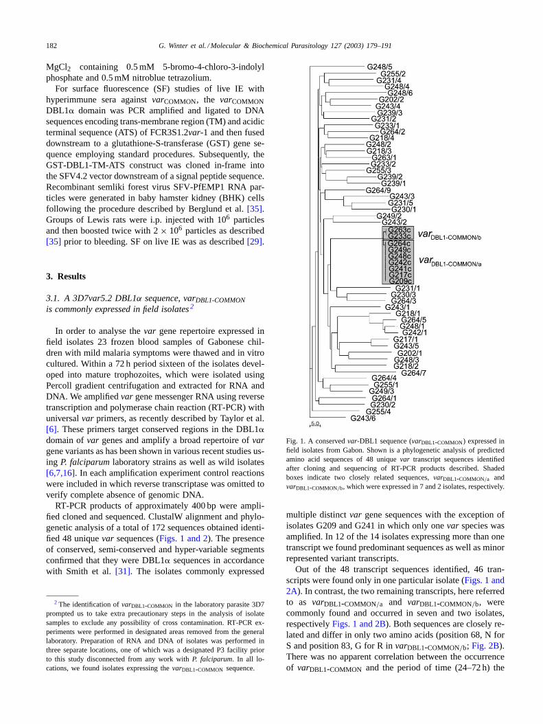
RT-PCR products of approximately 400 bp were ampli-fied cloned and sequenced. ClustalW alignment and phylo-genetic analysis of a total of 172 sequences obtained identi-fied 48 uniquevar sequences (Figs. 1 and 2). The presenceof conserved, semi-conserved and hyper-variable segmentsconfirmed that they were DBL1� sequences in accordancewith Smith et al.[31]. The isolates commonly expressed
2 The identification ofvarDBL1-COMMON in the laboratory parasite 3D7prompted us to take extra precautionary steps in the analysis of isolatesamples to exclude any possibility of cross contamination. RT-PCR ex-periments were performed in designated areas removed from the generallaboratory. Preparation of RNA and DNA of isolates was performed inthree separate locations, one of which was a designated P3 facility priorto this study disconnected from any work withP. falciparum. In all lo-cations, we found isolates expressing thevarDBL1-COMMON sequence.
Fig. 1. A conservedvar-DBL1 sequence (varDBL1-COMMON) expressed infield isolates from Gabon. Shown is a phylogenetic analysis of predictedamino acid sequences of 48 uniquevar transcript sequences identifiedafter cloning and sequencing of RT-PCR products described. Shadedboxes indicate two closely related sequences,varDBL1-COMMON/a andvarDBL1-COMMON/b, which were expressed in 7 and 2 isolates, respectively.
multiple distinctvar gene sequences with the exception ofisolates G209 and G241 in which only onevar species wasamplified. In 12 of the 14 isolates expressing more than onetranscript we found predominant sequences as well as minorrepresented variant transcripts.
Out of the 48 transcript sequences identified, 46 tran-scripts were found only in one particular isolate (Figs. 1 and2A). In contrast, the two remaining transcripts, here referredto as varDBL1-COMMON/a and varDBL1-COMMON/b, werecommonly found and occurred in seven and two isolates,respectivelyFigs. 1 and 2B). Both sequences are closely re-lated and differ in only two amino acids (position 68, N forS and position 83, G for R invarDBL1-COMMON/b; Fig. 2B).There was no apparent correlation between the occurrenceof varDBL1-COMMON and the period of time (24–72 h) the

G.
Win
ter
et
al./M
ole
cula
r&
Bio
che
mica
lP
ara
sitolog
y1
27
(20
03
)1
79
–1
91
183
Fig. 2. Predicted amino acid sequences ofvar transcripts identified in field isolates. RT-PCR products described inFig. 1 were cloned and 48var transcript sequences identified. (A) Alignment of 46sequences, which were unique to each isolate. Conserved and semi-conserved regions are indicated by shaded background. Shown to the left is the frequency of each sequence as a percentage of the totalnumber of sequence reads per isolate. (B) Alignment of two closely related DBL1-sequences,varDBL1-COMMON/a and varDBL1-COMMON/b, which were expressed in seven and two isolates, respectively.The sequences differ in two positions as indicated.

184 G. Winter et al. / Molecular & Biochemical Parasitology 127 (2003) 179–191
Fig. 3. Occurrence ofvarDBL1-COMMON transcript in isolates. Bars represent the frequency ofvarDBL1-COMMON sequence reads as a percentage of thetotal number of sequence reads per isolate. The actual numbers ofvarDBL1-COMMON sequences to the total number of sequence reads per isolate aregiven at the top of each bar.
patient samples were in culture prior to the harvest oftrophozoites. In isolates that expressedvarDBL1-COMMONthis variant was found as a dominantly amplified tran-script (Fig. 3) judged by the percentage of sequence clonescontaining thevarDBL1-COMMON sequence. The relativelyunbiased amplification ofvar genes by the PCR primersused has been shown previously[6,7]. Likewise, in PCRexperiments with genomic DNA of avarDBL1-COMMONcontaining parasite (3D7S8, see below) we did not observeany biased amplification of thevarDBL1-COMMON sequence(not shown). Thus, the amplification ofvarDBL1-COMMONtranscript was unlikely a result of contaminating ring stageparasites known to express multiple transcripts. In someisolatesvarDBL1-COMMON appeared to be exclusively ex-pressed as it was found in ten out of ten sequenced recom-binant plasmids (isolate G241 and G209;Fig. 3). However,with isolates G248 and G263 we initially obtained a similarexclusive amplification of thevarDBL1-COMMON sequence.Repeated RT-PCR experiments then amplified other minorvar mRNA suggesting that RT-PCR amplification may insome instances overstate the dominance of a particularvartranscript due to either general primer bias or statisticalevents in early cycles of the amplification.
Fig. 4. varDBL1-COMMON in genomic DNA of clinical isolates. ThevarDBL-COMMON sequence was PCR amplified from genomic DNA of clinical isolateswith specific primers vdc-1 and vdc-2 and separated by 2% agarose gel electrophoresis as outlined inSection 2. Arrows at the top indicate isolates thatcontained also detectable amounts ofvarDBL1-COMMON transcript. The arrow to the left indicates specific PCR-product of 264 bp.
BLAST analysis of the International Malaria GenomeDatabase, which is based on the 3D7 parasite, identifieda varDBL1-COMMON containingvar gene, here designatedvarCOMMON located at the telomeric end of chromosome5 (var5.2). The open reading frame encodes 3165 aminoacids and contains a semi-conserved head-structure (NTS-,a DBL1� and a CIDR1� domain) followed by a DBL2�/C2domain and further five DBL domains (DBL3�, DBL4ε,DBL5�, DBL6�, DBL7ε). varCOMMON is a pseudo-vargene truncated in the DBL7ε domain and lacking a putativetrans-membrane domain, an intron and the exon2 domain.
3.2. Presence of varDBL1-COMMON in genomicDNA of field isolates
We investigated the presence ofvarDBL1-COMMON ingenomic DNA of the isolates using PCR amplificationwith specific primers vdc-1 and vdc-2, mapping thevarDBL1-COMMON sequence. As expected a specific prod-uct of 264 bp was amplified in all nine isolates that tran-scribed varDBL1-COMMON (Fig. 4). In contrast, gel anal-ysis of repeated PCR attempts with isolates in whichvarDBL1-COMMON transcript was not detected, either no

G. Winter et al. / Molecular & Biochemical Parasitology 127 (2003) 179–191 185
Fig. 5. PCR-amplification of a 1.9 kbvarCOMMON sequence in clinical isolates. Genomic DNA of isolates G242, G249, G241 and the laboratory parasite3D7S8 were used in PCR experiments withvarDBL1-COMMON specific downstream primer vdc-3 and degenerate primer DBL�-2 directed against theDBL� domain ofvar genes as described inSection 2. (A) PCR products separated by agarose gel electrophoresis and stained with ethidium-bromide. (B)Southern blot analysis of the same gel with a probe targeting a conserved DBL1� sequence invar genes (seeSection 2). The arrow indicates amplifiedvar products.
(G202, G218, G230, G239, G255) or comparably lowamount of PCR product (G231, G243) was obtained.This result suggests that in parasites, which contain avarDBL1-COMMON comprisingvar gene, it is constitutivelytranscribed. Low amounts of PCR-product obtained inisolates G231 and G243 may be the result of minor repre-sented parasite genotypes that contain thevarDBL1-COMMONsequence but of which the expressed transcript was notdetectable in the RT-PCR.
3.3. varCOMMON gene is conserved in field isolatesover at least 2.1 kb
In order to obtain sequence information downstream ofvarDBL1-COMMON we designed degenerate primer targetingconsensus sequences of thevar gene DBL� domains as out-lined by Smith et al.[31]. We conducted PCR experimentson genomic DNA obtained from three isolates (G241, G242and G249) as well as the parasite 3D7S8, a clone obtained bymicromanipulation from 3D7AH1 parasites. We used a spe-cific upstream primer vdc-3 matching a variable region in thevarDBL1-COMMON sequence and a degenerate downstreamprimer DBL�-2 targeting a semi-conserved region locatedin the DBL� domain. We obtained several reaction products,which were screened in Southern blots using a probe target-ing a conserved region in the DBL1� domain (Fig. 5). Anamplification product of approximately 1.9 kb hybridised inall of the tested isolates and a 3D7 derived clone 3D7S8 (seebelow). Sequence analysis of the 1.9 kb products revealed inisolates G242 and G249 a completely identical sequence tothevarCOMMON gene. Thus, both isolates expressvar genescontaining a highly conserved sequence of at least 2.1 kbafter including the entirevarCOMMON sequence (Fig. 6).
However, thevar-sequence amplified with G241 DNA didnot containvarDBL1-COMMON but a differentvar sequence,which revealed some homology in the hypervariable regionof the DBL1 domain withvarCOMMON. The absence of avarCOMMON related amplification product in G241 suggeststhat somevarDBL1-COMMON comprisingvar genes diverge
Fig. 6. Schematic outline of 3D7varCOMMON, the varCOMMON relatedgenes of field isolates G242 and G249, and FCR3varCSA. ThevarCOMMON
related genes expressed in G242 and G249 contain a 2.1 kb sequenceidentical in sequence to 3D7varCOMMON spanning the DBL1� and DBL2�domains (indicated by shaded background). The hypothetical structuralsimilarity to the 3D7varCOMMON downstream of DBL2� is indicatedby dashed domain symbols. A comparative sequence analysis of thepredicted amino acid (aa) sequences of 3D7varCOMMON and FCRvarCSA
was performed (ClustalW and Pustell matrix analysis). The exon1 ofboth genes encodes 62% identical amino acids with a 5′ prime regionof moderate homology (54% predicted aa identical) spanning DBL1 andDBL4 domains and a 3′ prime region of high homology (85% predicted aaidentical) spanning DBL5 and DBL7 (indicated by background gradient).

186 G. Winter et al. / Molecular & Biochemical Parasitology 127 (2003) 179–191
downstream of the conserved DBL1� domain. Alternatively,in some isolates the truncation locus of thevarCOMMON re-lated gene may be upstream of the DBL2� domain and weretherefore not amplified.
3.4. varCOMMON is transcribed in a placenta-bindingclone (3D7S8)
The varCOMMON gene is structurally similar to recentlyidentified PfEMP1 variants expressed in placenta-bindingparasites[21,32,36] and homologous to the PfEMP1 ex-pressed in the CSA binding FCR3CSA strain[23,37].Until recently, varCOMMON related genes were also foundtranscribed in placental isolates suggesting a role of thesegenes in the binding of parasites to the placenta tissue[22,23].
We conducted experiments to investigate a placenta-bind-ing capacity ofvarCOMMON expressing parasites. In theprogeny of a gametocyte producing, 3D7AH1 derivedclone 3D7S8 we identified thevarCOMMON sequence asa predominant transcript in mature stage trophozoites.The varDBL1-COMMON sequence was found in 42 out of44 sequence clones as a result of two separate RT-PCRexperiments suggesting that the presence ofvarCOMMONtranscript was not due to contaminating ring stageparasites.
In two separate experiments using sections of human pla-centa tissue we found massive adherence of 3D7S8 IE tothe syncytiotrophoblast surfaces and at syncytial bridges(157± 56 and 129± 16 parasites mm−2 tissue), which iscomparable to results obtained previously in our laboratorywith placenta-binding parasite TM284S2 (224± 88 mm−2)and parasite FCR3CSA (125±23 mm−2) [32]. Intriguingly,placenta-binding parasites do not recognize CD36 in favourof receptors present in the placenta tissue[32,38,39]. In ac-cordance with these data we found in standard assays withCD36 expressing CHO-cells[40] no significant binding of3D7S8 IE compared to an established CD36 binding cellclone FCR3S1.2 (data not shown).
Glycosaminoglycans (GAGs) e.g. chondroitin sulfateA (CSA), HA and nonimmune IgG have previously beenshown to mediate adhesion of other placental parasites[21,32,36]. We investigated a role of these receptor candi-dates in the binding of 3D7S8. Pre-incubation of the pla-centa with enzymes hyaluronidase or chondroitinase ABCdid not affect binding of 3D7S8 IE to the placenta. Likewise,no reduction of binding was observed when parasites werepre-incubated with CSA or HA (50, 100 and 500�g/ml). Incontrast, under the conditions used the adherence of GAGrecognising parasites FCR3CSA (CSA) and CS2 (HA) issignificantly reduced[21,32,39]. We also found no effecton binding of the IE with protein A or after pre-incubationof placenta with 20 mg ml−1 non-immune IgG (data notshown). Thus, 3D7S8 binding to placental-tissue appearsto be mediated by receptor(s) other than those previouslyidentified.
Fig. 7. 3D7S8varCOMMON is truncated lacking an intron and exon2 do-main. PCR and RT-PCR experiments with genomic DNA and RNA of3D7S8 were performed using a specific downstream primer vc/DBL7 andtwo upstream primers vc-rev1 and vc-rev2. Primer pair vc/DBL7+vc-rev1amplifies a 352 bp segment of thevarCOMMON open reading frame(ORF) directly preceding a putative truncation locus. Primer pairvc/DBL7+ vc-rev2 amplifies a 598 bp sequence stretching 223 bp down-stream of the ORF into the telomeric repeat unit. A third primer setvc/DBL7+ L5 amplifies a putative sequence spanning exon1 and exon2.The presence of 3D7S8varCOMMON transcript was verified by RT-PCRwith primers vdc-1 and vdc-2, which amplify a 264 bp sequence of3D7varDBL1-COMMON. Products were separated by agarose gel elec-trophoresis and stained with ethidium bromide. The positions of the primertarget sequences in the 3D7varCOMMON gene are schematically outlinedat the top of the figure. For further details, seeSection 2.
3.5. varCOMMON in clone 3D7S8 is a truncatedpseudo-var gene
varCOMMON in the 3D7 parasite does not encode atrans-membrane domain or the internal ATS sequence (seeabove) and thus is unlikely to be exposed on the parasite sur-face. To verify that the placental binding of the 3D7S8 wasnot mediated by a full lengthvarCOMMON gene as a result ofa genomic rearrangement we conducted a series of PCR andRT-PCR experiments with 3D7S8-derived template DNAor RNA. We used a specific upstream primer, vc/DBL7and either of two specific downstream primers, vc-rev1 andvc-rev2 to amplify segments of thevarCOMMON gene. Theprimer positions are schematically outlined inFig. 7. Primervc-rev1 targets a sequence in the open reading frame directlypreceding the truncation locus. Primer vc-rev2 matches atelomeric repeat sequence located 223 bp downstream of theopen reading frame. In a third set of experiments, we usedprimer vc/DBL7 and primer L5, previously shown to binda conserved sequence in the exon2 ofvar genes[19]. As isshown inFig. 7, PCR with downstream primers vc-rev1 andvc-rev2 on 3D7S8 genomic DNA amplified the expected

G. Winter et al. / Molecular & Biochemical Parasitology 127 (2003) 179–191 187
products of 352 and 598 bp, respectively. PCR with down-stream primer L5 resulted in a 1.2 kb product, which aftersequencing and GenBank Blast retrieved a 3D7 sequencereferred to as avar-like hypothetical protein encoding gene(GenBank accession no. AL031747) of which two copiesare located on chromosome 9 and the chromosome BLOB.These genes are characterised by an exon1–intron–exon2structure expected forvar genes. The exon1 exhibits somehomology to the 3′ prime end of thevarCOMMON gene andencodes an ATS domain in the exon2 domain resulting inthe unspecific amplification product and thus providing anintrinsic positive control. In contrast, no product was ob-tained containing thevarCOMMON sequence in accordancewith the absence of the exon2 in thevarCOMMON gene. Wesimilarly conducted RT-PCR experiments on 3D7S8 RNAisolated from mature trophozoites using the upstream primervc/DBL7 and either of the downstream primers vc-rev1,vc-rev2 or the primer L5. As a control for the presence of thevarDBL1-COMMON sequence we also included an experimentusing specific PCR primers vdc-1 and vdc-2, which targetsequences in thevarDBL1-COMMON region. In each of theamplification experiments we included controls for the ab-sence of genomic DNA in which reverse-transcriptase wasomitted.
Primer set vc/DBL7 and vc-rev1 amplified the predicted352 bp product located immediately prior to the truncationlocus in thevarCOMMON gene. No product was obtained us-ing primer set vc/DBL7 and vc-rev2. This was anticipatedas the vc-rev2 primer targets a sequence in the telomericrepeat structure downstream of the open reading. Likewise,no product was obtained with primer set vc/DBL7 and L5.Amplification with specific primers vdc-1 and vdc-2 target-ing thevarDBL1-COMMON sequence resulted in the expected264 bp product. No amplification occurred in experimentsomitting reverse transcriptase confirming the complete ab-sence of genomic DNA.
In summary, these results indicate that thevarCOMMONtranscript contains the entire predicted open reading frameas deduced from the genomic database but is truncated up-stream of the trans-membrane domain encoding region.
3.6. A PfEMP1 variant other than varCOMMON is exposedat the surface of 3D7S8 infected erythrocytes
In RT-PCR experiments with the universal primers wewere unable to amplify a dominantly expressedvar geneother than varCOMMON. However, whilst recognizing abroad range ofvar genes the universal primers used in thisstudy may only amplify a subset of all possiblevar genevariants. A recent study has indicated that in contrast toearlier reports[18,41] trophozoites can contain multiplevar transcripts[42]. In order to investigate that a PfEMP1variant is expressed on the surface of 3D7S8 infectedIE, we conducted surface labelling experiments using thebiotin-derivative NHS-LC-Biotin. As a size reference weused the clonal parasite FCR3S1.2 of which the surface
Fig. 8. PfEMP1 variants exposed at the surface of erythrocytes in-fected with FCR3S1.2 or 3D7S8 parasites. Erythrocytes bearing tropho-zoite stage 3D7S8 or FCR3S1.2 parasites were surface biotinylated, en-riched by magnetic cell sorting and sequentially extracted with 1% TritonX-100/PBS and 2% SDS/PBS as described inSection 2. SDS-extractscontaining Triton X-100 insoluble polypeptides were separated by 5%SDS–PAGE, transferred onto a nitrocellulose membrane and either probedwith Extravidin-alkaline phosphatase conjugate to detect surface proteinsor probed with a purified IgG fraction from pooled sera of malaria pa-tients in Malawi. All lanes shown were separated in the same gel. Arrowsindicate position of surface exposed PfEMP1 variants in FCR3S1.2 and3D7S8 parasites. Asterisks indicate position of alpha- and beta-spectrin.
exposed variantvar-1 has previously been extensively char-acterised[24]. Labelled cells were sequentially extractedwith 2% Triton X-100 and 1% SDS. Under these conditionssurface-labelled PfEMP1 is recovered in the SDS fraction[43]. Western blots with SDS-extracts of the FCR3S1.2parasite revealed a prominently labelled band in the sizerange of approximately 280 kDa, as was expected forFCR3S1.2var1 (Fig. 8). Western blots of the 3D7S8 revealedsimilarly a prominently labelled band of, however, lower ap-parent molecular weight than its counterpart in FCR3S1.2.This is notably smaller than the expected size of 368 kDapredicted for the translation product of thevarCOMMONgene.
As expected of surface-exposed PfEMP1 variants thesebands were also recognised by sera from malaria patientsin Malawi (Fig. 8), which contain reactive antibodiesagainst PfEMP1 variants from various sources[41]. Inter-estingly, the sera recognised further minor bands of slightlylower and higher molecular weight to the surface labelledPfEMP1, which likewise varied in size between the twoparasites. These proteins were not labelled by the biotinreagent and thus may represent internal modifications of thesurface exposed PfEMP1 variant or distinct polypeptides.These results suggest that 3D7S8 trophozoites contain atleast twovar transcripts and expose a PfEMP1 other thanvarCOMMON at the surface.

188 G. Winter et al. / Molecular & Biochemical Parasitology 127 (2003) 179–191
3.7. Antibodies to DBL1α varCOMMON react with IE ofFCR3CSA but not 3D7S8
To further confirm thatvarCOMMON is not present on thesurface of 3D7S8 IE we raised hyperimmune sera againstthe recombinant DBL1� domain ofvarCOMMON PfEMP1fused to GST. In four independent indirect immunfluo-rescence experiments with IE of FCR3CSA, we founda strong, punctuate surface-fluorescence (titer 1/2–1/20)suggesting cross reactivity of the sera with the surface ex-pressed PfEMP1. The reaction was not anticipated as thehomology of FCR3varCSA andvarCOMMON DBL1 domainsis relatively low (52%). By contrast, we did not obtain anyreaction at the IE surface of 3D7S8 or FCR3S1.2 or withcontrol-sera generated to GST (1/2–1/20; data not shown)thus confirming the absence ofvarCOMMON at the 3D7S8surface.
4. Discussion
The variation in antigenicity of the IE surface, the switchof PfEMP1 variants, provides a logical framework of howthe parasite evades immune clearance while retaining theprimary function of adhesion. Indeed, studies have shownpolymorphism of thevar gene repertoire in natural parasitepopulations in which the sequence variability was found tobe similar within and between isolates[6,7,15,16,44,45].In this context, our observation of a highly conservedvar-sequence expressed in field isolates and in the labora-tory isolate 3D7 is surprising.
In RT-PCR experimentsvarCOMMON was a predominantlyamplified var-transcript in the mature trophozoite stage ofclone 3D7S8. We analysed the expression ofvarCOMMON inthe 3D7S8 clone in detail in order to gain insight into howvarCOMMON related genes might be expressed in the field.The 3D7varCOMMON gene (var5.2) belongs to a class ofDBL� containingvar genes that were previously implicatedin placenta binding. It is located immediately upstream ofthe telomere repeat sequences on chromosome 5 and istruncated in the DBL7 domain lacking intron and exon2coding region. The observation that 3D7S8 parasites bindto placental tissue in comparable numbers to IE of for ex-ample FCR3CSA and containvarCOMMON as a dominantlyamplified transcript initially suggested a possible role ofvarCOMMON PfEMP1 in the binding to placenta. To investi-gate a possible genomic rearrangement in 3D7S8 parasites,which could have resulted into a full-lengthvarCOMMONgene we conducted PCR and RT-PCR experiments withspecific primers. Our data confirmed a truncation in theDBL7 domain. In the absence of a potential trans-membraneanchor sequence and the internal ATS-domain a putativevarCOMMON protein is unlikely to be exposed on the sur-face of IE. In agreement with this, we identified a dominantPfEMP1 on the surface of IE of 3D7S8, which is of anapparent molecular weight lower than that of the previ-
ously characterised FCR3S1.2var-1 (MW 259 kDa) andthus notably smaller than a putative translation product ofthe varCOMMON gene (368 kDa). Furthermore, sera raisedagainst the DBL1� domain of varCOMMON did not reactwith the IE of 3D7S8. Taken together, these data suggestthat 3D7S8 mature trophozoites transcribe at least twovargenes and expose a dominant, yet unidentified PfEMP1on the IE surface that is different tovarCOMMON. Thecomparably low molecular weight of the surface-expressedvar/PfEMP1 is noteworthy as all previously reportedplacenta-bindingvar genes belong to a group of largevar-types.
Numerous earlier studies onvar gene expression inP. falciparum have suggested that mature stages expressonly one variant whereby the selection from an existingvar gene pool is likely to be random at large. A recentstudy, however, has shown that trophozoites can containmultiple transcripts[42]. Our detailed analysis on PfEMP1expression in 3D7S8 is in agreement with this. The ex-pression ofvarCOMMON may not be subject to the sametight control mechanism as is found for othervar genesand may be explained with the absence of an intron andexon2 structure. Deitsch et al.[20] have previously shownthat the controlled expression of a singlevar-type in aparasite involves the silencing of othervar genes duringthe transformation from early to mature stage trophozoites.Control elements required to suppress the transcriptionof a particularvar gene are located in thevar intron do-main. The constitutive transcription ofvarCOMMON relatedgenes in field isolates suggests that these genes are likewisetruncated.
Several studies have reported the amplification ofvarCOMMON related gene transcripts in placental parasitesindicating a role of this gene family in placental malaria,as it is suggested for the structurally related FCR3varCSAgene family[22,23,46]. A recent, albeit preliminary studyreported thatvarCOMMON related transcript containing aDBL� domain and thevarDBL1-COMMON sequence wasexpressed in 4 out of 5 placental isolates from Malawi[23]. It is noteworthy that none of these samples expressedFCR3varCSA relatedvar, which, as the authors conclude,does not support a major role of FCR3varCSA gene familyin placental malaria. Consistent with this data is a very re-cent report in which specific primers were used to RT-PCRamplify FCR3varCSA RNA in placental malaria isolates;only 9% of the isolates (3 out of 33) amplified FCR3varCSAtranscript. Intriguingly, FCRvarCSA transcript was alsoamplified in 24% of samples obtained from non-pregnantindividuals. The authors postulated that in non-pregnantindividuals the level of expression may be lower than inpregnant individuals. In the same study, RT-PCR with de-generate primers against the DBL� domain identified 43%of the amplified products asvarCOMMON typevar genes. Bycontrast, the blood samples analysed in the present reportwere collected fromchildrenof different ages. Most impor-tantly, in all samples expressingvarCOMMON it was found

G. Winter et al. / Molecular & Biochemical Parasitology 127 (2003) 179–191 189
as a dominantly amplified transcript in Percoll-enrichedtrophozoite fractions. In amplification experiments with3D7S8 genomic DNA under conditions following essen-tially the RT-PCR protocol we did not observe any biastowards thevarDBL1-COMMON sequence (not shown). Thus,amplification of thevarDBL1-COMMON sequence was un-likely due to contaminating ring stage parasites. The findingof varCOMMON related var as a common transcript doestherefore not support a link betweenvarCOMMON typegenes and placental malaria. On the contrary, our studysuggests thatvarCOMMON typevar genes may be expressedin any field isolate of old and new world origin. Reports(including this one) ofvarDBL1-COMMON transcripts in iso-lates from geographical origins as diverse as Brazil, India,Gabon and Malawi are consistent with this interpretation.In addition our report and the study by Rowe et al.[23] aGeneBank BLAST withvarDBL1-COMMON retrieved severalsequence entries identical with eithervarDBL1-COMMON/aor varDBL1-COMMON/b. varDBL1-COMMON/a transcript wasrecently identified in malaria patients in Brazil (GenBankaccession no. AAL11664)[44]. varDBL1-COMMON/b is iden-tical in sequence to a partial cDNA sequence obtainedfrom isolate R35S5 Rourkela, India (Chattopadhya, unpub-lished; GenBank accession no. AAL09630). In addition,we have recently identifiedvarDBL1-COMMON in an isolatefrom a malaria infected child from Uganda (Johan Nor-mark, unpublished). The finding that all three subtypes,varCOMMON, FCR3varCSA, and CS2, are also expressedin non-placental isolates raises doubts about the role ofDBL� containingvar types in placental malaria ([22], thisreport).
varCOMMON and FCR3varCSA exhibit a striking structuralsimilarity and are highly homologous as to sequence in the 3′half of the exon1, which spans from DBL5 to DBL7, albeitbeing clearly distinct in sequence in the 5′ prime half span-ning DBL1–DBL4 [22,23]. Furthermore, FCRvarCSA andvarCOMMON are likewise located in the sub-telomeric regionof P. falciparumchromosomes suggesting a functional linkconnecting bothvar-types. It has to be pointed out, though,that in contrast tovarCOMMON the FCR3varCSA was reportedto contain a common intron and exon2 structural domainfound in othervar genes[21]. Most expressedvar genes arelocated in this chromosome area where ectopic recombina-tion events occur more frequently than in internal parts ofthe chromosomes[5,12]. The conservation of FCR3varCSAtype, which are highly similar or identical in the 5′ region,andvarCOMMON type genes would therefore argue for a sim-ilar underlying mechanism that withdraws these genes fromfrequent sequence exchange with othervars. Conservationof varCOMMON appears to be linked to a location adjacent toa disrupted telomere structure. This allows the assumptionthat FCR3varCSA related genes may similarly be adjunctor in close vicinity to an incomplete telomere complex. In-deed, in the FCR3 strain, breakage and subsequent healingof chromosome 10 in the FCR3 strain has previously beenreported[47,48].
The recombination ofvar genes requires the cluster for-mation of chromosomal ends in sexual and asexual stagesvia relatively conserved repeat sequences in the telomereregion [49]. Both truncated copies ofvarCOMMON end intelomere repeat repeats, which were presumably de novosynthesized after chromosome breakage. As a result, thetelomers of chromosomes 5 and 6 are lacking commontelomere-associated sequences (TASs) which are implicatedin the alignment of chromosomal ends[47,50]. TAS-bindingproteins may be relevant components to stabilise cluster for-mation promoting ectopic recombination events of telom-ere associatedvar genes. Consequently, the replacement ofTASs in chromosomes 5 and 6 byvarCOMMON copies wouldwithdraw these chromosomal ends from high-frequent re-combination events and in effect lead to the conservation ofthevarCOMMON genes. Likewise, this mechanism may applyto varCOMMON related genes in field isolates. Alternatively,as was pointed out by Rowe et al.[23], the conservation ofvarCOMMON related genes could be caused by the directionof the gene, which is towards the telomere while the morecommon orientation ofvar genes is towards the centromer.Consequently,varCOMMON may be unable to align with themajority of opposite-directedvar genes and may thereforebe excluded from frequent recombination events.
Previous studies on genomic DNA have shown that somevar genes from different isolates share very similar or iden-tical DBL1�-sequences[7,14,16,45]. The vastvar generepertoire ofP. falciparumpopulations is a result of frequentrecombination events rather than genetic drift[14,16,45].
As a consequence,var genes of different parasites appearto be assembled in a ‘mosaic-like’ fashion and often sharesmall sequence segments comprising both variable andsemi-conserved regions. Various studies have consistentlyestimated a similarity in the DBL1� domain within andbetween different isolates of approximately 50%. A recentstudy examined DBL1� sequences of isolates obtained inthe Asian–Pacific region[7]. Any two of these isolatesshared on average two identical or very similar DBL1�sequences. A similar result was obtained when the se-quences were compared against a globalvar gene database.However, the sequence similarity of relatively fewvargenes from different isolates does not compare with theprevalence ofvarCOMMON genes described in this report.varDBL1-COMMON transcripts were present in 56% of theexamined isolates (9 of 16) of which at least two of the re-spectivevar genes were completely conserved over at least2.1 kb. This degree of conservation allows the conclusionthat varCOMMON related genes are less frequently involvedin recombination events.
The function ofvarCOMMON related genes for the parasiteis at present unclear. It has to be noted that some isolatesdid not contain thevarDBL1-COMMON transcript suggestingthat varCOMMON type genes are not essential for parasitesurvival. The parasites that do not express a gene of thevarCOMMON type may, however, contain othervargenes withsimilar structural characteristics and functions.
190 G. Winter et al. / Molecular & Biochemical Parasitology 127 (2003) 179–191
Acknowledgements
This work was funded by grants from the European Union(QLRT-PL-1999-30109 and QLK2-1999-01293 (Euromal-vac I)), and the Swedish Research Council. Gerhard Winterwas supported by the Wenner-Grenska Foundation. KirstenFlick was recipient of a Deutsche Forschungsgemeinschaftstipend (grant Fl328/1-1).
References
[1] Wahlgren M, Fernandez V, Chen Q, Svärd S, Hagblom P. Waves ofmalarial var-iations. Cell 1999;96:603–6.
[2] Brown KN, Brown IN. Immunity to malaria: antigenic variation inchronic infections ofPlasmodium knowlesi. Nature 1965;208:1286–8.
[3] Hayward RE, Tiwari B, Piper KP, Baruch DI, Day KP. Virulenceand transmission success of the malarial parasitePlasmodiumfalciparum. Proc Natl Acad Sci USA 1999;96:4563–8.
[4] Baruch DI, Pasloske BL, Singh HB, et al. Cloning thePlasmodiumfalciparum gene encoding PfEMP1, a malarial variant antigen andadherence receptor on the surface of parasitized human erythrocytes.Cell 1995;82:77–87.
[5] Hernandez-Rivas R, Mattei D, Sterkers Y, Peterson DS, WellemsTE, Scherf A. Expressedvar genes are found inPlasmodiumfalciparum subtelomeric regions. Mol Cell Biol 1997;17:604–11.
[6] Taylor HM, Kyes SA, Harris D, Kriek N, Newbold CI. A studyof var gene transcription in vitro using universalvar gene primers.Mol Biochem Parasitol 2000;105:13–23.
[7] Fowler EV, Peters JM, Gatton ML, Chen N, Cheng Q. Geneticdiversity of the DBLalpha region inPlasmodium falciparumvar genes among Asia-Pacific isolates. Mol Biochem Parasitol2002;120:117–26.
[8] Chen Q, Barragan A, Fernandez V, et al. Identification ofPlasmod-ium falciparum erythrocyte membrane protein 1 (PfEMP1) as therosetting ligand of the malaria parasiteP. falciparum. J Exp Med1998;187:15–23.
[9] Gardner MJ, Hall N, Fung E. Genome sequence of the humanmalaria parasitePlasmodium falciparum. Nature 2002;419:498–511.
[10] Fischer K, Horrocks P, Preuss M, et al. Expression ofvar geneslocated within polymorphic subtelomeric domains ofPlasmodiumfalciparum chromosomes. Mol Cell Biol 1997;17:3679–86.
[11] Thompson JK, Rubio JP, Caruana S, Brockman A, Wickham ME,Cowman AF. The chromosomal organization of thePlasmodiumfalciparum var gene family is conserved. Mol Biochem Parasitol1997;87:49–60.
[12] Rubio JP, Thompson JK, Cowman AF. Thevar genes ofPlas-modium falciparumare located in the subtelomeric region of mostchromosomes. EMBO J 1996;15:4069–77.
[13] de Bruin D, Lanzer M, Ravetch JV. The polymorphic subtelom-eric regions of Plasmodium falciparumchromosomes containarrays of repetitive sequence elements. Proc Natl Acad Sci USA1994;91:619–23.
[14] Freitas-Junior LH, Bottius E, Pirrit LA, et al. Frequent ectopicrecombination of virulence factor genes in telomeric chromosomeclusters ofP. falciparum. Nature 2000;407:1018–22.
[15] Kyes S, Taylor H, Craig A, Marsh K, Newbold C. Genomic rep-resentation ofvar gene sequences inPlasmodium falciparumfieldisolates from different geographic regions. Mol Biochem Parasitol1997;87:235–8.
[16] Taylor HM, Kyes SA, Newbold CI.Var gene diversity inPlasmod-ium falciparumis generated by frequent recombination events. MolBiochem Parasitol 2000;110:391–7.
[17] Smith JD, Chitnis CE, Craig AG, et al. Switches in expressionof Plasmodium falciparum vargenes correlate with changes in
antigenic and cytoadherent phenotypes of infected erythrocytes. Cell1995;82:101–10.
[18] Scherf A, Hernandez-Rivas R, Buffet P, et al. Antigenic varia-tion in malaria: in situ switching, relaxed and mutually exclusivetranscription ofvar genes during intra-erythrocytic development inPlasmodium falciparum. EMBO J 1998;17:5418–26.
[19] Chen Q, Fernandez V, Sundström A, et al. Developmental selec-tion of var gene expression inPlasmodium falciparum. Nature1998;394:392–5.
[20] Deitsch KW, Calderwood MS, Wellems TE. Malaria: cooperativesilencing elements invar genes. Nature 2001;412:875–6.
[21] Buffet PA, Gamain B, Scheidig C, et al.Plasmodium falciparumdo-main mediating adhesion to chondroitin sulfate A: a receptor for hu-man placental infection. Proc Natl Acad Sci USA 1999;96:12743–8.
[22] Fried M, Duffy PE. Two DBLgamma subtypes are commonlyexpressed by placental isolates ofPlasmodium falciparum. MolBiochem Parasitol 2002;122:201–10.
[23] Rowe JA, Kyes SA, Rogerson SJ, Babiker HA, Raza A. Identifica-tion of a conservedPlasmodium falciparum vargene implicated inmalaria in pregnancy. J Infect Dis 2002;185:1207–11.
[24] Fernandez V, Treutiger CJ, Nash GB, Wahlgren M. Multiple adhe-sive phenotypes linked to rosetting and binding of erythrocytes inPlasmodium falciparummalaria. Infect Immun 1998;66:2969–75.
[25] Kun JF, Schmidt-Ott RJ, Lehman LG, et al. Merozoite surfaceantigen 1 and 2 genotypes and rosetting ofPlasmodium falciparumin severe and mild malaria in Lambarene, Gabon. Trans R Soc TropMed Hyg 1998;92:110–4.
[26] Scholander C, Carlson J, Kremsner PG, Wahlgren M. Extensiveimmunoglobulin binding ofPlasmodium falciparum-infected ery-throcytes in a group of children with moderate anemia. InfectImmun 1998;66:361–3.
[27] Sylla EH, Kun JF, Kremsner PG. Mosquito distribution and entomo-logical inoculation rates in three malaria-endemic areas in Gabon.Trans R Soc Trop Med Hyg 2000;94:652–6.
[28] Trager W, Jensen JB. Human malaria parasites in continuous culture.Science 1976;193:673–5.
[29] Schlichterle M, Wahlgren M, Perlmann H, Scherf A, editors.Methods in malaria research. 3rd ed.http://www.malaria.mr4.org/mr4pages/ProtocolBook/MethodsIn Malaria Research.PDF, 2000.
[30] Chomczynski P, Sacchi N. Single-step method of RNA isolation byacid guanidinium thiocyanate–phenol–chloroform extraction. AnalBiochem 1987;162:156–9.
[31] Smith JD, Subramanian G, Gamain B, Baruch DI, Miller LH.Classification of adhesive domains in thePlasmodium falciparumerythrocyte membrane protein 1 family. Mol Biochem Parasitol2000;110:293–310.
[32] Flick K, Scholander C, Chen Q, et al. Role of nonimmune IgGbound to PfEMP1 in placental malaria. Science 2001;293:2098–100.
[33] Staalsoe T, Giha HA, Dodoo D, Theander TG, Hviid L. Detectionof antibodies to variant antigens onPlasmodium falciparum-infectederythrocytes by flow cytometry. Cytometry 1999;35:329–36.
[34] Taylor TE, Molyneux ME, Wirima JJ, Borgstein A, GoldringJD, Hommel M. Intravenous immunoglobulin in the treatment ofpaediatric cerebral malaria. Clin Exp Immunol 1992;90:357–62.
[35] Berglund P, Smerdou C, Fleeton MN, Tubulekas I, Liljestrom P.Enhancing immune responses using suicidal DNA vaccines. NatBiotechnol 1998;16:562–5.
[36] Reeder JC, Cowman AF, Davern KM, et al. The adhesion ofPlas-modium falciparum-infected erythrocytes to chondroitin sulfate Ais mediated byP. falciparumerythrocyte membrane protein 1. ProcNatl Acad Sci USA 1999;96:5198–202.
[37] Vazquez-Macias A, Martinez-Cruz P, Castaneda-Patlan MC, et al.A distinct 5′ flanking var gene region regulatesPlasmodium fal-ciparum variant erythrocyte surface antigen expression in placentalmalaria. Mol Microbiol 2002;45:155–67.
[38] Fried M, Duffy PE. Adherence ofPlasmodium falciparumto chon-droitin sulfate A in the human placenta. Science 1996;272:1502–4.

G. Winter et al. / Molecular & Biochemical Parasitology 127 (2003) 179–191 191
[39] Beeson JG, Rogerson SJ, Cooke BM, et al. Adhesion ofPlasmod-ium falciparum-infected erythrocytes to hyaluronic acid in placentalmalaria. Nat Med 2000;6:86–90.
[40] Heddini A, Pettersson F, Kai O, et al. Fresh isolates from chil-dren with severePlasmodium falciparummalaria bind to multiplereceptors. Infect Immun 2001;69:5849–56.
[41] Chen Q, Fernandez V, Sundstrom A, Schlichtherle M, Datta S,Hagblom P, Wahlgren M. Developmental selection ofvar geneexpression inPlasmodium falciparum. Nature 1998;394:392–5.
[42] Duffy MF, Brown GV, Basuki W, et al. Transcription of multiplevar genes by individual, trophozoite-stagePlasmodium falciparumcells expressing a chondroitin sulphate A binding phenotype. MolMicrobiol 2002;43:1285–93.
[43] Fernandez V, Hommel M, Chen Q, Hagblom P, Wahlgren M.Small, clonally variant antigens expressed on the surface of thePlasmodium falciparum-infected erythrocyte are encoded be therifgene family and are the target of human immune responses. J ExpMed 1999;190:1393–403.
[44] Kirchgatter K, Mosbach R, del Portillo HA.Plasmodium falci-parum: DBL-1 var sequence analysis in field isolates from centralBrazil. Exp Parasitol 2000;95:154–7.
[45] Ward CP, Clottey GT, Dorris M, Ji DD, Arnot DE. Analysis ofPlasmodium falciparumPfEMP-1/var genes suggests that recom-bination rearranges constrained sequences. Mol Biochem Parasitol1999;102:167–77.
[46] Salanti A, Jensen AT, Zornig HD, et al. A sub-family of commonand highly conservedPlasmodium falciparum vargenes. MolBiochem Parasitol 2002;122:111–5.
[47] Figueiredo LM, Freitas-Junior LH, Bottius E, Olivo-Marin JC,Scherf A. A central role forPlasmodium falciparumsubtelomericregions in spatial positioning and telomere length regulation. EMBOJ 2002;21:815–24.
[48] Scherf A, Petersen C, Carter R, et al. Characterization of aPlasmodium falciparummutant that has deleted the majority ofthe gametocyte-specific Pf11-1 locus. Mem Inst Oswaldo Cruz1992;87:91–4.
[49] Scherf A, Figueiredo LM, Freitas-Junior LH. Plasmodium telomeres:a pathogen’s perspective. Curr Opin Microbiol 2001;4:409–14.
[50] O’Donnell RA, Freitas-Junior LH, Preiser PR, et al. A geneticscreen for improved plasmid segregation reveals a role for Rep20in the interaction ofPlasmodium falciparumchromosomes. EMBOJ 2002;21:1231–9.
Copyright © 2022 FDOKUMEN