The Importance of Correlation between CBCT Analysis ... - MDPI
Upload
khangminh22Category
view
0download
0
Virginia Commonwealth University Virginia Commonwealth University
VCU Scholars Compass VCU Scholars Compass
Theses and Dissertations Graduate School
2021
COMPARING THE SENSITIVITY AND SPECIFICITY OF CBCT AND COMPARING THE SENSITIVITY AND SPECIFICITY OF CBCT AND
MRI IN DETECTING OSTEOARTHRITIS OF THE TMJ: A MRI IN DETECTING OSTEOARTHRITIS OF THE TMJ: A
SYSTEMATIC REVIEW SYSTEMATIC REVIEW
Gabriel R. Saavedra Virginia Commonwealth University
Follow this and additional works at: https://scholarscompass.vcu.edu/etd
Part of the Dentistry Commons, and the Life Sciences Commons
© Gabriel R Saavedra
Downloaded from Downloaded from https://scholarscompass.vcu.edu/etd/6867
This Thesis is brought to you for free and open access by the Graduate School at VCU Scholars Compass. It has been accepted for inclusion in Theses and Dissertations by an authorized administrator of VCU Scholars Compass. For more information, please contact [email protected].

© Gabriel R. Saavedra 2021 All Rights Reserved

ii
COMPARING THE SENSITIVITY AND SPECIFICITY OF CBCT AND MRI IN
DETECTING OSTEOARTHRITIS OF THE TMJ: A SYSTEMATIC REVIEW
A thesis submitted in partial fulfillment of the requirements for the degree of Master of
Science at Virginia Commonwealth University.
by
Gabriel R. Saavedra
B.S., Biology, George Mason University, 2017
Director: Sonali A. Rathore, D.D.S., M.S.
Associate Professor, Oral Diagnostic Sciences
Virginia Commonwealth University
Richmond, Virginia
November, 2021

iii
Acknowledgements
First, I would like to thank Dr. Rathore for giving me the opportunity to conduct
research at the dental school at VCU. She has always been encouraging me to push myself
and has been committed to my growth and development. I am grateful for her guidance
and patience throughout the whole process.
I would also like to express thanks to my project members, Dr. Ather and Dr. Best.
Dr. Ather was a light for us whenever we encountered an obstacle in our path, and Dr.
Best was always willing to provide his expertise and insight. I also would like to give a
special thanks to Dr. Bencharit, who assisted us in the earlier stages of the project.
In addition, I would like to thank my committee members, Dr. McGinn and Dr.
Simpson for their advice and input on how to make this project a success.
I am also grateful to Dr. Jacobs, Dr. Laskin, and Ms. Brody, who provided their
assistance in guiding me through the overall research process.
Lastly, I would like to thank my family and friends for their support and
encouragement throughout my academic journey. I especially thank my parents, without
whom I would not be able to pursue my goals, and a special thanks to Ms. Yang for her
technical assistance.
iv
Table of Contents
List of Tables ...................................................................................................................... vi
List of Figures .................................................................................................................... vii
List of Abbreviations ........................................................................................................ viii
Abstract............................................................................................................................... ix
Introduction ........................................................................................................................ 1
Methods ............................................................................................................................... 6
Focused question ............................................................................................................. 6
Search strategy ................................................................................................................. 6
Management of references .............................................................................................. 7
Inclusion/exclusion criteria ............................................................................................. 7
Study selection ................................................................................................................. 8
Data extraction ................................................................................................................. 8
Data synthesis & statistical analysis ................................................................................ 8
Results ............................................................................................................................... 10
Study selection ............................................................................................................... 10
Study characteristics ...................................................................................................... 10
Results of individual studies ........................................................................................... 11
Quantitative analysis ..................................................................................................... 12
Discussion .......................................................................................................................... 14
Implications ................................................................................................................... 20
Limitations ..................................................................................................................... 20
Recommendations ......................................................................................................... 20

v
Implications for practice ................................................................................................ 21
Implications for research ............................................................................................... 21
Conclusion ...................................................................................................................... 22
Bibliography ...................................................................................................................... 30

vi
List of Tables
Table 1: Description of selected studies ............................................................................ 24
Table 2: Kaimal et al. results: Diagnostic accuracy of MRI compared to CT for DJD ..... 25
Table 3: Dias et al. results: Diagnostic accuracy of CBCT compared to RDC/TMD ........ 26
Table 4: Talaat et al. results: Diagnostic accuracy of CBCT compared to RDC/TMD ..... 27

vii
List of Figures
Figure 1: Flowchart of articles ........................................................................................... 23
Figure 2: Forest plot for sensitivity of CBCT diagnosing RDC/TMD ............................... 28
Figure 3: Forest plot for specificity of CBCT diagnosing RDC/TMD ............................... 29

viii
List of Abbreviations
TMJ
TMD
OA
DJD
CBCT
MRI
CT
RDC/TMD
DC/TMD
PICO
MeSH
Temporomandibular Joint
Temporomandibular Disorders
Osteoarthritis
Degenerative Joint Disease
Cone-beam Computed Tomography
Magnetic Resonance Imaging
Computed Tomography
Research Diagnostic Criteria for Temporomandibular Disorders
Diagnostic Criteria for Temporomandibular Disorders
Patient/Population, Intervention, Comparison, Outcome
Medical Subject Headings

ix
Abstract
COMPARING THE SENSITIVITY AND SPECIFICITY OF CBCT AND MRI IN
DETECTING OSTEOARTHRITIS OF THE TMJ: A SYSTEMATIC REVIEW
By Gabriel R. Saavedra, B.S.
A thesis submitted in partial fulfillment of the requirements for the degree of Master of
Science at Virginia Commonwealth University.
Virginia Commonwealth University, 2021
Director: Sonali A. Rathore, D.D.S., M.S.
Associate Professor, Oral Diagnostic Sciences
The aim of this systematic review was to compare the sensitivity and specificity of
cone-beam computed tomography (CBCT) and magnetic resonance imaging (MRI) in
detecting osteoarthritis (OA) of the temporomandibular joint (TMJ). This was done by
using computed tomography (CT) and the research diagnostic criteria for
temporomandibular disorders (RDC/TMD) as reference methods and using CBCT and
MRI as index methods. A specific search strategy was developed and applied to these
electronic databases: PubMed, Embase, DOSS, and Cochrane. The search results returned
802 articles, which were then narrowed down using the inclusion/exclusion criteria, to
four final articles that were included in this review. Two of these articles used CBCT as
x
their index method, and the other two used MRI. The sensitivity and specificity for CBCT
was calculated to be moderate. Regarding MRI, we were not able to retrieve the raw data
necessary so sensitivity and specificity were unable to be calculated. It was concluded that
while CBCT and MRI show promise in their use as a diagnostic tool in the diagnosis of OA
of the TMJ, more studies are needed to fully evaluate their validity.

1
Introduction
The temporomandibular joint (TMJ) connects the mandible to the temporal bone
of the skull and allows for the complex function of jaw movement. This is essential to a
high quality of life and allows for functions such as mastication, communication, and
yawning. Regarding joint movements, the TMJ allows for movements such as mandibular
elevation, depression, protrusion, retrusion, and lateral deviation. The muscles involved
that allow for these movements include the temporalis, masseter, and the lateral and
medial pterygoid muscles. The TMJ is innervated by the mandibular and facial nerves1.
Any problems affecting the TMJ can thus impact its functions negatively.
Temporomandibular disorders (TMD) are defined as problems that affect the TMJ, the
associated muscles, or any other hard or soft tissue components surrounding it2. Typical
forms of TMD include arthralgia, myalgia, myofascial pain, disc displacement disorders,
degenerative joint disease, subluxation, and headache attributed to TMD3. The
combination of these problems can often result in a broad variety of disorders with a
complex etiology, making it difficult to diagnose and treat properly. In order to help
patients get the best care possible, it is essential to improve diagnostic ability of TMD.
One of the most common forms of TMD is osteoarthritis (OA), also known as
degenerative joint disease (DJD). OA or DJD is characterized by the chronic degradation
of the hard and soft tissues around a joint. This leads to symptoms such as pain and joint
dysfunction4. OA is considered to be the most common joint disorder in the world, with
the most commonly affected joints being the hands, hips, and knees5. It also tends to affect
women more so than men, with the main cause believed to be hormonal factors6.
2
Regarding OA of the TMJ, it is estimated to affect about 15% of the world’s
population and can be caused by various factors such as age, genetics, and trauma7. While
its diagnosis may be more obvious in late-stage OA, its early stage is where the difficulty
lies8. Treatment can include oral appliance therapy, pharmacotherapy, physical therapy,
intraarticular injections, or surgery. Due to its broad factors of causation, shared
symptoms with other TMD’s, and its difficult diagnosis, it is critical that this topic is not
overlooked to prevent patients from being undiagnosed or misdiagnosed.
Current diagnostic methodologies involve looking at the patient history and a
physical exam7. In most instances, diagnostic imaging is used as an adjunct to confirming
a clinical diagnosis. A panoramic radiograph is often used first for initial screening
purposes. From there, a determination will be made to see if further imaging is required
to confirm the diagnosis. Clinicians generally also use the research diagnostic criteria for
temporomandibular disorders (RDC/TMD); a set of criteria created in 1992 to
standardize processes for the diagnosis of TMD and assist clinicians in their diagnosis3.
Various imaging modalities are used in the diagnosis of osteoarthritis. These range
from the traditional modalities like panoramic imaging and computed tomography (CT),
to newer modalities like magnetic resonance imaging (MRI) and cone-beam computed
tomography (CBCT). A panoramic radiograph provides an overview of the dentition, jaws,
and TMJs in one 2-D image. It is best used for general observation of structures, as its
diagnostic value is limited due to superimposition of overlying bony structures and
variable obliquity of the condyle9. Thus, it is often not enough to fully discern if OA is
present10. However, it is readily available in most dental offices and is inexpensive.
Newer methods of diagnosis include using CBCT and MRI. In an effort to improve
the diagnosis of OA of the TMJ and to prevent more misdiagnosed or undiagnosed

3
patients, these are two imaging modalities that should not be overlooked. However, while
there are studies that discuss their potential benefits, there are not many studies that
provide definitive evidence as to which is the best modality in the detection of OA of the
TMJ.
CBCT is an imaging technology that emits a cone-shaped source of ionizing
radiation to produce multiple images, which are then stacked and reconstructed to obtain
a 3D composite image11. This allows it to overcome many of the original limitations of the
standard radiograph such as tissue overlapping and superimposition12. It can be used in
various applications, including oral and maxillofacial surgery, endodontics, implant
dentistry, orthodontics, periodontics, forensic dentistry, and TMJ imaging13. CBCT
benefits in exceling in hard tissue visualization such as skeletal and dental tissues and is
also cost-effective. However, CBCT uses radiation and artifacts involving image distortion
can be an issue in images14. Compared to CT, CBCT machines are much smaller and
affordable, allowing it to gain traction in the dental field around the late 1990’s11.
The application of MRI in dentistry has significantly increased in the recent years.
MRI is an imaging technology that functions by generating a local magnetic field that
aligns hydrogen nuclei to that field, then using radio frequencies to cause the nuclei to
resonate. The movement of the particles back to their original state allows a detailed
image to be produced15. MRI benefits in exceling in soft tissue visualization such as
masticatory muscles, ligaments, and the cartilaginous disc of the TMJ, as well as not
involving any radiation14. However, it is expensive and may be problematic for patients
that have any metal-based implants or have claustrophobia. MRI machines are not
currently found in dental offices due to their size and costs, making it not as readily

4
available as CBCT machines. However, referring patients to an imaging center that has an
MRI machine is an option if needed.
The typical imaging features of OA of the TMJ include articular surface flattening,
sclerosis, cortical thickening and irregularity, osteophytes, subcortical lucencies (areas of
low density), and ossified intra-articular bodies9. Therefore, the ability of an imaging
modality to detect these features is crucial to have an accurate diagnosis of OA. The ideal
choice would be an imaging modality that is able to detect the most features while having
the highest accuracy.
There are also non-imaging methods to diagnose OA of the TMJ, such as
arthroscopy. Arthroscopy is a minimally-invasive procedure that involves making a small
incision at the joint and viewing it directly or through a camera for diagnosis. However,
since newer non-invasive methods have been able to successfully manage OA of the TMJ,
it is not seen as commonly today8.
When comparing any two imaging modalities, a reference standard is needed to
compare them. A reference method is important when comparing two different variables,
as you need a standard or “true” value to have a baseline to compare to. We used
computed tomography (CT) as one of our reference standards, as it is currently
considered to be the gold standard for the diagnosis of DJD of the TMJ16. CT functions
similarly to CBCT, involving multiple x-ray images being taken at different angles of a
subject. However, it uses fan-beam x-rays, capturing multiple slices of the subject,
resulting in a longer image acquisition process and thus higher radiation exposure17.
The RDC/TMD criteria was also used as a reference method for the study, as it is a
widely used diagnostic system used to assist in the classification of TMD, and has been
demonstrated to be reliable. There is also a newer evidence-based version, termed the

5
diagnostic criteria for temporomandibular disorders (DC/TMD), which was published in
2015. It is comprised of 2 assessment components: Axis I involves pain and joint
assessment, while Axis II involves distress and pain disability3. Using these criteria has
been recommended for use in clinical and research settings. The goal of DC/TMD is to
standardize diagnostic criteria for classifying subtypes of TMD.
Sensitivity and specificity are the two main variables this study will focus on
regarding the imaging modalities. Sensitivity is a measure of how often a test generates a
positive result in those who actually have the condition, while specificity is a measure of
how often a test generates a negative result in those who do not actually have the
condition. In this study, we will use sensitivity and specificity to compare the two imaging
modalities.
The aim of this study is to compare the sensitivity and specificity of CBCT and MRI
in detecting osteoarthritis of the TMJ.

6
Methods
A systematic review is a type of review that follows a series of methodical steps to
evaluate relevant current literature to answer a question or issue18. Its purpose is to
summarize its findings to make evidence more accessible to other researchers and
determine the current literature landscape of the topic. Our systematic review used a two-
phase selection process following the Preferred Reporting Items for Systematic Reviews
and Meta-Analyses (PRISMA) guidelines. PRISMA is a guideline originally created in
2009 to improve the reporting of systematic reviews. It involves a checklist of items
recommended to report in systematic reviews19.
Focused question
Compare the sensitivity and specificity of CBCT and MRI in the diagnosis of OA of
the TMJ.
Search strategy
First, we identified our research question using the PICO framework. PICO is a
mnemonic device for population/problem, intervention/exposure, comparison, and
outcome20. A PICO table was created, which involves a table that has the PICO criteria on
one axis and our concepts and keywords for our question on the other. After working
through the table, we identified our key concepts and were able to formulate a proper
question for our search. We then developed our search strategy specifically for each
database used in our search: PubMed, Embase, DOSS, and Cochrane. We did this by
combining keywords, MeSH terms, and the appropriate search modifiers. Once the search
query was finalized, we performed the search on all the databases and the results were

7
compiled. A manual search of the reference lists of identified relevant articles was also
performed, but no additional articles were found. A grey literature (literature published
outside of traditional scientific journals) and hand search was not performed. The final
electronic database search was conducted on December 01, 2020.
Management of references
References were managed through Mendeley, a reference manager software.
Mendeley was used to document the articles that would be included in the paper. Any
duplicate results were removed.
Inclusion/exclusion criteria
We made an inclusion/exclusion criteria to determine which articles would be
most relevant to our study. The articles selected for this review contained and evaluated:
(1) Osteoarthritis of the TMJ; (2) Involved the use of CBCT or MRI as the index method;
(3) Used CT or RDC/TMD as a reference method; (4) Was an original in vivo clinical
study; (5) Was conducted on live human participants; (6) Had a minimum of 10
participants; (7) Involved participants who are 18 and older; (8) Was published after the
year 2000; (9) Was in English. The imaging features of the TMJ were also taken note of
if available, such as condylar flattening, condylar erosion, osteophyte formation, joint
effusion, sub-chondral cysts, and sclerosis of the condyle. These parameters were selected
after we discussed and determined what factors would be important to our study.
We did not select articles for this review that only included articles that discussed
only participants with trauma, infection, or a tumor of the TMJ, as well as any
publications that were review papers, case reports, part of a conference summary, or
posters.

8
Study selection
All articles were screened through a 2-stage process involving 2 independent
reviewers (GRS and SAR). In the first stage of the screening, the title and abstract of each
article were examined and evaluated against the inclusion/exclusion criteria. Articles that
did not meet the criteria were not included in the study. After the initial title abstract
review, a third independent reviewer (SB) was called in as a tiebreaker if there were any
disagreement between the two reviewers. The same two reviewers participated in stage
two of the screening process. The selected articles from stage one were independently
reviewed and the full text was assessed. Final selection was based on determining if the
articles met the criteria. The majority of the articles that did not pass the second stage of
screen typically did not involve sensitivity and specificity of the compared imaging
modalities, or did not use CT or RDC/TMD as the reference method.
Data extraction
The relevant information was collected from the included articles. This included
the following data: study characteristics (authors, year of publication, study design),
population characteristics (sample size, age of participants, sex) index method (CBCT,
MRI), reference method (CT, RDC/TMD), and outcome characteristics. In the case of a
couple articles, the required data was not able to be retrieved despite attempts in
contacting the original authors.
Data synthesis & statistical analysis
Data were extracted and placed into Excel spreadsheets, where the appropriate
summary statistics were calculated. The data were summarized with counts, percentages,
9
or means and standard deviations, as appropriate. All analyses were performed by Excel
or by SAS software (version 9.4, SAS Institute Inc., Cary NC). Meta-analysis was
performed using the MetaDAS macro in SAS software21. Meta-analysis, in this case,
combines the estimates of sensitivity and specificity across studies using a random-effects
model to yield a single overall estimate.

10
Results
Study selection
The final database search yielded a total of 802 results after duplicates were
removed. After the initial review of the title and abstracts, 780 articles were excluded,
leaving 22 potential articles remaining. A full-text evaluation was then performed, which
resulted in 18 more articles that were excluded. The references of these articles were also
considered, but none met the required criteria. The remaining 4 articles were the articles
that were included in this review. A flowchart summarizing the selection process is seen
in Figure 1.
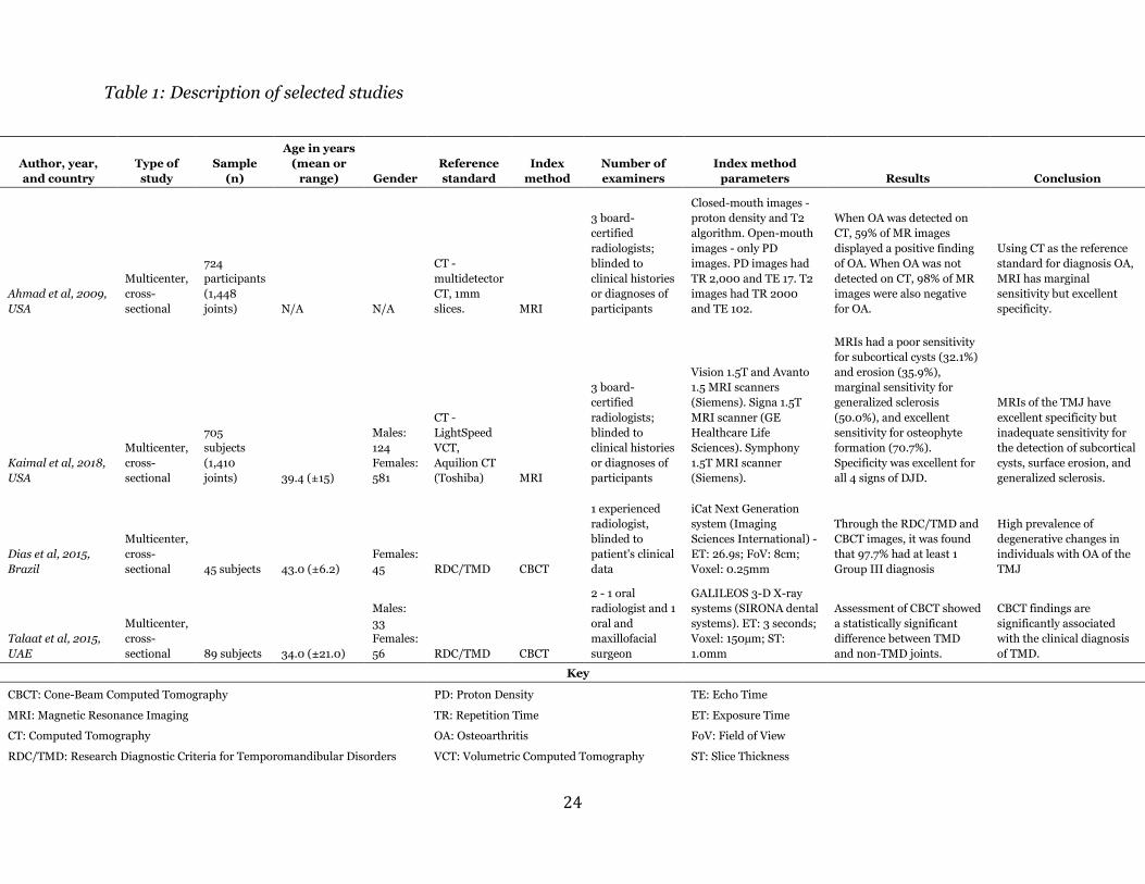
Study characteristics
The total sample size of all the studies was 1,563 subjects. The publishing range of
the selected studies was between 2009 and 2018, with 2 of them being from the USA, 1
from Brazil, and 1 from the UAE. 2 studies used CT as their reference method, while the
other 2 used RDC/TMD. CBCT was used in 2 studies as their index method, while MRI
was used in 2 studies. Sample sizes ranged from 45 to 724. Due to one of the studies not
providing demographic information, 839 participants were considered. From that
population, 81% were females. From the original sample size, we also only considered
patients that were classified with OA or osteoarthrosis of the TMJ. Control groups were
included as well. The study characteristics of the selected articles are summarized in Table
1.

11
Results of individual studies
All of the selected studies used either CBCT or MRI to confirm a diagnostic test,
using CT or RDC/TMD as the reference method. Some articles also discussed other results
that were not relevant to our study, but we will focus on only the relevant parts in this
study.
Ahmad et al.22 aimed to develop comprehensive image analysis criteria for the
RDC/TMD validation project. 724 study participants (1435 joints) were enrolled and
assessed with CT and MR imaging. When referenced to CT imaging, it was concluded that
MRI had poor to marginal sensitivity (59.4%, 95%CI=53.7 to 64.9%), but excellent
specificity (98.0, 95%CI=97.0 to 98.8) in diagnosing OA of the TMJ.
Kaimal et al.16 aimed to determine the diagnostic accuracy of MRI for detecting
DJD of TMJ, using CT as a reference method. 705 subjects (1410 joints) were evaluated
by CT and MR imaging. Imaging criteria was established that included subcortical cysts,
erosion, osteophytes, and sclerosis. When compared against their target values for
sensitivity and specificity, it was concluded that MRIs had below-target sensitivity but
above-target specificity in detecting all the reference CT imaging criteria. Their results are
summarized in Table 2. In this case, sensitivity is a measure of how often a MRI generates
a positive sign of those with DJD in those who actually have the condition, while
specificity is a measure of how often an MRI generates a negative sign of DJD in those
who do not actually have the condition. Sensitivity ranged from 32% to 71%, depending
upon the sign and specificity was at least 98% across all signs.
Dias et al.23 aimed to evaluate the presence of degenerative bone changes of the
TMJ in individuals with sleep bruxism (teeth grinding). 45 subjects were evaluated using

12
CBCT and RDC/TMD as the reference method (see Table 3). 19 subjects were classified
with OA and 18 with osteoarthrosis in at least 1 of the joints. In the 19 subjects identified
with OA by the reference method (RDC/TMD) only 10 were positive on the CBCT image,
yielding a sensitivity of 53% (CI = 30 to 75%). In the 28 subjects without OA, 26 were
negative on CBCT, yielding a specificity of 93% (CI = 83% to 100%). Dias et al. also
observed that there was a high prevalence of degenerative changes with individuals who
had OA of the TMJ.
Talaat et al.24 aimed to compare bony changes of TMD using CBCT, using
RDC/TMD as the reference method (see Table 4). 89 subjects were enrolled in the study
and assessed using CBCT and classified based on their RDC/TMD diagnosis. 20 subjects
were classified with OA according to RDC/TMD. In the 40 joints identified with OA by
the reference method (RDC/TMD) 36 were positive on the CBCT image, yielding a
sensitivity of 90% (CI = 81 to 99%). In the 86 joints in subjects without OA, 55 were
negative on CBCT, yielding a specificity of 64% (CI = 54% to 74%). Talaat et al. concluded
that assessment by CBCT showed a statistically significant difference between non-TMD
and TMD joints. It was concluded that CBCT findings are significantly associated with the
RDC/TMD clinical diagnosis of TMD.
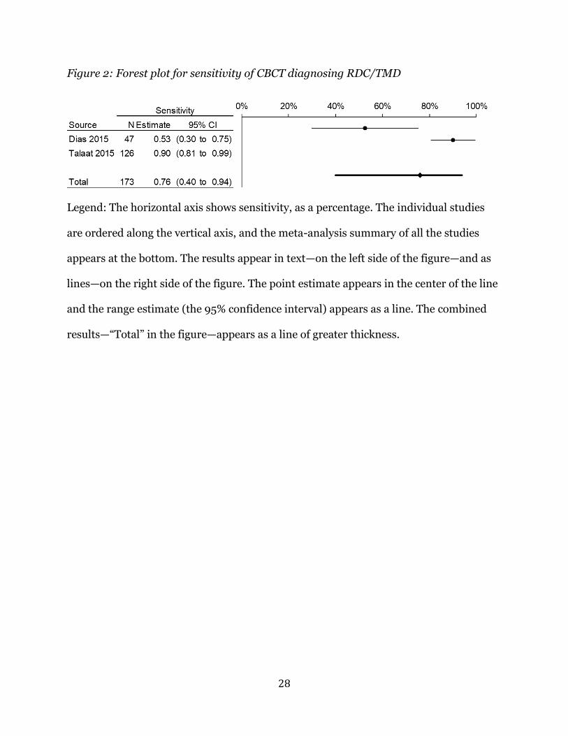
Quantitative analysis
The results from the two CBCT studies were combined using meta-analysis
software. The result is summarized in forest plots—a method for displaying the results of
several papers into one image. The combined sensitivity across the Dias et al. and Talaat
et al. studies was 76% (95% CI = 40% to 94%, Figure 2) and specificity was 84% (CI = 52%
to 96%, Figure 3). In the forest plots the horizontal axis is the estimate of interest, here

13
either the sensitivity or the specificity. The individual studies are ordered along the
vertical axis, and the meta-analysis summary of all the studies appears at the bottom. The
results appear in text—on the left side of the figure—and as lines—on the right side of the
figure. The point estimate appears in the center of the line and the range estimate (the
95% confidence interval) appears as a line. The combined results—“Total” in the figure—
appears as a line of greater thickness.
For example, the line for the combined estimate of sensitivity extends from a lower
bound of 40% to an upper bound of 94% and is centered on the combined estimate of
76%. Informally, this combined estimate is formed by the meta-analysis “averaging” of
the two study’s individual values—53% and 90% in this case. Note that these disparate
study findings result in a relatively wide confidence interval on the combined (total)
estimate.

14
Discussion
There is a growing trend in the field of dentistry on the use of CBCT and MRI for
diagnostic purposes; however, there is no consensus on the use of CBCT or MRI as
diagnostic tools for TMJ DJD. The present systematic review attempted at analyzing all
the in vivo studies conducted in the literature to assess evidence for the sensitivity and
specificity of CBCT and MRI imaging in the detection of OA of TMJ. Both the above-
mentioned imaging modalities were compared to reference diagnostic methods such as
CT and RDC/TMD criteria, which have been used as a standard in previous studies. We
found 4 articles that met the required criteria and were selected to be included in this
study.
CBCT has found use in various fields of dentistry, such as maxillofacial, sinonasal,
and TMJ bone imaging, with its most widespread application being in diagnostic
imaging25. It is also used in dental implant applications, being useful for presurgical
diagnosis and planning26. MRI has found use in the dental field mostly involving imaging
involving the TMJ, soft tissues, tumors, salivary glands, and maxillary sinuses27.
Previous studies discussed the various benefits of CBCT and MRI gives over
conventional imaging techniques in TMD diagnosis, but evidence supporting these claims
have been inconclusive. Ahmad et al.22 discusses that while CBCT has clear benefits and
may surpass CT, further studies are needed to fully determine its efficacy. Kaimal et al.16
also discusses how MRI has promising applications, but ultimately still needs CT to
confirm diagnoses of DJD. It is evident that there is varying evidence and opinions on the
usefulness of CBCT and MRI in TMD diagnosis, but this demonstrates that more studies
are needed before making a conclusive argument.

15
While diagnostic imaging does have its benefits, it should not be used just because
the technology is available. TMD imaging alone without any form of standardization in
interpretation can lead to varying results28. A cost-benefit analysis should be considered
first, as performing imaging analysis can be costly, involve taking more time if a patient
needs to be referred somewhere, and can involve radiation. These are factors that should
be considered before deciding to use diagnostic imaging. In addition to this, Petersson29
states there it is generally unclear when patients with TMD should undergo examination
with imaging methods. However, Talmaceanu et al.30 discusses that imaging techniques
are an essential step in the diagnosis of TMD due to its complex anatomy and pathology.
It is evident that there are conflicting opinions on whether radiographic imaging should
be a considered standard in diagnosis of OA of the TMJ.
Although CBCT and MRI have been emerging with great potential in the dental
field, there are still some constraints to surpass until they can gain widespread use. MRI’s
biggest constraints involve the size of the machine, cost, and patients who may have
claustrophobia. It currently would not be feasible to have an MRI machine in every dental
office. Therefore, until it becomes more practical to be used in dental offices, its
exploration and applications will remain limited. On the other hand, CBCT has been
gaining some traction with its incorporation in dental offices. It is much smaller and can
be incorporated into a multimodal system that allows panoramic and CBCT imaging26,
therefore is much more feasible to be included in dental offices. However, it does expose
the patient to radiation and therefore should not be used needlessly.
CT is considered the gold standard for diagnosing OA of the TMJ16. For this reason,
it was used as a reference method. CT uses a narrow fan-shaped X-ray beam and multiple
exposures around an object to reveal its internal structures31. This allows the observer to

16
view morphology in 3-D as opposed to a conventional radiograph which is in 2-D. While
CT seems to be the most reliable method to diagnose OA of the TMJ, its main drawback
is cost and radiation, which is a sizable amount higher than a regular radiograph. While
CBCT radiation is lower than CT, its radiation can vary widely, from the equivalent of 2
to 200 panoramic radiographs26. Therefore, a plan should be set in place to find a suitable
replacement that is more cost-efficient and involves less radiation, yet still being a reliable
tool for diagnostic imaging. The RDC/TMD was also considered as a reference method in
this study due to its wide acceptance and long-standing use as a tool in the diagnosis of
common forms of TMD. It is considered by some in the dental community to also be a
gold standard for its use as a validity diagnostic classification tool32. Having these two
widely accepted approaches as reference methods allows for a more valid comparison of
imaging modalities.
A diagnostic test such as diagnostic imaging should be used with a valid purpose
and have a reliable way to ensure that the disease or condition it is testing for is true. In
this case, diagnostic imaging is considered to help clinicians diagnose a disease,
specifically OA of the TMJ. With this in mind, several factors should be considered to
ensure a valid diagnosis. A gold standard being used as a reference method is essential.
As mentioned before, CT and RDC/TMD meets those criteria for our study. Validity can
be defined as the extent to which a test measures what it is supposed to measure; e.g.,
accuracy. Specifically, validity is measured by sensitivity and specificity33. Sensitivity can
be defined by the ability of a diagnostic test to determine if a diseased individual tests
positive. Specificity on the other hand is the ability of a diagnostic test to determine if an
non-diseased individual tests negative34. With our main goal to be to compare the
sensitivity and specificity of CBCT and MRI in diagnosing OA of the TMJ, these

17
parameters in essence determine the validity of each imaging modality and therefore were
considered in this study.
Kaimal et al.16 aimed to determine the diagnostic accuracy of panoramic
radiography and MRI in detection of signs of TMJ DJD, using CT as a reference standard.
DJD was defined as having at least one of the 4 signs: a subcortical cyst, surface erosion,
osteophyte formation, or generalized sclerosis. The sensitivity and specificity values for
MRIs, respectively, were found to be: subcortical cysts, 32% and 100%; erosion, 35% and
99%; osteophytes, 71% and 98%; sclerosis, 50% and 100% (Table 2). Using their target
values for sensitivity and specificity of ≥70% and ≥95%, MRIs had below-target specificity
and above-target sensitivity in all features except in osteophyte detection, where
sensitivity was adequate. It was recommended that CT still be used for diagnosis to avoid
false-negatives that may occur with MRI. This points towards that MRI is not quite ready
to replace CT as the gold standard, however, more research is necessary. Despite attempts
to contact the authors, raw data could not be obtained to perform data analysis.
Ahmad et al.22 aimed to develop comprehensive TMJ diagnostic criteria for image
analysis involving panoramic radiography and MRI, using CT as the reference standard.
In regards to OA diagnosis, reliability of MRI was fair, with positive percent agreement
(the percentage of patients with a positive test that actually have the disease33) being 59%.
MRIs had marginal sensitivity but excellent specificity. Image analysis criteria was able
to be developed for the assessment of OA using CT, but MRI was only considered for
evaluating disc position and effusion, in which it was good to excellent. This demonstrates
that more studies need to be conducted to determine the efficacy of MRI in diagnosing
OA of the TMJ. This is in agreement with Kaimal et al.16, which also did not have enough
evidence about the efficacy of MRI usage in this aspect. Different imaging machines were

18
used for each study, which may lead to a more varied result. Both studies involved 3
board-certified radiologist who reviewed images independently and blind to patient’s
history. One difference to note is that Ahmad et al.22 considered more osseous
components than Kaimal et al.16, including: hyperplasia, flattening of the articular
surface/eminence, subcortical sclerosis or cyst, surface erosion, osteophytes, generalized
sclerosis, loose joint bodies, and deviation in form. Condylar position and ankylosis, as
well as condylar edema, were also taken into account. This may account for some
heterogeneity between the two studies.
Dias et al.23 classified TMD of their participants using RDC/TMD and used CBCT
as their index method. Image analysis criteria was based on the criteria described by
Ahmad et al.22, specifically: planning, erosion, osteophytes, and sclerosis. Images were
evaluated by an experienced radiologist. The sensitivity and specificity of CBCT for
diagnosing OA of the TMJ according to these criteria was calculated to be 52.63% and
92.86%, respectively (Table 3). This illustrates that CBCT has excellent specificity but
adequate sensitivity in the diagnosis of OA of the TMJ.
Talaat et al.24 classified OA of the TMJ of their participants according to the
RDC/TMD for TMD’s Group IIb, IIc, and III. Diagnosis of TMD was also confirmed by
reviewing patient history and symptoms, a clinical examination, and radiographic
examination, including MRI imaging. The sensitivity and specificity were calculated to be
90% and 64%, respectively (Table 4). Interestingly, in contrast to Dias et al.23 results,
excellent sensitivity was observed, but with just adequate specificity. This heterogeneity
can possibly be explained due to the two studies having different imaging machines,
different image examining methods, or differences in participant positioning/presence of

19
artifacts. This could also be possible due to using Talaat et al.24 using MRI to confirm
findings, while Dias et al.23 did not use such as method.
While there is a general consensus agreeing upon CT and RDC/TMD as viable
reference standards for the diagnosis of OA of the TMJ, there are some who argue
otherwise. Many researchers agree that CT is the gold standard for OA of the TMJ
diagnosis, while others have varying opinions about the subject. Dias et al.23 states that
RDC/TMD is the gold standard for TMD diagnosis, however, Ahmad et al.22 argues
RDC/TMD for image applications is limited. Boeddinghaus et al.25 also states that MRI is
the reference standard for TMJ imaging and that CBCT is not a good substitute for MRI.
With all these varied statements from different articles, it is clear that there is no fully
unified consensus of what the gold standard is for diagnosis or what imaging modality is
best for diagnosis of OA of the TMJ. Diagnosis should be based on a clear and
methodological criterion that can be applied for any case. This information only
highlights the need for more information in this subject, with the main goal to be to
improve diagnostics to better help patients who may be suffering from OA of the TMJ.
Having varying methodologies and using different imaging modalities may lead to
incorrect diagnoses, which may lead to undiagnosed patients who may not be able to get
the help that they need. This is further seen in the clinical aspect as well, as many dentists
also feel ambiguous when it comes to diagnosing TMD, with only 25-50% of dentists that
feel positive about it32. Therefore, it is of utmost importance that more light is shed on
finding the most reliable method to diagnose OA of the TMJ properly.

20
Implications
CBCT demonstrated moderate pooled sensitivity and specificity for diagnosis of
OA of the TMJ, i.e., 76% and 84%, respectively. Variable CBCT data does not allow a clear
conclusion of its sensitivity and specificity to be drawn. However, more studies in this
area may be able to expand upon this data and allow for a more conclusive result.
Regarding the validity of MRI in the diagnosis of OA of the TMJ, as we were not
able to gather the information needed for MRI data analysis, we were unfortunately
unable to determine its sensitivity and specificity. However, past studies have shown
potential and further studies could potentially showcase further how it could benefit in
diagnosis of OA of the TMJ.
Limitations
It can be observed that the literature is generally lacking, specifically in comparing
sensitivity and specificity values for imaging modalities, and therefore, our data was
limited. The available literature was mostly heterogeneous, which may be due to various
factors such as machines used, number of examiners used, protocol followed, and
interobserver agreement differences. Having this heterogeneity may influence data
analysis negatively as having low consistency in data will lead to a result that is not
consistent with other studies.
Recommendations
Future studies are recommended to consider other reference methods than CT or
RDC/TMD, if another reliable method is available. Exploring other ways to measure the

21
validity of an imaging modality other than sensitivity and specificity may also be of
benefit.
Implications for practice
As the systematic review addressed a focused question on sensitivity and
specificity, combined with the fact that existing literature is scarce and significantly
heterogeneous, the results of our study should not be used to recommend one modality
over another. Further, well-designed and controlled studies are required on this
understudied topic, which has huge clinical implications, given the rise in in TMJ
disorders.
Implications for research
As mentioned previously, there is a tremendous need for more research
investigating the two modalities directly as well as with other reference standards for
diagnosing OA of TMJ. There are only select studies, which have investigated MRI for
diagnosing OA of TMJ. This might be attributed to the limited feasibility of using MRI in
dental settings as a result of size of equipment along with financial considerations.
However, given the trend towards minimally invasive diagnostics and avoidance of
harmful radiation from CT and CBCT, research on MRI as a TMJ diagnostic modality
holds great potential.
Amongst the studies included in this systematic review, there is a lack of
standardization in outcomes and outcome measures, which makes direct comparisons
difficult and external validity of the results questionable. This could be addressed in
future research by predefining and adopting a core outcome set for diagnosing OA of TMJ.

22
It is also important to mention the healthcare settings where the studies are being
performed as these can affect the applicability of the results and its translation into
clinical practice. E.g., Studies on more commonly available diagnostic modality CBCT
might be performed in a primary dental care setting whereas research on MRI might be
performed in a hospital-based setting, which does not have translational relevance from
a clinical practice point of view.
There are several other confounding moderators, which need to be taken in
account and should be reported in future research on TMJ disease. These include patient
factors (such as age, gender, dental history, medical history), operator factors (number of
investigators, experience and calibration), and technical factors (e.g. KvP, ma, FoV, and
voxel size for CBCT scan, viewing conditions for the images). Having these factors
reported in future studies will help other researchers and clinicians to understand the
outcomes better and will also improve the applicability and generalizability of the results.
Conclusion
Diagnosis of OA of the TMJ is often difficult and determining if CBCT or MRI are
beneficial in diagnosis may help alleviate the issue. CBCT sensitivity and specificity for
the diagnosis of OA of the TMJ was determined for our studies to be moderate for both
parameters. MRI sensitivity and specificity were not able to be pooled due to a lack of
access to raw data. It can be concluded that while CBCT and MRI show promise in being
used to diagnose OA of the TMJ, more well-designed studies are needed to substantiate
their validity.

23
Figure 1: Flowchart of articles
Note that in order to be eligible for this systematic review, an article needed to: evaluate
osteoarthritis in live adult human participants, have at least 10 participants, use either
CBCT or MRI as the index method, use either CT or RDC/TMD as the reference method,
provide estimates of sensitivity and specificity, be published in English, and be
published after 2000.
PubMed (n = 518)
Sc
ree
nin
g
Inc
lud
ed
E
lig
ibilit
y
Ide
nti
fic
ati
on
Records after duplicates removed (n = 802)
Records screened (n = 802)
Records excluded (n = 780)
Full-text articles assessed for eligibility
(n = 22)
Full-text articles excluded (n = 18)
Studies included in qualitative synthesis
(n = 4)
Studies included in quantitative synthesis
(meta-analysis) (n = 2)
Embase (n = 394)
DOSS (n = 261)
Cochrane (n = 38)
24
Table 1: Description of selected studies
Author, year,
and country
Type of
study
Sample
(n)
Age in years
(mean or
range) Gender
Reference
standard
Index
method
Number of
examiners
Index method
parameters Results Conclusion
Ahmad et al, 2009,
USA
Multicenter,
cross-
sectional
724
participants
(1,448
joints) N/A N/A
CT -
multidetector
CT, 1mm
slices. MRI
3 board-
certified
radiologists;
blinded to
clinical histories
or diagnoses of
participants
Closed-mouth images -
proton density and T2
algorithm. Open-mouth
images - only PD
images. PD images had
TR 2,000 and TE 17. T2
images had TR 2000
and TE 102.
When OA was detected on
CT, 59% of MR images
displayed a positive finding
of OA. When OA was not
detected on CT, 98% of MR
images were also negative
for OA.
Using CT as the reference
standard for diagnosis OA,
MRI has marginal
sensitivity but excellent
specificity.
Kaimal et al, 2018,
USA
Multicenter,
cross-
sectional
705
subjects
(1,410
joints) 39.4 (±15)
Males:
124
Females:
581
CT -
LightSpeed
VCT,
Aquilion CT
(Toshiba) MRI
3 board-
certified
radiologists;
blinded to
clinical histories
or diagnoses of
participants
Vision 1.5T and Avanto
1.5 MRI scanners
(Siemens). Signa 1.5T
MRI scanner (GE
Healthcare Life
Sciences). Symphony
1.5T MRI scanner
(Siemens).
MRIs had a poor sensitivity
for subcortical cysts (32.1%)
and erosion (35.9%),
marginal sensitivity for
generalized sclerosis
(50.0%), and excellent
sensitivity for osteophyte
formation (70.7%).
Specificity was excellent for
all 4 signs of DJD.
MRIs of the TMJ have
excellent specificity but
inadequate sensitivity for
the detection of subcortical
cysts, surface erosion, and
generalized sclerosis.
Dias et al, 2015,
Brazil
Multicenter,
cross-
sectional 45 subjects 43.0 (±6.2)
Females:
45 RDC/TMD CBCT
1 experienced
radiologist,
blinded to
patient's clinical
data
iCat Next Generation
system (Imaging
Sciences International) -
ET: 26.9s; FoV: 8cm;
Voxel: 0.25mm
Through the RDC/TMD and
CBCT images, it was found
that 97.7% had at least 1
Group III diagnosis
High prevalence of
degenerative changes in
individuals with OA of the
TMJ
Talaat et al, 2015,
UAE
Multicenter,
cross-
sectional 89 subjects 34.0 (±21.0)
Males:
33
Females:
56 RDC/TMD CBCT
2 - 1 oral
radiologist and 1
oral and
maxillofacial
surgeon
GALILEOS 3-D X-ray
systems (SIRONA dental
systems). ET: 3 seconds;
Voxel: 150µm; ST:
1.0mm
Assessment of CBCT showed
a statistically significant
difference between TMD
and non-TMD joints.
CBCT findings are
significantly associated
with the clinical diagnosis
of TMD.
Key
CBCT: Cone-Beam Computed Tomography PD: Proton Density TE: Echo Time
MRI: Magnetic Resonance Imaging TR: Repetition Time ET: Exposure Time
CT: Computed Tomography OA: Osteoarthritis FoV: Field of View
RDC/TMD: Research Diagnostic Criteria for Temporomandibular Disorders VCT: Volumetric Computed Tomography ST: Slice Thickness

25
Table 2: Kaimal et al. results: Diagnostic accuracy of MRI compared to CT for DJD
Signs of DJD Sensitivity (%) 95% CI Specificity (%) 95% CI
Subcortical cysts (n = 56) 32.1 17.6-51.1 99.9 99.0-100.0
Surface erosion (n = 256) 35.9 28.1-44.6 99.0 97.7-99.5
Osteophyte formation (n = 184) 70.7 60.6-79.0 97.9 96.4-98.8
Generalized sclerosis (n = 24) 50.0 24.4-75 .6 99.7 98.9-99.9
Note: From Table 3 of Kaimal et al.

26
Table 3: Dias et al. results: Diagnostic accuracy of CBCT compared to RDC/TMD
RDC/TMD
CBCT + – Total
+ 10 2 12
– 9 26 35
Total 19 28 47
Estimate 95% CI
Sensitivity= 53% 30% 75%
Specificity= 93% 83% 100%
Notes: Values were extracted from Table 2 of the Hilgenberg-Sydney summary35 of the
Dias et al. results.

27
Table 4: Talaat et al. results: Diagnostic accuracy of CBCT compared to RDC/TMD
RDC/TMD
CBCT + – Total
+ 36 31 67
– 4 55 59
Total 40 86 126
Estimate 95% CI
Sensitivity= 90% 81% 99%
Specificity= 64% 54% 74%
Notes: Values were extracted from Table 2 of the Hilgenberg-Sydney summary35 of the
Talaat et al. results.
28
Figure 2: Forest plot for sensitivity of CBCT diagnosing RDC/TMD
Legend: The horizontal axis shows sensitivity, as a percentage. The individual studies
are ordered along the vertical axis, and the meta-analysis summary of all the studies
appears at the bottom. The results appear in text—on the left side of the figure—and as
lines—on the right side of the figure. The point estimate appears in the center of the line
and the range estimate (the 95% confidence interval) appears as a line. The combined
results—“Total” in the figure—appears as a line of greater thickness.

29
Figure 3: Forest plot for specificity of CBCT diagnosing RDC/TMD
Legend: The horizontal axis shows specificity, as a percentage. The individual studies
are ordered along the vertical axis, and the meta-analysis summary of all the studies
appears at the bottom. The results appear in text—on the left side of the figure—and as
lines—on the right side of the figure. The point estimate appears in the center of the line
and the range estimate (the 95% confidence interval) appears as a line. The combined
results—“Total” in the figure—appears as a line of greater thickness.

30
Bibliography
1. Bordoni B, Varacallo M. Anatomy, Head and Neck, Temporomandibular Joint.
StatPearls. Published online 2019:1-9.
http://www.ncbi.nlm.nih.gov/pubmed/30860721
2. Liu F, Steinkeler A. Epidemiology, diagnosis, and treatment of
temporomandibular disorders. Dent Clin North Am. 2013;57(3):465-479.
doi:10.1016/j.cden.2013.04.006
3. Schiffman E, Ohrbach R, Truelove E, et al. Diagnostic Criteria for
Temporomandibular Disorders (DC/TMD) for Clinical and Research
Applications: Recommendations of the International RDC/TMD Consortium
Network* and Orofacial Pain Special Interest Group†. J Oral Facial Pain
Headache. 2015;28(1):6-27.
4. Wang XD, Zhang JN, Gan YH, Zhou YH. Current understanding of pathogenesis
and treatment of TMJ osteoarthritis. J Dent Res. 2015;94(5):666-673.
doi:10.1177/0022034515574770
5. Arden N, Nevitt MC. Osteoarthritis: Epidemiology. Best Pract Res Clin
Rheumatol. 2006;20(1):3-25. doi:10.1016/j.berh.2005.09.007
6. Zhang Y, Jordan JM. Epidemiology of osteoarthritis. Clin Ger. 2012;26(3):523-
536. doi:10.1007/978-94-007-5061-6_29
7. Kalladka M, Quek S, Heir G, Eliav E, Mupparapu M, Viswanath A.
Temporomandibular joint osteoarthritis: diagnosis and long-term conservative
management: a topic review. J Indian Prosthodont Soc. 2014;14(1):6-15.

31
doi:10.1007/s13191-013-0321-3
8. Tanaka E, Detamore MS, Mercuri LG. Degenerative disorders of the
temporomandibular joint: etiology, diagnosis, and treatment. J Dent Res.
2008;87(4):296-307. doi:10.1177/154405910808700406
9. Whyte A, Boeddinghaus R, Bartley A, Vijeyaendra R. Imaging of the
temporomandibular joint. Clin Radiol. 2020;76(1).
doi:10.1016/j.crad.2020.06.020
10. Larheim TA, Hol C, Ottersen MK, Mork-Knutsen BB, Arvidsson LZ. The Role of
Imaging in the Diagnosis of Temporomandibular Joint Pathology. Oral
Maxillofac Surg Clin North Am. 2018;30(3):239-249.
doi:10.1016/j.coms.2018.04.001
11. Scarfe WC, Farman AG. What is Cone-Beam CT and How Does it Work? Dent Clin
North Am. 2008;52(4):707-730. doi:10.1016/j.cden.2008.05.005
12. Al-Saleh MAQ, Alsufyani NA, Saltaji H, Jaremko JL, Major PW. MRI and CBCT
image registration of temporomandibular joint: A systematic review. J
Otolaryngol - Head Neck Surg. 2016;45(1):1-7. doi:10.1186/s40463-016-0144-4
13. Kumar M, Shanavas M, Sidappa A, Kiran M. Cone beam computed tomography -
know its secrets. J Int oral Heal JIOH. 2015;7(2):64-68.
http://www.ncbi.nlm.nih.gov/pubmed/25859112%0Ahttp://www.pubmedcentral
.nih.gov/articlerender.fcgi?artid=PMC4377156
14. Hunter A, Kalathingal S. Diagnostic imaging for temporomandibular disorders
and orofacial pain. Dent Clin North Am. 2013;57(3):405-418.

32
doi:10.1016/j.cden.2013.04.008
15. Berger A. Magnetic resonance imaging. BMJ. 2002;36(5):341-346.
doi:10.2345/0899-8205(2002)36[341:TFOMRI]2.0.CO;2
16. Kaimal S, Ahmad M, Kang W, Nixdorf D, Schiffman EL. Diagnostic accuracy of
panoramic signs of TMJ degenerative joint disease. Gen Dent. 2018;66(4):34-40.
https://pubmed-ncbi-nlm-nih-gov.proxy.library.vcu.edu/29964246/
17. Mazonakis M, Damilakis J. Computed tomography: What and how does it
measure? Eur J Radiol. 2016;85(8):1499-1504. doi:10.1016/j.ejrad.2016.03.002
18. Ganeshkumar P, Gopalakrishnan S. Systematic reviews and meta-analysis:
Understanding the best evidence in primary healthcare. J Fam Med Prim Care.
2013;2(1):9. doi:10.4103/2249-4863.109934
19. Liberati A, Altman DG, Tetzlaff J, et al. The PRISMA statement for reporting
systematic reviews and meta-analyses of studies that evaluate healthcare
interventions: explanation and elaboration. BMJ. 2009;339.
doi:10.1136/bmj.b2700
20. Brown D. A Review of the PubMed PICO Tool: Using Evidence-Based Practice in
Health Education. Health Promot Pract. 2020;21(4):496-498.
doi:10.1177/1524839919893361
21. Software for meta-analysis of DTA studies.
https://methods.cochrane.org/sdt/software-meta-analysis-dta-studies
22. Ahmad M, Hollender L, Anderson Q, et al. Research diagnostic criteria for
33
temporomandibular disorders (RDC/TMD): development of image analysis
criteria and examiner reliability for image analysis. Oral Surg Oral Med Oral
Pathol Oral Radiol Endod. 2009;107(6):844-860.
doi:10.1016/j.tripleo.2009.02.023
23. Dias GM, Bonato LL, Guimarães JP, et al. A study of the association between sleep
bruxism, low quality of sleep, and degenerative changes of the
temporomandibular joint. J Craniofac Surg. 2015;26(8):2347-2350.
doi:10.1097/SCS.0000000000002084
24. Talaat W, Al Bayatti S, Al Kawas S. CBCT analysis of bony changes associated with
temporomandibular disorders. Cranio - J Craniomandib Pract. 2016;34(2):88-
94. doi:10.1179/2151090315Y.0000000002
25. Boeddinghaus R, Whyte A. Trends in maxillofacial imaging. Clin Radiol.
2018;73(1):4-18. doi:10.1016/j.crad.2017.02.015
26. Jacobs R, Salmon B, Codari M, Hassan B, Bornstein MM. Cone beam computed
tomography in implant dentistry: Recommendations for clinical use. BMC Oral
Health. 2018;18(1):1-16. doi:10.1186/s12903-018-0523-5
27. Demirturk Kocasarac H, Geha H, Gaalaas LR, Nixdorf DR. MRI for Dental
Applications. Dent Clin North Am. 2018;62(3):467-480.
doi:10.1016/j.cden.2018.03.006
28. Sinha V, Gupta H, Mehrotra D, et al. Efficacy of plain radiographs, CT scan, MRI
and ultra sonography in temporomandibular joint disorders. Natl J Maxillofac
Surg. 2012;3(1):2. doi:10.4103/0975-5950.102138
34
29. Petersson A. What you can and cannot see in TMJ imaging - an overview related
to the RDC/TMD diagnostic system. J Oral Rehabil. 2010;37(10):771-778.
doi:10.1111/j.1365-2842.2010.02108.x
30. Talmaceanu D, Lenghel LM, Bolog N, et al. Imaging modalities for
temporomandibular joint disorders: An update. Clujul Med. 2018;91(3):280-287.
doi:10.15386/cjmed-970
31. Shah N, Bansal N, Logani A. Recent advances in imaging technologies in
dentistry. World J Radiol. 2014;6(10):794. doi:10.4329/wjr.v6.i10.794
32. Hasanain F, Durham J, Moufti A, Steen IN, Wassell RW. Adapting the diagnostic
definitions of the RDC/TMD to routine clinical practice: a feasibility study. J
Dent. 2009;37(12):955-962. doi:10.1016/j.jdent.2009.08.001
33. Parikh R, Mathai A, Parikh S, Sekhar GC, Thomas R. Understanding and using
sensitivity, specificity and predictive values. Indian J Ophthalmol.
2008;56(4):341. doi:10.4103/0301-4738.41424
34. Trevethan R. Sensitivity, Specificity, and Predictive Values: Foundations,
Pliabilities, and Pitfalls in Research and Practice. Front Public Heal.
2017;5(November):1-7. doi:10.3389/fpubh.2017.00307
35. Hilgenberg-Sydney PB, Bonotto DV, Stechman-Neto J, et al. SyStematic review
diagnostic validity of CT to assess degenerative temporomandibular joint disease:
A systematic review. Dentomaxillofacial Radiol. 2018;47(5):47.
doi:10.1259/DMFR.20170389/ASSET/IMAGES/LARGE/DMFR.20170389.G003.
JPEG

35
Vita
Gabriel R. Saavedra was born on September 21, 1994 in Falls Church, Virginia. He
attended and graduated high school from Lake Braddock High School in Burke, Virginia.
He then went on to receive his Bachelor of Science in Biology from George Mason
University, Fairfax, Virginia in 2017. His interest in healthcare and biomedical sciences
then led him to enroll in the Pre-Medical Graduate Health Sciences certificate program at
Virginia Commonwealth University, Richmond, Virginia in 2018, with his goal being
becoming a dentist. In 2020, Gabriel continued his academic journey and began to work
in Dr. Sonali Rathore’s lab at the dental school at VCU, as he works on his Master of
Science in Anatomy and Neurobiology.
Copyright © 2022 FDOKUMEN