The Importance of Correlation between CBCT Analysis ... - MDPI
14
Citation: Mikic, M.; Vlahovic, Z.; Stevanovi´ c, M.; Arsic, Z.; Mladenovic, R. The Importance of Correlation between CBCT Analysis of Bone Density and Primary Stability When Choosing the Design of Dental Implants—Ex Vivo Study. Tomography 2022, 8, 1293–1306. https://doi.org/10.3390/ tomography8030107 Academic Editor: Emilio Quaia Received: 12 April 2022 Accepted: 9 May 2022 Published: 11 May 2022 Publisher’s Note: MDPI stays neutral with regard to jurisdictional claims in published maps and institutional affil- iations. Copyright: © 2022 by the authors. Licensee MDPI, Basel, Switzerland. This article is an open access article distributed under the terms and conditions of the Creative Commons Attribution (CC BY) license (https:// creativecommons.org/licenses/by/ 4.0/). Article The Importance of Correlation between CBCT Analysis of Bone Density and Primary Stability When Choosing the Design of Dental Implants—Ex Vivo Study Mirko Mikic 1 , Zoran Vlahovic 2 , Momir Stevanovi´ c 3 , Zoran Arsic 2 and Rasa Mladenovic 3, * 1 Department of Dentistry, Faculty of Medicine, University of Montenegro, 81000 Podgorica, Montenegro; [email protected] 2 Department of Dentistry, Faculty of Medicine, University of Pristina, 38220 Kosovska Mitrovica, Serbia; [email protected] (Z.V.); [email protected] (Z.A.) 3 Department of Dentistry, Faculty of Medical Sciences, University of Kragujevac, 34000 Kragujevac, Serbia; [email protected] * Correspondence: [email protected]; Tel.: +381-695302256 Abstract: This study aims to determine the correlation between the mean value of bone density measured on the CBCT device and the primary stability of dental implants determined by resonant frequency analysis. An experimental study was conducted on a material of animal origin: bovine femur and pig ribs. Two types of implants were used in this study: self-tapping and non-self- tapping of the same dimensions. Results of the experimental study showed a statistically significant correlation between bone density expressed in HU units and the primary stability of self-tapping and non-self- tapping dental implants expressed in ISQ units in bovine femur bones and self-tapping implants and pig rib bones. There was no statistically significant correlation between non-self-tapping dental implants in pig rib bones. Self-tapping and non-self-tapping implants did not show statistical significance in the primary stability in bones of different qualities. The analysis of bone density from CBCT images in the software of the apparatus expressed in HU units can be used to predict the degree of primary stability of self-tapping and non-self-tapping dental implants in bones of densities D1 and D2, and self-tapping dental implants in bones of the lower quality D4. Keywords: bone density; CBCT; primary stability; osseointegration; dental implants 1. Introduction Caries and periodontitis represent the most common causes of tooth loss. The replace- ment of the missing teeth is very important for the patient both from the health and from the psychosocial aspect [1–5]. Implant therapy, aimed at replacing the missing teeth, has been used successfully for the past 50 years and has also been recognized as an effective treatment option. The introduction of osseointegrated titanium implants back in 1965 resulted in the expansive development of implantology [6]. Numerous studies have shown that implant therapy is considered a predictable type of dental therapy with a very high average success rate of around 90–95% [7–11]. 1.1. Osseointegration The most important factor and prerequisite for achieving osseointegration, and thus the successful implant therapy, is the stability of the implant, which depends on the quality of the bone at the implant site, implant design and surgical implantation technique [12,13]. In order to achieve the appropriate conditions for osseointegration (or functional ankylosis), the implant must show proper initial fixation (primary stability) following the implantation [14]. The following factors determine the value of primary implant stability: Tomography 2022, 8, 1293–1306. https://doi.org/10.3390/tomography8030107 https://www.mdpi.com/journal/tomography
-
Upload
khangminh22 -
Category
Documents
-
view
0 -
download
0
Transcript of The Importance of Correlation between CBCT Analysis ... - MDPI

Citation: Mikic, M.; Vlahovic, Z.;
Stevanovic, M.; Arsic, Z.; Mladenovic,
R. The Importance of Correlation
between CBCT Analysis of Bone
Density and Primary Stability When
Choosing the Design of Dental
Implants—Ex Vivo Study.
Tomography 2022, 8, 1293–1306.
https://doi.org/10.3390/
tomography8030107
Academic Editor: Emilio Quaia
Received: 12 April 2022
Accepted: 9 May 2022
Published: 11 May 2022
Publisher’s Note: MDPI stays neutral
with regard to jurisdictional claims in
published maps and institutional affil-
iations.
Copyright: © 2022 by the authors.
Licensee MDPI, Basel, Switzerland.
This article is an open access article
distributed under the terms and
conditions of the Creative Commons
Attribution (CC BY) license (https://
creativecommons.org/licenses/by/
4.0/).
Article
The Importance of Correlation between CBCT Analysis of BoneDensity and Primary Stability When Choosing the Design ofDental Implants—Ex Vivo StudyMirko Mikic 1 , Zoran Vlahovic 2, Momir Stevanovic 3 , Zoran Arsic 2 and Rasa Mladenovic 3,*
1 Department of Dentistry, Faculty of Medicine, University of Montenegro, 81000 Podgorica, Montenegro;[email protected]
2 Department of Dentistry, Faculty of Medicine, University of Pristina, 38220 Kosovska Mitrovica, Serbia;[email protected] (Z.V.); [email protected] (Z.A.)
3 Department of Dentistry, Faculty of Medical Sciences, University of Kragujevac, 34000 Kragujevac, Serbia;[email protected]
* Correspondence: [email protected]; Tel.: +381-695302256
Abstract: This study aims to determine the correlation between the mean value of bone densitymeasured on the CBCT device and the primary stability of dental implants determined by resonantfrequency analysis. An experimental study was conducted on a material of animal origin: bovinefemur and pig ribs. Two types of implants were used in this study: self-tapping and non-self-tapping of the same dimensions. Results of the experimental study showed a statistically significantcorrelation between bone density expressed in HU units and the primary stability of self-tappingand non-self- tapping dental implants expressed in ISQ units in bovine femur bones and self-tappingimplants and pig rib bones. There was no statistically significant correlation between non-self-tappingdental implants in pig rib bones. Self-tapping and non-self-tapping implants did not show statisticalsignificance in the primary stability in bones of different qualities. The analysis of bone density fromCBCT images in the software of the apparatus expressed in HU units can be used to predict thedegree of primary stability of self-tapping and non-self-tapping dental implants in bones of densitiesD1 and D2, and self-tapping dental implants in bones of the lower quality D4.
Keywords: bone density; CBCT; primary stability; osseointegration; dental implants
1. Introduction
Caries and periodontitis represent the most common causes of tooth loss. The replace-ment of the missing teeth is very important for the patient both from the health and fromthe psychosocial aspect [1–5].
Implant therapy, aimed at replacing the missing teeth, has been used successfullyfor the past 50 years and has also been recognized as an effective treatment option. Theintroduction of osseointegrated titanium implants back in 1965 resulted in the expansivedevelopment of implantology [6]. Numerous studies have shown that implant therapy isconsidered a predictable type of dental therapy with a very high average success rate ofaround 90–95% [7–11].
1.1. Osseointegration
The most important factor and prerequisite for achieving osseointegration, and thusthe successful implant therapy, is the stability of the implant, which depends on the qualityof the bone at the implant site, implant design and surgical implantation technique [12,13].
In order to achieve the appropriate conditions for osseointegration (or functionalankylosis), the implant must show proper initial fixation (primary stability) following theimplantation [14].
The following factors determine the value of primary implant stability:
Tomography 2022, 8, 1293–1306. https://doi.org/10.3390/tomography8030107 https://www.mdpi.com/journal/tomography

Tomography 2022, 8 1294
• quantity and quality of bone tissue at the implant site,• implant design, and• surgical implantation technique [15].
1.2. Alveolar Bone
Bone density stands as a significant predictor for the success of implant therapy [16].Therefore, the evaluation of bone density represents an integral part of preimplantologicalclinical and radiographic examination.
Methods that enable a three-dimensional radiological presentation of the alveolarextensions of the upper and lower jaw include Computed Tomography (CT) and ConeBeam Computer Tomography (CBCT), which are the preferred methods for the analysis ofbone density in the preimplantation phase [17].
Misch and Kircos [18] in 1999, and Northon and Gamble [19] in 2001 proposed aclassification of bone density based on CT images using interactive software, with the dataon bone quality at the site of the future implant being obtained based on the objective andquantitative result expressed in Hounsfield units.
1.3. Application of CBCT
Adequate radiological imaging during the planning process must provide qualityimaging, and allow for realistic analysis and qualitative and quantitative measurements ofthe upper and lower jaw. Qualitative measurements, as well as bone density measurements,can be assessed and presented both visually and numerically using modern dental imagingComputed Tomography (CT) and Cone Beam Computer Tomography (CBCT) [20–23].
The principle of operation of the CBCT device is based on measuring the attenua-tion of X-rays that are absorbed differently by passing through different types of tissues.By passing through different tissues, the radiation weakens due to the absorption andscattering of X-rays. Detectors, after measurement, convert rays into electrical signals.The computer software synthesizes the image based on the data obtained from the detec-tor. The synthesized image consists of the image matrix and its volume element (voxel)within which the pixel image element is created. A voxel is three-dimensional, and a pixelis two-dimensional.
In addition to using voxels of smaller dimensions to increase the accuracy of the HUnumber, algorithms are being developed that try to solve the problem of estimating thecoefficient of linear attenuation for areas that are not fully recorded. [24]
CBCT imaging contains high spatial resolution images with voxel reconstructed CBCTdata ranging between 0.07 and 0.4 mm [23]. Depending on the CBCT, an accuracy level of200 µm should be feasible, however, with certain deviations [25].
The clinician has a software overview of the mean values of HU units in the given cylin-der, i.e., in and around the virtually positioned implant, depending on the set parameters.
The advantages of CBCT over CT are reflected in the lower radiation to the patientduring exposure, easier installation and lower cost of the device. Since its launch, CBCThas grown exponentially with over 85 different CBCT models being available [26].
In addition to the proven variations in the obtained HU values using CBCT andCT [27,28], there has been an increasing number of studies that use software analysis ofbone density using CBCT to evaluate bone density [29–32].
1.4. Implant Design
As the design, shape and dimensions of implants can alter surgical outcomes (primarystability, bone compression) as well as biomechanical parameters (force distribution duringocclusal function), various designs of commercially available implant systems have beendeveloped with a view to providing optimal implant therapy to patients [33].
Implant macrodesign also applies to the shape and design of the thread, as well asthe geometry, angle, slope, depth, thickness (width) and spacing of the thread. The most
Tomography 2022, 8 1295
important role of the macrodesign is to provide adequate stability after implantation, butalso to ensure interaction with bone tissue through osseointegration [34,35].
1.5. Primary Implant Stability
The absence of clinical mobility of the implant following implantation represents thestability of the implant. Achieving and maintaining the stability of implants stands as a pre-requisite for successful osseointegration and the clinical outcome of dental implant therapy.The Resonance Frequency Analysis (RFA) method was first introduced in 1996 (Meredith)and is a non-invasive diagnostic method that enables clinical measurement of implantstability as well as the monitoring of the biological tissue response and osseointegration asa function of time [36,37].
With higher bone density (HU) values and a higher primary implant stability measuredin ISQ units, Hausfield units can be used as a diagnostic parameter to assess possibleimplant stability. [38–40].
The models of the bovine rib and pork femur are analogous to human bone density, thesoftware of the CBCT device on which the study was performed enables the virtual (guided)planning of the implant position that fully corresponds to the actual implant position builtinto the model in the next phase of the study. In this way, we obtained a direct relationshipbetween the mean value of the bone density around the implant and the primary stability inISQ units. The study was planned ex vivo because, in the following phases of the completeproject, pathophysiological examinations of bone models were performed to examine therelationship between bone density and primary stability in more detail.
The authors considered the prevalence and accessibility of CBCT devices in clinicalpractice, whereby the therapists can assess bone density and select the appropriate implantin relation to the conditions, while utilizing adequate preimplant analysis and planning.
Clinical application is best reflected in the fact that the therapists may decide to use anon-tapping or more invasive self-tapping implant, or conduct additional procedures suchas bone condensation or underprep drilling.
This study was performed under the hypothesis “Analysis of bone density of CBCTimage (Cone Beam Computer Tomography) in the software of the device expressed inHU units (Hausfield Units—HU) can predict the value of primary implant stability, whichstands as one of the basic factors for successful osseointegration, thus guiding the choice ofthe implant design”.
The aims of the study:
• Determine the correlation between the mean value of the bone density measured onthe CBCT device and the primary stability of self-tapping and non-self-tapping dentalimplants determined by resonant frequency analysis on samples of pig ribs and abovine femur;
• Compare the obtained values of primary stability on self-tapping and non-self-tappingimplants installed on samples of pig ribs and bovine femur samples.
2. Materials and Methods
This experimental study dealt with the correlation between radiological analysis ofbone density and the primary stability of dental implants of different designs on animalorigin material.
2.1. Experimental Animal Models
The experimental study used a bovine femur as a model of the human lower jaw (bonedensity D1/D2) and pork ribs of equal cortical thickness of 2 mm as a bone model of thehuman upper jaw (bone density D3/D4) [41].
All samples were obtained from experimental animals—males (due to higher bonedensity analogous to humans), six months old. Samples were provided from the localslaughterhouse. In order to preserve and minimize changes in the physical properties ofthe bone, the samples were prepared according to the instructions established by Sedline
Tomography 2022, 8 1296
and Hirch, which means that the bone was kept moist at all times, stored frozen in saline at−10 ◦C and used over the next 3–4 weeks [42].
For the purposes of the study, 20 samples of pork ribs and 20 samples of bovine femurwere used.
2.2. Implants Used in the Study
In the experimental part of the study, two types of implants were used:
• self-tapping Bredent Narrow SKY (Bredent®, Weissenhorner Str. 2, 89250 Senden -Germany) dental implants, with the following dimensions: 3.5 × 10 mm, and
• non-self-tapping NobelReplace Conical Connection (Nobel Biocare, Nobel BiocareServices AG, P.O. Box, CH-8058 Zürich-Flughafen, Switzerland) with the followingdimensions: 3.5 × 10 mm.
Implants had the same dimensions but different macro design of the threads. Bothtypes of implants are recommended by the manufacturers for placement in bones ofdifferent quality.
The Bredent Narrow SKY features a conical, cylindrical implant shape, with doubleself-tapping compression threads [43]. NobelReplace Conical Connection is characterizedby a conical shape, with non-aggressive, non-self-tapping threads [44,45].
In this experimental study, a total of 80 implants were used, that is 40 self-tapping and40 non-tapping implants.
2.3. Individual Stent Fabrication
An individual stent or guide was made for each part of the rib and femur samplesused in the study, using the appropriate material (3D Resin—Bredent, Germany) with twosleeves for the pilot drill with a diameter of 2.25 mm. Material for the experimental part ofthe study, bones of animal origin, were prepared and dimensionally adjusted. The preparedmaterial, bones, were stamped with condensing silicones in order to obtain working models,cast from hard gypsum, used to make guides.
The guide was made of self-binding two-component acrylate 3D Resin—Bredent,Germany, which is thermally and dimensionally stable and recommended for laboratorydevelopment of guides in navigation implantology. Due to the release of heat during thebonding process, the making of the guide was done indirectly on a model, in order to avoidthe possible influence of heat on the bone surface.
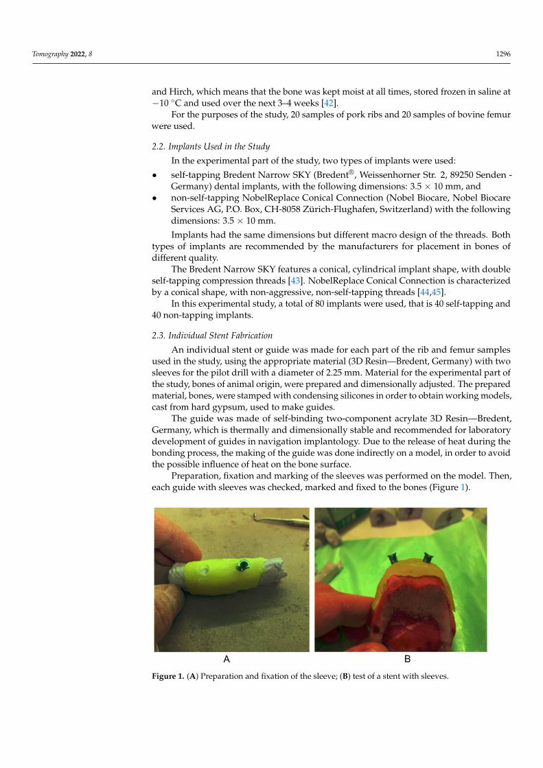
Preparation, fixation and marking of the sleeves was performed on the model. Then,each guide with sleeves was checked, marked and fixed to the bones (Figure 1).
Tomography 2022, 8, FOR PEER REVIEW 5
Figure 1. (A) Preparation and fixation of the sleeve; (B) test of a stent with sleeves.
2.4. Radiographic Analysis of Bone Density Experimental material, parts of pig ribs and bovine femurs, with prepared, fixed and
marked sleeves were recorded individually on a specially designed and adjusted stand of the CBCT device Planmeca 3D Promax—Asentajankatu 6, FI-00880 Helsinki, Finland.
All samples were recorded under the same conditions, 200 μm voxel, 90 kV, 10 mA, 36.4 s, 3112 mGy/cm2. Multiplanar reconstruction was performed in Romexis software (Planmeca Romexis 5.3.4.39, Asentajankatu 6, FI-00880 Helsinki, Finland) with associated mathematical software algorithms for reducing CBCT artifacts. Prior to the start of the study, the calibration was performed by an authorized Planmeca support (Beam check, QA test and flat field calibration test).
According to the recommendations from the literature, we used smaller voxels (200 μm) and the latest version of Romexis software with accompanying algorithms to scatter scattered signs.
Three-dimensional reconstruction of the recorded samples was performed using the software Romexis (Planmeca Romexis 5.3.4.39, Asentajankatu 6, FI-00880 Helsinki, Fin-land) of the CBCT device.
In the software of the CBCT device, implants with sleeves, Bredent Narrow SKY and NobelReplace Conical Connection, dimensions 3.5 × 10 mm were selected from the im-plant base.
The selected implants are virtually placed so that the longitudinal axis of the implant coincides with the axis and shape of the sleeve.
After the virtual positioning of the implant, the program automatically produced the values of the average bone density, expressed in Hounsfield units in the cylinder, that is, inside the virtual implant and 1 mm in the surrounding bone. The mean value of bone density was used for the purposes of the research (Figure 2).
Figure 1. (A) Preparation and fixation of the sleeve; (B) test of a stent with sleeves.

Tomography 2022, 8 1297
2.4. Radiographic Analysis of Bone Density
Experimental material, parts of pig ribs and bovine femurs, with prepared, fixed andmarked sleeves were recorded individually on a specially designed and adjusted stand ofthe CBCT device Planmeca 3D Promax—Asentajankatu 6, FI-00880 Helsinki, Finland.
All samples were recorded under the same conditions, 200 µm voxel, 90 kV, 10 mA,36.4 s, 3112 mGy/cm2. Multiplanar reconstruction was performed in Romexis software(Planmeca Romexis 5.3.4.39, Asentajankatu 6, FI-00880 Helsinki, Finland) with associatedmathematical software algorithms for reducing CBCT artifacts. Prior to the start of thestudy, the calibration was performed by an authorized Planmeca support (Beam check, QAtest and flat field calibration test).
According to the recommendations from the literature, we used smaller voxels (200 µm)and the latest version of Romexis software with accompanying algorithms to scatter scat-tered signs.
Three-dimensional reconstruction of the recorded samples was performed using thesoftware Romexis (Planmeca Romexis 5.3.4.39, Asentajankatu 6, FI-00880 Helsinki, Finland)of the CBCT device.
In the software of the CBCT device, implants with sleeves, Bredent Narrow SKYand NobelReplace Conical Connection, dimensions 3.5 × 10 mm were selected from theimplant base.
The selected implants are virtually placed so that the longitudinal axis of the implantcoincides with the axis and shape of the sleeve.
After the virtual positioning of the implant, the program automatically produced thevalues of the average bone density, expressed in Hounsfield units in the cylinder, that is,inside the virtual implant and 1 mm in the surrounding bone. The mean value of bonedensity was used for the purposes of the research (Figure 2).
Tomography 2022, 8, FOR PEER REVIEW 6
Figure 2. Software evaluations of bone volume expressed in HU.
2.5. Procedure for Experimental Implant Placement After checking the position of the guide, the experimental bone was fixed and the
experimental implant placement was initiated. The procedure involved preparation of the bearing in the bone and placement of the implant in the bearing, according to the manu-facturer’s instructions, and specialized implant surgical sets Bredent® and Nobel Biocare.
The pilot drill was put through the sleeves from the navigation implantology set to a depth of 10 mm. This was followed by a preparation without the use of guides, using drills for the appropriate bone type and implant system (Figure 3).
Figure 3. Bone preparation for implant bearing: (A) pilot drill through the sleeve; (B) preparation after the guide was removed.
One Bredent Narrow SKY self-tapping and one NobelReplace Conical Connection non-self-tapping implant were installed in each bovine femur and pig rib sample. The implants were placed in the bearing mechanically with a set torque of 35 N/cm2.
Figure 2. Software evaluations of bone volume expressed in HU.
2.5. Procedure for Experimental Implant Placement
After checking the position of the guide, the experimental bone was fixed and theexperimental implant placement was initiated. The procedure involved preparation ofthe bearing in the bone and placement of the implant in the bearing, according to the

Tomography 2022, 8 1298
manufacturer’s instructions, and specialized implant surgical sets Bredent® and NobelBiocare.
The pilot drill was put through the sleeves from the navigation implantology set to adepth of 10 mm. This was followed by a preparation without the use of guides, using drillsfor the appropriate bone type and implant system (Figure 3).
Tomography 2022, 8, FOR PEER REVIEW 6
Figure 2. Software evaluations of bone volume expressed in HU.
2.5. Procedure for Experimental Implant Placement After checking the position of the guide, the experimental bone was fixed and the
experimental implant placement was initiated. The procedure involved preparation of the bearing in the bone and placement of the implant in the bearing, according to the manu-facturer’s instructions, and specialized implant surgical sets Bredent® and Nobel Biocare.
The pilot drill was put through the sleeves from the navigation implantology set to a depth of 10 mm. This was followed by a preparation without the use of guides, using drills for the appropriate bone type and implant system (Figure 3).
Figure 3. Bone preparation for implant bearing: (A) pilot drill through the sleeve; (B) preparation after the guide was removed.
One Bredent Narrow SKY self-tapping and one NobelReplace Conical Connection non-self-tapping implant were installed in each bovine femur and pig rib sample. The implants were placed in the bearing mechanically with a set torque of 35 N/cm2.
Figure 3. Bone preparation for implant bearing: (A) pilot drill through the sleeve; (B) preparationafter the guide was removed.
One Bredent Narrow SKY self-tapping and one NobelReplace Conical Connectionnon-self-tapping implant were installed in each bovine femur and pig rib sample. Theimplants were placed in the bearing mechanically with a set torque of 35 N/cm2.
2.6. Primary Stability Measurement Procedure
After placement of both implants, measurements of primary implant stability wereperformed using resonant frequency analysis (RFA), via Osstell mentor device (IntegrationDiagnostics AB, Stampgatan 14, 411 01 Göteborg, Sweden).
The implant is placed and tightened manually, using a force of 4 to 5 Ncm anda suitable SmartPeg, type 49 for Bredent Narrow SKY and type 60 for NobelReplaceConical Connection.
The Osstell mentor probe is placed at the right angle and with a distance of 2–3 mm,next to the SmartPeg, in four positions: buccal, oral, mesial and distal. There are twothreads on the top of the device. After switching on, the first thread becomes a magnet thatexcites the magnet on the Smartpeg. The second thread registers the vibration produced bySmartpeg. After a short sound, the ISQ value is read on the register of the device. Duringthis study, we used the mean ISQ value obtained from four different directions.
2.7. Statistical Analysis
Descriptive statistical methods, methods for testing statistical hypotheses, methodsfor testing correlations, and methods for examining the correlation between outcomes andpotential predictors were used to analyze primary data. Depending on the type of variables,the data description is given as n (%) or as ±sd.
T-test was used as a method for testing statistical hypotheses. The Pearson linearcorrelation coefficient was used to examine the correlation of the two variables.
Statistical hypotheses were tested at the level of statistical significance of 0.05.The obtained data were then statistically processed to obtain a correlation between the
mean value of bone volume density and the value of primary stability of implants.

Tomography 2022, 8 1299
The following measurements were performed during the study:
• Bone densities based on CBCT images in HU units;• Primary stability of dental implants in ISQ units.
3. Results
An experimental study on the material of animal origin was conducted using20 samples of pig ribs and 20 samples of bovine femurs, in which two implants wereinstalled. In this study, a total of 40 self-tapping implants and 40 non-self-tapping implants(50%) were installed.
Table 1 shows the distribution of mean values of the bone density expressed inHounsfield units in relation to the bovine femur and pork rib.
Table 1. Overview of bone density values expressed in HU units relative to the bone.
HU n –x sd Med Min Max
Bovine femur 40 851.8 193.0 827.2 422.9 1236.9Pig rib 40 255.7 66.1 254.1 99.7 388.6
The arithmetic mean and standard deviation of bone density expressed in HU units inthe bovine femur stood at 851.8 ± 193.0, while in the pig rib it was 255.7 ± 66.1, which is astatistically significant difference (t = 18,478; p < 0.001).
Figure 4 shows the correlation between the mean values of bone density measured ona CBCT device and expressed in HU units and the primary stability of self-tapping dentalimplants installed in a pig rib.
Tomography 2022, 8, FOR PEER REVIEW 8
Figure 5 shows the correlation between the mean values of bone density measured on a CBCT device and expressed in HU units and the primary stability of non-self-tapping dental implants in pig ribs.
Figure 4. Correlation between bone density and primary stability of self-tapping implants in pig ribs.
Figure 5. Correlation between bone density and primary stability of non-self-tapping implants in pig ribs.
In non-self-tapping implants in the pig rib, there is no statistically significant corre-lation between the bone density, expressed in HU units, and the primary stability of the dental implants, expressed in ISQ units (r = 0.318; p = 0.172).
Table 2 shows the values of primary stability of the self-tapping and non-self-tapping implants in the pig rib.
Figure 4. Correlation between bone density and primary stability of self-tapping implants in pig ribs.
In self-tapping implants in the pig rib, there is a statistically significant mean positivecorrelation between the bone density expressed in HU units and the primary stability ofthe dental implants expressed in ISQ units (r = 0.506; p = 0.023).

Tomography 2022, 8 1300
Figure 5 shows the correlation between the mean values of bone density measured ona CBCT device and expressed in HU units and the primary stability of non-self-tappingdental implants in pig ribs.
Tomography 2022, 8, FOR PEER REVIEW 8
Figure 5 shows the correlation between the mean values of bone density measured on a CBCT device and expressed in HU units and the primary stability of non-self-tapping dental implants in pig ribs.
Figure 4. Correlation between bone density and primary stability of self-tapping implants in pig ribs.
Figure 5. Correlation between bone density and primary stability of non-self-tapping implants in pig ribs.
In non-self-tapping implants in the pig rib, there is no statistically significant corre-lation between the bone density, expressed in HU units, and the primary stability of the dental implants, expressed in ISQ units (r = 0.318; p = 0.172).
Table 2 shows the values of primary stability of the self-tapping and non-self-tapping implants in the pig rib.
Figure 5. Correlation between bone density and primary stability of non-self-tapping implants inpig ribs.
In non-self-tapping implants in the pig rib, there is no statistically significant corre-lation between the bone density, expressed in HU units, and the primary stability of thedental implants, expressed in ISQ units (r = 0.318; p = 0.172).
Table 2 shows the values of primary stability of the self-tapping and non-self-tappingimplants in the pig rib.
Table 2. ISQ value in pig rib in relation to implant type.
ISQ n –x sd Med Min Max
Self-tapping 20 68.2 3.8 68.0 59.0 73.0Non-self-tapping 20 67.0 4.5 68.5 54.0 72.0
The arithmetic mean and standard deviation of the values of the primary stability ofthe self-tapping implants expressed in ISQ units in the pig rib was 68.2 ± 3.8, while in thenon-self-tapping implants it was 67.0 ± 4.5, which is not a statistically significant difference(t = 0.947; p = 0.350).
Mean bone density values measured on the CBCT device, expressed in HU units, andtheir correlation to the primary stability of the self-tapping dental implants installed in thebovine femur are given in Figure 6.

Tomography 2022, 8 1301
Tomography 2022, 8, FOR PEER REVIEW 9
Table 2. ISQ value in pig rib in relation to implant type.
ISQ n x sd Med Min Max Self-tapping 20 68.2 3.8 68.0 59.0 73.0
Non-self-tapping 20 67.0 4.5 68.5 54.0 72.0
The arithmetic mean and standard deviation of the values of the primary stability of the self-tapping implants expressed in ISQ units in the pig rib was 68.2 ± 3.8, while in the non-self-tapping implants it was 67.0 ± 4.5, which is not a statistically significant difference (t = 0.947; p = 0.350).
Mean bone density values measured on the CBCT device, expressed in HU units, and their correlation to the primary stability of the self-tapping dental implants installed in the bovine femur are given in Figure 6.
Figure 6. Correlation between bone density and primary stability of self-tapping implants in bovine femur.
In the self-tapping implants installed in the bovine femur, there is a statistically sig-nificant strong positive correlation between the bone density expressed in HU units and the primary implant stability expressed in ISQ units (r = 0.880; p < 0.001).
Figure 7 shows the correlation between the mean values of the bone density meas-ured using a CBCT device and expressed in HU units and the primary stability of the non-self-tapping dental implants in the bovine femur.
In the non-self-tapping implants in the bovine femur, there is a statistically significant mean positive correlation between the bone density expressed in HU units and the pri-mary implant stability expressed in ISQ units (r = 0.584; p = 0.007).
The values of the primary stability of the self-tapping and non-self-tapping implants in the bovine femur are shown in Table 3.
The arithmetic mean and standard deviation of the primary stability of the self-tap-ping implants in the bovine femur was 75.8 ± 3.4 ISQ, while in the non-self-tapping im-plants it was 74.2 ± 3.9 ISQ, which does not represent a statistically significant difference (t = 1381; p = 0.175).
Figure 6. Correlation between bone density and primary stability of self-tapping implants inbovine femur.
In the self-tapping implants installed in the bovine femur, there is a statisticallysignificant strong positive correlation between the bone density expressed in HU units andthe primary implant stability expressed in ISQ units (r = 0.880; p < 0.001).
Figure 7 shows the correlation between the mean values of the bone density measuredusing a CBCT device and expressed in HU units and the primary stability of the non-self-tapping dental implants in the bovine femur.
Tomography 2022, 8, FOR PEER REVIEW 10
Figure 7. Correlation between bone density and primary stability of non-self-tapping implants in bovine femur.
Table 3. The value of ISQ in bovine femur with respect to the implant type.
ISQ n x sd Med Min Max Self-tapping 20 75.8 3.4 76.0 69.0 82.0
Non-self-tapping 20 74.2 3.9 75.0 66.0 80.0
4. Discussion In today’s modern dental therapy, implant therapy represents the therapy of choice,
indicated for all types of toothlessness. With the development of implantology and through the use of a multidisciplinary medical approach, most contraindications for im-plant therapy have been translated from absolute to relative contraindications, with a ten-dency to reduce complications and increase the success rate. The success rate of implant therapy, according to most studies, ranges from 90% to 95%.
There are numerous factors that influence the success of implant therapy, and are usually related to the characteristics of the patient, the type of implant and the skills of the therapist [46–52].
The experimental part of this study, which was performed on the material of animal origin, pig ribs and a bovine femur, assessed the influence of bone density, based on CBCT images, on the primary stability in different designs of dental implants, self-tapping and non-self-tapping.
The expansive development of radiological diagnostics and its application in implan-tology has enabled objective pre-implant preparation as well as the selection of the opti-mal treatment plan. Qualitative and objective bone analysis was enabled with the intro-duction of a three-dimensional multiplanar overview of the jaw bone tegmen, first by us-ing Computed Tomography (CT), then by using Multilayer Computed Tomography (MSCT) and by applying Cone Computed Tomography (CBCT) to implant practices [53,54].
The evaluation of the quality of the bone density in both the experimental part of the study on bones of animal origin, as well as in the clinical part of the study on humans, was performed on the basis of the software analysis of CBCT images and expressed in HU units. The validity of the assessment of the bone density expressed in HU units, based on CBCT and CT images has been the focus of numerous studies. Armstrong (2006) and
Figure 7. Correlation between bone density and primary stability of non-self-tapping implants inbovine femur.

Tomography 2022, 8 1302
In the non-self-tapping implants in the bovine femur, there is a statistically significantmean positive correlation between the bone density expressed in HU units and the primaryimplant stability expressed in ISQ units (r = 0.584; p = 0.007).
The values of the primary stability of the self-tapping and non-self-tapping implantsin the bovine femur are shown in Table 3.
Table 3. The value of ISQ in bovine femur with respect to the implant type.
ISQ n –x sd Med Min Max
Self-tapping 20 75.8 3.4 76.0 69.0 82.0Non-self-tapping 20 74.2 3.9 75.0 66.0 80.0
The arithmetic mean and standard deviation of the primary stability of the self-tappingimplants in the bovine femur was 75.8 ± 3.4 ISQ, while in the non-self-tapping implants itwas 74.2 ± 3.9 ISQ, which does not represent a statistically significant difference (t = 1381;p = 0.175).
4. Discussion
In today’s modern dental therapy, implant therapy represents the therapy of choice,indicated for all types of toothlessness. With the development of implantology and throughthe use of a multidisciplinary medical approach, most contraindications for implant therapyhave been translated from absolute to relative contraindications, with a tendency to reducecomplications and increase the success rate. The success rate of implant therapy, accordingto most studies, ranges from 90% to 95%.
There are numerous factors that influence the success of implant therapy, and areusually related to the characteristics of the patient, the type of implant and the skills of thetherapist [46–52].
The experimental part of this study, which was performed on the material of animalorigin, pig ribs and a bovine femur, assessed the influence of bone density, based on CBCTimages, on the primary stability in different designs of dental implants, self-tapping andnon-self-tapping.
The expansive development of radiological diagnostics and its application in implan-tology has enabled objective pre-implant preparation as well as the selection of the optimaltreatment plan. Qualitative and objective bone analysis was enabled with the introductionof a three-dimensional multiplanar overview of the jaw bone tegmen, first by using Com-puted Tomography (CT), then by using Multilayer Computed Tomography (MSCT) and byapplying Cone Computed Tomography (CBCT) to implant practices [53,54].
The evaluation of the quality of the bone density in both the experimental part of thestudy on bones of animal origin, as well as in the clinical part of the study on humans, wasperformed on the basis of the software analysis of CBCT images and expressed in HU units.The validity of the assessment of the bone density expressed in HU units, based on CBCTand CT images has been the focus of numerous studies. Armstrong (2006) and Arisanet al. (2013) pointed out that there is a difference in the values of HU units in the analysisof material recorded under the same conditions on CT and CBCT [54,55]. The researchfor the assessment of the bone density, expressed in HU units, can include the analysis ofCT images and the analysis of CBCT images as valid methods for the evaluation of bonedensity and is the method of choice in the preimplantology phase. Due to its availability,lower radiation dose, and simpler installation, CBCT represents a more common methodin the dental practice [53,56–62].
In the experimental study, the average bone density expressed in HU units in thebovine femur, which was used as a human lower jaw model, was 851.8 HU. The lowestobtained average value of the bovine femur bone density amounted to 442.9 HU, while thehighest was 1236.9 HU. All of the obtained values correspond to the bone quality classifiedin categories D1 and D2, according to Misch [23], and Q1, Q2 and Q3 according to Norton

Tomography 2022, 8 1303
and Gamble [24], which, according to these authors, can be found locally in the alveolarextensions of the lower jaw.
The average bone density in the pig rib, which was used as a model of the humanupper jaw, was 255.7 HU. The minimum value was 99.7 HU and the maximum 388.6 HU.The average value of the pork rib bone quality corresponds to the bone quality classified incategory D4 according to Misch [18] and Q4 according to Norton and Gamble [19], which,according to these authors, can be found in the lateral region of the upper jaw. The partialvolume effect, as described in the literature, can explain the registered minimum valueof the average bone density on one sample of pork rib without a noticeable deviationof the primary stability and a macroscopically noticeable lower strength on that sample.When different types of tissues are found in one voxel, in this case, soft tissue cavities andbone beams, with different X-ray attenuation coefficients, this sometimes affects the imagequality and may result in an inaccurate CT or CBCT reading [57].
The results of the presented experimental study did not show a significant statisticaldifference in the primary stability between the self-tapping and non-self-tapping implantsin both types of bone tissue. In an in vitro study aimed at examining the correlation of theprimary stability of two types of implants in different types of bone tissue, Bilhan et al.came to similar results, and that is that there is no significant statistical difference (2015).The results of the values of the primary stability in their study are slightly higher for bothtypes of implants compared to our study, which can be explained by the use of implantsof a larger diameter and length [58]. Falco et al. (2018), whose study was also performedon a material of animal origin, showed a statistically significant difference in the primarystability in the self-tapping implants compared to non-self-tapping implants in a low-density D4 bone, while in other bone types there was no significant difference [59]. Otherstudies dealing with the primary stability of implants of different designs conclude that themain determinants of primary stability are the bone density and implant macrodesign, andthat self-tapping implants are recommended for lower-density bones and for immediateimplantation [60–64].
According to the results of the presented experimental study on pig ribs, there is astatistically significant mean positive correlation between the bone density expressed inHU units and the primary stability of self-tapping dental implants expressed in ISQ units(r = 0.506; p = 0.023). While there was no statistically significant difference in this type ofbone when it comes to non-self-tapping dental implants, Isoda et al. (2012), in a similarin vitro study using a material of animal origin, demonstrated a significant positive correla-tion between the bone density measured on a CBCT device and the primary stability ofself-tapping dental implants [65]. Möhlhenrich et al. (2019) published similar results on thecorrelation between bone density measured on the CBCT device and the primary implantstability [66]. The difference in the correlation between the bone density assessment andprimary stability in the two types of implants in the lower-density bone can be explained bythe macrodesign of the thread. In their review paper, when analyzing thirteen papers thatanalyzed the correlation between different factors and the primary stability of implants,Marquezan et al. (2012) concluded that self-tapping implants with more aggressive threadsshow a better primary stability compared to non-self-tapping dental implants [67].
In self-tapping implants installed in the bovine femur, there is a statistically significantstrong, and in non-self-tapping, a moderately strong positive correlation between the bonedensity expressed in HU units and the primary implant stability expressed in ISQ units inthe presented study. Fuster-Torres et al. (2001) and Isoda et al. (2012) reported a positivecorrelation between the bone density expressed in HU units and the primary implantstability, which coincides with the results of the presented study [65,68].
5. Conclusions
On the basis of the presented results, we can conclude the following:
• By analyzing the density of the bone tissue in the CBCT images in the software of thedevice expressed in HU units, we cannot predict the degree of the primary stability
Tomography 2022, 8 1304
of the non-self-tapping dental implants in bones of a lower quality D4, according toMisch, and Q4, according to Norton and Gamble;
• Self-tapping and non-self-tapping dental implants installed in D4- and Q4-qualitybones do not show a significant statistical difference in the primary stability;
• By analyzing the density of the bone tissue in the CBCT images in the software of thedevice expressed in Hausfield units, we can predict the degree of the primary stabilityof the self-tapping dental implants in bones of the densities D1, D2 and Q1–Q3;
• By analyzing the density of the bone tissue in the CBCT images in the software of thedevice expressed in HU units, we can predict the degree of the primary stability of thenon-self-tapping dental implants in bones of the densities D1, D2 and Q1–Q3.
Author Contributions: Writing—review and editing, M.M.; Conceptualization, Z.V.; methodology,Z.A. and M.S.; Supervision, R.M. All authors have read and agreed to the published version ofthe manuscript.
Funding: This research received no external funding.
Institutional Review Board Statement: The research was approved by the Ethics Committee of theClinical Centre of Montenegro no. 03/01-4536/1 from March 2018.
Informed Consent Statement: Not applicable.
Data Availability Statement: Data available on request from authors.
Conflicts of Interest: The authors declare no conflict of interest.
References1. Barbato, P.R.; Nagano, H.C.M.; Zanchet, F.N.; Boing, A.F.; Peres, M.A. Tooth loss and associated socioeconomic, demographic,
and dental-care factors in Brazilian adults: An analysis of the Brazilian Oral Health Survey, 2002–2003. Cad. Saude Publica 2007,23, 1803–1814. [CrossRef] [PubMed]
2. Moreira, C.H.C.; Zanatta, F.B.; Antoniazzi, R.P.; Meneguetti, P.C.; Rösing, C.K. Criteria adopted by dentists to indicate theextraction of periodontally involved teeth. J. Appl. Oral Sci. 2007, 15, 437–441. [CrossRef] [PubMed]
3. Cahen, P.; Frank, R.; Turlot, J. A Survey of the Reasons for Dental Extractions in France. J. Dent. Res. 1985, 64, 1087–1093.[CrossRef] [PubMed]
4. Ong, G.; Yeo, J.-F.; Bhole, S. A survey of reasons for extraction of permanent teeth in Singapore. Community Dent. Oral Epidemiol.1996, 24, 124–127. [CrossRef]
5. Nuvvula, S.; Chava, V.K.; Nuvvula, S. Primary culprit for tooth loss!! J. Indian Soc. Periodontol. 2016, 20, 222–224.6. Adell, R.; Lehholm, U.; Rockler, B.; Branemark, P.I. A I5-year study of osseointegrated implants in the treatment of the edentulous
jaw. Int. J. Oral Surg. 1981, 10, 387–416. [CrossRef]7. Chrcanovic, B.R.; Albrektsson, T.; Wennerberg, A. Bone Quality and Quantity and Dental Implant Failure: A Systematic Review
and Meta-analysis. Int. J. Prosthodont. 2017, 30, 219–237. [CrossRef]8. Chrcanovic, B.R.; Albrektsson, T.; Wennerberg, A. Dental implants inserted in male versus female patients: A systematic review
and meta-analysis. J. Oral Rehabil. 2015, 42, 709–722. [CrossRef]9. Chrcanovic, B.R.; Albrektsson, T.; Wennerberg, A. Dental implants inserted in fresh extraction sockets versus healed sites: A
systematic review and meta-analysis. J. Dent. 2015, 43, 16–41. [CrossRef]10. Vlahovic, Z.; Mikic, M. 3D Printing Guide Implant Placement:A Case Report. Balk. J. Dent. Med. 2017, 21, 65–68. [CrossRef]11. Esposito, M.; Grusovin, M.G.; Polyzos, I.P.; Felice, P.; Worthington, H.V. Timing of implant placement after tooth extraction:
Immediate, immediate-delayed or delayed implants? A Cochrane systematic review. Eur. J. Oral Implantol. 2010, 3, 189–205.[PubMed]
12. Manivasagam, V.K.; Popat, K.C. Hydrothermally treated titanium surfaces for enhanced osteogenic differentiation of adiposederived stem cells. Mater. Sci. Eng. C 2021, 128, 112315. [CrossRef] [PubMed]
13. Heinemann, F.; Hasan, I.; Bourauel, C.; Biffar, R.; Mundt, T. Bone stability around dental implants: Treatment related factors. Ann.Anat.-Anat. Anz. 2015, 199, 3–8. [CrossRef] [PubMed]
14. Sayed, M.E.; Mugri, M.H.; Almasri, M.A.; Al-Ahmari, M.M.; Bhandi, S.; Madapusi, T.B.; Varadarajan, S.; Raj, A.T.; Reda, R.;Testarelli, L.; et al. Role of Stem Cells in Augmenting Dental Implant Osseointegration: A Systematic Review. Coatings 2021, 11,1035. [CrossRef]
15. Al-Sabbagh, M.; Eldomiaty, W.; Khabbaz, Y. Can Osseointegration Be Achieved Without Primary Stability? Dent. Clin. North Am.2019, 63, 461–473. [CrossRef]
16. Punn, K.K.; Wong, F.H.W. Importance of measurement of bone density in the management of osteoporosis. Singap. Med. J. 1990,31, 390–396.
Tomography 2022, 8 1305
17. Krolo, I.; Zadravec, D. Dentalna radiologija. Zagreb Med. Nakl. 2016, 12, 197–212.18. Misch, C.E.; Kircos, L.T. Diagnostic imaging and techniques. In Contemporary Implant Dentistry, 2nd ed.; Misch, C.E., Ed.; Mosby:
St. Louis, MO, USA, 1999; pp. 73–87.19. Norton, R.M.; Gamble, C. Bone classification: An objective scale of bone density using the computerized tomography scan. Clin.
Oral Implant. Res. 2001, 12, 79–84. [CrossRef]20. Tsiklakis, K.; Donta, C.; Gavala, S.; Karayianni, K.; Kamenopoulou, V.; Hourdakis, C.J. Dose reduction in maxillofacial imaging
using low dose Cone Beam CT. Eur. J. Radiol. 2005, 56, 413–417. [CrossRef]21. Alaqeely, R.; Babay, N.; AlQutub, M. Dental implant primary stability in different regions of the Jawbone: CBCT-based 3D finite
element analysis. Saudi Dent. J. 2020, 32, 101–107. [CrossRef]22. Vyas, R.; Talluri, S.; Vaddamanu, S.K.; Apparaju, V.; Ahuja, S.; Kanji, M.A. Evaluating cortico-cancellous ratio using virtual
implant planning and its relation with immediate and long-term stability of a dental implant—A CBCT-assisted prospectiveobservational clinical study. Niger. J. Clin. Pract. 2019, 22, 982–987. [CrossRef] [PubMed]
23. Jain, S.; Choudhary, K.; Nagi, R.; Shukla, S.; Kaur, N.; Grover, D. New evolution of cone-beam computed tomography in dentistry:Combining digital technologies. Imaging Sci. Dent. 2019, 49, 179–190. [CrossRef] [PubMed]
24. Tanimoto, H.; Arai, Y. The effect of voxel size on image reconstruction in cone-beam computed tomography. Oral Radiol. 2009, 25,149–153. [CrossRef]
25. Jacobs, R.; Salmon, B.; Codari, M.; Hassan, B.; Bornstein, M.M. Cone beam computed tomography in implant dentistry:Recommendations for clinical use. BMC Oral Health 2018, 18, 88. [CrossRef]
26. Quereshy, F.A.; Savell, T.A.; Palomo, J.M. Applications of Cone Beam Computed Tomography in the Practice of Oral andMaxillofacial Surgery. J. Oral Maxillofac. Surg. 2008, 66, 791–796. [CrossRef]
27. Bogaerde, L.V.; Pedretti, G.; Sennerby, L.; Meredith, N. Immediate/Early Function of Neoss Implants Placed in Maxillas andPosterior Mandibles: An 18-Month Prospective Case Series Study. Clin. Implant. Dent. Relat. Res. 2010, 12, e83–e94. [CrossRef]
28. Sennerby, L.; Andersson, P.; Verrocchi, D.; Viinamaki, R. One-year outcomes of Neoss bimodal implants. A prospective clinical,radiographic, and RFA study. Clin. Implant. Dent. Relat. Res. 2012, 14, 313–320. [CrossRef]
29. Tantanapornkul, W.; Okouchi, K.; Fujiwara, Y.; Yamashiro, M.; Maruoka, Y.; Ohbayashi, N.; Kurabayashi, T. A comparativestudy of conebeam computed tomography and conventional panoramic radiography in assessing the topographic correlationbetween the mandibular canal and impacted third molars. Oral Surg. Oral Med. Oral Pathol. Oral Radiol. Endod. 2007, 103, 253–259.[CrossRef]
30. Osorio, F.; Perilla, M.; Doyle, D.J.; Palomo, J.M. Cone Beam Computed Tomography: An Innovative Tool for Airway Assessment.Anesth. Analg. 2008, 106, 1803–1807. [CrossRef]
31. Fokas, G.; Vaughn, V.M.; Scarfe, W.C.; Bornstein, M.M. Accuracy of linear measurements on CBCT images related to presurgicalimplant treatment planning: A systematic review. Clin. Oral Implant. Res. 2018, 29, 393–415. [CrossRef]
32. Mozzo, P.; Procacci, C.; Tacconi, A.; Martini, P.T.; Andreis, I.A.B. A new volumetric CT machine for dental imaging based on thecone-beam technique: Preliminary results. Eur. Radiol. 1998, 8, 1558–1564. [CrossRef] [PubMed]
33. Gaviria, L.; Salcido, J.P.; Guda, T.; Ong, J.L. Current trends in dental implants. J. Korean Assoc. Oral Maxillofac. Surg. 2014, 40,50–60. [CrossRef] [PubMed]
34. de Andrade, C.L.; Carvalho, M.A.; Bordin, D.; da Silva, W.J.; Del Bel Cury, A.A.; Sotto-Maior, B.S. Biomechanical Behavior of theDental Implant Macrodesign. Int. J. Oral Maxillofac. Implant. 2017, 32, 264–270. [CrossRef] [PubMed]
35. Lin, C.P.; Lu, S.H.; Zhu, J.X.; Hu, H.C.; Yue, Z.G.; Tang, Z.H. Influence of thread shapes of custo made root-analogue implants onstress distribution of peri-implant bone: A three-dimensional finite element analysis. Beijing Da Xue Xue Bao Yi Xue Ban 2019, 51,1130–1137. [PubMed]
36. Bavetta, G.; Bavetta, G.; Randazzo, V.; Cavataio, A.; Paderni, C.; Grassia, V.; Dipalma, G.; Isacco, C.G.; Scarano, A.; De Vito, D.;et al. A Retrospective Study on Insertion Torque and Implant Stability Quotient (ISQ) as Stability Parameters for ImmediateLoading of Implants in Fresh Extraction Sockets. BioMed Res. Int. 2019, 2019, 9720419. [CrossRef]
37. Meredith, N.; Alleyne, D.; Cawley, P. Quantitative determination of the stability of the implant–tissue interface using re-so-nancefrequency analysis. Clin. Oral Implant. Res. 1996, 7, 261–267. [CrossRef]
38. Noaman, A.T.; Bede, S.Y. The Effect of Bone Density Measured by Cone Beam Computed Tomography and Implant Dimensionson the Stability of Dental Implants. J. Craniofacial Surg. 2021. epub ahead of print. [CrossRef]
39. Hakim, S.G.; Glanz, J.; Ofer, M.; Steller, D.; Sieg, P. Correlation of cone beam CT-derived bone density parameters with primaryimplant stability assessed by peak insertion torque and periotest in the maxilla. J. Cranio-Maxillofac. Surg. 2019, 47, 461–467.[CrossRef]
40. Al-Jamal, M.F.J.; Al-Jumaily, H.A. Can the Bone Density Estimated by CBCT Predict the Primary Stability of Dental Implants? ANew Measurement Protocol. J. Craniofacial Surg. 2021, 32, e171–e174. [CrossRef]
41. Cummaudo, M. Histomorphometric analysis of osteocyte lacunae in human and pig: Exploring its potential forspecies discrimi-nation. Int. J. Leg. Med. 2019, 133, 711–718. [CrossRef]
42. González-Garcia, R.; Monje, F.; Moreno-Garcia, C. Predictability of the resonance frequency analysis in the survival of dentalimplants placed in the anterior non-atrophied edentulous mandible. Med. Oral Patol. Oral Cir. Bucal. 2011, 16, 664–669. [CrossRef][PubMed]
43. Petersson, A.; Sennerby, L. On standard calibration of ISQ transducer pegs. Integr. Diagn. Update 2016, 1, 1–3.
Tomography 2022, 8 1306
44. Jaramillo, R.; Santos, R.; Lázaro, P.; Romero, M.; Rios-Santos, J.V.; Bullón, P.; Fernández-Palacín, A.; Herrero-Climent, M.Comparative analysis of 2 resonance frequency measurement devices: Osstell Mentor and Osstell ISQ. Implant. Dent. 2014, 23,351–356. [CrossRef] [PubMed]
45. Karabuda, Z.C.; Arιsan, V.; Abdel-Haq, J. Stability, marginal bone loss and survival of standard and modified sand-blasted,acid-etched implants in bilateral edentulous spaces: A prospective 15-month evaluation. Clin. Oral Implant. Res. 2011, 22, 840–849.[CrossRef]
46. Kim, S.H.; Kim, S.J.; Lee, K.W.; Han, D.H. The effects of local factorson the survival rate of dental implants: A 19 year retrospectivestudy. J. Korean Acad. Prosthodont. 2010, 48, 28–40. [CrossRef]
47. Esposito, M.; Grusovin, M.G.; Coulthard, P.; Thomsen, P.; Worthington, H.V. A 5-year follow-up comparative analysis of theefficacy of various osseointegrated dental implant systems: A systematic review of randomized controlled clinical trials. Int. J.Oral Maxillofac. Implant. 2005, 20, 557–568.
48. Renouard, F.; Nisand, D. Impact of implant length and diameter on survival rates. Clin. Oral Implant. Res. 2006, 17, 35–51.[CrossRef]
49. Arsalanloo, Z.; Telchi, R.; Osgouie, K.G. Optimum Selection of the Dental Implants according to Length and Diameter Parametersby FE Method in the Anterior Position. Int. J. Biosci. Biochem. Bioinform. 2014, 4, 265–269. [CrossRef]
50. Busenlechner, D.; Fürhauser, R.; Haas, R.; Watzek, G.; Mailath, G.; Pommer, B. Long-term implant success at the academy for oralimplantology: 8- year follow-up and risk factor analysis. J. Periodontal. Implant. Sci. 2014, 44, 102–108. [CrossRef]
51. Yesildal, R.; Karabudak, F.; Bayındır, F.; Zamanlou, H.; Yıldırım, M.P.; Sagsöz, N.P.; Sen, S. Effect of implant diameter and lengthon stress distribution for titanium and zirconia implants by using finite element analysis (FEA). Open Access Libr. J. 2015, 2, 1–7.[CrossRef]
52. Philip, J.M.; Abraham, H.M.; Jain, A.R.; Venkatakrishnan, C. The effect of implant and abutment diameter on peri-implant bonestress: A three-dimensional finite element analysis. J. Oral Res. Rev. 2016, 8, 49–52. [CrossRef]
53. Omami, G.; Al Yafi, F. Should Cone Beam Computed Tomography Be Routinely Obtained in Implant Planning? Dent. Clin. N.Am. 2019, 63, 363–379. [CrossRef] [PubMed]
54. Armstrong, R.T. Acceptability of Cone Beam CT vs. Multi-Detector CT for 3D Anatomic Model Construction. J. Oral Maxillofac.Surg. 2006, 64, 37. [CrossRef]
55. Arisan, V.; Karabuda, Z.C.; Avsever, H.; Özdemir, T. Conventional Multi-Slice Computed Tomography (CT) and Cone-Beam CT(CBCT) for Computer-Assisted Implant Placement. Part I: Relationship of Radiographic Gray Density and Implant Stability. Clin.Implant. Dent. Relat. Res. 2013, 15, 893–906. [CrossRef] [PubMed]
56. Wada, M.; Tsuiki, Y.; Suganami, T.; Ikebe, K.; Sogo, M.; Okuno, I.; Maeda, Y. The correlation between the bone characters obtainedby CBCT and primary stability of the implants. Int. J. Implant. Dent. 2015, 1, 1–7. [CrossRef]
57. Molen, A.D. Considerations in the use of cone-beam computed tomography for buccal bone measurements. Am. J. Orthod.Dentofac. Orthop. 2010, 137, 130–135. [CrossRef]
58. Bilhan, H.; Bilmenoglu, C.; Urgun, A.C.; Ates, G.; Bural, C.; Cilingir, A.; Geckili, O. Comparison of the Primary Stability of TwoImplant Designs in Two Different Bone Types: An In Vitro Study. Int. J. Oral Maxillofac. Implant. 2015, 30, 1036–1040. [CrossRef]
59. Falco, A.; Berardini, M.; Trisi, P. Correlation Between Implant Geometry, Implant Surface, Insertion Torque, and Primary Stability:In Vitro Biomechanical Analysis. Int. J. Oral Maxillofac. Implant. 2018, 33, 824–830. [CrossRef]
60. Cochran, D.; Stavropoulos, A.; Obrecht, M.; Pippenger, B.; Dard, M. A Comparison of Tapered and Nontapered Implants in theMinipig. Int. J. Oral Maxillofac. Implant. 2016, 31, 1341–1347. [CrossRef]
61. Norton, M.R. The Influence of Low Insertion Torque on Primary Stability, Implant Survival, and Maintenance of Marginal BoneLevels: A Closed-Cohort Prospective Study. Int. J. Oral Maxillofac. Implant. 2017, 32, 849–857. [CrossRef]
62. Schliephake, H.; Rödiger, M.; Phillips, K.; McGlumphy, E.A.; Chacon, G.E.; Larsen, P. Early loading of surface modified implantsin the posterior mandible—5 year results of an open prospective non-controlled study. J. Clin. Periodontol. 2012, 39, 188–195.[CrossRef] [PubMed]
63. Abuhussein, H.; Pagni, G.; Rebaudi, A.; Wang, H.L. Effect of thread pattern upon implant osseointegration. Clin. Oral Impl. Res.2010, 21, 129–136. [CrossRef] [PubMed]
64. McCullough, J.J.; Klokkevold, P.R. The effect of implant macro-thread design on implant stability in the early post-operativeperiod: A randomized, controlled pilot study. Clin. Oral Implant. Res. 2017, 28, 1218–1226. [CrossRef] [PubMed]
65. Isoda, K.; Ayukawa, Y.; Tsukiyama, Y.; Sogo, M.; Matsushita, Y.; Koyano, K. Correlation between the bone density estimatedby cone-beam computed tomography and the primary stability of dental implants. Clin. Oral Implant. Res. 2012, 23, 832–836.[CrossRef] [PubMed]
66. Möhlhenrich, S.C.; Heussen, N.; Winterhalder, P.; Prescher, A.; Hölzle, F.; Modabber, A.; Wolf, M.; Kniha, K. Predicting primarystability of orthodontic mini-implants, according to position, screw-size, and bone quality, in the maxilla of aged patients: Acadaveric study. Eur. J. Oral Sci. 2019, 127, 462–471. [CrossRef] [PubMed]
67. Marquezan, M.; Osório, A.; Sant’Anna, E.; Souza, M.M.; Maia, L. Does bone mineral density influence the primary stability ofdental implants? A systematic review. Clin. Oral Implant. Res. 2012, 23, 767–774. [CrossRef]
68. Fuster-Torres, M.A.; Peñarrocha-Diago, M.; Peñarrocha-Oltra, D. Correlations Between Bone Density Values from Cone BeamComputed Tomography, Maximum Insertion Torque, and Resonance Frequency Analysis at Implant Placement: A Pilot Study.Int. J. Oral Maxillofac. Implant. 2011, 26, 1051–1056.