Sequential innovations with unobservable follow-on investments
Upload
khangminh22Category
view
4download
0
Appl. Sci. 2021, 11, 4504. https://doi.org/10.3390/app11104504 www.mdpi.com/journal/applsci
Case Report
Mandibular Coronoid Process Hypertrophy: Diagnosis and
20-Year Follow-Up with CBCT, MRI and EMG Evaluations
Fabrizia d’Apuzzo, Giuseppe Minervini, Vincenzo Grassia, Rossana Patricia Rotolo, Letizia Perillo *
and Ludovica Nucci
Multidisciplinary Department of Medical-Surgical and Dental Specialties, University of Campania Luigi
Vanvitelli, via Luigi De Crecchio 6, 80138 Naples, Italy; [email protected] (F.d.);
[email protected] (G.M.); [email protected] (V.G.);
[email protected] (R.P.R.); [email protected] (L.N.)
* Correspondence: [email protected]; Tel.: +39-081-566-5495 or +39-333-4027-273
Abstract: Coronoid process hypertrophy (CPH) consists of an abnormal volumetric increment of
the mandibular coronoid process; as this process grows gradually, the infratemporal space needed
for the rotation and translation of the mandible is reduced, which results in a reduction of the range
of mouth opening and lateral excursion, limiting mouth opening. The purpose of this case report
was to describe a rare case of hypertrophy of coronoid processes with associated
temporomandibular ankylosis, monitored for over 20 years. The patient was first visited when he
had a facial trauma at the age of 4. Then he was followed through clinical, functional, instrumental,
bi-dimensional and three-dimensional radiological evaluations up to the age of 24. Physical therapy
was initiated at the age of 10 to improve the condition of the masticatory muscles, while at the age
of 14, Transcutaneous Electrical Nerve Stimulations were performed to reduce muscle tension and,
a bite plane was delivered to control the parafunctional activity of the jaw in the night and self-
control instruction was provided for daytime habits. The adult patient has not accepted surgical
intervention; thus, the future objective is to continue monitoring over the years to avoid a
detrimental progression of the medical condition through physical and functional therapies while
waiting for patient consent to surgery if needed.
Keywords: temporomandibular joint; temporomandibular disorders; hypertrophy of the coronoid
process; joint ankylosis; early diagnosis; mandibular movements; physical therapy; magnetic
resonance imaging; cone-beam computed tomography; electromyography
1. Introduction
Coronoid process hypertrophy (CPH), also defined as giant coronoid syndrome [1],
was firstly described by Lagenbeck in 1853 [2] and consists of an abnormal volumetric
increment of the mandibular coronoid process [1]; as this process grows gradually, the
infratemporal space needed for the rotation and translation of the mandible is reduced,
which results in a reduction of the range of mouth-opening and lateral excursion, limiting
mouth opening.
CPH affects both genders with a male to female ratio of 5:1 and a peak of prevalence
at around 25 years of age [1]. It can be mono or bilateral, although the bilateral form seems
to be more frequent. The etiology of CPH is still not conclusive and in literature, different
causes are reported [1]. In the clinical diagnosis of CPH, mouth-opening reduction (<20
mm) is quite always present [1]; however, the diagnosis should be confirmed by imaging
of the head and neck region [3]. Cone-beam computed tomography (CBCT) can be
considered the gold standard for radiologic diagnosis, providing detailed information
about the length and width of the coronoid process and its relation to the zygomatic bone
and arch [3–6]. Additionally, panoramic radiographs can be used as a routine examination
Citation: D’Apuzzo, F.; Minervini,
G.; Grassia, V.; Rotolo, R.P.; Perillo,
L.; Nucci, L. Mandibular Coronoid
Process Hypertrophy: Diagnosis and
20-Year Follow-Up with CBCT, MRI
and EMG Evaluations. Appl. Sci.
2021, 11, 4504. https://doi.org/
10.3390/app11104504
Academic Editor: Rosalia Maria
Leonardi
Received: 23 April 2021
Accepted: 11 May 2021
Published: 14 May 2021
Publisher’s Note: MDPI stays
neutral with regard to jurisdictional
claims in published maps and
institutional affiliations.
Copyright: © 2021 by the authors.
Licensee MDPI, Basel, Switzerland.
This article is an open access article
distributed under the terms and
conditions of the Creative Commons
Attribution (CC BY) license
(http://creativecommons.org/licenses
/by/4.0/).

Appl. Sci. 2021, 11, 4504 2 of 11
for all patients with limited mouth opening to first assessed enlarged coronoid processes
through Levandoski panographic analysis [7]. The limited mouth opening can be
categorized as intra-articular or extra-articular [3,8,9], and the muscular component is
usually involved in CPH due to its role as a stabilizer of the craniomandibular system
during movements [7,8].
An early diagnosis is advisable to monitor and limit the progression of hypertrophy
and consequent limited movements in oral multifunction [1].
However, this is not always feasible, due to delayed visits of patients stemming from
the painless nature of CPH, and to general dentists being unaware of this rare TMJ
pathology.
Regarding the type of treatment, it mainly depends on pathological severity,
although surgery should be considered—usually an intraoral coronoidectomy together
with postsurgical physiotherapy at the end of growth to achieve a normal range of
mandibular opening maintained in the long-term period.
Thus, most authors agree that patients should be treated at the end of craniofacial
skeletal growth to prevent any relapse with growth asymmetries [5,10–12].
However, a non-surgical treatment approach, with monitoring during growth until
the major age, may be achievable with good results when an early diagnosis is performed.
The available literature showed a lack of studies presenting various therapies and
long-term follow-up to evaluate treatment stability.
Thus, the purpose of this study was to describe a rare case of hypertrophy of coronoid
processes with associated temporomandibular ankylosis, monitored for over 20 years. For
this patient, strict monitoring was performed together with non-surgical treatments at
different stages of growth through several clinical and imaging evaluations.
2. Clinical Case
After falling, a 4-year-old child had facial trauma and was referred to the First Aid
Center of the Maresca Hospital Presidium in Torre del Greco, near Naples, in January
2001. He presented a chin cut that was sutured with three stitches; during this visit, no
damage of the temporomandibular joint was diagnosed.
About 1 year after the incident, a schoolteacher noticed the difficulty of the child
while eating. Thus, the parents were suggested to go to the Orthodontic Program of the
Multidisciplinary Department of Medical-Surgical and Dental Specialties of the
University of Campania Luigi Vanvitelli in Naples. A full visit with occlusal and
functional examinations was performed by an orthodontist. The intraoral evaluation
revealed mouth opening reduced (13 mm), lateral mandibular movements of 3 mm and a
protrusion of 2 mm without pain or clicking sounds in the temporomandibular joints
(TMJ) (Figure 1).
Figure 1. Reduced mouth opening (13 mm) at the first visit (age 5).

Appl. Sci. 2021, 11, 4504 3 of 11
After excluding a muscle contracture through functional exercises to decontract TMJ
and masticatory muscles, a possible diagnosis of coronoid process hypertrophy was
provided through bi-dimensional panoramic X-ray evaluation that showed larger and
higher coronoid processes over the zygomatic arch with normal bone trabeculae in the
processes.
A clinical reevaluation and further radiological diagnostic examinations were
performed at the age of 7. The patient was in mixed dentition and the elongation of the
coronoid processes was confirmed by panoramic X-ray. Levandoski panographic
analysis, described in Kubota et al. [7], was performed on the panoramic X-ray to measure
the elongation or hypertrophy of the coronoid process through the evaluation of three
reference points: condylion (Cd), gonion (Go) and koronion (Kr) [13]. The ratio of Kr-
Go/Cd-Go on the right side was 1.10 and 1.09 on the left side (Figure 2).
Figure 2. Diagram of the Levandoski panographic analysis on the panoramic X-ray. Line 1 is the
maxillary vertical midline. Lines 2, 3 and 4 are perpendicular to line 1 as they cross the lower
border of the symphysis of the mandible (Go′), the tip of the condyle (Cd′) and the tip of the
coronoid process (Kr′), respectively (age 7).
At the age of 10, the previous diagnosis was confirmed after clinical monitoring
supported by a three-dimensional evaluation through CBCT and Magnetic Resonance
Imaging (MRI). More in detail, CBCT scans showed bilateral enlargement of the coronoid
processes and their close relationships with the anterior part of the zygomatic arches
(Figure 3a–d). The MRI images confirmed a correct bilateral condyle–disk relationship.
Bilateral TMJ fibrosis anchyloses seemed to be revealed, differentiating CPH from chronic
disk displacement without reduction (Figure 4a,b).
Figure 3. (a–d) CBCT scans: (a) right side—closed mouth; (b) right side—open mouth; (c) left side–
closed mouth; (d) left side—open mouth (age: 10 years).
Appl. Sci. 2021, 11, 4504 4 of 11
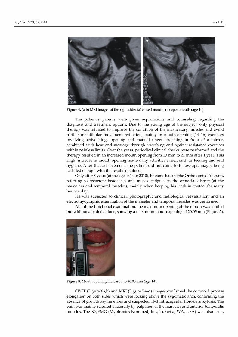
Figure 4. (a,b) MRI images at the right side: (a) closed mouth; (b) open mouth (age 10).
The patient’s parents were given explanations and counseling regarding the
diagnosis and treatment options. Due to the young age of the subject, only physical
therapy was initiated to improve the condition of the masticatory muscles and avoid
further mandibular movement reduction, mainly in mouth-opening [14–16] exercises
involving active hinge opening and manual finger stretching in front of a mirror,
combined with heat and massage through stretching and against-resistance exercises
within painless limits. Over the years, periodical clinical checks were performed and the
therapy resulted in an increased mouth opening from 13 mm to 21 mm after 1 year. This
slight increase in mouth opening made daily activities easier, such as feeding and oral
hygiene. After that achievement, the patient did not come to follow-ups, maybe being
satisfied enough with the results obtained.
Only after 8 years (at the age of 14 in 2010), he came back to the Orthodontic Program,
referring to recurrent headaches and muscle fatigues in the orofacial district (at the
masseters and temporal muscles), mainly when keeping his teeth in contact for many
hours a day.
He was subjected to clinical, photographic and radiological reevaluation, and an
electromyographic examination of the masseter and temporal muscles was performed.
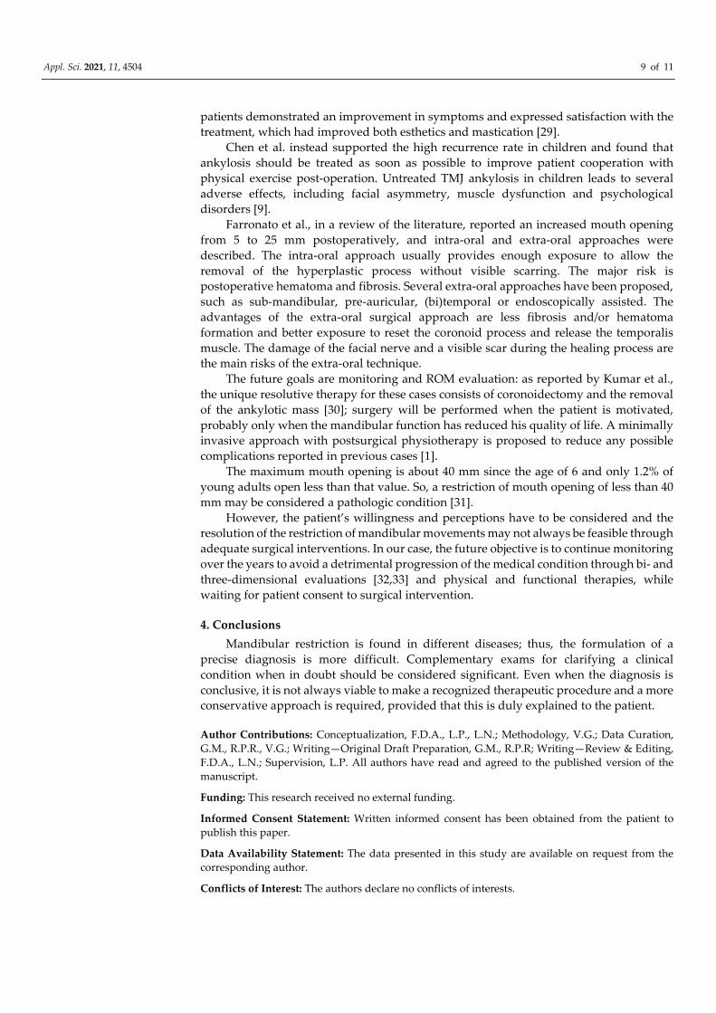
About the functional examination, the maximum opening of the mouth was limited
but without any deflections, showing a maximum mouth opening of 20.05 mm (Figure 5).
Figure 5. Mouth opening increased to 20.05 mm (age 14).
CBCT (Figure 6a,b) and MRI (Figure 7a–d) images confirmed the coronoid process
elongation on both sides which were locking above the zygomatic arch, confirming the
absence of growth asymmetries and suspected TMJ intracapsular fibrosis ankylosis. The
pain was mainly referred bilaterally by palpation of the masseter and anterior temporalis
muscles. The K7/EMG (Myotronics-Noromed, Inc., Tukwila, WA, USA) was also used,

Appl. Sci. 2021, 11, 4504 5 of 11
and it showed dystonia of all masticatory muscles and hypoactivity of the anterior right
temporalis at rest.
Figure 6. (a,b) CBCT examinations at closed mouth: (a) right side; (b) left side (age 14).
Figure 7. (a–d) MRI images: (a) right side—closed mouth; (b) right side—open mouth; (c) left
side—closed mouth; (d) left side—open mouth (age 14).
The patient accepted treatment with Transcutaneous Electrical Nerve Stimulation
(TENS) to reduce muscle tension, a bite plane to control the parafunctional activity of the
jaw at night and self-control instruction for daytime habits. The patient was treated 30 min
daily, two times weekly for 3 months, obtaining a significant reduction in algic
symptomatology.
However, it was reiterated that no conservative therapy could solve the patient’s
condition except bilateral coronoidectomy and removal of the ankylotic mass with
ostectomy at the end of growth, then allowing correct mandibular movements together
with better mouth opening.
In 2020, in a long-term follow-up of 19 years, the patient was recalled for a monitoring
check-up at the age of 23. A complete check-up was carried out. At the functional
examination, the range of movement (ROM) was unchanged compared to the previous
evaluation of 9 years before. Intraoral examination showed severe tooth decays at second
and third lower molars on the left side that were extracted despite the limited opening of
the mouth (Figure 8). On the panoramic X-ray, the Levandoski panographic analysis was
used to compare measurements with the previous analysis performed at the age of 7. The
ratio of Kr-Go/Cd-Go on both sides was increased with a value of 1.11 on the right and
1.10 on the left side.

Appl. Sci. 2021, 11, 4504 6 of 11
Figure 8. Diagram of the Levandoski panographic analysis on the panoramic X-ray (age 23).
CBCT imaging was obtained to evaluate the coronoid process changes in comparison
to scans of 13 years before (Figure 9). The mouth opening remained constant whereas the
joint disks and condyle at the MRI examination showed an ankylotic aspect despite the
acceptable position (Figure 10).
Figure 9. (a,b) CBCT examinations at closed mouth: (a) right side; (b) left side (age 23).

Appl. Sci. 2021, 11, 4504 7 of 11
Figure 10. (a–d). MRI images: (a) right side—closed mouth; (b) right side—open mouth; (c) left
side—closed mouth; (d) left side—open mouth (age 23).
The K7/EMG confirmed a mild hypotony of the masseter and anterior right temporal
muscles (Figure 11).
Figure 11. EMG/K7 analysis at rest (age 23). The following muscles were recorded: left temporalis
anterior (LTA), left masseter (LMM), right masseter (RMM), right temporalis anterior (RTA).
Considering the adult age of the patient and his diagnostic records over time, surgical
intervention of coronoidectomy was suggested as the gold standard of therapy to resolve
the limited mouth opening. In this regard, new, minimally invasive surgical techniques
associated with early postsurgical physiotherapy are available to reduce the main possible
complications [1]. This was referred to the patient, along with the provision of all detailed
information on treatment modalities and the consequent benefits to his oral functions.
Nevertheless, the patient denied surgery for correction of the hypertrophy of the coronoid
process, arguing that, at the time, the condition did not cause any reduction of his quality
of daily life. He preferred to delay the surgical intervention to the future even though he
understood the benefits of this approach.
3. Discussion
CPH is often misdiagnosed due to its rarity and the absence of pain [12], even if
restrictions in mouth opening and mandibular movements are present since childhood
[17]. Thus, a deep knowledge of any clinical and instrumental analyses available to
monitor this condition during growth is necessary to make an early diagnosis and correct

Appl. Sci. 2021, 11, 4504 8 of 11
treatment plan. The normal range of mouth opening is 53–58 mm, measured between the
incisal edges of the maxillary and mandibular teeth.
Diagnosis of CPH is effective mostly through imaging [17]. First, a panoramic X-ray
is recommended [2,17], and if there are any suspicions, the subsequent step is the
prescription of three-dimensional imaging such as CBCT and MRI to detect any
intraarticular derangements in hard or soft tissues [5,18]. In particular, MRI may help
differentiate CPH from chronic disk displacement without reduction [17,18]. The etiology
of coronoid hypertrophy is not well-described, but it has been found in association with
many conditions [4,7,19]. In this case, it may be supposed that the trauma was the main
cause for the onset of the pathological process [1]. Studies carried out on postnatal
surgeries on laboratory animals showed that changes in muscular connection result in
changes in size and form of the coronoid process and deficits in some myogenic factors or
in the expression of specific genes that can provoke muscular and articular disorders
[17,20]. Patients with restricted mouth opening tend to eat softer foods, which may lead
to reduced temporalis muscle activity. This results in fatigue and chronic hyperemia,
further stimulating an inflammatory response. The chronic persistence of inflammatory
cells in the muscles induces an excess of cytokines and growth factors, which contributes
to the formation of permanent fibrosis [6,21]. So, increased duration of TMJ ankylosis
leads to an increased incidence of coronoid elongation.
The surgical management of CPH patients is very complex, being that the treatment
of associated TMJ ankylosis is highly challenging for surgeons [9]. The major problems
faced during such surgeries are the risk of excessive blood loss with all of the
complications of blood transfusions [7], joint reankylosis [3], function reduction, growth
defect postoperatively [8] and the mandibular inability to intubate in an emergency [22].
Obtaining a satisfactory outcome depends largely on proper post-operative rehabilitation
[2] to avoid relapse, and to emphasize results, it is suggested to start physiotherapy 5–7
days after surgery [2,9,23].
It can be difficult to define the time to administer treatment in infants, although most
authors agree that, except in patients with very severe limitations in mouth opening, it is
better to perform the operation once the growth process has finished, in order to avoid
recurrence, deformity or even restricted movements [2,12,24].
Kumar et al. demonstrated that patients >16 years old showed greater improvement
in mouth opening at >1-year follow-up compared to patients aged <16 years [6]. The rate
of joint reankylosis in a child group was 19.1%, which was significantly greater than that
in the adult group (7.3%; p < 0.05) [9]. Yang et al. demonstrated that the rate of facial
growth and function after the reconstruction of the mandibular condyle in children with
ankylosis seemed to be less than on the undisturbed side, even after surgical treatment of
the ankylosis [25].
A review of the literature found that vertical amplitude of normal mandibular
movements for the production of speech is equal to one-third of the amplitude of the
maximum opening of the jaw, around 7–18 mm with a slight anteroposterior component
of 2–3 mm [26]. Moreover, a typical chewing pattern consists of a few cycles on one side,
after which the food bolus is moved to the other side by tongue and cheeks for a similar
number of cycles. Each cycle commences with an opening phase in which the teeth move
away from the intercuspal position along a path close to the median line but gradually
approaches the food bolus side. Rarely does the total movement exceed the minimum
necessary to grasp the food bolus; on average, the opening phase involves about 18 mm
incisal movement [27]. Effectively, our patient did not refer to any problems during
chewing function.
Thus, physical and manual therapy (including joint mobilization and the
manipulation of soft tissues) is amongst the 10 most common treatments used for
temporomandibular disorders to improve range of motion and promote exercise to
maintain healthy function [5,28]. Mazzetto et al. showed that after physical therapy,
Appl. Sci. 2021, 11, 4504 9 of 11
patients demonstrated an improvement in symptoms and expressed satisfaction with the
treatment, which had improved both esthetics and mastication [29].
Chen et al. instead supported the high recurrence rate in children and found that
ankylosis should be treated as soon as possible to improve patient cooperation with
physical exercise post-operation. Untreated TMJ ankylosis in children leads to several
adverse effects, including facial asymmetry, muscle dysfunction and psychological
disorders [9].
Farronato et al., in a review of the literature, reported an increased mouth opening
from 5 to 25 mm postoperatively, and intra-oral and extra-oral approaches were
described. The intra-oral approach usually provides enough exposure to allow the
removal of the hyperplastic process without visible scarring. The major risk is
postoperative hematoma and fibrosis. Several extra-oral approaches have been proposed,
such as sub-mandibular, pre-auricular, (bi)temporal or endoscopically assisted. The
advantages of the extra-oral surgical approach are less fibrosis and/or hematoma
formation and better exposure to reset the coronoid process and release the temporalis
muscle. The damage of the facial nerve and a visible scar during the healing process are
the main risks of the extra-oral technique.
The future goals are monitoring and ROM evaluation: as reported by Kumar et al.,
the unique resolutive therapy for these cases consists of coronoidectomy and the removal
of the ankylotic mass [30]; surgery will be performed when the patient is motivated,
probably only when the mandibular function has reduced his quality of life. A minimally
invasive approach with postsurgical physiotherapy is proposed to reduce any possible
complications reported in previous cases [1].
The maximum mouth opening is about 40 mm since the age of 6 and only 1.2% of
young adults open less than that value. So, a restriction of mouth opening of less than 40
mm may be considered a pathologic condition [31].
However, the patient’s willingness and perceptions have to be considered and the
resolution of the restriction of mandibular movements may not always be feasible through
adequate surgical interventions. In our case, the future objective is to continue monitoring
over the years to avoid a detrimental progression of the medical condition through bi- and
three-dimensional evaluations [32,33] and physical and functional therapies, while
waiting for patient consent to surgical intervention.
4. Conclusions
Mandibular restriction is found in different diseases; thus, the formulation of a
precise diagnosis is more difficult. Complementary exams for clarifying a clinical
condition when in doubt should be considered significant. Even when the diagnosis is
conclusive, it is not always viable to make a recognized therapeutic procedure and a more
conservative approach is required, provided that this is duly explained to the patient.
Author Contributions: Conceptualization, F.D.A., L.P., L.N.; Methodology, V.G.; Data Curation,
G.M., R.P.R., V.G.; Writing—Original Draft Preparation, G.M., R.P.R; Writing—Review & Editing,
F.D.A., L.N.; Supervision, L.P. All authors have read and agreed to the published version of the
manuscript.
Funding: This research received no external funding.
Informed Consent Statement: Written informed consent has been obtained from the patient to
publish this paper.
Data Availability Statement: The data presented in this study are available on request from the
corresponding author.
Conflicts of Interest: The authors declare no conflicts of interests.

Appl. Sci. 2021, 11, 4504 10 of 11
References
1. Farronato, M.; Lucchina, A.G.; Mortellaro, C.; Fama, A.; Galbiati, G.; Farronato, G.; Maspero, C. Bilateral hyperplasia of the
coronoid process in pediatric patients: What is the gold standard for treatment? J. Craniofac. Surg. 2019, 30, 1058–1063.
2. Fernández Ferro, M.; Fernandez Sanroman, J.; Sandoval Gutierrez, J.; Costas López, A.; López de Sánchez, A.; Etayo Pérez, A.
Treatment of bilateral hyperplasia of the coronoid process of the mandible. Presentation of a case and review of the literature.
Med. Oral Patol. Oral Cir. Bucal. 2008, 13, 595–598.
3. Lehman, H.; Fleissig, Y.; Abid-El-Raziq, D.; Nitzan, D. Limited mouth opening of unknown cause cured by diagnostic
coronoidectomy: A new clinical entity? Br. J. Oral Maxillofac. Surg. 2015, 53, 230–234.
4. Capote, A.; Rodríguez, F.J.; Blasco, A.; Muñoz, M.F. Jacob’s disease associated with temporomandibular joint dysfunction: A
case report. Med. Oral Patología Oral Cirugia Bucal 2005, 10, 210–214.
5. Minervini, G.; Lucchese, A.; Perillo, L.; Serpico, R.; Minervini, G. Unilateral superior condylar neck fracture with dislocation in
a child treated with an acrylic splint in the upper arch for functional repositioning of the mandible. CRANIO 2016, 35, 337–341.
6. Bravo-Hammett, S.; Nucci, L.; Christou, T.; Aristizabal, J.F.; Kau, C.H. 3D Analysis of Facial Morphology of a Colombian
Population Compared to Adult Caucasians. Eur. J. Dent. 2020, 14, 342–351.
7. Kubota, Y.; Takenoshita, Y.; Takamori, K.; Kanamoto, M.; Shirasuna, K. Levandoski panographic analysis in the diagnosis of
hyperplasia of the coronoid process. Br. J. Oral Maxillofac. Surg. 1999, 37, 409–411.
8. Wang, W.; Xu, B.; Zhang, B.; Lou, H. Temporomandibular joint ankylosis contributing to coronoid process hyperplasia. Int. J.
Oral Maxillofac. Surg. 2016, 45, 1229–1233.
9. Chen, S.; He, Y.; An, J.-G.; Zhang, Y. Recurrence-Related Factors of Temporomandibular Joint Ankylosis: A 10-Year Experience.
J. Oral Maxillofac. Surg. 2019, 77, 2512–2521.
10. Maspero, C.; Giannini, L.; Terzi, L.; Sesso, G. Bilateral hyperplasia of the coronoid process in pediatric patients. Dent. Cadmos.
2012, 80, 559–563.
11. Satoh, K.; Ohno, S.; Aizawa, T.; Imamura, M.; Mizutani, H. Bilateral Coronoid Hyperplasia in an Adolescent: Report of a Case
and Review of the Literature. J. Oral Maxillofac. Surg. 2006, 64, 334–338.
12. Mano, T.; Ueyama, Y.; Koyama, T.; Nishiyama, A.; Matsumura, T. Trismus due to bilateral coronoid hyperplasia in a child: Case
report. J. Oral Maxillofac. Surg. 2005, 63, 399–401.
13. Çorumlu, U.; Kopuz, C.; Demir, M.T.; Pirzirenli, M.E. Bilateral elongated mandibular coronoid process in an Anatolian skull.
Anat. Cell Biol. 2016, 49, 217–220.
14. McLoughlin, P.M.; Hopper, C.; Bowley, N.B. Hyperplasia of the mandibular coronoid process: an analysis of 31 cases and a
review of the literature. J. Oral Maxillofac. Surg. 1995, 53(3):250-5.
15. Marji, F.P.; Anstadt, E.; Davit, A.; Goldstein, J.A.; Losee, J.E. Pediatric Mandibular Condylar Fractures With Concomitant
Cervical Spine Injury: A Treatment Protocol for Prevention of Temporomandibular Joint Ankylosis. J. Craniofac. Surg. 2020, 31,
e248–e250.
16. Fariña, R.; Canto, L.; Gunckel, R.; Alister, J.P.; Uribe, F. Temporomandibular Joint Ankylosis: Algorithm of Treatment. J.
Craniofac. Surg. 2018, 29, 427–431.
17. Ghazizadeh, M.; Sheikhi, M.; Salehi, M.M.; Khaleghi, A. Bilateral coronoid hyperplasia causing painless limitation of
mandibular movement. Radiol. Case Rep. 2018, 13, 112–117.
18. Minervini, G.; Nucci, L.; Lanza, A.; Femiano, F.; Contaldo, M.; Grassia, V. Temporomandibular disc displacement with
reduction treated with anterior repositioning splint: A 2-year clinical and magnetic resonance imaging (MRI) follow-up. J. Biol.
Regul. Homeost. Agents. Supplement, D. 2020, 34, 151–160.
19. Chang, C.C.; Allori, A.C.; Wang, E.; Fariña, R.; Warren, S.M.; Grayson, B.H.; McCarthy, J.G. A Quantitative Three-Dimensional
Analysis of Coronoid Hypertrophy in Pediatric Craniofacial Malformations. Plast. Reconstr. Surg. 2012, 129, 312e–318e.
20. Anthwal, N.; Peters, H.; Tucker, A.S. Species-specific modifications of mandible shape reveal independent mechanisms for
growth and initiation of the coronoid. EvoDevo 2015, 6, 35.
21. Moccia, S.; Nucci, L.; Spagnuolo, C.; D’Apuzzo, F.; Piancino, M.G.; Minervini, G. Polyphenols as Potential Agents in the
Management of Temporomandibular Disorders. Appl. Sci. 2020, 10, 5305.
22. Costello, B.J.; Edwards, S.P. Pediatric Mandibular Hypomobility: Current Management and Controversies. Oral Maxillofac. Surg.
Clin. N. Am. 2005, 17, 455–466.
23. Gibbons, A.; Abulhoul, S. Use of a Therabite appliance in the management of bilateral mandibular coronoid hyperplasia. Br. J.
Oral Maxillofac. Surg. 2007, 45, 505–506.
24. Contaldo, M.; Della Vella, F.; Raimondo, E.; Minervini, G.; Buljubasic, M.; Ogodescu, A.; Sinescu, C.; Serpico, R. Early Childhood
Oral Health Impact Scale (ECOHIS): Literature review and Italian validation. Int. J. Dent. Hyg. 2020, 18, 396–402.
25. Yang, Y.; Li, Y.; Jiang, N.; Bi, R.; Zhu, S. Grafts of autogenous coronoid process to reconstruct the mandibular condyle in children
with unilateral ankylosis of the temporomandibular joint: Long-term effects on mandibular growth. Br. J. Oral Maxillofac. Surg.
2018, 56, 107–112.
26. Bianchini, E.M.; De Andrade, C.R. A Model of Mandibular Movements during Speech: Normative Pilot Study for the Brazilian
Portuguese Language. CRANIO 2006, 24, 197–206.
27. Brown, T. Mandibular Movements. Monogr. Oral Sci. 1975, 4, 126–150.
28. Armijo-Olivo, S.; Pitance, L.; Singh, V.; Neto, F.; Thie, N.; Michelotti, A. Effectiveness of Manual Therapy and Therapeutic
Exercise for Temporomandibular Disorders: Systematic Review and Meta-Analysis. Phys. Ther. 2016, 96, 9–25.

Appl. Sci. 2021, 11, 4504 11 of 11
29. Mazzetto, M.O.; Hotta, T.H. Hypertrophy of the mandibular coronoid process and structural alterations of the condyles
associated with limited buccal opening: Case report. Braz. Dent. J. 2007, 18, 171–174.
30. Kumar, P.; Singh, V.; Agrawal, A.; Bhagol, A.; Bali, R. Incremental increase in percentage mouth opening after coronoidectomy
in temporomandibular joint ankylosis. Int. J. Oral Maxillofac. Surg. 2015, 44, 859–863.
31. Okeson, J.P. Management of Temporomandibular Disorders and Occlusion, 8th ed.; Elsevier Health Sciences: Amsterdam, The
Netherlands, 2019, 1–23.
32. Dekel, E.; Nucci, L.; Weill, T.; Flores-Mir, C.; Becker, A.; Perillo, L.; Chaushu, S. Impaction of maxillary canines and its effect on
the position of adjacent teeth and canine development: A cone-beam computed tomography study. Am. J. Orthod. Dentofac.
Orthop. 2021, 159, e135–e147.
33. Grassia, V.; D’Apuzzo, F.; Ferrulli, V.E.; Matarese, G.; Femiano, F.; Perillo, L. Dento-skeletal effects of mixed palatal expansion
evaluated by postero-anterior cephalometric analysis. Eur. J. Paediatr. Dent. 2014, 15, 59–62.
Copyright © 2022 FDOKUMEN