Control of Leg Movements Driven by EMG Activity of Shoulder Muscles
9
HUMAN NEUROSCIENCE ORIGINAL RESEARCH ARTICLE published: 20 October 2014 doi: 10.3389/fnhum.2014.00838 Control of leg movements driven by EMG activity of shoulder muscles Valentina La Scaleia 1,2 *, Francesca Sylos-Labini 1,2 ,Thomas Hoellinger 3 , Letian Wang 4 , Guy Cheron 3 , Francesco Lacquaniti 1,2,5 and Yuri P. Ivanenko 1 1 Laboratory of Neuromotor Physiology, Santa Lucia Foundation, Rome, Italy 2 Centre of Space Bio-Medicine, University of RomeTor Vergata, Rome, Italy 3 Laboratory of Neurophysiology and Movement Biomechanics, Université Libre de Bruxelles, Brussels, Belgium 4 Department of Biomechanical Engineering, University ofTwente, Enschede, Netherlands 5 Department of Systems Medicine, University of RomeTor Vergata, Rome, Italy Edited by: Ana Bengoetxea, Universidad del País Vasco-Euskal Herriko Unibertsitatea (UPV/EHU), Spain Reviewed by: Vincent C. K. Cheung, Massachusetts Institute ofTechnology, USA Tomoyoshi Komiyama, Chiba University, Japan *Correspondence: Valentina La Scaleia, Laboratory of Neuromotor Physiology, IRCCS Fondazione Santa Lucia, 306 via Ardeatina, Rome 00179, Italy e-mail: [email protected] During human walking, there exists a functional neural coupling between arms and legs, and between cervical and lumbosacral pattern generators. Here, we present a novel approach for associating the electromyographic (EMG) activity from upper limb muscles with leg kinematics. Our methodology takes advantage of the high involvement of shoulder muscles in most locomotor-related movements and of the natural co-ordination between arms and legs. Nine healthy subjects were asked to walk at different constant and variable speeds (3–5 km/h), while EMG activity of shoulder (deltoid) muscles and the kinematics of walking were recorded.To ensure a high level of EMG activity in deltoid, the subjects performed slightly larger arm swinging than they usually do. The temporal structure of the burst-like EMG activity was used to predict the spatiotemporal kinematic pattern of the forthcoming step. A comparison of actual and predicted stride leg kinematics showed a high degree of correspondence (r > 0.9). This algorithm has been also implemented in pilot experiments for controlling avatar walking in a virtual reality setup and an exoskeleton dur- ing over-ground stepping.The proposed approach may have important implications for the design of human–machine interfaces and neuroprosthetic technologies such as those of assistive lower limb exoskeletons. Keywords: arm–leg co-ordination, quadrupedal locomotion, EMG patterns, gait kinematics, neuroprosthetic technology INTRODUCTION Neuroprosthetic devices based on brain–machine interface (BMI) technology have the potential to restore mobility to both upper and lower extremities and to enable walking (Millán et al., 2010; Lebedev et al., 2011; Cheron et al., 2012; Wolpaw, 2013). Exoskeleton robotic devices are extensively developed in recent years to provide new possibilities for severely paralyzed patients to walk (Sale et al., 2012; Malcolm et al., 2013; Wang et al., 2013; Sylos-Labini et al., 2014b). The development of practi- cal applications for gait assistance may involve a wide range of potential control implementations, from using trunk movements (Wang et al., 2013) or myoelectric signals (del-Ama et al., 2012; Alcaide-Aguirre et al., 2013; Gordon et al., 2013) to implement- ing BMI-based technology (Fitzsimmons et al., 2009; Gwin et al., 2011). To develop effective neuroprosthetic devices for human beings, BMI research has to address a number of issues related to improving the quality of neuronal recordings, and extend- ing the BMI approach to a broad range of motor and sensory functions (Lebedev et al., 2011). In human beings, non-invasive electroencephalogram-based brain–machine interfacing for pro- viding a reliable control of walking in the exoskeleton is still very limited (Gwin et al., 2011; Cheron et al., 2012; Wagner et al., 2012), and alternative or supplementary control strategies should be considered in parallel. The current study investigates a novel approach for associating the upper limb electromyo- graphic (EMG) activity with leg kinematics. This approach holds promise for applications in the field of assistive lower limb exoskeletons. The co-ordination of limb and body segments in locomo- tion arises from stereotyped coupling of cervical and lumbosacral spinal segment outputs (Ballesteros et al., 1965; Murray et al., 1967; Hogue, 1969; Wannier et al., 2001; Zehr and Duysens, 2004; Iva- nenko et al., 2006; De Sèze et al., 2008; Barthelemy and Nielsen, 2010; Meyns et al., 2013; Sylos-Labini et al., 2014a). The co- ordination between arms and legs during human locomotion shares many features with that in quadrupeds (Falgairolle et al., 2006; Patrick et al., 2009; Juvin et al., 2012; MacLellan et al., 2012). Due to the natural arm–leg co-ordination in human walking, the leg movement control can be derived from or reflected in the EMGs of arm muscles. In particular, a group of the upper arm muscles was shown to be active during many locomotion tasks in human beings (Ivanenko et al., 2006; Kuhtz-Buschbeck and Jing, 2012). Taking into account, the high involvement of the deltoid muscle in most locomotor-related movements in human beings, we took advantage of the natural arm–leg co-ordination for investigating the control of stepping using the upper limb EMG activity. Here, Frontiers in Human Neuroscience www.frontiersin.org October 2014 |Volume 8 | Article 838 | 1
-
Upload
independent -
Category
Documents
-
view
4 -
download
0
Transcript of Control of Leg Movements Driven by EMG Activity of Shoulder Muscles

HUMAN NEUROSCIENCEORIGINAL RESEARCH ARTICLE
published: 20 October 2014doi: 10.3389/fnhum.2014.00838
Control of leg movements driven by EMG activity ofshoulder musclesValentina La Scaleia1,2*, Francesca Sylos-Labini 1,2,Thomas Hoellinger 3, Letian Wang4, Guy Cheron3,Francesco Lacquaniti 1,2,5 andYuri P. Ivanenko1
1 Laboratory of Neuromotor Physiology, Santa Lucia Foundation, Rome, Italy2 Centre of Space Bio-Medicine, University of Rome Tor Vergata, Rome, Italy3 Laboratory of Neurophysiology and Movement Biomechanics, Université Libre de Bruxelles, Brussels, Belgium4 Department of Biomechanical Engineering, University of Twente, Enschede, Netherlands5 Department of Systems Medicine, University of Rome Tor Vergata, Rome, Italy
Edited by:Ana Bengoetxea, Universidad del PaísVasco-Euskal Herriko Unibertsitatea(UPV/EHU), Spain
Reviewed by:Vincent C. K. Cheung, MassachusettsInstitute of Technology, USATomoyoshi Komiyama, ChibaUniversity, Japan
*Correspondence:Valentina La Scaleia, Laboratory ofNeuromotor Physiology, IRCCSFondazione Santa Lucia, 306 viaArdeatina, Rome 00179, Italye-mail: [email protected]
During human walking, there exists a functional neural coupling between arms and legs,and between cervical and lumbosacral pattern generators. Here, we present a novelapproach for associating the electromyographic (EMG) activity from upper limb muscleswith leg kinematics. Our methodology takes advantage of the high involvement of shouldermuscles in most locomotor-related movements and of the natural co-ordination betweenarms and legs. Nine healthy subjects were asked to walk at different constant and variablespeeds (3–5 km/h), while EMG activity of shoulder (deltoid) muscles and the kinematicsof walking were recorded. To ensure a high level of EMG activity in deltoid, the subjectsperformed slightly larger arm swinging than they usually do. The temporal structure of theburst-like EMG activity was used to predict the spatiotemporal kinematic pattern of theforthcoming step. A comparison of actual and predicted stride leg kinematics showed ahigh degree of correspondence (r > 0.9).This algorithm has been also implemented in pilotexperiments for controlling avatar walking in a virtual reality setup and an exoskeleton dur-ing over-ground stepping. The proposed approach may have important implications for thedesign of human–machine interfaces and neuroprosthetic technologies such as those ofassistive lower limb exoskeletons.
Keywords: arm–leg co-ordination, quadrupedal locomotion, EMG patterns, gait kinematics, neuroprosthetictechnology
INTRODUCTIONNeuroprosthetic devices based on brain–machine interface (BMI)technology have the potential to restore mobility to both upperand lower extremities and to enable walking (Millán et al.,2010; Lebedev et al., 2011; Cheron et al., 2012; Wolpaw, 2013).Exoskeleton robotic devices are extensively developed in recentyears to provide new possibilities for severely paralyzed patientsto walk (Sale et al., 2012; Malcolm et al., 2013; Wang et al.,2013; Sylos-Labini et al., 2014b). The development of practi-cal applications for gait assistance may involve a wide range ofpotential control implementations, from using trunk movements(Wang et al., 2013) or myoelectric signals (del-Ama et al., 2012;Alcaide-Aguirre et al., 2013; Gordon et al., 2013) to implement-ing BMI-based technology (Fitzsimmons et al., 2009; Gwin et al.,2011). To develop effective neuroprosthetic devices for humanbeings, BMI research has to address a number of issues relatedto improving the quality of neuronal recordings, and extend-ing the BMI approach to a broad range of motor and sensoryfunctions (Lebedev et al., 2011). In human beings, non-invasiveelectroencephalogram-based brain–machine interfacing for pro-viding a reliable control of walking in the exoskeleton is stillvery limited (Gwin et al., 2011; Cheron et al., 2012; Wagneret al., 2012), and alternative or supplementary control strategies
should be considered in parallel. The current study investigatesa novel approach for associating the upper limb electromyo-graphic (EMG) activity with leg kinematics. This approach holdspromise for applications in the field of assistive lower limbexoskeletons.
The co-ordination of limb and body segments in locomo-tion arises from stereotyped coupling of cervical and lumbosacralspinal segment outputs (Ballesteros et al., 1965; Murray et al., 1967;Hogue, 1969; Wannier et al., 2001; Zehr and Duysens, 2004; Iva-nenko et al., 2006; De Sèze et al., 2008; Barthelemy and Nielsen,2010; Meyns et al., 2013; Sylos-Labini et al., 2014a). The co-ordination between arms and legs during human locomotionshares many features with that in quadrupeds (Falgairolle et al.,2006; Patrick et al., 2009; Juvin et al., 2012; MacLellan et al., 2012).Due to the natural arm–leg co-ordination in human walking, theleg movement control can be derived from or reflected in the EMGsof arm muscles. In particular, a group of the upper arm muscleswas shown to be active during many locomotion tasks in humanbeings (Ivanenko et al., 2006; Kuhtz-Buschbeck and Jing, 2012).Taking into account, the high involvement of the deltoid musclein most locomotor-related movements in human beings, we tookadvantage of the natural arm–leg co-ordination for investigatingthe control of stepping using the upper limb EMG activity. Here,
Frontiers in Human Neuroscience www.frontiersin.org October 2014 | Volume 8 | Article 838 | 1

La Scaleia et al. Arm EMG-based assisted gait
we tested the hypothesis that the temporal structure of EMG activ-ity of shoulder muscles can be used to predict the spatiotemporalkinematic pattern of the forthcoming step during actual gait at 3–5 km/h (and we investigated the accuracy of this prediction) and tocontrol avatar walking in a virtual reality setup or an exoskeletonduring over-ground stepping.
MATERIALS AND METHODSPARTICIPANTS AND PROTOCOLSTwo main protocols were implemented. In the first series of exper-iments performed in Brussels (protocol 1), we investigated thepossibility of extracting lower limb kinematic data from the gait-related rhythmic EMG activity of arm muscles. To this end, ninehealthy volunteers were enrolled [age range 23–33 years, fourmales and five females, leg length 0.85± 0.04 m (mean± SD),height 1.75± 0.10 m, weight 72± 7 kg]. In the second series ofexperiments performed in Rome (protocol 2), we implementedthe on-line control of avatar walking and also over-ground walk-ing in the exoskeleton. For this protocol, eight healthy volunteerswere enrolled (age range 25–53 years, five males and three females,leg length 0.83± 0.04 m, height 1.77± 0.09 m, weight 69± 8 kg).None of the subjects had any known neurological or motor dis-order. These protocols were approved by the Ethics Committeesof the Université Libre de Bruxelles (Belgium) and FondazioneSanta Lucia (Rome, Italy), respectively, and all subjects gave theirinformed written consent prior to participation.
EXPERIMENTAL SETUP AND DATA ANALYSISProtocol 1In the first protocol, the subjects were asked to walk on a treadmill(Cosmed Treadmill T150) at different constant speeds (3, 4, and5 km/h) and at a variable speed (ramp-and-hold velocity profile,increasing from 3 to 5 km/h at 0.1 km/h/s), four trials total (3 con-stant speeds+ 1 trial at a variable speed). On average, 10–15 strideswere recorded in each trial during walking at a constant speed (thatcorresponds to a walking distance of ~15–20 m) and about 50strides during walking at a variable speed (distance ~70 m). Dur-ing normal walking, the amplitude of arm swinging depends onthe walking speed (Webb et al., 1994) and, at slow speeds, the activ-ity of arm muscles decreases significantly (Ivanenko et al., 2006;Kuhtz-Buschbeck and Jing, 2012). Therefore, to obtain a higherlevel of arm EMG activity, the subjects were asked to performslightly larger arm swinging than they usually do. In the prelimi-nary training sessions (2–3 min of walking), we verified that thesemovements evoked consistent EMG activation of shoulder mus-cles at all walking speeds (3–5 km/h). We recorded kinematic databilaterally at 100 Hz by means of the Vicon system (Vicon, Oxford,UK) with 10 Bonita cameras spaced around the treadmill. Infraredreflective markers (diameter 15 mm) were attached on each side ofthe subjects to the skin (using Vicon BioMind asymmetric model),in particular, overlying the following landmarks used to calculateleg kinematics: greater trochanter (GT), lateral femur epicondyle(LE), lateral malleolus (LM), and fifth metatarsophalangeal joint(VM). EMG activity of the anterior deltoid (DELTa) and poste-rior deltoid (DELTp) muscles was recorded bilaterally by meansof surface electrodes. The EMG data were recorded with the wire-less BTS Freeemg system (BTS Bioengineering, Milano, Italy) and
digitized at 1000 Hz. Sampling of kinematic and EMG data weresynchronized.
Data processingOur approach uses the timing of the burst-like EMG activity ofshoulder muscles (by applying the peak detection algorithm) topredict the spatiotemporal kinematic pattern of the forthcomingstep. Prior to application of the peak detection algorithm, the EMGdata were pre-processed: high-pass filtered at 30 Hz, rectified, andfinally low-pass filtered (all filters, zero-lag fourth order Butter-worth). Low-pass filtering was performed at different frequencies(1÷ 5 Hz) to achieve the best correlation between actual and pre-dicted leg kinematics. Despite some inter-individual variability,periods of EMG activity of DELTa and DELTp during normalwalking tend to be alternating and correspond to those of the con-tralateral upper limb (Ivanenko et al., 2006; Kuhtz-Buschbeck andJing, 2012), as well as multi-muscle synergy-based control inter-face may be more efficient than a single-muscle control (Lunardiniet al., 2014). Therefore, bilateral EMGs of synergistic muscles (Iva-nenko et al., 2006; Kuhtz-Buschbeck and Jing, 2012) were summed(Figure 1A):
EMG1 = DELTaright + DELTpleft (1)
EMG2 = DELTaleft + DELTpright (2)
the peaks of EMG1 and EMG2 occur around the beginning of theswing phase of the right and left legs, respectively (Figure 1A).Nevertheless, we also compared the performance of our algorithmusing all four muscles (Eqs 1 and 2) and only pairs of contralateralmuscles (DELTaleft and DELTaright; DELTpleft and DELTpright).
The elevation angles of the thigh (GT-LE), shank (LE-LM), andfoot (LM-VM) segments (Lacquaniti et al., 2002; Cheron et al.,2012) were calculated as the angles between the segment projectedon the sagittal plane and the vertical (positive in the forward direc-tion, i.e., when the distal marker falls anterior to the proximalone). We divided the recorded kinematic and EMG data into gaitcycles (maximum shank elevation angle as the beginning of thegait cycle), then interpolated each stride to 200 time points, andfinally averaged across gait cycles (for each trial). These ensemble-averaged (thigh, shank, and foot) elevation angles were used asa reference (template) for predicting leg kinematics from shoul-der muscle EMGs. The amplitude of arm oscillations was assessedas the peak-to-peak amplitude of anterior–posterior wrist markermovements relative to the shoulder averaged across both (left andright) sides of the body and across all strides of the trial.
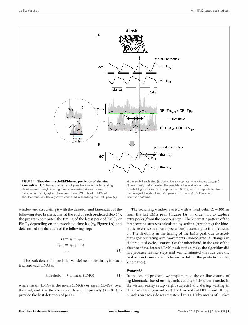
Prediction of leg kinematics based on shoulder muscle EMG activityThe algorithm used to predict the kinematics of the followingstep from the temporal structure of pre-processed EMG activityis schematically depicted in Figure 1. In essence, it predicts strideduration from the temporal distance between EMG peaks, andthe kinematic pattern of a reference template (derived from aver-ages of the collected data) is then temporally stretched to fit thepredicted duration so as to generate the predicted segment ele-vation angles. The algorithm started from the second recordedstep and consisted in detecting the timing of the EMG1 or EMG2
peak (exceeding the pre-defined threshold) in the appropriate time
Frontiers in Human Neuroscience www.frontiersin.org October 2014 | Volume 8 | Article 838 | 2
La Scaleia et al. Arm EMG-based assisted gait
FIGURE 1 | Shoulder muscle EMG-based prediction of steppingkinematics. (A) Schematic algorithm. Upper traces – actual left and rightshank elevation angles during three consecutive strides. Lowertraces – rectified (gray) and low-pass filtered (2 Hz, black) EMGs ofshoulder muscles. The algorithm consisted in searching the EMG peak (τi)
at the end of each step (ti) during the appropriate time window [(τi−1 +∆,ti), see insert] that exceeded the pre-defined individually adjustedthreshold (green line). Each step duration (Ti, Ti+1, etc.,) was predicted fromthe timing of the shoulder EMG peaks (Ti = τi − τi−1). (B) Predictedkinematic patterns.
window and associating it with the duration and kinematics of thefollowing step. In particular, at the end of each predicted step (ti),the program computed the timing of the latest peak of EMG1 orEMG2 depending on the associated time lag (τi, Figure 1A) anddetermined the duration of the following step:
Ti = τi − τi−1
Ti+1 = τi+1 − τi
. . . (3)
The peak detection threshold was defined individually for eachtrial and each EMG as
threshold = k ×mean (EMG) (4)
where mean (EMG) is the mean (EMG1) or mean (EMG2) overthe trial, and k is the coefficient found empirically (k = 0.8) toprovide the best detection of peaks.
The searching window started with a fixed delay ∆= 200 msfrom the last EMG peak (Figure 1A) in order not to captureextra peaks (from the previous step). The kinematic pattern of theforthcoming step was calculated by scaling (stretching) the kine-matic reference template (see above) according to the predictedTi. The flexibility in the timing of the EMG peak due to accel-erating/decelerating arm movements allowed gradual changes inthe predicted cycle duration. On the other hand, in the case of theabsence of the detected EMG peak at the time ti, the algorithm didnot produce further steps and was terminated (in such case thetrial was not considered to be successful for the prediction of legkinematics).
Protocol 2In the second protocol, we implemented the on-line control ofleg kinematics based on rhythmic activity of shoulder muscles inthe virtual reality setup (eight subjects) and during walking inthe exoskeleton (one subject). EMG activity of DELTa and DELTpmuscles on each side was registered at 500 Hz by means of surface
Frontiers in Human Neuroscience www.frontiersin.org October 2014 | Volume 8 | Article 838 | 3

La Scaleia et al. Arm EMG-based assisted gait
electrodes with the wireless Delsys Trigno EMG system (DelsysInc., Boston, MA, USA), bandwidth of 20–450 Hz, and overall gainof 1000. LabView software was used to collect and process the EMGdata and to predict leg kinematics in real time. The predicted kine-matic signals (thigh, shank, and foot elevation angles) were sent at100 Hz using the UDP protocol to the virtual environment (usingXVR software of VRMedia S.r.l.) to animate walking avatar (thirdperson viewpoint). The algorithm was similar (Figure 1, the low-pass filter was performed at 2 Hz, based on the results of the firstprotocol, see Results) although, in addition to the on-line step-by-step control of walking kinematics, it also included gait initiationand gait termination steps. The threshold for EMG peak detection(Eq. 4) was adjusted individually for each subject, computing themean EMG activity during alternating arm movements at a self-selected rhythm in the preliminary trial. The subjects (in standingposition) were instructed to swing their arms at approximatelythe same amplitude at self-selected, slow, and fast frequency, aswell as at a variable frequency. We instructed the subjects to varythe frequency approximately in the range between the slow andfast frequencies performed in the previous trials. The duration ofeach trial was 1 min and three trials were performed at each armfrequency.
In a pilot experiment, one subject was also trained to controlan exoskeleton during over-ground stepping along an 8-m walk-way. The detailed description of the exoskeleton (called MIND-WALKER, https://www.mindwalker-project.eu) is provided else-where (Wang et al., 2013). Briefly, knee and hip exoskeleton joints(powered by series elastic actuators) followed pre-defined jointangles (based on the walking patterns of the same subject walkingin the MINDWALKER exoskeleton without assistance) providedwith variable joint impedances (Wang et al., 2013). The algorithmwas similar to that used in the first protocol and detected the tim-ing of EMG peaks. The output of the LabView software triggeredand determined the initiation and the duration of the swing phaseof each leg.
STATISTICSDescriptive statistics included means± SD of the mean. The effi-ciency and accuracy of the predicting algorithm during walking ata constant speed (3, 4 and 5 km/h) were assessed using two para-meters: the number of successful trials (if 10 consecutive strideswere successfully predicted) and correlation between predictedand actual segment elevation angles. The efficiency of the methodduring walking at a variable speed (3–5 km/h) was assessed usingthe correlation between predicted and actual segment elevationangles, and the correlation between predicted and actual stridedurations. In protocol 2, since the actual leg kinematics was notperformed, the efficiency of the on-line algorithm using a virtualreality setup was assessed by the percentage of successful trials (ifthe algorithm produced alternating uninterrupted steps during the1-min trial) during slow, self-selected, and fast arm movements.Statistics on Pearson’s correlation coefficients was performed onthe normally distributed, Z -transformed values.
A repeated measure (RM) ANOVA was used to evaluate pre-diction (correlation coefficients between predicted and actual limbsegment elevation angles), and post hoc Tukey’s HSD test was usedto determine statistical significance. In one subject, the algorithm
failed to predict limb kinematics (presumably due to low-EMGactivity, see Results) and his data were not included. In anothersubject, it failed at a high speed (5 km/h) and the missing data forthis condition for the ANOVA were replaced by the unweightedmean value estimated from all other subjects. Reported results areconsidered significant for p < 0.05.
RESULTSSHOULDER MUSCLE ACTIVITY DURING WALKINGThe shoulder muscles we monitored (bilateral anterior and poste-rior deltoid) showed rhythmical EMG signals during walking in allsubjects (on average, the amplitude of the main rectified EMG peakwas 7.0± 1.7 µV, all muscles and all speed being pooled together),although in a few cases their activity was small (<3 µV). Since thesubjects were asked to perform approximately the same amplitudeof reciprocal arm swinging [forward arm swing reverses to back-ward arm swing in the middle of the gait cycle (Kuhtz-Buschbeckand Jing, 2012)], EMG activity did not decrease with decreas-ing walking speed, as it normally occurs during walking (Ivanenkoet al., 2006). On average, the anterior–posterior arm (wrist marker)oscillations were 50± 12 cm at 3 km/h, 45± 9 cm at 4 km/h, and41± 10 cm at 5 km/h (for instance, during normal walking at 4–5 km/h, arm oscillations are ~35–40 cm, Murray et al., 1967; Webbet al., 1994; Ford et al., 2007), and the mean amplitude of deltoidmuscle EMGs was 7.6± 1.7 µV at 3 km/h, 7.0± 1.7 µV at 4 km/h,and 6.1± 1.3 µV at 5 km/h.
Figure 2A illustrates an example of shoulder muscle EMGsignals during walking at 4 km/h. Typically, the deltoid mus-cle demonstrated alternating activity during walking: alternationoccurred both between left and right sides of the body and betweenanterior and posterior bellies of the deltoid (DELTa and DELTp).However, there could be an additional smaller second burst ofactivity over the gait cycle, as well as some inter-individual variabil-ity in the timing of the main EMG bursts [see also Ballesteros et al.(1965), Hogue (1969), Ivanenko et al. (2006), Kuhtz-Buschbeckand Jing (2012)]. Nevertheless, in most cases, there were promi-nent peaks of EMG1 and EMG2 around the beginning of the swingphase of the right and left legs, respectively (Figures 1A and 2A),which allowed us to associate this phasic alternating pattern of theupper limb EMG activity during arm–leg co-ordination with thespatiotemporal pattern of gait kinematics.
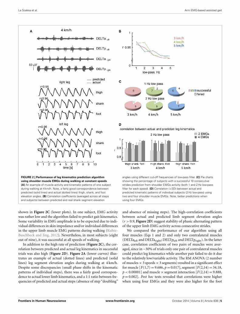
PREDICTING LEG KINEMATICS FROM SHOULDER MUSCLE EMGsDURING WALKINGWe used the rectified and filtered EMG data to detect the timingof shoulder muscle EMG peaks in the concomitant and previoussteps (see Materials and Methods) to predict the limb kinemat-ics in the forthcoming step. On the whole, the best correlationbetween actual and predicted kinematics was observed at 1–2 Hzlow-pass filtering (r > 0.95, Figure 2B), with slightly lower per-centage of successful trials at 1 Hz low-pass at faster walking speeds(Figure 2C, presumably because the cut-off frequency was closeto the step cycle frequency and the algorithm failed to detect EMGpeak in time and was terminated), so that in the following we willpresent the results obtained at 2 Hz low-pass.
The percentage of subjects with a successful prediction of con-secutive strides (2 Hz low-pass) from shoulder EMGs activity is
Frontiers in Human Neuroscience www.frontiersin.org October 2014 | Volume 8 | Article 838 | 4
La Scaleia et al. Arm EMG-based assisted gait
FIGURE 2 | Performance of leg kinematics prediction algorithmusing shoulder muscle EMGs during walking at constant speeds.(A) An example of muscle activity and kinematic patterns of one subjectduring walking at 4 km/h. Note, a fairly good correspondence betweenpredicted (solid lines) and actual (dotted lines) thigh, shank, and footelevation angles. (B) Correlation coefficients (averaged across all stepsand subjects) between predicted and real shank segment elevation
angles using different cut-off frequencies of low-pass filter. (C) Pie chartsshowing the percentage of subjects with a successful 10 consecutivestrides prediction from shoulder EMGs activity (both 1 and 2 Hz low-passfilter for each speed). (D) Correlation (+SD) between actual andpredicted kinematic patterns of individual subjects (2 Hz low-pass) usingtwo and four shoulder muscle EMGs. Note, better predictions whenusing four EMGs.
shown in Figure 2C (lower plots). In one subject, EMG activitywas rather low and the algorithm failed to predict gait kinematics.Some variability in EMG amplitude is to be expected due to indi-vidual differences in skin impedance and/or individual differencesin the upper limb muscle EMG patterns during walking (Kuhtz-Buschbeck and Jing, 2012). Nevertheless, in most subjects (eightout of nine), it was successful at all speeds of walking.
In addition to the high rate of prediction (Figure 2C), the cor-relation between predicted and actual leg kinematics in successfultrials was also high (Figure 2D). Figure 2A (lower curves) illus-trates an example of actual (dotted lines) and predicted (solidlines) leg segment elevation angles during walking at 4 km/h.Despite some discrepancies (small phase shifts in the kinematicpatterns of individual steps), there was a fairly good correspon-dence to actual lower limb kinematics, and a 1:1 ratio between fre-quencies of predicted and actual steps (absence of step “doubling”
and absence of missing steps). The high-correlation coefficientsbetween actual and predicted limb segment elevation angles(r > 0.9, Figure 2D) suggest stability of phasic alternating patternof the upper limb EMG activity across consecutive strides.
We compared the performance of our algorithm using allfour muscles (Eqs 1 and 2) and only two contralateral muscles(DELTaleft and DELTaright; DELTpleft and DELTpright). In the lattercase, correlation coefficients of two pairs of muscles were aver-aged, since in ~30% of trials only one pair of contralateral musclescould predict leg kinematics while another pair failed to do it dueto the relatively low/variable activity. The RM ANOVA (2 numberof muscles× 3 speeds× 3 segments) resulted in a significant effectfor muscle [F(1,7)= 9.686, p= 0.017], segment [F(2,14)= 34.18,p < 0.00001] and muscle× segment interaction [F(2,14)= 9.888,p= 0.002]. Post hoc tests revealed that correlations were higherwhen using four EMGs and they were also higher for the foot
Frontiers in Human Neuroscience www.frontiersin.org October 2014 | Volume 8 | Article 838 | 5

La Scaleia et al. Arm EMG-based assisted gait
elevation angle relative to the shank and thigh elevation angles(Figure 2D). Thus, both the percentage of successful trials andthe correlation coefficients were higher when using four shouldermuscles for prediction compared with only two muscles.
We also investigated the performance of the proposed leg kine-matics prediction algorithm during walking at a variable speed(3–5 km/h). Figure 3B shows an example of cycle durations ofall individual strides in one subject (left panel) and all successful(n= 8) subjects (right panel). The actual stride duration variedbetween 0.85 and 1.43 s, while the predicted stride duration alsovaried in a similar though slightly larger range (between 0.7 and1.55 s, Figure 3B). The correlation between the reference templateand individual cycles used for computing the template was high(on average 0.98± 0.01, the data for all segments were pooledtogether) consistent with a relatively low inter-stride variabilityof the segment elevation angles (Borghese et al., 1996; Bianchiet al., 1998). Therefore, inter-stride variability in the predictedstride durations and leg kinematics is likely related (at least inpart) to inter-stride variability in the EMG patterns that has beendocumented for both leg and arm muscle activity during human
walking (Kang and Dingwell, 2009; Kuhtz-Buschbeck and Jing,2012; Zelik et al., 2014). On the whole, the relationship betweenpredicted and actual stride duration was linear (Figure 3B) andcorrelation between predicted and actual leg kinematics washigh (r > 0.95, Figure 3C). RM ANOVA resulted in a significanteffect for segment [F(2,14)= 7.789, p= 0.0053] and post hoc testsrevealed higher correlations for the foot elevation angle relative tothe shank segment (p= 0.0045).
CONTROLLING VIRTUAL AVATAR AND EXOSKELETONThe suggested algorithm has also been implemented in thepilot experiments to trigger steps and control avatar walking(Figure 4A) and an exoskeleton during over-ground stepping(Figure 4B). Each trial consisted of the three locomotor-relatedphases controlled by the timing of EMG peaks: gait initiation,walking at a variable speed, and gait termination. In the absenceof the EMG peak, the virtual avatar or exoskeleton did not producefurther steps and gait termination was performed. The exoskeletonwas tested only in one trained subject since typically the wearer hasto use crutches to guarantee lateral stability (Wang et al., 2013).
FIGURE 3 | Performance of leg kinematics prediction algorithm usingshoulder muscle EMGs at variable walking speed (3–5 km/h). (A) Anexample of muscle activity and kinematic patterns of one subject duringwalking at a variable speed (ramp velocity profile, seven cycles in the middleof the trial are shown). (B) Relationship between predicted and actual stride
duration for one representative subject (left) and all subjects (right). Each pointcorresponds to individual stride and data from each subject were displayedwith different colors. Changes of predicted stride duration are fitted by a linearfunction. (C) Correlation (+SD, n=8 subjects) between actual and predictedleg kinematics.
Frontiers in Human Neuroscience www.frontiersin.org October 2014 | Volume 8 | Article 838 | 6

La Scaleia et al. Arm EMG-based assisted gait
A B
FIGURE 4 | On-line shoulder muscle EMG control of leg movements.(A) Controlling of walking avatar in a virtual reality setup (third personviewpoint). To control the timing and duration of individual steps, the subjectproduced alternating arm swinging movements in standing position (upperpanel). Lower panel – pie charts showing the percentage of trials with asuccessful 1-min test for producing stepping (if the algorithm predictedconsecutive uninterrupted steps during the 1-min trial) using alternating arm
swinging at different frequencies (n=8 subjects, 24 trials total for eachcondition). (B) Arm EMG-based control of stepping in the exoskeleton by thehealthy subject. Upper traces – rectified (gray) and low-pass filtered (black)EMGs of shoulder muscles. Each step duration and initiation were calculatedand triggered based on the timing of the shoulder EMG peaks. Bottomtraces – knee and hip joint angle kinematic patterns of eight consecutivesteps along a 9-m walkway.
Nevertheless, this subject succeeded to use alternating EMG burstsof shoulder muscles to trigger 7–10 consecutive strides along a 8-mwalkway (Figure 4B). Again, the percentage of successful trials forcontrolling virtual avatar at slow, self-selected, and fast frequencyof arm movements was high (Figure 4A, lower panel) and simi-lar to that found in the first experiment (Figure 2C) even thoughdifferent subjects participated in the two protocols.
DISCUSSIONARM SWINGING DURING LOCOMOTIONArm swing during human walking is not an entirely passive move-ment, as also shown by the observation that upper limb musclesshow rhythmic activity contributing to arm swing (Ballesteroset al., 1965; Hogue, 1969; Ivanenko et al., 2006). Interestingly,rhythmic muscle activity continues to some extent even when thearm is immobilized (Kuhtz-Buschbeck and Jing, 2012). Duringwalking, the EMG activity associated with arm swing is feeble com-pared with the EMG activity during maximum voluntary contrac-tions. For instance, the mean amplitude values depend on speedand are lower at 4 km/h than at 6 km/h (Kuhtz-Buschbeck and Jing,2012). However, even during fast walking (6 km/h), they remainwell below 5% of maximum voluntary contraction, and yet, EMGpatterns show prominent peaks around specific phases of the gait
cycle. During running (7–12 km/h), the activity of shoulder mus-cles increases ~2–3 times relative to walking at 5 km/h (Cappelliniet al., 2006) and also demonstrates a strong coupling between cer-vical and lumbosacral spinal motoneuron output (Ivanenko et al.,2008).
Previous studies describe this behavior both as a biomechani-cal advantage (Ortega et al., 2008; Park, 2008; Collins et al., 2009;Meyns et al., 2013) and a neural/evolutionary mechanism basedon propriospinal pathways connecting distinct spinal segments,as seen in invertebrates and quadrupedal mammals (Falgairolleet al., 2006). Moreover, many features of quadrupedal arm–legco-ordination are conserved across different locomotor tasks inhuman beings (Balter and Zehr, 2007; Patrick et al., 2012), includ-ing a reciprocal pattern of influences between the co-ordinationof reaching and walking (Chiovetto and Giese, 2013), balance cor-rective responses (Forero and Misiaszek, 2014), or quadrupedallimb co-ordination during obstacle avoidance (Dietz and Michel,2009).
GAIT ASSISTED BY EMG ACTIVITY IN UPPER LIMB MUSCLESOur study investigated the possibility of extracting spatiotempo-ral kinematic data from the gait-related rhythmic activity of armmuscles in order to provide a reliable control of walking. The
Frontiers in Human Neuroscience www.frontiersin.org October 2014 | Volume 8 | Article 838 | 7
La Scaleia et al. Arm EMG-based assisted gait
results showed that, even with relatively low intensity of upperlimb muscle activation, the temporal structure of EMG activityis sufficiently reliable to reproduce the spatiotemporal kinematicpattern of leg movements during walking at constant and variablespeed (Figures 2 and 3). A comparison of actual and predictedstride leg kinematics showed a high degree of correspondence(r > 0.9). The suggested algorithm has been also implementedin pilot experiments for controlling avatar walking in a virtualreality environment and walking in the exoskeleton (Figure 4).Subjects were able to generate walking kinematics of the avatar forat least 1 min.
Myoelectric signals represent one of the measurable outputsof central nervous system activity. Thus, our study showed thatthis type of brain–computer interface can be used to link shoul-der muscle EMG activity to leg movements (Figures 2 and3) and to control ambulation within a virtual reality environ-ment (Figure 4A), suggesting that a myoelectric-controlled lowerextremity prosthesis for ambulation may be feasible. While theexample in Figure 4A involves only four shoulder muscles andthe example in Figure 4B involves two muscles, the implementedon-line algorithm can include the sum of bilateral EMGs of syner-gistic muscles (trapezius, latissimus dorsi, posterior and anteriorportions of deltoid muscle, etc., individually adjusted for eachpatient to provide maximum comfort/efficiency) that are normallyactive during walking (Ivanenko et al., 2006; Kuhtz-Buschbeck andJing, 2012). In fact, multi-muscle synergy-based control interfacemay be more efficient than a single-muscle control (Lunardiniet al., 2014), as it also tended to be the case in our experimentswhen comparing predictions from two EMGs vs. four EMGs(Figure 2D), although further investigations are needed to com-pare different approaches, especially in neurological injuries withimpaired inter-limb co-ordination.
Currently, many research projects are trying to apply novel,physiologically inspired control methods to provide an intuitiveway for a patient to command an exoskeleton. Despite its deceiv-ing simplicity, it is worth stressing that the implemented algorithm(Figure 1) takes into account a natural coupling of leg and armmovements during normal walking. Automaticity of arm swing-ing may be beneficial for the control of rhythmic leg movements,as opposed to a step-by-step voluntary control of muscles that aretypically not involved in locomotion (e.g., when using push but-tons or finger tapping for triggering stepping of a leg assistive robotthat requires continuous cognitive resources, Lisi et al., 2014).
Finally, in addition to gait assistive aspects of exoskeletonrobotic devices in severely paralyzed individuals, the proposedapproach may also be beneficial for gait rehabilitation in lesssevere paresis of the lower limbs. Rhythmic upper limb muscleactivation has an excitatory effect on lower limb muscle acti-vation during locomotor-like tasks (Ferris et al., 2006; Massaadet al., 2014; Sylos-Labini et al., 2014a). To ensure a high level ofEMG activity in deltoid (especially at a lower walking speed), thesubjects performed slightly larger arm swinging than they usu-ally do, which may require some additional voluntary control.Nevertheless, a special neural coupling occurs between arms andlegs when arms move in alternation (Massaad et al., 2014), aswell as active engagement of supraspinal motor areas reinforcesCPG circuitry functioning (van den Brand et al., 2012; Solopova
et al., 2014). This suggests that locomotor rehabilitation therapyafter neurological injury should incorporate simultaneous armand leg rhythmic exercise to take advantage of neural coupling.Such investigations stimulate new developments of neuropros-thetic technology and provide further insights into how we canintegrate biological principles of control with an electromechan-ical exoskeleton that augments human performance for both gaitassistive and rehabilitation technology.
ACKNOWLEDGMENTSThe authors thank Barbara La Scaleia for the help with usingthe UDP protocol and software for controlling virtual avatar andMathieu Duvinage for the help with experiments. This work wassupported by the Italian Health Ministry, Italian Ministry of Uni-versity and Research (PRIN project), Italian Space Agency (CRU-SOE and COREA grants), and European Union FP7-ICT program(MINDWALKER grant #247959). Valentina La Scaleia is sup-ported by the Ph.D. program in Neurosciences of the Universityof Rome Tor Vergata.
REFERENCESAlcaide-Aguirre, R. E., Morgenroth, D. C., and Ferris, D. P. (2013). Motor control
and learning with lower-limb myoelectric control in amputees. J. Rehabil. Res.Dev. 50, 687. doi:10.1682/JRRD.2012.06.0115
Ballesteros, M. L., Buchthal, F., and Rosenfalck, P. (1965). The pattern of muscu-lar activity during the arm swing of natural walking. Acta Physiol. Scand. 63,296–310. doi:10.1111/j.1748-1716.1965.tb04069.x
Balter, J. E., and Zehr, E. P. (2007). Neural coupling between the arms and legs dur-ing rhythmic locomotor-like cycling movement. J. Neurophysiol. 97, 1809–1818.doi:10.1152/jn.01038.2006
Barthelemy, D., and Nielsen, J. B. (2010). Corticospinal contribution to arm muscleactivity during human walking. J. Physiol. (Lond.) 588, 967–979. doi:10.1113/jphysiol.2009.185520
Bianchi, L., Angelini, D., and Lacquaniti, F. (1998). Individual characteris-tics of human walking mechanics. Pflugers Arch. 436, 343–356. doi:10.1007/s004240050642
Borghese, N. A., Bianchi, L., and Lacquaniti, F. (1996). Kinematic determinants ofhuman locomotion. J. Physiol. (Lond.) 494(Pt 3), 863–879.
Cappellini, G., Ivanenko, Y. P., Poppele, R. E., and Lacquaniti, F. (2006). Motorpatterns in human walking and running. J. Neurophysiol. 95, 3426–3437.doi:10.1152/jn.00081.2006
Cheron, G., Duvinage, M., De Saedeleer, C., Castermans, T., Bengoetxea, A.,Petieau, M., et al. (2012). From spinal central pattern generators to corticalnetwork: integrated BCI for walking rehabilitation. Neural Plast. 2012, 375148.doi:10.1155/2012/375148
Chiovetto, E., and Giese, M. A. (2013). Kinematics of the coordination of pointingduring locomotion. PLoS ONE 8:e79555. doi:10.1371/journal.pone.0079555
Collins, S. H., Adamczyk, P. G., and Kuo, A. D. (2009). Dynamic arm swinging inhuman walking. Proc. Biol. Sci. 276, 3679–3688. doi:10.1098/rspb.2009.0664
De Sèze, M., Falgairolle, M.,Viel, S., Assaiante, C., and Cazalets, J.-R. (2008). Sequen-tial activation of axial muscles during different forms of rhythmic behavior inman. Exp. Brain Res. 185, 237–247. doi:10.1007/s00221-007-1146-2
del-Ama, A. J., Moreno, J. C., Gil-Agudo, A., de-los-Reyes, A., and Pons, J. L. (2012).Online assessment of human-robot interaction for hybrid control of walking.Sensors 12, 215–225. doi:10.3390/s120100215
Dietz,V., and Michel, J. (2009). Human bipeds use quadrupedal coordination duringlocomotion. Ann. N. Y. Acad. Sci. 1164, 97–103. doi:10.1111/j.1749-6632.2008.03710.x
Falgairolle, M., de Seze, M., Juvin, L., Morin, D., and Cazalets, J.-R. (2006). Coor-dinated network functioning in the spinal cord: an evolutionary perspective.J. Physiol. Paris 100, 304–316. doi:10.1016/j.jphysparis.2007.05.003
Ferris, D. P., Huang, H. J., and Kao, P.-C. (2006). Moving the arms to activatethe legs. Exerc. Sport Sci. Rev. 34, 113–120. doi:10.1249/00003677-200607000-00005
Frontiers in Human Neuroscience www.frontiersin.org October 2014 | Volume 8 | Article 838 | 8

La Scaleia et al. Arm EMG-based assisted gait
Fitzsimmons, N. A., Lebedev, M. A., Peikon, I. D., and Nicolelis, M. A. L. (2009).Extracting kinematic parameters for monkey bipedal walking from cortical neu-ronal ensemble activity. Front. Integr. Neurosci. 3:3. doi:10.3389/neuro.07.003.2009
Ford, M. P., Wagenaar, R. C., and Newell, K. M. (2007). Arm constraint and walkingin healthy adults. Gait Posture 26, 135–141. doi:10.1016/j.gaitpost.2006.08.008
Forero, J., and Misiaszek, J. E. (2014). Balance corrective responses to unex-pected perturbations at the arms during treadmill walking. J. Neurophysiol. 112,1790–1800. doi:10.1152/jn.00719.2013
Gordon, K. E., Kinnaird, C. R., and Ferris, D. P. (2013). Locomotor adapta-tion to a soleus EMG-controlled antagonistic exoskeleton. J. Neurophysiol. 109,1804–1814. doi:10.1152/jn.01128.2011
Gwin, J. T., Gramann, K., Makeig, S., and Ferris, D. P. (2011). Electrocortical activ-ity is coupled to gait cycle phase during treadmill walking. Neuroimage 54,1289–1296. doi:10.1016/j.neuroimage.2010.08.066
Hogue, R. E. (1969). Upper-extremity muscular activity at different cadences andinclines during normal gait. Phys. Ther. 49, 963–972.
Ivanenko, Y. P., Cappellini, G., Poppele, R. E., and Lacquaniti, F. (2008). Spa-tiotemporal organization of alpha-motoneuron activity in the human spinalcord during different gaits and gait transitions. Eur. J. Neurosci. 27, 3351–3368.doi:10.1111/j.1460-9568.2008.06289.x
Ivanenko, Y. P., Poppele, R. E., and Lacquaniti, F. (2006). Spinal cord maps of spa-tiotemporal alpha-motoneuron activation in humans walking at different speeds.J. Neurophysiol. 95, 602–618. doi:10.1152/jn.00767.2005
Juvin, L., Gal, J.-P. L., Simmers, J., and Morin, D. (2012). Cervicolumbar coordina-tion in mammalian quadrupedal locomotion: role of spinal thoracic circuitryand limb sensory inputs. J. Neurosci. 32, 953–965. doi:10.1523/JNEUROSCI.4640-11.2012
Kang, H. G., and Dingwell, J. B. (2009). Dynamics and stability of muscle activationsduring walking in healthy young and older adults. J. Biomech. 42, 2231–2237.doi:10.1016/j.jbiomech.2009.06.038
Kuhtz-Buschbeck, J. P., and Jing, B. (2012). Activity of upper limb muscles dur-ing human walking. J. Electromyogr. Kinesiol. 22, 199–206. doi:10.1016/j.jelekin.2011.08.014
Lacquaniti, F., Ivanenko, Y. P., and Zago, M. (2002). Kinematic control of walking.Arch. Ital. Biol. 140, 263–272.
Lebedev, M. A., Tate, A. J., Hanson, T. L., Li, Z., O’Doherty, J. E., Winans, J. A., et al.(2011). Future developments in brain-machine interface research. Clinics (SaoPaulo) 66(Suppl. 1), 25–32. doi:10.1590/S1807-59322011001300004
Lisi, G., Noda, T., and Morimoto, J. (2014). Decoding the ERD/ERS: influenceof afferent input induced by a leg assistive robot. Front. Syst. Neurosci. 8:85.doi:10.3389/fnsys.2014.00085
Lunardini, F., Casellato, C., Berger, D., Bertucco, M., d’Avella, A., Sanger, T., et al.(2014). Multi-muscle synergy-based control of a robotic device: a promisingapproach for helping people with movement disorders. Neural Control of Move-ment Society Annual Meeting, Amsterdam.
MacLellan, M. J., Ivanenko, Y. P., Cappellini, G., Sylos Labini, F., and Lacquaniti, F.(2012). Features of hand-foot crawling behavior in human adults. J. Neurophys-iol. 107, 114–125. doi:10.1152/jn.00693.2011
Malcolm, P., Derave, W., Galle, S., and De Clercq, D. (2013). A simple exoskeletonthat assists plantarflexion can reduce the metabolic cost of human walking. PLoSONE 8:e56137. doi:10.1371/journal.pone.0056137
Massaad, F., Levin, O., Meyns, P., Drijkoningen, D., Swinnen, S. P., and Duysens, J.(2014). Arm sway holds sway: locomotor-like modulation of leg reflexes whenarms swing in alternation. Neuroscience 258, 34–46. doi:10.1016/j.neuroscience.2013.10.007
Meyns, P., Bruijn, S. M., and Duysens, J. (2013). The how and why of armswing during human walking. Gait Posture 38, 555–562. doi:10.1016/j.gaitpost.2013.02.006
Millán, J. D. R., Rupp, R., Müller-Putz, G. R., Murray-Smith, R., Giugliemma,C., Tangermann, M., et al. (2010). Combining brain-computer interfaces andassistive technologies: state-of-the-art and challenges. Front. Neurosci. 4:161.doi:10.3389/fnins.2010.00161
Murray, M. P., Sepic, S. B., and Barnard, E. J. (1967). Patterns of sagittal rotation ofthe upper limbs in walking. Phys. Ther. 47, 272–284.
Ortega, J. D., Fehlman, L. A., and Farley, C. T. (2008). Effects of aging and arm swingon the metabolic cost of stability in human walking. J. Biomech. 41, 3303–3308.doi:10.1016/j.jbiomech.2008.06.039
Park, J. (2008). Synthesis of natural arm swing motion in human bipedal walking.J. Biomech. 41, 1417–1426. doi:10.1016/j.jbiomech.2008.02.031
Patrick, S. K., Noah, J. A., and Yang, J. F. (2009). Interlimb coordination in humancrawling reveals similarities in development and neural control with quadrupeds.J. Neurophysiol. 101, 603–613. doi:10.1152/jn.91125.2008
Patrick, S. K., Noah, J. A., and Yang, J. F. (2012). Developmental constraints ofquadrupedal coordination across crawling styles in human infants. J. Neuro-physiol. 107, 3050–3061. doi:10.1152/jn.00029.2012
Sale, P., Franceschini, M., Waldner, A., and Hesse, S. (2012). Use of the robot assistedgait therapy in rehabilitation of patients with stroke and spinal cord injury. Eur.J. Phys. Rehabil. Med. 48, 111–121.
Solopova, I. A., Selionov, V. A., Kazennikov, O. V., and Ivanenko, Y. P. (2014). Effectsof transcranial magnetic stimulation during voluntary and non-voluntary step-ping movements in humans. Neurosci. Lett. 579, 64–69. doi:10.1016/j.neulet.2014.07.015
Sylos-Labini, F., Ivanenko, Y. P., Maclellan, M. J., Cappellini, G., Poppele, R. E., andLacquaniti, F. (2014a). Locomotor-like leg movements evoked by rhythmic armmovements in humans. PLoS ONE 9:e90775. doi:10.1371/journal.pone.0090775
Sylos-Labini, F., La Scaleia, V., d’Avella, A., Pisotta, I., Tamburella, F., Scivoletto, G.,et al. (2014b). EMG patterns during assisted walking in the exoskeleton. Front.Hum. Neurosci. 8:423. doi:10.3389/fnhum.2014.00423
van den Brand, R., Heutschi, J., Barraud, Q., DiGiovanna, J., Bartholdi, K., Huerli-mann, M., et al. (2012). Restoring voluntary control of locomotion after para-lyzing spinal cord injury. Science 336, 1182–1185. doi:10.1126/science.1217416
Wagner, J., Solis-Escalante, T., Grieshofer, P., Neuper, C., Müller-Putz, G., andScherer, R. (2012). Level of participation in robotic-assisted treadmill walkingmodulates midline sensorimotor EEG rhythms in able-bodied subjects. Neu-roimage 63, 1203–1211. doi:10.1016/j.neuroimage.2012.08.019
Wang, L., Wang, S., van Asseldonk, E. H. F., and van der Kooij, H. (2013).“Actively controlled lateral gait assistance in a lower limb exoskeleton,” in 2013IEEE/RSJ International Conference on Intelligent Robots and Systems (IROS),Tokyo, 965–970.
Wannier, T., Bastiaanse, C., Colombo, G., and Dietz, V. (2001). Arm to leg coordi-nation in humans during walking, creeping and swimming activities. Exp. BrainRes. 141, 375–379. doi:10.1007/s002210100875
Webb, D., Tuttle, R. H., and Baksh, M. (1994). Pendular activity of human upperlimbs during slow and normal walking. Am. J. Phys. Anthropol. 93, 477–489.doi:10.1002/ajpa.1330930407
Wolpaw, J. R. (2013). Brain-computer interfaces. Handb. Clin. Neurol. 110, 67–74.doi:10.1016/B978-0-444-52901-5.00006-X
Zehr, E. P., and Duysens, J. (2004). Regulation of arm and leg movement duringhuman locomotion. Neuroscientist. 10,347–361. doi:10.1177/1073858404264680
Zelik, K. E., La Scaleia, V., Ivanenko, Y. P., and Lacquaniti, F. (2014). Can mod-ular strategies simplify neural control of multidirectional human locomotion?J. Neurophysiol. 111, 1686–1702. doi:10.1152/jn.00776.2013
Conflict of Interest Statement: The authors declare that the research was conductedin the absence of any commercial or financial relationships that could be construedas a potential conflict of interest.
Received: 08 May 2014; accepted: 01 October 2014; published online: 20 October 2014.Citation: La Scaleia V, Sylos-Labini F, Hoellinger T, Wang L, Cheron G, Lacquaniti Fand Ivanenko YP (2014) Control of leg movements driven by EMG activity of shouldermuscles. Front. Hum. Neurosci. 8:838. doi: 10.3389/fnhum.2014.00838This article was submitted to the journal Frontiers in Human Neuroscience.Copyright © 2014 La Scaleia, Sylos-Labini, Hoellinger , Wang , Cheron, Lacquanitiand Ivanenko. This is an open-access article distributed under the terms of the CreativeCommons Attribution License (CC BY). The use, distribution or reproduction in otherforums is permitted, provided the original author(s) or licensor are credited and thatthe original publication in this journal is cited, in accordance with accepted academicpractice. No use, distribution or reproduction is permitted which does not comply withthese terms.
Frontiers in Human Neuroscience www.frontiersin.org October 2014 | Volume 8 | Article 838 | 9