The Shoulder
31
8 The Shoulder Raymond M. Carroll The shoulder joint, as it is commonly called, is not a single joint but a complex arrangement of bones, ligaments, and musculotendinous units that is more aptly called the shoulder girdle. The primary role of the shoul- der girdle is to provide a tremendous range of motion for positioning the upper extremity in space. The shoulder girdle also provides power and support for the upper extremity throughout and at the extremes of the range of motion. Many shoulder girdle problems stem from overuse inju- ries during activities such as pitching a baseball or serving a tennis ball that exploit both the power and range of motion of the shoulder girdle. This chapter reviews the anatomy of the shoulder girdle and provides an approach to evaluating and treating common shoulder problems. Functional Anatomy The shoulder girdle includes three bones (scapula, clavicle, and proximal humerus) (Fig. 8-1), three joints (glenohumeral, acromioclavicular, and sternoclavicular), an additional articulation (scapulothoracic), and some 17 musculotendinous units. These individual elements function in a synchro- nous and interdependent manner to maximize the power and range of motion of the shoulder girdle. The clavicle is the bony strut that links the upper appendicular skeleton to the axial skeleton at the sternum (sterno- clavicular joint). The Glenohumeral Joint The glenohumeral (GH) joint is the articulation of the proximal humeral epiphysis (ball) with the glenoid fossa (socket) of the scapula. This joint contributes to the majority of motion in the shoulder girdle. As only 20% to 30% of the humeral head is in contact with the glenoid fossa at any point in the shoulder’s arc of motion and the radius of curvature of the glenoid is greater than that of the humeral head, there is little inherent bony stability of the GH joint. As a result, the soft tissues surrounding the joint 333
-
Upload
khangminh22 -
Category
Documents
-
view
0 -
download
0
Transcript of The Shoulder

8The Shoulder
Raymond M. Carroll
The shoulder joint, as it is commonly called, is not a single joint but a complex arrangement of bones, ligaments, and musculotendinous units that is more aptly called the shoulder girdle. The primary role of the shoul-der girdle is to provide a tremendous range of motion for positioning the upper extremity in space. The shoulder girdle also provides power and support for the upper extremity throughout and at the extremes of the range of motion. Many shoulder girdle problems stem from overuse inju-ries during activities such as pitching a baseball or serving a tennis ball that exploit both the power and range of motion of the shoulder girdle. This chapter reviews the anatomy of the shoulder girdle and provides an approach to evaluating and treating common shoulder problems.
Functional Anatomy
The shoulder girdle includes three bones (scapula, clavicle, and proximal humerus) (Fig. 8-1), three joints (glenohumeral, acromioclavicular, and sternoclavicular), an additional articulation (scapulothoracic), and some 17 musculotendinous units. These individual elements function in a synchro-nous and interdependent manner to maximize the power and range of motion of the shoulder girdle. The clavicle is the bony strut that links the upper appendicular skeleton to the axial skeleton at the sternum (sterno-clavicular joint).
The Glenohumeral JointThe glenohumeral (GH) joint is the articulation of the proximal humeral epiphysis (ball) with the glenoid fossa (socket) of the scapula. This joint contributes to the majority of motion in the shoulder girdle. As only 20% to 30% of the humeral head is in contact with the glenoid fossa at any point in the shoulder’s arc of motion and the radius of curvature of the glenoid is greater than that of the humeral head, there is little inherent bony stability of the GH joint. As a result, the soft tissues surrounding the joint
333

334 R.M. Carroll
are responsible for maintaining joint stability and congruity while allowing a tremendous range of motion. These soft tissue stabilizers include the joint capsule, glenohumeral ligaments, glenoid labrum, long head of the biceps tendon, and the rotator cuff musculature. The burden placed upon these soft tissues leads to the majority of degenerative and traumatic conditions affecting the shoulder girdle.
The Glenohumeral LigamentsThe capsule of the shoulder is a specialized structure that contains distinct thickenings referred to as ligaments (Fig. 8-2). The glenohumeral ligaments are named for their origin from the glenoid rim. This ligamentous complex includes the superior glenohumeral ligament (SGHL), the middle glenohu-meral ligament (MGHL), the anterior band of the inferior glenohumeral ligament (AIGHL), and the posterior band of the inferior glenohumeral ligament (PIGHL). These ligaments function as static stabilizers of the gle-nohumeral joint. The SGHL is the primary restraint to inferior translation and external rotation with the arm in adduction. The MGHL is the primary
Figure 8-1. Anterior view of the shoulder demonstrates the skeletal anatomy and two of the four articulations, the glenohumeral and acromioclavicular joints.
8. The Shoulder 335
stabilizer to anterior translation with the arm in 45 degrees of abduction. The inferior glenohumeral ligament complex includes an anterior band, a posterior band, and an intervening sling or pouch. The inferior glenohu-meral ligament complex becomes taut when the arm is abducted to 90 degrees. In this position, the anterior band resists anterior translation with external rotation and the posterior band resists posterior translation with internal rotation forces. The sling supports the humeral head.
Figure 8-2. In this cutaway view of the shoulder joint, the humeral head has been removed, allowing visualization of the interior of the normal glenohumeral anatomy. Notice the discrete ligaments that constitute the anterior shoulder capsule, namely the superior (SGHL), middle (MGHL), and anterior inferior glenohumeral ligaments. In this illustration, the most important anterior restrain-ing structure, the inferior glenohumeral ligament complex (IGHLC), is shown further subdivided into having anterior (AB) and posterior (PB) bands and an axillary pouch (AP). (From Rockwood CA Jr, Matsen FA III (eds) The Shoulder, vol 1. Philadelphia: Saunders, 1990. Reprinted by permission.)

336 R.M. Carroll
The LabrumThe labrum is a fibrous structure of variable anatomy that attaches to the rim of the glenoid cartilage through a fi brocartilaginous zone, increasing the depth of the glenoid concavity by 50%. The labrum functions to increase the surface contact area with the humeral head; to act as a static stabilizer through a buttress effect; and to serve as an attachment site for the shoulder capsule, glenohumeral ligaments, and long head of the biceps tendon. The labrum has a variable cross-sectional anatomy, the superior aspect of the labrum being more triangular shaped and well defined and the inferior aspect of the labrum more rounded and less distinct. Common anatomic variations include a sublabral hole (foramen) or an absent labrum in the anterosuperior quadrant of the glenoid. The combination of a cord-like MGHL and absent anterosuperior labrum has been termed a Buford complex. Additionally, the labrum may attach directly to the rim of the surface of the glenoid cartilage or it may have a reflected, or meniscal-type, attachment.
The Rotator IntervalThe rotator interval is the triangular region between the superior aspect of the subscapularis tendon and the anterior aspect of the supraspinatus tendon whose base is the coracoid. The rotator interval includes a number of fi brous structures including the coracohumeral ligament, the SGHL, and the transverse humeral ligament. The coracohumeral ligament (CHL) is the most significant structure in the rotator interval and is extraarticular. It originates from the lateral base of the coracoid, fanning out to envelop the supraspinatus tendon inserting on the greater tuberosity and to envelop the subscapularis tendon inserting on the lesser tuberosity. The CHL is a primary restraint to inferior translation and external rotation in the adducted arm. The transverse humeral ligament forms the apex of the rotator interval and contributes to the superior soft tissue sling that stabi-lizes the long head of the biceps tendon as it passes through the interval to enter (or exit) the glenohumeral joint.
The Long Head of the Biceps TendonThe long head of the biceps tendon (LHBT) remains somewhat enigmatic with respect to its function in the shoulder girdle, but it is nonetheless a potential source of pain and disability. The long head of the biceps enters/exits the glenohumeral joint at the rotator interval by way of the bicipital groove and is an intraarticular structure. The LHBT originates from the superior glenoid tubercle and blends with the fibers of the superior labrum. This intimate relationship of the LGBT with the superior labrum is a sig-nificant source of morbidity in the throwing athlete. Although there are
8. The Shoulder 337
conflicting data, the long head of the biceps is thought to be a humeral head depressor and may contribute to glenohumeral instability. Potentially more relevant is the theory of the “peel-back” mechanism of superior labral tears or SLAP (superior labrum anteroposterior) tears. This theory suggests that the LHBT and superior labrum detach from the superior glenoid in the late cocking position of a baseball pitch as the LHBT becomes taut and “peels back” the superior labrum off the glenoid rim. Whether or not this theory is correct, SLAP tears can be a significant problem in the throwing athlete. Tendonitis of the LHBT is also a common source of morbidity in the shoulder and is often a component of the impingement syndrome.
The Rotator CuffThe rotator cuff consists of four muscle–tendon units including the sub-scapularis, supraspinatus, infraspinatus, and teres minor. These muscles originate on the scapula and insert onto the tuberosities of the proximal humerus. The subscapularis originates on the anterior surface of the scapula and inserts onto the lesser tuberosity. The remaining rotator cuff muscles originate from the posterior surface of the scapula and insert along the greater tuberosity. The roles of the rotator cuff are to keep the humeral head centered in the glenoid fossa throughout the range of shoulder motion and to contribute to the rotation and elevation of the extremity. As such, the rotator cuff is the primary dynamic stabilizer of the glenohumeral joint. Traumatic and overuse injuries to the rotator cuff are the most common problems in the shoulder girdle.
The Subacromial SpaceThe subacromial space is a potential space beneath the acromion and above the rotator cuff. The subacromial bursa outlines the subacromial space and provides frictionless gliding of the rotator cuff beneath the acromion and coracoacromial arch. Bony osteophytes on the undersurface of the anterior acromion have been postulated to narrow the subacromial space, irritate the subacromial bursa, and contribute to rotator cuff tears.
The Acromioclavicular JointThe acromioclavicular (AC) joint is a true diarthrodial joint containing a fi brocartilaginous disk. The AC joint helps link the appendicular skeleton with the axial skeleton through the clavicle. Because there is little intrinsic bony stability to the AC joint, a number of ligaments and other soft tissues serve to stabilize this articulation (Fig. 8-3). The superior AC ligament is the most important horizontal stabilizer. The coracoclavicular (CC) ligaments, consisting of the conoid ligament (medial) and the trapezoid

338 R.M. Carroll
ligament (lateral), provide the primary restraint to vertical displacement of the clavicle. A significant amount of rotation occurs in the clavicle throughout the arc of elevation of the upper extremity. Approximately 10% of this rotation occurs at the acromioclavicular joint.
The Sternoclavicular JointThe sternoclavicular (SC) joint is the only bony connection between the upper appendicular skeleton and the axial skeleton and has the least bony stability of any major joint. The majority of clavicular rotation occurs at the sternoclavicular joint, but less than 50% of the bulbous medial clavicle is in contact with the shallow sternal articular fossa. Thus, the soft tissues provide stability to the sternoclavicular joint. The ligamentous anatomy of the SC joint includes the intraarticular disk ligament, the costoclavicular
ClavicleConoid lig.
Trapezoid lig.
Coraco-clavicularligament
Acromioclavicular ligament
Acromion
Coraco-acromialligament
Humerus
Scapula
Intertubercularsynovial sheath
Capsule
Opening of sub-scapular bursa
Coracohumeral ligamentCoracoid process
Figure 8-3. In this anterior view, note the acromioclavicular joint surrounded by the capsule (acromioclavicular ligament), in addition to the supporting cora-coclavicular ligaments, the conoid and trapezoid. (From Rockwood CA Jr, Matsen FA III (eds) The Shoulder, vol 1. Philadelphia: Saunders, 1990. Reprinted by permission.)

8. The Shoulder 339
ligament, the interclavicular ligament, and the capsular ligament. Of these, the posterior sternoclavicular joint capsule has been shown to be the most important structure for preventing both anterior and posterior displace-ment of the medial clavicle.
The Scapulothoracic ArticulationThe scapulothoracic articulation includes the scapula, posterior thorax, and interposed bursae, which provide frictionless motion between the scapula and posterior thorax. The scapulothoracic articulation provides a significant percentage of motion to the shoulder girdle. Specifically, the glenohumeral joint and scapulothoracic articulation function in a synchro-nous fashion to provide full forward elevation of the upper extremity in a 2 : 1 ratio. The scapular stabilizer muscles include the trapezius, levator scapulae, rhomboids, latissimus dorsi, and serratus anterior. Dysfunction of scapulothoracic motion, seen clinically as scapular winging, may be a result of nerve injury or muscle dysfunction. Damage to the spinal acces-sory nerve results in trapezius dysfunction and lateral scapular winging. Long thoracic nerve injury leads to serratus anterior dysfunction and medial scapular winging. Pain and loss of motion in the glenohumeral joint can lead to overuse and fatigue of the scapular stabilizer muscles, resulting in scapular winging.
The Brachial PlexusThe brachial plexus is composed of the ventral rami of cervical roots C5, C6, C7, and C8 and ventral thoracic root T1. With the exception of the spinal accessory nerve (XI), which innervates the trapezius, all the muscles contributing to the function of the shoulder girdle and upper extremity are innervated by nerves originating from the brachial plexus. The brachial plexus includes fi ve nerve roots, three trunks (superior, middle, and infe-rior), six divisions (three anterior, three posterior), three cords (lateral, medial, and posterior), and six terminal branches (musculocutaneous, ulnar, medial cord branch to median nerve, lateral cord branch to median nerve, axillary, and radial). With the exception of the divisions, nerves originate from each level of the brachial plexus to innervate muscles of the shoulder girdle. Brachial plexus injuries are relatively common with trau-matic shoulder girdle injuries such as proximal humerus fractures, gleno-humeral dislocations, and fracture-dislocations.
Clinical Examination of the Shoulder Girdle
The history of present illness is critical in evaluation of shoulder girdle pathology and should be used to develop a reasonable differential diagno-sis based on the patient’s story and the epidemiology of shoulder pathology.

340 R.M. Carroll
For example, a high-school athlete with activity-related shoulder pain is more likely to have instability or labral pathology than a rotator cuff tear. Conversely, a 65-year-old who has shoulder pain with activities of daily living is more likely to have rotator cuff disease than a labral tear or insta-bility. The physical examination is used to narrow the differential diagnosis and make the defi nitive diagnosis. Most of the time an accurate diagnosis can be made using only the history and physical examination. Indiscrimi-nate use of imaging studies and additional testing is not recommended. Before ordering additional studies, the examiner must have a clear under-standing of how the study will contribute to the evaluation and treatment of the patient.
HistoryPatients with shoulder pathology most often complain of pain, stiffness, instability, and weakness. When pain is the chief complaint, the examiner must characterize the pain, with particular attention to location. Pain from the glenohumeral joint and its surrounding soft tissues typically is localized to the anterosuperior aspect of the shoulder. Localization of the pain to the deltoid insertion in the arm is common in rotator cuff or subacromial pathology. Pain emanating from the neck or to the posterior scapular region is often due to cervical spine disease. Pain and crepitation in the periscapular region, however, may be related to scapulothoracic bursitis.
The timing and frequency of shoulder pain must also be given careful consideration. Activity-related pain can provide valuable clues as to the underlying diagnosis. Pain with overhead activities of daily living is common in rotator cuff pathology. Pain with sporting activities such as swimming, throwing, or serving is often related to the labrum or glenohumeral liga-ments. Night pain is often reported with shoulder girdle pathology, espe-cially in the setting of rotator cuff tears. Patients often report the inability to sleep on the affected side. Rest pain is uncommon but may occur with severe arthropathy or radicular pain from the cervical spine. If rest pain is the predominant complaint, the examiner should consider infection or malignancy as a possible source of pain.
The relationship of pain to injury is important to establish. Pain that begins with a traumatic event such as a fall on an outstretched hand, direct blow to the shoulder, or shoulder dislocation may represent significant damage to the rotator cuff, ligaments, or bony structures. Pain that begins days or weeks after a seemingly innocuous event such as shoveling snow; trimming hedges, or painting may represent tendonitis or early capsulitis. Pain that begins more insidiously or over time is more likely to be related to degenerative lesions of the shoulder girdle such as rotator cuff tears or osteoarthritis.
Complaints of shoulder instability are relatively common. The patient may describe the shoulder “slipping out of place” or “getting stuck” in

8. The Shoulder 341
extreme positions. It is important to establish whether a frank shoulder dislocation was ever documented. True traumatic shoulder dislocations are the result of significant trauma and require a manipulative reduction. Unfortunately, subsequent dislocations may occur with less trauma. Patients who have shoulders that “slip out of place” and “slide back in” on their own are more likely to have multidirectional instability as opposed to traumatic instability.
Weakness or loss of shoulder function is an infrequent complaint. In the absence of pain, a neurologic origin of the deteriorating function should be considered. Insidious onset of pain with deteriorating function may represent a degenerative condition of the shoulder or adhesive capsulitis.
A careful review of systems is important to document as there are a number of disease processes remote from the shoulder girdle that can result in shoulder pain. Cervical spine pathology, cardiac disease, gallbladder disease, and lung disease (pancoast tumor) can present with shoulder pain. A history of cancer is also important to document because metastatic cancer can present with shoulder pain and lesions in the shoulder girdle.
Functional AssessmentIn addition to establishing the history of present illness, it is imperative to establish the functional status of the patient. Important patient factors to note include the handedness (right, left, or ambidextrous) of the patient; the vocation of the patient; extracurricular/sporting activities enjoyed by the patient, and, most importantly, the expectations of the patient with regard to the shoulder problem. Understanding the patient’s functional demands and expectations allows the clinician to prescribe appropriate treatment regimens and to provide reasonable expectations for functional recovery.
InspectionThe physical examination begins with inspection of the shoulder girdle. The region must be adequately exposed for the examination. The inspec-tion begins with assessment of symmetry between the involved and unin-volved shoulder girdles. Gross deformity such as distal clavicle prominence in an AC separation, prior surgical incisions, skin discoloration, or open wounds are readily appreciated. A more subtle finding is muscle atrophy, which may be the result of disuse or injury. Patients with long-standing rotator cuff tears often have atrophy of the supraspinatus and infraspinatus fossae, resulting in prominence of the spine of the scapula. Traumatic injuries can produce subtle deformities. In the setting of an anterior dislo-cation, the anterior aspect of the shoulder may appear “full” and the pos-terior aspect may lose its normal contour, making the posterior acromion

342 R.M. Carroll
appear more prominent. Inspection should continue through the entire exam as some deformities such as scapular winging may only be revealed during provocative testing.
PalpationThe primary importance of palpation is to localize the source of pain. Palpation of bony prominences and superficial joints yields the most infor-mation. In the absence of trauma, palpation includes the SC joint, AC joint, the greater and lesser tuberosities, and the intertubercular or bicipital groove. Tenderness on palpation at any of these sites can be a valuable clue in making a diagnosis. When the presenting complaint is neck or periscap-ular pain, palpation of the posterior elements of the cervical spine and bony elements of the scapula is warranted. In the setting of trauma, palpa-tion of all bony structures and areas of deformity is critical to localize the zone of injury.
Range of MotionThe evaluation of range of motion is straightforward. The examiner directs the motions and observes for symmetry. The standard motions include forward elevation, external rotation, internal rotation, and abduction. Forward elevation occurs in the plane of the scapula and is a combination of scapulothoracic and glenohumeral motion. Loss of glenohumeral motion can lead to scapulothoracic substitution and scapular winging. External rotation is evaluated with the arms at the side to prevent scapulothoracic contribution to rotation. Internal rotation is evaluated by having the patient place his hands as high as possible along the midline of the back. Internal rotation is graded by the approximate vertebral level the patient is able to reach. Assessment of abduction, including internal and external rotation in abduction, is critical for unmasking subtle losses of motion. Baseball pitchers often lose some internal rotation in abduction while gaining exter-nal rotation in abduction in their throwing arm. There is no net loss of motion, only a resetting of the range of motion relative to the nonthrowing shoulder.
When loss of active motion is identified, the examiner must assess the passive range of motion. If there is loss of active and passive motion, there is likely a soft tissue contracture or a physical block to motion (dislocation, loose body, or osteophyte). In the absence of trauma, loss of both active and passive motion usually represents adhesive capsulitis (frozen shoulder) or arthropathy. If there is loss of active motion with preserved passive motion, the examiner must consider tendon (rotator cuff) rupture or, potentially, nerve damage. When examining the rotator cuff muscles, the examiner must appreciate lag signs.
8. The Shoulder 343
A lag sign can be documented when the patient has a loss of active motion with preservation of passive motion. The examiner positions the shoulder at the end range of full passive motion and instructs the patient to maintain the position. If the patient is unable to maintain the position and the extremity falls away, the patient is considered to have a positive lag sign. The hornblower’s sign is the lag sign for the abducted, externally rotated position and is suggestive of a massive rotator cuff tear involving the posterior cuff.
Strength AssessmentThe relative strength of muscle groups can be assessed during the physical examination. To assess strength, the examiner manually resists the patient’s active motion in a defined plane such as abduction, adduction, or internal or external rotation. Asymmetrical weakness on the involved side can provide additional diagnostic information. Weakness or paralysis of the scapular stabilizer muscles can be assessed by having the patient perform pushups against a wall. Scapular winging can be elicited using this technique.
Neurologic ExaminationIn the absence of trauma or brachial plexopathies, most neurologic lesions about the shoulder involve a peripheral nerve. Common peripheral neu-ropathies in the shoulder girdle include the suprascapular, spinal accessory, and long thoracic nerves. Although these conditions can be painful, many patients report dysfunction or cosmetic deformity as the presenting com-plaint. These lesions are appreciated during the inspection, range of motion, and strength testing of the shoulder girdle. Suprascapular neuropathy can occur at the level of the suprascapular notch or proximal and involve both the supra and infraspinatus tendons, resulting in prominence of the scapu-lar spine and weakness of forward elevation and external rotation. Supra-scapular nerve lesions at the level of the spinoglenoid notch involve only the infraspinatus muscle, resulting in atrophy of the infraspinatus fossa and weakness of external rotation. Spinal accessory nerve injury is often iatro-genic from a posterior cervical node biopsy or a radical neck dissection for malignancy. Injury to the spinal accessory nerve (cranial nerve XI) results in trapezius dysfunction and lateral scapular winging. Long thoracic nerve injury is thought to be secondary to traction or contusion and affects the serratus anterior muscle, resulting in medial scapular winging. Medial or lateral refers to the direction toward which the inferior border of the scapula is directed. Nerve lesions in the shoulder girdle should be further evaluated with electromyography (EMG) and nerve conduction testing. The majority of these nerve lesions (except iatrogenic laceration) recover without surgical intervention.

344 R.M. Carroll
Special Tests and SignsA variety of special tests or maneuvers have been described to evaluate individual structures or reveal specific pathology. A few of these tests and signs are reviewed here.
Rotator Cuff
The most commonly used tests attempt to recreate the pain that occurs with rotator cuff impingement under the coracoacromial (CA) arch by rotat-ing the greater tuberosity under the acromion. The painful arc sign occurs when the patient experiences pain while elevating the upper extremity from 70 to 120 degrees. The Neer impingement sign is positive when shoulder pain is reproduced as the upper extremity is passively elevated in the scapular plane with the scapula stabilized (Fig. 8-4). Hawkins’s impinge-
Figure 8-4. Impingement of the rotator cuff is demonstrated by passively elevating the shoulder against the fixed scapula. Pain suggests the possibility of mechanical compression of the rotator cuff against the anterior inferior acromion, a process known as impingement. (From DeLee JC, Drez D Jr. Orthopaedic Sports Medi-cine: Principles and Practice, vol 1. Philadelphia: Saunders, 1994. Reprinted by permission.)

8. The Shoulder 345
ment sign is tested by passively internally rotating the humerus when the arm is at 90 degrees of forward flexion with the elbow flexed. A positive test is defined as shoulder pain with this maneuver. The drop-arm test is performed by placing the upper extremity at shoulder level (90 degrees) in the scapular plane with the thumb pointing downward. The test is consid-ered positive when the patient is unable to maintain the extremity in this position and is indicative of superior rotator cuff pathology.
Two tests have been described to evaluate the subscapularis. The lift-off test is performed by having the patient place the hands behind the back with the arm internally rotated and the elbow flexed. The patient is then asked to lift the hands off the back without extending the elbows. If the patient is unable to perform the lift-off, the test is considered positive and indicative of subscapularis insufficiency. For patients who are unable to reach behind their back, the belly-press test can be used to evaluate the subscapularis. The belly-press test is performed by having the patient place the hands on the abdomen and, while pressing the hands to the abdomen, bringing the elbows anterior to the coronal plane of the body. Inability to perform the belly-press maneuver is a positive test.
Biceps Tendon (Long Head)
Speed’s test is used to evaluate the long head of the biceps tendon. The test is performed by having the patient maintain forward elevation of the upper extremity at shoulder height against resistance with the elbow extended and the forearm supinated. The test is considered positive when pain is produced in the area of the bicipital groove with this maneuver.
The active compression test, or O’Brien’s test, is used to evaluate the superior labral–biceps tendon complex. The test is performed in two steps. The upper extremity is brought to shoulder height in forward flexion with the forearm fully pronated (thumb down) and adducted approximately 15 degrees. The patient resists the examiner’s downward pressure from this position. If this maneuver elicits pain in the shoulder, the test is repeated with the forearm supinated. If the pain is reduced or absent with the second maneuver, the test is considered positive. A positive test indicates that the biceps tendon–superior labral complex is torn or detached from the supe-rior glenoid.
Shoulder Instability
A number of tests have been described to evaluate shoulder instability. All the following tests can be performed with the patient is supine on the examining table. The apprehension test is performed with the shoulder abducted to 90 degrees and externally rotated to 90 degrees in the coronal plane of the body. From this position, the examiner continues to externally rotate the shoulder. If the patient experiences apprehension (fear of the shoulder dislocating), the test is considered positive. If the patient has a

346 R.M. Carroll
positive apprehension test, the examiner can reduce the subluxated humeral head by applying a posterior-directed force against the proximal humerus, thereby reducing the humeral head. If the apprehension is relieved, the relocation test is positive. The examiner can then release the proximal humerus. If apprehension recurs with release of the posterior-directed force, the release test is positive.
The load-and-shift test is used to assess the direction and degree of shoulder laxity. The examiner uses one hand to apply a longitudinal load to the humerus directed toward the glenohumeral joint. This hand is located at the elbow with the elbow flexed. The other hand is used to apply a perpendicular force to the proximal humeral shaft in an attempt to shift (subluxate or dislocate) the humeral head relative to the glenoid. The test is performed while maintaining the upper extremity in the coronal plane of the body. The degree of abduction/rotation and the direction of the applied force can be varied to evaluate the various glenohumeral liga-ments. The test is graded by the examiner who determines through tactile sense whether the humeral head translates to the glenoid rim (1+); over the glenoid rim but spontaneously reduces (2+); or over the rim requiring manual reduction (3+).
Imaging Studies and Other Diagnostic TestsThe use of routine imaging studies and tests to evaluate the shoulder girdle for diagnostic purposes is not recommended. At the conclusion of the history and physical examination, the examiner should have a reasonable diagnosis. Additional tests or studies are used to answer specific questions. If the clinical diagnosis is frozen shoulder but the examiner is concerned that the patient has glenohumeral arthritis, it is reasonable to order radio-graphs to rule out osteoarthritis because the natural history and treatment of osteoarthritis and adhesive capsulitis are dissimilar. If the clinical diag-nosis is rotator cuff impingement or tendonitis, there is no reason to obtain further studies initially as they will not change the recommended course of treatment.
Radiographs
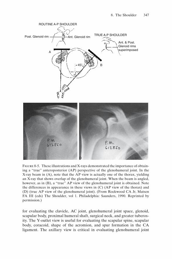
The standard shoulder series includes an anteroposterior (AP) X-ray in the plane of the scapula, a Y-outlet view, and an axillary view. This series of X-rays is mandatory in the evaluation of shoulder girdle trauma. Unfortu-nately, the axillary view is often not obtained, yet it is the most sensitive for documenting shoulder dislocations. AP views with the humerus inter-nally and externally rotated may be used to critically evaluate greater tuberosity fractures or calcific tendonitis. The scapula is approximately 45 degrees oblique to the coronal plane of the body; therefore, to obtain a true AP view of the glenohumeral joint, the X-ray beam must be obliquely oriented to the coronal plane of the body (Fig. 8-5). The AP view is useful
8. The Shoulder 347
for evaluating the clavicle, AC joint, glenohumeral joint space, glenoid, scapular body, proximal humeral shaft, surgical neck, and greater tuberos-ity. The Y-outlet view is useful for evaluating the scapular spine, scapular body, coracoid, shape of the acromion, and spur formation in the CA ligament. The axillary view is critical in evaluating glenohumeral joint
ROUTINE A-P SHOULDER
Post. Glenoid rim Ant. Glenoid rim
A
C D
B45°
TRUE A-P SHOULDER
Ant. & Post.Glenoid rimssuperimposed
Figure 8-5. These illustrations and X-rays demonstrated the importance of obtain-ing a “true” anteroposterior (AP) perspective of the glenohumeral joint. In the X-ray beam in (A), note that the AP view is actually one of the thorax, yielding an X-ray that shows overlap of the glenohumeral joint. When the beam is angled, however, as in (B), a “true” AP view of the glenohumeral joint is obtained. Note the differences in appearance in these views in (C) (AP view of the thorax) and (D) (true AP view of the glenohumeral joint). (From Rockwood CA Jr, Matsen FA III (eds) The Shoulder, vol 1. Philadelphia: Saunders, 1990. Reprinted by permission.)
348 R.M. Carroll
congruence. Anterior or posterior dislocations are best seen on the axillary view.
Magnetic Resonance Imaging
The magnetic resonance imaging (MRI) scan is commonly employed to evaluate the soft tissues of the shoulder girdle and is considered is the gold standard for evaluating the rotator cuff tendons. Subacromial fl uid, tendon inflammation, and rotator cuff tears are all visible with MR imaging. MRI scans can be performed with an arthrogram (intraarticular contrast dye) to better delineate intraarticular structures such as labral tears and articular-sided partial rotator cuff tears. Standard MRI views of the shoul-der include coronal oblique, sagittal oblique, and axial cuts. The coronal and sagittal views are termed oblique because they are obtained in the plane of the scapula that is oblique to the coronal and sagittal planes of the body. Although the MRI scan is a powerful tool in the evaluation of shoulder problems, it is a very sensitive test. Positive findings, therefore, may correlate poorly with a patient’s clinical presentation. For example, MRI scans have been obtained on patients with normal, painfree shoulders and documented that not only did a large number of these healthy patients have rotator cuff tears, but that the incidence of such asymptomatic tears increased with increasing age. It is therefore important to treat the patient and NOT the MRI scan.
Computerized Tomography
Computerized tomography (CT) scans are useful in the evaluation of bony abnormalities. In the setting of complex or comminuted shoulder girdle fractures, CT scanning with or without image reconstruction is a powerful tool for clinical decision making or preoperative planning. CT scans are also valuable in assessing bony deficiencies such as glenoid wear before reconstructive shoulder surgery. The CT scan has been demonstrated to be superior to the axillary radiograph in evaluating the glenoid before prosthetic arthroplasty.
Electrodiagnostic Testing
Electromyography (EMG) and nerve conduction velocity (NCV) testing are commonly used to evaluate neurologic lesions of the shoulder girdle. EMG testing involves placing small needle electrodes into the muscles to record resting potentials and firing patterns. NCV testing is used to docu-ment the speed with which an impulse is conducted along a peripheral nerve. Abnormalities such as a conduction block may indicate severe nerve injury. Electrodiagnostic testing is useful in documenting both the pres-ence and recovery of peripheral nerve lesions.
8. The Shoulder 349
Evaluation and Treatment of Common Shoulder Problems
The majority of common shoulder girdle problems result from degenera-tive changes, overuse, or traumatic injury. Atraumatic shoulder pain is common and includes rotator cuff disease, arthropathy, adhesive capsulitis, calcific tendonitis, and multidirectional instability. Most atraumatic shoul-der pain is initially treated with activity modification, antiinflammatory medication, and physical therapy. Treatment regimens may vary depending on the specific diagnosis. Calcific tendonitis, for example, responds well to subacromial corticosteroid injections. The physical therapy prescription may also vary depending on the diagnosis. Patients with adhesive capsulitis require stretching exercises, in contrast to patients with rotator cuff ten-donitis who are treated with rotator cuff strengthening exercises. Surgical treatment in the atraumatic population is generally reserved for those patients who fail to respond to nonoperative treatment regimens. A basic algorithm for the evaluation of atraumatic shoulder pain is provided in Figure 8-6.
Traumatic injuries to the shoulder girdle are common and include both soft tissue and bony injury. Treatment is individualized based on the age of the patient, functional status of the patient, and the severity of the injury. Depending on the injury, nonoperative or operative treatment may be appropriate. Common traumatic injuries to the shoulder girdle include shoulder dislocations, AC joint injuries, clavicle fractures, and proximal humerus fractures, and these are reviewed in the skeletal trauma chapter. Contrary to popular belief, traumatic rotator cuff tears are relatively uncommon.
Rotator Cuff DiseaseDegenerative and overuse injuries of the rotator cuff are common sources of shoulder pain and disability. Anterosuperior shoulder pain emanating from the rotator cuff (RC) under the coracoacromial arch has historically been called impingement syndrome. Impingement syndrome encompasses a spectrum of pathology in the subacromial region including subacromial bursitis, RC tendinopathy, partial-thickness RC tears, and full-thickness RC tears. Partial- and full-thickness tears of the rotator cuff become more prevalent with increasing age. It is unusual for patients under the age of 40 years to present with RC tears in the absence of significant trauma. Conversely, older patients may present with massive RC tears after an innocuous event. There are two main theories that attempt to explain such degenerative cuff tears. The external impingement model suggests an extrinsic cause of RC tears such as abrasion of the anterosuperior cuff

350 R.M. Carroll
Fig
ure
8-6
. A
lgor
ithm
ic a
ppro
ach
to t
he d
iagn
osis
and
tre
atm
ent
of a
trau
mat
ic s
houl
der
pain
.
8. The Shoulder 351
under the acromion and coracoacromial arch. The intrinsic model suggests that a relatively poor blood supply to the critical zone of the rotator cuff in combination with high stresses across the cuff leads to RC tears. The true pathophysiology likely results from a combination of these models.
History
The chief complaint is usually anterosuperior shoulder pain, which often radiates to the lateral deltoid region. The pain is typically worse with over-head activities and at night. The patient may recall a minor traumatic event, or the pain may have started insidiously.
Examination
Inspection of the shoulder girdle usually reveals symmetry, but patients with degenerative cuff tears may present with atrophy of the supra or infraspinatus fossae. The patient typically has discrete tenderness at the cuff insertion on the greater tuberosity. The active range of motion is gen-erally normal; however, some patients with RC tears may exhibit loss of active motion with preservation of passive motion. In this setting, the clini-cian may be able to document lag signs. Strength testing may reveal weak-ness of the supraspinatus or infraspinatus tendons. Special tests include the Neer and Hawkins’s impingement signs. If the patient has concomitant biceps tendon pathology, there may be tenderness at the bicipital groove, and Speed’s test may be positive. Tenderness over the AC joint may indi-cate that the AC joint is contributing to the painful condition. A cross-body adduction test recreating pain at the AC joint is considered confirmatory.
Differential Diagnosis
The differential diagnosis varies with the age of the patient. In olderpatients, the differential diagnosis includes arthritis, cervical spine pathol-ogy, metastatic disease, and visceral pathology such as cardiac disease. In younger patients, instability and labral pathology should be considered. In any age group, the differential diagnosis includes adhesive capsulitis, cal-cific tendonitis, and a variety of other less common shoulder problems (avascular necrosis, scapulothoracic dysfunction, and infection).
Radiographic Evaluation and Magnetic Resonance Imaging
The AP radiograph may reveal sclerosis and cyst formation of the greater tuberosity. The Y-outlet view shows the acromial morphology with poten-tial narrowing of the subacromial space. In patients with long-standing RC tears, the distance between the humerus and acromion may be narrowed. The axillary view illustrates the joint space and may reveal an os acromiale. An MRI scan is useful for a number of reasons. Confirmation of RC disease (and exclusion of other etiologies) is reassuring, but not necessary.
352 R.M. Carroll
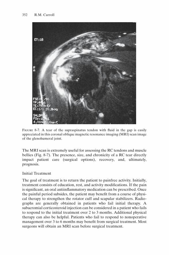
The MRI scan is extremely useful for assessing the RC tendons and muscle bellies (Fig. 8-7). The presence, size, and chronicity of a RC tear directly impact patient care (surgical options), recovery, and, ultimately, prognosis.
Initial Treatment
The goal of treatment is to return the patient to painfree activity. Initially, treatment consists of education, rest, and activity modifications. If the pain is significant, an oral antiinflammatory medication can be prescribed. Once the painful period subsides, the patient may benefit from a course of physi-cal therapy to strengthen the rotator cuff and scapular stabilizers. Radio-graphs are generally obtained in patients who fail initial therapy. A subacromial corticosteroid injection can be considered in a patient who fails to respond to the initial treatment over 2 to 3 months. Additional physical therapy can also be helpful. Patients who fail to respond to nonoperative management over 3 to 6 months may benefit from surgical treatment. Most surgeons will obtain an MRI scan before surgical treatment.
Figure 8-7. A tear of the supraspinatus tendon with fl uid in the gap is easily appreciated in this coronal oblique magnetic resonance imaging (MRI) scan image of the glenohumeral joint.

8. The Shoulder 353
Surgical Treatment
In the absence of a RC tear, most surgeons recommend a subacromial decompression performed either open or arthroscopically. This procedure involves removing the inflamed subacromial bursa and shaving the under-surface of the acromion (acromioplasty) to create more room in the sub-acromial space for the rotator cuff. Patients who have reparable RC tears are treated with primary repair, and most surgeons perform an acromioplasty. Care should be taken to preserve the CA ligament in patients with large tears and multiple tendon tears to prevent superior migration of the humeral head. There are a variety of options for patients with irreparable tears, including arthroscopic debridement, partial tendon repair, and tendon transfers. Patients with irreparable RC tears and arthropathy may be candidates for shoulder arthroplasty with a humeral head replacement. If biceps tendon pathology is found at the time of surgery, either tenodesis or tenolysis can be performed. Patients who are noted to have AC joint arthropathy and pain before surgery may benefi t from a distal clavicle resection. Recovery from RC surgery can take from 4 to 6 months. The goal of early (4–6 weeks) postoperative physical therapy is recovery of passive shoulder motion. Restoration of strength and function is the goal of subsequent postoperative therapy. Failure of the patient to adhere to postoperative physical therapy can result in a poor outcome.
OsteoarthritisDegenerative or osteoarthritis occurs in the glenohumeral joint but is less common than in the hip or knee joints. Osteoarthritis of the glenohumeral joint has the same pathophysiology as in other joints with progressive articular cartilage destruction.
History
Patients with early osteoarthritis may have a clinical syndrome that is vir-tually indistinguishable from impingement syndrome. In patients with advanced osteoarthritis, pain is more likely to be chronic, occur at rest, and be resistant to standard analgesics and antiinflammatory medications. In addition, loss of shoulder motion is a common complaint.
Examination
Patients with early osteoarthritis (OA) may examine similarly to those with impingement syndrome. In more-advanced OA, generalized disuse atrophy of the shoulder girdle may be noticeable. In general, active motion is decreased in all planes but loss of external rotation is often the most dra-matic. Passive motion is similarly decreased.

354 R.M. Carroll
Differential Diagnosis
Adhesive capsulitis and inflammatory arthropathy can have similar pre-sentations. The examiner must have a high index of suspicion for locked posterior shoulder dislocations in older patients who are poor historians as a result of dementia or stroke.
Radiographs
A standard shoulder series is recommended. Joint space narrowing, sub-chondral sclerosis, osteophytes, and subchondral cyst formation are classic fi ndings in osteoarthritis and are best seen on the AP and axillary view (Fig. 8-8). In the glenohumeral joint, inferior humeral osteophytes pre-dominate. Often, eccentric posterior glenoid wear is present. MRI scans are generally not used in the evaluation of OA. A CT scan to assess the glenoid for eccentric wear or bone loss is common during preoperative evaluation for shoulder arthroplasty.
Figure 8-8. All the classic findings of osteoarthritis are present in this true AP X-ray of the glenohumeral joint, including joint space narrowing, subchondral sclerosis, osteophyte formation, and subchondral cyst formation.
8. The Shoulder 355
Treatment
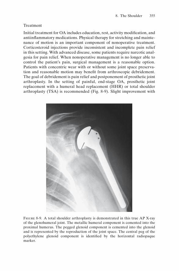
Initial treatment for OA includes education, rest, activity modification, and antiinflammatory medications. Physical therapy for stretching and mainte-nance of motion is an important component of nonoperative treatment. Corticosteroid injections provide inconsistent and incomplete pain relief in this setting. With advanced disease, some patients require narcotic anal-gesia for pain relief. When nonoperative management is no longer able to control the patient’s pain, surgical management is a reasonable option. Patients with concentric wear with or without some joint space preserva-tion and reasonable motion may benefit from arthroscopic debridement. The goal of debridement is pain relief and postponement of prosthetic joint arthroplasty. In the setting of painful, end-stage OA, prosthetic joint replacement with a humeral head replacement (HHR) or total shoulder arthroplasty (TSA) is recommended (Fig. 8-9). Slight improvement with
Figure 8-9. A total shoulder arthroplasty is demonstrated in this true AP X-ray of the glenohumeral joint. The metallic humeral component is cemented into the proximal humerus. The pegged glenoid component is cemented into the glenoid and is represented by the reproduction of the joint space. The central peg of the polyethylene glenoid component is identified by the horizontal radiopaque marker.
356 R.M. Carroll
respect to motion and pain relief has been demonstrated with TSA relative to HHR. Total shoulder arthroplasty introduces the risk of glenoid-sided prosthetic loosening and wear, which may require revision surgery. Humeral head replacement can fail as a result of inadequate pain relief.
Miscellaneous ArthropathyA variety of other disease processes can lead to glenohumeral joint destruc-tion. Inflammatory arthropathy such as rheumatoid arthritis can lead to joint destruction as a result of synovial disease. Although the clinical pre-sentation may be similar to osteoarthritis with pain and loss of motion, there are some important differences. In particular, rheumatoid arthritis can result in rotator cuff deficiency and incompetence. In these patients, total shoulder arthroplasty is contraindicated because glenoid loosening in the setting of rotator cuff deficiency is a common problem. Progressive bony destruction of the humeral head and glenoid can result from rheu-matoid arthritis, making prosthetic arthroplasty difficult if not impossible. Avascular necrosis can occur as a result of trauma, corticosteroid use, alcoholism, and other less common etiologies. Avascular necrosis of the humeral head can lead to pain and loss of motion in the glenohumeral joint. Humeral head replacement is an option for patients with humeral head collapse and chronic pain. Total shoulder arthroplasty is indicated when secondary destruction of the glenoid is present. Charcot or neuropathic arthropathy is typically a painless condition that results in severe joint destruction. Charcot arthropathy in the glenohumeral joint is commonly related to a cervical spine syrinx. There are no reliable surgical options for Charcot arthropathy.
Adhesive CapsulitisAdhesive capsulitis, or frozen shoulder, is a painful condition in which the synovial lining of the glenohumeral joint is inflamed. Adhesive capsulitis is a clinical diagnosis in which examination reveals an equal loss of active and passive motion. Primary adhesive capsulitis is idiopathic, meaning that no trigger can be identified; it occurs in middle-aged persons and is associ-ated with diabetes. Secondary adhesive capsulitis implies that a trigger or cause of the disease process can be identified. Trauma, surgery, and con-comitant shoulder girdle pathology may result in secondary adhesive capsulitis.
History
The patient reports an insidious onset of shoulder pain. Pain often occurs during rotational movements such as reaching behind the back, putting on a coat, or fastening a bra. Often the patient may recall a minor event that
8. The Shoulder 357
precipitated the condition. It is important to obtain a past medical and surgical history to identify possible risk factors. Insulin-dependent diabe-tes is a strong risk factor for adhesive capsulitis.
Examination
In the absence of prior trauma or surgery to the shoulder girdle, the inspec-tion and palpation portions of the examination are usually unremarkable. Active motion can be extremely limited in all planes of motion, and passive motion is similarly restricted. The patient often experiences pain at the end range of motion (active or passive).
Differential Diagnosis
Early adhesive capsulitis can mimic impingement. Subtle losses of internal and external rotation in abduction may be the only clues to differentiate between the two diagnoses. Unrecognized trauma (locked posterior shoul-der dislocations) and glenohumeral joint arthropathy can mimic adhesive capsulitis.
Radiographs
A standard shoulder series is useful in excluding other diagnoses; however, there are no radiographic findings for adhesive capsulitis. Further studies are generally not indicated unless additional pathology is suspected.
Treatment
Once the diagnosis is made, education of the patient is paramount. In general, the treatment of adhesive capsulitis is twofold: treatment of the synovial inflammation and restoration of motion. Antiinflammatory medi-cations can be used, but a corticosteroid injection into the glenohumeral joint space is more efficient and effective for treating the synovial inflam-mation. The patient must start a stretching program to regain motion in all planes. Initially, supervised physical therapy is helpful, but the patient must independently perform a battery of home stretching exercises daily. A gradual restoration of motion is the anticipated course. In patients who fail to respond to nonoperative treatment over the course of 3 to 6 months, surgery may be a reasonable option. Historically, patients with diabetes have a higher failure rate of nonoperative treatment compared to patients without risk factors. Additionally, patients with secondary adhesive capsu-litis from trauma or prior shoulder surgery often fail to respond fully to nonoperative treatment.
Manipulation of the shoulder under anesthesia was once the preferred treatment and continues to be a reasonable option. Proximal humerus

358 R.M. Carroll
fractures can occur with manipulations under anesthesia, however, and osteoporosis is a risk factor for this complication. Arthroscopic adhesioly-sis is a more-invasive, yet more-anatomic, procedure. Arthroscopic adhe-siolysis involves releasing the shoulder capsule under direct vision with some form of electrofrequency device. Because of the risk of axillary nerve damage, most surgeons prefer to gently manipulate the shoulder in abduc-tion to release the inferior capsule. Aggressive physical therapy with active-assisted and active range of motion is mandatory to maintain the postoperative range of motion. Shoulder strengthening and resistance therapy is instituted only after restoration of full, active shoulder motion.
Calcific TendonitisCalcific tendonitis of the rotator cuff is a painful condition of the shoulder girdle and is a common clinical problem (Figure 8.10). The etiology of calcific tendonitis is a matter of debate. The pathogenesis of calcifying tendonitis includes various stages of tendon degeneration, calcium deposi-tion, and calcium resorption. In the formative phase of calcium deposition, there may be little or no pain. Typically, the resorptive phase is more painful and clinically relevant.
Figure 8-10. A calcium deposit is present in the supraspinatus tendon immedi-ately medial to its attachment site on the greater tuberosity in this true AP X-ray of the glenohumeral joint.

8. The Shoulder 359
History
In the resorptive phase, the patient may present with an acute onset of severe shoulder pain. In the formative phase, the patient may present with more chronic symptoms that mimic impingement syndrome.
Examination
Acute bursitis in the resorptive phase may lead to fullness of the antero-superior shoulder, but otherwise the inspection is typically unremarkable. There may be tenderness at the rotator cuff insertion corresponding to the calcium deposition. There may be a loss of active motion secondary to pain, but passive motion, although painful, is generally preserved. Impingement signs are often positive.
Differential Diagnosis
The differential diagnosis includes rotator cuff disease and adhesive cap-sulitis. Referred pain from cardiac origin or other visceral organs and radicular pain from the cervical spine should be considered.
Radiographs
The appearance of calcific tendonitis on radiographs varies depending on the phase of the disease. In the formative phase, the calcium deposit is usually well circumscribed and easily identified. In the resorptive phase, the deposit may appear fl uffy and less well defined. In addition to the standard shoulder series, internal and external rotational views (AP) can be helpful for identifying more subtle deposits. Additional studies are not usually indicated.
Treatment
Treatment generally involves pain management. Noninvasive treatment options include antiinflammatory medications and extracorporeal shock wave therapy. More-invasive options include corticosteroid injections and lavage therapy. Surgical treatment is a last resort and involves arthroscopic debridement of the calcium deposit.
Multidirectional InstabilityShoulder instability is a complex problem with a spectrum of pathology ranging from atraumatic multidirectional shoulder instability to traumatic, unidirectional shoulder dislocations. Multidirectional instability (MDI) generally refers to shoulder pain and disability caused by excessive laxity of the static shoulder stabilizers (capsule and glenohumeral ligaments).

360 R.M. Carroll
History
In the overhead athlete (pitchers, swimmers, and volleyball players), MDI can present with activity-related pain, scapular winging, and occasionally with neurologic symptoms down the arm. Other patients may present with shoulder subluxations and dislocations that may easily reduce on their own but are a significant source of disability and distress to the patient.
Examination
Scapular winging may be noticeable on inspection during range of motion and strength testing. The active and passive ranges of motion are often excessive compared to the average shoulder. Additionally, the patient may exhibit generalized ligamentous laxity at other joints. The sulcus sign (hol-lowing of the subacromial region with downward traction on the arm) may be noticeable and indicative of shoulder laxity. Provocative shoulder testing such as the apprehension test may produce pain rather than apprehension. This pain is often related to secondary rotator cuff irritation. Other patients may have true apprehension. Load-and-shift testing often reveals sublux-ation or dislocation in multiple directions.
Differential Diagnosis
The differential diagnosis includes rotator cuff disease, labral pathology, and peripheral nerve injury in the setting of scapular winging.
Radiographs
The standard radiographs are typically unremarkable, although bony abnormalities such as glenoid hypoplasia can be identified. Patients who have had previous traumatic anterior shoulder dislocation may have a pos-terosuperior impression fracture of the humeral head (Hill–Sachs lesion) or a bony deficiency of the anteroinferior glenoid rim (bony Bankart lesion). An MRI arthrogram can be useful to exclude labral injury (Bankart lesion) and document the patulous capsule.
Treatment
The mainstay of treatment for MDI is rehabilitation. Physical therapy is focused on strengthening the dynamic stabilizers of the shoulder girdle, including the rotator cuff and scapular stabilizers. More-specialized therapy can be prescribed for athletes and is based on their specific sport and needs. Patients who fail rehabilitation may be candidates for surgical treat-ment. In most cases, rehabilitation should be continued for at least 6 to 12 months. Surgical treatment involves decreasing the volume of the shoulder joint by surgically altering the capsule (capsulorraphy). Surgery may be
8. The Shoulder 361
performed by arthroscopic or open methods. Arthroscopic methods tend to preserve motion better and may be preferable in athletes who would not tolerate minor losses of motion. Open surgical treatments historically have had lower rates of recurrent instability. Criticisms of open procedures such as the inferior capsular shift include loss of motion and potential subscapu-laris deficiency.
Summary
The shoulder is a complex structure that provides tremendous versatility and power to the upper extremity. The majority of painful shoulder girdle conditions are readily diagnosed with a thorough history and physical examination. Successful treatment of shoulder girdle problems is often accomplished by following a relatively simple algorithm of rest, activity modification, nonsteroidal antiinflammatory drug therapy, and physical therapy. More-invasive treatment options such as arthroscopic and open surgery are highly effective in appropriately selected patients.
Suggested ReadingsNorris TR. Orthopaedic Knowledge Update: Shoulder and Elbow, 2nd ed. Rose-
mont, IL: American Academy of Orthopaedic Surgeons, 2002.Rockwood CA, Matsen F, Wirth M. The Shoulder, 3rd ed. Philadephia: Saunders,
2004.
Questions
Note: Answers are provided at the end of the book before the index.
8-1. Which of these articulations is not a true diarthrodial joint?a. Sternoclavicularb. Acromioclavicularc. Scapulothoracicd. Glenohumerale. Hip joint
8-2. The primary, passive restraint to anterior displacement of the humeral head when the shoulder is abducted and externally rotated to 90 degrees is the:a. Coracohumeral ligamentb. Anterior band of the inferior glenohumeral ligament complexc. Superior glenohumeral ligamentd. Long head of the biceps tendone. Posterior band of the inferior glenohumeral ligament complex

362 R.M. Carroll
8-3. Which of the following radiographic findings is not a hallmark of glenohumeral osteoarthritis?a. Central erosion of the glenoidb. Osteophyte of the inferior humeral headc. Subchondral sclerosisd. Joint space narrowinge. Subchondral cysts
8-4. Which of these peripheral nerves does not exit from the brachial plexus?a. Long thoracic nerveb. Thoracodorsal nervec. Axillary nerved. Suprascapular nervee. Spinal accessory nerve
8-5. The rotator cuff includes all the following muscles except:a. Supraspinatusb. Teres minorc. Deltoidd. Subscapularise. Infraspinatus
8-6. What is the most common origin of shoulder pain that does not originate within the shoulder girdle?a. The cervical spineb. The heartc. The gallbladderd. The lungse. The thoracic spine
8-7. Which of the following procedures is not an option for the treatment of glenohumeral osteoarthritis?a. Total shoulder arthroplastyb. Humeral head replacementc. Arthroscopic debridementd. Arthroscopic labral repaire. Fusion
8-8. What is the best noninvasive study to evaluate the integrity of the rotator cuff tendons?a. Computerized tomography scanb. Plain AP radiograph in the plane of the scapulac. Electromyography studyd. Magnetic resonance imaging arthrograme. Magnetic resonance imaging scan
8-9. All the following are treatment options for adhesive capsulitis except:a. Arthroscopic rotator cuff repairb. Arthroscopic adhesiolysis

8. The Shoulder 363
c. Manipulation under anesthesiad. Glenohumeral corticosteroid injectione. Physical therapy
8-10. Which of the following statements is not true of rotator cuff tears?a. Most rotator cuff tears are degenerative in natureb. Most rotator cuff tears involve the supraspinatus tendonc. Some rotator cuff tears can be asymptomaticd. Some rotator cuff tears can be treated without surgerye. Most rotator cuff tears are the result of high-energy trauma