Muscular load characterization during isometric shoulder abductions with varying force
10
This article appeared in a journal published by Elsevier. The attached copy is furnished to the author for internal non-commercial research and education use, including for instruction at the authors institution and sharing with colleagues. Other uses, including reproduction and distribution, or selling or licensing copies, or posting to personal, institutional or third party websites are prohibited. In most cases authors are permitted to post their version of the article (e.g. in Word or Tex form) to their personal website or institutional repository. Authors requiring further information regarding Elsevier’s archiving and manuscript policies are encouraged to visit: http://www.elsevier.com/copyright
-
Upload
independent -
Category
Documents
-
view
4 -
download
0
Transcript of Muscular load characterization during isometric shoulder abductions with varying force

This article appeared in a journal published by Elsevier. The attachedcopy is furnished to the author for internal non-commercial researchand education use, including for instruction at the authors institution
and sharing with colleagues.
Other uses, including reproduction and distribution, or selling orlicensing copies, or posting to personal, institutional or third party
websites are prohibited.
In most cases authors are permitted to post their version of thearticle (e.g. in Word or Tex form) to their personal website orinstitutional repository. Authors requiring further information
regarding Elsevier’s archiving and manuscript policies areencouraged to visit:
http://www.elsevier.com/copyright
Author's personal copy
Muscular load characterization during isometric shoulderabductions with varying force
Hardianto Iridiastadi a, Maury A. Nussbaum b,*, Jaap H. van Dieen c
a Department of Industrial Engineering, Institut Teknologi Bandung, Indonesiab Department of Industrial and Systems Engineering, Virginia Tech, Blacksburg, VA, USA
c Faculty of Human Movement Sciences, Vrije Universiteit Amsterdam, The Netherlands
Received 27 July 2006; received in revised form 29 January 2007; accepted 29 January 2007
Abstract
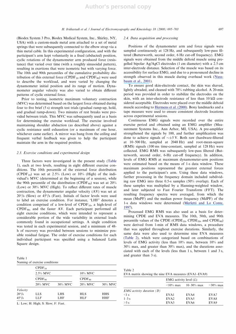
This study sought to characterize muscle loading and fatigue during static shoulder abductions with varying force. In a supine pos-ture, participants maintained fixed shoulder abductions against a time-varying external resistance, generated by a dynamometer-springmechanism. Patterns (cumulative distribution) of the external resistance were varied by selecting different 10th and 90th percentiles of thedistribution. Dynamometer angular velocities were also varied, to reflect different rates of cyclic muscle contraction. The degree of localfatigue development was assessed by common measures, including endurance time, strength reduction, and perceived discomfort. Myo-electric (EMG) signals were continuously obtained from the middle deltoid muscle throughout experimental exercise (60 min max).Changes in EMG root-mean-square (RMS) and spectral measures (derived from 1-s windows at peaks in the cyclic contractions) wereused as manifestations of muscle fatigue. For each minute, the RMS signal was further reduced using two methods, the cumulative prob-ability distribution of EMG (CPDE) and exposure variation analysis (EVA). The former resulted in three percentile values (10th, 50th,and 90th), whereas the latter method resulted in 10 different measures (grouped by EMG activity level and duration). A main finding ofthe study was the applicability of several common fatigue indicators for these cyclic, repetitive exertions. Overall, the use of CPDE andEVA to characterize task differences and predict muscle fatigue was found to have limited value.� 2007 Elsevier Ltd. All rights reserved.
Keywords: Localized muscle fatigue; Electromyography; CPDE; EVA; Exposure assessment
1. Introduction
Muscle fatigue has been defined as any reduction of theforce producing capacity of muscle as a consequence ofmuscle activity (Christensen, 1986). Fatigue is of particularimportance in ergonomics, because of effects on perfor-mance as well as assumed effects on health. Muscle fatiguehas mainly been studied under isometric continuous orintermittent constant force conditions. The external loadis defined in these experiments as the force produced bythe subject on the environment, usually in percentages ofmaximum voluntary contraction force (MVC). Productionof this force requires muscle activity leading to a reduction
in the force producing capacity and eventually a failure toproduce the required force. The rate of fatigue develop-ment is reflected in the time to force failure or endurancetime. Electromyographic (EMG) based fatigue indicators,reflecting the signal’s frequency content, can be used to pre-dict the point of force failure, where such failure is deter-mined as an inability to continue a task or based on afixed level of force decrement (Hagberg, 1981; Dieenet al., 1993; Dieen et al., 1998; Merletti and Roy, 1996).It is believed that, during fatiguing contractions, shifts inEMG frequency spectra are partly attributable to adecrease in muscle fiber conduction velocity (Luttmannet al., 2000). It should also be noted that muscle fatiguein turn affects muscle activity. In isometric constant forcecontractions, the central drive increases to compensatefor loss of force producing capacity. This is usually
1050-6411/$ - see front matter � 2007 Elsevier Ltd. All rights reserved.
doi:10.1016/j.jelekin.2007.01.011
* Corresponding author. Tel.: +1 540 231 6053; fax: +1 540 231 3322.E-mail address: [email protected] (M.A. Nussbaum).
Available online at www.sciencedirect.com
Journal of Electromyography and Kinesiology 18 (2008) 695–703
www.elsevier.com/locate/jelekin

Author's personal copy
reflected in an increase of the EMG amplitude during thecontraction, although the EMG amplitude is also affectedby other factors such as the change in frequency content.Such increases in activity do not, however, occur consis-tently. In some subjects, muscle activity will decrease (tem-porarily), presumably because load sharing between musclechanges (Dieen et al., 1993; Zijdewind et al., 1995; McLeanand Goudy, 2004). Such changes in load sharing in sus-tained activities have been shown to slow down fatiguedevelopment (Dieen et al., 1993) and to disturb the rela-tionship between the endurance time and EMG based fati-gue indicators (McLean and Goudy, 2004).
It is conceivable that fatigue-related changes in muscleactivation over time are less predictable under less con-straining external loads than the typical constant force con-tractions commonly studied. The aim of the present study,therefore, was to describe changes in muscle activation asestimated from the EMG amplitude over time and thedevelopment of fatigue in force varying isometric contrac-tions. Also of interest were the potentials to describe mus-cle activity (and hence characterize complex tasks inergonomic applications) using two relatively recent EMGdata processing methods, specifically the cumulative prob-ability distribution of EMG (CPDE) and exposure varia-tion analysis (EVA). The former employed threepercentile values (10th, 50th, and 90th) to characterizethe EMG amplitude, while the latter classified the EMGamplitude into 10 groups according to magnitude andduration. Further, it was of interest to determine if taskdescriptors yielded by these methods were related to com-mon indicators of local fatigue.
2. Methods
2.1. Participants
Eight college-age students (4 males, 4 females) participated inthis study, with mean (SD) age, stature, and body mass of 21.4
(1.6) years, 170.1 (5.0) cm, and 62.7 (1.0) kg, respectively. Allreported no recent (12 months) history of musculoskeletal injuriesand performing physical exercise 2–4 times a week. Participantsprovided informed consent prior to their participation, usingprocedures approved by the Virginia Tech Institutional ReviewBoard. An initial (practice) session was given that allowed forfamiliarization with the experimental procedures.
2.2. Experimental procedures and apparatus
In a supine posture (Fig. 1), the participants abducted theirright (dominant) arm to 90�, and placed the arm on a metalplatform supported by ball-bearings. This setup minimized fric-tional and gravitational effects. A padded strap was worn proxi-mally to the elbow, and was attached to a mechanism generatingan external force. The mechanism consisted of a dynamometer
Fig. 1. Experimental setup. Dynamometer (A) moves back and forth withangular velocity and range of motion that are varied according to theexperimental condition. A light metal wire connects the dynamometer, aset of springs (B), a force transducer (C), and a padded strap (D) worn atthe elbow. With the arm maintained in a fixed position, movements of thedynamometer result in variable (cyclic) loading.
Nomenclature
AV dynamometer angular velocityCPDE cumulative probability distribution of EMGCPDE10/50/90 10th, 50th, or 90th percentile of CPDECPDF dynamometer cumulative probability distribu-
tion of (external) forceCPDF10/90 10th or 90th percentile of CPDFD EMG activity durationEMG electromyographyET endurance timeEVA exposure variation analysisEVA1–9
cumulative duration of EVA relative to totalrecording period
FFT Fast Fourier transform
L EMG activity levelMnPF/MdPF mean/median power frequencyMVC maximum voluntary contractionRCPDE rates of CPDE changeREVA rates of EVA changeRMnPF/RMdPF rates of Mn/MdPF changeRMS root-mean-square of EMG signalsRMVC rates of MVC changeRPD ratings of perceived discomfortRRMS rates of RMS increaseRRPD rates of RPD changeRSumEVA rates of SumEVA changeSumEVA summation across EVA ‘cells’
696 H. Iridiastadi et al. / Journal of Electromyography and Kinesiology 18 (2008) 695–703
Author's personal copy
(Biodex System 3 Pro, Biodex Medical System, Inc., Shirley, NY,USA) with a custom-made metal arm attached to a set of metalsprings that were subsequently connected to the elbow strap via athin metal cable. In this experimental configuration, and with theparticipant’s arm kept voluntarily in a fixed (abducted) position,cyclic rotations of the dynamometer arm produced force (resis-tance) that varied over time (with a roughly sinusoidal pattern),resulting in exertions that were isometric but with varying force.The 10th and 90th percentiles of the cumulative probability dis-tribution of this external force (CPDF10 and CPDF90) were usedto describe the workload, and were varied by changing thedynamometer initial position and its range of motion. Dyna-mometer angular velocity was also varied to obtain differentpatterns of cyclic external force.
Prior to testing, isometric maximum voluntary contraction(MVC) was determined based on the largest force obtained duringfour to five brief (5 s) strength test trials (gradual ramp-up, hold,and gradual ramp-down). Two minutes of rest breaks were pro-vided between trials. This MVC was subsequently used as a basisfor determining the exercise workload. The exercise involvedmaintaining shoulder abduction (as described above) against acyclic resistance until exhaustion (or a maximum of one hour,whichever came earlier). A mirror was hung from the ceiling andfrequent verbal feedback was given to help the participantmaintain the arm in the required position.
2.3. Exercise conditions and experimental design
Three factors were investigated in the present study (Table1), each at two levels, resulting in eight different exercise con-ditions. The 10th percentile of the external force distribution(CPDF10) was set at 2.5% (Low) or 10% (High) of the indi-vidual’s MVC (determined at the beginning of a session), whilethe 90th percentile of the distribution (CPDF90) was set at 20%(Low) or 30% MVC (High). To reflect different rates of musclecontraction, the dynamometer angular velocity (AV) was set at20�/s (Slow) or 45�/s (Fast). Initials of factor levels were usedto label an exercise condition. For instance, ‘LHF’ denotes acondition comprised of a low-level of CPDF10, a high-level ofCPDF90, and the faster AV. Each participant performed alleight exercise conditions, which were intended to represent aconsiderable portion of the wide variability in external loadscommonly found in occupational settings. A single conditionwas tested in each experimental session, and a minimum of 48-h of recovery was provided between sessions to minimize pos-sible residual fatigue. The order of exercise conditions for eachindividual participant was specified using a balanced LatinSquare design.
2.4. Data acquisition and processing
Positions of the dynamometer arm and force signals weresampled continuously at 128 Hz, and subsequently low-pass fil-tered (Butterworth, second order, 6 Hz cut-off frequency). EMGsignals were obtained from the middle deltoid muscle using pre-gelled bipolar Ag/AgCl electrodes (1 cm diameter) with a 2.5 cminter-electrode distance. Selection of the muscle was based on itsaccessibility for surface EMG, and due to a pronounced decline instrength observed in this muscle during overhead work (Nuss-baum et al., 2001).
To ensure good skin-electrode contact, the skin was shaved,lightly abraded, and cleaned with 70% rubbing alcohol. A 20-minperiod was provided in order to stabilize the electrodes on theskin, with an inter-electrode resistance of less than 10 kX con-sidered acceptable. Electrodes were placed over the middle deltoidmuscle according to Hermens et al. (2000). Bony landmarks and atape measure were used to ensure consistent electrode locationsacross experimental sessions.
Continuous EMG signals were recorded over the entireexercise period and obtained using an EMG amplifier (Mea-surement Systems Inc., Ann Arbor, MI, USA). A pre-amplifierstrengthened the signals by 100, and further amplification wasdone to achieve signals of �±5 V. Both raw (hardware-filteredat 10–500 Hz, sampled at 2048 Hz) and root-mean-square(RMS) signals (100 ms time-constant, sampled at 128 Hz) werecollected. EMG RMS was subsequently low-pass filtered (But-terworth, second order, 6-Hz cut-off frequency). In addition,levels of EMG RMS at maximum dynamometer-arm positionswere estimated based on the means of 1-s data window. Thesemaximum positions represented the greatest external forcesapplied to the participant’s arm. Using these data windows,further processing in the frequency domain included subdivid-ing raw EMG into three 0.5-s samples (50% overlap). Each ofthese samples was multiplied by a Hanning-weighted window,and later subjected to Fast Fourier Transform (FFT). Theresulting frequency spectra were averaged, from which themean (MnPF) and the median power frequency (MdPF) of the1-s data windows were determined (Merletti and Lo Conte,1997).
Processed EMG RMS was also used as a basis for deter-mining CPDE and EVA measures. The 10th, 50th, and 90thpercentile values of the CPDE (CPDE10, CPDE50, and CPDE90)were derived from 1-min of RMS data windows, a procedurethat was applied throughout exercise durations. Similarly, thesame data were also used to determine nine EVA measures(Table 2), which were categorized based on combinations oflevels of EMG activity (less than 10% max, between 10% and30% max, and greater than 30% max), and the durations asso-ciated with each of the levels (less than 1 s, between 1 and 3 s,and greater than 3 s).
Table 1Naming of exercise conditions
CPDF10
2.5% MVC 10% MVC
CPDF90 CPDF90
20% MVC 30% MVC 20% MVC 30% MVC
Velocity
20�/s LLS LHS HLS HHS45�/s LLF LHF HLF HHF
L: Low; H: High; S: Slow; F: Fast.
Table 2EVA matrix showing the nine EVA measures (EVA1–EVA9)
EMG activity level (L)
<10% max 10–30% max >30% max
EMG activity duration (D)
<1 s EVA1 EVA4 EVA71–3 s EVA2 EVA5 EVA8>3 s EVA3 EVA6 EVA9
H. Iridiastadi et al. / Journal of Electromyography and Kinesiology 18 (2008) 695–703 697
Author's personal copy
2.5. Dependent measures and data analysis
Several common objective and subjective measures were usedto assess the degree of exercise-induced fatigue. Endurance time(exercise duration) was noted at the end of an exercise, whereasrates of MVC reduction (RMVC) were calculated according to(MVCend of exercise �MVCstart of exercise)/endurance time. Ratings ofperceived discomfort (RPD), using a common 10-point scale(Borg, 1990), were obtained every four minutes throughout anexercise, and were later expressed as rates of RPD increase (RRPD)using simple linear regressions.
Changes in EMG (obtained at peak dynamometer-arm posi-tions, see explanations above) were used as manifestations of localfatigue, and were defined as rates of RMS increase (RRMS) andrates of decrease in mean and median power frequency (RMnPF
and RMdPF). These rates were obtained based on slopes of linearregressions. CPDEs and EVAs derived from the initial minute ofexercise characterized (non-fatigued) muscle loading, whereasrates of change for these measures (RCPDE and REVA) wereexpected to indicate fatigue. Note that an additional EVA-basedmeasure was determined as the absolute sum of all the nine EVAs(Sum EVA), and its changes overtime was expressed as RSumEVA.
Due to the relatively small number of participants andpotential departures from normality, non-parametric (RankF)ANOVA was used to test the effects of the independent measures.Stepwise multiple linear regression was employed to establishspecific relationships between local fatigue measures and the ini-tial CPDEs and EVAs. Mellow’s Cp criterion was used in order todetermine the best subset of predictors. Significance of all statis-tical tests was based on P < 0.05. Omega squared (x2), a measureof effect size, was further used to determine the relative sensitivityof the different measures of local fatigue (Keppel, 1991). Presen-tation orders of the exercise conditions could potentially affect theresponse variables, but initial analysis did not indicate any sig-nificant effects (P > 0.10).
3. Results
3.1. Endurance and common indicators of muscle fatigue
The exercise conditions resulted in considerably differentdegrees of muscle fatigue. Endurance times ranged from 20to 60 min, while mean rates of strength reduction (RMVC)varied between 0.3% and 2%/min. Similar variability acrossconditions was also found for changes in subjective percep-tions of discomfort (RRPD). The light exercise conditionsproduced RRPD of about 0.1 units/min, whereas the mostfatiguing conditions yielded RRPD of 6–8 times greater.Differences in external workload and rates of muscle con-traction were generally associated with changes in thesemeasures of fatigue (P < 0.05), though the effect of AVon RMVC only approached significance (P = 0.06).
3.2. EMG amplitude and spectral measures
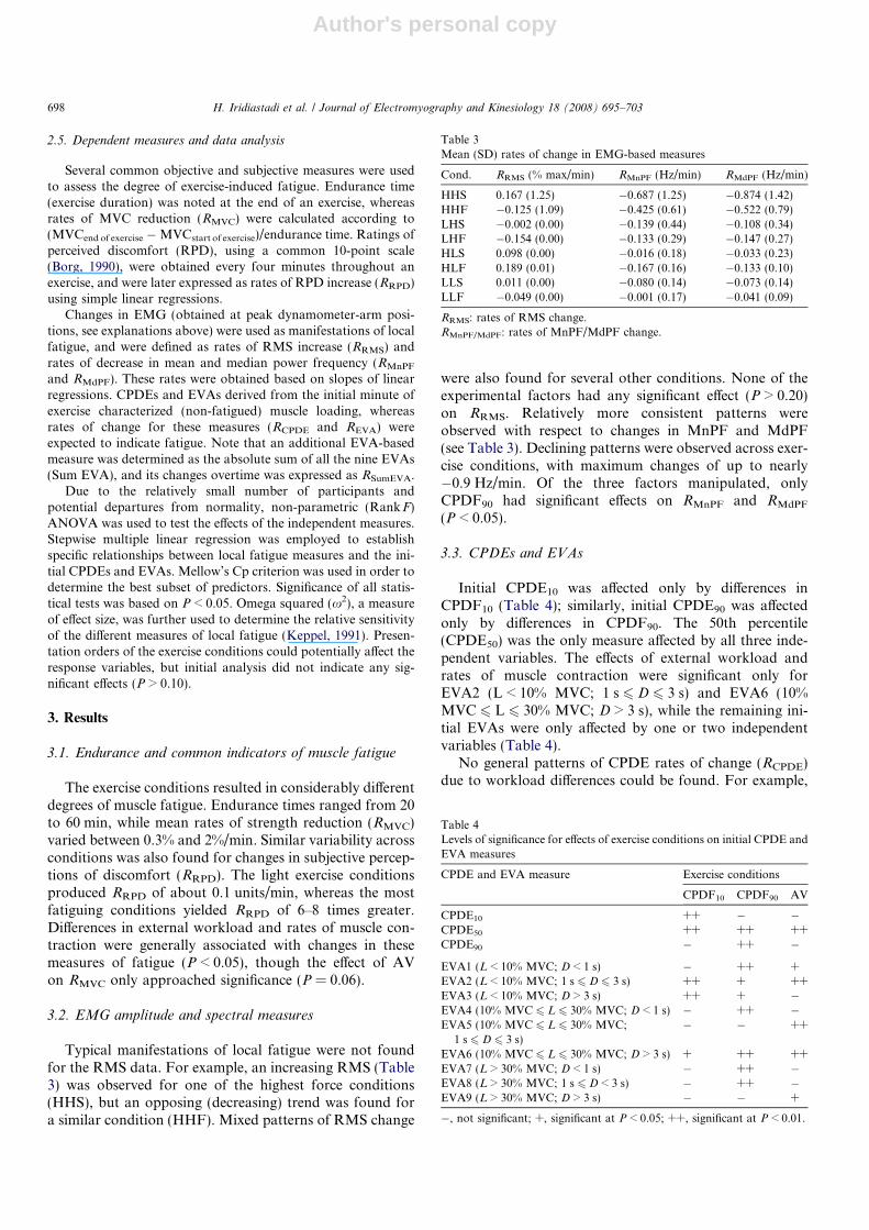
Typical manifestations of local fatigue were not foundfor the RMS data. For example, an increasing RMS (Table3) was observed for one of the highest force conditions(HHS), but an opposing (decreasing) trend was found fora similar condition (HHF). Mixed patterns of RMS change
were also found for several other conditions. None of theexperimental factors had any significant effect (P > 0.20)on RRMS. Relatively more consistent patterns wereobserved with respect to changes in MnPF and MdPF(see Table 3). Declining patterns were observed across exer-cise conditions, with maximum changes of up to nearly�0.9 Hz/min. Of the three factors manipulated, onlyCPDF90 had significant effects on RMnPF and RMdPF
(P < 0.05).
3.3. CPDEs and EVAs
Initial CPDE10 was affected only by differences inCPDF10 (Table 4); similarly, initial CPDE90 was affectedonly by differences in CPDF90. The 50th percentile(CPDE50) was the only measure affected by all three inde-pendent variables. The effects of external workload andrates of muscle contraction were significant only forEVA2 (L < 10% MVC; 1 s 6 D 6 3 s) and EVA6 (10%MVC 6 L 6 30% MVC; D > 3 s), while the remaining ini-tial EVAs were only affected by one or two independentvariables (Table 4).
No general patterns of CPDE rates of change (RCPDE)due to workload differences could be found. For example,
Table 3Mean (SD) rates of change in EMG-based measures
Cond. RRMS (% max/min) RMnPF (Hz/min) RMdPF (Hz/min)
HHS 0.167 (1.25) �0.687 (1.25) �0.874 (1.42)HHF �0.125 (1.09) �0.425 (0.61) �0.522 (0.79)LHS �0.002 (0.00) �0.139 (0.44) �0.108 (0.34)LHF �0.154 (0.00) �0.133 (0.29) �0.147 (0.27)HLS 0.098 (0.00) �0.016 (0.18) �0.033 (0.23)HLF 0.189 (0.01) �0.167 (0.16) �0.133 (0.10)LLS 0.011 (0.00) �0.080 (0.14) �0.073 (0.14)LLF �0.049 (0.00) �0.001 (0.17) �0.041 (0.09)
RRMS: rates of RMS change.RMnPF/MdPF: rates of MnPF/MdPF change.
Table 4Levels of significance for effects of exercise conditions on initial CPDE andEVA measures
CPDE and EVA measure Exercise conditions
CPDF10 CPDF90 AV
CPDE10 ++ � �CPDE50 ++ ++ ++CPDE90 � ++ �
EVA1 (L < 10% MVC; D < 1 s) � ++ +EVA2 (L < 10% MVC; 1 s 6 D 6 3 s) ++ + ++EVA3 (L < 10% MVC; D > 3 s) ++ + �EVA4 (10% MVC 6 L 6 30% MVC; D < 1 s) � ++ �EVA5 (10% MVC 6 L 6 30% MVC;
1 s 6 D 6 3 s)� � ++
EVA6 (10% MVC 6 L 6 30% MVC; D > 3 s) + ++ ++EVA7 (L > 30% MVC; D < 1 s) � ++ �EVA8 (L > 30% MVC; 1 s 6 D < 3 s) � ++ �EVA9 (L > 30% MVC; D > 3 s) � � +
�, not significant; +, significant at P < 0.05; ++, significant at P < 0.01.
698 H. Iridiastadi et al. / Journal of Electromyography and Kinesiology 18 (2008) 695–703

Author's personal copy
a comparison between conditions with the highest forcelevels differing only in AV (HHS vs. HHF) showed positivevalues for HHS condition, but positive as well as negativevalues for the other. Differences in force level (CPDF10 andCPDF90) did not produce changes in CPDE measures(P > 0.45), with the exception of a significant effect ofCPDF90 on CPDE10 (P < 0.05) (Table 5). Changes in AVdid not result in significantly different CPDEs (P > 0.08).A similar result was found with respect to the effects ofexternal muscle loading on rates of EVA change. Of the10 EVAs, RSumEVA was the only measure that changed as
a result of differences in CPDF10 and CPDF90 (P < 0.01).This EVA measure, however, was not influenced bychanges in AV.
3.4. Relationships between fatigue measures and initial
CPDEs or EVAs
Several regression models (Table 6) were generated toestimate measures of muscle fatigue as a function of initialCPDEs or EVAs. All CPDE measures were included in themodels, whereas up to four EVA measures were includedas predictors. The coefficients of determination (adjustedR2) were generally low, ranging from 0.10 to 0.45, and weregenerally higher for RMVC and RRPD than for other fatiguemeasures.
3.5. Sensitivity of measures to exercise conditions
A broad range of effect sizes (x2) was observed acrossthe different indicators of muscle fatigue (Table 7), withmoderate to large sensitivity (x2 > 0.30) typically shownfor non-EMG-based measures. Relatively low sensitivitywas found for EMG RMS and most CPDE measures.Across EVA measures, the effect sizes were generally low,though RSumEVA seemed to have relatively high sensitivityto changes in CPDF10 and CPDF90 (x2 > 0.50).
4. Discussion
This study aimed to describe changes in muscle activa-tion during isometric shoulder abduction with varyingforce. Such efforts were utilized as a logical next step in
Table 6Fatigue measures as a function of initial CPDEs and EVAs (parameters and performance of multiple regression models)
Measure Intercept CPDE10 CPDE50 CPDE90 Adj. R2 SE
ET 67.51 �1.87 0.66 �0.83 0.18 6.58RMVC �0.430 0.181 �0.187 0.097 0.31 0.30RRPD �0.089 0.045 �0.030 0.020 0.25 0.10RRMS 0.0053 �0.0001 0.0004 0.0004 0.10 0.002RMnPF 0.204 �0.059 0.048 �0.024 0.15 0.13RMdPF 0.307 �0.081 0.078 �0.039 0.24 0.14
Measure Intercept EVA2 EVA3 EVA4 EVA5 EVA6 EVA7 EVA8 EVA9 Adj. R2 SE
ET 50.96 �203.94 0.17 6.62RMVC 2.53 �2.88 �11.13 �1.33 8.03 0.45 0.27RRPD 0.35 �1.07 2.20 0.53 0.30 0.10RRMS 0.00 0.05 �0.05 �0.02 0.21 0.00RMnPF �0.43 0.90 1.96 �2.43 0.21 0.13RMdPF �0.21 1.46 2.38 �1.34 �3.30 0.33 0.14
ET = Endurance time (min)RMVC = Rates of MVC change (%/min).RRPD = Rates of RPD change (unit/min).RRMS = Rates of RMS change (% MVC/min).RMnPF/MdPF = Rates of MnPF/MdPF change (Hz/min).CPDE10/50/90 = 10th/50th/90th percentile of CPDE (% MVC).EVA1–9 = Cumulative duration of EVA1–9 relative to total recording period (%).
Table 5Levels of significance for effects of exercise conditions on CPDE and EVArates of change
CPDE and EVA measure Exercise conditions
CPDF10 CPDF90 AV
RCPDE10 � + �RCPDE50 � � �RCPDE90 � � �
REVA1 (L < 10% MVC; D < 1 s) � � �REVA2 (L < 10% MVC;1 s 6 D 6 3 s) � � �REVA3 (L < 10% MVC; D > 3 s) � � +REVA4 (10% MVC 6 L 6 30% MVC; D < 1 s) + � �REVA5 (10% MVC 6 L 6 30% MVC;
1 s 6 D 6 3 s)� � +
REVA6 (10% MVC 6 L 6 30% MVC; D > 3 s) � � �REVA7 (L > 30% MVC; D < 1 s) � � �REVA8 (L > 30% MVC; 1 s 6 D 6 3 s) � � +REVA9 (L > 30% MVC; D > 3 s) � + �RSumEVA ++ ++ �SumEVA: sum of absolute EVA.�, not significant; +, significant at P < 0.05; ++, significant at P < 0.01.
H. Iridiastadi et al. / Journal of Electromyography and Kinesiology 18 (2008) 695–703 699

Author's personal copy
understanding short-term responses beyond pure staticcontractions. In addition to conventional EMG-based indi-cators, more recent EMG signal processing methods wereused, to determine their ability in describing different pat-terns of external load and as indicators of fatiguedevelopment.
A broad range of local fatigue was induced using thecurrent protocols, and was manifested by changes in sev-eral commonly used measures. Endurance times variedmarkedly, ranging from less than 20 min for the mostfatiguing conditions to 60 min for the lightest conditions.As a ‘gold standard’ (Gerdle et al., 2000), substantialdeclines (14–26%) in muscle strength were present in allconditions, equaling rates of �0.3%/min to more than�2%/min. Data on perceived local discomfort also sug-gested different rates of fatigue, varying from roughly 0.1to 0.4 unit/min.
While muscle fatigue did occur, and developed at sub-stantially different rates, such a phenomenon was notindicated by concomitant changes in EMG amplitude.In contrast, this discriminating characteristic was demon-strated by using spectral measures. Employing EMGbased measures for evaluating dynamic activities hasbeen criticized on methodological grounds (Ducheneand Goubel, 1993; Roy et al., 1998), though several stud-ies employing such a method have suggested its feasibil-ity (e.g., Masuda et al., 2001; Morlock et al., 1997;Nussbaum, 2001; Potvin and Bent, 1997). This study,however, showed that the choice of fatigue assessmentmethod based on EMG mean or median power fre-quency is worth considering, even for muscle loadingsthat are ‘‘quasi’’ static.
Comparisons among fatigue measures noted aboveshowed greater sensitivity for endurance time, strength,and discomfort rating, relative to EMG amplitude andspectral indicators. The former measures were highlydependent on differences in distributions of external loadand, to a lesser extent, rates of muscle contraction. Otherevidence has shown a good agreement between subjectiveand objective measures of fatigue (e.g., Dedering et al.,1999; Grant et al., 1994; Jørgensen et al., 1988), thougha contrasting finding with respect to these two types offatigue indicator has also been reported (e.g., Dederinget al., 1999). When compared to EMG-derived measures,higher sensitivity for subjective ratings can probably beattributed to the fact that an individual’s perception dur-ing muscular exertions is based on more complete infor-mation facilitated by the peripheral components, thecentral nervous system, and the central cardiovascularand respiratory functions (Grant et al., 1994). Reportsin the literature (e.g., Ahsberg and Gamberale, 1998;Dieen and Heijblom, 1996; Vøllestad, 1997) also seemto suggest the adequacy of muscle strength and subjectiveratings, particularly with respect to their sensitivity to dif-ferences in task conditions.
This investigation also evaluated CPDE and EVA asdata reduction methods, with an expectation that thesemethods could be utilized to describe external exposures(Hagberg, 1979; Mathiassen and Winkel, 1991) and asmanifestations of local fatigue development (Hagberg,1979; Hagberg and Jonsson, 1975; Nakata et al., 1992).The assumption was that acute responses were (surro-gate) indicators of chronic musculoskeletal symptomsand problems (Nussbaum et al., 2001). Adequate expo-sure metrics, therefore, should be capable of indicatingthe degree of short and (potentially) long-term effectsassociated with the exposure. To this end, however, thetwo methods did not seem to fulfill the above purposes.The potential of using initial CPDEs and EVAs to char-acterize muscle loading was limited, and using changes inCPDE and EVA-based measures to assess the develop-ment of local fatigue is probably of little value, draw-backs that have been noted in earlier studies(Christensen, 1986; Hagberg and Sundelin, 1986; Linde-rhed, 1993; Mathiassen and Winkel, 1991; Winkel andBendix, 1984).
It should be noted that, specific to EVA, results of thisstudy indicated a general observation, in which positiverates of change for some EVAs were simultaneously (andsomewhat consistently) accompanied by negative valuesfor others. The overall (absolute) magnitude of thesechanges (SumEVA) was closely associated with differencesin external workload, with sensitivity (x2) equal to orgreater than 0.4. This finding suggests that variability ofmyoelectrical activities is probably a more important esti-mator of fatigue, as opposed to changes in the magnitudeof EMG amplitudes. Variability of muscular activitiesmay provide important physiological and etiological infor-mation (Mathiassen and Winkel, 1991), and has been
Table 7Sensitivity of dependent measures (x2)
Measure Exercise conditions
CPDF10 CPDF90 AV
ET 0.82 0.85 0.42RMVC 0.72 0.64 0.11RRPD 0.88 0.92 0.30
RRMS* 0.07 0.08
RMnPF 0.55 0.40 *
RMdPF 0.57 0.41 *
RCPDE10* 0.30 0.16
RCPDE50* * 0.10
RCPDE90* * 0.21
REVA1* * 0.03
REVA2* * *
REVA3 0.09 * 0.31REVA4 0.31 * 0.18REVA5
* * 0.23REVA6
* * *
REVA7* * 0.04
REVA8* 0.14 0.27
REVA9* 0.25 *
RSumEVA 0.51 0.70 *
All CPDEs and EVAs represent rates of change.* Negative x2 due to F-values <1.
700 H. Iridiastadi et al. / Journal of Electromyography and Kinesiology 18 (2008) 695–703

Author's personal copy
assumed to lead to reduced risk of musculoskeletal disor-ders (Moller et al., 2004).
Another issue that is worth considering is the design ofEVA matrix, since it could affect study results. The matrixselected here was based on ‘‘assigned’’ loading parameters.Though different classification could have been used (e.g.,Mathiassen and Winkel, 1991), there was currently nowell-established classification scheme available. A morerecent study (Dan et al., 2003) combined EVA cells into‘‘more manageable’’ clusters, an approach that had thepotential to discriminate workers with different muscleloading characteristics. No firm criteria, however, werechosen as a basis for selecting EVA cell boundaries.Another slightly different approach in building an EVAmatrix has been based on the associations between EVA-based measures and the presence of musculoskeletal symp-toms/problems. A few investigations (e.g., Hagg andAstrom, 1997; Jensen et al., 1993; Merletti and Lo Conte,1997; Veiersted et al., 1993) have addressed this relation-ship by investigating certain aspects of an EVA matrix,including periods of zero activity (EMG gaps) and veryshort EMG durations (EMG jerks). These studies typicallydemonstrated the ability of the measures to discriminateexposure patterns between occupational tasks, or even dif-ferentiate groups of healthy workers vs. those with disor-ders. The investigators further argued that differences inEMG gaps or jerks may provide important etiologicalinformation, but agreed that the actual effects (or if themeasures can indeed predict future problems) remainunknown. These and findings from the present work pointtoward the potential value of the EVA, though muchfuture work is still needed.
A critical issue worth discussing pertains to the choiceof muscle, particularly since it is not known if activity ofthe middle deltoid muscle is representative of typicalshoulder muscle functions. The shoulder girdle is a com-plex structure (Kadefors et al., 1976). Movements andcontrol of this shoulder joint are accomplished via thework of several muscles, including the infraspinatus, tra-pezius, deltoid, and supraspinatus, with the latter twomuscles playing an important role during shoulder abduc-tions. It is thus important to provide the arguments for(and against) obtaining surface EMG data from only asingle muscle.
With regard to the present work, a major advantage ofmeasuring muscle activities from the middle deltoid is easeof measurement. Recall that participants in this studyadopted a supine posture with the intention of eliminatingthe effect of arm weight. As a result, obtaining EMG fromthe dorsal muscles would have been impaired by skin con-tact with the supporting device. Another benefit gainedincluded simple interpretations of EMG data. Difficultymay arise, for example, when two different muscles areinvestigated, but yielded conflicting results. There is cur-rently no accepted method in deriving conclusions (i.e. rel-ative fatigue rates induced by task parameters) if more thanone muscle is involved. It should be noted that several com-
mon indicators of fatigue examined in this study (e.g.,changes in strength or discomfort rating) were not influ-enced by the choice of muscle, though these measures wereconsidered secondary.
A likely drawback of investigating only the middle del-toid muscle is the assumption of consistent recruitment(load sharing) between multiple muscles and the associatedchanges in EMG-based measures. Note, though, that suchload sharing is often assumed in biomechanical models.This drawback, however, presented an opportunity if itwas found that a single measure from a major agonist givesresults comparable to other local fatigue measures (e.g.,endurance time or strength changes). Comparable resultswould justify the use of a single muscle (representing func-tion of the synergistic group), while conflicting resultswould emphasize a need for EMG recordings obtainedfrom multiple muscles. To a certain extent, this study indi-cated correspondence between the two data processingmethods (particularly the EVA) and the common measuresof fatigue. This finding suggests the potential of using themiddle deltoid for rough assessments of shoulder abduc-tion efforts, and perhaps the use of a major agonist ingeneral.
5. Conclusions
This investigation aimed to characterize the develop-ment of local fatigue during static work with varying exter-nal forces. A primary finding of the study was thatcommon indicators (endurance, reductions in musclestrength, and change in perceived discomfort) could begenerally employed for fatigue assessment purposes, forefforts that are cyclic and repetitive. In contrast to EMGRMS, spectral measures (MnPF and MdPF) were associ-ated with fatigue development. For the cyclic, repetitivecontractions examined, use of CPDE to describe task dif-ferences and muscle fatigue was of limited value. Thepotential of employing EVA for these purposes was alsomarginal. In short, the use of these two more recentEMG signal processing methods to characterize externalexposure is still worth considering, but their use as a basisfor indicating fatigue development is questionable. Itshould be noted, however, that an EVA-derived measure(RSumEVA) did show some relationships with local fatiguedevelopment. Further research on this is warranted, partic-ularly in terms of how EVA ‘cells’ should be grouped.
References
Ahsberg E, Gamberale F. Perceived fatigue during physical work: anexperimental evaluation of fatigue inventory. Int J Ind Ergon 1998;21:117–31.
Borg G. Psychophysical scaling with applications in physical work and theperception of exertion. Scand J Work Environ Health 1990;16(Suppl. 1):55–8.
Christensen H. Muscle activity and fatigue in the shoulder muscles ofassembly-plant employees. Scand J Work Environ Health 1986;12:582–7.
H. Iridiastadi et al. / Journal of Electromyography and Kinesiology 18 (2008) 695–703 701
Author's personal copy
Dan A, Thomas CM, John RC, Linda MA. Method for quantitativelyassessing physical risk factors during variable noncyclic work. Scand JWork Environ Health 2003;39:354–62.
Dedering A, Nemeth G, Harms-Ringdahl K. Correlation betweenelectromyographic spectral changes and subjective assessment oflumbar muscle fatigue in subjects without pain from the lower back.Clin Biomech 1999;14:103–11.
Dieen JHv, Heijblom P. Reproducibility of isometric trunk extensiontorque, trunk extensor endurance, and related electromyographycparameters in the context of their clinical applicability. J Orthop Res1996;14:139–43.
Dieen JHv, Oude Vrielink HHE, Housheer FAF, Lotters FJB, TouissaintHM. Trunk extensor endurance and its relationship to electromyo-gram parameters. Eur J Appl Physiol 1993;66:388–96.
Dieen v-JH, Oude Vrielink HHE, Toussaint HM. An investigation intothe relevance of the pattern of temporal activation with respect toerector spinae muscle endurance. Eur J Appl Physiol 1993;66:70–5.
Dieen v-JH, Heijblom P, Bunkens H. Extrapolation of time series of EMGpower spectrum parameters in isometric endurance tests of trunkextensor muscles. J Electromyogr Kinesiol 1998;8:35–44.
Duchene J, Goubel F. Surface electromyogram during voluntary con-traction: processing tools and relation to physiological events. Crit RevBiomed Eng 1993;21:313–97.
Gerdle B, Larsson B, Karlsson S. Criterion validation of surface EMGvariables as fatigue indicators using peak torque. A study of repetitivemaximum isokinetic knee extension. J Electromyogr Kinesiol 2000;10:225–32.
Grant KA, Habes DJ, Putz-Anderson V. Psychophysical and EMGcorrelates of force exertion in manual work. Int J Ind Ergon 1994;13:31–9.
Hagberg M. The amplitude distribution of surface EMG in static andintermittent static muscular performance. Eur J Appl Physiol 1979;40:265–72.
Hagberg M. Electromyographic signs of shoulder muscular fatigue in twoelevated arm positions. Am J Phys Med 1981;60:111–21.
Hagberg M, Jonsson B. The amplitude distribution of the myoelectric signalin an ergonomic study of the deltoid muscle. Ergonomics 1975;18:311–9.
Hagberg M, Sundelin G. Discomfort and load on the upper trapezius musclewhen operating a word processor. Ergonomics 1986;29:1637–45.
Hagg GM, Astrom A. Load pattern and pressure pain threshold in theupper trapezius muscle and psychosocial factors in medical secretarieswith and without shoulder/neck disorders. Int Arch Occup EnvironHealth 1997;69:423–32.
Hermens HJ, Freriks B, Disselhorst-Klug C, Rau G. Development ofrecommendations for SEMG sensors and sensor placement proce-dures. J Electromyogr Kinesiol 2000;10:361–74.
Jensen C, Nilsen K, Hansen K, Westgaard RH. Trapezius muscle load as arisk indicator for occupational shoulder-neck complaints. Int ArchOccup Environ Health 1993;64:415–23.
Jørgensen K, Fallentin N, Krough-Lund C, Jensen B. Electromyographyand fatigue during prolonged, low- level static contractions. Eur JAppl Physiol 1988;57:316–21.
Kadefors R, Petersen I, Herberts P. Muscular reaction to welding work:an electromyographic investigation. Ergonomics 1976;19:543–58.
Keppel G. Design and analysis: a researcher’s handbook. Prentice-Hall,Inc.; 1991.
Linderhed H. A new dimension to amplitude analysis of EMG. Int J IndErgon 1993;11:243–7.
Luttmann A, Jager M, Laurig W. Electromyographical indication ofmuscular fatigue in occupational field studies. Int J Ind Ergon 2000;25:645–60.
Masuda T, Kizuka T, Zhe JY, Yamada H, Saitou K, Sadoyama T, et al..Influence of contraction force and speed on muscle fiber conductionvelocity during dynamic voluntary exercise. J Electromyogr Kinesiol2001;11:85–94.
Mathiassen ES, Winkel J. Quantifying variation in physical load usingexposure-vs-time data. Ergonomics 1991;34:1455–68.
McLean L, Goudy N. Neuromuscular response to sustained low-levelmuscle activation: within- and between-synergist substitution in thetriceps surae muscles. Eur J Appl Physiol 2004;91:204–16.
Merletti R, Lo Conte LR. Surface EMG signal processing duringisometric contractions. J Electromyogr Kinesiol 1997;7:241–50.
Merletti R, Roy SH. Myoelectric and mechanical manifestations of musclefatigue in voluntary contractions. JOSPT 1996;94:342–53.
Moller T, Mathiassen ES, Franzon H, Kihlberg S. Job enlargement andmechanical exposure variability in cyclic assembly work. Ergonomics2004;47:19–40.
Morlock MM, Bonin V, Muller G, Schneider E. Trunk muscle fatigue andassociated EMG changes during a dynamic iso-inertial test. Eur J ApplPhysiol 1997;76:75–80.
Nakata M, Hagner I-M, Jonsson B. Perceived musculoskeletal discomfortand electromyography during repetitive light work. J ElectromyogrKinesiol 1992;2:103–11.
Nussbaum MA. Static and dynamic myoelectric measures of shouldermuscle fatigue during intermittent dynamic exertions of low tomoderate intensity. Eur J Appl Physiol 2001;85:299–309.
Nussbaum MA, Clark LL, Lanza MA, Rice KM. Fatigue and endurancelimits during intermittent overhead work. Am Ind Hyg Assoc J2001;62:446–56.
Potvin JR, Bent LR. A validation technique using surface EMG signalsfrom dynamic contractions to quantify muscle fatigue during repetitivetasks. J Electromyogr Kinesiol 1997;7:131–9.
Roy SH, Bonato P, Knaflitz M. EMG assessment of back muscle functionduring cyclical lifting. J Electromyogr Kinesiol 1998;8:233–45.
Veiersted KB, Westgaard RH, Andersen P. Electromyographic evaluationof muscular work pattern as a predictor of trapezius myalgia. Scand JWork Environ Health 1993;19:284–90.
Vøllestad NK. Measurement of human muscle fatigue. J NeurosciMethods 1997;74:219–27.
Winkel J, Bendix T. A method for electromyographic analysis of muscularcontraction frequencies. Eur J Appl Physiol 1984;53:112–7.
Zijdewind I, Kernell D, Kukulka CG. Spatial differences in fatigue-associated electromyographic behaviour of the human first dorsalinterosseus muscle. J Physiol (London) 1995;483:499–509.
Hardianto Iridiastadi graduated from InstitutTeknologi Bandung, Indonesia, obtained hisM.S. (1997) in Industrial Engineering fromLouisiana State University, and received hisPh.D. (2004) in Industrial and Systems Engi-neering from Virginia Tech. He is currently anAssistant Professor at the Department ofIndustrial Engineering of Institut TeknologiBandung, Indonesia. His research interests arein the areas of occupational ergonomics,transportation safety and human error, andwork design.
Maury A. Nussbaum received an M.S. (1989) inBioengineering and a Ph.D. (1994) in Industrialand Operations Engineering from The Uni-versity of Michigan. He is currently a Professorof Industrial and Systems Engineering at Vir-ginia Tech. His research interests are (in noparticular order): occupational biomechanicsand ergonomics, localized muscle fatigue,aging, and postural control.
702 H. Iridiastadi et al. / Journal of Electromyography and Kinesiology 18 (2008) 695–703

Author's personal copy
Jaap H. van Dieen obtained a Ph.D. in HumanMovement Sciences from the Faculty ofHuman Movement Sciences at the ‘Vrije Uni-versiteit Amsterdam’ the Netherlands in 1993.Since 1996 he has been affiliated to this faculty,since 2002 as professor of biomechanics. He ischairing the ergonomics program at this facultyand leads a research group focusing onmechanical aspects of musculoskeletal injuries.His main research interest is on the interactionof muscle coordination, fatigue, disorders, jointload and stability. He has (co-) authored over
120 papers in international scientific journals and numerous abstracts and
book chapters in the international literature and technical reports andpublications in Dutch. He is an editor of the European Journal of AppliedPhysiology, section editor of Human Movement Sciences and serves onthe editorial advisory board of the Journal of Electromyography andKinesiology and the editorial board of Clinical Biomechanics and is cur-rently a member of the boards of ISEK and the Dutch Society for HumanMovement Sciences.
H. Iridiastadi et al. / Journal of Electromyography and Kinesiology 18 (2008) 695–703 703