Adaptations to isolated shoulder fatigue during simulated repetitive work. Part II: Recovery
8
Adaptations to isolated shoulder fatigue during simulated repetitive work. Part II: Recovery Alison C. McDonald, Calvin T.F. Tse, Peter J. Keir ⇑ Occupational Biomechanics Laboratory, Department of Kinesiology, McMaster University, Hamilton, ON L8S 4K1, Canada article info Article history: Received 2 January 2015 Received in revised form 18 April 2015 Accepted 25 May 2015 Available online xxxx Keywords: Occupational Co-ordination Movement Kinematics EMG Control abstract The shoulder allows kinematic and muscular changes to facilitate continued task performance during prolonged repetitive work. The purpose of this work was to examine changes during simulated repetitive work in response to a fatigue protocol. Participants performed 20 one-minute work cycles comprised of 4 shoulder centric tasks, a fatigue protocol, followed by 60 additional cycles. The fatigue protocol targeted the anterior deltoid and cycled between static and dynamic actions. EMG was collected from 14 upper extremity and back muscles and three-dimensional motion was captured during each work cycle. Participants completed post-fatigue work despite EMG manifestations of muscle fatigue, reduced flexion strength (by 28%), and increased perceived exertion (3 times). Throughout the post-fatigue work cycles, participants maintained performance via kinematic and muscular adaptations, such as reduced gleno- humeral flexion and scapular rotation which were task specific and varied throughout the hour of sim- ulated work. By the end of 60 post-fatigue work cycles, signs of fatigue persisted in the anterior deltoid and developed in the middle deltoid, yet perceived exertion and strength returned to pre-fatigue levels. Recovery from fatigue elicits changes in muscle activity and movement patterns that may not be perceived by the worker which has important implications for injury risk. Ó 2015 Elsevier Ltd. All rights reserved. 1. Introduction Industrial workplaces are often characterized by low load, repetitive and prolonged tasks. Repetitive work, elevated arm pos- tures, constrained workplaces and periods of sustained muscle activity act in combination as risk factors for developing shoulder pain and disorders, stressing the need to understand muscular and kinematic responses to these exposures (Hanvold et al., 2012; Ferguson et al., 2013; Nordander et al., 2009; Svendsen et al., 2004). Much of the evidence for the risk of repetitive injuries in the workplace comes from cross-sectional and longitudinal epi- demiological studies, making it difficult to characterize causal rela- tionships. Understanding responses to repetitive work can become even more challenging as workers’ functional capacities change with muscle fatigue. Muscle fatigue can be defined as a combination of increased per- ceived effort and an eventual decline in force production ability (Enoka and Stuart, 1992). Muscle fatigue can be quantified through changes in muscle activity and maximum force output (Enoka and Duchateau, 2008). Quantifying the development of fatigue and vari- ations in fatigue during workplace tasks is difficult. Workers are typ- ically required to generate submaximal efforts, allowing tasks to be successfully completed even in the presence of fatigue-reduced mus- cle capacity. With fatigue, the force generating capacity of muscle is reduced, effectively increasing the relative demands of the task. A change in capacity would impact the maximal acceptable effort for a task, which is also dependent on the duty cycle, or the percentage of time a worker is actively engaged in the task, further emphasizing the effect of fatigue on workers (Potvin, 2012). Repetitive work has been shown to impair recovery when rest opportunities within the day and between work shifts are inade- quate (Elliott et al., 2008, 2009). Both muscle fatigue and recovery are time-dependent processes but each proceeds at a different rate (Lucidi and Lehman, 1992; Vollestad and Sejersted, 1988), with recovery statistically modeled to occur 10–15 times slower than the fatiguing process itself (Frey-Law et al., 2012). Long recovery times for fatigued muscles is especially relevant in the shoulder given the frequent demands placed on the postural and stabilizing muscles of the shoulder complex, specifically, the rotator cuff mus- cles (Karduna et al., 1996; Labriola et al., 2004). In an endurance study of the trapezius muscle, individuals exhibiting greater http://dx.doi.org/10.1016/j.jelekin.2015.05.005 1050-6411/Ó 2015 Elsevier Ltd. All rights reserved. ⇑ Corresponding author at: McMaster University, Department of Kinesiology, Ivor Wynne Centre, Room 212, 1280 Main Street West, Hamilton, ON L8S4K1, Canada. Tel.: +1 905 525 9140x23543. E-mail address: [email protected] (P.J. Keir). Journal of Electromyography and Kinesiology xxx (2015) xxx–xxx Contents lists available at ScienceDirect Journal of Electromyography and Kinesiology journal homepage: www.elsevier.com/locate/jelekin Please cite this article in press as: McDonald AC et al. Adaptations to isolated shoulder fatigue during simulated repetitive work. Part II: Recovery. J Elec- tromyogr Kinesiol (2015), http://dx.doi.org/10.1016/j.jelekin.2015.05.005
-
Upload
independent -
Category
Documents
-
view
2 -
download
0
Transcript of Adaptations to isolated shoulder fatigue during simulated repetitive work. Part II: Recovery

Journal of Electromyography and Kinesiology xxx (2015) xxx–xxx
Contents lists available at ScienceDirect
Journal of Electromyography and Kinesiology
journal homepage: www.elsevier .com/locate / je lek in
Adaptations to isolated shoulder fatigue during simulated repetitivework. Part II: Recovery
http://dx.doi.org/10.1016/j.jelekin.2015.05.0051050-6411/� 2015 Elsevier Ltd. All rights reserved.
⇑ Corresponding author at: McMaster University, Department of Kinesiology, IvorWynne Centre, Room 212, 1280 Main Street West, Hamilton, ON L8S4K1, Canada.Tel.: +1 905 525 9140x23543.
E-mail address: [email protected] (P.J. Keir).
Please cite this article in press as: McDonald AC et al. Adaptations to isolated shoulder fatigue during simulated repetitive work. Part II: Recoverytromyogr Kinesiol (2015), http://dx.doi.org/10.1016/j.jelekin.2015.05.005
Alison C. McDonald, Calvin T.F. Tse, Peter J. Keir ⇑Occupational Biomechanics Laboratory, Department of Kinesiology, McMaster University, Hamilton, ON L8S 4K1, Canada
a r t i c l e i n f o a b s t r a c t
Article history:Received 2 January 2015Received in revised form 18 April 2015Accepted 25 May 2015Available online xxxx
Keywords:OccupationalCo-ordinationMovementKinematicsEMGControl
The shoulder allows kinematic and muscular changes to facilitate continued task performance duringprolonged repetitive work. The purpose of this work was to examine changes during simulated repetitivework in response to a fatigue protocol. Participants performed 20 one-minute work cycles comprised of 4shoulder centric tasks, a fatigue protocol, followed by 60 additional cycles. The fatigue protocol targetedthe anterior deltoid and cycled between static and dynamic actions. EMG was collected from 14 upperextremity and back muscles and three-dimensional motion was captured during each work cycle.Participants completed post-fatigue work despite EMG manifestations of muscle fatigue, reduced flexionstrength (by 28%), and increased perceived exertion (�3 times). Throughout the post-fatigue work cycles,participants maintained performance via kinematic and muscular adaptations, such as reduced gleno-humeral flexion and scapular rotation which were task specific and varied throughout the hour of sim-ulated work. By the end of 60 post-fatigue work cycles, signs of fatigue persisted in the anteriordeltoid and developed in the middle deltoid, yet perceived exertion and strength returned topre-fatigue levels. Recovery from fatigue elicits changes in muscle activity and movement patterns thatmay not be perceived by the worker which has important implications for injury risk.
� 2015 Elsevier Ltd. All rights reserved.
1. Introduction
Industrial workplaces are often characterized by low load,repetitive and prolonged tasks. Repetitive work, elevated arm pos-tures, constrained workplaces and periods of sustained muscleactivity act in combination as risk factors for developing shoulderpain and disorders, stressing the need to understand muscularand kinematic responses to these exposures (Hanvold et al.,2012; Ferguson et al., 2013; Nordander et al., 2009; Svendsenet al., 2004). Much of the evidence for the risk of repetitive injuriesin the workplace comes from cross-sectional and longitudinal epi-demiological studies, making it difficult to characterize causal rela-tionships. Understanding responses to repetitive work can becomeeven more challenging as workers’ functional capacities changewith muscle fatigue.
Muscle fatigue can be defined as a combination of increased per-ceived effort and an eventual decline in force production ability(Enoka and Stuart, 1992). Muscle fatigue can be quantified through
changes in muscle activity and maximum force output (Enoka andDuchateau, 2008). Quantifying the development of fatigue and vari-ations in fatigue during workplace tasks is difficult. Workers are typ-ically required to generate submaximal efforts, allowing tasks to besuccessfully completedeven in the presence of fatigue-reduced mus-cle capacity. With fatigue, the force generating capacity of muscle isreduced, effectively increasing the relative demands of the task. Achange in capacity would impact the maximal acceptable effort fora task, which is also dependent on the duty cycle, or the percentageof time a worker is actively engaged in the task, further emphasizingthe effect of fatigue on workers (Potvin, 2012).
Repetitive work has been shown to impair recovery when restopportunities within the day and between work shifts are inade-quate (Elliott et al., 2008, 2009). Both muscle fatigue and recoveryare time-dependent processes but each proceeds at a different rate(Lucidi and Lehman, 1992; Vollestad and Sejersted, 1988), withrecovery statistically modeled to occur 10–15 times slower thanthe fatiguing process itself (Frey-Law et al., 2012). Long recoverytimes for fatigued muscles is especially relevant in the shouldergiven the frequent demands placed on the postural and stabilizingmuscles of the shoulder complex, specifically, the rotator cuff mus-cles (Karduna et al., 1996; Labriola et al., 2004). In an endurancestudy of the trapezius muscle, individuals exhibiting greater
. J Elec-

2 A.C. McDonald et al. / Journal of Electromyography and Kinesiology xxx (2015) xxx–xxx
changes in muscle activity had longer endurance times than thosewith more uniform activity, suggesting that variability in load dis-tribution may allow for recovery during a sustained exertion(Farina et al., 2008). Numerous multi-joint movement strategiesare possible with the large range of motion of the shoulder andadditional degrees of freedom from the elbow and forearm(Culham and Peat, 1993). In the upper extremity, the presence ofmuscle fatigue also impacts kinematics, such as scapulohumeralrhythm, scapular motion and glenohumeral range of motion(Endo et al., 2001; McQuade et al., 1998).
In the workplace the specific changes in both scapulothoracic andglenohumeral kinematics would be impacted by task design, as theyare sensitive to the angle of humeral elevation during simple move-ments (Ebaugh et al., 2006a,b; Tsai et al., 2003). In more complexrepetitive pointing tasks, participants maintained performance withstrategies such as changing their inter-segmental movement timingand compensating for altered shoulder position by varying theirelbow and wrist movements (Fuller et al., 2009, 2011). Changes inmuscle activity have been assessed by variability andco-dependence between muscles and joints (Fedorowich et al.,2013). Several measures of variability in motion and muscle activityhave been related to fatigue, experience, and pain; however, out-comes seem to be dependent on the specific tasks and variables mea-sured (Fedorowich et al., 2013; Fuller et al., 2011; Madeleine et al.,2008a,b; Qin et al., 2014). Although these immediate responses tofatigue protocols have been examined, how these adaptations changeover time remains unknown. Understanding the response over timewill give insight into the changing demands of repetitive work.
This study is the second part of an investigation examining theimmediate and prolonged kinematic and muscular responses tomuscle fatigue during repetitive work. In the first paper (Tseet al., submitted), we examined the immediate response in the firsteight minutes of ‘‘fatigued’’ work. The purpose of this paper was tofocus on how the response of the shoulder complex changed overone hour of simulated repetitive work. We hypothesized thatthroughout the post-fatigue period, muscular and kinematic adap-tations would occur to reduce the load on the fatigued muscles. Wealso hypothesized that kinematics and muscle activity wouldreturn to pre-fatigue values by the end of 60 work cycles afterthe fatiguing protocol.
2. Methods
2.1. Participants
Twelve right-hand dominant men (20–24 years, 76.5 ± 8.5 kg,177.9 ± 6.8 cm), free from upper limb or shoulder pathologieswithin the last year, participated in this study. The HamiltonIntegrated Research Ethics Board approved this study and all par-ticipants provided informed written consent. Participant informa-tion including age, mass, height, umbilicus height, andacromioclavicular height were recorded.
2.2. Protocol
Detailed methods are described in the companion paper (Tseet al., submitted) and are included in the supplementary files. Inbrief, participants performed 20 simulated, repetitive work cyclesbefore and 60 identical work cycles after a fatiguing protocol.Each work cycle consisted of four tasks performed on a customapparatus: (1) handle pull (2 kg, 10 repetitions), (2) cap rotation(6 revolutions – 3 clockwise, 3 counter-clockwise), (3) drill press(50% of maximum in the anterior axis, 10 s), (4) handle push(2 kg, 10 repetitions). Each work cycle was repeated every 60 s;participants were instructed to perform the tasks at their own pace
Please cite this article in press as: McDonald AC et al. Adaptations to isolated stromyogr Kinesiol (2015), http://dx.doi.org/10.1016/j.jelekin.2015.05.005
within the 60 s. The tasks were chosen to simulate industrial tasksand specific durations were designed to create an 80–90% dutycycle. The fatigue protocol targeted the anterior deltoid and cycledbetween a static hold (60 s at 45� of glenohumeral flexion) and adynamic task (20 repetitions of glenohumeral flexion from 0� to90�) using 25% of their maximum isometric flexion strength.Participants repeated this cycle until one of two stoppage criteriawere met: (1) verbal declaration of inability to continue, (2) failureto perform either task with adequate form despite verbal encour-agement (Ebaugh et al., 2006a). To quantify fatigue and recoverythroughout the protocol, a maximal flexion exertion (digital forcegauge, Mark-10, NY, USA) and a static, submaximal 5-s exertionwere performed at four time points (baseline, pre-fatigue,post-fatigue, post 60 work cycles). Surface EMG (Trigno, Delsys,Natick, MA, USA) was used to measure muscle activity from 14muscles (primarily on the right side except where noted): anterior,middle and posterior deltoid, biceps brachii, triceps brachii, bilat-eral upper and lower trapezius, infraspinatus, latissimus dorsi,sternal and clavicular heads of pectoralis major, serratus anterior.A passive motion capture system using eleven 4-megapixel resolu-tion cameras and 30 reflective markers placed on the upperextremity and trunk was used to track three-dimensional motionduring the work cycles (Cortex v4.1.1.1408 and Raptor-4 cameras,Motion Analysis Corp., Santa Rosa, CA). EMG and kinematic datawere recorded continuously for each 60 s work cycle.Participants were asked for their rating of perceived exertion(RPE) every second work cycle.
2.3. Data analysis
Work cycles were divided into the four constituent tasks withthe handle push and handle pull tasks further divided into loadand return phases. Only tasks 1, 3 and 4 were included in the anal-ysis. Task 2 was included to increase the duty cycle and add a com-plex task above shoulder height; the cap rotation set-up did notinclude force or position data and was not analyzed. EMG datawere linear enveloped (dual pass, 2nd order Butterworth filter,fc = 4 Hz) and were normalized to maximal EMG from maximalvoluntary exertions. Mean muscle activity and median absolutedeviation (MAD) were calculated for each muscle in each task.Median absolute deviation (MAD) was calculated as a measure ofvariability in both muscle activity and joint angles (Bosch et al.,2012). Marker data were imported into Visual 3D (C-Motion,Germantown, MD, USA) and the following segments were mod-eled: pelvis, thorax, clavicle, scapula, humerus, forearm and hand.Local coordinate systems were computed in accordance with ISBrecommendations and joint angles were calculated for each task(Wu et al., 2002, 2005). Joint angles were dual-pass filtered witha 2nd order Butterworth filter (fc = 10 Hz). Mean and MAD werealso calculated for each joint angle for each task. There were no sig-nificant differences between the last 8 pre-fatigue work cycles thatwere greater than 0.5% MVE or 1�, thus they were averaged to gen-erate one pre-fatigue value for each variable. To evaluate thechanges throughout the post-fatigue work-cycles, the mean ofevery second set of four work cycles was computed (Fig. 1).Using the reduced data set, one-way repeated measures ANOVAswere performed on the mean and MAD for each muscle and jointangle in each task. Preplanned comparisons were made betweenpre-fatigue and post-fatigue variables using Tukey’s HSD tests.Effect sizes were calculated with Eta-squared (g2) tests and arereported for significant variables. All statistical analyses used analpha level of 0.05 (SPSS v20.0, IBM, NY, USA).
To assess changes in EMG frequency in the static reference con-tractions, a power spectral analysis was performed on the middle3-s window for each muscle using a Fast Fourier Transformationand the median power frequency (MDF) was calculated (0.125 s
houlder fatigue during simulated repetitive work. Part II: Recovery. J Elec-
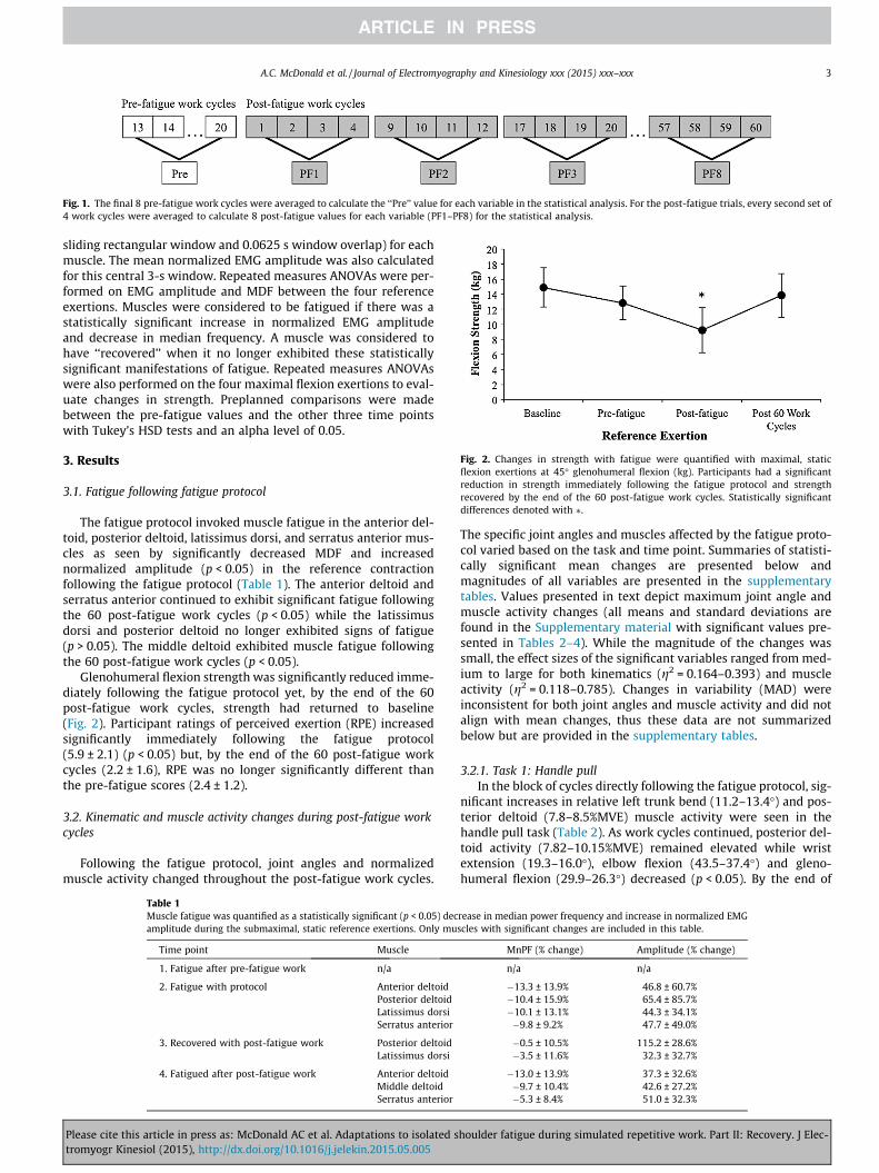
Fig. 1. The final 8 pre-fatigue work cycles were averaged to calculate the ‘‘Pre’’ value for each variable in the statistical analysis. For the post-fatigue trials, every second set of4 work cycles were averaged to calculate 8 post-fatigue values for each variable (PF1–PF8) for the statistical analysis.
Fig. 2. Changes in strength with fatigue were quantified with maximal, staticflexion exertions at 45� glenohumeral flexion (kg). Participants had a significantreduction in strength immediately following the fatigue protocol and strengthrecovered by the end of the 60 post-fatigue work cycles. Statistically significantdifferences denoted with ⁄.
A.C. McDonald et al. / Journal of Electromyography and Kinesiology xxx (2015) xxx–xxx 3
sliding rectangular window and 0.0625 s window overlap) for eachmuscle. The mean normalized EMG amplitude was also calculatedfor this central 3-s window. Repeated measures ANOVAs were per-formed on EMG amplitude and MDF between the four referenceexertions. Muscles were considered to be fatigued if there was astatistically significant increase in normalized EMG amplitudeand decrease in median frequency. A muscle was considered tohave ‘‘recovered’’ when it no longer exhibited these statisticallysignificant manifestations of fatigue. Repeated measures ANOVAswere also performed on the four maximal flexion exertions to eval-uate changes in strength. Preplanned comparisons were madebetween the pre-fatigue values and the other three time pointswith Tukey’s HSD tests and an alpha level of 0.05.
3. Results
3.1. Fatigue following fatigue protocol
The fatigue protocol invoked muscle fatigue in the anterior del-toid, posterior deltoid, latissimus dorsi, and serratus anterior mus-cles as seen by significantly decreased MDF and increasednormalized amplitude (p < 0.05) in the reference contractionfollowing the fatigue protocol (Table 1). The anterior deltoid andserratus anterior continued to exhibit significant fatigue followingthe 60 post-fatigue work cycles (p < 0.05) while the latissimusdorsi and posterior deltoid no longer exhibited signs of fatigue(p > 0.05). The middle deltoid exhibited muscle fatigue followingthe 60 post-fatigue work cycles (p < 0.05).
Glenohumeral flexion strength was significantly reduced imme-diately following the fatigue protocol yet, by the end of the 60post-fatigue work cycles, strength had returned to baseline(Fig. 2). Participant ratings of perceived exertion (RPE) increasedsignificantly immediately following the fatigue protocol(5.9 ± 2.1) (p < 0.05) but, by the end of the 60 post-fatigue workcycles (2.2 ± 1.6), RPE was no longer significantly different thanthe pre-fatigue scores (2.4 ± 1.2).
3.2. Kinematic and muscle activity changes during post-fatigue workcycles
Following the fatigue protocol, joint angles and normalizedmuscle activity changed throughout the post-fatigue work cycles.
Table 1Muscle fatigue was quantified as a statistically significant (p < 0.05) decamplitude during the submaximal, static reference exertions. Only mu
Time point Muscle
1. Fatigue after pre-fatigue work n/a
2. Fatigue with protocol Anterior deltoidPosterior deltoidLatissimus dorsiSerratus anterior
3. Recovered with post-fatigue work Posterior deltoidLatissimus dorsi
4. Fatigued after post-fatigue work Anterior deltoidMiddle deltoidSerratus anterior
Please cite this article in press as: McDonald AC et al. Adaptations to isolated stromyogr Kinesiol (2015), http://dx.doi.org/10.1016/j.jelekin.2015.05.005
The specific joint angles and muscles affected by the fatigue proto-col varied based on the task and time point. Summaries of statisti-cally significant mean changes are presented below andmagnitudes of all variables are presented in the supplementarytables. Values presented in text depict maximum joint angle andmuscle activity changes (all means and standard deviations arefound in the Supplementary material with significant values pre-sented in Tables 2–4). While the magnitude of the changes wassmall, the effect sizes of the significant variables ranged from med-ium to large for both kinematics (g2 = 0.164–0.393) and muscleactivity (g2 = 0.118–0.785). Changes in variability (MAD) wereinconsistent for both joint angles and muscle activity and did notalign with mean changes, thus these data are not summarizedbelow but are provided in the supplementary tables.
3.2.1. Task 1: Handle pullIn the block of cycles directly following the fatigue protocol, sig-
nificant increases in relative left trunk bend (11.2–13.4�) and pos-terior deltoid (7.8–8.5%MVE) muscle activity were seen in thehandle pull task (Table 2). As work cycles continued, posterior del-toid activity (7.82–10.15%MVE) remained elevated while wristextension (19.3–16.0�), elbow flexion (43.5–37.4�) and gleno-humeral flexion (29.9–26.3�) decreased (p < 0.05). By the end of
rease in median power frequency and increase in normalized EMGscles with significant changes are included in this table.
MnPF (% change) Amplitude (% change)
n/a n/a
�13.3 ± 13.9% 46.8 ± 60.7%�10.4 ± 15.9% 65.4 ± 85.7%�10.1 ± 13.1% 44.3 ± 34.1%�9.8 ± 9.2% 47.7 ± 49.0%
�0.5 ± 10.5% 115.2 ± 28.6%�3.5 ± 11.6% 32.3 ± 32.7%
�13.0 ± 13.9% 37.3 ± 32.6%�9.7 ± 10.4% 42.6 ± 27.2%�5.3 ± 8.4% 51.0 ± 32.3%
houlder fatigue during simulated repetitive work. Part II: Recovery. J Elec-

Table 2Statistically significant changes in mean joint angle in handle pull task (task 1) in the post fatigue (PF1–PF8) work cycles compared to pre-fatigue work cycles. Significant changesare denoted with bold font, rows without notation had a main effect and no significant post hoc tests. Magnitudes of the significant and non-significant changes can be found inthe Supplementary Tables.
PRE PF1 PF2 PF3 PF4 PF5 PF6 PF7 PF8
AnglePull Wrist extension 19.8 ± 4.8 18.8 ± 5.4 16.0 ± 5.51 14.65 ± 5.8 15.3 ± 7.2 15.0 ± 7.9 16.1 ± 6.7 13.6 ± 5.2 16.0 ± 6.1
Elbow extension �43.5 ± 8.2 �42.7 ± 13.8 �37.4 ± 8.1 �36.2 ± 12.4 �37.8 ± 11.3 �34.5 ± 10.8 �39.0 ± 12.1 �33.8 ± 7.7 �37.7 ± 11.1GH extension �29.9 ± 6.4 �30.9 ± 6.7 �27.8 ± 7.0 �29.2 ± 5.1 �29.4 ± 6.7 �26.3 ± 5.1 �26.5 ± 5.8 �28.5 ± 5.2 �26.0 ± 4.6Absolute righttrunk bend
�0.9 ± 4.5 �2.1 ± 3.7 �1.6 ± 4.1 �1.2 ± 3.5 0.4 ± 5.3 �0.6 ± 5.1 �0.2 ± 5.3 �0.1 ± 4.9 0.5 ± 5.3
Relative left trunkrotation
11.2 ± 3.8 13.4 ± 3.2 11.8 ± 3.5 11.1 ± 3.6 9.6 ± 4.8 11.0 ± 3.8 10.3 ± 3.7 10.9 ± 3.9 10.7 ± 4.2
Return Wrist extension 20.6 ± 5.1 20.8 ± 5.6 15.7 ± 5.2 14.3 ± 7.0 16.1 ± 7.2 15.8 ± 8.7 15.4 ± 6.4 15.2 ± 6.7 16.1 ± 5.8Elbow extension �44.8 ± 7.6 �49.2 ± 13.4 �37.5 ± 10.4 �36.3 ± 14.9 �40.0 ± 11.6 �36.9 ± 12.7 �37.6 ± 11.7 �38.3 ± 9.1 �38.8 ± 10.8Absolute righttrunk bend
�0.7 ± 4.6 �1.6 ± 3.5 �1.2 ± 4.1 �1.0 ± 3.5 0.5 ± 5.3 �0.8 ± 5.1 �0.4 ± 5.2 0.2 ± 4.8 0.6 ± 5.4
MusclePull Posterior deltoid 7.82 ± 4.44 8.50 ± 4.62 8.82 ± 4.61 9.11 ± 4.56 9.40 ± 4.60 9.56 ± 4.78 9.77 ± 4.93 9.97 ± 5.04 10.15 ± 5.09
Right uppertrapezius
8.88 ± 3.57 8.16 ± 2.85 7.35 ± 2.98 7.21 ± 2.76 6.87 ± 2.59 6.31 ± 2.72 6.60 ± 2.55 6.67 ± 2.88 7.42 ± 4.17
Return Posterior deltoid 7.63 ± 4.13 8.27 ± 4.53 8.53 ± 4.63 8.77 ± 4.72 9.08 ± 4.72 9.23 ± 4.96 9.47 ± 5.03 9.68 ± 5.13 9.90 ± 5.21
Table 3Statistically significant changes in mean joint angle in the drill task (task 3) in the post fatigue (PF1–PF8) work cycles compared to pre-fatigue work cycles. Significant post hocchanges are denoted with bold font, rows without notation had a main effect but no significant post hoc tests. Magnitudes of the significant and non-significant changes can befound in the Supplementary Tables.
PRE PF1 PF2 PF3 PF4 PF5 PF6 PF7 PF8
AngleDrill GH extension �31.5 ± 8.5 �23.9 ± 9.7 �25.4 ± 7.7 �25.9 ± 10.6 �26.8 ± 8.8 �29.4 ± 9.5 �25.4 ± 8.9 �27.2 ± 9.0 �27.3 ± 8.6
Scapular inferiorrotation
�32.3 ± 10.8 �35.0 ± 10.4 �37.0 ± 10.7 �36.9 ± 11.3 �37.0 ± 11.5 �37.5 ± 11.9 �37.3 ± 10.6 �37.4 ± 10.4 �38.0 ± 11.0
SC protraction �4.4 ± 5.7 �6.8 ± 6.6 �8.9 ± 7.4 �7.9 ± 7.3 �7.3 ± 6.4 �8.3 ± 7.3 �7.8 ± 6.7 �7.8 ± 5.9 �8.3 ± 5.8
MuscleDrill Anterior deltoid 13.1 ± 4.1 17.1 ± 4.8 17.7 ± 5.0 18.2 ± 5.8 17.5 ± 7.4 17.9 ± 6.5 17.0 ± 6.4 16.3 ± 4.5 15.8 ± 4.5
Infraspinatus 10.2 ± 5.4 11.5 ± 7.5 12.7 ± 7.0 13.4 ± 7.5 12.8 ± 8.0 13.2 ± 8.3 12.5 ± 7.6 11.6 ± 5.7 11.3 ± 5.9Latissimus dorsi 4.3 ± 2.2 4.5 ± 2.1 4.5 ± 2.1 4.4 ± 2.1 4.1 ± 2.2 4.1 ± 2.3 4.1 ± 2.2 4.0 ± 2.2 4.0 ± 2.2Middle deltoid 17.2 ± 5.4 20.1 ± 7.3 21.0 ± 7.0 20.9 ± 8.4 20.9 ± 8.6 21.7 ± 9.4 20.4 ± 8.0 19.4 ± 6.8 20.0 ± 6.8Posterior deltoid 8.0 ± 3.9 9.2 ± 4.2 9.6 ± 4.5 9.7 ± 4.5 9.9 ± 4.7 10.4 ± 5.1 10.3 ± 4.9 10.4 ± 5.0 10.8 ± 5.2Pectoralis majorsternal
2.4 ± 2.2 2.3 ± 2.2 2.2 ± 2.1 2.2 ± 2.1 2.1 ± 2.1 2.2 ± 2.1 2.1 ± 2.2 2.0 ± 2.0 2.1 ± 2.1
Triceps 3.4 ± 2.0 4.2 ± 2.4 4.2 ± 2.3 4.2 ± 2.3 4.4 ± 3.1 4.6 ± 3.2 4.4 ± 3.0 4.3 ± 2.8 4.5 ± 3.0
4 A.C. McDonald et al. / Journal of Electromyography and Kinesiology xxx (2015) xxx–xxx
the 60 post-fatigue cycles there was reduced wrist extension(19.8–16.0�), elbow flexion (43.5–37.7�) and glenohumeral flexion(29.9–26.0�) (p < 0.05) while trunk rotation returned to itspre-fatigue angle. In the return phase of the task, no changes wereseen immediately following the fatigue protocol, but, as work con-tinued, there was reduced wrist extension (20.6–14.3�) and elbowflexion (44.8–36.3�) along with increased posterior deltoid activity(7.63–9.90%MVE) (p < 0.05).
3.2.2. Task 3: DrillIn the block of work cycles immediately following the fatigue
protocol, there were significant increases in anterior (13.1–17.1%MVE) and middle (17.2–20.1%MVE) deltoid activity as wellas decreased glenohumeral flexion (31.5–23.9�) (Fig. 3 andTable 3). These changes are seen graphically in Fig. 3 from their ini-tial pre-fatigue values as they rise and fall throughout the recoveryprocess (from the first post-fatigue block, PF1, to the last block,PF8). As work cycles progressed, increased scapular superior rota-tion (32.2–38.0�) and sternoclavicular retraction (4.4–8.9�) werealso seen, accompanied by increased infraspinatus (10.2–13.4%MVE) and posterior deltoid activity (8.0–10.8% MVE). In thefinal work cycles, changes in the scapular and sternoclavicularangles persisted; however, there was no longer a significant reduc-tion in glenohumeral flexion (p < 0.05). Along with these
Please cite this article in press as: McDonald AC et al. Adaptations to isolated stromyogr Kinesiol (2015), http://dx.doi.org/10.1016/j.jelekin.2015.05.005
statistically significant changes, there were several trends in jointangle changes during this task. These joint angle changes work inconcert, and vary throughout the post-fatigue work cycles to main-tain hand position (Fig. 4).
3.2.3. Task 4: Handle pushImmediately following the fatigue protocol, there was a signif-
icant reduction in glenohumeral flexion (32.4–29.0�) withincreased scapular superior rotation (28.2–31.1�) and increasedsternoclavicular retraction (1.9 to �0.7�). These kinematic changeswere accompanied with increased triceps activity (12.5–16.1%MVE) (Table 4). As the work cycles progressed, posterior del-toid activity (8.2–10.0%MVE) also increased while middle deltoid(23.6–19.0%MVE) and latissimus dorsi activity (4.4–3.7% MVE)decreased. By the end of the 60 work cycles, the initial kinematicchanges were accompanied by increased absolute right trunk bend(�1.2 to 0.7�) and increased relative left trunk bend (0.04 to �2.0�)(p < 0.05).
During the return phase of this task, there was an immediateincrease in the activity of the triceps (5.9–7.4%MVE), middle del-toid (15.8–17.6%MVE) and posterior deltoid (7.7–8.5%MVE), aswell as increased superior scapular rotation (28.9–31.7�) anddecreased sternoclavicular protraction (2.2–0.0�). More kinematicchanges developed as the work cycles progressed including
houlder fatigue during simulated repetitive work. Part II: Recovery. J Elec-

Table 4Statistically significant changes in mean joint angle in handle push task (task 4) in the post fatigue (PF1–PF8) work cycles compared to pre-fatigue work cycles. Significantchanges are denoted with bold font, rows without notation had a main effect with no significant post hoc tests. Magnitudes of the significant and non-significant changes can befound in the Supplementary Tables.
PRE PF1 PF2 PF3 PF4 PF5 PF6 PF7 PF8
AnglePush Wrist extension 29.1 ± 8.7 30.2 ± 8.3 24.8 ± 9.3 23.4 ± 9.7 23.3 ± 9.8 26.1 ± 11.0 23.8 ± 10.5 24.2 ± 10.9 24.5 ± 9.4
GH extension �32.4 ± 9.8 �29.0 ± 9.3 �28.6 ± 8.5 �29.2 ± 7.7 �27.6 ± 8.1 �28.4 ± 7.8 �27.3 ± 7.6 �26.8 ± 6.9 �27.3 ± 6.2Scapular inferiorrotation
�28.2 ± 10.6 �31.1 ± 10.5 �31.7 ± 11.4 �31.4 ± 10.2 �31.3 ± 11.3 -32.9 ± 10.8 �33.1 ± 10.7 �31.7 ± 9.7 �32.5 ± 10.1
SC protraction 1.9 ± 5.6 �0.5 ± 6.0 �0.2 ± 5.6 �0.4 ± 6.0 0.2 ± 5.0 �0.5 ± 5.0 �1.0 ± 5.4 �0.7 ± 6.0 �0.3 ± 4.4Absolute righttrunk bend
�1.2 ± 2.0 �1.8 ± 1.8 �0.7 ± 2.9 �0.7 ± 2.8 �0.5 ± 3.3 0.3 ± 3.5 �0.02 ± 2.2 0.01 ± 2.3 0.7 ± 3.0
Relative righttrunk bend
0.04 ± 2.0 1.0 ± 2.8 �0.6 ± 4.1 �0.3 ± 2.1 �0.3 ± 2.7 �1.7 ± 3.2 �1.1 ± 2.7 �1.2 ± 2.1 �2.0 ± 2.0
Return Elbow extension �46.7 ± 6.1 �44.2 ± 7.0 �38.4 ± 8.9 �37.3 ± 9.0 �37.1 ± 8.6 �39.2 ± 12.5 �38.0 ± 9.9 �38.9 ± 11.2 �38.7 ± 9.7GH extension �33.7 ± 11.0 �31.3 ± 9.9 �29.7 ± 9.0 �30.0 ± 9.7 �27.3 ± 8.3 �28.3 ± 8.4 �28.2 ± 7.4 �27.1 ± 7.4 �26.6 ± 7.2Scapular Inferiorrotation
�28.9 ± 10.5 �31.7 ± 10.5 �32.2 ± 11.3 �31.9 ± 9.9 �31.4 ± 11.3 �33.1 ± 10.4 �33.5 ± 10.7 �32.2 ± 9.7 �32.6 ± 10.4
Scapular Internalrotation
26.5 ± 4.4 25.5 ± 3.6 25.6 ± 4.6 24.6 ± 4.0 23.6 ± 4.3 24.5 ± 5.6 24.0 ± 4.3 23.4 ± 4.9 24.1 ± 4.3
SC protraction 2.2 ± 5.7 0.03 ± 5.7 0.2 ± 5.3 �0.01 ± 5.8 0.2 ± 5.0 �0.3 ± 5.1 �0.6 ± 5.3 �0.5 ± 5.7 �0.3 ± 4.3Absolute Righttrunk bend
�1.6 ± 1.9 �2.2 ± 1.9 �1.2 ± 3.1 �1.0 ± 2.7 �0.6 ± 3.3 0.2 ± 3.4 �0.4 ± 2.2 �0.3 ± 2.2 0.7 ± 2.9
Relative righttrunk bend
0.5 ± 2.1 1.4 ± 2.8 �0.2 ± 4.2 0.03 ± 2.1 �0.1 ± 2.9 �1.7 ± 3.5 �0.7 ± 2.6 �0.9 ± 1.8 �2.0 ± 1.9
MusclePush Latissimus dorsi 4.4 ± 2.5 4.2 ± 2.3 4.2 ± 2.2 4.0 ± 2.1 4.0 ± 2.3 3.7 ± 2.1 3.8 ± 2.1 3.8 ± 2.2 3.7 ± 2.06
Middle deltoid 23.6 ± 9.2 22.6 ± 10.5 22.0 ± 10.1 20.5 ± 8.4 20.2 ± 8.8 20.0 ± 7.7 20.3 ± 9.0 20.1 ± 7.4 19.0 ± 7.9Posterior deltoid 8.2 ± 4.1 9.1 ± 4.4 9.1 ± 4.5 9.2 ± 4.6 9.4 ± 4.7 9.6 ± 4.8 9.8 ± 5.0 10.0 ± 5.0 10.0 ± 5.0Triceps 12.5 ± 8.0 16.1 ± 9.2 15.1 ± 9.2 15.2 ± 8.3 15.3 ± 9.1 15.5 ± 8.3 15.4 ± 8.1 15.5 ± 7.7 14.5 ± 8.4
Return Latissimus dorsi 3.9 ± 2.1 4.1 ± 2.1 4.1 ± 2.0 3.8 ± 1.9 3.7 ± 2.0 3.7 ± 2.0 3.6 ± 1.9 3.8 ± 2.0 3.7 ± 1.9Middle deltoid 15.8 ± 5.9 17.6 ± 7.5 17.6 ± 7.5 16.2 ± 6.6 15.9 ± 6.6 16.4 ± 6.2 16.4 ± 6.3 15.9 ± 6.2 15.4 ± 6.0Posterior deltoid 7.7 ± 4.1 8.5 ± 4.4 8.8 ± 4.5 8.9 ± 4.7 9.2 ± 4.8 9.4 ± 4.8 9.5 ± 4.9 9.8 ± 5.1 10.0 ± 5.1Triceps 5.9 ± 2.7 7.4 ± 3.3 7.5 ± 3.6 7.4 ± 3.2 7.3 ± 3.6 7.4 ± 3.3 7.5 ± 3.1 7.5 ± 3.1 7.2 ± 3.4
Fig. 3. In the drill task during the post-fatigue work cycles, glenohumeral flexion(dashed black line) decreased, which coincided with increased glenohumeralabduction (solid black line), middle deltoid (red line) and posterior deltoid (blueline) muscle activity. By the end of the post-fatigue work cycles the middle deltoidexhibited significant signs of muscle fatigue (p < 0.05). Axes have been adjusted foreach variable and work cycles are shown from pre-fatigue (PRE) to post-fatigue 8(PF1–PF8) in a clockwise direction. (For interpretation of the references to color inthis figure legend, the reader is referred to the web version of this article.)
A.C. McDonald et al. / Journal of Electromyography and Kinesiology xxx (2015) xxx–xxx 5
reduced elbow (46.7–37.1�) and glenohumeral flexion (33.7–26.6�)and increased scapular superior rotation (28.9–33.5�). These
Please cite this article in press as: McDonald AC et al. Adaptations to isolated stromyogr Kinesiol (2015), http://dx.doi.org/10.1016/j.jelekin.2015.05.005
kinematic changes persisted throughout the final work cycles(p < 0.05).
4. Discussion
Following the fatigue protocol, all participants completed the60 post-fatigue work cycles, despite physiological signs of musclefatigue, reduced strength and increased ratings of perceived exer-tion. During the post-fatigue work cycles, joint angle and muscleactivity changed over time. By the end of the protocol, strengthand perceived exertion had returned to pre-fatigue levels, yet somemuscles exhibited signs of fatigue. After the additional hour ofwork, the latissimus dorsi and posterior deltoid showed recovery,fatigue persisted in the anterior deltoid and serratus anterior,and fatigue had developed in the middle deltoid. Based on this evi-dence, participants were able to use the mobility of the shouldercomplex and upper extremity to adapt to reduced physical capac-ity and allow recovery in some muscles. Despite the constrainednature of the pushing and pulling tasks, we saw significant kine-matic and muscular changes during the post-fatigue work cycles.In the drill task, participants reduced their glenohumeral flexionangle, effectively reducing the demand on the fatigued muscle.Amongst other kinematic changes, participants increased gleno-humeral abduction to compensate for this change. This action alsocorresponded with increased middle and posterior deltoid activity(Fig. 3). The fatigue protocol induced fatigue in the anterior andposterior deltoids; as time progressed, the posterior deltoid recov-ered, which allowed it to compensate for reduced middle deltoidcapacity. The greatest abduction occurred during the laterpost-fatigue work cycles (PF7–PF8), corresponding with increasedposterior deltoid activity (Fig. 3). Given that that the posterior
houlder fatigue during simulated repetitive work. Part II: Recovery. J Elec-
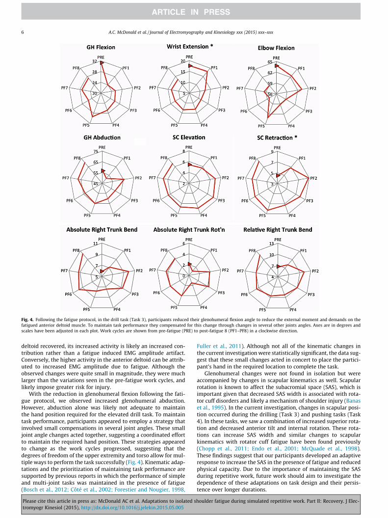
Fig. 4. Following the fatigue protocol, in the drill task (Task 3), participants reduced their glenohumeral flexion angle to reduce the external moment and demands on thefatigued anterior deltoid muscle. To maintain task performance they compensated for this change through changes in several other joints angles. Axes are in degrees andscales have been adjusted in each plot. Work cycles are shown from pre-fatigue (PRE) to post-fatigue 8 (PF1–PF8) in a clockwise direction.
6 A.C. McDonald et al. / Journal of Electromyography and Kinesiology xxx (2015) xxx–xxx
deltoid recovered, its increased activity is likely an increased con-tribution rather than a fatigue induced EMG amplitude artifact.Conversely, the higher activity in the anterior deltoid can be attrib-uted to increased EMG amplitude due to fatigue. Although theobserved changes were quite small in magnitude, they were muchlarger than the variations seen in the pre-fatigue work cycles, andlikely impose greater risk for injury.
With the reduction in glenohumeral flexion following the fati-gue protocol, we observed increased glenohumeral abduction.However, abduction alone was likely not adequate to maintainthe hand position required for the elevated drill task. To maintaintask performance, participants appeared to employ a strategy thatinvolved small compensations in several joint angles. These smalljoint angle changes acted together, suggesting a coordinated effortto maintain the required hand position. These strategies appearedto change as the work cycles progressed, suggesting that thedegrees of freedom of the upper extremity and torso allow for mul-tiple ways to perform the task successfully (Fig. 4). Kinematic adap-tations and the prioritization of maintaining task performance aresupported by previous reports in which the performance of simpleand multi-joint tasks was maintained in the presence of fatigue(Bosch et al., 2012; Côté et al., 2002; Forestier and Nougier, 1998,
Please cite this article in press as: McDonald AC et al. Adaptations to isolated stromyogr Kinesiol (2015), http://dx.doi.org/10.1016/j.jelekin.2015.05.005
Fuller et al., 2011). Although not all of the kinematic changes inthe current investigation were statistically significant, the data sug-gest that these small changes acted in concert to place the partici-pant’s hand in the required location to complete the task.
Glenohumeral changes were not found in isolation but wereaccompanied by changes in scapular kinematics as well. Scapularrotation is known to affect the subacromial space (SAS), which isimportant given that decreased SAS width is associated with rota-tor cuff disorders and likely a mechanism of shoulder injury (Banaset al., 1995). In the current investigation, changes in scapular posi-tion occurred during the drilling (Task 3) and pushing tasks (Task4). In these tasks, we saw a combination of increased superior rota-tion and decreased anterior tilt and internal rotation. These rota-tions can increase SAS width and similar changes to scapularkinematics with rotator cuff fatigue have been found previously(Chopp et al., 2011; Endo et al., 2001; McQuade et al., 1998).These findings suggest that our participants developed an adaptiveresponse to increase the SAS in the presence of fatigue and reducedphysical capacity. Due to the importance of maintaining the SASduring repetitive work, future work should aim to investigate thedependence of these adaptations on task design and their persis-tence over longer durations.
houlder fatigue during simulated repetitive work. Part II: Recovery. J Elec-

A.C. McDonald et al. / Journal of Electromyography and Kinesiology xxx (2015) xxx–xxx 7
We evaluated variability in both motion and muscle activity asvariability has become a focus in workplace exposures, fatigue andinjury risk. We found that the changes in variability (median abso-lute deviation) observed throughout the post-fatigue work cycleswere dependent on the variable (muscle, joint angle) and thework-cycle. The usefulness of exposure variability on fatigueappears to be dependent on definitions and measurement of bothvariability and fatigue, as well as the specific tasks involved(Luger et al., 2014). Variability seems inherent in the mobility ofthe shoulder complex, which affords many kinematic and musclestrategies to complete a given task (Srinivasan and Mathiassen,2012). Multiple control strategies create challenges when usingjob rotation to increase exposure variability when muscle overlapbetween tasks is difficult to determine (Keir et al., 2011). In simu-lated cutting tasks, experienced butchers had greater kinematicvariability but reduced EMG variability (Madeleine et al., 2008a).Our mixed findings agree with the previous studies and supportthe notion that variability with fatigue and recovery will be depen-dent on the context of its measurement (Qin et al., 2014).
There are limitations to the current study. The multiple degreesof freedom of the upper extremity and trunk made it possible forparticipants to combine multiple small changes in joint anglesand muscle activity to compensate for fatigue. Although we believethat these adaptations are important to understand, they are diffi-cult to quantify with traditional statistical analyses. Another chal-lenge in this analysis was the vast amount of data, and thus onlysummary variables have been presented. Future work will aim todevelop analyses methods that are more sensitive to small changesand can incorporate more data to better understand the complexresponse to fatigue over time.
5. Conclusion
In the presence of muscle fatigue and reduced muscle capacity,participants were able to coordinate muscular and kinematic adap-tations to maintain task performance for an hour of simulatedwork. These findings highlight the importance of not only examin-ing the immediate response to fatigue but also how the responsechanges over time. We also found that, while individuals continuedworking with signs of muscle fatigue, they did not perceive fatigue,as reflected in their lowered perceived exertion scores. Adaptationsto isolated muscle fatigue change over time, kinematic and muscu-lar changes allow recovery but individuals may not perceive exist-ing fatigue, which may contribute to overuse injuries in theworkplace.
Acknowledgements
This study was supported by funding from the Centre ofResearch Expertise for the Prevention of MusculoskeletalDisorder (CRE-MSD) and Automotive Partnership Canada (APC).
Appendix A. Supplementary material
Supplementary methods and data associated with this articlecan be found, in the online version, at http://dx.doi.org/10.1016/j.jelekin.2015.05.005.
References
Banas MP, Miller RJ, Totterman S. Relationship between the lateral acromion angleand rotator cuff disease. J Shoulder Elbow Surg 1995;4(6):454–61.
Bosch T, Mathiassen SE, Hallman D, de Looze MP, Lyskov E, Visser B, et al. Temporalstrategy and performance during a fatiguing short-cycle repetitive task.Ergonomics 2012;55(8):863–73.
Please cite this article in press as: McDonald AC et al. Adaptations to isolated stromyogr Kinesiol (2015), http://dx.doi.org/10.1016/j.jelekin.2015.05.005
Chopp JN, Fischer SL, Dickerson CR. The specificity of fatiguing protocols affectsscapular orientation: implications for subacromial impingement. Clin Biomech2011;26:40–5.
Côté JN, Mathieu PA, Levin MF, Feldman AG. Movement reorganization tocompensate for fatigue during sawing. Exp Brain Res 2002;146:394–8.
Culham E, Peat M. Functional anatomy of the shoulder complex. J Orthop SportsPhys Ther 1993;18(1):342–50.
Ebaugh DD, McClure PW, Karduna AR. Scapulothorasic and glenohumeralkinematics following an external rotation fatigue protocol. J Orthop SportsPhys Ther 2006a;16:224–35.
Ebaugh DD, McClure PW, Karduna AR. Effects of shoulder muscle fatigue caused byrepetitive overhead activities on scapulothoracic and glenohumeral kinematics.J Electromyogr Kinesiol 2006b;16(3):224–35.
Elliott MB, Barr AE, Kietrys DM, Al-Shatti T, Amin M, Barbe MF. Peripheral neuritisand increased spinal cord neurochemicals are induced in a model of repetitivemotion injury with low force and repetition exposure. Brain Res2008;1218:103–13.
Elliott MB, Barr AE, Clark BD, Amin M, Amin S, Barbe MF. High force reaching taskinduced widespread inflammation, increased spinal cord neurochemicals andneuropathic pain. Neuroscience 2009;158:922–31.
Endo K, Ikata T, Katoh S, Takeda Y. Radiographic assessment of scapular rotationaltilt in chronic shoulder impingement syndrome. J Orthop Sci 2001;6:3–10.
Enoka RM, Duchateau J. Muscle fatigue: what, why and how it influences musclefunction. J Physiol 2008;586:11–23.
Enoka RM, Stuart DG. Neurobiology of muscle fatigue. J Appl Physiol1992;72(5):1631–48.
Farina D, Leclerc F, Arendt-Nielsen L, Buttelli O, Madeleine P. The change in spatialdistribution of upper trapezius muscle activity is correlated to contractionduration. J Electromyogr Kinesiol 2008;18(1):16–25.
Fedorowich L, Emery K, Gervasi B, Côté JN. Gender differences in neck/shouldermuscular patterns in response to repetitive motion induced fatigue. JElectromyogr Kinesiol 2013;23:1183–9.
Ferguson SA, Allread WG, Le P, Rose J, Marras WS. Shoulder muscle fatigue duringrepetitive tasks as measured by electromyography and near-infraredspectroscopy. Hum Factors 2013;55(6):1077–87.
Forestier N, Nougier V. The effects of muscular fatigue on the coordination of amultijoint movement in human. Neurosci Lett 1998;252:187–90.
Frey-Law LA, Looft JM, Heitsman J. A three-compartment muscle fatigue modelaccurately predicts joint-specific maximum endurance times for sustainedisometric tasks. J Biomech 2012;45(10):1803–8.
Fuller JR, Lomond KV, Fung J, Côté JN. Posture-movement changes followingrepetitive motion-induced shoulder muscle fatigue. J Electromyogr Kinesiol2009;19:1043–52.
Fuller JR, Fung J, Côté JN. Time-dependent adaptations to posture and movementcharacteristics during the development of repetitive reaching induced fatigue.Exp Brain Res 2011;211:133–43.
Hanvold TN, Wærsted M, Veiersted KB. Long periods with uninterrupted muscleactivity related to neck and shoulder pain. Work 2012;41(Suppl 1):2535–8.
Karduna AR, Williams GR, Williams JL, Iannotti JP. Kinematics of the glenohumeraljoint: influences of muscle forces, ligamentous constraints, and articulargeometry. J Orthop Res 1996;14(6):986–93.
Keir PJ, Sanei K, Holmes MWR. Task rotation effects on upper extremity and backmuscle activity. Appl Ergon 2011;42:814–9.
Labriola JE, Jolly JT, McMahon PJ, Debski RE. Active stability of the glenohumeraljoint decreases in the apprehension position. Clin Biomech 2004;19(8):801–9.
Lucidi CA, Lehman SL. Adaptation to fatigue of long duration in human wristmovements. J Appl Physiol 1992;73:2596–603.
Luger T, Bosch T, Veeger D, de Looze M. The influence of task variation onmanifestation of fatigue is ambiguous – a literature review. Ergonomics2014;57(2):162–74.
Madeleine P, Mathiassen SE, Arendt-Nielsen L. Changes in the degree of motorvariability associated with experimental and chronic neck-shoulder pain duringa standardized repetitive arm movement. Exp Brain Res 2008a;185:689–98.
Madeleine P, Voigt M, Mathiassen SE. The size of cycle-to-cycle variability inbiomechanical exposure among butchers performing a standardized cuttingtask. Ergonomics 2008b;51(7):1078–95.
McQuade KJ, Dawson J, Smidt GL. Scapulothoracic muscle fatigue associated withalterations in scapulohumeral rhythm kinematics during maximum resistiveshoulder elevation. J Orthop Sports Phys Ther 1998;28(2):74–80.
Nordander C, Ohlsson K, Akesson I, Arvidsson I, Balogh I, Hansson G, et al. Risk ofmusculoskeletal disorders among females and males in repetitive/constrainedwork. Ergonomics 2009;52(10):1226–39.
Potvin JR. Predicting maximum acceptable efforts for repetitive tasks: an equationbased on duty cycle. Hum Factors: J Hum Factors Ergon Soc 2012;54(2):175–88.
Qin J, Lin JH, Faber GS, Buchholz B, Xu X. Upper extremity kinematic and kineticadaptations during a fatiguing repetitive task. J Electromyogr Kinesiol2014;24:404–11.
Srinivasan D, Mathiassen SE. Motor variability in occupational health andperformance. Clin Biomech 2012;27:979–93.
Svendsen SW, Gelineck J, Mathiassen SE, Bonde JP, Frich LH, Stengaard-Pedersen K,et al. Work above shoulder level and degenerative alterations of the rotator cufftendons: a magnetic resonance imaging study. Arthritis Rheum2004;50(10):3314–22.
houlder fatigue during simulated repetitive work. Part II: Recovery. J Elec-

8 A.C. McDonald et al. / Journal of Electromyography and Kinesiology xxx (2015) xxx–xxx
Tsai N, McClure PW, Karduna AW. Effects of muscle fatigue on 3-Dimensionalscapular kinematics. Arch Phys Med Rehabil 2003;84:1000–5.
Vollestad NK, Sejersted OM. Biochemical correlates of fatigue a brief review. Eur JAppl Physiol 1988;57:336–47.
Wu G, Siegler S, Allard P, Kirtley C, Leardini A, Rosenbaum D, et al. ISBrecommendation on definitions of joint coordinate system of various jointsfor the reporting of human joint motion—part I: ankle, hip, and spine. J Biomech2002;25:543–8.
Wu G, van der Helm FCT, Veeger HEJ, Makhsous M, Van Roy P, Anglin C, et al. ISBrecommendation on definitions of joint coordinate systems of various joints forthe reporting of human joint motion—Part II: shoulder, elbow, wrist and hand. JBiomech 2005;38(5):981–92.
Alison McDonald received a Bachelor of Science(Kinesiology, Honors Cooperative Program, 2011)from the University of Waterloo. She started her MScat McMaster University in 2011 and is currently aPhD student holding an NSERC post-graduate schol-arship.
Please cite this article in press as: McDonald AC et al. Adaptations to isolated stromyogr Kinesiol (2015), http://dx.doi.org/10.1016/j.jelekin.2015.05.005
Calvin Tse received his Bachelor of ScienceKinesiology (Hons., 2014) from McMaster University.He is continuing his studies at McMaster Universityas an MSc student in the Department of Kinesiology.He hopes to continue his research in upper extremitybiomechanics and digital modelling.
Peter Keir received his PhD from the University ofWaterloo. He is currently a Professor in theDepartment of Kinesiology at McMaster University inHamilton, Ontario. His research examines upperextremity mechanics and function using EMG,imaging and modelling to determine the mecha-nisms of work-related musculoskeletal disorders,with an emphasis on carpal tunnel syndrome andmuscle-related injuries of the arm and hand. He is aformer President of the Canadian Society forBiomechanics.
houlder fatigue during simulated repetitive work. Part II: Recovery. J Elec-