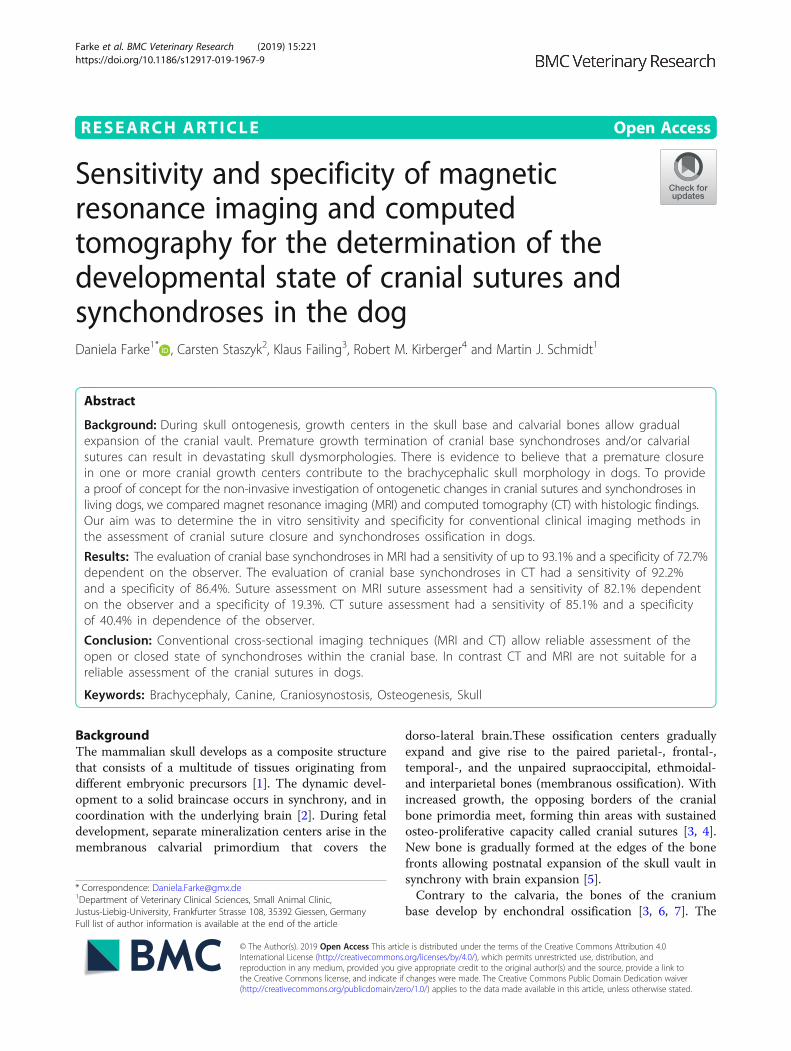
Sensitivity and specificity of magnetic resonance imaging and ...
14
RESEARCH ARTICLE Open Access Sensitivity and specificity of magnetic resonance imaging and computed tomography for the determination of the developmental state of cranial sutures and synchondroses in the dog Daniela Farke 1* , Carsten Staszyk 2 , Klaus Failing 3 , Robert M. Kirberger 4 and Martin J. Schmidt 1 Abstract Background: During skull ontogenesis, growth centers in the skull base and calvarial bones allow gradual expansion of the cranial vault. Premature growth termination of cranial base synchondroses and/or calvarial sutures can result in devastating skull dysmorphologies. There is evidence to believe that a premature closure in one or more cranial growth centers contribute to the brachycephalic skull morphology in dogs. To provide a proof of concept for the non-invasive investigation of ontogenetic changes in cranial sutures and synchondroses in living dogs, we compared magnet resonance imaging (MRI) and computed tomography (CT) with histologic findings. Our aim was to determine the in vitro sensitivity and specificity for conventional clinical imaging methods in the assessment of cranial suture closure and synchondroses ossification in dogs. Results: The evaluation of cranial base synchondroses in MRI had a sensitivity of up to 93.1% and a specificity of 72.7% dependent on the observer. The evaluation of cranial base synchondroses in CT had a sensitivity of 92.2% and a specificity of 86.4%. Suture assessment on MRI suture assessment had a sensitivity of 82.1% dependent on the observer and a specificity of 19.3%. CT suture assessment had a sensitivity of 85.1% and a specificity of 40.4% in dependence of the observer. Conclusion: Conventional cross-sectional imaging techniques (MRI and CT) allow reliable assessment of the open or closed state of synchondroses within the cranial base. In contrast CT and MRI are not suitable for a reliable assessment of the cranial sutures in dogs. Keywords: Brachycephaly, Canine, Craniosynostosis, Osteogenesis, Skull Background The mammalian skull develops as a composite structure that consists of a multitude of tissues originating from different embryonic precursors [1]. The dynamic devel- opment to a solid braincase occurs in synchrony, and in coordination with the underlying brain [2]. During fetal development, separate mineralization centers arise in the membranous calvarial primordium that covers the dorso-lateral brain.These ossification centers gradually expand and give rise to the paired parietal-, frontal-, temporal-, and the unpaired supraoccipital, ethmoidal- and interparietal bones (membranous ossification). With increased growth, the opposing borders of the cranial bone primordia meet, forming thin areas with sustained osteo-proliferative capacity called cranial sutures [3, 4]. New bone is gradually formed at the edges of the bone fronts allowing postnatal expansion of the skull vault in synchrony with brain expansion [5]. Contrary to the calvaria, the bones of the cranium base develop by enchondral ossification [3, 6, 7]. The © The Author(s). 2019 Open Access This article is distributed under the terms of the Creative Commons Attribution 4.0 International License (http://creativecommons.org/licenses/by/4.0/), which permits unrestricted use, distribution, and reproduction in any medium, provided you give appropriate credit to the original author(s) and the source, provide a link to the Creative Commons license, and indicate if changes were made. The Creative Commons Public Domain Dedication waiver (http://creativecommons.org/publicdomain/zero/1.0/) applies to the data made available in this article, unless otherwise stated. * Correspondence: [email protected] 1 Department of Veterinary Clinical Sciences, Small Animal Clinic, Justus-Liebig-University, Frankfurter Strasse 108, 35392 Giessen, Germany Full list of author information is available at the end of the article Farke et al. BMC Veterinary Research (2019) 15:221 https://doi.org/10.1186/s12917-019-1967-9
-
Upload
khangminh22 -
Category
Documents
-
view
1 -
download
0
Transcript of Sensitivity and specificity of magnetic resonance imaging and ...
RESEARCH ARTICLE Open Access
Sensitivity and specificity of magneticresonance imaging and computedtomography for the determination of thedevelopmental state of cranial sutures andsynchondroses in the dogDaniela Farke1* , Carsten Staszyk2, Klaus Failing3, Robert M. Kirberger4 and Martin J. Schmidt1
Abstract
Background: During skull ontogenesis, growth centers in the skull base and calvarial bones allow gradualexpansion of the cranial vault. Premature growth termination of cranial base synchondroses and/or calvarialsutures can result in devastating skull dysmorphologies. There is evidence to believe that a premature closurein one or more cranial growth centers contribute to the brachycephalic skull morphology in dogs. To providea proof of concept for the non-invasive investigation of ontogenetic changes in cranial sutures and synchondroses inliving dogs, we compared magnet resonance imaging (MRI) and computed tomography (CT) with histologic findings.Our aim was to determine the in vitro sensitivity and specificity for conventional clinical imaging methods inthe assessment of cranial suture closure and synchondroses ossification in dogs.
Results: The evaluation of cranial base synchondroses in MRI had a sensitivity of up to 93.1% and a specificity of 72.7%dependent on the observer. The evaluation of cranial base synchondroses in CT had a sensitivity of 92.2%and a specificity of 86.4%. Suture assessment on MRI suture assessment had a sensitivity of 82.1% dependenton the observer and a specificity of 19.3%. CT suture assessment had a sensitivity of 85.1% and a specificityof 40.4% in dependence of the observer.
Conclusion: Conventional cross-sectional imaging techniques (MRI and CT) allow reliable assessment of theopen or closed state of synchondroses within the cranial base. In contrast CT and MRI are not suitable for areliable assessment of the cranial sutures in dogs.
Keywords: Brachycephaly, Canine, Craniosynostosis, Osteogenesis, Skull
BackgroundThe mammalian skull develops as a composite structurethat consists of a multitude of tissues originating fromdifferent embryonic precursors [1]. The dynamic devel-opment to a solid braincase occurs in synchrony, and incoordination with the underlying brain [2]. During fetaldevelopment, separate mineralization centers arise in themembranous calvarial primordium that covers the
dorso-lateral brain.These ossification centers graduallyexpand and give rise to the paired parietal-, frontal-,temporal-, and the unpaired supraoccipital, ethmoidal-and interparietal bones (membranous ossification). Withincreased growth, the opposing borders of the cranialbone primordia meet, forming thin areas with sustainedosteo-proliferative capacity called cranial sutures [3, 4].New bone is gradually formed at the edges of the bonefronts allowing postnatal expansion of the skull vault insynchrony with brain expansion [5].Contrary to the calvaria, the bones of the cranium
base develop by enchondral ossification [3, 6, 7]. The
© The Author(s). 2019 Open Access This article is distributed under the terms of the Creative Commons Attribution 4.0International License (http://creativecommons.org/licenses/by/4.0/), which permits unrestricted use, distribution, andreproduction in any medium, provided you give appropriate credit to the original author(s) and the source, provide a link tothe Creative Commons license, and indicate if changes were made. The Creative Commons Public Domain Dedication waiver(http://creativecommons.org/publicdomain/zero/1.0/) applies to the data made available in this article, unless otherwise stated.
* Correspondence: [email protected] of Veterinary Clinical Sciences, Small Animal Clinic,Justus-Liebig-University, Frankfurter Strasse 108, 35392 Giessen, GermanyFull list of author information is available at the end of the article
Farke et al. BMC Veterinary Research (2019) 15:221 https://doi.org/10.1186/s12917-019-1967-9

basioccipital, basisphenoid and presphenoid bonesemerge from cartilaginous precursors that ossify duringfetal development. Cartilaginous segments termed syn-chondroses persist between the ossification centers, con-sisting of two mirror-image growth plates arranged inopposing directions. Analogous to endochondral growthplates in long bones, synchondroses of the skull basegrow through ongoing chondrocyte proliferation andgradual osseous transformation allowing the expansionof the cranial base along its rostro-caudal axis [1, 2, 8].The growth of individual bones is regulated within
these centers by complex signal cascades, involving mul-tiple receptors and transcription factors within bothkinds of growth centers [9]. Any disturbance in theseprocesses has been shown in children to lead to prema-ture closure of both, cranial base synchondroses and/orsutures (craniosynostoses) and thereby to devastatingskull dysmorphologies [10]. Regulation of growth andclosure of both, sutures and synchondroses of the skullhave been extensively studies in laboratory rodents [11,12], but not so in dogs. Even the knowledge of generaltemporary evolution of skull sutures and synchondrosesin companion animals is limited. Two older investiga-tions determined closure times for the cranium growthcenters in dogs at the age of 12 months [13, 14]. Two re-cent studies documented a higher incidence of closed fa-cial sutures and premature closure times for the spheno-occipital synchondrosis in brachycephalic dogs com-pared to mesaticephalic dogs [15, 16]. The authors ofboth studies suggest that the temporal variation ofgrowth termination has a substantial influence for thedevelopment of a brachycephalic head morphology. Inlight of these new insights, the question arises as towhether these variations are part of a physiologicspectrum or, at least partially, a pathological condition.It should be considered that different forms of patho-logic craniosynostoses and grades might contribute tothe brachycephalic skull morphology in dogs in the sameway as it does in cats [17]. Breeding of phenotypes basedon pathological genetic defects clearly known to be asso-ciated with neurological or craniofacial diseases as inhumans [18] would be prohibited by the German animalprotection law and would also be unethical.Cross sectional diagnostic imaging methods (MRI and
CT) would allow the examination of large cohorts ofdogs of different breeds in vivo. However, the value ofthese imaging methods for the assessment of synchon-drosal and sutural status has never been determined.The aim of the present study was, therefore, to com-
pare imaging findings with histological preparations inorder to determine the sensitivity and specificity for con-ventional clinical CT and MRI in the assessment of theopen or closed status of cranial synchondroses and su-tures in dogs.
ResultsHistologyDue to the fragility of the structures, especially in veryyoung puppies, twenty-two sutures and five synchon-droses were lost during sample preparation. 433 suturesand 112 synchondroses could be histologically evaluated.
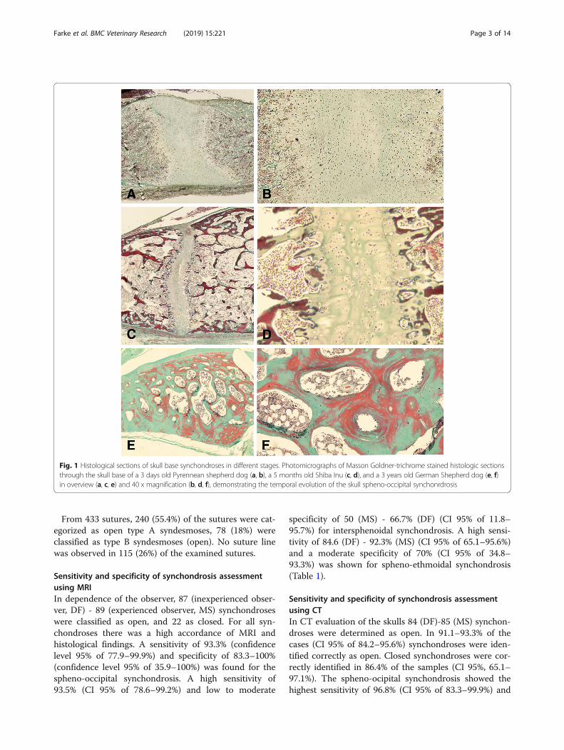
Synchondroses morphology and statusIn all immature dogs, a double-sided arrangement ofchondrocytes in a hyaline homogenous cartilage matrixwas observed between the bone tissue of the basicranialbones. The cartilage consisted of chondrocytes beingdistributed into a central resting zone, as well as bilateralproliferating, and hypertrophic zones (Fig. 1 A, B) [19].In the fourth zone in the peripheral portion of synchon-drosis, osteoblasts and blood vessels invaded the area ofcartilage. In older dogs, the resting and proliferativelayer within the synchondrosis gradually decreased, lead-ing to a relative narrowing of the growth center (Fig. 1cand d). At the chondro-osseous junction differentiatedhypertrophic chondrocytes are replaced by bony trabec-ula that, eventually, fill the growth plate leading to acontinuous medullary cavity (Fig. 1e and f).90 synchondroses (80.4%) were evaluated as closed,
and 22 (19.6%) were assessed as open. The spheno-occipital synchondroses were classified as open in 31(83.78%) specimen as closed in 6 (16.22%). Of the 38 ex-amined inter-sphenoidal synchondroses, 32 (84.21%)were classified as open, and 6 (15.79%) as closed. Of 37examined spheno-ethmoidal synchondroses, 27 (72.97%)were classified as open and 10 (27.03%) as closed.
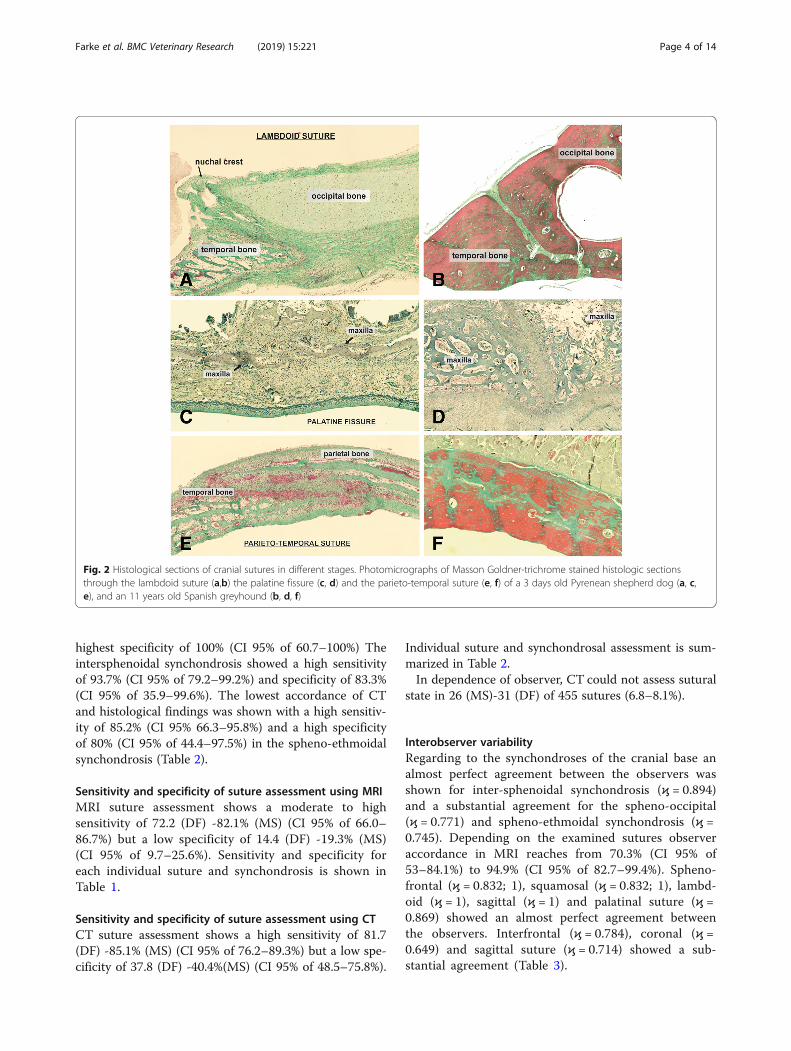
Suture morphology and statusSuture morphology ranged from straight-edged plane su-tures (lambda suture; Fig. 2 A, B) or butt-sutures (palat-ine fissure, Fig. 2c and d) over simple overlapping(sphenofrontal and squamosal suture, Fig. 2e and f; cor-onal suture, Fig. 3a and b) to serrated sutures, with thebone edges having a saw-like appearance (sagittal suture,Fig. 3c and d). The gap in between the bony edges arefilled with collagen and elongated fibrocytes. Two typesof open sutures were identified according to the type ofconnective tissue, and cellular components, which domi-nated the sutural space. In young dogs the sutures con-tained loosely arranged connective tissue showing acollagen fiber orientation preferentially parallel to thesutural alignment (Fig. 4a). These sutures also featuredhigh amounts osteoblasts (> 50 in a mean of 3 FOV) andfibroblasts (> 20 in 3 FOV) (type A; Fig. 4a). In olderdogs (> 7 months), sutures rather contained a moredense connective tissue with collagen fiber orientationbeing orientated oblique to perpendicular to the sutureline, with a low numbers of osteoblasts (< 10 in 3 FOV)and fibroblasts (< 20 in 3 FOV) (type B, Fig. 4b).
Farke et al. BMC Veterinary Research (2019) 15:221 Page 2 of 14
From 433 sutures, 240 (55.4%) of the sutures were cat-egorized as open type A syndesmoses, 78 (18%) wereclassified as type B syndesmoses (open). No suture linewas observed in 115 (26%) of the examined sutures.
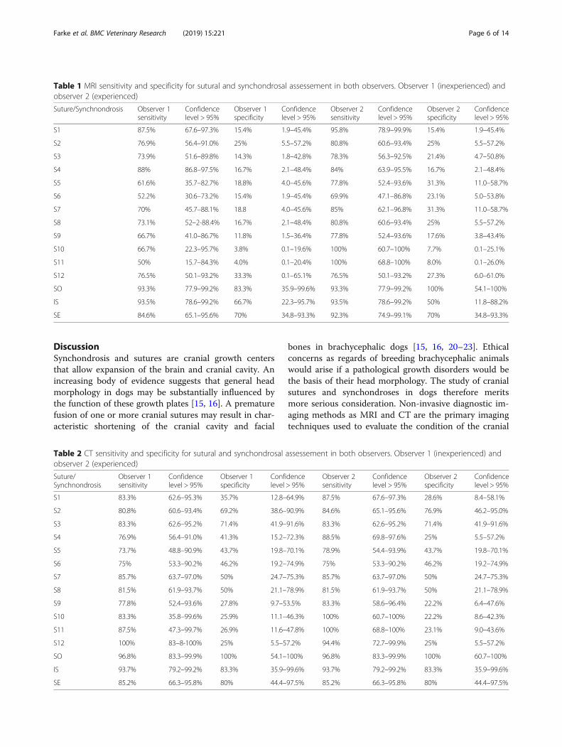
Sensitivity and specificity of synchondrosis assessmentusing MRIIn dependence of the observer, 87 (inexperienced obser-ver, DF) - 89 (experienced observer, MS) synchondroseswere classified as open, and 22 as closed. For all syn-chondroses there was a high accordance of MRI andhistological findings. A sensitivity of 93.3% (confidencelevel 95% of 77.9–99.9%) and specificity of 83.3–100%(confidence level 95% of 35.9–100%) was found for thespheno-occipital synchondrosis. A high sensitivity of93.5% (CI 95% of 78.6–99.2%) and low to moderate
specificity of 50 (MS) - 66.7% (DF) (CI 95% of 11.8–95.7%) for intersphenoidal synchondrosis. A high sensi-tivity of 84.6 (DF) - 92.3% (MS) (CI 95% of 65.1–95.6%)and a moderate specificity of 70% (CI 95% of 34.8–93.3%) was shown for spheno-ethmoidal synchondrosis(Table 1).
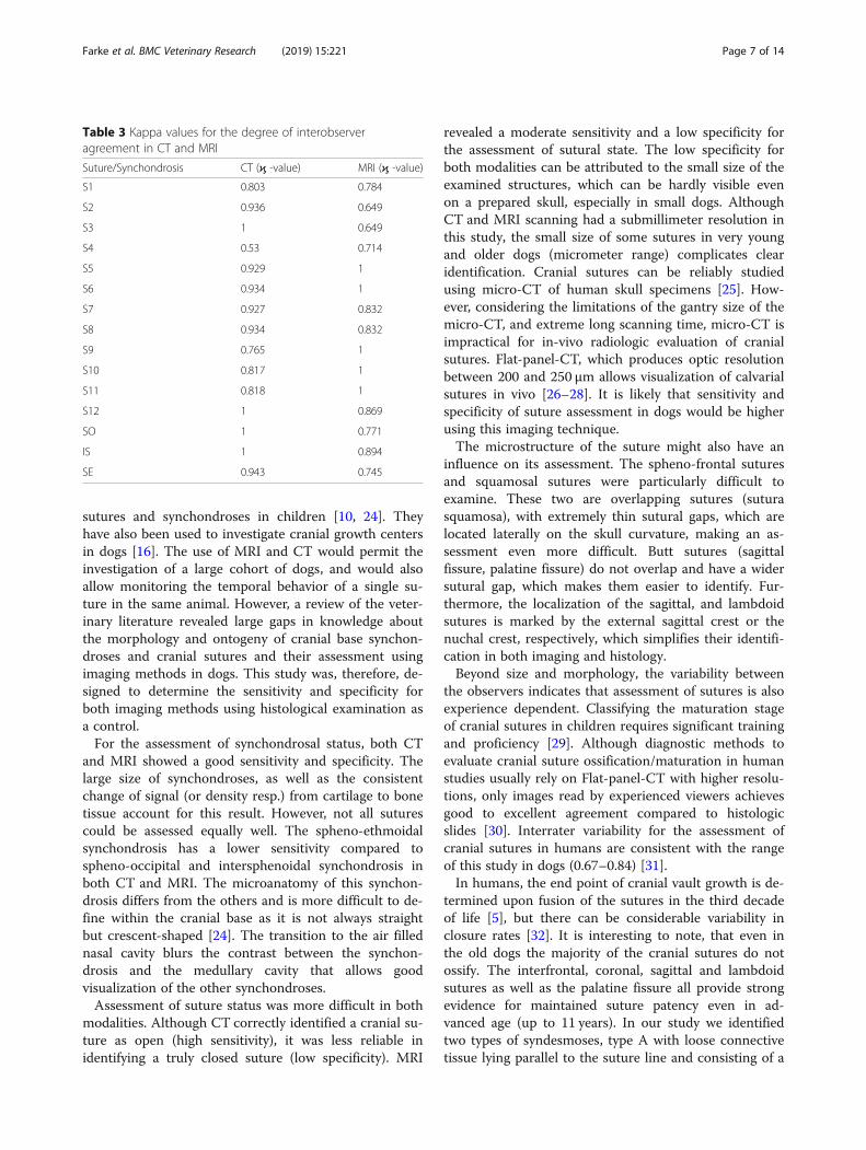
Sensitivity and specificity of synchondrosis assessmentusing CTIn CT evaluation of the skulls 84 (DF)-85 (MS) synchon-droses were determined as open. In 91.1–93.3% of thecases (CI 95% of 84.2–95.6%) synchondroses were iden-tified correctly as open. Closed synchondroses were cor-rectly identified in 86.4% of the samples (CI 95%, 65.1–97.1%). The spheno-ocipital synchondrosis showed thehighest sensitivity of 96.8% (CI 95% of 83.3–99.9%) and
Fig. 1 Histological sections of skull base synchondroses in different stages. Photomicrographs of Masson Goldner-trichrome stained histologic sectionsthrough the skull base of a 3 days old Pyrennean shepherd dog (a, b), a 5 months old Shiba Inu (c, d), and a 3 years old German Shepherd dog (e, f)in overview (a, c, e) and 40 x magnification (b, d, f), demonstrating the temporal evolution of the skull spheno-occipital synchonrdrosis
Farke et al. BMC Veterinary Research (2019) 15:221 Page 3 of 14
highest specificity of 100% (CI 95% of 60.7–100%) Theintersphenoidal synchondrosis showed a high sensitivityof 93.7% (CI 95% of 79.2–99.2%) and specificity of 83.3%(CI 95% of 35.9–99.6%). The lowest accordance of CTand histological findings was shown with a high sensitiv-ity of 85.2% (CI 95% 66.3–95.8%) and a high specificityof 80% (CI 95% of 44.4–97.5%) in the spheno-ethmoidalsynchondrosis (Table 2).
Sensitivity and specificity of suture assessment using MRIMRI suture assessment shows a moderate to highsensitivity of 72.2 (DF) -82.1% (MS) (CI 95% of 66.0–86.7%) but a low specificity of 14.4 (DF) -19.3% (MS)(CI 95% of 9.7–25.6%). Sensitivity and specificity foreach individual suture and synchondrosis is shown inTable 1.
Sensitivity and specificity of suture assessment using CTCT suture assessment shows a high sensitivity of 81.7(DF) -85.1% (MS) (CI 95% of 76.2–89.3%) but a low spe-cificity of 37.8 (DF) -40.4%(MS) (CI 95% of 48.5–75.8%).
Individual suture and synchondrosal assessment is sum-marized in Table 2.In dependence of observer, CT could not assess sutural
state in 26 (MS)-31 (DF) of 455 sutures (6.8–8.1%).
Interobserver variabilityRegarding to the synchondroses of the cranial base analmost perfect agreement between the observers wasshown for inter-sphenoidal synchondrosis (ϗ = 0.894)and a substantial agreement for the spheno-occipital(ϗ = 0.771) and spheno-ethmoidal synchondrosis (ϗ =0.745). Depending on the examined sutures observeraccordance in MRI reaches from 70.3% (CI 95% of53–84.1%) to 94.9% (CI 95% of 82.7–99.4%). Spheno-frontal (ϗ = 0.832; 1), squamosal (ϗ = 0.832; 1), lambd-oid (ϗ = 1), sagittal (ϗ = 1) and palatinal suture (ϗ =0.869) showed an almost perfect agreement betweenthe observers. Interfrontal (ϗ = 0.784), coronal (ϗ =0.649) and sagittal suture (ϗ = 0.714) showed a sub-stantial agreement (Table 3).
Fig. 2 Histological sections of cranial sutures in different stages. Photomicrographs of Masson Goldner-trichrome stained histologic sectionsthrough the lambdoid suture (a,b) the palatine fissure (c, d) and the parieto-temporal suture (e, f) of a 3 days old Pyrenean shepherd dog (a, c,e), and an 11 years old Spanish greyhound (b, d, f)
Farke et al. BMC Veterinary Research (2019) 15:221 Page 4 of 14

Fig. 4 Histological sections of cranial sutures in different stages. Photomicrographs of Masson Goldner-trichrome stained histologic sectionsthrough the sagittal suture of a 3 days old Pyrenean shepherd dog (a), and an 11 years old Spanish greyhound (b)
Fig. 3 Histological sections of cranial sutures in different stages. Photomicrographs of Masson Goldner-trichrome stained histologic sectionsthrough the coronal suture (a,b) the sagittal (c, d) and the metopic suture (e, f) of a 3 days old Pyrenean shepherd dog (a, c, e), and an 11 yearsold Spanish greyhound (b, d, f)
Farke et al. BMC Veterinary Research (2019) 15:221 Page 5 of 14
DiscussionSynchondrosis and sutures are cranial growth centersthat allow expansion of the brain and cranial cavity. Anincreasing body of evidence suggests that general headmorphology in dogs may be substantially influenced bythe function of these growth plates [15, 16]. A prematurefusion of one or more cranial sutures may result in char-acteristic shortening of the cranial cavity and facial
bones in brachycephalic dogs [15, 16, 20–23]. Ethicalconcerns as regards of breeding brachycephalic animalswould arise if a pathological growth disorders would bethe basis of their head morphology. The study of cranialsutures and synchondroses in dogs therefore meritsmore serious consideration. Non-invasive diagnostic im-aging methods as MRI and CT are the primary imagingtechniques used to evaluate the condition of the cranial
Table 1 MRI sensitivity and specificity for sutural and synchondrosal assessement in both observers. Observer 1 (inexperienced) andobserver 2 (experienced)
Suture/Synchnondrosis Observer 1sensitivity
Confidencelevel > 95%
Observer 1specificity
Confidencelevel > 95%
Observer 2sensitivity
Confidencelevel > 95%
Observer 2specificity
Confidencelevel > 95%
S1 87.5% 67.6–97.3% 15.4% 1.9–45.4% 95.8% 78.9–99.9% 15.4% 1.9–45.4%
S2 76.9% 56.4–91.0% 25% 5.5–57.2% 80.8% 60.6–93.4% 25% 5.5–57.2%
S3 73.9% 51.6–89.8% 14.3% 1.8–42.8% 78.3% 56.3–92.5% 21.4% 4.7–50.8%
S4 88% 86.8–97.5% 16.7% 2.1–48.4% 84% 63.9–95.5% 16.7% 2.1–48.4%
S5 61.6% 35.7–82.7% 18.8% 4.0–45.6% 77.8% 52.4–93.6% 31.3% 11.0–58.7%
S6 52.2% 30.6–73.2% 15.4% 1.9–45.4% 69.9% 47.1–86.8% 23.1% 5.0–53.8%
S7 70% 45.7–88.1% 18.8 4.0–45.6% 85% 62.1–96.8% 31.3% 11.0–58.7%
S8 73.1% 52–2-88.4% 16.7% 2.1–48.4% 80.8% 60.6–93.4% 25% 5.5–57.2%
S9 66.7% 41.0–86.7% 11.8% 1.5–36.4% 77.8% 52.4–93.6% 17.6% 3.8–43.4%
S10 66.7% 22.3–95.7% 3.8% 0.1–19.6% 100% 60.7–100% 7.7% 0.1–25.1%
S11 50% 15.7–84.3% 4.0% 0.1–20.4% 100% 68.8–100% 8.0% 0.1–26.0%
S12 76.5% 50.1–93.2% 33.3% 0.1–65.1% 76.5% 50.1–93.2% 27.3% 6.0–61.0%
SO 93.3% 77.9–99.2% 83.3% 35.9–99.6% 93.3% 77.9–99.2% 100% 54.1–100%
IS 93.5% 78.6–99.2% 66.7% 22.3–95.7% 93.5% 78.6–99.2% 50% 11.8–88.2%
SE 84.6% 65.1–95.6% 70% 34.8–93.3% 92.3% 74.9–99.1% 70% 34.8–93.3%
Table 2 CT sensitivity and specificity for sutural and synchondrosal assessement in both observers. Observer 1 (inexperienced) andobserver 2 (experienced)
Suture/Synchnondrosis
Observer 1sensitivity
Confidencelevel > 95%
Observer 1specificity
Confidencelevel > 95%
Observer 2sensitivity
Confidencelevel > 95%
Observer 2specificity
Confidencelevel > 95%
S1 83.3% 62.6–95.3% 35.7% 12.8–64.9% 87.5% 67.6–97.3% 28.6% 8.4–58.1%
S2 80.8% 60.6–93.4% 69.2% 38.6–90.9% 84.6% 65.1–95.6% 76.9% 46.2–95.0%
S3 83.3% 62.6–95.2% 71.4% 41.9–91.6% 83.3% 62.6–95.2% 71.4% 41.9–91.6%
S4 76.9% 56.4–91.0% 41.3% 15.2–72.3% 88.5% 69.8–97.6% 25% 5.5–57.2%
S5 73.7% 48.8–90.9% 43.7% 19.8–70.1% 78.9% 54.4–93.9% 43.7% 19.8–70.1%
S6 75% 53.3–90.2% 46.2% 19.2–74.9% 75% 53.3–90.2% 46.2% 19.2–74.9%
S7 85.7% 63.7–97.0% 50% 24.7–75.3% 85.7% 63.7–97.0% 50% 24.7–75.3%
S8 81.5% 61.9–93.7% 50% 21.1–78.9% 81.5% 61.9–93.7% 50% 21.1–78.9%
S9 77.8% 52.4–93.6% 27.8% 9.7–53.5% 83.3% 58.6–96.4% 22.2% 6.4–47.6%
S10 83.3% 35.8–99.6% 25.9% 11.1–46.3% 100% 60.7–100% 22.2% 8.6–42.3%
S11 87.5% 47.3–99.7% 26.9% 11.6–47.8% 100% 68.8–100% 23.1% 9.0–43.6%
S12 100% 83–8-100% 25% 5.5–57.2% 94.4% 72.7–99.9% 25% 5.5–57.2%
SO 96.8% 83.3–99.9% 100% 54.1–100% 96.8% 83.3–99.9% 100% 60.7–100%
IS 93.7% 79.2–99.2% 83.3% 35.9–99.6% 93.7% 79.2–99.2% 83.3% 35.9–99.6%
SE 85.2% 66.3–95.8% 80% 44.4–97.5% 85.2% 66.3–95.8% 80% 44.4–97.5%
Farke et al. BMC Veterinary Research (2019) 15:221 Page 6 of 14
sutures and synchondroses in children [10, 24]. Theyhave also been used to investigate cranial growth centersin dogs [16]. The use of MRI and CT would permit theinvestigation of a large cohort of dogs, and would alsoallow monitoring the temporal behavior of a single su-ture in the same animal. However, a review of the veter-inary literature revealed large gaps in knowledge aboutthe morphology and ontogeny of cranial base synchon-droses and cranial sutures and their assessment usingimaging methods in dogs. This study was, therefore, de-signed to determine the sensitivity and specificity forboth imaging methods using histological examination asa control.For the assessment of synchondrosal status, both CT
and MRI showed a good sensitivity and specificity. Thelarge size of synchondroses, as well as the consistentchange of signal (or density resp.) from cartilage to bonetissue account for this result. However, not all suturescould be assessed equally well. The spheno-ethmoidalsynchondrosis has a lower sensitivity compared tospheno-occipital and intersphenoidal synchondrosis inboth CT and MRI. The microanatomy of this synchon-drosis differs from the others and is more difficult to de-fine within the cranial base as it is not always straightbut crescent-shaped [24]. The transition to the air fillednasal cavity blurs the contrast between the synchon-drosis and the medullary cavity that allows goodvisualization of the other synchondroses.Assessment of suture status was more difficult in both
modalities. Although CT correctly identified a cranial su-ture as open (high sensitivity), it was less reliable inidentifying a truly closed suture (low specificity). MRI
revealed a moderate sensitivity and a low specificity forthe assessment of sutural state. The low specificity forboth modalities can be attributed to the small size of theexamined structures, which can be hardly visible evenon a prepared skull, especially in small dogs. AlthoughCT and MRI scanning had a submillimeter resolution inthis study, the small size of some sutures in very youngand older dogs (micrometer range) complicates clearidentification. Cranial sutures can be reliably studiedusing micro-CT of human skull specimens [25]. How-ever, considering the limitations of the gantry size of themicro-CT, and extreme long scanning time, micro-CT isimpractical for in-vivo radiologic evaluation of cranialsutures. Flat-panel-CT, which produces optic resolutionbetween 200 and 250 μm allows visualization of calvarialsutures in vivo [26–28]. It is likely that sensitivity andspecificity of suture assessment in dogs would be higherusing this imaging technique.The microstructure of the suture might also have an
influence on its assessment. The spheno-frontal suturesand squamosal sutures were particularly difficult toexamine. These two are overlapping sutures (suturasquamosa), with extremely thin sutural gaps, which arelocated laterally on the skull curvature, making an as-sessment even more difficult. Butt sutures (sagittalfissure, palatine fissure) do not overlap and have a widersutural gap, which makes them easier to identify. Fur-thermore, the localization of the sagittal, and lambdoidsutures is marked by the external sagittal crest or thenuchal crest, respectively, which simplifies their identifi-cation in both imaging and histology.Beyond size and morphology, the variability between
the observers indicates that assessment of sutures is alsoexperience dependent. Classifying the maturation stageof cranial sutures in children requires significant trainingand proficiency [29]. Although diagnostic methods toevaluate cranial suture ossification/maturation in humanstudies usually rely on Flat-panel-CT with higher resolu-tions, only images read by experienced viewers achievesgood to excellent agreement compared to histologicslides [30]. Interrater variability for the assessment ofcranial sutures in humans are consistent with the rangeof this study in dogs (0.67–0.84) [31].In humans, the end point of cranial vault growth is de-
termined upon fusion of the sutures in the third decadeof life [5], but there can be considerable variability inclosure rates [32]. It is interesting to note, that even inthe old dogs the majority of the cranial sutures do notossify. The interfrontal, coronal, sagittal and lambdoidsutures as well as the palatine fissure all provide strongevidence for maintained suture patency even in ad-vanced age (up to 11 years). In our study we identifiedtwo types of syndesmoses, type A with loose connectivetissue lying parallel to the suture line and consisting of a
Table 3 Kappa values for the degree of interobserveragreement in CT and MRI
Suture/Synchondrosis CT (ϗ -value) MRI (ϗ -value)
S1 0.803 0.784
S2 0.936 0.649
S3 1 0.649
S4 0.53 0.714
S5 0.929 1
S6 0.934 1
S7 0.927 0.832
S8 0.934 0.832
S9 0.765 1
S10 0.817 1
S11 0.818 1
S12 1 0.869
SO 1 0.771
IS 1 0.894
SE 0.943 0.745
Farke et al. BMC Veterinary Research (2019) 15:221 Page 7 of 14
high amount of osteoblaststs at the sutural edges. TypeB was considered to have dense connective tissue whichis orientated in a ninty degree to the suture line andcontained low numbers of osteoblasts. Exemplary find-ings of coronal, parieto-sphenoidal, parieto-interparietaland lamboid type B syndesmoses in older dogs (3 to 10years) lead to the suggestion that this might be a form ofinactive (functional closed) suture. The sutural space isreduced to ~ 100-200 μm but not bridged with bony tis-sue and remain as a syndesmosis. Cranial sutures inmammals do not necessarily fuse when growths stops orslows down suggesting that they have an additional role[18]. The transformation of the sutural structure allowsflexibility and energy absorption in the skull bones andreduces the risk of skull fractures in mature [33], whichis why they can remain in mature animals. The fact thatthe end-point of sutural development in the dog is notnecessarily determined upon fusion demonstrates theimpossibility to determine the physiological end point ofbone growth in the suture on the basis of imaging tech-niques. A pathological craniosynostosis on the otherhand might be diagnosed using CT or MRI.
Our study presents one important limitation. Histo-logical sections without visible sutures were rated asfused. This finding might be based on the presence of atruly ossified suture, or because the suture was left outduring sample cutting. Relatively simple at birth, themicroarchitecture of many sutures gain complexity dur-ing growth. We observed a three-dimensional structureand their internal course must not necessarily corres-pond to the visible suture on the surface. The validity ofour results might have increased further if the whole ex-cised bone/suture sample would have been fully sliced,but we refrained from the examination of the whole su-ture course as this process is extremely time consuming.
ConclusionConventional imaging techniques are very useful to as-sess the open or closed status of synchondroses withinthe cranial base. Sutural closure is difficult to diagnosein MRI and CT. Assessment is dependent on observerexperience. Histologic examination remains the goldstandard for suture assessment.
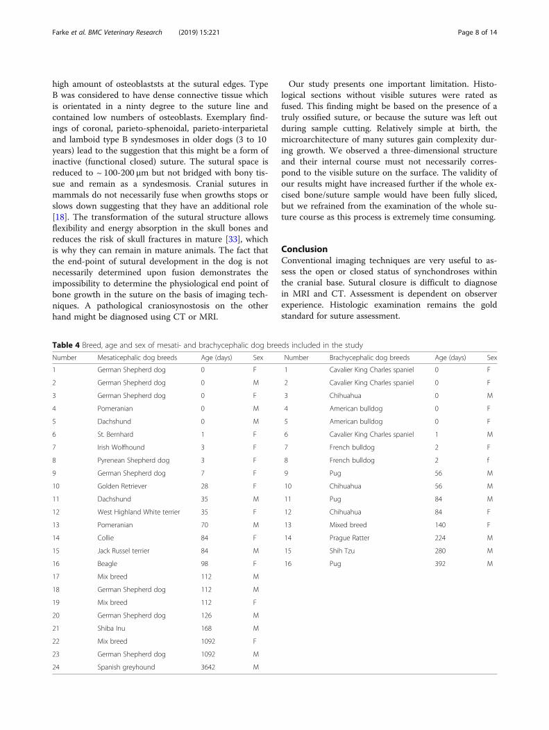
Table 4 Breed, age and sex of mesati- and brachycephalic dog breeds included in the study
Number Mesaticephalic dog breeds Age (days) Sex Number Brachycephalic dog breeds Age (days) Sex
1 German Shepherd dog 0 F 1 Cavalier King Charles spaniel 0 F
2 German Shepherd dog 0 M 2 Cavalier King Charles spaniel 0 F
3 German Shepherd dog 0 F 3 Chihuahua 0 M
4 Pomeranian 0 M 4 American bulldog 0 F
5 Dachshund 0 M 5 American bulldog 0 F
6 St. Bernhard 1 F 6 Cavalier King Charles spaniel 1 M
7 Irish Wolfhound 3 F 7 French bulldog 2 F
8 Pyrenean Shepherd dog 3 F 8 French bulldog 2 f
9 German Shepherd dog 7 F 9 Pug 56 M
10 Golden Retriever 28 F 10 Chihuahua 56 M
11 Dachshund 35 M 11 Pug 84 M
12 West Highland White terrier 35 F 12 Chihuahua 84 F
13 Pomeranian 70 M 13 Mixed breed 140 F
14 Collie 84 F 14 Prague Ratter 224 M
15 Jack Russel terrier 84 M 15 Shih Tzu 280 M
16 Beagle 98 F 16 Pug 392 M
17 Mix breed 112 M
18 German Shepherd dog 112 M
19 Mix breed 112 F
20 German Shepherd dog 126 M
21 Shiba Inu 168 M
22 Mix breed 1092 F
23 German Shepherd dog 1092 M
24 Spanish greyhound 3642 M
Farke et al. BMC Veterinary Research (2019) 15:221 Page 8 of 14

MethodsThe aim of the present study was, therefore, to compareimaging findings with histological preparations in orderto determine the sensitivity and specificity for conven-tional clinical CT and MRI in the assessment of the openor closed status of cranial synchondroses and sutures indogs.
AnimalsThe heads of 40 dogs of different breeds (24 mesati-cephalic- and 16 brachycephalic dogs), were collectedfrom the Department of Veterinary Clinical Sciences,Clinic for Small Animals and from the Clinic forGynecology, Andrology and Obstetrics of the Justus-Liebig-University, Giessen (Table 4). The dogs were
euthanized or died due to diseases unrelated to theskull and central nervous system. Age and sex was re-corded. Written consent was obtained from allowners that donated their animals for the study andactual dogs remained anonymous.
Imaging techniquesPost mortem MRI of the head was obtained using a highfield scanner (Gyroscan Intera, 1.0 T, Phillips, Hamburg,Germany). Dorsal, sagittal, and transvers T2-weighted(W) images with a 2mm slice thickness and a 0.2 mmslice interval were acquired (T2-Turbospin echo, echotime (TE) 120 ms, repetition time (TR): 2900 ms. T1-FFE weighted dorsal and transversal images with a slicethickness of 1 mm were obtained (TR 25ms TE 6.9 ms).Field of view was 120 × 120 mm in small dogs and 210 ×
Fig. 5 Overview of cranial sutures in the skull of a 4.5-months-old German shepherd dog. Three-dimensional volumetric reconstructions of the CTdata of the skull of a 4.5-months-old German shepherd dog in dorsal view (a), frontal view (b), left lateral view (c) and caudal view (d). S1 = interfrontal(metopic) suture, S2/3 = left and right fronto-parietal (coronal) suture, S4 = sagittal suture, S5 = left spheno-frontal suture, S6/8 = left and rightsquamosal suture, S9 = parieto-interparietal suture, S10/11 = left and right lambdoid suture
Farke et al. BMC Veterinary Research (2019) 15:221 Page 9 of 14
210 mm in large dogs. Matrix was 288 × 288 in smalldogs and 384 × 384 in large dogs leading to a pixel sizebetween 0.625 × 0.625 mm and 0.54 × 0.54 mm.Post mortem CT examination was performed using a
sixteen slice CT scanner (Philips Brilliance 16, Phillips,Hamburg, Germany). Transvers images with a slicethickness of 0.8 mm were obtained using 120 kV, 321mA and a field of view of 133mm. Data were processedusing a bone algorithm (window width 2500 and windowlevel 500).
Image analysisAll images were retrieved from the relevant picture ar-chiving system and evaluated by a board certified neur-ologist (MJS) and a doctoral student (DF). Theexperiments were performed using anonymized and ran-domized image data sets. The observers were blinded toage and breed of the dog. Studies were evaluated withopen source DICOM viewing software and windowlevels, window widths, and magnification were adjustedas needed in order to optimize visualization of bone.The following cranial growth centers were examined:
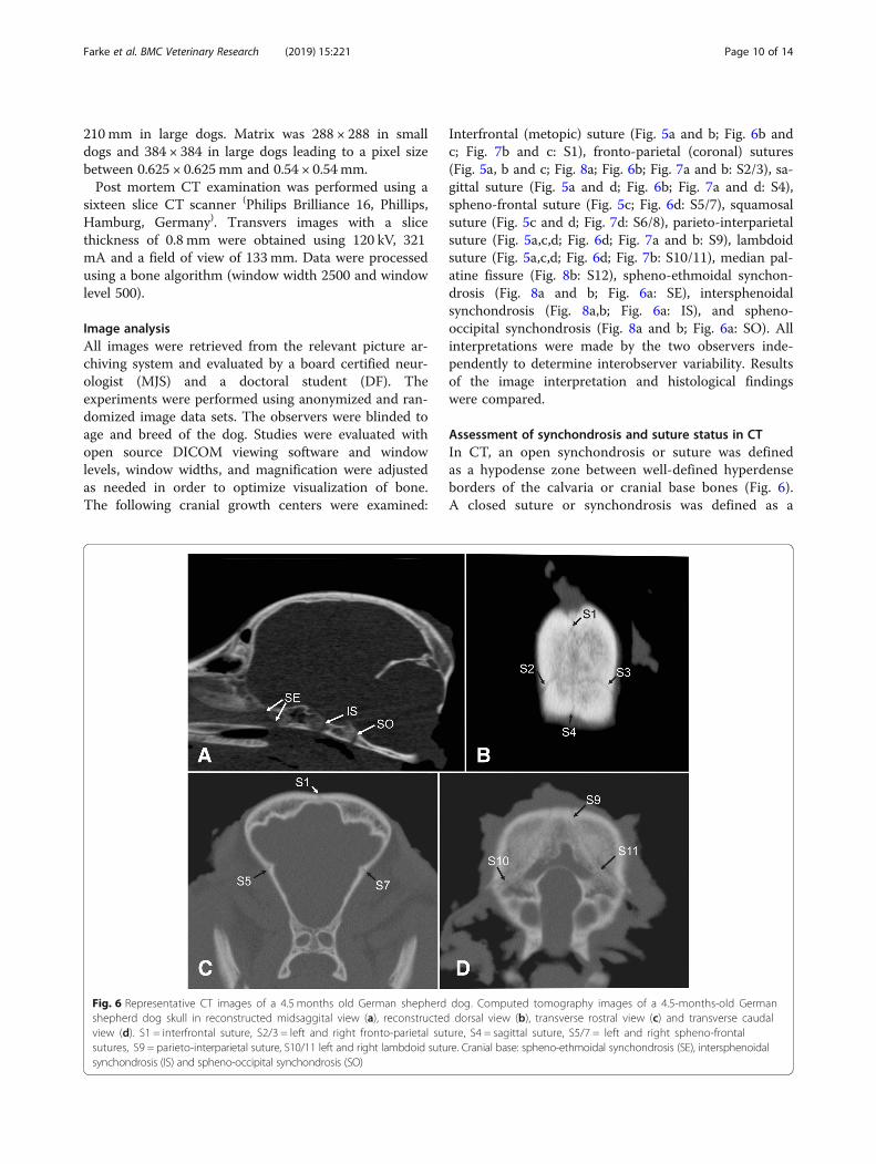
Interfrontal (metopic) suture (Fig. 5a and b; Fig. 6b andc; Fig. 7b and c: S1), fronto-parietal (coronal) sutures(Fig. 5a, b and c; Fig. 8a; Fig. 6b; Fig. 7a and b: S2/3), sa-gittal suture (Fig. 5a and d; Fig. 6b; Fig. 7a and d: S4),spheno-frontal suture (Fig. 5c; Fig. 6d: S5/7), squamosalsuture (Fig. 5c and d; Fig. 7d: S6/8), parieto-interparietalsuture (Fig. 5a,c,d; Fig. 6d; Fig. 7a and b: S9), lambdoidsuture (Fig. 5a,c,d; Fig. 6d; Fig. 7b: S10/11), median pal-atine fissure (Fig. 8b: S12), spheno-ethmoidal synchon-drosis (Fig. 8a and b; Fig. 6a: SE), intersphenoidalsynchondrosis (Fig. 8a,b; Fig. 6a: IS), and spheno-occipital synchondrosis (Fig. 8a and b; Fig. 6a: SO). Allinterpretations were made by the two observers inde-pendently to determine interobserver variability. Resultsof the image interpretation and histological findingswere compared.
Assessment of synchondrosis and suture status in CTIn CT, an open synchondrosis or suture was definedas a hypodense zone between well-defined hyperdenseborders of the calvaria or cranial base bones (Fig. 6).A closed suture or synchondrosis was defined as a
Fig. 6 Representative CT images of a 4.5 months old German shepherd dog. Computed tomography images of a 4.5-months-old Germanshepherd dog skull in reconstructed midsaggital view (a), reconstructed dorsal view (b), transverse rostral view (c) and transverse caudalview (d). S1 = interfrontal suture, S2/3 = left and right fronto-parietal suture, S4 = sagittal suture, S5/7 = left and right spheno-frontalsutures, S9 = parieto-interparietal suture, S10/11 left and right lambdoid suture. Cranial base: spheno-ethmoidal synchondrosis (SE), intersphenoidalsynchondrosis (IS) and spheno-occipital synchondrosis (SO)
Farke et al. BMC Veterinary Research (2019) 15:221 Page 10 of 14
continuous hyperdense bone, without interruption bya hypodense structure. Partial closure was defined asa non-continuous hypodense bone and the presenceof bone-isointense bridges within this hypointensebone structure.
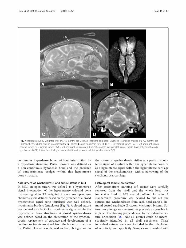
Assessment of synchondrosis and suture status in MRIIn MRI, an open suture was defined as a hypointensesignal interruption of the hyperintense calvarial bonemarrow signal in T2 weighted images. An open syn-chondrosis was defined based on the presence of a broadhyperintense signal zone (cartilage) with well defined,hypointense borders (endplates) (Fig. 7). A closed suturewas defined as a lack of a hypointense signal within thehyperintense bony structures. A closed synchondrosiswas defined based on the obliteration of the synchon-drosis, replacement of cartilage and development of acontinuous isointense signal from the bone marrow cav-ity. Partial closure was defined as bony bridges within
the suture or synchondrosis, visible as a partial hypoin-tense signal of a suture within the hyperintense bone, oras a hypointense signal within the hyperintense cartilagesignal of the synchondrosis, with a narrowing of thesynchondrosal cartilage.
Histological sample preparationAfter postmortem scanning soft tissues were carefullyremoved from the skull and the whole head wasimmersion fixed in 10% neutral buffered formalin. Astandardized procedure was devised to cut out thesutures and synchondroses from each head using a dia-mond coated sawblade (Proxxon Micromot System). Su-ture morphology was assessed as precisely as possible ina plane of sectioning perpendicular to the individual su-ture orientation [18]. Not all sutures could be macro-scopically identified in all skull specimens. Theseindividual sutures were not included in the calculationof sensitivity and specificity. Samples were washed with
Fig. 7 Representative T2 weighted MRI of a 4.5 months old German shepherd dog head. Magnetic resonance images of a 4.5-months-oldGerman shepherd dog skull in in a midsagittal (a), dorsal (b), and transverse view (c, d). S1 = interfrontal suture, S2/3 = left and right fronto-parietal suture, S4 = sagittal suture, S6/8 = left and right squamosal suture, S9 = parieto-interparietal suture, Cranial base: spheno-ethmoidalsynchondrosis (SE), intersphenoidal synchondrosis (IS) and spheno-occipital synchondrosis (SO)
Farke et al. BMC Veterinary Research (2019) 15:221 Page 11 of 14
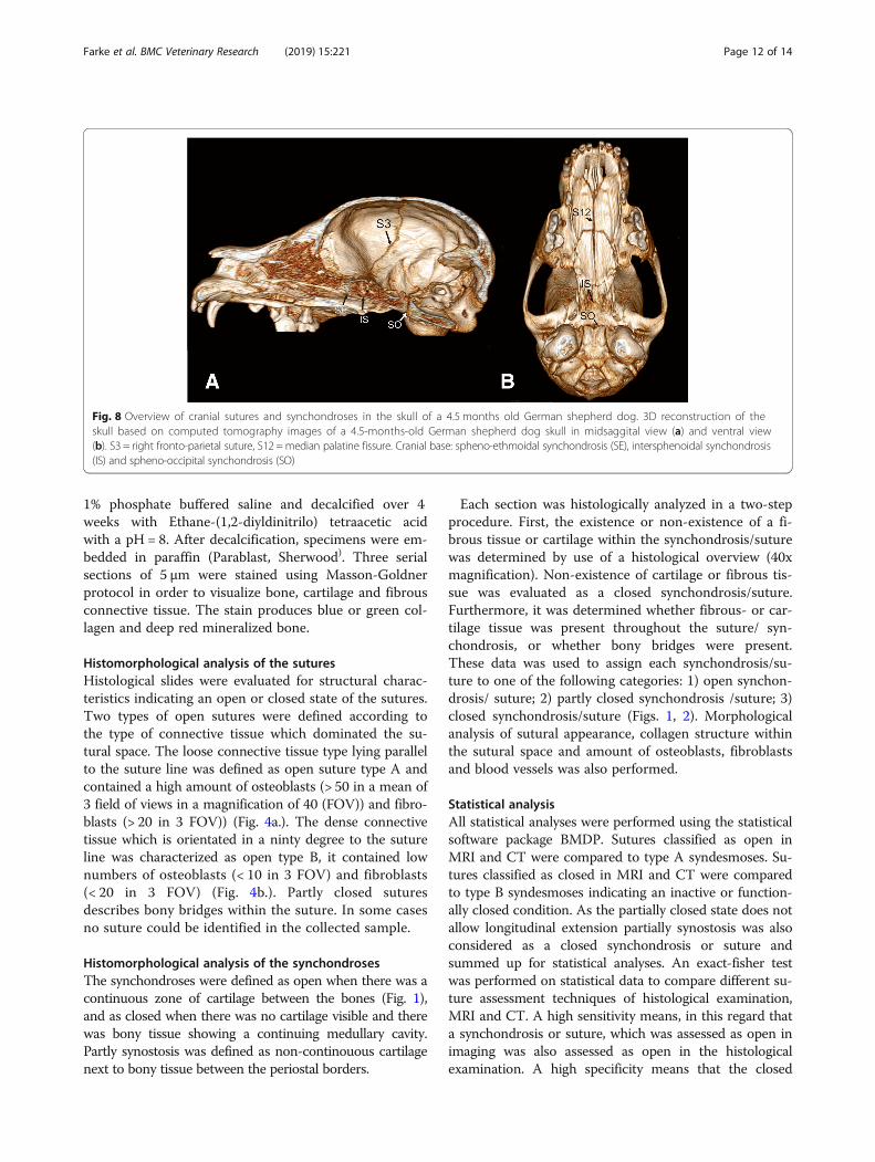
1% phosphate buffered saline and decalcified over 4weeks with Ethane-(1,2-diyldinitrilo) tetraacetic acidwith a pH = 8. After decalcification, specimens were em-bedded in paraffin (Parablast, Sherwood). Three serialsections of 5 μm were stained using Masson-Goldnerprotocol in order to visualize bone, cartilage and fibrousconnective tissue. The stain produces blue or green col-lagen and deep red mineralized bone.
Histomorphological analysis of the suturesHistological slides were evaluated for structural charac-teristics indicating an open or closed state of the sutures.Two types of open sutures were defined according tothe type of connective tissue which dominated the su-tural space. The loose connective tissue type lying parallelto the suture line was defined as open suture type A andcontained a high amount of osteoblasts (> 50 in a mean of3 field of views in a magnification of 40 (FOV)) and fibro-blasts (> 20 in 3 FOV)) (Fig. 4a.). The dense connectivetissue which is orientated in a ninty degree to the sutureline was characterized as open type B, it contained lownumbers of osteoblasts (< 10 in 3 FOV) and fibroblasts(< 20 in 3 FOV) (Fig. 4b.). Partly closed suturesdescribes bony bridges within the suture. In some casesno suture could be identified in the collected sample.
Histomorphological analysis of the synchondrosesThe synchondroses were defined as open when there was acontinuous zone of cartilage between the bones (Fig. 1),and as closed when there was no cartilage visible and therewas bony tissue showing a continuing medullary cavity.Partly synostosis was defined as non-continouous cartilagenext to bony tissue between the periostal borders.
Each section was histologically analyzed in a two-stepprocedure. First, the existence or non-existence of a fi-brous tissue or cartilage within the synchondrosis/suturewas determined by use of a histological overview (40xmagnification). Non-existence of cartilage or fibrous tis-sue was evaluated as a closed synchondrosis/suture.Furthermore, it was determined whether fibrous- or car-tilage tissue was present throughout the suture/ syn-chondrosis, or whether bony bridges were present.These data was used to assign each synchondrosis/su-ture to one of the following categories: 1) open synchon-drosis/ suture; 2) partly closed synchondrosis /suture; 3)closed synchondrosis/suture (Figs. 1, 2). Morphologicalanalysis of sutural appearance, collagen structure withinthe sutural space and amount of osteoblasts, fibroblastsand blood vessels was also performed.
Statistical analysisAll statistical analyses were performed using the statisticalsoftware package BMDP. Sutures classified as open inMRI and CT were compared to type A syndesmoses. Su-tures classified as closed in MRI and CT were comparedto type B syndesmoses indicating an inactive or function-ally closed condition. As the partially closed state does notallow longitudinal extension partially synostosis was alsoconsidered as a closed synchondrosis or suture andsummed up for statistical analyses. An exact-fisher testwas performed on statistical data to compare different su-ture assessment techniques of histological examination,MRI and CT. A high sensitivity means, in this regard thata synchondrosis or suture, which was assessed as open inimaging was also assessed as open in the histologicalexamination. A high specificity means that the closed
Fig. 8 Overview of cranial sutures and synchondroses in the skull of a 4.5 months old German shepherd dog. 3D reconstruction of theskull based on computed tomography images of a 4.5-months-old German shepherd dog skull in midsaggital view (a) and ventral view(b). S3 = right fronto-parietal suture, S12 =median palatine fissure. Cranial base: spheno-ethmoidal synchondrosis (SE), intersphenoidal synchondrosis(IS) and spheno-occipital synchondrosis (SO)
Farke et al. BMC Veterinary Research (2019) 15:221 Page 12 of 14

status of a synchondrosis or suture in MRI and CT wasconfirmed by histological evaluation. Sensitivity and speci-ficity in MRI and CT were defined as high (> 80), moder-ate (65–79%) or low (< 64%). Interobserver variability wasobtained using the kappa coefficient. A kappa value < 0.2implies slight agreement, 0.21–0.4 fair agreement, 0.41–0.6 moderate agreement, 0.61–0.8 substantial agreementand > 0.81 almost perfect agreement.
AbbreviationsCl 95%: Confidence level of 95%; CT: Computed tomography; DF: DanielaFarke; F: Female; FOV: Field of view; IS: Intersphenoidal synchondrosis;M: Male; MJS: Martin Jürgen Schmidt; MRI: Magnetic resonance imaging;S1: Interfrontal suture (metopic suture); S10: Left lambdoid suture; S11: Rightlambdoid suture; S12: Medial palatine fissure; S2: Left fronto-parietal suture(coronal suture); S3: Right fronto-parietal suture (coronal suture); S4: Saggitalsuture; S5: Left spheno-frontal suture; S6: Left squamosal suture; S7: Rightspheno-frontal suture; S8: Right squamosal suture; S9: Parieto-interparietalsuture; SE: Spheno-ethmoidal synchondrosis; SO: Spheno-occipitalsynchondrosis; μm: micrometer; ϗ: kappa value
AcknowledgementsWe thank Andreas Schaubmar for his help with statistics.
Authors’ contributionsAll authors helped to draft the manuscript and participated in its design. DFcollected the data for the study. MJS and DF analyzed the imaging data. CSand DF performed the histological interpretation. RK reviewed the manuscript.KF performed the statistical analysis of the data. All authors read and approvedthe final manuscript.
FundingThe study was sponsored by the Society for Canine Research (Gesellschaftzur Förderung Kynologischer Forschung e.V).
Availability of data and materialsData and materials are available from the corresponding author onreasonable request.
Ethics approval and consent to participateAll research procedures were approved by the Ethical Animal Care and UseCommittee of the Justus-Liebig University of Giessen. All owners gave writ-ten consent to perform post mortem examination.
Consent for publicationNot applicable for this study.
Competing interestsThe authors declare that they have no competing interests.
Author details1Department of Veterinary Clinical Sciences, Small Animal Clinic,Justus-Liebig-University, Frankfurter Strasse 108, 35392 Giessen, Germany.2Institute of Veterinary-Anatomy, -Histology, and –Embryology,Justus-Liebig-University, Frankfurter Strasse 98, 35392 Giessen, Germany.3Department of Biomathematics, Justus-Liebig-University, Frankfurter Strasse95, 35392 Giessen, Germany. 4Department of Companion Animal ClinicalStudies, Faculty of Veterinary Science, University of Pretoria, Private Bag X04,Onderstepoort, Pretoria 0110, South Africa.
Received: 19 April 2018 Accepted: 17 June 2019
References1. Kuratani S. Craniofacial development and the evolution of vertebrates. The
old problemns on a new background. Zool Sci. 2005;22:1–19.2. Morriss-Kay GM, Wilkie AO. Growth of the normal skull vault and its
alteration in craniosynostosis: insights from human genetics andexperimental studies. J Anat. 2005;207:637–53.
3. Johansen VA, Hall SH. Morphogenesis of the mouse coronal suture. ActaAnat. 1982;114:58–67.
4. Decker JD, Hall SH. Light and electron microscopy of the newborn sagittalsuture. Anat Rec. 1985;212:81–9.
5. Opperman LA. Cranial sutures as intramembranous bone growth sites. DevDyn. 2000;219(4):472–85.
6. Nickel R, Schummer A, Seiferle E. Aktiver Bewegungsapparat. In: Lehrbuchder Anatomie der Haustiere. Bd. 1. 8th ed. Berlin: Parey; 2003.
7. Liebich HG. Funktionelle Histologie der Haussäugetiere - Lehrbuch undFarbatlas für Studium und Praxis. 4th ed. Stuttgart: Schattauer; 2004.
8. Schliemann H. Zur Morphologie und Entwicklung des Craniums von Canislupus f. familiaris L. Gegenbaurs Morphol Jahrb. 1965;109:501–603.
9. Erlebacher A, Filvaroff EH, Gitelman SE, Derynck R. Toward a molecularunderstanding of skeletal development. Cell. 1995;10:371–8.
10. Kim HJ, Roh HG, Lee IW. Craniosynostosis : updates in radiologic diagnosis. JKorean Neurosurg Soc. 2016;59:219–26.
11. Kolpakova-Hart E, McBratney-Owen B, Hou B, Fukai N, Nicolae C, Zhou J,Olsen BR. Growth of cranial synchondroses and sutures requires polycystin-1. Dev Biol. 2008;321(2):407–19.
12. Wilkie AO, Morriss-Kay GM. Genetics of craniofacial development andmalformation. Nat Rev Genet. 2001;2:458–68.
13. Ussow SS. Über alters- und wachstumsveränderungen am Knochengerüstder Haussäugetiere. Prakt Tierheilk. 1901;27:341–9.
14. Ellenberger W, Baum H. Handbuch der vergleichenden Anatomie derHaustiere. In: Kopf der Fleischfresser. 18th ed. Berlin: Springer-Verlag; 1977.
15. Geiger M, Haussman S. Cranial suture closure in domestic dog breeds andits relationships to skull morphology. Anat Rec. 2016;299:412–23.
16. Schmidt MJ, Volk H, Klingler M, Failing K, Kramer M, Ondreka N. Comparison ofcranial base synchondrosis in cavalier king Charles spaniels, brachycephalic andmesaticephalic dogs. Vet Radiol Ultrasound. 2013;54:497–503.
17. Schmidt MJ, Kampschulte M, Enderlein S, Gorgas D, Lang J, Ludewig E,Fischer A, Meyer-Lindenberg A, Schaubmar AR, Failing K, Ondreka N. Therelationship between brachycephalic head features in modern Persian catsand Dysmorphologies of the skull and internal hydrocephalus. J Vet IntMed. 2017;31:1487–501.
18. Rice DP. Craniofacial sutures: development, disease and treatment. FrontOral Biol. 2008;12:15–18.
19. Topczewska JM, Shoela RA, Tomaszewski JP, Mirmira RB, Gosain AK. Themorphogenesis of cranial sutures in zebrafish. PLoS One. 2016;11:e0165775.
20. Hall BA. Bones and cartilage: developmental and evolutionary skeletalbiology. New York: Elsevier Academic Press; 2005. p. 760.
21. Schmidt MJ, Neumann AC, Amort KH, Failing K, Kramer M. Cephalometricmeasurements and determination of general skull type of cavalier kingCharles spaniels. Vet Radiol Ultrasound. 2011;52:436–40.
22. Knowler SP, McFadyen AK, Freeman C, et al. Quantitative analysis of Chiari-like malformation and Syringomyelia in the griffon Bruxellois dog. PLoSONE. 2014;9:e88120.
23. Rusbridge C, Knowler SP, Pieterse L, McFadyen AK. Chiari-likemalformationin the griffon bruxellois. J Small Anim Pract. 2009;50:386–93.
24. Tejszerska D, Wolański W, Larysz D, Gzik M, Sacha E. Morphological analysisof the skull shape in craniosynostosis. Acta Bioeng Biomech. 2011;13:35–40.
25. Anderson PJ, Netherway DJ, David DJ, Self P. Scanning electron microscopeand micro-CT evaluation of cranial sutures in health and disease. J CraniofacSurg. 2006;17:909–19.
26. Furuya Y, Edwards MS, Alpers CE, Tress BM, Ousterhout DK, Norman D.Computerized tomography of cranial sutures part 1″ comparison of sutureanatomy in children and adults. J Neurosurg. 1984;61(1):53–8.
27. William F, Perrin JGM. The Ecyclopedia of marine mammals. In: SkullAnatomy Elsevier, vol. 21; 2009. p. 1033–47.
28. Harth S. Estimating age by assessing the ossificationdegree of cranialsutures with the aid of flat- panel- CT. Leg Med (Tokyo). 2009;11:186–9.
29. Angelieri F, Cevidanes LH, Franchi L, Gonçalves JR, Benavides E, JA MN Jr.Midpalatal suture maturation: classification method for individualassessment before rapid maxillary expansion. Am J Orthod DentofacOrthop. 2013;144:759–69.
30. Pilgram TK, Vannier MW, Marsh JL, Kraemer BB, Rayne SC, Gado MH, Moran CJ,McAlister WH, Shackelford GD, Hardesty RA. Binary nature and radiographicidentifiability of craniosynostosis. Investig Radiol. 1994;29:890–6.
31. Kwak KH, Kim SS, Kim YI, Kim YD. Quantitative evaluation ofmidpalatal suture maturation via fractal analysis. Korean J Orthod.2016;46:323–30.
Farke et al. BMC Veterinary Research (2019) 15:221 Page 13 of 14
32. Sun Z, Lee E, Herring SW. Cranial Sutures and Bones: Growth and Fusion inRelation to Masticatory Strain. Anat Rec A Discov Mol Cell Evol Biol. 2004;276:150.
33. Herring SW, Mucci RJ. In vivo strain in cranial sutures: the zygomatic arch. JMorph. 1991;207:225.
Publisher’s NoteSpringer Nature remains neutral with regard to jurisdictional claims inpublished maps and institutional affiliations.
Farke et al. BMC Veterinary Research (2019) 15:221 Page 14 of 14