Hidden genetic diversity in the green alga Spirogyra (Zygnematophyceae, Streptophyta)

Glycobiology vol. 18 no. 3 pp. 250–259, 2008doi:10.1093/glycob/cwm139Advanced Access publication January 3, 2008
A preponderantly 4-sulfated, 3-linked galactan from the green algaCodium isthmocladum
Eduardo H C Farias2,3, Vitor H Pomin2,4,5,Ana-Paula Valente5,6, Helena B Nader7,Hugo A O Rocha1,3, and Paulo A S Mourao1,4,5
3Laboratorio de Biotecnologia de Polımeros Naturais - BIOPOL,Departamento de Bioquımica, Universidade Federal do Rio Grande do Norte,Natal, RN, 59072-970; 4Laboratorio de Tecido Conjuntivo, HospitalUniversitario Clementino Fraga Filho; 5Instituto de Bioquımica Medica,Centro de Ciencias da Saude, Universidade Federal do Rio de Janeiro, CaixaPostal 68041, Rio de Janeiro, RJ, 21941-590; 6Centro Nacional deRessonancia Magnetica Nuclear Jiri Jonas, Universidade Federal do Rio deJaneiro, Rio de Janeiro, RJ, 21941-590; and 7Departamento de Bioquımica,Universidade Federal de Sao Paulo, SP, 04044-020, Brazil
Received on October 9, 2007; revised on November 28, 2007; accepted onDecember 28, 2007
The green algae of the genus Codium have recently beendemonstrated to be an important source of sulfated galac-tans from the marine environment. Here, a sulfated galactanwas isolated from the species Codium isthmocladum and itsstructure was studied by a combination of chemical analysesand NMR spectroscopy. Two fractions (SG 1, ∼14 kDa, andSG 2, ∼20 kDa) were derived from this highly polydisperseand heterogeneous polysaccharide. Both exhibited similarstructures in 1H 1D NMR spectra. The structural features ofSG 2 and its desulfated derivative were analyzed by COSY,TOCSY, DEPT-HSQC, HSQC, and HMBC. This sulfatedgalactan is composed preponderantly of 4-sulfated, 3-linkedβ-D-galactopyranosyl units. In minor amounts, it is sulfatedand glycosylated at C-6. Pyruvate groups are also found,forming five-membered cyclic ketals as 3,4-O-(1’carboxy)-ethylidene-β-D-galactose residues. A comparison of sulfatedgalactans from different marine taxonomic groups revealedsimilar backbones of 3-β-D-Galp-1.
Keywords: Codium isthmocladum/green alga/greenseaweed/phylogeny/pyruvylated-sulfated galactan
Introduction
The sulfated polysaccharides comprise a complex group ofmacromolecules with a large range of important biologicalactivities (Verli 2007). These anionic polymers can occur asglycosaminoglycans, widely distributed in vertebrate tissues(Mathews 1975). In marine organisms (invertebrates and al-gae), the sulfated polysaccharides are found predominantly assulfated fucans or sulfated galactans. The sulfated fucans areconstituted mainly of α-L-fucose residues in which the species-specific molecular structures vary in the glycosidic position and
1To whom correspondence should be addressed: e-mail: [email protected] two authors contributed equally to the work.
in the pattern of sulfation (Berteau and Mulloy 2003; Mourao2004, 2007). The most common source of sulfated fucans isthe cell wall of brown algae (Phaeophyta) (Pereira et al. 1999;Rocha et al. 2005), but they can also be found in the egg jellylayer of sea urchins (Mulloy et al. 1994; Alves et al. 1997)and in the body wall of the sea cucumber (Echinodermata,Holothuroidea)(Ribeiro et al. 1994).
The most common source of sulfated galactans is the cellwall of red algae (Rhodophyta) (Lahaye 2001; van de Veldeet al. 2004). Like the sulfated fucans, the sulfated galactanscan also be isolated from the outer layer of the sea-urchin egg(Echinodermata, Echinoidea) (Alves et al. 1997), or extractedfrom other classes of invertebrates, such as tunicates (Urochor-data, Ascidiacea) (Pavao et al. 1989; Albano et al. 1990; Santoset al. 1992) and clams (Mollusca, Bivalvia) (Amornrut et al.1999). Recently, a novel sulfated galactan was isolated from seagrass (Aquino et al. 2005), leading for the first time to a descrip-tion of sulfated polysaccharides in the marine angiosperms, agroup of vascular plants. Thus, sulfated galactans with differentstructures are widely distributed among marine organisms fromdistant phyla.
Recently, the green algae, particularly the genus Codium(Bryopsidales, Chlorophyta) (Love and Percival 1964;Matsubara et al. 2001; Bilan et al. 2007), have proved to beanother important source of sulfated galactans in the marineenvironment. Fewer structures of sulfated polysaccharides fromgreen seaweed are known than are those from red or brownalgae. In the genus Codium, different species demonstrate struc-tural heterogeneity and complexity in their sulfated galactans.Bilan et al. (2007) described a complex and highly pyruvylatedand sulfated galactan from C. yezoense, composed essentiallyof linear backbone segments of 3-linked β-D-galactopyranosylunits, ramified by short oligosaccharides attached by linkages atC-6. Sulfate groups were localized mainly at C-4 and in loweramounts at C-6. A few pyruvate groups (about 25% of totalresidues) were present, forming 3,4-O-(1′-carboxy)-ethylidene-β-D-galactopyranose residues located at nonreducing terminalsof the chains of galactose. The sulfated galactans from C. frag-ile and C. cylindricum were heterogeneous polymers. In addi-tion to its galactose content, C. fragile also contained arabinoseresidues (sulfated arabinogalactan) (Love and Percival 1964)and C. cylindricum contained glucose residues, probably form-ing a sulfated glucogalactan (Matsubara et al. 2001).
Here, we describe the isolation and structural characterizationof the sulfated galactan from Codium isthmocladum, a green algathat occurs abundantly along the coast of Natal, Rio Grande doNorte, Brazil. We analyzed this sulfated galactan by a combi-nation of chemical methods (desulfation and methylation re-actions and measurements of sulfate content) and nuclear mag-netic resonance (NMR) spectroscopy (1H 1D and 2D correlationspectroscopy (COSY), total correlation spectroscopy (TOCSY),
C© The Author 2008. Published by Oxford University Press. All rights reserved. For permissions, please e-mail: [email protected] 250
at UFR
J on June 8, 2013http://glycob.oxfordjournals.org/
Dow
nloaded from

A preponderantly 4-sulfated, 3-linked galactan from the green alga Codium isthmocladum
distortionless enhancement by polarization transfer-hetero-nuclear single quantum coherence (DEPT-HSQC), HSQC,and heteronuclear multiple bound correlation (HMBC)). Thesulfated galactan from C. isthmocladum is composed preponder-antly of 3-linked β-D-galactopyranose residues that are exten-sively 4-sulfated and occasionally 6-sulfated. In minor amounts,the β-D-galactopyranosyl units are 6-linked. Also, based onHMBC experiments, the sulfated galactans from C. isthmo-cladum contain pyruvate groups that form five-membered cyclicketals located in 3,4-O-(1′carboxy)-ethylidene-β-D-galactoseunits.
A comparison among different sulfated polysaccharides in-dicates that sulfated galactans with glycosidic linkage (β1→3)occur in some taxonomic groups of marine eukaryotic organ-isms (rhodophytes, chlorophytes, angiosperms, echinoderms,and molluscs). The sulfated galactans found among thesephyla differ in sulfation sites, with a marked tendency toward4-sulfation in algae and 2-sulfation in invertebrate animals. Inminor amounts, 6-sulfation is dispersed throughout the phylo-genetic tree.
Results and discussion
Purification of the sulfated galactans from the green algaC. isthmocladumThe polysaccharides extracted from the alga with maxatasedigestion (see the Materials and methods section) were frac-tionated by precipitation with increasing volumes of acetone(1:0.3, 1:0.5, 1:0.7, 1:0.9, and 1:1.2, v/v of sample and acetone).All the precipitated fractions, except the one that was obtainedwith 1:1.2 acetone, revealed a heterogeneous content of sugars(Table I). Mannose was the major constituent in fractions pre-cipitated with 1:0.3 and 1:0.5 acetone. Galactose and arabinosewere equally predominant in fractions precipitated with 1:0.7and 1:0.9 acetone. The precipitate obtained with 1:1.2 acetonecontained primarily one type of sugar, arabinose.
The mixture of polysaccharides obtained by precipitationwith 1:0.9 acetone was purified additionally by ion-exchangechromatography (Bayer Lewatit) (Figure 1A). The fractioneluted with 0.3 M NaCl contained almost 60% of the totalpolysaccharides applied to the column. However, the low sul-
fate content indicated that these polysaccharides were composedpredominantly of neutral residues. The fractions eluted with2.0 M and 3.0 M NaCl contained ∼16% and ∼13% of thetotal polysaccharides applied to the Lewatit column. Thesefractions were highly sulfated with a molar ratio of 1.4 and1.9 sulfate/residue, respectively (Table I). The sugar analysis ofthese two fractions revealed mainly galactopyranose residues(Table I) and they were denominated as SG 1 and SG 2.
The purity of these fractions was demonstrated by agarosegel electrophoresis (Figure 1B). For both fractions, there wasonly a single band in the gel, indicating purified polysaccharideswhen compared with the polysaccharides precipitated with 1:0.9acetone and with the crude polysaccharide before the serialprecipitation with acetone. In conclusion, these two fractionscontained exclusively sulfated galactan and were chosen forsubsequent structural studies.
The molecular masses of SG1 and SG2 were determinedby PAGE (Figure 1C). Their electrophoretic mobilities werecompared with different molecular markers as indicated inthe figure. Both fractions of sulfated galactan showed adispersive migration due to a heterogeneous molecular weightas is the characteristic for the sulfated polysaccharides. How-ever, the predominance of bands at ∼14 kDa and ∼20 kDa canbe noted for SG1 and SG2, respectively. Obviously, the greatermolecular weight of SG 2 together with its higher sulfate con-tent (Table I) explains its elution from the Lewatit column witha higher saline concentration (3.0 M NaCl) than that observedfor SG 1 (2.0 M NaCl) (Figure 1A).
Presence of 4- and 6-sulfation in the galactansIn an initial attempt to determine the structure of the sulfatedgalactan obtained from the green alga, we methylated the nativepolysaccharides and their chemically desulfated derivatives. Inspite of limitations to this type of methodology, such as partialdesulfation and methylation or preferential sites of methylationin the sugar ring, this analysis provides valuable informationabout the structure (Table II).
In fact, comparing the methyl galactitols produced from thenative sulfated galactan and desulfated derivative, we can ob-serve the disappearance of the 2-O-methyl galactitol and asignificant increase in 2,4-di-O-methyl galactitol (indicative of4-sulfation) and of the 2,4,6-tri-O-methyl galactitol (indicative
Table I. Sugar and sulfate contents of the fractions precipitated with different volumes of acetone and average molecular mass of the fractions of sulfated galactansfrom the green alga C. isthmocladum
Fractions (v/v)a Composition (molar ratios)b Average molecular massc (kDa)
Gal Ara Man (SO4)/total sugars
1:0.3 0.29 0.14 0.57 − −1:0.5 0.07 0.34 0.59 − −1:0.7 0.43 0.52 0.05 − −1:0.9 0.42 0.50 0.08 − −1:1.2 0.14 0.78 0.08 −Sulfated galactan 1 (SG 1) 1.00 <0.01 <0.01 1.4 ∼14Sulfated galactan 2 (SG 2) 1.00 <0.01 <0.01 1.9 ∼20
aThe fractions were obtained by adding different volumes of acetone (1:0.3, 1:0.5, 1:0.7, 1:0.9, and 1:1.2, v/v of sample and acetone) to the solution of the crudepolysaccharide. The sulfated galactans (SG 1 and SG 2) were obtained from the fraction precipitated by 1:9 acetone using ion-exchange chromatography on BayerLewatit resin (see Figure 1A).bThe sugar contents were determined by GLC-MS of alditol acetates as described under the Materials and methods section. Standard sugars were fucose, glucose,rhamnose, arabinose, mannose, galactose, and xylose converted to their respective alditol acetate derivatives.cThe average molecular masses (kDa) were estimated by PAGE (see Figure 1C).
251
at UFR
J on June 8, 2013http://glycob.oxfordjournals.org/
Dow
nloaded from

Eduardo H C Farias et al.
Fig. 1. Purification of the fractions of sulfated galactan (SG 1 and SG 2) from C. isthmocladum by ion-exchange chromatography (A) and electrophoretic analysisby agarose gel (B) and polyacrylamide gel (C). (A) The fraction precipitated with 1:0.9 acetone (∼12 mg) was applied to a Lewatit column and the elution wascarried out in a stepwise system, initializing with 0.3 M NaCl, followed by 0.5 M, 0.7 M, 1.0 M, 1.5 M, 2.0 M, 3.0 M, and 4.0 M NaCl. The eluted fractions wereanalyzed for their hexose content (black bars) and for sulfate content (white bars). (B) The crude polysaccharides before acetone precipitation (Total), the fractionprecipitated with 1:0.9 acetone, and SG 1 and SG 2 were applied to a 0.5% agarose gel in 1,3-diaminopropane:acetate (pH 9.0) and run for 1 h at 110 V as indicatedby the arrow. The sulfated polysaccharides in the gel were fixed, dried, and stained as described under the Materials and methods section. (C) SG 1 and SG 2(∼10 µg of each) were analyzed by PAGE as described under the Materials and methods section. The molecular weights (MW-Da) of standard compounds areindicated at the right. These standards were: high-molecular-weight dextran sulfate (∼500 kDa), chondroitin 6-sulfate from shark cartilage (∼60 kDa), chondroitin4-sulfate from whale cartilage (∼40 kDa), unfractionated heparin from porcine intestinal mucosa (∼15 kDa), low-molecular-weight heparin (∼7.5 kDa), andlow-molecular-weight dextran sulfate (∼8 kDa).
of 4- and 6-sulfation). The production of significant amounts of2,4,6-tri-O-methyl galactitol from the methylation of the desul-fated galactans suggests the predominance of 3-linked galac-tose units. In addition, the production of significant amounts of
2,4- and 2,6-di-O-methyl galactitols may indicate partial desul-fation of the molecules, the presence of branching residuesor the presence of other types of substituents, as discussedbelow.
Table II. Methylated galactose derivatives obtained from native and desulfated SG 1 and SG 2 from the green alga C. isthmocladum
Methylated sugarsa
(as alditol acetates)tcR (min) Total peak areab (%)
SG 1 SG 2
Native Desulfated Native Desulfated
2,4,6-Met3-Gal 34.4 10 34 7 362,6-Met2-Gal 36.9 40 41 32 382,4-Met2-Gal 40.8 <1 25 <1 222-Met-Gal 42.3 50 <1 61 4
4-substituted 49% – 51% –6-substituted 24% – 29% –
aAfter the methylation reaction, the methylated galactans were hydrolyzed and the products analyzed as their alditol acetate derivatives by GLC-MS using a DB-1capillary column (25 m × 0.3 mm).bThe proportions of the methylated acetates are based on the integral of each peak compared with the sum of integrals.cRetention time of the alditol acetate derivatives in the GLC-MS on the DB-1 capillary column (25 m × 0.3 mm).
252
at UFR
J on June 8, 2013http://glycob.oxfordjournals.org/
Dow
nloaded from

A preponderantly 4-sulfated, 3-linked galactan from the green alga Codium isthmocladum
Fig. 2. 1H NMR spectra at 400 MHz of the native SG 1 (A), native SG 2 (C) from C. isthmocladum, and the desulfated derivatives of SG 1 (B) and SG 2 (D).About 5 mg of each were dissolved in 0.5 mL D2O and the 1D NMR spectra were recorded at 50◦C. The residual water signal was suppressed by presaturation.Chemical shifts are relative to external trimethylsilylpropionic acid at 0 ppm. The H4 signals correspond to 4-sulfated galactose units. The β-H1 signals correspondto the β-anomeric protons. The signals denoted by A and B correspond to 3-linked and 6-linked galactose units, respectively. The integrals of each anomeric peakof the desulfated galactan are indicated under the peak. Pyr-CH3 indicates methyl signals from pyruvate groups.
Preponderance of 4-sulfated, 3-linkedβ-D-galactopyranose residuesFor a more detailed structural analysis of the sulfated galactansfrom the green alga, we employed one-dimensional and two-dimensional NMR spectroscopy. The fractions of native sulfatedgalactans and their desulfated derivatives produced similar sig-nals in the 1H 1D NMR spectra (Figure 2), which indicatessimilar structures for SG 1 and SG 2. Therefore, the 2D NMRspectra were recorded exclusively with fraction SG 2 and itsdesulfated derivative.
The 1H-signals between 4.4 and 5.0 ppm in the spectra of thesulfated galactan contained a mixture of signals of H-1 from theβ-anomers of galactopyranoses and of H-4 from the sugar rings;these exhibited a down-field shift (∼0.6 ppm) due to sulfation(Pomin, Pereira, et al. 2005; Pomin, Valente, et al. 2005; Bilanet al. 2007). This conclusion is reinforced by the analysis of the1H/13C DEPT-HSQC of the native polysaccharide (Figure 3A)and the disappearance of the signal at δH/δC 4.94/77.8 ppm, afterthe desulfation reaction (Figure 3B).
The 1H 1D spectra (Figure 2C and D) and especially the1H/13C DEPT-HSQC (Figure 3) showed two preponderantlyanomeric signals, denominated as A and B, for the native sul-fated galactan SG 2, as well as for its desulfated derivative.Through the 2D COSY spectra (Figure 4A and C) and TOCSY
spectra (Figure 4B and D), it was possible to trace the spin sys-tems of these two signals, especially for the desulfated SG 2(Figure 4A and B). Thus, we obtained the 1H chemical shiftsindicated in Table III. Only the chemical shifts of H-6 couldnot be determined. However, these values were easily deduceddue to the negative-phase signals relative to CH2 (blue-contourpeaks in Figure 3B) in the 1H/13C DEPT-HSQC spectrum. Weidentified two signals of H-6/C-6 associated with spin systemsA and B (preponderant and minority blue signals, respectively).The analysis of the 13C chemical-shift values indicated unequiv-ocally that the units designated A and B (structures 1 and 2 inTable III, respectively) were associated with 3- and 6-linkedβ-D-galactopyranose residues, respectively, as indicated by thetypical low-field shift of carbons (∼10 ppm) in sites of glyco-sylation (Table III). This 13C shift was also seen in referencecompounds of 3- and 6-linked β-D-galactosyl units (structures5 and 6 in Table III) and in galactose residues located at nonre-ducing terminals (structure 7 in Table III).
The spin system traced for the native SG 2 was more complexdue to the greater heterogeneity of the polymer. However, weidentified two spin systems, denoted by A and B (Figure 4C andD) (Table III). Again, it was difficult to identify the signals thatcorrespond to H-6, but the DEPT-HSQC spectrum (Figure 3A)was especially useful for this assignment (blue-contour peaks).
253
at UFR
J on June 8, 2013http://glycob.oxfordjournals.org/
Dow
nloaded from

Eduardo H C Farias et al.
Fig. 3. 1H/13C DEPT-HSQC spectra of the native SG 2 (A) and its desulfated derivative (B). The assignments were based on 2D NMR experiments (COSY andTOCSY), and HSQC of the depyruvylated and desulfated derivative (see supplementary material). The blue-contour peaks are due to the negative phase from CH2groups, and the black-contour peaks are due to the positive phase from CH and CH3 groups. The values of chemical shifts are relative to externaltrimethylsilylpropionic acid at 0 ppm for 1H and methanol for 13C. The signals are denoted by A for 3-linked and by B for 6-linked β-D-galactopyranosyl units. Thepeaks denoted by A3-Pyr, A4-Pyr and A5∗ indicate 1H-chemical shifts of H3, H4, and H5 of the 3,4-(1′carboxy)-ethylidene-β-D-galactopyranose residues,respectively. The peaks denoted by A3′, A, B4′, and A5′ correspond respectively to signals from H3 and H4 of the 4-sulfated, 3-linked β-D-galactopyranosyl units;H4 of the 4-sulfated, 6-linked β-D-galactopyranosyl units; and H5 of the 4-sulfated, 3-linked β-D-galactopyranosyl units.
Signals fromglycosylated 6-position (δH,H′ /δC 4.36, 4.01/69.9ppm of units denominated as B) and unsubstituted 6-position(units A) were identified by comparison with the spectra of thedesulfated SG 2 (Figure 3B). Moreover, we identified anothersignal (δH,H′ /δC 4.42, 4.32/66.8 ppm) with a typical 1H low-fieldshift (∼0.6 ppm) that indicates 6-sulfation (Figure 3A).
The 6-sulfation is necessarily associated with the system A,while the system B is glycosylated at this site. The system Ais mainly sulfated at C-4, as indicated by the typical low-fieldshift (∼0.65 ppm) of the H-4 (structure 3 in Table III). But,there are also minor amounts of nonsulfated units, as indicatedby the methylation analysis. The system B is mainly 4-sulfated
Fig. 4. Strips of the anomeric regions (expansions from 5.1 to 4.5 ppm) from the COSY (A and C) and TOCSY (B and D) spectra of the desulfated galactan 2(A and B) and the native SG 2 (C and D) from C. isthmocladum. About 5 mg of each were dissolved in 0.5 mL D2O and the 2D NMR spectra were recorded at50◦C at 400 MHz. The spin systems are denoted by A for 3-linked and by B for 6-linked β-galactose units. The peaks denoted by A3-Pyr, 4-Pyr and A5∗ indicate1H-chemical shifts of H3, H4, and H5 of the 3,4-(1′carboxy)-ethylidene-β-D-galactopyranose residues, respectively. The peaks denoted by A4′ and B4′ correspondto signals H4 of the 4-sulfated, 3-linked, and 6-linked galactosyl units, respectively.
254
at UFR
J on June 8, 2013http://glycob.oxfordjournals.org/
Dow
nloaded from
A preponderantly 4-sulfated, 3-linked galactan from the green alga Codium isthmocladum
Table III. 1H and 13C chemical shifts (ppm) for native SG 2 from the green alga C. isthmocladum and its desulfated derivative
Polysaccharide Structure 1H and 13C chemical shifta (ppm)
H-1 H-2 H-3 H-4 H-5 H-6,6′
C-1 C-2 C-3 C-4 C-5 C-6
Desulfated galactan from C. isthmocladum (1) Unit Ac: 3-β-D-Galp-1 4.81 3.64 3.92 4.39 3.86 3.94,3.85102.6 73.8 82.9 67.1 75.2 60.3
(2) Unit Bd:6-β-D-Galp-1 4.62 3.82 3.89 4.38 4.09 4.36,4.01103.1 70.1 70.2 67.0 73.9 69.9
Native galactan from C. Isthmocladum (3.1) Unit A: 3-β-D-Galp-4(SO4)-1 (preponderant) 4.84 3.71 4.16 4.94 3.95 3.96,3.86103.1 71.9 77.6 77.8 74.8 60.4
(3.2) Unit A: 3-β-D-Galp-4,6-di(SO4)-1 (minor) 4.84 3.71 ND 4.94 ND 4.42,4.32103.1 71.9 ND 77.8 ND 66.8
(4) Unit B: 6-β-D-Galp-4(SO4)-1 (minor) 4.63 3.83 3.89 4.94 4.13 4.36,4.01103.2 70.2 70.2 77.8 73.7 69.9
Desulfated galactan from C. yezoenseb (5) 3-β-D-Galp-1 4.70 3.79 3.86 4.21 3.73 3.79105.4 71.5 83.4 69.8 76.1 62.3
(6) 6-β-D-Galp-1 4.47 3.57 3.69 3.97 3.92 4.04,3.91104.8 72.2 73.9 69.9 74.8 70.5
(7) β-D-Galp-1 4.61 3.61 3.68 3.93 3.69 3.79105.7 72.5 73.9 70.0 76.5 62.3
aChemical shifts are relative to external trimethylsilylpropionic acid at 0 ppm for 1H and methanol for 13C. Values in boldface indicate sulfate position and those initalic indicate glycosylated positions.bReference values from Bilan et al. (2007).c3-linked -β-D-galactopyranose.d6-linked -β-D-galactopyranose.ND, not determined.
(as indicated by the down-field shift of H-4 and structure4 in Table III), but nonsulfated units also co-exist with theseunits.
In synthesis, these results indicate, for the sulfated galactanfrom the green alga, a preponderant constitution of 3-β-D-Galp-1, about 80% of the total residues, as indicated by the integralsof the NMR signals (Figure 2B and D). Most of these units are4-sulfated, but, in minor amounts, there are also units sulfated atthe 6-position, as well as nonsulfated units. The sulfated galactanalso contains 6-linked units of β-D-Galp-4(SO4), and, in minoramounts, 6-linked nonsulfated galactopyranose residues. Thenuclear overhauser enhancement spectroscopy (NOESY) spec-tra (data not shown) did not reveal a clear distribution patternfor these minority residues in the structure of the polymer.
Occurrence of 3,4-O-(1′carboxy)-ethylidene residuesin the sulfated galactansCuriously, in addition to the 1H- and 13C-signals from the galac-tose ring and its anomeric protons and carbons, the native SG 1and SG 2 and their desulfated derivatives showed an intensehigh-field 1H-signal at ∼1.7 ppm (Figure 2), which stronglyindicates methyl groups. Furthermore, in the methylation anal-ysis the desulfated galactan was still highly substituted at C-3and C-4, as observed from the production of 2,6-di-O-methylgalactitols (Table II). These data together suggest the presenceof an extra-ring methyl group bound to C-3 and/or C-4 of thegalactose ring.
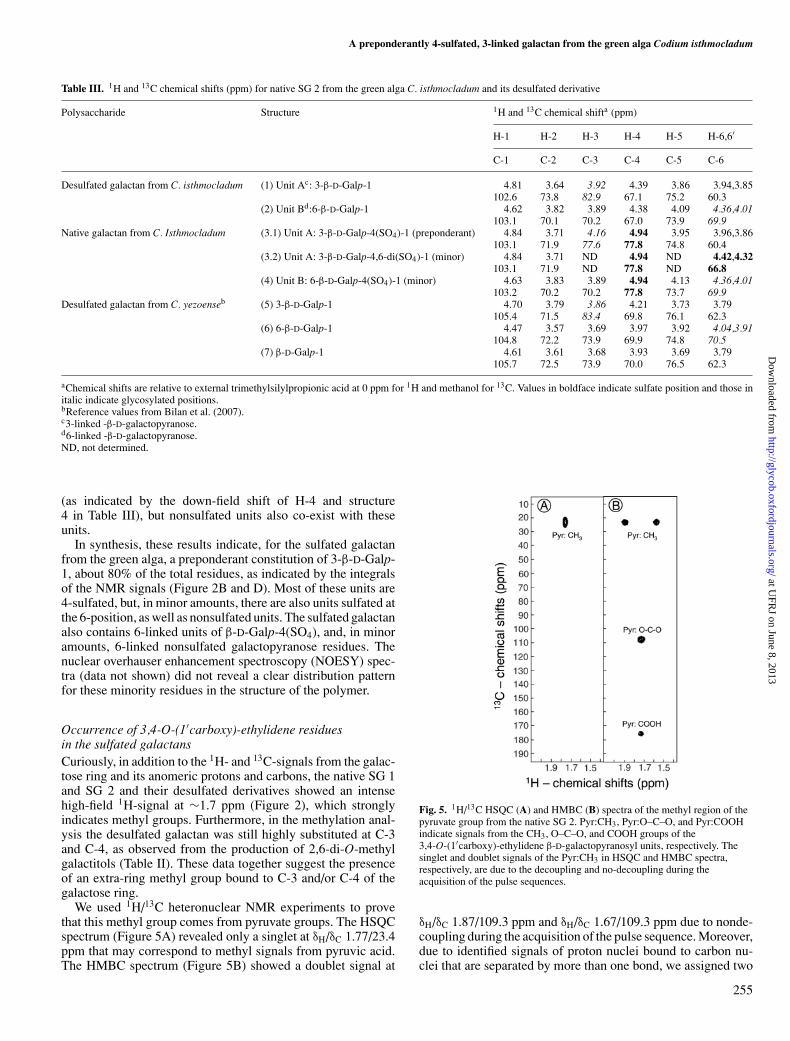
We used 1H/13C heteronuclear NMR experiments to provethat this methyl group comes from pyruvate groups. The HSQCspectrum (Figure 5A) revealed only a singlet at δH/δC 1.77/23.4ppm that may correspond to methyl signals from pyruvic acid.The HMBC spectrum (Figure 5B) showed a doublet signal at
Fig. 5. 1H/13C HSQC (A) and HMBC (B) spectra of the methyl region of thepyruvate group from the native SG 2. Pyr:CH3, Pyr:O–C–O, and Pyr:COOHindicate signals from the CH3, O–C–O, and COOH groups of the3,4-O-(1′carboxy)-ethylidene β-D-galactopyranosyl units, respectively. Thesinglet and doublet signals of the Pyr:CH3 in HSQC and HMBC spectra,respectively, are due to the decoupling and no-decoupling during theacquisition of the pulse sequences.
δH/δC 1.87/109.3 ppm and δH/δC 1.67/109.3 ppm due to nonde-coupling during the acquisition of the pulse sequence. Moreover,due to identified signals of proton nuclei bound to carbon nu-clei that are separated by more than one bond, we assigned two
255
at UFR
J on June 8, 2013http://glycob.oxfordjournals.org/
Dow
nloaded from
Eduardo H C Farias et al.
Table IV. 1H and 13C chemical shiftsa of the correlation peaks, δH/δC (ppm),of pyruvate involved in cyclic ketals with nonreducing-terminalgalactopyranoses derived from the 1H/13C HMBC spectra
Reference valuesb
Pyruvylated Five-membered Six-memberedgalactose found in ring (O-3 and O-4 ring (O-4 and O-6
Chemical group C. isthmocladum substituted) substituted)
CH3 1.77/23.4 1.62/26.4 1.48/26.4O-C-O 1.77/109.1 1.62/108.3 1.48/102.0COOH 1.77/176 1.62/178.5 1.48/177.2
aChemical shifts are relative to external trimethylsilylpropionic acid at 0 ppmfor 1H and methanol for 13C.bData from Bilan et al. (2007).
other signals coupled to the δH 1.77 ppm frequency. These peaksresonate at δC 109.1 ppm and δC 176.6 ppm and correspond re-spectively to the groups O–C–O and COOH of the pyruvate(Figure 5B and Table IV). The low-field proton chemical shiftof this system (∼1.77 ppm) clearly reveals a typical pyruvate in-volved in a five-membered cyclic ketal including O-3 and O-4 ofthe nonreducing-terminal galactoses, instead of a six-memberedcyclic ketal including O-4 and O-6 positions (Shashkov et al.2000; Bilan et al. 2007). These data together indicate the pres-ence of galactose residues with 3,4-O-(1′carboxy)-ethylidenecyclic ketals.
The occurrence of 3,4-O-(1′carboxy)-ethylidene-galactoseresidues in the sulfated galactans from the green alga allows usto reinterpret the methylation analysis of these polysaccharides.The observation that significant amounts of 2,6-di-O-methylderivative obtained from the desulfated galactans (Table II) maybe explained by the presence of pyruvylated groups substitutedat 3- and 4-positions of the galactoses that are located at nonre-ducing ends of the polysaccharide. In addition, a re-analysisof the NMR spectra (TOCSY and DEPT-HSQC) of the spinsystem A reveals an additional heterogeneity compatible with3,4-O-(1′carboxy)-ethylidene-β-D-galactopyranosyl units fromnonreducing terminals, especially coincident signals of H-3 andH-4 of pyruvated units and the low-field shift of the adjacentH-5 (denoted by A5∗) (Figures 3 and 4).
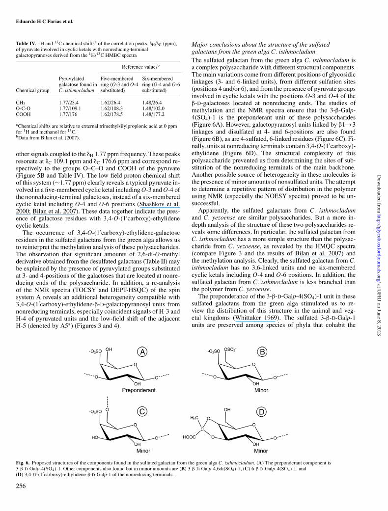
Major conclusions about the structure of the sulfatedgalactans from the green alga C. isthmocladumThe sulfated galactan from the green alga C. isthmocladum isa complex polysaccharide with different structural components.The main variations come from different positions of glycosidiclinkages (3- and 6-linked units), from different sulfation sites(positions 4 and/or 6), and from the presence of pyruvate groupsinvolved in cyclic ketals with the positions O-3 and O-4 of theβ-D-galactoses located at nonreducing ends. The studies ofmethylation and the NMR spectra ensure that the 3-β-Galp-4(SO4)-1 is the preponderant unit of these polysaccharides(Figure 6A). However, galactopyranosyl units linked by β1→3linkages and disulfated at 4- and 6-positions are also found(Figure 6B), as are 4-sulfated, 6-linked residues (Figure 6C). Fi-nally, units at nonreducing terminals contain 3,4-O-(1′carboxy)-ethylidene (Figure 6D). The structural complexity of thispolysaccharide prevented us from determining the sites of sub-stitution of the nonreducing terminals of the main backbone.Another possible source of heterogeneity in these molecules isthe presence of minor amounts of nonsulfated units. The attemptto determine a repetitive pattern of distribution in the polymerusing NMR (especially the NOESY spectra) proved to be un-successful.
Apparently, the sulfated galactans from C. isthmocladumand C. yezoense are similar polysaccharides. But a more in-depth analysis of the structure of these two polysaccharides re-veals some differences. In particular, the sulfated galactan fromC. isthmocladum has a more simple structure than the polysac-charide from C. yezoense, as revealed by the HMQC spectra(compare Figure 3 and the results of Bilan et al. 2007) andthe methylation analysis. Clearly, the sulfated galactan from C.isthmocladum has no 3,6-linked units and no six-memberedcyclic ketals including O-4 and O-6 positions. In addition, thesulfated galactan from C. isthmocladum is less branched thanthe polymer from C. yezoense.
The preponderance of the 3-β-D-Galp-4(SO4)-1 unit in thesesulfated galactans from the green alga stimulated us to re-view the distribution of this structure in the animal and veg-etal kingdoms (Whittaker 1969). The sulfated 3-β-D-Galp-1units are preserved among species of phyla that cohabit the
Fig. 6. Proposed structures of the components found in the sulfated galactan from the green alga C. isthmocladum. (A) The preponderant component is3-β-D-Galp-4(SO4)-1. Other components also found but in minor amounts are (B) 3-β-D-Galp-4,6di(SO4)-1, (C) 6-β-D-Galp-4(SO4)-1, and(D) 3,4-O-(1′carboxy)-ethylidene-β-D-Galp-1 of the nonreducing terminals.
256
at UFR
J on June 8, 2013http://glycob.oxfordjournals.org/
Dow
nloaded from

A preponderantly 4-sulfated, 3-linked galactan from the green alga Codium isthmocladum
marine environment. These species appear among brown algae(Phaephyta), green algae (Chlorophyta), red seaweeds(Rhodophyta), marine seagrass (Angiospermae, Spermato-phyta), invertebrates [sea urchins (Echinodermata, Echinoidea),clams (Mollusca, Bivalvia), tunicates (Urochordata, Ascidi-acea)], and vertebrates such as fishes (Teleostei, Chordata) thatexpress keratan sulfate, although 6-sulfation is less evident inthis glycosaminoglycan (Scudder et al. 1986). Although the3-β-D-Galp-1 units are preserved in these phyla, the preferen-tial sulfation site varies: 4-sulfation in the algae and marineangiosperm and 2- or 6-sulfation in the galactans from inverte-brates and in the keratan sulfate (and the marine angiosperm).
Materials and methods
Extraction of polysaccharidesThe green alga C. isthmocladum was collected from the sublit-toral coast of Natal, Rio Grande do Norte, Brazil. Immediatelyafter collection, the alga was air-dried at 50◦C (under ventila-tion) and ground in a blender. The seaweed was then treatedwith ethanol and acetone to remove pigments and lipids, re-spectively. One hundred grams of delipidated, dry powderedalga was suspended in 500 mL of an aqueous solution of0.25 M NaCl and adjusted to pH 8.0 with NaOH. About 20 mgof maxatase, an alkaline protease from Esporobacillus (Biobras,Montes Claros, Minas Gerais, Brazil) was added to the solutionfor proteolytic digestion. After incubation for 18 h at 60◦Cunder agitation, the mixture was filtered through cheesecloth.The filtrate was fractionated through a serial precipitation withincreasing volumes of acetone (1:0.3, 1:0.5, 1:0.7, 1:0.9, and1:1.2, v/v). For each precipitation, the volume of ice-cold ace-tone was added under gentle agitation and maintained at 4◦C for24 h. The pellets formed in each suspension were collected bycentrifugation separately (10,000 × g for 20 min), dried undervacuum, resuspended in distilled water, and analyzed for chem-ical composition (content of sugars and sulfate molar ratio wereestimated as described below). The measurements are indicatedin Table I.
Chemical analyses of the precipitates obtained with differentvolumes of acetoneTotal sugar content of each pellet was measured bythe phenol–H2SO4 reaction (Dubois et al. 1956), us-ing D-galactose as standard. After acid hydrolysis ofthe polysaccharides (6 M HCl for 4 h at 100◦C),the sulfate contents were measured by the toluidinemethod as previously described (Nader and Dietrich 1977).Again, the polysaccharides of each pellet were hydrolyzed(2 M HCl for 2 h at 100◦C) and the type of hexose was deter-mined by gas–liquid chromatography of alditol acetate deriva-tives (Kircher 1960), as described below. As standards, fucose,glucose, rhamnose, arabinose, mannose, galactose, and xylosewere converted to their respective alditol acetates.
Purification of the sulfated galactanTo obtain the sulfated galactan, the precipitate formed with 1:0.9acetone was fractionated by ion-exchange chromatography. Af-ter centrifugation and drying, 12 mg was dissolved in 5 mLof distilled water and applied to a column (19.0 × 4.5 cm)of MP500 Lewatit resin (Bayer Chemicals, Sao Paulo, Brazil).
First, the column was washed with 300 mL of distilled waterand developed by a stepwise gradient system using 300 mL ofaqueous solutions with different concentrations of NaCl (0.3 M,0.5 M, 0.7 M, 1.0 M, 1.5 M, 2.0 M, 3.0 M, and 4.0 M) at1 mL/min. The first 300 mL at each step was collected in asingle recipient; then, a second recipient was used to collectan additional 300 mL of the same saline molarity to checkfor the complete elution of the polysaccharides. To precipitatethe polysaccharides, two volumes (600 mL) of methanol wereadded to both recipients for each step. However, only the first300 mL of the steps at 0.3 M, 2.0 M, and 3.0 M precipitatedpolysaccharides when methanol was added. These suspensionswere centrifuged separately (10,000 × g for 20 min), dried un-der vacuum, dissolved in distilled water to a final concentrationof 10 mg/mL, and analyzed for hexose (Dubois et al. 1956) andsulfate molar ratio (Nader and Dietrich 1977).
Agarose and polyacrylamide gel electrophoresisThe precipitate with 1:0.9 acetone and the fractions of sul-fated galactan (SG 1 and SG 2) were analyzed by agarosegel electrophoresis as described previously (Dietrich CP andDietrich SMC 1977). The samples (∼20 µg) were appliedto a 5-mm-thick 0.5% agarose gel and run for 1 h at 110 Vin 0.05 M 1,3-diaminopropane–acetate (pH 9.0). The sulfatedpolysaccharides in the gel were fixed with 0.1% N-cetyl-N,N,N-trimethylammonium bromide solution. After 12 h, thegel was dried and stained with 0.1% toluidine blue in aceticacid:ethanol:water (0.1:5:5, v/v).
The molecular masses of the sulfated galactan (SG 1 andSG 2) were estimated by PAGE in comparison with the elec-trophoretic mobility of standard compounds (Pomin, Pereira,et al. 2005). The sulfated polysaccharides (∼10 µg of each)were applied to a 1-mm-thick 10% polyacrylamide slab gel in0.02 M sodium barbital (pH 8.6). After electrophoresis (100 Vfor 30 min), the sulfated polysaccharides were stained with0.1% toluidine blue in 1% acetic acid and washed for about1 h in 1% acetic acid. The molecular-mass markers used werehigh-molecular-weight dextran sulfate (∼500 kDa), chondroitin6-sulfate from shark cartilage (∼60 kDa), chondroitin 4-sulfatefrom whale cartilage (∼40 kDa), unfractionated heparin fromporcine intestinal mucosa (∼15 kDa), low-molecular-weight-heparin (∼7.5 kDa), and low-molecular-weight dextran sulfate(∼8 kDa).
Desulfation and methylation reactionsDesulfation of the sulfated galactan was performed as detailedpreviously (Mourao and Perlin 1987; Vieira et al. 1991). About20 mg of each sulfated galactan (SG 1 and SG 2) was dissolvedin 5 mL of distilled water and mixed with 1 g (dry weight) ofDowex 50-W (H+, 200–400 mesh). After neutralization withpyridine, solutions were lyophilized. The resulting pyridiniumsalts were dissolved in 2.5 mL of dimethyl sulfoxide:methanol(9:1, v/v). The mixtures were heated at 80◦C for 4 h, and thedesulfated products were exhaustively dialyzed against distilledwater and lyophilized. The extent of desulfation was estimatedby the molar ratio of sulfate/total sugars. This method allows usto detect desulfation down to a molar ratio of ≤0.1 sulfate/totalsugar. About 5 mg of each desulfated fraction was obtained.The native and desulfated galactans were subjected to threerounds of methylation, as described (Ciucanu and Kerek 1984)
257
at UFR
J on June 8, 2013http://glycob.oxfordjournals.org/
Dow
nloaded from

Eduardo H C Farias et al.
and with the modifications suggested by Patankar et al. (1993).The methylated galactans were hydrolyzed with 6.0 M trifluo-roacetic acid for 5 h at 100◦C, reduced with borohydride, andthe alditols were acetylated with 1:1 acetic anhydride/pyridine(Kircher 1960). The alditol acetates from the methylated sugarswere dissolved in chloroform and analyzed in a Hewlett-Packardgas chromatography/mass spectrometry unit, model 5987-A. In-jection was made in the splitless mode in a DB-1 capillary col-umn (25 m × 0.3 mm). The column was programmed to run at120◦C for 2 min, then raised to 230◦C at 2◦C/min, and held for5 min.
NMR experiments1H and 13C, one-dimensional and two-dimensional spectra ofthe native sulfated galactan and the desulfated galactan wererecorded using a Bruker DRX 400 MHz apparatus with atriple resonance probe as detailed previously (Pomin, Valente,et al. 2005). About 5 mg of each sample was dissolved in0.5 mL 99.9% deuterium oxide (Cambridge Isotope Laboratory,Cambridge, MA). All spectra were recorded at 50◦C with HODsuppression by presaturation. The 1D 1H NMR spectra wererecorded with 16 scans. The 2D 1H/1H COSY, TOCSY, NOESY,and 1H/13C HSQC spectra were recorded using states-time pro-portion phase incrementation (states-TPPI) for quadrature de-tection in the indirect dimension. The TOCSY spectra were runwith 4046 × 400 points with a spin-lock field of 10 kHz and amixed time of 80 ms. The NOESY spectra were recorded witha mixing time of 100 ms. The 1H/13C DEPT-HSQC and 1H/13CHSQC spectra were run with 1024 × 256 points and globally op-timized alternating phase rectangular pulses (GARP) for decou-pling. The 1H/13C HMBC spectrum was recorded with 1024 ×256 points, with a 60-ms delay for evolution of long-range cou-plings and was set with no decoupling during acquisition time.Chemical shifts are displayed relative to external trimethylsi-lylpropionic acid at 0 ppm for 1H and relative to methanolfor 13C.
Supplementary Data
Supplementary data for this article is available online athttp://glycob.oxfordjournals.org.
Funding
Conselho Nacional de Desenvolvimento Cientıfico e Tec-nologico (CNPq), Fundacao de Amparo a Pesquisa do Estado doRio de Janeiro (FAPERJ) and Coordenacao de Aperfeicoamentode Pessoal de Nıvel Superior (CAPES).
Acknowledgements
We are grateful to Anderson Pinheiro and Fabiana Albernazfrom the Centro Nacional de Ressonancia Magnetica NuclearJiri Jonas, CCS, Brazil for help with the NMR experimentsand figures and especially to Martha Sorenson for editing themanuscript.
Conflict of interest statement
None declared.
Abbreviations
COSY, correlation spectroscopy; DEPT, distortionless enhance-ment by polarization transfer; HMBC, heteronuclear multiplebound coherence; HSQC, heteronuclear single quantum coher-ence; NMR, nuclear magnetic resonance; NOESY, nuclear over-hauser enhancement spectroscopy; PAGE, polyacrylamide gelelectrophoresis; SG, sulfated galactan; TOCSY, total correlationspectroscopy..
References
Albano RM, Pavao MSG, Mourao PAS, Mulloy B. 1990. Structural studies ofa sulfated L-galactan from Styela plicata (Tunicate): Analysis of the Smith-degraded polysaccharide. Carbohydr Res. 208:163–174.
Alves AP, Mulloy B, Diniz JA, Mourao PAS. 1997. Sulfated polysaccharidesfrom the egg jelly layer are species-specific inducers of acrosomal reactionin sperms of sea urchins. J Biol Chem. 272:6965–6971.
Amornrut C, Toida T, Imanari T, Woo E-R, Park H, Linhardt R, Wu SJ, KimYS. 1999. A new sulfated beta-galactan from clams with anti-HIV activity.Carbohydr Res. 321:121–127.
Aquino RS, Landeira-Fernandez AM, Valente A-P, Andrade LR, Mourao PAS.2005. Occurrence of sulfated galactans in marine angiosperms: Evolutionaryimplications. Glycobiology. 15:11–20.
Berteau O and Mulloy B. 2003. Sulfated fucans, fresh perspectives: Structures,functions, and biological properties of sulfated fucans and an overviewof enzymes active toward this class of polysaccharide. Glycobiology.13:29R–40R.
Bilan MI, Vinogradova EV, Shashkov AS, Usov AI. 2007. Structure of a highlypyruvylated galactan sulfate from the Pacific green alga Codium yezoense(Bryopsidales, Chlorophyta). Carbohydr Res. 342:586–596.
Ciucanu I and Kerek F. 1984. Rapid and simultaneous methylation of fattyand hydroxy fatty acids for gas chromatographic analysis. J Chromatogr.286:179–185.
Dietrich CP and Dietrich SMC. 1977. Electrophoretic behaviour of acidicmucopolysaccharides by agarose gel electrophoresis. J Chromatogr.130:299–304.
Dubois M, Gilles KA, Hamilton JK, Rebers PA, Smith F. 1956. Colorimetricmethod for determination of sugars and related substances. Anal Chem. 28:350–354.
Estevez JM, Ciancia M, Cerezo AS. 2004. The system of low-molecular-weightcarrageenans and agaroids from the room-temperature-extracted fraction ofKappaphycus alvarezii. Carbohydr Res. 339:2575–2592.
Kircher HW. 1960. Gas–liquid partition chromatography of methylated sugars.Anal Chem. 32:1103–1106.
Lahaye M. 2001. Development on gelling algal galactans, their structure andphysico-chemistry. J Appl Phycol. 13:173–184.
Love J and Percival E. 1964. The polysaccharides of green seaweed Codiumfragile: Part III. A β-1,4-linked mannan. J Chem Soc. 3345–3350.
Mathews MB. 1975. Connective Tissue, Macromolecular Structure and Evolu-tion. Berlin: Springer.
Matsubara K, Matsuura Y, Bacic A, Liao M-L, Hori K, Miyazawa K. 2001.Anticoagulant properties of a sulfated galactan preparation from a marinegreen alga. Codium cylindricum Biol Macromol. 28:395–399.
Mourao PAS. 2004. Use of sulfated fucans as anticoagulant and antithromboticagents: Future perspectives. Curr Pharm Des. 10:967–981.
Mourao PAS. 2007. A carbohydrate-based mechanism of species recognition insea urchin fertilization. Braz J Med Biol Res. 40:5–17.
Mourao PA and Perlin AS. 1987. Structural features of sulfated glycansfrom the tunic of Styela plicata (Chordata-Tunicata). A unique occur-rence of L-galactose in sulfated polysaccharides. Eur J Biochem. 166:431–436.
Mulloy B, Ribeiro A-C, Alves A-C, Vieira RP, Mourao PAS. 1994. Sulfatedfucans from echinoderms have a regular tetrasaccharide repeating unit de-fined by specific patterns of sulfation at the O-2 and O-4 positions. J BiolChem. 296:22113–22123.
Murano E, Toffanin R, Cecere E, Rizzo R, Knutsen SH. 1997. Investigation ofthe carrageenans extracted from Solieria filiformis and Agardhiella subulatafrom Mar Piccolo, Taranto. Mar Chem. 58:319–325.
Nader HB and Dietrich CP. 1977. Determination of sulfate after chromatographyand toluidine blue complexation. Anal Biochem. 78:112–118.
258
at UFR
J on June 8, 2013http://glycob.oxfordjournals.org/
Dow
nloaded from
A preponderantly 4-sulfated, 3-linked galactan from the green alga Codium isthmocladum
Patankar MS, Oehninger S, Barnett T, Williams RL, Clark GF. 1993. A revisedstructure for fucoidan may explain some of its biological activities. J BiolChem. 268:21770–21776.
Pavao MSG, Albano RM, Lawsom AM, Mourao PAS. 1989. Structural het-erogeneity among unique sulfated L-galactans from different species ofascidians (tunicates). J Biol Chem. 264:9972–9979.
Pavao MSG, Mourao PA, Mulloy B. 1990. Structure of a unique sulfatedalpha-L-galactofucan from the tunicate Clavelina. Carbohydr Res. 208:153–161.
Pereira MS, Mulloy B, Mourao PAS. 1999. Structure and anticoagulant ac-tivity of sulfated fucans. Comparison between the regular, repetitive andlinear fucans from echinoderms with the more heterogeneous and branchedpolymers from brown algae. J Biol Chem. 274:7656–7667.
Pereira MG, Benevides NMB, Melo MRS, Valente A-P, Melo FR, Mourao PA.2005. Structure and anticoagulant activity of a sulfated galactan from thered alga, Gelidium crinale. Is there a specific structural requirement for theanticoagulant action? Carbohydr Res. 340:2015–2023.
Pomin VH, Pereira MS, Valente A-P, Tollefsen DM, Pavao MSG, MouraoPA. 2005. Selective cleavage and anticoagulant activity of a sulfated fucan:Stereospecific removal of a 2-sulfated ester from the polysaccharide bymild acid hydrolysis, preparation of oligosaccharides, and heparin cofactorII-dependent anticoagulant activity. Glycobiology. 15:369–381.
Pomin VH, Valente A-P, Pereira MS, Mourao PA. 2005. Mild acid hydrolysisof sulfated fucans: A selective 2-desulfation reaction and an alternativeapproach for preparing tailored sulfated oligosaccharides. Glycobiology.15:1376–1385.
Ribeiro A-C, Vieira RP, Mourao PAS, Mulloy B. 1994. A sulfated alpha-L-fucanfrom sea cucumber. Carbohydr Res. 255:225–240.
Rocha HA, Moraes FA, Trindade ES, Franco CR, Torquato RJ, Veiga SS, ValenteA-P, Mourao PA, Leite EL, Nader HB et al. 2005. Structural and hemostaticactivities of a sulfated galactofucan from the Brown alga Spatoglossumschroederi. An ideal antithrombotic agent? J Biol Chem. 280:41278–41288.
Santos JA, Mulloy B, Mourao PAS. 1992. Structural diversity among sulfatedalpha-L-galactans from ascidians (tunicates). Studies on the species Cionaintestinalis and Herdmania monus. Eur J Biochem. 204:669–677.
Scudder P, Tanq PW, Hounsell EF, Lawson AM, Mehmet H, Feizi T. 1986.Isolation and characterization of sulphated oligosaccharides released frombovine corneal keratin sulphate by the action of endo-beta-galactosidase.Eur J Biochem. 157:365–373.
Shashkov AS, Senchenkova SN, Vinogradov EV, Zatonsky GV, Knirel YA,Literacka E, Kaca W. 2000. Full structure of the O-specific polysaccharideof Proteus mirabilis O24 containing 3,4-O-[(S)-1-carboxyethylidene]-D-galactose. Carbohydr Res. 329:453–457.
van de Velde F, Pereira L, Rollema HS. 2004. The revised NMR chemical shiftdata of carrageenans. Carbohydr Res. 339:2309–2313.
Verli H. 2007. Insights into Carbohydrate Structure and Biological Function.Kerala (India): Transworld Research Network.
Vieira RP, Mulloy B, Mourao PA. 1991. Structure of a fucose-branched chon-droitin sulfate from sea cucumber. Evidence for the presence of 3-O-sulfo-beta-D-glucuronosyl residues. J Biol Chem. 266:13530–13536.
Whittaker RH. 1969. New concepts of kingdoms of organisms. Science.163:150–160.
259
at UFR
J on June 8, 2013http://glycob.oxfordjournals.org/
Dow
nloaded from
Copyright © 2022 FDOKUMEN