Sulfated Polysaccharides in the Freshwater Green Macroalga ...
10
ORIGINAL RESEARCH published: 13 November 2017 doi: 10.3389/fpls.2017.01927 Edited by: Wim Van den Ende, KU Leuven, Belgium Reviewed by: Koenraad Muylaert, KU Leuven, Belgium Céline Laroche, Université Clermont Auvergne, France *Correspondence: Marina Ciancia [email protected] José M. Estevez [email protected] Specialty section: This article was submitted to Plant Physiology, a section of the journal Frontiers in Plant Science Received: 16 August 2017 Accepted: 25 October 2017 Published: 13 November 2017 Citation: Arata PX, Alberghina J, Confalonieri V, Errea MI, Estevez JM and Ciancia M (2017) Sulfated Polysaccharides in the Freshwater Green Macroalga Cladophora surera Not Linked to Salinity Adaptation. Front. Plant Sci. 8:1927. doi: 10.3389/fpls.2017.01927 Sulfated Polysaccharides in the Freshwater Green Macroalga Cladophora surera Not Linked to Salinity Adaptation Paula X. Arata 1 , Josefina Alberghina 2 , Viviana Confalonieri 2 , María I. Errea 3 , José M. Estevez 4 * and Marina Ciancia 1,5 * 1 Universidad de Buenos Aires, Facultad de Agronomía, Departamento de Biología Aplicada y Alimentos, Cátedra de Química de Biomoléculas, Buenos Aires, Argentina, 2 Universidad de Buenos Aires, Facultad de Ciencias Exactas y Naturales, Departamento de Ecología, Genética y Evolución, Instituto IEGEBA (UBA-CONICET), Buenos Aires, Argentina, 3 Instituto Tecnológico de Buenos Aires, Departamento de Ingeniería Química, Buenos Aires, Argentina, 4 Fundación Instituto Leloir-IIBBA-CONICET, Buenos Aires, Argentina, 5 CONICET-Universidad de Buenos Aires, Centro de Investigación de Hidratos de Carbono (CIHIDECAR), Buenos Aires, Argentina The presence of sulfated polysaccharides in cell walls of seaweeds is considered to be a consequence of the physiological adaptation to the high salinity of the marine environment. Recently, it was found that sulfated polysaccharides were present in certain freshwater Cladophora species and some vascular plants. Cladophora (Ulvophyceae, Chlorophyta) is one of the largest genera of green algae that are able to grow in both, seas and freshwater courses. Previous studies carried out on the water-soluble polysaccharides of the marine species C. falklandica established the presence of sulfated xylogalactoarabinans constituted by a backbone of 4-linked β-L-arabinopyranose units partially sulfated mainly on C3 and also on C2 with partial glycosylation, mostly on C2, with terminal β-D-xylopyranose or β-D-galactofuranose units. Besides, minor amounts of 3-, 6- and/or 3,6-linked β-D-galactan structures, with galactose in the pyranosic form were detected. In this work, the main water soluble cell wall polysaccharides from the freshwater alga Cladophora surera were characterized. It was found that this green alga biosynthesizes sulfated polysaccharides, with a structure similar to those found in marine species of this genus. Calibration of molecular clock with fossil data suggests that colonization of freshwater environments occurred during the Miocene by its ancestor. Therefore, the presence of sulfated polysaccharides in the freshwater green macroalga C. surera could be, in this case, an adaptation to transient desiccation and changes in ionic strength. Retention of sulfated polysaccharides at the cell walls may represent a snapshot of an evolutionary event, and, thus constitutes an excellent model for further studies on the mechanisms of sulfation on cell wall polysaccharides and environmental stress co-evolution. Keywords: green alga, Cladophora, cell walls, sulfated polysaccharides, freshwater environment Frontiers in Plant Science | www.frontiersin.org 1 November 2017 | Volume 8 | Article 1927
-
Upload
khangminh22 -
Category
Documents
-
view
1 -
download
0
Transcript of Sulfated Polysaccharides in the Freshwater Green Macroalga ...
fpls-08-01927 November 10, 2017 Time: 16:57 # 1
ORIGINAL RESEARCHpublished: 13 November 2017doi: 10.3389/fpls.2017.01927
Edited by:Wim Van den Ende,
KU Leuven, Belgium
Reviewed by:Koenraad Muylaert,
KU Leuven, BelgiumCéline Laroche,
Université Clermont Auvergne, France
*Correspondence:Marina Ciancia
[email protected]é M. Estevez
Specialty section:This article was submitted to
Plant Physiology,a section of the journal
Frontiers in Plant Science
Received: 16 August 2017Accepted: 25 October 2017
Published: 13 November 2017
Citation:Arata PX, Alberghina J,
Confalonieri V, Errea MI, Estevez JMand Ciancia M (2017) Sulfated
Polysaccharides in the FreshwaterGreen Macroalga Cladophora surera
Not Linked to Salinity Adaptation.Front. Plant Sci. 8:1927.
doi: 10.3389/fpls.2017.01927
Sulfated Polysaccharides in theFreshwater Green MacroalgaCladophora surera Not Linked toSalinity AdaptationPaula X. Arata1, Josefina Alberghina2, Viviana Confalonieri2, María I. Errea3,José M. Estevez4* and Marina Ciancia1,5*
1 Universidad de Buenos Aires, Facultad de Agronomía, Departamento de Biología Aplicada y Alimentos, Cátedra deQuímica de Biomoléculas, Buenos Aires, Argentina, 2 Universidad de Buenos Aires, Facultad de Ciencias Exactas yNaturales, Departamento de Ecología, Genética y Evolución, Instituto IEGEBA (UBA-CONICET), Buenos Aires, Argentina,3 Instituto Tecnológico de Buenos Aires, Departamento de Ingeniería Química, Buenos Aires, Argentina, 4 Fundación InstitutoLeloir-IIBBA-CONICET, Buenos Aires, Argentina, 5 CONICET-Universidad de Buenos Aires, Centro de Investigación deHidratos de Carbono (CIHIDECAR), Buenos Aires, Argentina
The presence of sulfated polysaccharides in cell walls of seaweeds is consideredto be a consequence of the physiological adaptation to the high salinity of themarine environment. Recently, it was found that sulfated polysaccharides were presentin certain freshwater Cladophora species and some vascular plants. Cladophora(Ulvophyceae, Chlorophyta) is one of the largest genera of green algae that areable to grow in both, seas and freshwater courses. Previous studies carried out onthe water-soluble polysaccharides of the marine species C. falklandica establishedthe presence of sulfated xylogalactoarabinans constituted by a backbone of 4-linkedβ-L-arabinopyranose units partially sulfated mainly on C3 and also on C2 with partialglycosylation, mostly on C2, with terminal β-D-xylopyranose or β-D-galactofuranoseunits. Besides, minor amounts of 3-, 6- and/or 3,6-linked β-D-galactan structures, withgalactose in the pyranosic form were detected. In this work, the main water soluble cellwall polysaccharides from the freshwater alga Cladophora surera were characterized. Itwas found that this green alga biosynthesizes sulfated polysaccharides, with a structuresimilar to those found in marine species of this genus. Calibration of molecular clockwith fossil data suggests that colonization of freshwater environments occurred duringthe Miocene by its ancestor. Therefore, the presence of sulfated polysaccharides in thefreshwater green macroalga C. surera could be, in this case, an adaptation to transientdesiccation and changes in ionic strength. Retention of sulfated polysaccharides at thecell walls may represent a snapshot of an evolutionary event, and, thus constitutesan excellent model for further studies on the mechanisms of sulfation on cell wallpolysaccharides and environmental stress co-evolution.
Keywords: green alga, Cladophora, cell walls, sulfated polysaccharides, freshwater environment
Frontiers in Plant Science | www.frontiersin.org 1 November 2017 | Volume 8 | Article 1927
fpls-08-01927 November 10, 2017 Time: 16:57 # 2
Arata et al. Sulfated Polysaccharides from Freshwater Cladophora
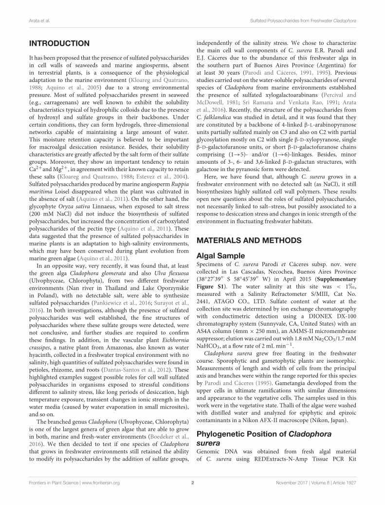
INTRODUCTION
It has been proposed that the presence of sulfated polysaccharidesin cell walls of seaweeds and marine angiosperms, absentin terrestrial plants, is a consequence of the physiologicaladaptation to the marine environment (Kloareg and Quatrano,1988; Aquino et al., 2005) due to a strong environmentalpressure. Most of sulfated polysaccharides present in seaweed(e.g., carrageenans) are well known to exhibit the solubilitycharacteristics typical of hydrophilic colloids due to the presenceof hydroxyl and sulfate groups in their backbones. Undercertain conditions, they can form hydrogels, three-dimensionalnetworks capable of maintaining a large amount of water.This moisture retention capacity is believed to be importantfor macroalgal desiccation resistance. Besides, their solubilitycharacteristics are greatly affected by the salt form of their sulfategroups. Moreover, they show an important tendency to retainCa2+and Mg2+, in agreement with their known capacity to retainthese salts (Kloareg and Quatrano, 1988; Estevez et al., 2004).Sulfated polysaccharides produced by marine angiosperm Ruppiamaritima Loisel disappeared when the plant was cultivated inthe absence of salt (Aquino et al., 2011). On the other hand, theglycophyte Oryza sativa Linnaeus, when exposed to salt stress(200 mM NaCl) did not induce the biosynthesis of sulfatedpolysaccharides, but increased the concentration of carboxylatedpolysaccharides of the pectin type (Aquino et al., 2011). Thesedata suggested that the presence of sulfated polysaccharides inmarine plants is an adaptation to high-salinity environments,which may have been conserved during plant evolution frommarine green algae (Aquino et al., 2011).
In an opposite way, very recently, it was found that, at leastthe green alga Cladophora glomerata and also Ulva flexuosa(Ulvophyceae, Chlorophyta), from two different freshwaterenvironments (Nan river in Thailand and Lake Oporzynskiein Poland), with no detectable salt, were able to synthesizesulfated polysaccharides (Pankiewicz et al., 2016; Surayot et al.,2016). In both investigations, although the presence of sulfatedpolysaccharides was well established, the fine structures ofpolysaccharides where these sulfate groups were detected, werenot conclusive, and further studies are required to confirmthese findings. In addition, in the vascular plant Eichhorniacrassipes, a native plant from Amazonas, also known as waterhyacinth, collected in a freshwater tropical environment with nosalinity, high quantities of sulfated polysaccharides were found inpetioles, rhizome, and roots (Dantas-Santos et al., 2012). Thesehighlighted examples suggest possible roles for cell wall sulfatedpolysaccharides in organisms exposed to stressful conditionsdifferent to salinity stress, like long periods of desiccation, hightemperature exposure, transient changes in ionic strength in thewater media (caused by water evaporation in small microsites),and so on.
The branched genus Cladophora (Ulvophyceae, Chlorophyta)is one of the largest genera of green algae that are able to growin both, marine and fresh-water environments (Boedeker et al.,2016). We then decided to test if one species of Cladophorathat grows in freshwater environments still retained the abilityto modify its polysaccharides by the addition of sulfate groups,
independently of the salinity stress. We chose to characterizethe main cell wall components of C. surera E.R. Parodi andE.J. Cáceres due to the abundance of this freshwater alga inthe southern part of Buenos Aires Province (Argentina) forat least 30 years (Parodi and Cáceres, 1991, 1995). Previousstudies carried out on the water-soluble polysaccharides of severalspecies of Cladophora from marine environments establishedthe presence of sulfated xylogalactoarabinans (Percival andMcDowell, 1981; Sri Ramana and Venkata Rao, 1991; Arataet al., 2016). Recently, the structure of the polysaccharides fromC. falklandica was studied in detail, and it was found that theyare constituted by a backbone of 4-linked β-L-arabinopyranoseunits partially sulfated mainly on C3 and also on C2 with partialglycosylation mostly on C2 with single β-D-xylopyranose, singleβ-D-galactofuranose units, or short β-D-galactofuranose chainscomprising (1→5)- and/or (1→6)-linkages. Besides, minoramounts of 3-, 6- and 3,6-linked β-D-galactan structures, withgalactose in the pyranosic form were detected.
Here, we have found that, although C. surera grows in afreshwater environment with no detected salt (as NaCl), it stillbiosynthesizes highly sulfated cell wall polymers. These resultsopen new questions about the roles of sulfated polysaccharides,not necessarily linked to salt–stress, but possibly associated to aresponse to desiccation stress and changes in ionic strength of theenvironment in fluctuating freshwater habitats.
MATERIALS AND METHODS
Algal SampleSpecimens of C. surera Parodi et Cáceres subsp. nov. werecollected in Las Cascadas, Necochea, Buenos Aires Province(38◦27′39′′ S 58◦45′39′′ W) in April 2015 (SupplementaryFigure S1). The water salinity at this site was < 1h,measured with a Salinity Refractometer S/MIII, Cat No.2441, ATAGO CO., LTD. Sulfate content of water at thecollection site was determined by ion exchange chromatographywith conductimetric detection using a DIONEX DX-100chromatography system (Sunnyvale, CA, United States) with anAS4A column (4mm × 250 mm), an AMMS-II micromembranesuppressor; elution was carried out with 1.8 mM Na2CO3/1.7 mMNaHCO3, at a flow rate of 2 mL min−1.
Cladophora surera grew free floating in the freshwatercourse. Sporophytic and gametophytic plants are isomorphic.Measurements of length and width of cells from the principalaxis and branches were within the range reported for this speciesby Parodi and Cáceres (1995). Gametangia developed from theupper cells in ultimate ramifications with similar dimensionsand appearance to the vegetative cells. The samples used in thiswork were in the vegetative state. Thalli of the algae were washedwith distilled water and analyzed for epiphytic and epizoiccontaminants in a Nikon AFX-II macroscope (Nikon, Japan).
Phylogenetic Position of CladophorasureraGenomic DNA was obtained from fresh algal materialof C. surera using REDExtracts-N-Amp Tissue PCR Kit
Frontiers in Plant Science | www.frontiersin.org 2 November 2017 | Volume 8 | Article 1927
fpls-08-01927 November 10, 2017 Time: 16:57 # 3
Arata et al. Sulfated Polysaccharides from Freshwater Cladophora
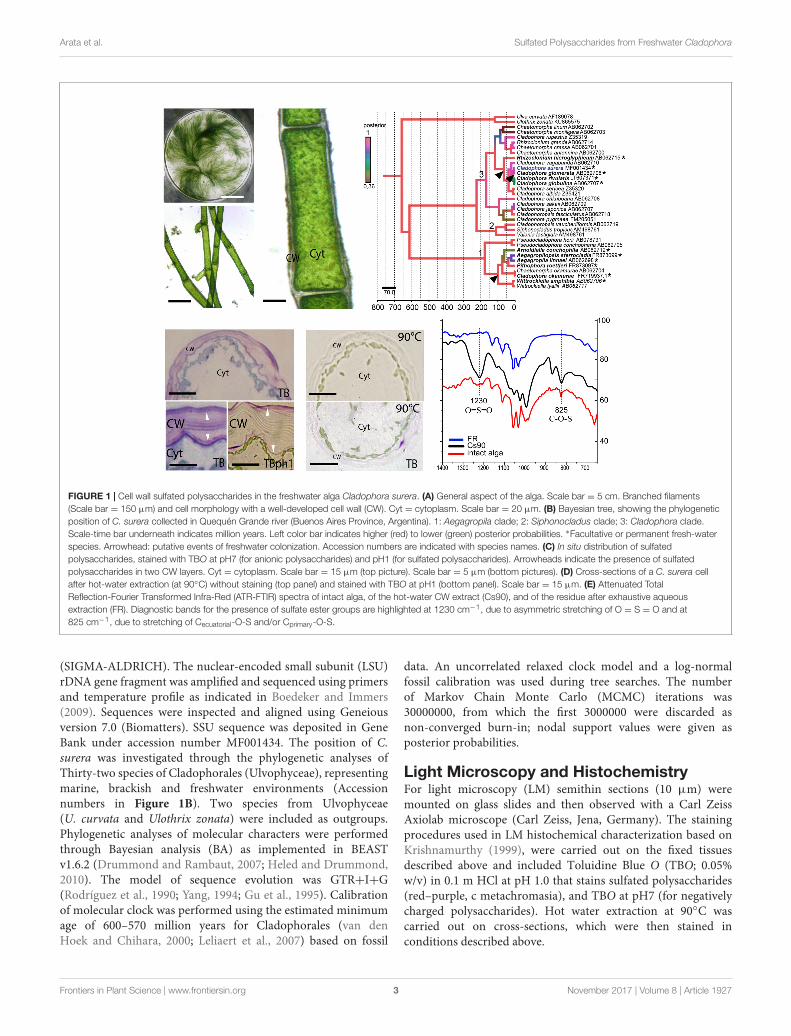
FIGURE 1 | Cell wall sulfated polysaccharides in the freshwater alga Cladophora surera. (A) General aspect of the alga. Scale bar = 5 cm. Branched filaments(Scale bar = 150 µm) and cell morphology with a well-developed cell wall (CW). Cyt = cytoplasm. Scale bar = 20 µm. (B) Bayesian tree, showing the phylogeneticposition of C. surera collected in Quequén Grande river (Buenos Aires Province, Argentina). 1: Aegagropila clade; 2: Siphonocladus clade; 3: Cladophora clade.Scale-time bar underneath indicates million years. Left color bar indicates higher (red) to lower (green) posterior probabilities. ∗Facultative or permanent fresh-waterspecies. Arrowhead: putative events of freshwater colonization. Accession numbers are indicated with species names. (C) In situ distribution of sulfatedpolysaccharides, stained with TBO at pH7 (for anionic polysaccharides) and pH1 (for sulfated polysaccharides). Arrowheads indicate the presence of sulfatedpolysaccharides in two CW layers. Cyt = cytoplasm. Scale bar = 15 µm (top picture). Scale bar = 5 µm (bottom pictures). (D) Cross-sections of a C. surera cellafter hot-water extraction (at 90◦C) without staining (top panel) and stained with TBO at pH1 (bottom panel). Scale bar = 15 µm. (E) Attenuated TotalReflection-Fourier Transformed Infra-Red (ATR-FTIR) spectra of intact alga, of the hot-water CW extract (Cs90), and of the residue after exhaustive aqueousextraction (FR). Diagnostic bands for the presence of sulfate ester groups are highlighted at 1230 cm−1, due to asymmetric stretching of O = S = O and at825 cm−1, due to stretching of Cecuatorial-O-S and/or Cprimary-O-S.
(SIGMA-ALDRICH). The nuclear-encoded small subunit (LSU)rDNA gene fragment was amplified and sequenced using primersand temperature profile as indicated in Boedeker and Immers(2009). Sequences were inspected and aligned using Geneiousversion 7.0 (Biomatters). SSU sequence was deposited in GeneBank under accession number MF001434. The position of C.surera was investigated through the phylogenetic analyses ofThirty-two species of Cladophorales (Ulvophyceae), representingmarine, brackish and freshwater environments (Accessionnumbers in Figure 1B). Two species from Ulvophyceae(U. curvata and Ulothrix zonata) were included as outgroups.Phylogenetic analyses of molecular characters were performedthrough Bayesian analysis (BA) as implemented in BEASTv1.6.2 (Drummond and Rambaut, 2007; Heled and Drummond,2010). The model of sequence evolution was GTR+I+G(Rodríguez et al., 1990; Yang, 1994; Gu et al., 1995). Calibrationof molecular clock was performed using the estimated minimumage of 600–570 million years for Cladophorales (van denHoek and Chihara, 2000; Leliaert et al., 2007) based on fossil
data. An uncorrelated relaxed clock model and a log-normalfossil calibration was used during tree searches. The numberof Markov Chain Monte Carlo (MCMC) iterations was30000000, from which the first 3000000 were discarded asnon-converged burn-in; nodal support values were given asposterior probabilities.
Light Microscopy and HistochemistryFor light microscopy (LM) semithin sections (10 µm) weremounted on glass slides and then observed with a Carl ZeissAxiolab microscope (Carl Zeiss, Jena, Germany). The stainingprocedures used in LM histochemical characterization based onKrishnamurthy (1999), were carried out on the fixed tissuesdescribed above and included Toluidine Blue O (TBO; 0.05%w/v) in 0.1 m HCl at pH 1.0 that stains sulfated polysaccharides(red–purple, c metachromasia), and TBO at pH7 (for negativelycharged polysaccharides). Hot water extraction at 90◦C wascarried out on cross-sections, which were then stained inconditions described above.
Frontiers in Plant Science | www.frontiersin.org 3 November 2017 | Volume 8 | Article 1927

fpls-08-01927 November 10, 2017 Time: 16:57 # 4
Arata et al. Sulfated Polysaccharides from Freshwater Cladophora
Extraction and Purification of thePolysaccharidesAlgal samples of C. surera were dried in open air. The milledalgae (100 g) were extracted twice with EtOH 70% (20g/L)for 3 h at room temperature. The residue from the alcoholextraction was extracted for 3 h with H2O (20 g/L) at 90◦C,giving extract Cs90 (20.1 g). Cs90 was dissolved in water(4.5 mg/ml). A residue (Ri) was separated from the supernatant,which was chromatographed on DEAE-Sephadex A-25. Thesupernatant was applied to a column (90 cm × 1.5 cm id),previously stabilized in H2O. The first elution solvent waswater and then NaCl solutions of increasing concentrationup to 4 M. Fractions of 4 ml were collected. Finally, thephase was boiled in 4 M NaCl solution. The presence ofcarbohydrates in the samples was detected by the phenolsulfuric acid method (Dubois et al., 1956); after obtainingblank readings, the eluant was replaced by another with higherconcentration of NaCl. Seven fractions (F1-F7) were obtained,dialyzed (molecular weight cut off 3,500) and freeze dried(Supplementary Figure S2).
Chemical CharacterizationThe total sugars content was analyzed by the phenol-sulfuricacid method (Dubois et al., 1956). Sulfate was determinedturbidimetrically (Dodgson and Price, 1962). Alternatively,ion exchange chromatography with conductimetric detectionwas used: the sample was hydrolyzed in 2 M CF3COOH at121◦C for 2 h, evaporated to dryness under nitrogen andredissolved in high purity water from a Milli-Q system.A DIONEX DX-100 chromatography system (Sunnyvale, CA,United States) was used with an AS4A column (4 × 250 mm),an AMMS-II micromembrane suppressor and a conductivitydetector, elution was carried out with 1.8 mM Na2CO3/1.7 mMNaHCO3, at a flow rate of 2 mL min−1. The absence ofpyruvic acid and uronic acids was confirmed using thecolorimetric determinations of Koepsell and Sharpe (1952)and Filisetti-Cozzi and Carpita (1991). The protein contentwas measured by microanalysis to determine the amountof nitrogen, a factor of 5 was applied to calculate theamount of protein, according to Angell et al. (2016). Theconfiguration of galactose and arabinose was determined bythe method of Cases et al. (1995) through their diastereomericacetylated 1-deoxy-1-(2-hydroxypropylamino) alditols. Todetermine the monosaccharide composition, samples werederivatized to the alditol acetates (Stevenson and Furneaux,1991).
Methylation AnalysisThe sample (10–20 mg) was converted into the correspondingtriethylammonium salt (Stevenson and Furneaux, 1991) andmethylated according to Ciucanu and Kerek (1984). The samplewas dissolved in dimethylsulfoxide; finely powdered NaOH wasused as base. The methylated samples were submitted to reductivehydrolysis and acetylation to give the alditol acetates in the sameway as the parent polysaccharides (Stevenson and Furneaux,1991).
Gas ChromatographyGC of the alditol acetates were carried out on a Agilent7890A gas-liquid chromatograph (Avondale, PA, United States)equipped with a flame ionization detector and fitted with afused silica column (0.25mm i.d. × 30 m) WCOT-coated with a0.20 mm film of SP-2330 (Supelco, Bellefonte, PA, United States).Chromatography was performed: from 200 to 240◦C at 2◦Cmin−1, followed by a 10-min hold for alditol acetates. For thepartially methylated alditol acetates, the initial temperature was160◦C, which was increased at 1◦C min−1 to 210◦C and then at2◦C min−1 to 230◦C. N2 was used as the carrier gas at a flowrate of 1 mL min−1 and the split ratio was 80:1. The injector anddetector temperature was 250◦C.
Gas Chromatography-MassSpectrometryGC–MS of the partially methylated alditol acetates wasperformed on a Agilent 7890A gas-liquid chromatographequipped the SP-2330 interfaced to a Agilent 5977A Series massspectrometer, working at 70 eV. The He flow rate was 1.3 mlmin−1, the injector temperature was 250◦C. Mass spectra wererecorded over a mass range of 30–500 amu.
Desulfation of Cs90The reaction was carried out by the microwave-assisted methoddescribed by Navarro et al. (2007). The sample (40 mg) wasconverted to the pyridinium salt and dissolved in 10 ml of DMSOcontaining 2% of pyridine. The mixture was heated for 10 sintervals and cooled to 50◦C (× 6). It was dyalized 3 days againsttap water and then 24 h against distilled water (MWCO 3,500)and lyofilized. An aliquot was methylated as described abovewithout previous isolation of the product.
Partial Acid Hydrolysis of RiThe reaction was carried out according to Bilan et al. (2007). Thesample (100 mg) was heated in 1% CH3COOH (20 mL) for 4 hat 100◦C, the solution was neutralized with NaHCO3, dialyzed,and lyophilized to give RiH (78.2 mg). RiH was fractionated byanion exchange chromatography on DEAE-Sephadex A-25 ina similar way as F1, in this case, five fractions were obtained(RiH-F1-RiH-F5).
ATR-FTIR SpectroscopySamples were analyzed on a Thermo Scientific Nicolet 6700spectrometer equipped with a smart ARK (AttenuatedReflectance Kit) accessory using a standard ZnSe crystaland a DTGS KBr detector. The spectra were recorded in the650–4000 cm−1 range, and the spectral resolution was 4 cm−1.Data were processed by using the software Origin Pro 9.0.0.
NMR Spectroscopy500 MHz 1H NMR, proton decoupled 125 MHz 13C NMRspectra, and two-dimensional NMR experiments (HSQC,HMBC, and COZY) were recorded on a Bruker AM500 atroom temperature, with external reference of TMS. The samples(20 mg) were exchanged in 99.9% D2O (0.5 mL) four times.
Frontiers in Plant Science | www.frontiersin.org 4 November 2017 | Volume 8 | Article 1927
fpls-08-01927 November 10, 2017 Time: 16:57 # 5
Arata et al. Sulfated Polysaccharides from Freshwater Cladophora
Chemical shifts were referenced to internal acetone (δH 2.175,δCH3 31.1). Parameters for 13C NMR spectra were as follows:pulse angle 51.4◦, acquisition time 0.56 s, relaxation delay 0.6 s,spectral width 29.4 kHz, and scans 25,000. For 1H NMR spectrathe parameters were: pulse angle 76◦, acquisition time 3 s,relaxation delay 3 s, spectral width 6250 Hz and scans 32. 2Dspectra were obtained using standard Bruker software.
RESULTS AND DISCUSSION
Cladophora surera BiosynthesizesSulfated PolysaccharidesSpecimens of the green alga were collected in a freshwaterenvironment (Quequén Grande river, 38◦27′39′′ S 58◦45′39′′ W)located in Buenos Aires Province, Argentina (SupplementaryFigure S1). Based on the morphological characters, it wasassigned to C. surera (Figure 1A; Parodi and Cáceres, 1991, 1995).
In order to gain further insight on the taxonomical identity,a molecular characterization of the SSU (nuclear-encoded smallsubunit rDNA) gene fragment of the collected species was carriedout and its phylogenetic position within the Cladophorales wasinvestigated. The Bayesian tree obtained (Figure 1B) recoveredwith high Posterior Probabilities (PP) the three major groupspreviously described by Boedeker et al. (2012): the “Aegagropilaclade,” the “Siphonocladus clade” and the “Cladophora clade.”C. surera is resolved within the latter group, and particularlywithin a lineage of four species that can be mainly foundin freshwater/brackish environments (Hanyuda et al., 2002;Boedeker et al., 2012). Within the order Cladophorales, theboundary between marine and freshwater environments wascrossed at least two to three times (van den Hoek, 1963;Logares et al., 2007; Hayakawa et al., 2012; Boedeker et al.,2016; Figure 1B). Calibration of molecular clock with fossil datasuggests that colonization of freshwater environments probablyoccurred 11.4 million years ago (MYA; 4–25 MYA 95% PP) bythe ancestor of the four freshwater species closely related toC. surera (Figure 1B); or even before, at about 65.5 MYA (35–115MYA 95%PP), during the diversification of the more basal speciesRhizoclonium hieroglyphicum.
Cladophora surera showed a thick cell wall with two fibrillar-like layers delimiting a middle amorphous region. TBO stainingsuggested the presence of sulfated polysaccharides concentratedin both marginal cell wall layers (Figure 1C). Hot waterextraction (at 90◦C) carried out on cross-sections greatlysuppressed TBO staining (Figure 1D). In agreement, the hot
water extract (Cs90) obtained from C. surera that represents20.1% (w/w) of the algal dry weight (Supplementary Figure S2)contained high amounts of sulfate (17%, as SO3Na, whichcorrespond to ∼4% w/w of the algal dry weight), in accordancewith the histochemical stainings.
Finally, Attenuated Total Reflection-Fourier TransformedInfra-Red (ATR-FTIR) spectrum of the dry milled alga, as wellas that of extract Cs90 (Figure 1E) showed the bands diagnosticfor the presence of sulfate ester groups at 1230 cm−1, which wasassigned to asymmetric stretching of O= S=O, and at 825 cm−1,due to stretching of Cecuatorial-O-S and/or Cprimary-O-S (PradoFernández et al., 2003). In agreement, after exhaustive sequentialextraction with water (at 90◦C) and with alkaline solutions,both IR bands (825 and 1230 cm−1) mostly disappeared inthe remaining cell wall residue (FR) confirming that sulfatedpolysaccharides are confined in the aqueous extracts (Figure 1E).
All these results together confirmed the presence of highquantities of sulfated polysaccharides in cell walls of freshwatermacroalga C. surera. The water salinity at the collection sitewas < 1h (w/w; as NaCl) and the sulfate content of the waterwas ∼1 mM, while usually in marine waters there are highsulfate concentrations (∼25–28 mM) (Bochenek et al., 2013).These results suggested that sulfated polysaccharides in someCladophora species (at least in C. glomerata, and now in C. surera)could be synthetized in response to other stress factors, likedesiccation and changes in ionic strength of the environment, notlinked to high salinity levels as a stable environmental condition.
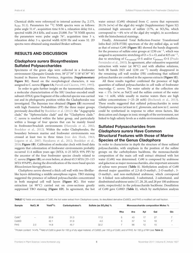
Sulfated Polysaccharides fromCladophora surera Have CommonStructural Features with those of MarineSpecies of the Genus CladophoraIn order to characterize in depth the structure of these sulfatedpolysaccharides, with emphasis in the position of the sulfategroups on the carbohydrates backbone, the monosaccharidecomposition of the main cell wall extract obtained with hotwater (Cs90) was determined. Cs90 is composed by arabinoseand galactose as major monosaccharides, also important amountsof xylose were present (Table 1). Methylation analysis of Cs90showed major quantities of 2,3-di-O-methyl-, 2-O-methyl-, 3-O-methyl-, and non-methylated arabinose, which correspondto 4-linked non-substituted, 3-substituted, 2-substituted, anddisubstituted arabinose units (17, 28, 20, and 28 per 100 arabinoseunits, respectively) in the polysaccharide backbone. Desulfationof Cs90 gave Cs90D (Table 1), which by methylation analysis
TABLE 1 | Yields and analyses of Cs90, the hot water extract from Cladophora surera, its desulfated derivative (Cs90D), and FiH3 a modified cell wall fraction.
Sample NaCl, M Yield2% Carbohydrates% Sulfate (as SO3Na) % Monosaccharide composition Moles %
Rha Ara Xyl Gal Glc
Cs901 – 20.9 31.3 17.3 5 43 17 27 7
Cs90D 42.1 51.9 4.0 3 57 14 18 7
FiH3 2.0 14.8 45.7 32.2 7 66 12 15 –
1Protein content: 14.4%. 2Yield of Cs90 is given for 100 g of dry algal material, of Cs90D, per 100 g of Cs90, and of FiH3, for 100 g of RiH (see Table 2).
Frontiers in Plant Science | www.frontiersin.org 5 November 2017 | Volume 8 | Article 1927
fpls-08-01927 November 10, 2017 Time: 16:57 # 6
Arata et al. Sulfated Polysaccharides from Freshwater Cladophora
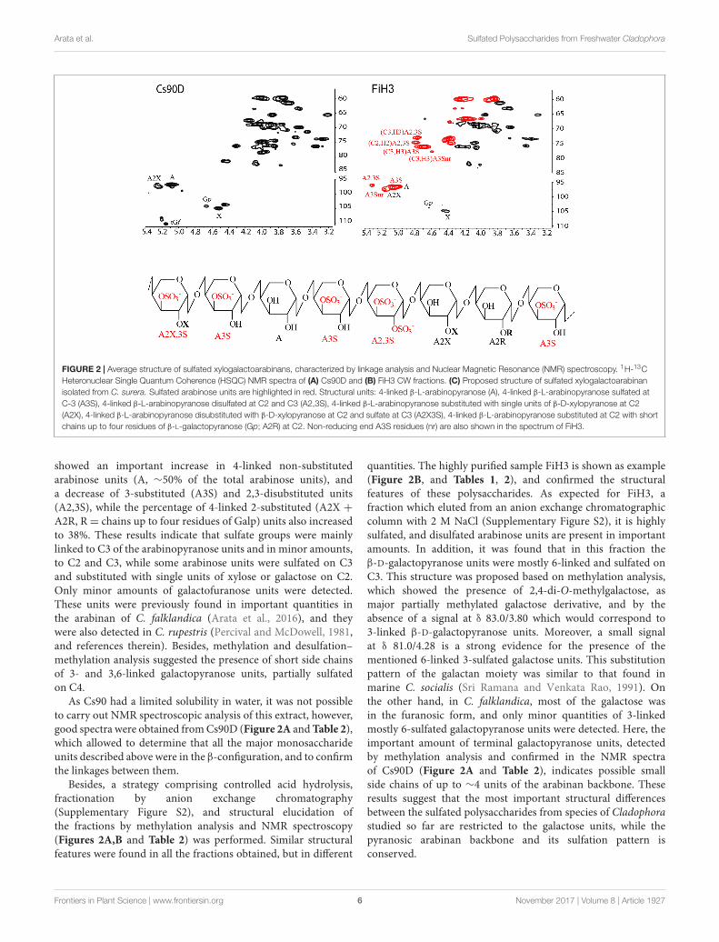
FIGURE 2 | Average structure of sulfated xylogalactoarabinans, characterized by linkage analysis and Nuclear Magnetic Resonance (NMR) spectroscopy. 1H-13CHeteronuclear Single Quantum Coherence (HSQC) NMR spectra of (A) Cs90D and (B) FiH3 CW fractions. (C) Proposed structure of sulfated xylogalactoarabinanisolated from C. surera. Sulfated arabinose units are highlighted in red. Structural units: 4-linked β-L-arabinopyranose (A), 4-linked β-L-arabinopyranose sulfated atC-3 (A3S), 4-linked β-L-arabinopyranose disulfated at C2 and C3 (A2,3S), 4-linked β-L-arabinopyranose substituted with single units of β-D-xylopyranose at C2(A2X), 4-linked β-L-arabinopyranose disubstituted with β-D-xylopyranose at C2 and sulfate at C3 (A2X3S), 4-linked β-L-arabinopyranose substituted at C2 with shortchains up to four residues of β-L-galactopyranose (Gp; A2R) at C2. Non-reducing end A3S residues (nr) are also shown in the spectrum of FiH3.
showed an important increase in 4-linked non-substitutedarabinose units (A, ∼50% of the total arabinose units), anda decrease of 3-substituted (A3S) and 2,3-disubstituted units(A2,3S), while the percentage of 4-linked 2-substituted (A2X +A2R, R= chains up to four residues of Galp) units also increasedto 38%. These results indicate that sulfate groups were mainlylinked to C3 of the arabinopyranose units and in minor amounts,to C2 and C3, while some arabinose units were sulfated on C3and substituted with single units of xylose or galactose on C2.Only minor amounts of galactofuranose units were detected.These units were previously found in important quantities inthe arabinan of C. falklandica (Arata et al., 2016), and theywere also detected in C. rupestris (Percival and McDowell, 1981,and references therein). Besides, methylation and desulfation–methylation analysis suggested the presence of short side chainsof 3- and 3,6-linked galactopyranose units, partially sulfatedon C4.
As Cs90 had a limited solubility in water, it was not possibleto carry out NMR spectroscopic analysis of this extract, however,good spectra were obtained from Cs90D (Figure 2A and Table 2),which allowed to determine that all the major monosaccharideunits described above were in the β-configuration, and to confirmthe linkages between them.
Besides, a strategy comprising controlled acid hydrolysis,fractionation by anion exchange chromatography(Supplementary Figure S2), and structural elucidation ofthe fractions by methylation analysis and NMR spectroscopy(Figures 2A,B and Table 2) was performed. Similar structuralfeatures were found in all the fractions obtained, but in different
quantities. The highly purified sample FiH3 is shown as example(Figure 2B, and Tables 1, 2), and confirmed the structuralfeatures of these polysaccharides. As expected for FiH3, afraction which eluted from an anion exchange chromatographiccolumn with 2 M NaCl (Supplementary Figure S2), it is highlysulfated, and disulfated arabinose units are present in importantamounts. In addition, it was found that in this fraction theβ-D-galactopyranose units were mostly 6-linked and sulfated onC3. This structure was proposed based on methylation analysis,which showed the presence of 2,4-di-O-methylgalactose, asmajor partially methylated galactose derivative, and by theabsence of a signal at δ 83.0/3.80 which would correspond to3-linked β-D-galactopyranose units. Moreover, a small signalat δ 81.0/4.28 is a strong evidence for the presence of thementioned 6-linked 3-sulfated galactose units. This substitutionpattern of the galactan moiety was similar to that found inmarine C. socialis (Sri Ramana and Venkata Rao, 1991). Onthe other hand, in C. falklandica, most of the galactose wasin the furanosic form, and only minor quantities of 3-linkedmostly 6-sulfated galactopyranose units were detected. Here, theimportant amount of terminal galactopyranose units, detectedby methylation analysis and confirmed in the NMR spectraof Cs90D (Figure 2A and Table 2), indicates possible smallside chains of up to ∼4 units of the arabinan backbone. Theseresults suggest that the most important structural differencesbetween the sulfated polysaccharides from species of Cladophorastudied so far are restricted to the galactose units, while thepyranosic arabinan backbone and its sulfation pattern isconserved.
Frontiers in Plant Science | www.frontiersin.org 6 November 2017 | Volume 8 | Article 1927

fpls-08-01927 November 10, 2017 Time: 16:57 # 7
Arata et al. Sulfated Polysaccharides from Freshwater Cladophora
TABLE 2 | Signal assignment of the HSQC NMR spectra of Cs90D and FiH3.
Structural unit Chemical shifts (ppm)
C-l/H-1 C-2/H-2 C-3/H-3 C-4/H-4 C-5/H-5,5′ C-6/H-6,6′
CS90D1
4-linked β-L-Arap (A) 97.3/5.03 69.1/3.89 68.9/4.02 75.2/4.02 60.4/4.01,3.81
4-linked β-L-Arap with β-D-Xylp on C2 (A2X) 98.4/5.18 78.3/4.02 67.9/4.14 75.2/4.02 60.4/4.01,3.81
T-β-D-Xylp (X) 105.7/4.45 74.1/3.33 76.3/3.39 70.2/3.58 66.0/3.88,3.24
T-β-D-Galf (Gf ) 108.8/4.98 82.0/4.04 77.7/3.97 84.5/3.96 71.6/3.75 63.7/3.58,3,65
5-linked β-D-Gal/(5G) 108.9/5.21 82.9/4.14 76.2/3.89 62.0/3.70
3-linked β-D-Galp (3G) 104.4/4.41 71.1/3.72 83.0/3.80 69.4/4.18 75.8/3.63 62.0/3.70
3,6-linked β-D-Galp (3,6G) 104.9/4.60 71.1/3.72 83.0/3.80 69.4/4.18 74.7/3.85 70.4/3.98, 3.86
TP-D-Galp (T-Gp) 103.6/4.51 71.7/3.47 73.6/3.61 75.8/3.65 62.0/3.70
FiH32,3,4
4-linked β-L-Arap (A) 97.3/5.03 69.1/3.89 68.9/4.02 75.4/4.02 60.4/4.03,3.85
4-linked β-L-Arap with β-D-Xylp on C2 (A2X) 98.4/5.18 78.3/4.02 67.9/4.14 75.4/4.02 60.4/4.03,3.85
T-β-D-Xylp (X) 105.7/4.45 74.1/3.33 76.3/3.39 70.2/3.58 66.0/3.88,3.24
4-linked β-L-Arap 3-sulfate (A3S) 97.6/5.09 67.4/4.12 76.7/4.63 74.2/4.34 60.9/3.78,4.20
4-linked β-L-Arap 2,3-disulfate (A2,3S) 96.8/5.34 75.2/4.73 73.6/4.71
Tn-β-L-Arap 3-sulfate (A3S-nr) 98.6/5.19 67.3/3.94 78.2/4.54 68.4/4.32 63.8/4.25,4,17
T-β-D-Galf (T-Gf ) 109.6/5.14 82.4/4.08 76.9/3.93 82.6/3.95 71.2/3.72 63.7/3.60,3,65
1Ratio Ara:Ara2X in Cs90D ∼1.0:0.5, calculated from the 1H NMR spectrum. 2Ratio Ara2.3S:Ara2X+TAra3Snr:Ara3S:Ara in Fi-H3 ∼1.0:1.2:6.0:1.5 calculated from the1H NMR spectrum. 3Peaks corresponding to sulfated carbons are highlighted in red. 4A signal at δ 81.0/4.28 was attributed to C3/H3 of 6-linked β-D-galactopyranose3-sulfate units.
In conclusion, sulfated xylogalactoarabinans from cell wallsof the freshwater alga C. surera have structural features similarto those reported for the marine species of this genus. Based onthe structural determination carried out by linkage analysis andNMR spectroscopy, it is proposed that C. surera biosynthesizessulfated xylogalactoarabinans as shown in Figure 2C.
Evolutionary Implications of thePresence of Sulfated Polysaccharides inC. sureraThe presence of sulfated polysaccharides in C. surera is inagreement with two recent studies on C. glomerata fromdifferent freshwater environments (Nan river in Thailand andLake Oporzynskie in Poland) (Pankiewicz et al., 2016; Surayotet al., 2016). In all three cases, they developed in freshwaterenvironments. Sulfated polysaccharides are widespread in thecell walls of marine angiosperms, in marine algae (green, red,and brown seaweeds), in the extracellular matrix of vertebratetissues, and in invertebrate species (Aquino et al., 2005; Ghandiand Mancera, 2008; Pomin and Mourao, 2008; Usov and Bilan,2009; Ciancia et al., 2010; Usov, 2011). In the green algaLamprothamnium papulosum (Characeae, Charophyta), whichgrows in water of fluctuating salinity, the extracellular sulfatedmucilage increases in sulfate content with increasing salinity(Shepherd and Beilby, 1999; Shepherd et al., 1999), while inbrown algae the concentrations of sulfate groups on fucanspositively correlate with increasing exposure to the atmospherein the intertidal zone, suggesting a role in desiccation resistance(Mabeau and Kloareg, 1987). Ectocarpus subulatus (Phaeophyta)isolated from a true freshwater environment (West and Kraft,
1996) undergoes major morphological, transcriptomic andmetabolic changes under variable salinities, including alterationof the expression of genes encoding enzymes potentially involvedin the sulfation or de-sulfation of cell wall polysaccharides(Dittami et al., 2012). It is now widely accepted that theoccurrence of sulfated polysaccharides in phylogenetically distanttaxa is a case of convergent adaptation to a broad range ofenvironmental conditions, such as stable high ionic media (e.g.,seawater for marine plants and algae) or transient changes insalt concentration linked to desiccation and high temperaturestresses (e.g., in some cases of freshwater macroalgae and vascularplants) (Mabeau and Kloareg, 1987; Shepherd and Beilby, 1999;Shepherd et al., 1999; Aquino et al., 2011; Dantas-Santos et al.,2012; Pankiewicz et al., 2016; Surayot et al., 2016). From aphysiological point of view, sulfated cell wall polysaccharidesfrom marine plants and marine algae may confer an adaptiveadvantage through possible structural and osmotic functionsthat are linked to an environmental pressure. Terrestrialization,and the subsequent taxonomic proliferation of land plants, waspredated by a divergence between the Chlorophyte (a groupof green algae) and the Streptophyte lineages (which includesland plants). This is strongly correlated with a change inhabitat preference from saline to freshwater conditions withlow or no salt, as well as exposure to transient changes inionic strength (e.g., caused by water evaporation in smallmicrosites). These transitions are accompanied by a majoralteration in cell wall polysaccharides composition such that,although the majority of marine Chlorophytes contain sulfatedcell wall polysaccharides, they are largely absent from most ofthe freshwater Charophycean green algae and their descendants.The latter green algae contain large amounts of negatively
Frontiers in Plant Science | www.frontiersin.org 7 November 2017 | Volume 8 | Article 1927
fpls-08-01927 November 10, 2017 Time: 16:57 # 8
Arata et al. Sulfated Polysaccharides from Freshwater Cladophora
charged pectins with carboxylated sugars (uronic acids) in theircell walls that could functionally replace some of the roles ofsulfated polysaccharides (Domozych et al., 2012). Further studiesto determine if sulfated polysaccharides are present in otherfreshwater algae are required to obtain a more comprehensivepicture about their possible functions in the cell wall.
CONCLUSION
Although further evidence is required, early emerging freshwaterand terrestrial organisms may have had a demand for sulfur thatwas in excess in the marine environment, but not in terrestrialand freshwater habitats (Raven, 1997). Such requirement forsulfate may have become a limiting factor and, therefore, mayhave been selected in organisms in a terrestrial environment,potentially resulting in the lack of carbohydrate sulfotransferasesand sulfatases from the genomes of freshwater algae and landplants and sulfated polysaccharides from their cell walls (Popperet al., 2011). The case of both freshwater Cladophora species mayindicate that not always the presence of sulfated polysaccharidesprovides an ionic barrier to high levels of salts, but it couldbe acting in response to other environmental factors likedesiccation and transient changes in ionic strength media. It isimportant to note, that the effect of transient desiccation couldgive an important temporary increase in the ionic strength ofthe medium. Also, the high temperatures, in which plants intropical regions develop, could cause desiccation. So, all thesefactors could be linked to high ionic strength environments,permanent or lasting only a short period of time. Hence, retentionof sulfation capacity in freshwater Cladophora species couldbe due to this factor, as well as to water retention capacity.Colonization of freshwater environments by the ancestor of thefour freshwater Cladophora species closely related to C. sureraprobably occurred during the Miocene. Retention of sulfatedpolysaccharides at the cell walls of freshwater green macroalgaemay represent a more general phenomenon, which constitutesan excellent model for further studies on the mechanismsof sulfation on cell wall polysaccharides and stress factors infreshwater environments co-evolution. Further genomic and
physiological studies are required to understand the molecularbasis of the retention of sulfated polysaccharides in C. surerain a freshwater environment. Finally, it is tempting to suggestfor future studies that sulfated polysaccharides could have, inthis case, unknown masked functions, not related to salinityadaptation.
AUTHOR CONTRIBUTIONS
PA, JA, and ME performed the experiments, VC analyzedand discussed the phylogenetic data, MC programmed theexperiments, JE and MC discussed the results and wrote themanuscript.
FUNDING
This work was supported by a grant from the University ofBuenos Aires (UBACYT 2014-2017, 20020130100576BA) andMinisterio de Ciencia, Tecnología e Innovación Productiva(PICT2014-0504).
ACKNOWLEDGMENTS
VC, JE, and MC are Research Member of the National ResearchCouncil of Argentina (CONICET). Authors are indebted to JuanPablo Basualdo for technical support.
SUPPLEMENTARY MATERIAL
The Supplementary Material for this article can be found onlineat: https://www.frontiersin.org/articles/10.3389/fpls.2017.01927/full#supplementary-material
FIGURE S1 | Maps showing the collecting site of the green macroalgaeCladophora surera located in Buenos Aires Province, Argentina (A). Specimens ofa green macroalgae were collected in a fresh-water environment of QuequénGrande river (38◦27′39′ ′ S 58◦45′39′ ′ W) (B).
REFERENCESAngell, A. R., Mata, L., de Nys, R., and Paul, N. A. (2016). The protein content
of seaweeds: a universal nitrogen-to-protein conversion factor of five. J. Appl.Phycol. 28, 511–524. doi: 10.1007/s10811-015-0650-1
Aquino, R. S., Grativol, C., and Mourão, P. A. S. (2011). Rising from the sea:correlations between sulfated polysaccharides and salinity in plants. PLOS ONE6:e18862. doi: 10.1371/journal.pone.0018862
Aquino, R. S., Landeira-Fernandez, A. M., Valente, A. P., Andrade, L. R., andMourao, P. A. (2005). Occurrence of sulfated galactans in marine angiosperms:evolutionary implications. Glycobiology 15, 11–20. doi: 10.1093/glycob/cwh138
Arata, P. X., Quintana, I., Raffo, M. P., and Ciancia, M. (2016). Novel sulfatedxylogalactoarabinans from green seaweed Cladophora falklandica: chemicalstructure and action on the fibrin network. Carbohydr. Polym. 154, 139–150.doi: 10.1016/j.carbpol.2016.07.088
Bilan, M. I., Vinogradova, E. V., Shashkov, A. S., and Usov, A. I. (2007).Structure of a highly pyruvylated galactan sulfate from the pacific green alga
Codium yezoense (Bryopsidales, Chlorophyta). Carbohydr. Res. 342, 586–596.doi: 10.1016/j.carres.2006.11.008
Bochenek, M., Etherington, G. J., Koprivova, A., Mugford, S. T., Bell, T. G.,Malin, G., et al. (2013). Transcriptome analysis of the sulfate deficiencyresponse in the marine microalga Emiliania huxleyi. New Phytol. 199, 650–662.doi: 10.1111/nph.12303
Boedeker, C., and Immers, A. (2009). No more lake balls (Aegagropila linnaeiKützing, Cladophoraphyceae, Chlorophyta) in the Netherlands? Aquat. Ecol.43, 891–902. doi: 10.1007/s10452-009-9231-1
Boedeker, C., Leliaert, F., and Zuccarello, G. C. (2016). Molecular phylogeny ofthe Cladophoraceae (Cladophorales, Ulvophyceae), with the resurrection ofAcrocladus Nägeli and Willeella Børgesen, and the description of Lubrica gen.nov. and Pseudorhizoclonium gen. nov. J. Phycol. 52, 905–928. doi: 10.1111/jpy.12457
Boedeker, C., O’Kelly, C. J., Star, W., and Leliaert, F. (2012). Molecular phylogenyand taxonomy of the Aegagropila clade (Cladophorales, Chlorophyta),including the description of Aegagropilopsis gen. nov. and Pseudocladophoragen. nov. J. Phycol. 48, 808–825. doi: 10.1078/1434-4610-00068
Frontiers in Plant Science | www.frontiersin.org 8 November 2017 | Volume 8 | Article 1927
fpls-08-01927 November 10, 2017 Time: 16:57 # 9
Arata et al. Sulfated Polysaccharides from Freshwater Cladophora
Cases, M. R., Cerezo, A. S., and Stortz, C. A. (1995). Separation andquantitation of enantiomeric galactoses and their mono-O-methylethers astheir diastereomeric acetylated 1-deoxy-1-(2-hydroxypropylamino) alditols.Carbohydr. Res. 269, 333–341. doi: 10.1016/0008-6215(94)00370-U
Ciancia, M., Quintana, I., and Cerezo, A. S. (2010). Overview of anticoagulantactivity of sulfated polysaccharides from seaweeds in relation to their structures,focusing on those of green seaweeds. Curr. Med. Chem. 17, 2503–2529.doi: 10.2174/092986710791556069
Ciucanu, I., and Kerek, K. (1984). A simple and rapid method for thepermethylation of carbohydrates. Carbohydr. Res. 134, 209–217. doi: 10.1016/0008-6215(84)85242-8
Dantas-Santos, N., Gomes, D. L., Costa, L. S., Cordeiro, S. L., Costa,M. S., Trindade, E. S., et al. (2012). Freshwater plants synthesize sulfatedpolysaccharides: heterogalactans from water hyacinth (Eicchornia crassipes).Int. J. Mol. Sci. 13, 961–976. doi: 10.3390/ijms13010961
Dittami, S. M., Gravot, A., Goulitquer, S., Rousvoal, S., Peters, A. F., Bouchereau, A.,et al. (2012). Towards deciphering dynamic changes and evolutionarymechanisms involved in the adaptation to low salinities in Ectocarpus (brownalgae). Plant J. 71, 366–377. doi: 10.1111/j.1365-313X.2012.04982.x
Dodgson, K. S., and Price, R. G. (1962). A note on the determination of theester sulphate content of sulphated polysaccharides. Biochem. J. 84, 106–110.PMCID: PMC1243628 doi: 10.1042/bj0840106
Domozych, D. S., Ciancia, M., Fangel, J., Mikkelsen, M. D., Ulvskov, P., and Willats,W. G. T. (2012). The cell walls of green algae: a journey through evolution anddiversity. Front. Plant Sci. 3:82. doi: 10.3389/fpls.2012.00082
Drummond, A. J., and Rambaut, A. (2007). Beast: Bayesian evolutionary analysisby sampling trees. BMC Evol. Biol. 7:214. doi: 10.1186/1471-2148-7-214
Dubois, M., Gilles, K. A., Hamilton, J. K., Rebers, P. A., and Smith, F. (1956).Colorimetric method of determination of sugars and related substances. Anal.Chem. 28, 350–356. doi: 10.1021/ac60111a017
Estevez, J. M., Ciancia, M., and Cerezo, A. S. (2004). The system of galactans of thered seaweed, Kappaphycus alvarezii, with emphasis on its minor constituents.Carbohydr. Res. 339, 2575–2592. doi: 10.1016/j.carres.2004.08.010
Filisetti-Cozzi, T. M., and Carpita, N. C. (1991). Measurement of uronic acidswithout interference from neutral sugars. Anal. Biochem. 197, 157–162.doi: 10.1016/0003-2697(91)90372-Z
Ghandi, N. S., and Mancera, R. L. (2008). The structure of glycosaminoglycansand their interactions with proteins. Chem. Biol. Drug Des. 72, 455–482.doi: 10.1111/j.1747-0285.2008.00741.x
Gu, X., Fu, Y. X., and Li, W. H. (1995). Maximum likelihood estimation of theheterogeneity of substitution rate among nucleotide sites. Mol. Biol. Evol. 12,546–557. doi: 10.1093/oxfordjournals.molbev.a040235
Hanyuda, T., Wakana, I., Arai, S., Miyaj, I. K., Watano, Y., and Ueda, K. (2002).Phylogenetic relationships between Cladophorales (Ulvophyceae, Chlorophyta)inferred from 18S rRNA gene sequences, with special reference to Aegagropilalinnei. J. Phycol 38, 564–571. doi: 10.1046/j.1529-8817.2002.01151.x
Hayakawa, Y. I., Ogawa, T., Yoshikawa, S., Ohki, K., and Kamiya, M.(2012). Genetic and ecophysiological diversity of Cladophora (Cladophorales,Ulvophyceae) in various salinity regimes. Phycol. Res. 60, 86–97. doi: 10.1111/j.1440-1835.2012.00641.x
Heled, J., and Drummond, A. J. (2010). Bayesian inference of species trees frommultilocus data. Mol. Biol. Evol. 27, 570–580. doi: 10.1093/molbev/msp274
Kloareg, B., and Quatrano, R. S. (1988). Structure of the cell wall of marine algaeand ecophysiological functions of the matrix polysaccharides. Oceanogr. Mar.Biol. Annu. Rev. 26, 259–315.
Koepsell, H. J., and Sharpe, E. S. (1952). Microdetermination of pyruvic andα-ketoglutaric acids. Arch. Biochem. Biophys. 38, 443–449. doi: 10.1016/0003-9861(52)90050-7
Krishnamurthy, K. V. (1999). “Transmission electron microscopic cytochemistry,”in Methods in Cell Wall Cytochemistry, ed. K. V. Krishnamurthy (Boca Raton,FL: CRC Press), 177–226.
Leliaert, F., De Clerck, O., Verbruggen, H., Boedeker, C., andCoppejans, E. (2007). Molecular phylogeny of the Siphonocladales(Chlorophyta: Cladophorophyceae). Mol. Phylogenet. Evol. 44, 1237–1256.doi: 10.1016/j.ympev.2007.04.016
Logares, R., Shalchian-Tabrizi, K., Boltovskoy, A., and Rengefors, K. (2007).Extensive dinoflagellate phylogenies indicate infrequent marine–freshwater
transitions. Mol. Phylogen. Evol. 45, 887–903. doi: 10.1016/j.ympev.2007.08.005
Mabeau, S., and Kloareg, B. (1987). Isolation and analysis of the cell walls of brownalgae: Fucus spiralis, F. ceranoides, F. vesiculosus, F. serratus, Bifurcaria bifurcataand Laminaria digitata. J. Exp. Bot. 38, 1573–1580. doi: 10.1093/jxb/38.9.1573
Navarro, D. A., Flores, M. L., and Stortz, C. A. (2007). Microwave-assisteddesulfation of sulfated polysaccharides. Carbohydr. Polym. 69, 742–747.doi: 10.1016/j.carbpol.2007.02.009
Pankiewicz, R., Łeska, B., Messyasz, L., Fabrowska, J., Sołoducha, M., andPikosz, M. (2016). First isolation of polysaccharidic ulvans from the cell wallsof freshwater algae. Algal Res. 19, 348–354. doi: 10.1016/j.algal.2016.02.025
Parodi, E. R., and Cáceres, E. J. (1991). Variation in number of apicalramifications and vegetative cell length in freshwater populations ofCladophora (Ulvophyceae, Chlorophyta). J. Phycol. 27, 628–633. doi: 10.1111/j.0022-3646.1991.00628.x
Parodi, E. R., and Cáceres, E. J. (1995). Life history of Cladophora surera sp. Nov.(Cladophorales, Ulvophyceae). Phycol. Res. 43, 223–231. doi: 10.1111/j.1440-1835.1995.tb00028.x
Percival, E., and McDowell, R. H. (1981). “Algal walls: composition andbiosynthesis,” in Encyclopedia of Plant Physiology, Vol. 13B, eds W. Tanner andF. A. Loewus (Berlin: Springer), 277–316.
Pomin, V. H., and Mourao, P. A. (2008). Structure, biology, evolution, and medicalimportance of sulfated fucans and galactans. Glycobiology 18, 1016–1027.doi: 10.1093/glycob/cwn085
Popper, Z. A., Michel, G., Hervé, C., Domozych, D. S., Willats, W. G. T., Tuohy,M. G., et al. (2011). Evolution and diversity of plant cell walls: from algae toflowering plants. Annu. Rev. Plant Biol. 62, 567–590. doi: 10.1146/annurev-arplant-042110-103809
Prado Fernández, J., Rodríguez Vazquez, J. A., Tojo, E., and Andrade, J. M. (2003).Quantitation of κ-, ι- and λ-carrageenan by midinfrared spectroscopy andPLS regression. Anal. Chim. Acta 480, 23–37. doi: 10.1016/S0003-2670(02)01592-1
Raven, J. A. (1997). The role of marine biota in the evolution of terrestrialbiota: gases and genes. Biogeochemistry 39, 139–164. doi: 10.1023/A:1005855528289
Rodríguez, F., Oliver, J. L., Marín, A., and Medina, J. R. (1990). The generalstochastic model of nucleotide substitution. J. Theor. Biol. 142, 485–501.doi: 10.1016/S0022-5193(05)80104-3
Shepherd, V. A., and Beilby, M. J. (1999). The effect of an extracellular mucilageon the response to osmotic shock in the charophyte alga Lamprothamniumpapulosum. J. Membr. Biol. 170, 229–242. doi: 10.1007/s002329900552
Shepherd, V. A., Beilby, M. J., and Heslop, D. J. (1999). Ecophysiology of thehypotonic response in the salt-tolerant charophyte alga Lamprothamniumpapulosum. Plant Cell Environ. 22, 333–346. doi: 10.1046/j.1365-3040.1999.00414.x
Sri Ramana, K., and Venkata Rao, E. (1991). Structural features of the sulphatedpolysaccharide from a green seaweed, Cladophora socialis. Phytochemistry 30,259–262. doi: 10.1016/0031-9422(91)84133-D
Stevenson, T. T., and Furneaux, R. H. (1991). Chemical methods for the analysis ofsulphated galactans from red algae. Carbohydr. Res. 210, 277–298. doi: 10.1016/0008-6215(91)80129-B
Surayot, U., Lee, J. H., Kanongnuch, C., Peerapornpisal, Y., Park, W. J.,and You, S. G. (2016). Structural characterization of sulfated arabinansextracted from Cladophora glomerata Kützing and their macrophage activation.Biosci. Biotechnol. Biochem. 80, 972–982. doi: 10.1080/09168451.2015.1132149
Usov, A. I. (2011). Polysaccharides of the red algae. Adv. Carbohydr. Chem.Biochem. 65, 115–217. doi: 10.1016/B978-0-12-385520-6.00004-2
Usov, A. I., and Bilan, M. I. (2009). Fucoidans-sulfated polysaccharidesof brown algae. Russ. Chem. Rev. 78, 785–799. doi: 10.1070/RC2009v078n08ABEH004063
van den Hoek, C. (1963). Revision of the European Species of Cladophora. Leiden:Brill Archive, 248.
van den Hoek, C., and Chihara, M. (2000). A taxonomic revision of the marinespecies of Cladophora (Chlorophyta) along the coasts of Japan and the RussianFar-east. Nat. Sci. Mus. Monogr. 19, 242.
Frontiers in Plant Science | www.frontiersin.org 9 November 2017 | Volume 8 | Article 1927

fpls-08-01927 November 10, 2017 Time: 16:57 # 10
Arata et al. Sulfated Polysaccharides from Freshwater Cladophora
West, J., and Kraft, G. (1996). Ectocarpus siliculosus (Dillwyn) Lyngb. from hopkinsriver falls, victoria-the first record of a freshwater brown alga in Australia.Muelleria 9, 29–33.
Yang, Z. (1994). Maximum likelihood phylogenetic estimation from DNAsequences with variable rates over sites: approximate methods. J. Mol. Evol. 39,306–314. doi: 10.1007/BF00160154
Conflict of Interest Statement: The authors declare that the research wasconducted in the absence of any commercial or financial relationships that couldbe construed as a potential conflict of interest.
The reviewer KM and handling Editor declared their shared affiliation.
Copyright © 2017 Arata, Alberghina, Confalonieri, Errea, Estevez andCiancia. This is an open-access article distributed under the terms of theCreative Commons Attribution License (CC BY). The use, distributionor reproduction in other forums is permitted, provided the originalauthor(s) or licensor are credited and that the original publication in thisjournal is cited, in accordance with accepted academic practice. No use,distribution or reproduction is permitted which does not comply with theseterms.
Frontiers in Plant Science | www.frontiersin.org 10 November 2017 | Volume 8 | Article 1927