Effect of light and temperature on biomass, photosynthesis and capsular polysaccharides in cultured...
10
Effect of light and temperature on biomass, photosynthesis and capsular polysaccharides in cultured phototrophic biofilms Francesca Di Pippo & Neil Tomas William Ellwood & Antonella Guzzon & Luisa Siliato & Ernesto Micheletti & Roberto De Philippis & Patrizia B. Albertano Received: 18 December 2010 / Revised and accepted: 7 March 2011 /Published online: 1 April 2011 # Springer Science+Business Media B.V. 2011 Abstract Phototrophic biofilms seem to be suitable candi- dates for tertiary wastewater treatment due to their high uptake capacity for nutrients and other pollutants, also taking into account the time and cost savings derived from easy procedures for biomass harvesting. Biomass accrual, structure, and physiology of biofilms affect the efficiency of nutrient removal by its microbial community. Here, we construct a biofilm consisting of a cyanobacterium Syn- echocystis sp. and the green alga Chlorococcum sp. and determine the effect of combined variations of irradiance and temperature on the biofilm structure and function. The two species were isolated from phototrophic biofilms naturally developing in an Italian wastewater treatment plant and grown in a microcosm designed for biofilm investigations. Phototrophic biomass accumulation, percent species composition, photosynthetic response and the amount and composition of capsular polysaccharides (CPS), including anionic residues, are reported. The results showed that biofilm development required relatively mod- erate irradiances (60 μmol photons m -2 s -1 ) below which development was arrested. Both light and temperature had a strong effect on the composition of each species to the biofilm. The CPS compositions also changed with temper- ature, light and species composition. The CPS of the green- algal-dominated biofilm had the higher uronic acid content indicating a potential to exploit green algae in the treatment of waste contaminated with heavy metals. Given the knowledge of the response of certain species to light and temperature combinations, it may be possible to construct biofilms of known species and CPS composition to use them for specific applications. Keywords Phototrophic biofilm . Water treatment . Matrix . Capsular polysaccharides . Uronic acids . Photosynthesis Introduction Phototrophic biofilms are matrix-enclosed attached micro- bial communities of phototrophs (cyanobacteria and micro- algae) and chemotrophs (Archaea, bacteria, fungi and protozoa), that can develop highly differentiated architec- tures and occur on most surfaces exposed to light given enough water including all submerged surface of outdoor wastewater treatment plants (WWTPs; Sládecková 1994; Congestri et al. 2006; Guzzon and Albertano 2009). From the 1990s, structure and function of biofilms has been increasingly investigated. Most of this research has fo- cussed on freshwater biofilms because of their ecological importance and of the high potential biotechnological applications of these complex microbial associations, e.g. biomass production as energy source, wastewater treatment (Schumacher et al. 2003) and bioremediation (Cohen 2002; Chaillan et al. 2006). Phototrophic biofilms seem to be suitable candidates for tertiary wastewater treatment due to F. Di Pippo (*) : A. Guzzon : L. Siliato : P. B. Albertano Department of Biology, University of Rome “Tor Vergata”, via della Ricerca scientifica s.n.c, 00133, Rome, Italy e-mail: [email protected] N. T. W. Ellwood Department of Geological Sciences, University of Rome “Roma Tre”, Rome, Italy E. Micheletti : R. De Philippis Department of Agricultural Biotechnology, University of Florence, Florence, Italy J Appl Phycol (2012) 24:211–220 DOI 10.1007/s10811-011-9669-0
-
Upload
independent -
Category
Documents
-
view
2 -
download
0
Transcript of Effect of light and temperature on biomass, photosynthesis and capsular polysaccharides in cultured...
Effect of light and temperature on biomass, photosynthesisand capsular polysaccharides in cultured phototrophicbiofilms
Francesca Di Pippo & Neil Tomas William Ellwood &
Antonella Guzzon & Luisa Siliato & Ernesto Micheletti &Roberto De Philippis & Patrizia B. Albertano
Received: 18 December 2010 /Revised and accepted: 7 March 2011 /Published online: 1 April 2011# Springer Science+Business Media B.V. 2011
Abstract Phototrophic biofilms seem to be suitable candi-dates for tertiary wastewater treatment due to their highuptake capacity for nutrients and other pollutants, alsotaking into account the time and cost savings derived fromeasy procedures for biomass harvesting. Biomass accrual,structure, and physiology of biofilms affect the efficiency ofnutrient removal by its microbial community. Here, weconstruct a biofilm consisting of a cyanobacterium Syn-echocystis sp. and the green alga Chlorococcum sp. anddetermine the effect of combined variations of irradianceand temperature on the biofilm structure and function. Thetwo species were isolated from phototrophic biofilmsnaturally developing in an Italian wastewater treatmentplant and grown in a microcosm designed for biofilminvestigations. Phototrophic biomass accumulation, percentspecies composition, photosynthetic response and theamount and composition of capsular polysaccharides(CPS), including anionic residues, are reported. The resultsshowed that biofilm development required relatively mod-erate irradiances (60 μmol photons m−2 s−1) below whichdevelopment was arrested. Both light and temperature had a
strong effect on the composition of each species to thebiofilm. The CPS compositions also changed with temper-ature, light and species composition. The CPS of the green-algal-dominated biofilm had the higher uronic acid contentindicating a potential to exploit green algae in the treatmentof waste contaminated with heavy metals. Given theknowledge of the response of certain species to light andtemperature combinations, it may be possible to constructbiofilms of known species and CPS composition to usethem for specific applications.
Keywords Phototrophic biofilm .Water treatment . Matrix .
Capsular polysaccharides . Uronic acids . Photosynthesis
Introduction
Phototrophic biofilms are matrix-enclosed attached micro-bial communities of phototrophs (cyanobacteria and micro-algae) and chemotrophs (Archaea, bacteria, fungi andprotozoa), that can develop highly differentiated architec-tures and occur on most surfaces exposed to light givenenough water including all submerged surface of outdoorwastewater treatment plants (WWTPs; Sládecková 1994;Congestri et al. 2006; Guzzon and Albertano 2009). Fromthe 1990s, structure and function of biofilms has beenincreasingly investigated. Most of this research has fo-cussed on freshwater biofilms because of their ecologicalimportance and of the high potential biotechnologicalapplications of these complex microbial associations, e.g.biomass production as energy source, wastewater treatment(Schumacher et al. 2003) and bioremediation (Cohen 2002;Chaillan et al. 2006). Phototrophic biofilms seem to besuitable candidates for tertiary wastewater treatment due to
F. Di Pippo (*) :A. Guzzon : L. Siliato : P. B. AlbertanoDepartment of Biology, University of Rome “Tor Vergata”,via della Ricerca scientifica s.n.c,00133, Rome, Italye-mail: [email protected]
N. T. W. EllwoodDepartment of Geological Sciences,University of Rome “Roma Tre”,Rome, Italy
E. Micheletti : R. De PhilippisDepartment of Agricultural Biotechnology,University of Florence,Florence, Italy
J Appl Phycol (2012) 24:211–220DOI 10.1007/s10811-011-9669-0
their high efficiency in removing inorganic nutrients, pollu-tants and xenobiotics (Hoffmann 1998; Guzzon et al. 2008).Moreover, field and culture experiments (Sládečková et al.1983; Sládecková 1994; Guzzon et al. 2008) conducted todate have highlighted how biomass production and nutrientremoval capacity of these periphytic communities arecomparable to those of suspended algal cultures which areusually employed in wastewater remediation (Hoffmann1998). The time and cost savings involved in biomassharvesting and cultivation of phototrophic biofilms makethem viable alternatives to suspended algae for wastewatertreatment (Hoffmann 1998; Gonzalez and Bashan 2000).
Phototrophic biofilms are efficient in removing nutrientsfrom wastewater firstly because they use nitrogen andphosphorus for their growth and may store these elementsinside cells when they are present in excess (Graham andWilcox 2000). Secondly, the highly hydrated matrix beingmainly made up of exopolysaccharides, is not onlyresponsible for the structural and functional biofilmintegrity (Mayer et al. 1999; Decho 2000; Flemming et al.2007), but is also an important nutrient storage zone. Thepresence of anionic residues in the matrix exopolysacchar-ides allows cation binding and accumulation (De Philippisand Vincenzini 1998; Pereira et al. 2009). All these featureshave led to the exploitation of phototrophic biofilms inwastewater treatment (Cohen 2002; Bender and Phillips2004; Roeselers et al. 2008). However, the potentialbiomass production of phototrophic biofilms cultured underdifferent light and temperature conditions it still to beelucidated. Community development, in terms of biomassaccrual and structure, and its physiology affect theefficiency of nutrient removal by microorganisms insidebiofilm and by the biofilm as a whole (Sabater et al. 2002).
To date, studies have unravelled a wide array of physicaland biological factors (irradiance, temperature, speciescomposition) as being the key factors in phototrophicbiofilm development, structural differentiation and physiol-ogy. Light has been shown to affect biofilm growth andphysiology as well as community composition and biomass(Hill 1996; Stevenson 1996; Sabater et al. 2002). This isbecause light is the principal energy source fuelling biofilmprimary production. Biofilm thickness causes a verticalgradient of light quantity and quality causing zonation(layering) of different species as a consequence, to havedifferent photosynthetic responses (Hill 1996). Light is alsoknown to exert an indirect effect on exopolysaccharideproduction by cultured diatoms and cyanobacteria, asproduction is closely linked to photosynthesis (Staats etal. 2000; Otero and Vincenzini 2003; Stal and Defarge2005). The effect of the temperature on photosynthesis isunderstood but there is a scarcity of information on theeffect of temperature on exopolysaccharide production byphototrophic biofilms with only a few studies on biofilms
containing diatoms (Wolfstein and Stal 2002) and cyano-bacteria (Moreno et al. 1998). Among the biological factorsthat affect biofilm functioning, community composition isfundamental, shifting in space and time according tophysical conditions such as light and temperature (Hill1996; Stevenson 1996). Another driving factor of biofilmfunction is the quantity and quality of exopolysaccharidesproduced by the biofilm. Studies of exopolysaccharidesproduced by cultured and natural communities have shownthat the proportions of monosaccharides, forming thecomplex anionic heteropolymers of the EPS matrix, dependon the specific organisms of the biofilm (Neu 1994;Bahulikar and Kroth 2008). Moreover, competition forlight and temperature may affect the relative abundancesof phototrophic species and this will affect the compo-sition and production of exopolysaccarides at biofilmlevel.
It was decided to perform culture experiments using amixed culture of two phototrophic species isolated from anItalian WWTP (Albertano et al. 1999) to create a syntheticphototrophic biofilm under different light and temperatureconditions in a semi-continuous flow-lane incubator,specifically designed for studying biofilm developmentunder closely controlled ambient conditions (Zippel et al.2007). The aim of this research was to assess the effect ofcombined variations of irradiance and temperature onstructure and functioning of the two-species culturedbiofilms. Phototrophic biomass accumulation and compo-sition, photosynthetic response and exopolysaccharidecomposition and production were estimated and resultsevaluated in view of the application of these artificialbiofilms in wastewater remediation.
Materials and methods
The two benthic freshwater phototrophs, the coccoidcyanobacterium Synechocystis sp. and the green algaChlorococcum sp., used in this study, were isolated frombiofilms grown in the sedimentation tank of the WWTPlocated in Fiumicino (Rome, Italy) for the Airport‘Leonardo da Vinci’. Each non-axenic stock culture wascultured in a 1,000-mL flask on 400 mL BG11 modifiedmedium (Stanier et al. 1971; Guzzon et al. 2005) andgrown in a controlled chamber at 20°C, at 60% RH andilluminated at 30μmol photons m−2 s−1 (PPFD was measuredwith LI-Cor model LI-185B radiometer equipped withQuantum LI-190SB sensor) following a light: dark regimeof 16:8 h. Four milliliters of each sample was taken every48 h and growth was estimated by measuring optical density(Kontron Uvikon 860 spectrophotometer) at 730 nm for thecyanobacterium and at 678 nm for the green alga. When thestationary phase was reached, a mixed two-species biofilm
212 J Appl Phycol (2012) 24:211–220
has been made after measurement of total cell biovolumes toensure uniform inoculation at the start of the experiment. Fouraliquots of ca.100 mL of inoculum were added to 3.9 L ofmedium and the resulting mixture was continuously pumped(for 72 h at 100 L h−1) through a flow-lane incubatorprototype. After this period, the medium was refreshed twicea week and the flow rate was set at 25 L h−1.
The flow-lane incubator prototype (PBI, Zippel and Neu2005; Zippel et al. 2007) was especially designed andconstructed by the Department of Inland Water Research,UFZ Centre for Environmental Research (Magdeburg,Germany) within the framework of the EU-project PHO-BIA (PHototrophic BIofilms and their potential Applica-tion) for the development and investigation of aquaticphototrophic biofilms. It consisted of four separate cham-bers (LCs) each measuring 120×10 cm with four photo-synthetic photon flux densities (PPFD): 15 (LC15), 30(LC30), 60 (LC60) and 120 (LC120)μmol photons m−2 s−1.The BG11-modified medium was pumped through the inletdevice within each lane with a flow rate of 25 Lh−1,resulting in a water velocity of 0.5 ms−1. Two temperaturesettings, 20 and 30°C, were tested for each PPFD.Polycarbonate slides (76×25×1 mm) were used as artificialsubstrata for the biofilm adhesion. A turbulence reducer anda water temperature sensor were located at the inlet deviceof each chamber and the flow rate was valve regulated togive a uniform laminar flow over the surface of the slides.Light was provided by fluorescent lamps (True-light 36 WAuralight, Sweden) connected to a timer for a 16:8 h light–dark cycle. Four light sensors (one incident and threetransmitted each composed of three diodes) were integratedper lane for the measure of incident light and formonitoring biomass accumulation as a function of percentlight absorbed. Biofilm growth was monitored and recordedwith three light sensors that were positioned directly underselected slides for monitoring the variations of transmit-tance (the percent of incident light attenuation through thebiofilm biomass) during the experiments (Zippel et al.2007). The development of the biofilm communities weremonitored up to 45 d and samples were taken at the initialadhesion (90% transmittance), active growth phase (50%)and mature stage (<10%). For details of these standardvalues and for more details of the incubator design, seeZippel et al. (2007).
Biomass estimation
Biofilms at the three stages of development were scrapedoff the polycarbonate slides to evaluate the biomass.Phototrophic biomass was assessed by determining Chl aconcentration and by calculating the biovolume of individ-ual taxa. Chl a was extracted overnight in 90% acetone inthe dark and then quantified spectrophotometrically accord-
ing to Jeffrey and Humphrey (1975). Biofilm samples forbiovolume calculation were fixed in 2% formaldehyde in0.1 M phosphate buffer (pH 7.2) and stored at 4°C.Preparation of samples for biovolume evaluation wascarried out according to Congestri et al. (2006). Thescrapings were sonicated twice for 3 min in a sonic waterbath to disaggregate the samples. Aliquots of suspensionswere diluted in phosphate buffer and left to settle for 24 hin 25-mL sedimentation chambers. Observations were madewith an inverted Zeiss Axiovert 100 microscope. A NikonCoolSnap digital photo-camera was used to acquire opticalfields and digital images. Measurements of selectedmorphometric parameters were performed manually ondigital images using Adobe Photoshop version CS3. Toestimate the biovolume of single cells standard equationsproposed for the different cyanobacterial and algal shapeswere used (Hillebrand et al. 1999). All biomass analyseswere performed in triplicate and per unit area calculateddividing biomass by the surface of sample substrate.
Determination of photosynthetic characteristics
Photosynthetic parameters of cultured biofilms at the threestages of development were assessed using a miniaturisedpulse amplitude modulated (Mini-PAM) fluorometer, usingWinControl Software (Walz GmbH, Germany) for opera-tion and data analysis. Mini-PAM fluorometry was used asa non-invasive method to monitor the photosynthetic charac-teristics of the biofilm according to Barranguet et al. (2004).Further detailed information describing the measurementsmade using the PAM technique is available in Genty et al.(1989). Three slides were sampled at the initial stage ofbiofilm development and positioned on a specificallydesigned grid with the PAM fiber-optic positioned 10 mmabove biofilm surface. After the measurements were made,the slides were put back in the same position in the lane andthen re-used for fluorescence assessment in the followingsamplings, i.e. at the active and mature stage of biofilmdevelopment. In order to take into account heterogeneityof microalgal distribution, the light utilization efficiencyΔF/F′m' was measured at 27 points per slide (randomlywithout bias) for three slides (81 replicates). Thereafter, rapidlight curves were made at three spots taken at random on eachslide giving nine replicates for each sampling.
The relative electron transport rate (rETR) was calculat-ed as follows: rel rETR ¼ ΔF=F 0m � E � 0:5 because it isassumed that light energy is equally distributed betweenboth photosystems (Perkins et al. 2006; Herlory et al.2007). Photosynthetic electron transport rate curves werefitted by means of an automatic spreadsheet based on linearregression for estimating ETR per light intensity and a Chi-square minimisation for the exponential function ETR=ETRmax (1−e−α E/ETRmax) proposed by Webb et al. (1974,
J Appl Phycol (2012) 24:211–220 213
in Henley 1993). From the fit of the maximum rate ofrelative ETR (relETRmax), the initial slope, i.e. photosyn-thetic efficiency (α) and the light saturation parameter (Ik=relETRmax/α) were calculated.
Capsular polysaccharide extraction, quantificationand composition
The bound/capsular polysaccharide extraction was performedfollowing the procedure described in Barranguet et al. (2004,2005). Cultured biofilms sampled at the active and maturestages of development were suspended in bi-distilled water,shaken for 5 min and centrifuged for 5 min at 3,500 rpm.The pellet was re-suspended in 0.1 M H2SO4 and incubatedat 95°C in a water bath for 30 min. After the incubation, thesamples were centrifuged for 5 min (3,500 rpm) and theresulting supernatant (considered as representative for theamount of ‘bound/capsular’ extracellular carbohydratesin the biofilm) was precipitated in cold 96% ethanol. Thecarbohydrate fractions were measured spectrophotometri-cally using the phenol–sulphuric acid method (Dubois1956). The carbohydrate concentration in the samples wasdetermined by comparing the measured absorption valueswith those of a calibration range of fresh glucose standardsof known concentrations. After centrifugation, the pelletwas stained with Alcian Blue at pH 2.5 and 0.5 to checkthe extraction efficiency of each eluent. Microscopicobservation of the stained samples allowed us todetermine the intactness of the cells and whether anycapsular carbohydrates had remained after the extraction.
CPS extracts were analysed for their monosaccharidecomposition using ion exchange chromatography (IEC).Freeze-dried (2N trifluoroacetic acid, 120°C for 45 min)capsular polysaccharide samples were eluted in H2OMilliQ, NaOH 0.185 M, CH3COONa (40 μg mL−1) witha flux of 1 mL min−1 and injected into a Carbon Pac A1ion exchange chromatography column connected to aDIONEX.
Results
Biomass accumulation
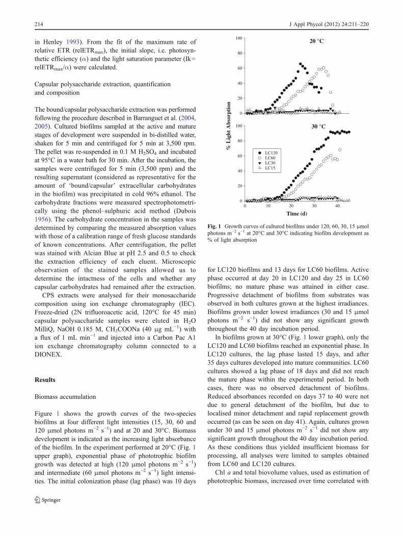
Figure 1 shows the growth curves of the two-speciesbiofilms at four different light intensities (15, 30, 60 and120 μmol photons m−2 s−1) and at 20 and 30°C. Biomassdevelopment is indicated as the increasing light absorbanceof the biofilm. In the experiment performed at 20°C (Fig. 1upper graph), exponential phase of phototrophic biofilmgrowth was detected at high (120 μmol photons m−2 s−1)and intermediate (60 μmol photons m−2 s−1) light intensi-ties. The initial colonization phase (lag phase) was 10 days
for LC120 biofilms and 13 days for LC60 biofilms. Activephase occurred at day 20 in LC120 and day 25 in LC60biofilms; no mature phase was attained in either case.Progressive detachment of biofilms from substrates wasobserved in both cultures grown at the highest irradiances.Biofilms grown under lowest irradiances (30 and 15 μmolphotons m−2 s−1) did not show any significant growththroughout the 40 day incubation period.
In biofilms grown at 30°C (Fig. 1 lower graph), only theLC120 and LC60 biofilms reached an exponential phase. InLC120 cultures, the lag phase lasted 15 days, and after35 days cultures developed into mature communities. LC60cultures showed a lag phase of 18 days and did not reachthe mature phase within the experimental period. In bothcases, there was no observed detachment of biofilms.Reduced absorbances recorded on days 37 to 40 were notdue to general detachment of the biofilm, but due tolocalised minor detachment and rapid replacement growthoccurred (as can be seen on day 41). Again, cultures grownunder 30 and 15 μmol photons m−2 s−1 did not show anysignificant growth throughout the 40 day incubation period.As these conditions thus yielded insufficient biomass forprocessing, all analyses were limited to samples obtainedfrom LC60 and LC120 cultures.
Chl a and total biovolume values, used as estimation ofphototrophic biomass, increased over time correlated with
30 °C
Time (d)
0 10 20 30 400
20
40
60
80
100
20 °C
% L
ight
Abs
orpt
ion
0
20
40
60
80
100
LC120 LC60 LC30 LC15
Fig. 1 Growth curves of cultured biofilms under 120, 60, 30, 15 μmolphotons m−2 s−1 at 20°C and 30°C indicating biofilm development as% of light absorption
214 J Appl Phycol (2012) 24:211–220
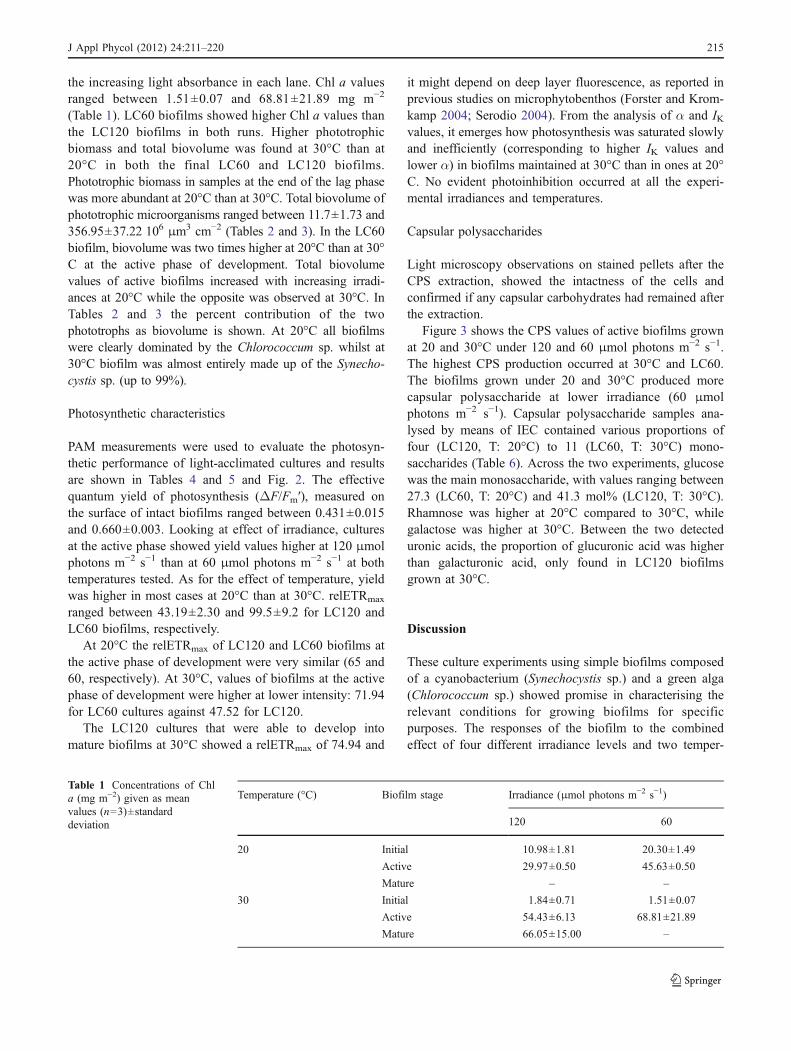
the increasing light absorbance in each lane. Chl a valuesranged between 1.51±0.07 and 68.81±21.89 mg m−2
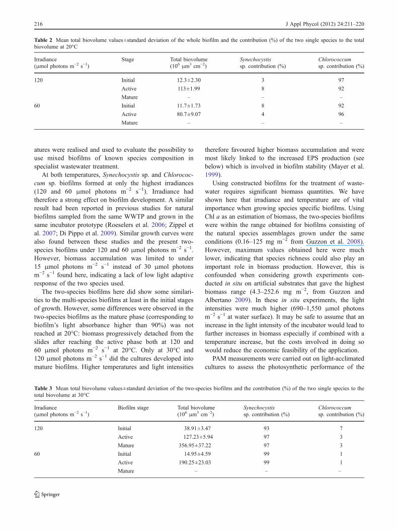
(Table 1). LC60 biofilms showed higher Chl a values thanthe LC120 biofilms in both runs. Higher phototrophicbiomass and total biovolume was found at 30°C than at20°C in both the final LC60 and LC120 biofilms.Phototrophic biomass in samples at the end of the lag phasewas more abundant at 20°C than at 30°C. Total biovolume ofphototrophic microorganisms ranged between 11.7±1.73 and356.95±37.22 106 μm3 cm−2 (Tables 2 and 3). In the LC60biofilm, biovolume was two times higher at 20°C than at 30°C at the active phase of development. Total biovolumevalues of active biofilms increased with increasing irradi-ances at 20°C while the opposite was observed at 30°C. InTables 2 and 3 the percent contribution of the twophototrophs as biovolume is shown. At 20°C all biofilmswere clearly dominated by the Chlorococcum sp. whilst at30°C biofilm was almost entirely made up of the Synecho-cystis sp. (up to 99%).
Photosynthetic characteristics
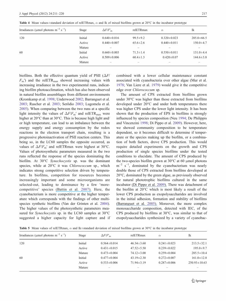
PAM measurements were used to evaluate the photosyn-thetic performance of light-acclimated cultures and resultsare shown in Tables 4 and 5 and Fig. 2. The effectivequantum yield of photosynthesis (ΔF/Fm′), measured onthe surface of intact biofilms ranged between 0.431±0.015and 0.660±0.003. Looking at effect of irradiance, culturesat the active phase showed yield values higher at 120 μmolphotons m−2 s−1 than at 60 μmol photons m−2 s−1 at bothtemperatures tested. As for the effect of temperature, yieldwas higher in most cases at 20°C than at 30°C. relETRmax
ranged between 43.19±2.30 and 99.5±9.2 for LC120 andLC60 biofilms, respectively.
At 20°C the relETRmax of LC120 and LC60 biofilms atthe active phase of development were very similar (65 and60, respectively). At 30°C, values of biofilms at the activephase of development were higher at lower intensity: 71.94for LC60 cultures against 47.52 for LC120.
The LC120 cultures that were able to develop intomature biofilms at 30°C showed a relETRmax of 74.94 and
it might depend on deep layer fluorescence, as reported inprevious studies on microphytobenthos (Forster and Krom-kamp 2004; Serodio 2004). From the analysis of α and IKvalues, it emerges how photosynthesis was saturated slowlyand inefficiently (corresponding to higher IK values andlower α) in biofilms maintained at 30°C than in ones at 20°C. No evident photoinhibition occurred at all the experi-mental irradiances and temperatures.
Capsular polysaccharides
Light microscopy observations on stained pellets after theCPS extraction, showed the intactness of the cells andconfirmed if any capsular carbohydrates had remained afterthe extraction.
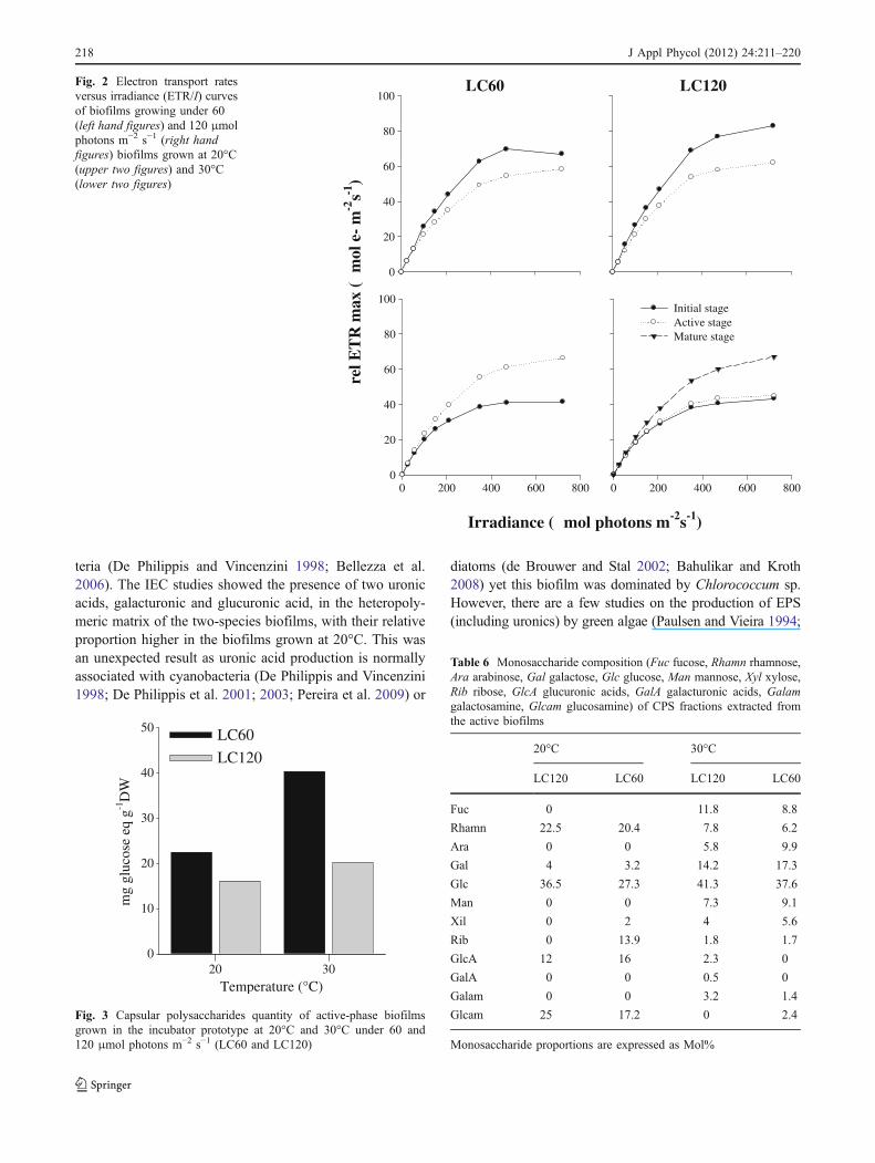
Figure 3 shows the CPS values of active biofilms grownat 20 and 30°C under 120 and 60 μmol photons m−2 s−1.The highest CPS production occurred at 30°C and LC60.The biofilms grown under 20 and 30°C produced morecapsular polysaccharide at lower irradiance (60 μmolphotons m−2 s−1). Capsular polysaccharide samples ana-lysed by means of IEC contained various proportions offour (LC120, T: 20°C) to 11 (LC60, T: 30°C) mono-saccharides (Table 6). Across the two experiments, glucosewas the main monosaccharide, with values ranging between27.3 (LC60, T: 20°C) and 41.3 mol% (LC120, T: 30°C).Rhamnose was higher at 20°C compared to 30°C, whilegalactose was higher at 30°C. Between the two detecteduronic acids, the proportion of glucuronic acid was higherthan galacturonic acid, only found in LC120 biofilmsgrown at 30°C.
Discussion
These culture experiments using simple biofilms composedof a cyanobacterium (Synechocystis sp.) and a green alga(Chlorococcum sp.) showed promise in characterising therelevant conditions for growing biofilms for specificpurposes. The responses of the biofilm to the combinedeffect of four different irradiance levels and two temper-
Temperature (°C) Biofilm stage Irradiance (μmol photons m−2 s−1)
120 60
20 Initial 10.98±1.81 20.30±1.49
Active 29.97±0.50 45.63±0.50
Mature – –
30 Initial 1.84±0.71 1.51±0.07
Active 54.43±6.13 68.81±21.89
Mature 66.05±15.00 –
Table 1 Concentrations of Chla (mg m−2) given as meanvalues (n=3)±standarddeviation
J Appl Phycol (2012) 24:211–220 215
atures were realised and used to evaluate the possibility touse mixed biofilms of known species composition inspecialist wastewater treatment.
At both temperatures, Synechocystis sp. and Chlorococ-cum sp. biofilms formed at only the highest irradiances(120 and 60 μmol photons m−2 s−1). Irradiance hadtherefore a strong effect on biofilm development. A similarresult had been reported in previous studies for naturalbiofilms sampled from the same WWTP and grown in thesame incubator prototype (Roeselers et al. 2006; Zippel etal. 2007; Di Pippo et al. 2009). Similar growth curves werealso found between these studies and the present two-species biofilms under 120 and 60 μmol photons m−2 s−1.However, biomass accumulation was limited to under15 μmol photons m−2 s−1 instead of 30 μmol photonsm−2 s−1 found here, indicating a lack of low light adaptiveresponse of the two species used.
The two-species biofilms here did show some similari-ties to the multi-species biofilms at least in the initial stagesof growth. However, some differences were observed in thetwo-species biofilms as the mature phase (corresponding tobiofilm’s light absorbance higher than 90%) was notreached at 20°C: biomass progressively detached from theslides after reaching the active phase both at 120 and60 μmol photons m−2 s−1 at 20°C. Only at 30°C and120 μmol photons m−2 s−1 did the cultures developed intomature biofilms. Higher temperatures and light intensities
therefore favoured higher biomass accumulation and weremost likely linked to the increased EPS production (seebelow) which is involved in biofilm stability (Mayer et al.1999).
Using constructed biofilms for the treatment of waste-water requires significant biomass quantities. We haveshown here that irradiance and temperature are of vitalimportance when growing species specific biofilms. UsingChl a as an estimation of biomass, the two-species biofilmswere within the range obtained for biofilms consisting ofthe natural species assemblages grown under the sameconditions (0.16–125 mg m−2 from Guzzon et al. 2008).However, maximum values obtained here were muchlower, indicating that species richness could also play animportant role in biomass production. However, this isconfounded when considering growth experiments con-ducted in situ on artificial substrates that gave the highestbiomass range (4.3–252.6 mg m−2, from Guzzon andAlbertano 2009). In these in situ experiments, the lightintensities were much higher (690–1,550 μmol photonsm−2 s−1 at water surface). It may be safe to assume that anincrease in the light intensity of the incubator would lead tofurther increases in biomass especially if combined with atemperature increase, but the costs involved in doing sowould reduce the economic feasibility of the application.
PAM measurements were carried out on light-acclimatedcultures to assess the photosynthetic performance of the
Table 2 Mean total biovolume values±standard deviation of the whole biofilm and the contribution (%) of the two single species to the totalbiovolume at 20°C
Irradiance(μmol photons m−2 s−1)
Stage Total biovolume(106 μm3 cm−2)
Synechocystissp. contribution (%)
Chlorococcumsp. contribution (%)
120 Initial 12.3±2.30 3 97
Active 113±1.99 8 92
Mature – – –
60 Initial 11.7±1.73 8 92
Active 80.7±9.07 4 96
Mature – – –
Table 3 Mean total biovolume values±standard deviation of the two-species biofilms and the contribution (%) of the two single species to thetotal biovolume at 30°C
Irradiance(μmol photons m−2 s−1)
Biofilm stage Total biovolume(106 μm3 cm−2)
Synechocystissp. contribution (%)
Chlorococcumsp. contribution (%)
120 Initial 38.91±3.47 93 7
Active 127.23±5.94 97 3
Mature 356.95±37.22 97 3
60 Initial 14.95±4.59 99 1
Active 190.25±23.03 99 1
Mature – – –
216 J Appl Phycol (2012) 24:211–220
biofilms. Both the effective quantum yield of PSII (ΔF/Fm′) and the relETRmax showed increasing values withincreasing irradiance in the two experimental runs, indicat-ing biofilm photoacclimation, which has also been observedin natural biofilm assemblages from different environments(Kromkamp et al. 1998; Underwood 2002; Barranguet et al.2003; Rascher et al. 2003; Serõdio 2003; Lugomela et al.2005). When comparing between the two runs at a specificlight intensity the values of ΔF/Fm′ and relETRmax werehigher at 20°C than at 30°C. This is because high light and/or high temperature, can lead to an imbalance between theenergy supply and energy consumption by the redoxreactions in the electron transport chain, resulting is aprogressive photoinactivation of PSII reaction centers. Thisbeing so, in the LC60 samples the opposite occurred, asvalues of ΔF/Fm′ and relETRmax were highest at 30°C.Values of photosynthetic parameters measured in the tworuns reflected the response of the species dominating thebiofilm. At 30°C Synechocystis sp. was the dominantspecies, while at 20°C it was Chlorococcum sp., whichindicates strong competitive selection driven by tempera-ture. In biofilms, competition for resources becomesincreasingly important and some microorganisms areselected-out, leading to dominance by a few ‘more-competitive’ species (Battin et al. 2007). Here, thecyanobacterium is more competitive at the higher temper-ature which corresponds with the findings of other multi-species synthetic biofilms (Van der Grinten et al. 2004).The higher values of the photosynthetic parameters mea-sured for Synechocystis sp. in the LC60 samples at 30°Csuggested a higher capacity for light capture and if
combined with a lower cellular maintenance constantassociated with cyanobacteria over other algae (Mur et al.1978; Van Liere et al. 1979) would give it the competitiveedge over Chlorococcum sp.
The amount of CPS extracted from biofilms grownunder 30°C was higher than those extracted from biofilmsdeveloped under 20°C and under both temperatures therewas higher CPS under the lower light intensity. It has beenshown that the production of EPS in biofilms is stronglyinfluenced by species composition (Neu 1994; De Philippisand Vincenzini 1998; Di Pippo et al. 2009). However, here,we showed community composition to be temperaturedependent, so it becomes difficult to determine if temper-ature or the species making up the biofilm, or a combina-tion of both factors, drove CPS production. This wouldrequire detailed experiments on the growth and CPSproduction of single species biofilms under the testedconditions to elucidate. The amount of CPS produced bythe two-species biofilm grown at 30°C at 60 μmol photonsm−2 s−1, dominated by the cyanobacterium was nearlydouble those of CPS extracted from biofilms developed at20°C, dominated by the green algae, as previously observedfor natural phototrophic biofilms cultured in the sameincubator (Di Pippo et al. 2009). There was detachment ofthe biofilm at 20°C which is most likely a result of thelower CPS production as exopolysaccharides are involvedin the initial adhesion, formation and stability of biofilms(Barranguet et al. 2005). Moreover, the more complexmonosaccharide composition, detected with IEC, of theCPS produced by biofilms at 30°C, was similar to that ofexopolysaccharides synthesized by a variety of cyanobac-
Table 4 Mean values±standard deviation of relETRmax, α and Ik of mixed biofilms grown at 20°C in the incubator prototype
Irradiances (μmol photons m−2 s−1) Stage ΔF/F′m relETRmax α Ik
120 Initial 0.640±0.016 99.5±9.2 0.320±0.023 285.0±66.5
Active 0.440±0.007 65.6±2.6 0.440±0.011 150.0±4.7
Mature – – – –
60 Initial 0.660±0.003 71.3±1.4 0.550±0.011 131.0±4.4
Active 0.509±0.006 60.4±1.3 0.420±0.07 144.6±3.8
Mature – – – –
Table 5 Mean values of relETRmax, α and Ik±standard deviation of mixed biofilms grown at 30°C in the incubator prototype
Irradiances (μmol photons m−2 s−1) Stage ΔF/F′m relETRmax α Ik
120 Initial 0.564±0.014 46.34±3.60 0.241±0.023 213.5±32.1
Active 0.431±0.015 47.52±5.50 0.239±0.022 195.0±9.7
Mature 0.473±0.004 74.12±3.00 0.259±0.004 285.5±10.4
60 Initial 0.477±0.004 43.19±2.30 0.272±0.007 161.0±12.8
Active 0.533±0.006 71.94±3.19 0.287±0.006 250.93±10.63
Mature – – – –
J Appl Phycol (2012) 24:211–220 217
teria (De Philippis and Vincenzini 1998; Bellezza et al.2006). The IEC studies showed the presence of two uronicacids, galacturonic and glucuronic acid, in the heteropoly-meric matrix of the two-species biofilms, with their relativeproportion higher in the biofilms grown at 20°C. This wasan unexpected result as uronic acid production is normallyassociated with cyanobacteria (De Philippis and Vincenzini1998; De Philippis et al. 2001; 2003; Pereira et al. 2009) or
diatoms (de Brouwer and Stal 2002; Bahulikar and Kroth2008) yet this biofilm was dominated by Chlorococcum sp.However, there are a few studies on the production of EPS(including uronics) by green algae (Paulsen and Vieira 1994;
LC60
rel E
TR
max
(m
ol e
- m
-2µ
µ
-1)
0
20
40
60
80
100LC120
0 200 400 600 8000
20
40
60
80
100
Irradiance ( mol photons m-2s
s
-1)
0 200 400 600 800
Initial stageActive stageMature stage
Fig. 2 Electron transport ratesversus irradiance (ETR/I) curvesof biofilms growing under 60(left hand figures) and 120 μmolphotons m−2 s−1 (right handfigures) biofilms grown at 20°C(upper two figures) and 30°C(lower two figures)
Temperature (°C)
mg
gluc
ose
eq g
-1D
W
0
10
20
20
30
30
40
50 LC60
LC120
Fig. 3 Capsular polysaccharides quantity of active-phase biofilmsgrown in the incubator prototype at 20°C and 30°C under 60 and120 μmol photons m−2 s−1 (LC60 and LC120)
Table 6 Monosaccharide composition (Fuc fucose, Rhamn rhamnose,Ara arabinose, Gal galactose, Glc glucose, Man mannose, Xyl xylose,Rib ribose, GlcA glucuronic acids, GalA galacturonic acids, Galamgalactosamine, Glcam glucosamine) of CPS fractions extracted fromthe active biofilms
20°C 30°C
LC120 LC60 LC120 LC60
Fuc 0 11.8 8.8
Rhamn 22.5 20.4 7.8 6.2
Ara 0 0 5.8 9.9
Gal 4 3.2 14.2 17.3
Glc 36.5 27.3 41.3 37.6
Man 0 0 7.3 9.1
Xil 0 2 4 5.6
Rib 0 13.9 1.8 1.7
GlcA 12 16 2.3 0
GalA 0 0 0.5 0
Galam 0 0 3.2 1.4
Glcam 25 17.2 0 2.4
Monosaccharide proportions are expressed as Mol%
218 J Appl Phycol (2012) 24:211–220
Lombardi et al. 2002; 2005) and so it seems that thisresult was not an anomaly. If not, then this result suggestsa wider potential for the use of green algae in watertreatment such as for the removal of heavy metals in wastewater treatment.
Although it is difficult to define the precise effect of lightor temperature on biofilm structure and function due tointerrelationships between metabolism and light saturation,it was possible to determine somewhat their synergisticeffect. It was shown that changes in ambient conditionscaused shifts in the species composition and that this had alarge effect on the exopolysaccharide amount and compo-sition. A potential application of this type of study wouldbe in construction of a biofilm with a known compositionand to have some control over the exopolysaccharideproduction in response to a particular need (e.g. heavymetal removal). It was also revealed that the potentiallyuseful properties of green algal CPS should be given moreconsideration as there is very little information available inthe literature to our knowledge.
References
Albertano P, Congestri R, Shubert LE (1999) Cyanobacterialbiofilms in sewage treatment plants along the Tyrrhenian coast(Mediterranean Sea), Italy. Arch Hydrobiol Algol Stud 94:13–24
Bahulikar RA, Kroth PG (2008) The complex extracellular poly-saccharides of mainly chain-forming freshwater diatom speciesfrom epilithic biofilms. J Phycol 44:1465–1475
Barranguet C, van den Ende FP, Rutgers M, Breure AM, GreijdanusM, Sinke JJ, Admiraal W (2003) Copper-induced modificationsof the trophic relations in riverine algal-bacterial biofilms.Environ Toxicol Chem 22:1340–1349
Barranguet C, van den Beusekom AAM, Veuger B, Neu TR, MandersEMM, Sinke JJ, Admiraal W (2004) Studying undisturbedautotrophic biofilms: still a technical challenge. Aquat MicrobEcol 34:1–9
Barranguet C, Veuger B, van Beusekom SAM, Marvan P, Sinke JJ,Admiraal W (2005) Divergent composition of algal-bacterialbiofilms developing under various external factors. Eur J Phycol40:1–8
Battin TJ, Sloan WT, Kjelleberg S, Daims H, Head IM, Curtis TP,Ebert L (2007) Microbial landscapes: new paths to biofilmresearch. Nat Rev Microbiol 5:76–81
Bellezza S, De Philippis R, Paradossi G, Albertano P (2006)Exopolysaccharides of two cyanobacterial strains from Romanhypogea. Geomicrobiol J 23:301–310
Bender J, Phillips P (2004) Microbial mats for multiple applica-tions in aquaculture and bioremediation. Bioresour Technol94:229–238
Chaillan F, Gugger M, Saliot A, Coute A, Oudot J (2006) Role ofcyanobacteria in the biodegradation of crude oil by a tropicalcyanobacterial mat. Chemosphere 62:1574–1582
Cohen Y (2002) Bioremediation of oil by marine microbial mats. IntMicrobiol 5:189–193
Congestri R, Di Pippo F, De Philippis R, Paradossi G, Albertano P(2006) Seasonal succession of phototrophic biofilms in an Italian
wastewater treatment plant: biovolume, spatial structure andexopolysaccharides. Aquat Microb Ecol 45:301–312
de Brouwer J, Stal LJ (2002) Daily fluctuations of exopolymers incultures of the benthic diatoms Cylindrotheca closterium andNitzschia sp. (Bacillariophyceae). J Phycol 38:464–472
De Philippis R, Vincenzini M (1998) Exocellular polysaccharidesfrom cyanobacteria and their possible applications. FEMSMicrobiol Rev 22:151–175
De Philippis R, Sili C, Paperi R, Vincenzini M (2001)Exopolysaccharide-producing cyanobacteria and their possibleexploitation: a review. J Appl Phycol 13:293–299
De Philippis R, Paperi R, Sili C, Vincenzini M (2003) Assessment ofthe metal removal capability of two capsulated cyanobacteria,Cyanospira capsulata and Nostoc PCC7936. J Appl Phycol15:155–161
Decho AW (2000) Microbial biofilms in intertidal systems: anoverview. Cont Shelf Res 20:1257–1273
Di Pippo F, Bohn A, Congestri R, de Philippis R, Albertano P (2009)Capsular polysaccharides of culture phototrophic biofilms.Biofouling 25:495–504
Dubois M (1956) Colorimetric method for determination of sugars andrelated substances. Anal Chem 28:350–356
Flemming HC, Neu TR, Wozniak DJ (2007) The EPS matrix: the“house of biofilm cells”. J Bacteriol 189:7945–7947
Forster RM, Kromkamp JC (2004) Modelling the effects of chlorop-hill fluorescence from subsurface layers on photosyntheticefficiency measurement in microphytobenthic algae. Mar EcolProg Ser 284:9–22
Genty B, Briantais JM, Baker NR (1989) The relationship betweenthe quantum yield of photosynthetic electron and quenchingof chlorophyll fluorescence. Biochem Biophys Acta 990:87–92
Gonzalez LE, Bashan Y (2000) Growth promotion of the microalgaeChlorella vulgaris when coimmobilized and cocultured inalginate beads with the plant growth-promoting bacteria Azospir-illum brasilense. Appl Environ Microbiol 66:1537–1541
Graham LE, Wilcox LW (2000) Algae. Prentice Hall, New Jersey, p640
Guzzon A, Albertano P (2009) Annual response of phototrophicbiofilms from an Italian wastewater treatment plant. ArchHydrobiol Algol Stud 131:87–102
Guzzon A, Congestri R, Albertano P (2005) Light-induced changes inphotosynthesis and structure of cyanobacterial cultured biofilmsfrom an Italian wastewater treatment plant. Arch Hydrobiol AlgolStud 117:223–238
Guzzon A, Bohn A, Diociaiuti M, Albertano P (2008) Culturedphototrophic biofilms for phosphorus removal in wastewatertreatment. Water Res 42:4357–4367
Henley WJ (1993) Measurement and interpretation of photosyntheticlight response curves in algae in the context of photoinhibitionand diel changes. J Phycol 29:729–739
Herlory O, Richard P, Blanchard GF (2007) Methodology of lightresponse curves: application of chlorophyll fluorescence tomicrophytobenthic biofilms. Mar Biol 153:91–101
Hill WR (1996) Effects of light. In: Stevenson RJ, Bothwell ML,Lowe RL (eds) Algal ecology—freshwater benthic ecosystems.Academic, USA, pp 121–148
Hillebrand H, Dürselen CD, Kirschtel D, Pollingher U, Zohary T(1999) Biovolume calculation for pelagic and benthic micro-algae. J Phycol 35:403–424
Hoffmann JP (1998) Wastewater treatment with suspended andnonsuspended algae. J Phycol 34:757–763
Jeffrey SW, Humphrey GF (1975) New spectrophotometric equationsfor determining chlorophylls a, b, c1 and c2 in higher plants,algae, and natural phytoplankton. Biochem Physiol Pflanzen167:191–194
J Appl Phycol (2012) 24:211–220 219

Kromkamp JC, Barranguet C, Peene J (1998) Determination ofmicrophytobenthos PSII quantum efficiency activity by meansof variable chlorophyll fluorescence. Mar Ecol Prog Ser 162:45–55
Lombardi AT, Vieira AAH, Sartori LA (2002) Mucilaginous capsuleadsorption and intracellular uptake of copper by Kirchneriellaaperta (Chlorococcales). J Phycol 38:332–337
Lombardi AT, Hidalgo TMR, Vieira AAH (2005) Copper complexingproperties of dissolved organic materials exuded by the freshwa-ter microalgae Scenedesmus acuminatus (Chlorophyceae). Che-mosphere 60:453–459
Lugomela C, Soderback E, Bjork M (2005) Photoacclimation ofphotosynthesis irradiance response curves and photosyntheticpigments in microalgae and cyanobacteria. J Phycol 38:17–38
Mayer C,Moritz R, Kirschner C, BorchardW,Maibaum R,Wingender J,Flemming HC (1999) The role of intermolecular interactions:studies onmodel systems for bacterial biofilms. Int J Biol Macromol26:3–16
Moreno J, Vargas MA, Olivares H, Rivas J, Guerrero GM (1998)Exopolysaccharide production by the cyanobacterium Anabaenasp. ATCC 33047 in batch and continuous culture. J Biotechnol60:175–182
Mur LR, Gons HJ, van Liere L (1978) Competition of the green algaScenedesmus and the blue-green alga Oscillatoria in light limitedenvironments. FEMS Microbiol Lett 1:335–338
Neu TR (1994) Biofilms and microbial mats. In: Krumbein WE,Paterson DM, Stal LJ (eds) Biostabilization of sediments. BIS,Oldenburg, pp 9–15
Otero A, Vincenzini M (2003) Extracellular polysaccharide synthesisby Nostoc strains as affected by N source and light intensity. JBiotechnol 102:143–152
Paulsen BS, Vieira AAH (1994) Structure of the capsular andextracellular polysaccharides produced by the desmid Spondylo-sium panduriforme (Chlorophyta). J Phycol 30:638–641
Pereira S, Zille A, Micheletti E, Moradas-Ferreira P, De Philippis R,Tamagnini P (2009) Complexity of cyanobacterial exopolysac-charides: composition, structures, inducing factors and putativegenes involved in their biosynthesis and assembly. FEMSMicrobiol Rev 33:917–941
Perkins RG, Mouget JL, Lefebvre S, Lavaud J (2006) Light responsecurve methodology and possible implications in the applicationof chlorophyll fluorescence to benthic diatoms. Mar Biol149:703–712
Rascher U, Lakatos M, Büdel B, Lüttge U (2003) Photosynthetic fieldcapacity of cyanobacteria of a tropical inselberg of the GuaianaHighlands. Eur J Phycol 38:247–256
Roeselers G, Zippel B, Staal M, van Loosdrecht MCM, Muyzer G(2006) On the reproducibility of microcosm experiments-different community composition in parallel phototrophic biofilmmicrocosms. FEMS Microbiol Ecol 58:169–178
Roeselers G, van Loosdrecht MCM, Muyzer G (2008) Phototrophicbiofilms and their potential applications. J Appl Phycol 20:227–235
Sabater S, Guasch H, Romaní A, Muñoz I (2002) The effect ofbiological factors on the efficiency of river biofilms in improvingwater quality. Hydrobiologia 469:149–156
Schumacher G, Blume T, Asekuolov I (2003) Bacteria reduction andnutrient removal in small wastewater treatment plants by an algalbiofilm. Water Sci Technol 47:195–202
Serodio J (2004) Analysis of variable chlorophyll fluorescence inmicrophytobenthos assemblages: implications of the use ofdepth-integrated measurements. Aquat Microb Ecol 36:137–152
Serõdio J (2003) A chlorophyll fluorescence index to estimate short-term rates of photosynthesis by intertidal microphytobenthos. JPhycol 39:33–46
Sládecková A (1994) The role of periphyton in waste treatmenttechnology. Int Ver Theor Angew Limnol Verh 25:1929–1932
Sládečková A, Marvan P, Vymazal J (1983) The utilization ofperiphyton in waterworks pre-treatment for nutrient removalfrom enriched influents. In: Wetzel RG (ed) Periphyton offreshwater ecosystems. Junk, The Hague, pp 299–303
Staats N, Stal LJ, de Winder B, Mur LR (2000) Oxygenicphotosynthesis as a driving process in exopolysaccharideproduction in benthic diatoms. Mar Ecol Prog Ser 193:261–269
Stal LJ, Defarge C (2005) Structure and dynamics of exopolymers inan intertidal diatom biofilm. Geomicrobiol J 22:341–352
Stanier RY, Kunisawa R, Mandel M, Cohen-Bazire G (1971)Purification and unicellular blue-green algae (Order Chroococ-cales). Bacteriol Rev 35:171–205
Stevenson RJ (1996) An introduction to algal ecology in freshwaterbenthic habitats. In: Stevenson RJ, Bothwell ML, Lowe RL (eds)Algal ecology—freshwater benthic ecosystems. Academic, NY,pp 321–340
Underwood GJC (2002) Adaptations of tropical marine microphyto-benthic assemblages along a gradient of light and nutrientavailability in Suva Lagoon, Fiji. Eur J Phycol 37:449–462
van der Grinten E, Janssen M, Simis SGH, Barranguet C, Admiral W(2004) Phosphate regime structure species composition incultured phototrophic biofilms. Freshw Biol 49:369–381
van Liere L, Mur LR, Gibson CE, Herdman M (1979) Growth andphysiology of Oscillatoria agardhii and some related species, asurvey. Dev Hydrobiol 2:67–77
Wolfstein K, Stal LJ (2002) Production of extracellular polymericsubstances (EPS) by benthic diatoms: effect of irradiance andtemperature. Mar Ecol Prog Ser 236:13–22
Zippel B, Neu TR (2005) Growth and structure of phototrophicbiofilms under controlled light conditions. Water Sci Technol52:203–209
Zippel B, Rijstenbil J, Neu TR (2007) A flow-lane incubator forstudying freshwater and marine phototrophic biofilms. J Micro-biol Meth 70:336–345
220 J Appl Phycol (2012) 24:211–220