The Arabidopsis At1g45130 and At3g52840 genes encode β-galactosidases with activity toward cell...
10
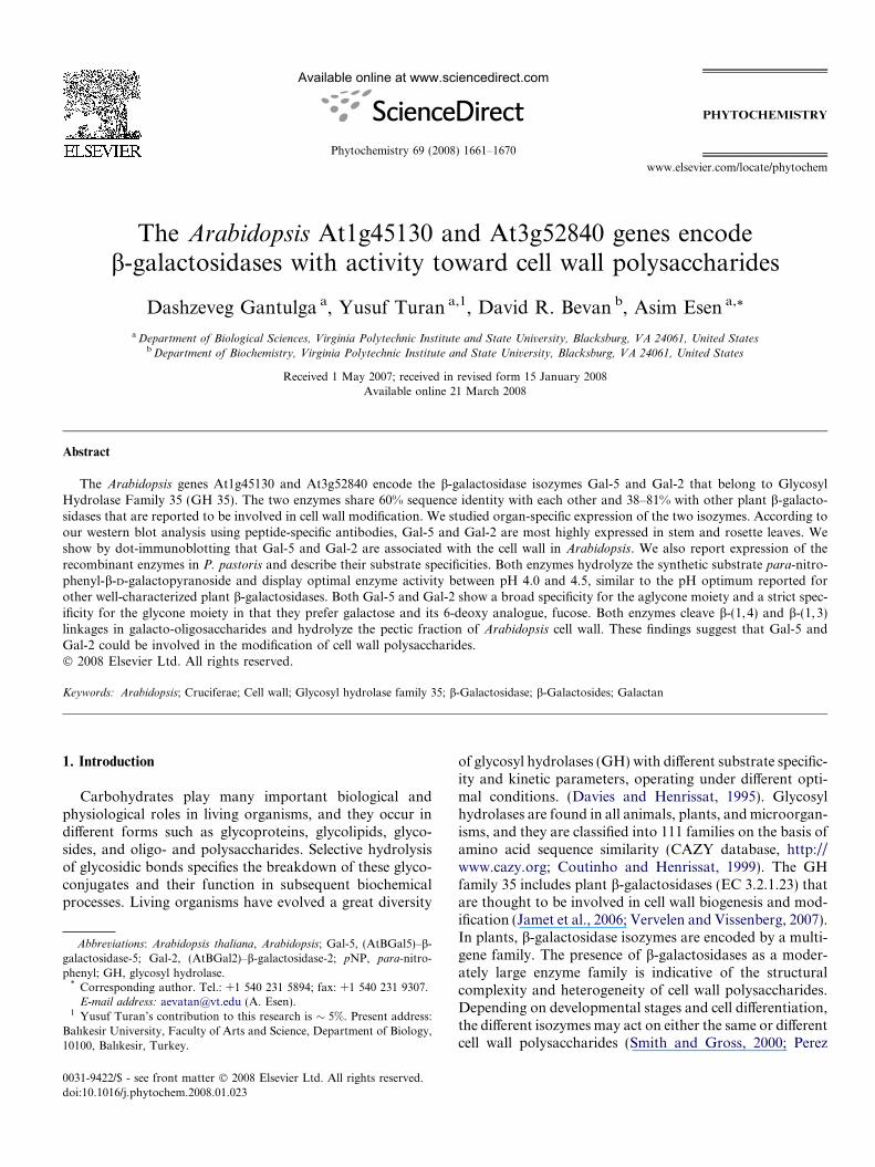
The Arabidopsis At1g45130 and At3g52840 genes encode b-galactosidases with activity toward cell wall polysaccharides Dashzeveg Gantulga a , Yusuf Turan a,1 , David R. Bevan b , Asim Esen a, * a Department of Biological Sciences, Virginia Polytechnic Institute and State University, Blacksburg, VA 24061, United States b Department of Biochemistry, Virginia Polytechnic Institute and State University, Blacksburg, VA 24061, United States Received 1 May 2007; received in revised form 15 January 2008 Available online 21 March 2008 Abstract The Arabidopsis genes At1g45130 and At3g52840 encode the b-galactosidase isozymes Gal-5 and Gal-2 that belong to Glycosyl Hydrolase Family 35 (GH 35). The two enzymes share 60% sequence identity with each other and 38–81% with other plant b-galacto- sidases that are reported to be involved in cell wall modification. We studied organ-specific expression of the two isozymes. According to our western blot analysis using peptide-specific antibodies, Gal-5 and Gal-2 are most highly expressed in stem and rosette leaves. We show by dot-immunoblotting that Gal-5 and Gal-2 are associated with the cell wall in Arabidopsis. We also report expression of the recombinant enzymes in P. pastoris and describe their substrate specificities. Both enzymes hydrolyze the synthetic substrate para-nitro- phenyl-b-D-galactopyranoside and display optimal enzyme activity between pH 4.0 and 4.5, similar to the pH optimum reported for other well-characterized plant b-galactosidases. Both Gal-5 and Gal-2 show a broad specificity for the aglycone moiety and a strict spec- ificity for the glycone moiety in that they prefer galactose and its 6-deoxy analogue, fucose. Both enzymes cleave b-(1, 4) and b-(1, 3) linkages in galacto-oligosaccharides and hydrolyze the pectic fraction of Arabidopsis cell wall. These findings suggest that Gal-5 and Gal-2 could be involved in the modification of cell wall polysaccharides. Ó 2008 Elsevier Ltd. All rights reserved. Keywords: Arabidopsis; Cruciferae; Cell wall; Glycosyl hydrolase family 35; b-Galactosidase; b-Galactosides; Galactan 1. Introduction Carbohydrates play many important biological and physiological roles in living organisms, and they occur in different forms such as glycoproteins, glycolipids, glyco- sides, and oligo- and polysaccharides. Selective hydrolysis of glycosidic bonds specifies the breakdown of these glyco- conjugates and their function in subsequent biochemical processes. Living organisms have evolved a great diversity of glycosyl hydrolases (GH) with different substrate specific- ity and kinetic parameters, operating under different opti- mal conditions. (Davies and Henrissat, 1995). Glycosyl hydrolases are found in all animals, plants, and microorgan- isms, and they are classified into 111 families on the basis of amino acid sequence similarity (CAZY database, http:// www.cazy.org; Coutinho and Henrissat, 1999). The GH family 35 includes plant b-galactosidases (EC 3.2.1.23) that are thought to be involved in cell wall biogenesis and mod- ification (Jamet et al., 2006; Vervelen and Vissenberg, 2007). In plants, b-galactosidase isozymes are encoded by a multi- gene family. The presence of b-galactosidases as a moder- ately large enzyme family is indicative of the structural complexity and heterogeneity of cell wall polysaccharides. Depending on developmental stages and cell differentiation, the different isozymes may act on either the same or different cell wall polysaccharides (Smith and Gross, 2000; Perez 0031-9422/$ - see front matter Ó 2008 Elsevier Ltd. All rights reserved. doi:10.1016/j.phytochem.2008.01.023 Abbreviations: Arabidopsis thaliana, Arabidopsis; Gal-5, (AtBGal5)–b- galactosidase-5; Gal-2, (AtBGal2)–b-galactosidase-2; pNP, para-nitro- phenyl; GH, glycosyl hydrolase. * Corresponding author. Tel.: +1 540 231 5894; fax: +1 540 231 9307. E-mail address: [email protected] (A. Esen). 1 Yusuf Turan’s contribution to this research is 5%. Present address: Balıkesir University, Faculty of Arts and Science, Department of Biology, 10100, Balıkesir, Turkey. www.elsevier.com/locate/phytochem Available online at www.sciencedirect.com Phytochemistry 69 (2008) 1661–1670 PHYTOCHEMISTRY
-
Upload
independent -
Category
Documents
-
view
3 -
download
0
Transcript of The Arabidopsis At1g45130 and At3g52840 genes encode β-galactosidases with activity toward cell...
Available online at www.sciencedirect.com
www.elsevier.com/locate/phytochem
Phytochemistry 69 (2008) 1661–1670
PHYTOCHEMISTRY
The Arabidopsis At1g45130 and At3g52840 genes encodeb-galactosidases with activity toward cell wall polysaccharides
Dashzeveg Gantulga a, Yusuf Turan a,1, David R. Bevan b, Asim Esen a,*
a Department of Biological Sciences, Virginia Polytechnic Institute and State University, Blacksburg, VA 24061, United Statesb Department of Biochemistry, Virginia Polytechnic Institute and State University, Blacksburg, VA 24061, United States
Received 1 May 2007; received in revised form 15 January 2008Available online 21 March 2008
Abstract
The Arabidopsis genes At1g45130 and At3g52840 encode the b-galactosidase isozymes Gal-5 and Gal-2 that belong to GlycosylHydrolase Family 35 (GH 35). The two enzymes share 60% sequence identity with each other and 38–81% with other plant b-galacto-sidases that are reported to be involved in cell wall modification. We studied organ-specific expression of the two isozymes. According toour western blot analysis using peptide-specific antibodies, Gal-5 and Gal-2 are most highly expressed in stem and rosette leaves. Weshow by dot-immunoblotting that Gal-5 and Gal-2 are associated with the cell wall in Arabidopsis. We also report expression of therecombinant enzymes in P. pastoris and describe their substrate specificities. Both enzymes hydrolyze the synthetic substrate para-nitro-phenyl-b-D-galactopyranoside and display optimal enzyme activity between pH 4.0 and 4.5, similar to the pH optimum reported forother well-characterized plant b-galactosidases. Both Gal-5 and Gal-2 show a broad specificity for the aglycone moiety and a strict spec-ificity for the glycone moiety in that they prefer galactose and its 6-deoxy analogue, fucose. Both enzymes cleave b-(1,4) and b-(1,3)linkages in galacto-oligosaccharides and hydrolyze the pectic fraction of Arabidopsis cell wall. These findings suggest that Gal-5 andGal-2 could be involved in the modification of cell wall polysaccharides.� 2008 Elsevier Ltd. All rights reserved.
Keywords: Arabidopsis; Cruciferae; Cell wall; Glycosyl hydrolase family 35; b-Galactosidase; b-Galactosides; Galactan
1. Introduction
Carbohydrates play many important biological andphysiological roles in living organisms, and they occur indifferent forms such as glycoproteins, glycolipids, glyco-sides, and oligo- and polysaccharides. Selective hydrolysisof glycosidic bonds specifies the breakdown of these glyco-conjugates and their function in subsequent biochemicalprocesses. Living organisms have evolved a great diversity
0031-9422/$ - see front matter � 2008 Elsevier Ltd. All rights reserved.
doi:10.1016/j.phytochem.2008.01.023
Abbreviations: Arabidopsis thaliana, Arabidopsis; Gal-5, (AtBGal5)–b-galactosidase-5; Gal-2, (AtBGal2)–b-galactosidase-2; pNP, para-nitro-phenyl; GH, glycosyl hydrolase.
* Corresponding author. Tel.: +1 540 231 5894; fax: +1 540 231 9307.E-mail address: [email protected] (A. Esen).
1 Yusuf Turan’s contribution to this research is � 5%. Present address:Balıkesir University, Faculty of Arts and Science, Department of Biology,10100, Balıkesir, Turkey.
of glycosyl hydrolases (GH) with different substrate specific-ity and kinetic parameters, operating under different opti-mal conditions. (Davies and Henrissat, 1995). Glycosylhydrolases are found in all animals, plants, and microorgan-isms, and they are classified into 111 families on the basis ofamino acid sequence similarity (CAZY database, http://www.cazy.org; Coutinho and Henrissat, 1999). The GHfamily 35 includes plant b-galactosidases (EC 3.2.1.23) thatare thought to be involved in cell wall biogenesis and mod-ification (Jamet et al., 2006; Vervelen and Vissenberg, 2007).In plants, b-galactosidase isozymes are encoded by a multi-gene family. The presence of b-galactosidases as a moder-ately large enzyme family is indicative of the structuralcomplexity and heterogeneity of cell wall polysaccharides.Depending on developmental stages and cell differentiation,the different isozymes may act on either the same or differentcell wall polysaccharides (Smith and Gross, 2000; Perez
1662 D. Gantulga et al. / Phytochemistry 69 (2008) 1661–1670
Almeida, 2004; Kotake et al., 2005; Ishimaru et al., 2005;Jamet et al., 2006; Chantarangsee et al., 2007).
A growing body of evidence suggests that GH Family 35b-galactosidases participate in loosening of the cell wallduring growth and degrade cell wall components during rip-ening of fruits and senescence. Recently, many b-galactosi-dases have been identified and purified from different plantsources. For example, Smith and Gross (2000) showed thata family of at least seven b-galactosidases is expressed dur-ing tomato fruit development. Furthermore, these authorsexpressed the tomato b-galactosidase isozyme 4 (TBG4) inyeast and studied its natural substrate specificity. TBG4hydrolyzed chelator-soluble pectin, alkali-soluble pectin,hemicellulose from tomato cell walls, and commercially pre-pared galactan. Li et al. (2001) purified five isoforms of b-galactosidases from mung bean and showed that they differwith respect to enzymatic characteristics and substrate spec-ificities. Kotake et al. (2005) isolated a b-galactosidase fromradish (RsbGal), which specifically hydrolyzed b-(1,6) andb-(1,3) linkages in arabinogalactan protein. b-Galactosi-dase I purified from ripe carambola (Averrhoa carambola)was active in hydrolyzing b-(1,4)-linked spruce and b-(1,6)/b-(1,3)-linked gum arabic galactan, and alkali-solublehemicelluloses of carambola cell wall (Balasubramaniamet al., 2005). Furthermore, papaya b-galactosidase isoformsdifferentially hydrolyzed cell wall during fruit ripening(Lazan et al., 2004).
Despite the status of Arabidopsis thaliana as a modelplant with a completely sequenced genome, not much isknown about its b-galactosidases. There are 17 putativeGH family 35 b-galactosidases (17 BGALs) in Arabidopsis
(Ahn et al., 2007). According to a recent microarray study,the b-galactosidase genes are among 765 genes specific tocell wall dynamics (Imoto et al., 2005). Perez Almeida(2004) studied expression of b-galactosidases by pro-moter-GUS fusion and found that temporal and spatialexpression of b-galactosidase genes are differentially regu-lated. She also reported that the galactosyl content of thecell wall increases and total b-galactosidase activitydecreases in Gal-1 knockout mutant. Iglesias et al. (2006)reported b-galactosidase activity on xyloglucan oligosac-charides in the apoplastic fluid. To corroborate furtherthe role of b-galactosidases in cell wall modification, it isessential to determine their substrate specificity, by isolat-ing and characterizing the proteins. To this end, Ahnet al. (2007) expressed Gal-4 (At5g56870), one of the seven-teen b-galactosidases, in Escherichia coli and insect cells. Itwas shown that in addition to synthetic substrates, Gal-4hydrolyzes b-(1, 3) and b-(1,4) linked galacto-oligosaccha-rides (Ahn et al., 2007).
It is likely that the 17 BGALs differ in temporal and spa-tial expression and in their ability to hydrolyze various cellwall polysaccharides. For annotating them and under-standing their function better, it is desirable to determinethe expression profile and substrate specificity of all 17BGALs. Currently, we are focused on the largest subfamily(Gal-1 through Gal-5 and Gal-12), which consists of six
genes, amongst the 17 BGALs. Of these, we studied Gal-5 and Gal-2 for this paper. First, we conducted databaseanalyses using microarray data available for these genes.Second, we studied spatial expression and subcellular local-ization by immunoblotting using peptide-specific antibod-ies. Third, we purified the recombinant proteins expressedin Pichia pastoris and investigated their substrate specificityusing synthetic galactosides and natural polysaccharidesderived from Arabidopsis cell wall.
2. Results and discussion
2.1. In silico analysis of Gal-5 and Gal-2
BLAST searches of the NCBI database using Gal-5 andGal-2 sequences as queries show that the sequences of 20other plant b-galactosidases share 38–81% identity withall Arabidopsis b-galactosidases except Gal-17. Gal-5 andGal-2 proteins share 60% sequence identity with eachother. The sequence differences among b-galactosidasessuggest differences in their enzymatic properties (e.g., sub-strate specificity) and biological functions.
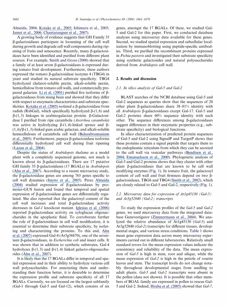
In silico characterization of predicted protein sequencesof Gal-5 and Gal-2 using SignalP and TargetP shows thatthese proteins contain a signal peptide that targets them tothe endoplasmic reticulum from which they can be secretedto the cell wall via vesicular pathways (Bendtsen et al.,2004; Emanuelsson et al., 2000). Phylogenetic analysis ofGal-5 and Gal-2 proteins shows that they cluster with otherplant b-galactosidases that are known to be cell wallmodifying enzymes (Fig. 1). In tomato fruit, the galactosylcontent of cell wall and fruit firmness depend on two b-galactosidases, TBG6 and TBG4 (Smith et al., 2002), whichare closely related to Gal-5 and Gal-2, respectively (Fig. 1).
2.2. Microarray data for expression of At1g45130 (Gal-5)
and At3g52840 (Gal-2) transcripts
To study the expression profiles of the Gal-5 and Gal-2
genes, we used microarray data from the integrated data-base Genevestigator (Zimmermann et al., 2004). We ana-lyzed the relative abundance of At1g45130 (Gal-5) andAt3g52840 (Gal-2) transcripts for different tissues, develop-mental stages, and various stress conditions. Table 1 showsmean gene expression data across many microarray exper-iments carried out in different laboratories. Relatively smallstandard errors for the mean expression values indicate theconsistency and reliability of the data. The mean expres-sion of Gal-5 is high in stem, root and silique, while themean expression of Gal-2 is high in the petiole of rosetteleaves and stem. The transcript levels do not change nota-bly throughout developmental stages from seedling toadult plants. Gal-5 and Gal-2 transcripts were absent inthe pollen (data not shown). It is possible that other mem-bers of BGAL family are expressed in pollen to rescue Gal-5 and Gal-2. Indeed, Hruba et al. (2005) showed that Gal-7,
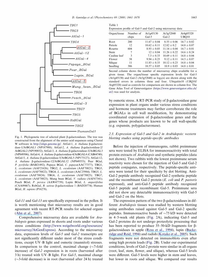
Fig. 1. Phylogenetic tree of selected plant b-galactosidases. The tree wasconstructed from the alignment of the amino acid sequences using ClustalW software in http://align.genome.jp/. AtGal-1, A. thaliana b-galactosi-dase-1/AtBGAL1 (NP187988); AtGal-2, A. thaliana b-galactosidase-2/AtBGAL2 (NP190852); AtGal-3, A. thaliana b-galactosidase-3/AtBGAL3(NP849506); AtGal-4, A. thaliana b-galactosidase-4/AtBGAL4 (CAB64740);AtGal-5, A. thaliana b-galactosidase-5/AtBGAL5 (NP175127); AtGal-12,A. thaliana b-galactosidase-12/AtBGAL12 (NP849553); Pear BGal,P. pyrifolia (BAB21492); Papaya BGal, C. papaya(AAC77377); TBG1,L. esculentum (AAF21626); TBG2, L. esculentum (AAF70821); TBG3,L. esculentum (AAF70822); TBG4, L. esculentum (AAC25984); TBG5, L.
esculentum (AAF70824); TBG6, L. esculentum (AAF70825); TBG7,L. esculentum (AAF70823); Mung bean BGal, V. radiata (AAF67341);Peach BGal, P. persica (AAW47739); Lupin BGal, L. angustifolius
(CAA09467); RsbGal, R. sativus b-galactosidase 1 (BAD20774); HumanBGal, H. sapiens (P16278).
Table 1Expression profile of Gal-5 and Gal-2 using microarray data
Organ/tissue Number ofchips
At1g45130Gal-5
At3g52840Gal-2
At4g05320UBQ10
Root 200 11.67 ± 0.04 8.55 ± 0.06 14.7 ± 0.02Petiole 12 10.62 ± 0.11 12.82 ± 0.2 14.8 ± 0.07Rosette 604 8.93 ± 0.05 11.14 ± 0.04 14.7 ± 0.01Stem 7 12 ± 0.04 11.26 ± 0.22 14.6 ± 0.24Cauline leaf 3 7.3 ± 0.33 10.69 ± 0.11 14.8 ± 0.04Flower 58 9.94 ± 0.25 9.12 ± 0.11 14.5 ± 0.07Silique 11 11.83 ± 0.33 10.12 ± 0.23 14.8 ± 0.04Seedling 386 10.57 ± 0.05 10.9 ± 0.03 14.8 ± 0.01
Second column shows the number of microarray chips available for agiven tissue. The organ/tissue specific expression levels for Gal-5(At1g45130) and Gal-2 (At3g52840) as log2(n) are shown along with thestandard errors in columns three and four. Ubiquitin10 (UBQ10/Atg05320) used as controls for comparison are shown in column five. TheGene Atlas Tool of Genevestigator (https://www.genevestigator.ethz.ch/at/) was used for analysis.
D. Gantulga et al. / Phytochemistry 69 (2008) 1661–1670 1663
Gal-11 and Gal-13 are specifically expressed in the pollen. Itis worth mentioning that microarray results are in goodagreement with recent RT-PCR results for selected organs(Ahn et al., 2007).
Comprehensive microarray data are available for Ara-bidopsis genes expressed in shoots and roots under variousstress conditions (http://www.weigelworld.org/resources/microarray/AtGenExpress). According to the microarraydata, expression levels of Gal-5 and Gal-2 transcripts arenot significantly different under most of the stress condi-tions, except UV–B light and osmotic (mannitol) stresses.In comparison to the control, maximal change (�7-foldincrease) of Gal-2 expression is in roots (harvested after3 h) treated with UV–B light. For Gal-5, maximal change(�3-fold decrease) is in root (harvested after 24 h) treated
by osmotic stress. A RT-PCR study of b-galactosidase geneexpression in plant organs under various stress conditionsand hormone treatments may further corroborate the roleof BGALs in cell wall modification, by demonstratingcoordinated expression of b-galactosidase genes and thegenes whose products are known to be cell wall-specific(e.g. expansin, polygalacturonase).
2.3. Expression of Gal-5 and Gal-2 in Arabidopsis: western
blotting studies using peptide-specific antibodies
Before the injection of immunogens, rabbit preimmunesera were tested by ELISA for immunoreactivity with totalprotein extracts of Arabidopsis and synthetic peptides (datanot shown). Two rabbits with the lowest preimmune serumreactivity were chosen for the injection of Gal-5 and Gal-2peptide conjugates, respectively. The peptide-specific anti-sera were tested for their specificity by dot blotting. Anti-Gal-2 peptide antibody recognized Gal-2 synthetic peptideand the recombinant Gal-2 protein (E. coli and P. pastoris
expressed), and anti-Gal-5 peptide antibody recognizedGal-5 peptide and recombinant Gal-5. Preimmune seradid not show any detectable immunoreactivity with Gal-5and Gal-2 on the blots.
The expression pattern of the two b-galactosidases in dif-ferent Arabidopsis tissues was studied by western blottingusing antibodies raised against Gal-5 and Gal-2 specificpeptides. Immunoreactive bands of �75 kD were detectedin 4–5-week old plants (Fig. 2A), indicating Gal-5 andGal-2 proteins do not undergo proteolytic processing thathas been reported to produce 35–50-kD fragments for b-galactosidases in apple (Ross et al., 1994), lupin (Bucke-ridge and Reid, 1994) and radish (Kotake et al., 2005). Suchfragments were not detected on immunoblots even afterusing high protein loads (Fig. 2B). Under our experimentalconditions, levels of Gal-2 protein were similar in all organs(root, leaf, stem, flower, and silique), whereas Gal-5 levelswere different. Gal-5 levels were higher in stem and leaves,but lower in roots and silique. We compared our results

Fig. 2. Organ-specific expression of Gal-5 and Gal-2. (A) Western blotand (B) Coomassie Blue stained SDS–PAGE. Total proteins (30 lg) fromArabidopsis (4 weeks old) root, petiole of rosette leaves, rosette leaves,stems, cauline leaves, flowers, and siliques were separated by 10% SDS–PAGE and transferred to nitrocellulose membranes. Identical membraneswere incubated with rabbit preimmune sera and immune antisera againstGal-5 and Gal-2 peptides. The arrow marks the position of immunore-active bands.
Fig. 3. Localization of Gal-5 and Gal-2 in cell wall fractions by dotblotting. Cell walls were isolated from Arabidopsis rosette leaves. Threefractions (S1, soluble; CW4, CaCl2-soluble; CW5, LiCl-soluble) with b-galactosidase activity were spotted on nitrocellulose strips at the sameplace multiple times. Identical membranes with protein spots wereincubated with anti-whole Gal-2, anti-Gal-5 peptide-, and anti-Gal-2peptide-specific antisera. Total protein spots were stained with CoomassieBlue R-250. These dot-immunoblotting data show the presence of Gal-5and Gal-2 in the cell wall.
1664 D. Gantulga et al. / Phytochemistry 69 (2008) 1661–1670
with the previous studies on expression of b-galactosidasegenes by Perez Almeida (2004) and Ahn et al. (2007). Theseauthors found Gal-2 transcripts with moderate levels ofexpression in all organs, which is in agreement with ourresult for Gal-2. In the case of Gal-5, they found higher levelof expression in roots than we did. We propose that this dif-ference in Gal-5 expression is due to the difference in tran-scriptional and translational stages of regulation, thoughwe do not exclude other factors, such as plant age, growthconditions, and extraction methods used in the experiments.It is worth mentioning that there is agreement between ourdata and those of Perez Almeida (2004) in that Gal-5 is notdetectable in mature roots, although Perez Almeida (2004)found Gal-5 expression in root elongation and root hairzones of juvenile plants.
2.4. Cell wall localization of Gal-5 and Gal-2 proteins: dot
blotting
We isolated cell wall from rosette leaves of Arabidopsis toconfirm the presence of Gal-5 and Gal-2 proteins in the cellwall. Five different fractions (S1, soluble 1; S2, soluble 2; S3,soluble 3; CW4, extractable with CaCl2; and CW5, extract-able with LiCl) were obtained. These fractions were assayedfor b-galactosidase activity using pNPGal as a substrate. S2and S3 fractions did not have detectable activity. Specificactivities of S1, CW4, and CW5 were 0.06, 0.12, and20.1 nmole pNP/min/mg, respectively, indicating that frac-
tion CW5 (LiCl-soluble) had the highest specific b-galacto-sidase activity. Fraction CW5 contained the lowest amountof protein (Fig. 3, bottom row) among the three fractions.The immunoblotting data showed that antiserum to intactGal-2 protein had high immunoreactivity (Fig. 3, top blot)with fraction CW4 (CaCl2-soluble) and weak immunoreac-tivity with fractions S1 and CW5. However, this antiserumis not specific for Gal-2; it recognizes also other cell wall-bound b-galactosidases. In contrast, the reactivity of theGal-2 peptide-specific antiserum was strongest with fractionCW5 (Fig. 3, third blot), indicating that Gal-2 is enriched inCW5 and it requires LiCl for complete release from the cellwall. In the case of Gal-5, the preimmune serum from therabbit immunized with Gal-5 peptide had considerablebackground activity with cell wall components (Fig. 3,second blot). Although the immune serum from the samerabbit reacted more strongly with fractions CW4 andCW5 than the preimmune serum, the difference betweenthe specific and the background reactions was not as strik-ing as for Gal-2. Taken together, our enzyme activity anddot-immunoblotting data indicate both Gal-5 and Gal-2are present in, and tightly associated with, the cell wall in
D. Gantulga et al. / Phytochemistry 69 (2008) 1661–1670 1665
Arabidopsis. ELISA data (not shown) also confirmed thatGal-5 and Gal-2 proteins were present in cell wall fractions,supporting our hypothesis that Gal-5 and Gal-2 proteinsare bound to the cell wall.
2.5. Expression of Gal-5 and Gal-2 in P. pastoris andpurification
The recombinant proteins were expressed under the con-trol of the AOX (alcohol oxidase) promoter in P. pastoris.Gal-5, 700 amino acids long, (79-kD protein, calculated)and Gal-2, 719 amino acids long, (81-kD protein, calcu-lated), were expressed and secreted into the culture med-ium. Optimization of induction and time-course studiesof expression were done to obtain the best expression levelfor recombinant Gal-5 and Gal-2. Results from the induc-tion time course (data not shown) showed that b-galactosi-dase activity was secreted into the culture medium and wasdetectable after 24 h of induction on 1% methanol, and itpeaked after 72–96 h. While b-galactosidase activity ofGal-5 and Gal-2 transformants increased during the courseof induction, no detectable activity was observed in thecontrol P. pastoris transformed with an empty vector.
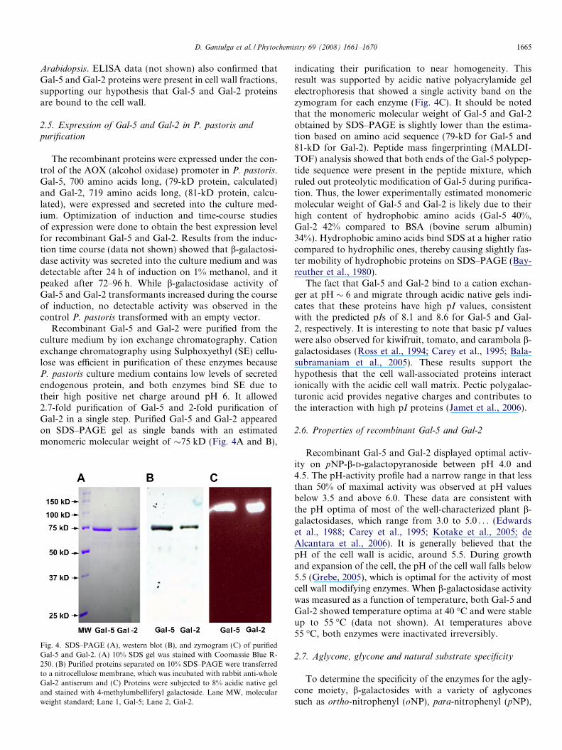
Recombinant Gal-5 and Gal-2 were purified from theculture medium by ion exchange chromatography. Cationexchange chromatography using Sulphoxyethyl (SE) cellu-lose was efficient in purification of these enzymes becauseP. pastoris culture medium contains low levels of secretedendogenous protein, and both enzymes bind SE due totheir high positive net charge around pH 6. It allowed2.7-fold purification of Gal-5 and 2-fold purification ofGal-2 in a single step. Purified Gal-5 and Gal-2 appearedon SDS–PAGE gel as single bands with an estimatedmonomeric molecular weight of �75 kD (Fig. 4A and B),
Fig. 4. SDS–PAGE (A), western blot (B), and zymogram (C) of purifiedGal-5 and Gal-2. (A) 10% SDS gel was stained with Coomassie Blue R-250. (B) Purified proteins separated on 10% SDS–PAGE were transferredto a nitrocellulose membrane, which was incubated with rabbit anti-wholeGal-2 antiserum and (C) Proteins were subjected to 8% acidic native geland stained with 4-methylumbelliferyl galactoside. Lane MW, molecularweight standard; Lane 1, Gal-5; Lane 2, Gal-2.
indicating their purification to near homogeneity. Thisresult was supported by acidic native polyacrylamide gelelectrophoresis that showed a single activity band on thezymogram for each enzyme (Fig. 4C). It should be notedthat the monomeric molecular weight of Gal-5 and Gal-2obtained by SDS–PAGE is slightly lower than the estima-tion based on amino acid sequence (79-kD for Gal-5 and81-kD for Gal-2). Peptide mass fingerprinting (MALDI-TOF) analysis showed that both ends of the Gal-5 polypep-tide sequence were present in the peptide mixture, whichruled out proteolytic modification of Gal-5 during purifica-tion. Thus, the lower experimentally estimated monomericmolecular weight of Gal-5 and Gal-2 is likely due to theirhigh content of hydrophobic amino acids (Gal-5 40%,Gal-2 42% compared to BSA (bovine serum albumin)34%). Hydrophobic amino acids bind SDS at a higher ratiocompared to hydrophilic ones, thereby causing slightly fas-ter mobility of hydrophobic proteins on SDS–PAGE (Bay-reuther et al., 1980).
The fact that Gal-5 and Gal-2 bind to a cation exchan-ger at pH � 6 and migrate through acidic native gels indi-cates that these proteins have high pI values, consistentwith the predicted pIs of 8.1 and 8.6 for Gal-5 and Gal-2, respectively. It is interesting to note that basic pI valueswere also observed for kiwifruit, tomato, and carambola b-galactosidases (Ross et al., 1994; Carey et al., 1995; Bala-subramaniam et al., 2005). These results support thehypothesis that the cell wall-associated proteins interactionically with the acidic cell wall matrix. Pectic polygalac-turonic acid provides negative charges and contributes tothe interaction with high pI proteins (Jamet et al., 2006).
2.6. Properties of recombinant Gal-5 and Gal-2
Recombinant Gal-5 and Gal-2 displayed optimal activ-ity on pNP-b-D-galactopyranoside between pH 4.0 and4.5. The pH-activity profile had a narrow range in that lessthan 50% of maximal activity was observed at pH valuesbelow 3.5 and above 6.0. These data are consistent withthe pH optima of most of the well-characterized plant b-galactosidases, which range from 3.0 to 5.0 . . . (Edwardset al., 1988; Carey et al., 1995; Kotake et al., 2005; deAlcantara et al., 2006). It is generally believed that thepH of the cell wall is acidic, around 5.5. During growthand expansion of the cell, the pH of the cell wall falls below5.5 (Grebe, 2005), which is optimal for the activity of mostcell wall modifying enzymes. When b-galactosidase activitywas measured as a function of temperature, both Gal-5 andGal-2 showed temperature optima at 40 �C and were stableup to 55 �C (data not shown). At temperatures above55 �C, both enzymes were inactivated irreversibly.
2.7. Aglycone, glycone and natural substrate specificity
To determine the specificity of the enzymes for the agly-cone moiety, b-galactosides with a variety of aglyconessuch as ortho-nitrophenyl (oNP), para-nitrophenyl (pNP),

1666 D. Gantulga et al. / Phytochemistry 69 (2008) 1661–1670
4-methylumbelliferyl (4MU), 5-bromo-4-chloro-3-indolyl(X) and 6-bromo-2-naphthyl (6BN) were tested. All ofthem were hydrolyzed by both Gal-5 and Gal-2, albeit withdifferent efficiencies (Table 2). These results indicatethat they have a broad specificity with respect to theaglycone moiety. To investigate the specificity for theglycone moiety, pNP-b-D-galactopyranoside (pNPGal),pNP-b-D-mannopyranoside, pNP-b-D-fucopyranoside,pNP-b-D-xylofuranoside, pNP-b-D-arabinopyranoside, andpNP-a-L-arabinopyranoside (pNPAra) were tested. Theresults are summarized in Table 3, which show that onlypNP-b-D-galactopyranoside and its 6-deoxy analoguepNP-b-D-fucopyranoside were hydrolyzed. Gal-5 andGal-2 failed to hydrolyze pNPAra, showing that the gly-cone specificity of these two enzymes is strict. Thus, Gal-5 and Gal-2 are highly specific for b-galactopyranosideand discriminate sugars based on the configuration of thehydroxyl group at C4 and C3 positions.
Kinetic parameters of Gal-5 and Gal-2 were determinedwith pNP-b-D-galactopyranoside. Km values for pNP-b-D-galactopyranoside for the two enzymes were similar(0.28 ± 0.06 mM for Gal-5 and 0.40 ± 0.02 mM for Gal-2), but kcatvalues were different (1.55 s�1 for Gal-5 and6.03 s�1 for Gal-2). Their catalytic efficiencies (kcat/Km) dif-fered to some extent (5.54 s�1 mM�1 for Gal-5 and15.21 s�1 mM�1 for Gal-2). Km values for oNPGal andpNPFuc were 0.83 ± 0.015 and 3.75 ± 0.55 mM for Gal-
Table 2Aglycone specificities of Gal-5 and Gal-2
Aglycone Relative activitya (%)
Gal-5 Gal-2
para-Nitrophenyl-(pNPGal) 100 100ortho-Nitrophenyl-(oNPGal) 61 724-Methylumbelliferyl-(4MUGal) 18 205-Bromo-4-chloro-3-indolyl-(X-Gal) 43 566-Bromonaphthyl-(6BNGal) 22 30
a Activities of Gal-5 and Gal-2 were assayed in reaction mixtures con-taining 2.5 mM substrate in NaOAc buffer pH 4.6. Aglycone specificity isexpressed as a percentage of activity against pNPGal (100% � 0.03 units(nkat)). For insoluble aglycones, amounts of galactose produced as aresult of hydrolysis were measured by the galactose dehydrogenase assay.
Table 3Sugar specificities of Gal-5 and Gal-2
Glycone Relative activitya (%)
Gal-5 Gal-2
pNP-b-D-galactopyranoside 100 100pNP-b-D-fucopyranoside 25 21pNP-b-D-glucopyranoside <1 <1pNP-b-D-mannopyranoside 0 0pNP-b-D-xylofuranoside 0 0pNP-b-D-arabinopyranoside 0 0pNP-a-L-arabinopyranoside 0 0
a Activities of Gal-5 and Gal-2 were assayed in reaction mixtures con-taining 2.5 mM substrate in NaOAc buffer pH 4.6. Sugar specificity isexpressed as a percentage of activity against pNPGal (100% � 0.03 unit(nkat)).
5, respectively, and 0.72 ± .016 and 6.4 ± 2.8 mM forGal-2, respectively.
Inhibitory effects of several sugars and sugar derivativeswere tested using pNPGal as a substrate. c-Galactonolac-tone and D-galactose were the most effective inhibitorsfor Gal-5 and Gal-2 activity. Their Ki values were 44 lMand 7.4 mM, respectively, for Gal-5 and 98 lM and4.5 mM for Gal-2. Also D-fucose, methyl-a-D-galactosideand raffinose were weaker inhibitors for both enzymeswhile pNPGlc, pNPAra, lactose, IPTG, galacturonic acid,L-arabinose, and D-mannose did not show any inhibitoryeffects. Ag+, Hg2+ and SDS strongly inhibited activity ofboth enzymes when pNPGal was used as a substrate.
b-Galactosidases from either different plants or withinthe same plant are known to differ considerably in theirlinkage specificity (Kotake et al., 2005; Ishimaru et al.,2005; Buckeridge et al., 2005). Using b-(1,4), b-(1,3) andb-(1,6) linked galacto-oligosaccharides, we investigatedthe linkage specificity of Gal-5 and Gal-2. As can be seenfrom Fig. 5, both Gal-5 and Gal-2 hydrolyze b-(1,4) (lanes3–4 and 5–6) and b-(1,3) (lanes 8–9 and 10–11) linkages,whereas the b-(1,6) linkages in galacto-oligosaccharideswere less susceptible to hydrolysis (lanes 13–16).
Ahn et al. (2007) showed that a member of the family,Gal-4, preferentially cleaves b-(1,4) and b-(1,3) linkages.Thus, the three Arabidopsis paralogs, Gal-5, Gal-2, andGal-4, might act on the same natural substrates with b-(1,4) and b-(1,3) linkages. To probe the natural substratespecificity of Gal-5 and Gal-2, more complex oligo-/poly-saccharides were tested and the results are shown in Table4. L-Arafase (a-L-arabinofuranosidase) pretreated (toremove arabinose) lupin galactan, a polymer of b-(1,4)
Fig. 5. Linkage specificity of Gal-5 and Gal-2. After hydrolysis ofgalactobioses and galactotrioses by Gal-5 and Gal-2, products wereseparated by TLC and developed with naphthoresorcinol (see methods).Lane 1, monogalactose, Lane2-b-(1,4)-linked galactobiose and galacto-triose; Lane 3 and 4, hydrolysis product of b-(1,4)-linked galactobiose andgalactotriose by Gal-5; Lane 5 and 6, hydrolysis product of b-(1,4)-linkedgalactobiose and galactotriose by Gal-2; Lane 7, b-(1,3)-linked galacto-biose and galactotriose; Lane 8 and 9, hydrolysis product of b-(1,3)-linkedgalactobiose and galactotriose by Gal-5; Lane 10 and 11, hydrolysisproduct of b-(1,3)-linked galactobiose and galactotriose by Gal-2; Lane12,b-(1,6)- linked galactobiose and galactotriose; Lane 13 and 14,hydrolysis product of b-(1,6)-linked galactobiose and galactotriose byGal-5; Lane 15 and 16, hydrolysis product of b-(1,6)-linked galactobioseand galactotriose by Gal-2.
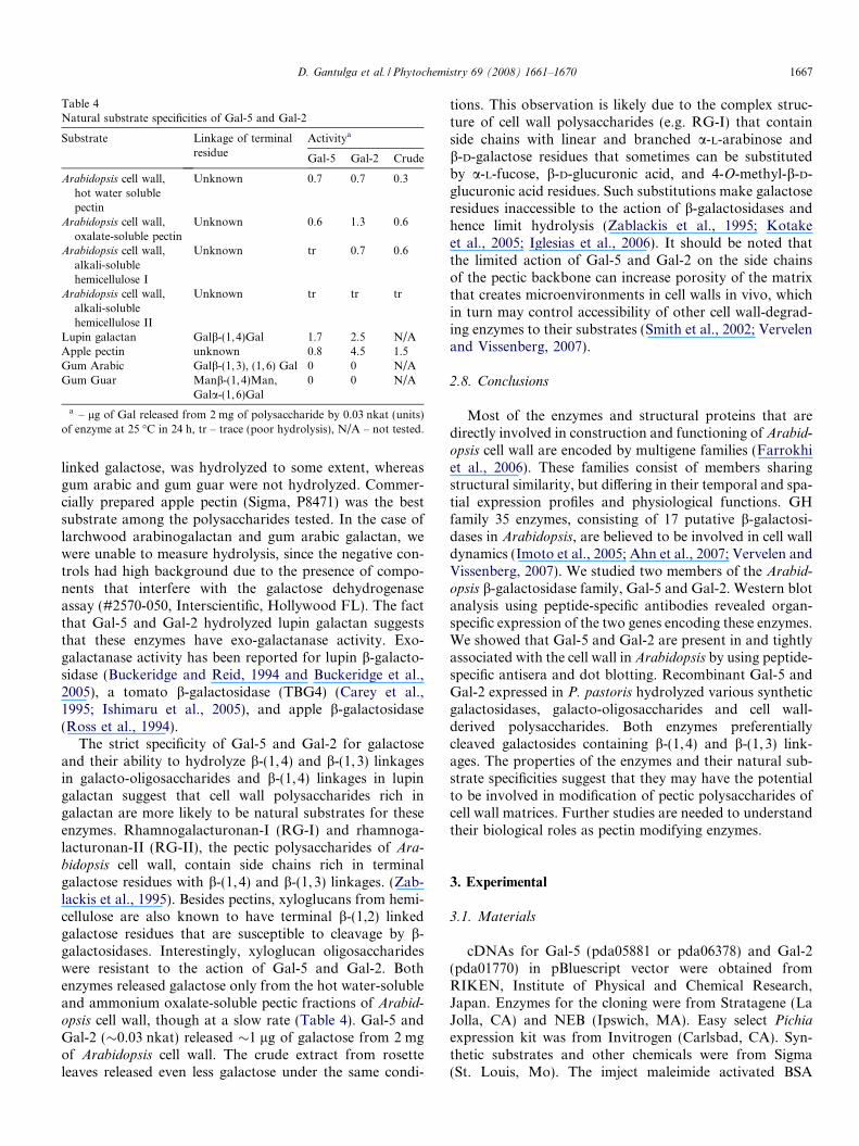
Table 4Natural substrate specificities of Gal-5 and Gal-2
Substrate Linkage of terminalresidue
Activitya
Gal-5 Gal-2 Crude
Arabidopsis cell wall,hot water solublepectin
Unknown 0.7 0.7 0.3
Arabidopsis cell wall,oxalate-soluble pectin
Unknown 0.6 1.3 0.6
Arabidopsis cell wall,alkali-solublehemicellulose I
Unknown tr 0.7 0.6
Arabidopsis cell wall,alkali-solublehemicellulose II
Unknown tr tr tr
Lupin galactan Galb-(1,4)Gal 1.7 2.5 N/AApple pectin unknown 0.8 4.5 1.5Gum Arabic Galb-(1,3), (1,6) Gal 0 0 N/AGum Guar Manb-(1,4)Man,
Gala-(1,6)Gal0 0 N/A
a – lg of Gal released from 2 mg of polysaccharide by 0.03 nkat (units)of enzyme at 25 �C in 24 h, tr – trace (poor hydrolysis), N/A – not tested.
D. Gantulga et al. / Phytochemistry 69 (2008) 1661–1670 1667
linked galactose, was hydrolyzed to some extent, whereasgum arabic and gum guar were not hydrolyzed. Commer-cially prepared apple pectin (Sigma, P8471) was the bestsubstrate among the polysaccharides tested. In the case oflarchwood arabinogalactan and gum arabic galactan, wewere unable to measure hydrolysis, since the negative con-trols had high background due to the presence of compo-nents that interfere with the galactose dehydrogenaseassay (#2570-050, Interscientific, Hollywood FL). The factthat Gal-5 and Gal-2 hydrolyzed lupin galactan suggeststhat these enzymes have exo-galactanase activity. Exo-galactanase activity has been reported for lupin b-galacto-sidase (Buckeridge and Reid, 1994 and Buckeridge et al.,2005), a tomato b-galactosidase (TBG4) (Carey et al.,1995; Ishimaru et al., 2005), and apple b-galactosidase(Ross et al., 1994).
The strict specificity of Gal-5 and Gal-2 for galactoseand their ability to hydrolyze b-(1,4) and b-(1,3) linkagesin galacto-oligosaccharides and b-(1,4) linkages in lupingalactan suggest that cell wall polysaccharides rich ingalactan are more likely to be natural substrates for theseenzymes. Rhamnogalacturonan-I (RG-I) and rhamnoga-lacturonan-II (RG-II), the pectic polysaccharides of Ara-
bidopsis cell wall, contain side chains rich in terminalgalactose residues with b-(1,4) and b-(1,3) linkages. (Zab-lackis et al., 1995). Besides pectins, xyloglucans from hemi-cellulose are also known to have terminal b-(1,2) linkedgalactose residues that are susceptible to cleavage by b-galactosidases. Interestingly, xyloglucan oligosaccharideswere resistant to the action of Gal-5 and Gal-2. Bothenzymes released galactose only from the hot water-solubleand ammonium oxalate-soluble pectic fractions of Arabid-
opsis cell wall, though at a slow rate (Table 4). Gal-5 andGal-2 (�0.03 nkat) released �1 lg of galactose from 2 mgof Arabidopsis cell wall. The crude extract from rosetteleaves released even less galactose under the same condi-
tions. This observation is likely due to the complex struc-ture of cell wall polysaccharides (e.g. RG-I) that containside chains with linear and branched a-L-arabinose andb-D-galactose residues that sometimes can be substitutedby a-L-fucose, b-D-glucuronic acid, and 4-O-methyl-b-D-glucuronic acid residues. Such substitutions make galactoseresidues inaccessible to the action of b-galactosidases andhence limit hydrolysis (Zablackis et al., 1995; Kotakeet al., 2005; Iglesias et al., 2006). It should be noted thatthe limited action of Gal-5 and Gal-2 on the side chainsof the pectic backbone can increase porosity of the matrixthat creates microenvironments in cell walls in vivo, whichin turn may control accessibility of other cell wall-degrad-ing enzymes to their substrates (Smith et al., 2002; Vervelenand Vissenberg, 2007).
2.8. Conclusions
Most of the enzymes and structural proteins that aredirectly involved in construction and functioning of Arabid-
opsis cell wall are encoded by multigene families (Farrokhiet al., 2006). These families consist of members sharingstructural similarity, but differing in their temporal and spa-tial expression profiles and physiological functions. GHfamily 35 enzymes, consisting of 17 putative b-galactosi-dases in Arabidopsis, are believed to be involved in cell walldynamics (Imoto et al., 2005; Ahn et al., 2007; Vervelen andVissenberg, 2007). We studied two members of the Arabid-
opsis b-galactosidase family, Gal-5 and Gal-2. Western blotanalysis using peptide-specific antibodies revealed organ-specific expression of the two genes encoding these enzymes.We showed that Gal-5 and Gal-2 are present in and tightlyassociated with the cell wall in Arabidopsis by using peptide-specific antisera and dot blotting. Recombinant Gal-5 andGal-2 expressed in P. pastoris hydrolyzed various syntheticgalactosidases, galacto-oligosaccharides and cell wall-derived polysaccharides. Both enzymes preferentiallycleaved galactosides containing b-(1,4) and b-(1, 3) link-ages. The properties of the enzymes and their natural sub-strate specificities suggest that they may have the potentialto be involved in modification of pectic polysaccharides ofcell wall matrices. Further studies are needed to understandtheir biological roles as pectin modifying enzymes.
3. Experimental
3.1. Materials
cDNAs for Gal-5 (pda05881 or pda06378) and Gal-2(pda01770) in pBluescript vector were obtained fromRIKEN, Institute of Physical and Chemical Research,Japan. Enzymes for the cloning were from Stratagene (LaJolla, CA) and NEB (Ipswich, MA). Easy select Pichiaexpression kit was from Invitrogen (Carlsbad, CA). Syn-thetic substrates and other chemicals were from Sigma(St. Louis, Mo). The imject maleimide activated BSA
1668 D. Gantulga et al. / Phytochemistry 69 (2008) 1661–1670
conjugation kit was from Pierce (Rockford, IL). Total Gal-actose Neonatal Screening Test Kit was from Interscientific(Hollywood, FL). Galactose dehydrogenase was fromRoche (Indianapolis, IN). Galacto-oligosaccharides werea gift from Dr. Yoichi Tsumuraya and Dr. ToshihisaKotake of Saitama University, Japan. Lupin galactanwas a gift from Dr. David Smith of USDA, Beltsville, MD.
3.2. Methods
3.2.1. Database analysis
Signal sequence predictions and subcellular targeting ofpredicted proteins were done using SignalP (Bendtsenet al., 2004) and TargetP (Emanuelsson et al., 2000).Amino acid sequences of b-galactosidases were alignedusing software at http://align.genome.jp. A phylogenetictree was constructed from the alignment using PAUP 4.0.
3.2.2. Microarray expression analysis
Expression profile analysis was done usingArabidopsis
gene expression datasets from the Genevestigator website(http://www.genevestigator.ethz.ch). Using Gene AtlasTool, the organ/tissue-specific expression levels forAt1g45130 (Gal-5) and At3g52840 (Gal-2) were estimatedby Genevestigator software along with Atg05320 (Ubiqui-tin10, UBQ10) as the control for comparison. A given genewas scored as ‘‘expressed” if data from the Digital North-ern Tool gave signal values higher than 200 with p < 0.06(Zimmermann et al., 2004). The p value for Gal-5 andGal-2 was p = 0.00164. Response Viewer Tool was usedto verify up- and down-regulated genes under different abi-otic and biotic stresses. Reliability and reproducibility ofanalyses were evaluated by the number of chips and repli-cates in individual experiments.
3.2.3. Plant materials
Arabidopsis seeds (Col-O) were obtained from the Ara-bidopsis Biological Resource Center (ABCR), Ohio StateUniversity Seed Stock Center (Columbus, OH). For germi-nation, seeds were surface-sterilized with 3% hypochloritefor 10 min followed by washes with dH2O (three times)and finally suspended in 0.1% agarose. Sterilized seeds werekept at 4 �C for 3–4 days and seedlings were germinated onhalf strength Murashige–Skoog salt-agar plates for 10–14days with 16 h day and 8 h night cycles. Seedlings weretransferred to soil and grown at 16/8 h day/night cycle.Plants were harvested when 4–5 weeks old, and immediatelyfrozen in liquid nitrogen and kept at �80 �C until use. Forwestern blot analysis, Arabidopsis tissue was ground withsand (0.3 g/1 g tissue). Total proteins were solubilized in6 M urea (1 g tissue: 2 ml solvent). Cell wall polysaccharideisolation was done as described in Li et al. (2001).
3.2.4. Expression of Gal-2 in E. coli and preparation ofrabbit antisera
The Gal-2 mature protein coding sequence was clonedinto pET21a vector and expressed in E. coli BL21 codon
plus cells. Cells were suspended in lysis buffer (50 mMTris–HCl pH 8.0, 100 mM NaCl, 0.02% SDS, 1 mMPMSF) and broken up using a French press. After exten-sive washing of soluble fractions with lysis buffer, insolubleproteins (the inclusion body fraction) were solubilized in6 M urea and separated on a 10% SDS–PAGE preparativegel. The gel was stained (30 min) with Coomassie brilliantblue R-250 and the band corresponding to the Gal-2 poly-peptide was excised. The excised band was destained inMeOH:H2O (1:1, v/v) with several changes of solutionand rehydrated in a minimum amount of 1X PBS at 4 �Covernight. After rehydration, the band was ground in apre-chilled mortar. Ground powder was suspended in 1XPBS containing 0.2% SDS and 0.5% 2-mercaptoethanol(v/v) and heated at 75 �C for 15 min. After cooling, the sus-pension was mixed with 1 volume of Freund’s CompleteAdjuvant (Sigma) and used for immunization. Rabbitanti-Gal-2 sera were raised by repeated injection of antigenmixed with Freund’s Incomplete Adjuvant at two-weekintervals. Synthetic peptides (Gal-2: CSGKIRAPTILMK-MIPTS and Gal-5: CSGVAFLTNYHMNAPAKVV) wereconjugated to the BSA using an imject maleimide activatedBSA kit (Pierce, Rockford, IL) according to the vendor’sprotocol. Peptide-specific antisera were raised by injectingBSA-conjugated synthetic peptides with Freund’sadjuvants. A small volume of trial bleeding was taken attwo-week intervals to monitor the change in antisera titerduring the course of immunization. All antisera werediluted twice with glycerol and stored at �20 �C untilusage.
3.3. Expression of recombinant Gal-5 and Gal-2 in P.
pastoris and purification
The mature protein coding sequences of Gal-5(S24 through N724) and Gal-2 (V28 throughK727) cDNAs were amplified by the primer pair 50-CAC-CGTGGTCACTTATGATCACAAAGC-30 and 50-CCA-ATGAAAGAGGGTAACAAAGGGC-30 for Gal-2 and50-AGGTGAATTCCAGTGTAGTAGTGTAACCTACG-30 and 50-TTTGCGGCCGCAAGTTAG TTTACTGAT-CTCTTCACAAC-30 for Gal-5 from cDNA inserts of plas-mids obtained from RIKEN, using the high-fidelity Pfu
Turbo DNA polymerase. The inserts were cloned intopPICZa Pichia expression vector to express Gal-5 andGal-2 as the yeast a factor secretion signal fusion proteinto facilitate secretion of recombinant proteins into culturemedium. After confirming the accuracy of the sequenceand the correct reading frame, linearized plasmids weretransformed into P. pastoris by electroporation. Recombi-nant enzyme production was under the control of the alco-hol oxidase (AOX) promoter induced by methanol.Production of recombinant proteins was monitored byassaying b-galactosidase activity toward pNPGal in culturesupernatant samples taken every 24 h. After 72 h of induc-tion, cells were pelleted and culture supernatant was usedfor further purification of the recombinant enzymes.
D. Gantulga et al. / Phytochemistry 69 (2008) 1661–1670 1669
Gal-5 and Gal-2 were purified from culture supernatantsby ion exchange chromatography using Sulphoxyethyl (SE)cellulose. In a typical experiment, 100 ml of culture super-natant was filtered, diluted five times with degassed dH2Oto reduce ionic strength of the medium and loaded ontothe column (1.5 cm � 3 cm) pre-equilibrated with bufferA (20 mM potassium phosphate, pH 6.0). After washingwith 10 column volumes of buffer A, bound proteins wereeluted in one step with 150 mM NaCl in buffer A (flow rate1 ml/min). All fractions were checked for b-galactosidaseactivity using pNPGal as substrate. Fractions with highestb-galactosidase activity were used for further experiments.Protein concentration was determined by the Bradfordmethod (Bradford, 1976) (Bio-Rad Protein Assay Reagentkit) using BSA as a standard.
3.3.1. Cell wall isolation and extraction of cell wall-bound
proteins
We isolated cell walls from rosette leaves of Arabidop-
sis using the procedure described by Feiz et al. (2006).Rosette leaves were ground in a blender whose cup wasdipped at intervals into liquid nitrogen to maintain lowtemperature during grinding. The cell wall fraction waswashed extensively with 3 L of wash buffer (5 mM NaO-Ac buffer, pH 4.6) on a metal net (75 lm pore size). Afterwashing, the cell wall fraction was lyophilized. The lyoph-ilized cell wall material was ground to a fine powder bygrinding in a blender, and was then used to extractwall-bound proteins. Five different (S1, soluble 1; S2, sol-uble 2; S3, soluble 3; CW4, extractable with CaCl2; andCW5, extractable with LiCl) fractions were obtained.These fractions were assayed for b-galactosidase activity.Of these, three fractions with b-galactosidase activity werefurther analyzed for immunoreactivity. They were spottedmultiple times on nitrocellulose strips to increase antigen(Gal-5 and Gal-2) concentration and incubated with pre-immune (control) and immune sera from rabbits immu-nized with whole Gal-2 polypeptide and unique peptidesderived from Gal-5 and Gal-2 sequences.
3.3.2. SDS–PAGE, native PAGE and western blotting
SDS–PAGE was performed as described by Laemmli(1970). Native PAGE was performed in acidic gels usingthe protocol on the website (http://wolfson.huji.ac.il/purifi-cation/Protocols/PAGE_Acidic.html). After electrophore-sis, the gel was rinsed in a wash buffer (100 mM acetatebuffer pH 4.6) for 2 � 15 min. The zymogram was devel-oped by incubating the gel in 0.5 mM 4-MUGal in washbuffer at 37 �C for 20 min and photographed under UVlight. For immunoblotting, the gel was soaked in a blottingbuffer (10 mM CAPS, pH 11 with 10% (v/v) MeOH) for2 � 15 min. Proteins were transferred onto a nitrocellulose(0.45 lm, Protran) membrane using a Bio-Rad Mini transblot cell at 50 V, at 4 �C overnight following the vendor’sprotocol. For immunodetection, 2000-times dilution ofanti-Gal-2 antiserum or 1000-times dilution of peptide-spe-cific antiserum was used as primary antibody and 2000-
times dilution of goat anti-rabbit antibody conjugated withperoxidase (A0545, Sigma, Saint Louis, MO) as secondaryantibody. Immunoreactive bands were visualized by thedeposition of 4-chloronaphthol after oxidation by HRP(horse radish peroxidase) using the substrate solution(21 ml of PBS pH 7.4, mixed with 5.5 ml of 3.3 mg/ml 4-chloronaphthol in 100% MeOH and 10 ll of 30% H2O2).
3.3.3. b-galactosidase activity assay
One hundred micro liters of 5 mM pNPGal in 100 mMNaOAc buffer pH 4.6, 80 ll H2O and 20 ll of the dilutedenzyme solution were mixed and incubated at 37 �C forup to 30 min. The reaction was stopped by adding 100 llof 1 M Na2CO3. The absorbance was measured at405 nm to quantify the amount of pNP released afterhydrolysis. This standard protocol was used for all activityassays with pNPGal, if not otherwise stated. Boiled enzymeor buffer solution was used as a control. One unit (nkat) ofenzyme activity is defined as an amount of enzyme that isable to produce 1 nmole of pNP per second at 37 �C.
For the determination of natural substrate specificity,galacto-oligosaccharides (20 mM), xyloglucan oligosaccha-rides (1 lg/ll) and polysaccharides (1% (w/v)) were pre-pared in H2O. Final concentration of substrates was4 mM for oligosaccharides and 0.5% for polysaccharidesin 100 mM acetate buffer pH 4.6. Reaction mixture wasincubated with 0.03 units/nkats enzymes at room tempera-ture for 24 h. Reaction mixtures containing no enzyme andno substrate were used as controls. Reactions for polysac-charides were stopped by adding 1 ml of 100% EtOH to0.4 ml of reaction mix to precipitate proteins and polysac-charides. After centrifugation, the supernatant was trans-ferred into a new microfuge tube and vacuum dried.Dried mixtures were dissolved in 100 ll of dH2O, and totalgalactose produced as a result of hydrolysis was quantifiedusing galactose dehydrogenase assay kit (Interscientific,Hollywood, FL). Products of hydrolysis of oligosaccha-rides were analyzed by thin layer chromatography (TLC)on silica gel 60 F254 (EM Science, Germany) using 3:2:1(v/v/v) n-BuOH:AcOH:H2O as the solvent and detectedby heating TLC plates after spraying with 0.2% (w/v)naphthoresorcinol in 1:19 H2SO4:EtOH (v/v). (Ahn et al.,2004).
Acknowledgements
We are grateful to Drs. Yoichi Tsumuraya and Dr.Toshihisa Kotake of Saitama University, Japan for provid-ing galacto-oligosaccharides, and Dr. David Smith ofUSDA, Beltsville, MD for providing lupin galactan. Theauthors also wish to thank Dr. Farooqahmed Kittur ofVirginia Tech for much help and critical reading of themanuscript. This research is funded by the Arabidopsis2010 Project of the National Science Foundation (MCB-0115937) A. Esen et al., and a research grant award fromthe Virginia Academy of Science to D. Gantulga.

1670 D. Gantulga et al. / Phytochemistry 69 (2008) 1661–1670
References
Ahn, Y.O., Mizutani, M., Hiromichi, S., Kanzo, S., 2004. Furcatinhydrolase from Viburnum furcatum blume is a novel disaccharide-specific acuminosidase in Glycosyl Hydrolase Family 1. J. Biol. Chem.279, 23405–23414.
Ahn, Y.O., Zheng, M., Winkel, B., Bevan, D.R., Esen, A., Shin-Han, S.,Benson, J., Peng, H., Miller, J.T., Cheng, C., Poulton, J.E., Shih, M.,2007. Functional genomic analysis of Arabidopsis thaliana GlycosideHydrolase Family 35. Phytochemistry 68, 1510–1520.
Balasubramaniam, S., Lee, H., Lazan, H., Othman, R., Ali, Z.M., 2005.Purification and properties of a b-galactosidase from carambola fruitwith significant activity toward cell wall polysaccharides. Phytochem-istry 66, 153–163.
Bayreuther, K., Bieseler, D.J., Ehring, R., Griesser, H.W., Mieschendahl,M., Muller-Hill, B., Triesch, I., 1980. Investigation of structure andfunction of lactose permease of Escherichia coli. Biochem. Soc. Trans.8, 675–676.
Bendtsen, J., Nielsen, H., von Heijne, G., Brunak, S., 2004. Improvedprediction of signal peptides: SignalP 3.0. J. Mol. Biol. 340, 783–795.
Bradford, M.M., 1976. A rapid and sensitive method for the quantitationof microgram quantities of protein utilizing the principle of protein-dye binding. Anal. Biochem. 72, 248–254.
Buckeridge, M.S., Reid, J.S., 1994. Purification and properties of a novelbeta-galactosidase or exo-(1->4)-beta-D galactanase from the cotyle-dons of germinated Lupinus angustifolius L. seeds. Planta 192 (4), 502–511.
Buckeridge, M.S., Huntcheon, I.S., Grant Reid, J.S., 2005. The role ofexo-(1-4)-b-galactanase in the mobilization of polysaccharides fromthe cotyledon cell wall of Lupinus angustifolus following germination.Ann. Botany 96, 435–444.
Carey, A.T., Holt, K., Picard, S., Wilde, R., Tucker, G.A., Bird, C.R.,Schuch, W., Seymour, G.B., 1995. Tomato exo-(1-4)-D-galactanase.Isolation, changes during ripening in normal and mutant tomato fruit,and characterization of related cDNA clone. Plant Physiol. 108, 1099–1107.
Chantarangsee, M., Fujimura, T., Fry, S.C., Ketudat-Cairns, J.R., 2007.Molecular characterization of b-galactosidases from germinating rice(Oryza sativa). Plant Sci. 173, 118–134.
Coutinho, P.M., Henrissat, B., 1999. Carbohydrate-active enzymes: anintegrated database approach. In: Gilbert, H.J., Davies, G., Henrissat,B., Svensson, B. (Eds.), Recent Advances in Carbohydrate Bioengi-neering. The Royal Society of Chemistry, Cambridge, pp. 3–12.
Davies, G., Henrissat, B., 1995. Structures and mechanism of glycosylhydrolases. Structure 3, 853–859.
de Alcantara, P.H.N., Martim, L., Silva, C.O., Dietrich, S.M.C.,Buckeridge, M.S., 2006. Purification of a b-galactosidase fromcotyledons of Hymenaea courbaril L. (Leguminosae). Enzyme proper-ties and biological function. Plant Physiol. Biochem. 44, 619–627.
Edwards, M., Bowman, Y., Dea, I., Reid, J., 1988. A b-D-galactosidasefrom Nasturtium (Tropaeolum majus L.) cotyledons. J. Biol. Chem. 9,4333–4337.
Emanuelsson, O., Nielsen, H., Brunak, S., von Heijne, G., 2000.Predicting subcellular localization of proteins based on their N-terminal amino acid sequence. J. Mol. Biol. 300, 1005–1016.
Farrokhi, N., Burton, R.A., Brownfield, L., Hrmova, M., Wilson, S.M.,Bacic, A., Fincher, G.B., 2006. Plant cell wall biogenesis: genetic,
biochemical and functional genomics approaches to the identificationof key genes. Plant Biotechnol. 4, 145–167.
Feiz, L., Irshad, M., Pont-Lezica, R.F., Canut, H., Jamet, E., 2006.Evaluation of cell wall preparations for proteomics: a new procedurefor purifying cell walls from Arabidopsis hypocotyls. Plant Methods 2,10.
Grebe, M., 2005. Growth by Auxin: when a weed needs acid. Science 310,60–61.
Hruba, P., Honys, D., Twell, D., Capkova, V., Tupy, J., 2005. Expressionof b-galactosidase and b-xylosidase genes during microspore andpollen development. Planta 220, 931–940.
Iglesias, N., Abelenda, J.A., Rodino, M., Samredro, J., Revilla, G., Zarra,I., 2006. Apoplastic glycosidase active against xyloglucan oligosaccha-rides of Arabidopsis thaliana. Plant Cell Physiol. 47 (1), 55–63.
Imoto, K., Yokoyama, R., Nishitani, K., 2005. Comprehensive approachto genes involved in cell wall modifications in Arabidopsis thaliana.Plant Mol. Biol. 58 (2), 177–192.
Ishimaru, M., Smith, D.L., Gross, K.C., 2005. Yeast expressed tomato b-galactosidase 1,4, and 5 have activity against synthetic and plant-derived cell wall substrates. HortScience 40, 1092.
Jamet, E., Canut, H., Boudart, G., Pont-Lezica, R.F., 2006. Cell wallproteins: a new insight through proteomics. Trends Plant Sci. 11, 33–39.
Kotake, T., Dina, S., Konichi, T., Kaneko, S., Igarashi, K., Samejima, M.,Watanbe, Y., Kimura, K., Tsumuraya, Y., 2005. Molecular cloning ofa b-galactosidase from radish that specifically hydrolyzes b-(1-3) andb-(1-6)-galactosyl residues of arabinogalactan protein. Plant Physiol.138, 1563–1576.
Laemmli, U.K., 1970. Cleavage of structural proteins during assembly ofthe head of bacteriophage T4. Nature 277, 680–685.
Lazan, H., Ng, S.Y., Goh, L.Y., Ali, Z.M., 2004. Papaya b-galactosidase/galactanase isoforms in differential cell wall hydroly-sis and fruit softening during ripening. Plant Physiol. Biochem. 42,847–853.
Li, S.C., Han, J.W., Chen, K.C., Chen, C.S., 2001. Purification andcharacterization of isoforms of b-galactosidases in mung bean seedling.Phytochemistry 57, 349–359.
Perez Almeida, I.B., 2004. Arabidopsis cell wall beta-galactosidase genefamily: expression, catalytic activities and biological function ingalactose dynamics. Dissertation. Purdue University ETD.
Ross, G.S., Wegrzyn, T., MacRae, E.A., Redgwell, R.J., 1994. Apple b-galactosidase activity against cell wall polysaccharides and character-ization of related cDNA clone. Plant Physiol. 106, 521–528.
Smith, D.L., Gross, K.C., 2000. A family of at least seven b-galactosidasegenes is expressed during tomato fruit development. Plant Physiol. 123,1173–1183.
Smith, D.L., Abbott, J.A., Gross, K.C., 2002. Down-regulation of tomatob-galactosidase 4 results in decreased fruit softening. Plant Physiol.129, 1755–1762.
Vervelen, J.P., Vissenberg, K., 2007. The expanding cell. Plant CellMonographs, vol. 5. Springer.
Zablackis, E., Huang, J., Muller, B., Darvill, A.G., Albersheim, P., 1995.Characterization of the cell wall polysaccharides of Arabidopsis
thaliana leaves. Plant Physiol. 107, 1129–1138.Zimmermann, P., Hirsch-Hoffmann, M., Hennig, L., Gruissem, W., 2004.
GENEVESTIGATOR. Arabidopsis microarray database and analysistool. Plant Physiol. 136, 2621–2632.